Embed Size (px)
Citation preview
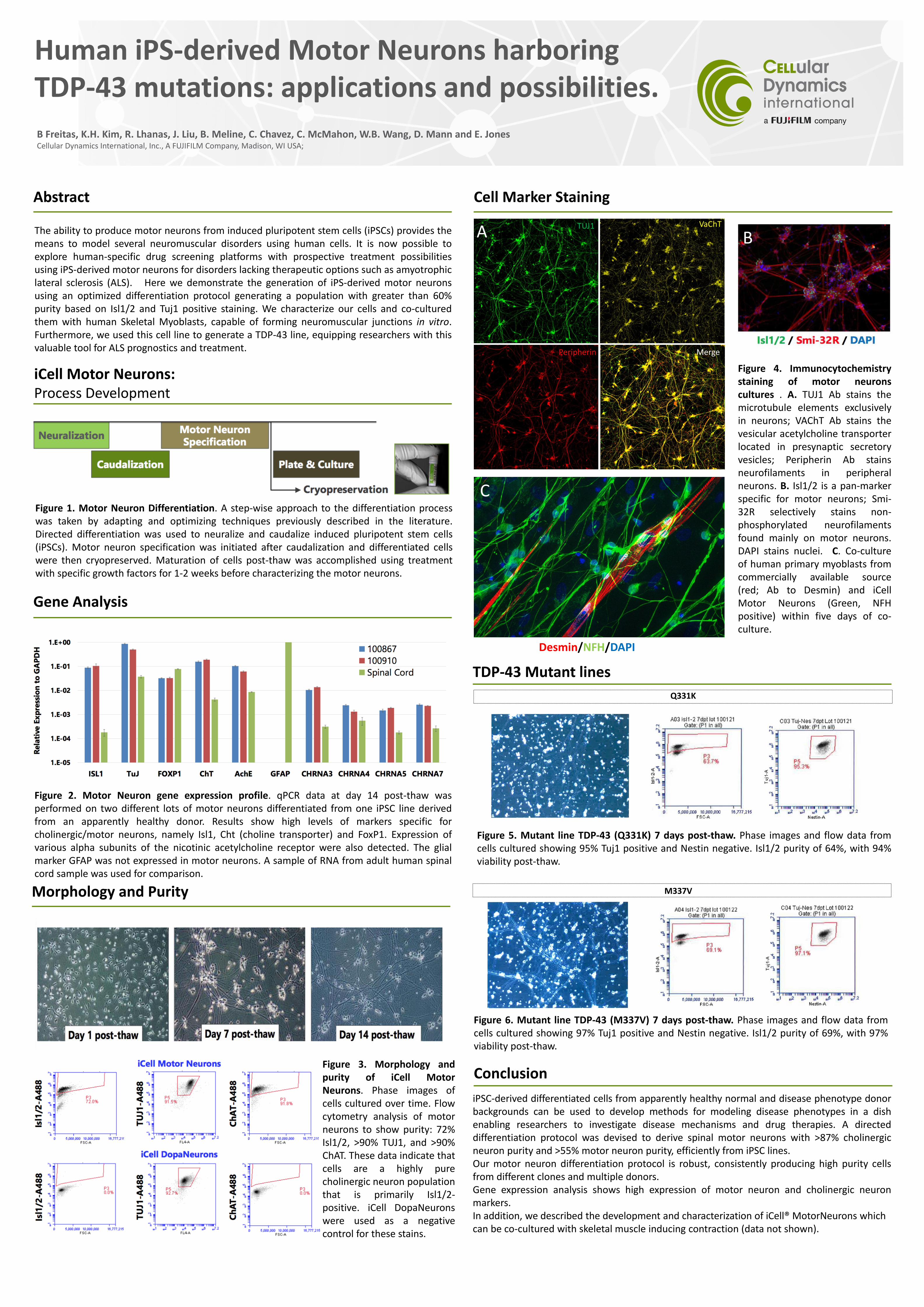
Human iPS-derived Motor Neurons harboring TDP-43 mutations: applications and possibilities.
B Freitas, K.H. Kim, R. Lhanas, J. Liu, B. Meline, C. Chavez, C. McMahon, W.B. Wang, D. Mann and E. JonesCellular Dynamics International, Inc., A FUJIFILM Company, Madison, WI USA;
Abstract
The ability to produce motor neurons from induced pluripotent stem cells (iPSCs) provides themeans to model several neuromuscular disorders using human cells. It is now possible toexplore human-specific drug screening platforms with prospective treatment possibilitiesusing iPS-derived motor neurons for disorders lacking therapeutic options such as amyotrophiclateral sclerosis (ALS). Here we demonstrate the generation of iPS-derived motor neuronsusing an optimized differentiation protocol generating a population with greater than 60%purity based on Isl1/2 and Tuj1 positive staining. We characterize our cells and co-culturedthem with human Skeletal Myoblasts, capable of forming neuromuscular junctions in vitro.Furthermore, we used this cell line to generate a TDP-43 line, equipping researchers with thisvaluable tool for ALS prognostics and treatment.
Conclusion
iPSC-derived differentiated cells from apparently healthy normal and disease phenotype donorbackgrounds can be used to develop methods for modeling disease phenotypes in a dishenabling researchers to investigate disease mechanisms and drug therapies. A directeddifferentiation protocol was devised to derive spinal motor neurons with >87% cholinergicneuron purity and >55% motor neuron purity, efficiently from iPSC lines.Our motor neuron differentiation protocol is robust, consistently producing high purity cellsfrom different clones and multiple donors.Gene expression analysis shows high expression of motor neuron and cholinergic neuronmarkers.In addition, we described the development and characterization of iCell® MotorNeurons which can be co-cultured with skeletal muscle inducing contraction (data not shown).
iCell Motor Neurons:Process Development
Gene Analysis
Q331K
TDP-43 Mutant lines
Cell Marker Staining
Figure 1. Motor Neuron Differentiation. A step-wise approach to the differentiation processwas taken by adapting and optimizing techniques previously described in the literature.Directed differentiation was used to neuralize and caudalize induced pluripotent stem cells(iPSCs). Motor neuron specification was initiated after caudalization and differentiated cellswere then cryopreserved. Maturation of cells post-thaw was accomplished using treatmentwith specific growth factors for 1-2 weeks before characterizing the motor neurons.
Figure 2. Motor Neuron gene expression profile. qPCR data at day 14 post-thaw wasperformed on two different lots of motor neurons differentiated from one iPSC line derivedfrom an apparently healthy donor. Results show high levels of markers specific forcholinergic/motor neurons, namely Isl1, Cht (choline transporter) and FoxP1. Expression ofvarious alpha subunits of the nicotinic acetylcholine receptor were also detected. The glialmarker GFAP was not expressed in motor neurons. A sample of RNA from adult human spinalcord sample was used for comparison.
Morphology and Purity
Figure 3. Morphology andpurity of iCell MotorNeurons. Phase images ofcells cultured over time. Flowcytometry analysis of motorneurons to show purity: 72%Isl1/2, >90% TUJ1, and >90%ChAT. These data indicate thatcells are a highly purecholinergic neuron populationthat is primarily Isl1/2-positive. iCell DopaNeuronswere used as a negativecontrol for these stains.
M337V
A B
C
Desmin/NFH/DAPI
Figure 4. Immunocytochemistrystaining of motor neuronscultures . A. TUJ1 Ab stains themicrotubule elements exclusivelyin neurons; VAChT Ab stains thevesicular acetylcholine transporterlocated in presynaptic secretoryvesicles; Peripherin Ab stainsneurofilaments in peripheralneurons. B. Isl1/2 is a pan-markerspecific for motor neurons; Smi-32R selectively stains non-phosphorylated neurofilamentsfound mainly on motor neurons.DAPI stains nuclei. C. Co-cultureof human primary myoblasts fromcommercially available source(red; Ab to Desmin) and iCellMotor Neurons (Green, NFHpositive) within five days of co-culture.
Figure 5. Mutant line TDP-43 (Q331K) 7 days post-thaw. Phase images and flow data fromcells cultured showing 95% Tuj1 positive and Nestin negative. Isl1/2 purity of 64%, with 94%viability post-thaw.
Figure 6. Mutant line TDP-43 (M337V) 7 days post-thaw. Phase images and flow data fromcells cultured showing 97% Tuj1 positive and Nestin negative. Isl1/2 purity of 69%, with 97%viability post-thaw.