Embed Size (px)
Citation preview

4
Human Leukocyte Antigen Class II in Stimulated Polymorphnuclear Neutrophils
Bahaa K. A. Abdel-Salam Zoology Department, Faculty of Science,
Minia University, El-Minia, Egypt
1. Introduction
1.1 Polymorphonuclear neutrophils (PMN)
Polymorphnuclear neutrophils (PMN) possess a very short half-life in the circulation because they constitutively undergo apoptosis (Gasmi et al., 1996; Stringer et al., 1996; Moulding et al., 1998). Under certain conditions PMN play an important role in the effectors arm of host immune defense through the clearance of immune complexes, the phagocytosis of opsonized particles, and the release of inflammatory mediators (Shen et al., 1987; Petroni et al., 1988; Lloyd et al., 1992). During the recent years the image of PMN has changed considerably. Traditionally considered to be the first line defense against bacterial infection. It became increasingly clear that PMN also participate in chronic inflammatory disease and regulation of the immune response when appropriately activated (Iking-Konert et al., 2002). Persistent Staphylococcus aureus-mediated local infection induces the local activation and transdifferentiation of PMN to cells with dendritic-like characteristics (Wagner et al., 2006).
1.2 Interleukin (IL)
Interleukin is involved in processes of cell activation, cell differentiation, proliferation and cell to cell interactions. Each IL acts on a specific group of cells that express the correct receptor for the IL. The same IL can be produced by different cell types and an individual IL may act on different cell types, eliciting variable biological responses depending on the particular cell and its environment (Bhandari, 2002).
1.2.1 Interleukin (IL)-2
IL-2 has been considered to be a lymphocyte-activating and growth-promoting factor, and has been widely studied on T cells and NK cells (Waldmann, 1989). Monocytes have been reported to express IL-2R� and to be activated by IL-2 for tumoricidal activity (Espinoza-Delgado et al., 1990). Thus far, PMN have not been studied for their interaction with IL-2, and their possession of IL-2R is unknown. The direct effect of IL-2 on PMN, especially the mechanisms involved in the activation of PMN, is unknown, although the ability of other immune cells to respond to IL-2 is well studied. Preliminary studies have shown that PMN have the capacity to respond to IL-2 with increased antifungal activity (Djeu et al., 1993).
www.intechopen.com
Histocompatibility
56
More importantly it have identified that PMN express surface receptors for IL-2, but only IL-2Rß and not IL-2R┙ is present (Djeu et al., 1993).
1.2.2 Interleukin (IL)-4
IL-4 production has been found to occur in thymocytes, mature T-cells, certain malignant T-cells, mast-cells and basophiles and occasionally, in transformed B-cells (Holter, 1997). It has an effect on B-cells, T-cells, monocytes, mast-cells, endothelial cells, and fibroblasts (Paul, 1991). Directly and/or indirectly, IL-2 has a prominent role in the regulation of IL-4 producing cells (Holter, 1997). IL-4 binds to a high-affinity cell-surface receptor (IL-4R) to exert its effects (Idzerda et al., 1990). It promotes the growth and differentiation of activated human B-lymphocytes and shares many biological functions with IL-13 (Aversa et al., 1993).
1.2.3 Interleukin (IL)-15
IL-15 is a pleiotropic cytokine which shares biological activities with IL-2. IL-15 uses both ┚- and ┛-chains of the IL-2 receptor for binding and signaling. The IL-15 receptor (IL-15R) complex also includes a specific ┙ subunit ( IL-15R┙) , distinct from the IL-2R┙ chain. IL-15R is expressed on various cells of the immune response, including T- and B-cells, NK cells and more recently, peripheral blood neutrophils (Girard et al., 1999).
IL-15 plays an important role in both innate and adaptive immunity. It induces T-cell proliferation and cytokine production, stimulates the locomotion and chemotaxis of T-cells and delays its apoptosis. IL-15 has been shown to stimulate the growth and cytotoxicity of NK cells and to induce antibody-dependent cell-mediated cytotoxicity. It was also demonstrated that this cytokine induces macrophages function and B-cell proliferation (Girard et al., 1999). Recent investigation has shown that IL-15 potentiated several antimicrobial functions of normal PMN involved in the innate immune response against invading pathogens (Casatella and McDonald, 2000). IL-15 was observed to enhance phagocytosis, NF-ќB activation, and IL-8 production and to delay apoptosis of these cells. In addition, IL-15 has been shown to prime the metabolic burst of PMN in response to N-formyl-methionyl-eucyl- phenylalanine (fMLP) (Casatella and McDonald, 2000; Girard et al.,
1996; McDonald et al., 1998; Mastroianni et al., 2000).
The main source of IL-15 are macrophages / monocytes and lymphocytes as well as neutrophils (Casatella and McDonald, 2000; Muro and Taha, 2001; Zissel and Baumer, 2000).
1.3 Human leukocyte antigen (HLA) class II
For surface molecules, there are several reports that PMN from a variety of species can express HLA class II (Vachiery et al., 1999; Radsak et al., 2000; Iking-Konert et al., 2001a; Iking-Konert et al., 2002; Tyler et al., 2006). Under certain stimulation murine neutrophils present Class II restricted antigen (Shauna et al., 2008). Coexpression of HLA class II would potentially endow PMN with the capacity to influence the adaptive immune system through antigen presentation. Previous investigations with mouse, human, and goat PMN defined conditions whereby HLA class II expression was observed following stimulation with IFN-┛, granulocyte macrophage-colony stimulating factor (GM-CSF), or IL-3 (Gosselin et al., 1993; Smith et al., 1995; Mudzinski et al., 1995; Reinisch et al., 1996) or in human patients,
www.intechopen.com

Human Leukocyte Antigen Class II in Stimulated Polymorphnuclear Neutrophils
57
with Wegener’s granulomatosis (Haensch et al., 1999; Iking-Konert et al., 2001b). In some cases, PMN expression of HLA II was reported as constitutive (Okuda et al., 1979; Vachiery et al., 1999), and human PMN were also shown to function as accessory cells for primed T-cell activation with protein antigens and superantigens (Okuda et al., 1980; Fanger et al., 1997; Iking-Konert et al., 2001a; Tyler et al., 2006). Bovine PMN expressed detectable levels of HLA class II on their surface, only when cocultured with peripheral blood mononuclear cells (PBMC). This observation suggested that PMN may up-regulate endogenous HLA class II expression or passively acquire HLA class II protein from other leukocytes.
To better understand the role of IL-2 in the activation of PMN and to explore the possibility that the activation of PMN by IL-2 may reflect the interaction between T-cells and PMN. These findings provide useful insight for understanding some mechanisms of T-cell and PMN interactions and the possible role of PMN in IL-2 immunotherapy. There is evidence that HLA class II-positive PMN are able to present the superantigen staphylococcus enterotoxin E (SEE) to T-cells in an HLA class II-dependent manner (Fanger et al., 1997). Moreover, PMN pulsed with peptide was shown to activate antigen-specific memory T-cells (Reali et al., 1996). In addition, PMN stimulated with GM-CSF and IFN-┛ has the capacity to present antigen to naive T-cells (Fanger et al., 1997). Those PMN with antigen-presenting capacity are thought to be a relatively mature population (Mudzinski et al., 1995). There is a large production of proinflammatory cytokines from both T-cells and monocytes following superantigen activation: T-cells produce IL-1┚, IFN-┛, TNF-┙, and IL-2 (Jupin et al., 1988; See et al., 1992a; Lagoo et al., 1994). While monocytes produce both IL-1┚ and IFN-┛ (See and Chow 1992; See et al., 1992; Trede et al., 1994). It has shown that human PMN can be induced to express HLA class II molecules both in vitro and in vivo. Specifically, in vitro incubation of human PMN from healthy donors with either GM-CSF, IFN-┛, or IL-3 resulted in low-level expression of HLA-DR (Gosselin et al., 1993; Smith et al., 1995). In rheumatoid arthritis, there is an evidence for T-cells activation, where PMN act as dendritic-like cells at the site of inflammation (Iking-Konert et al., 2005).
1.4 Aim of the study
The aim of this study was to insure that IL-2, IL-4 or IL- 15 stimulated PMN might be involved in T-cell proliferation by acquiring HLA class II antigens.
2. Materials and methods
2.1 Immunocytochemistry
Blood was taken by venous puncture using 7·5 ml heparin-coated tubes (Sarstedt; Nümbrecht, Germany) and was analyzed within 2 hours
The IL-2R of PMN could be detected by immunofluorescence. Freshly isolated PMN by two hypotonic/hypertonic lyses steps with 0.2%/1.6% saline were fixed on slides (2 X 10⁵ cells / slide) by a cytoSpin 4 centrifuge (Shandon; Frankfurt, Germany) and ice-cold methanol. Cells were incubated with 5% goat serum (Sigma; Saint Louis, MO, USA) in PBS followed by 2 g anti-CD25-FITC (Becton Dickinson; San Jose, USA), 2g anti-CD122-FITC (Serotec; Oxford, UK). Anti-mouse IgG-FITC and anti-CD66-FITC (Immunotech. Marseille, France) were used as positive and negative control respectively. The slides were examined by confocal laser microscopy (Leica, Bensheim, Germany) using Windows TC as software.
www.intechopen.com
Histocompatibility
58
2.2 Cytofluorometry
IL-2R was detected on PMN by direct and indirect extracellular fluorescence-activated cell sorter (FACS). In direct sets of experiments cells in whole blood were stained with 2μg anti-CD122-FITC and 2μg anti-CD25 (Serotic; Oxford, UK) as a markers for IL-2R┚ chain and IL-2R┙ chain. Primary an unlabeled antibody for IL-2R┚ chain was added to cells before staining with anti-CD122-FITC. Anti-mouse IgG-FITC and anti-CD66-FITC (Immunotech.; Marseille, France) were used as a negative and a positive control, respectively.
For HLA class II detection cells in whole blood were double labeled with 2g anti-CD66b-FITC (Immunotech.; Marseille, France) as a PMN marker and 2g PE-labeled antibodies to HLA DP-DQ-DR (Serotec; Oxford, UK), respectively, using standard procedures. They were analyzed by FACSCalibur and CellQuest software (Becton-Dickinson, Heidelberg, Germany). Results are expressed as percentage of positive cells in the respective gate or quadrant.
2.3 Cells purification and cultivation
For co-culture, cells were isolated by PolymorphPrep® (Nycomed; Oslo, Norway). PMN, Monocytes and T-cells fraction was further purified by adsorption to CD15, CD14 and CD3 beads (Miltenyi Biotech; Bergisch Gladbach, Germany), respectively, by magnetic cell separation using the devices supplied by Milteny Biotech (Bergisch-Gladbach, Germany).
Highly purified PMN (1 x 10⁶ /ml) were cultivated in AIM V (Gibco BRL; Paisley, Scotland)) with 2.5% autologous normal human serum, NHS (inactivated at 56˚C for 30 min.). T-cells and monocytes were cultured in RPMI 1640 (Gibco BRL; Paisley, Schottland) supplemented with 10% FCS (PAN Biotech GmbH; Aidenbach, Germany), 100U/ml penicillin/streptomycin (Gibco BRL; Paisley, Schottland), 2mM L-glutamine (Gibco BRL; Paisley, Schottland)), and 10mM HEPES (Gibco BRL; Paisley, Scottland). All the three types of cells were incubated at 37C and 5 % CO2 for the times indicated.
2.4 Stimulation of PMN
Highly purified PMN were placed into 24-well plate (NuncTm; Roskilde, Danmark)), 2ml/well, and incubated in the presence or absence of 10ng/ml IL-2 (Sigma; St Louis, MO, USA)) for about 48 hours at 37˚C with 5% CO2.
2.5 RNA isolation and reverse transcription-polymerase chain reaction (RT-PCR)
Total cellular RNA was isolated using the RNeasy kit from Qiagen (Hilden, Germany). For RT, 2 mg of RNA was incubated with 50 pmol random primer, 1 U RNase inhibitor, 10 pmol dNTP and 20 U Moloney Murine Leukemia Virus (MMuLV) reverse transcriptase (all purchased from Boehringer Mannheim, Germany) for 60 min at 37u, followed by 15 min at 94u. PCR for HLA class II was carried out in a Perkin-Elmer (UÈ berlingen, Germany) thermocycler as follows: 10 pmol dNTP was added to 5 ml of cDNA, followed by 50 pmol of the primers and 1 U Taq DNA polymerase in 2 mM MgCl2. After preheating to 94u for 10 min, 30 cycles for HLA class II were performed (30 seconds at 94u, 30 seconds at 60u, 60 seconds at 72u) followed by a final extension step at 72u for 10 min. The HLA-DR primer was used: sense: 5k-CGGATCCTTCGTGTCCCCAC-3k; antisense: 5k-
www.intechopen.com
Human Leukocyte Antigen Class II in Stimulated Polymorphnuclear Neutrophils
59
CTCCCCAACCCCGTAGTTGTGTCTGCA-3k, amplifying a 270-bp fragment (Nadler et al., 1994). This primer were synthesized by ARK Scientific Biosystems (Darmstadt, Germany). The PCR products were analyzed by gel electrophoresis (1.5% agarose) and staining with Sybr green (Molecular Probes; Leiden, the Netherlands). As a size marker, DNA molecular weight marker VI (Boehringer: Mannheim, Germany) was used. For sequencing, the PCR product was extracted by using the QIAquick gel extraction kit (Qiagen; Hilden, Germany); sequencing was performed using the ABI PRISM2 Big Dye2 Terminator Cycle Sequencing Ready Reaction Kit (PE Applied Biosystems; Warrington, UK). Data were measured and analyzed by Fluorescent Image Analyzer (FLA)-2000 (Becton Dickinson, San Jose, USA).
2.6 Coculture expreiments
Unstimulated and stimulated PMN (1x10³) or monocytes (1x10) in 100l were added per well of a 96-well concave-bottom plate (Greiner; Nuertingen, Germany). Then, 1x10⁴ T cells (100l) were added to each well together with 25ng Staphylococcal aueurs Enterotoxins A (Sigma; München, Germany). The effect of IL-2 stimulation was blocked by ant-IL-2R (Serotec; Oxford, UK). After coincubation for 4 days at 37C with 5% CO2, proliferation was tested by adding 1 mCi of 3H-thymidine (Amer-sham Life Science; Braunschweig, Germany) for 6-8 hours [3H]TdR incorporation into DNA was measured and expressed as counts per minute (cpm). The values represent the meanSD of 6-12 parallel wells.
2.7 Statistical analysis
Data were analyzed by student's t test. And a P value of 0.05 was a considered as the limit of significance.
3. Results
3.1 Visualization of the IL-2R chain by immunocytochemistry
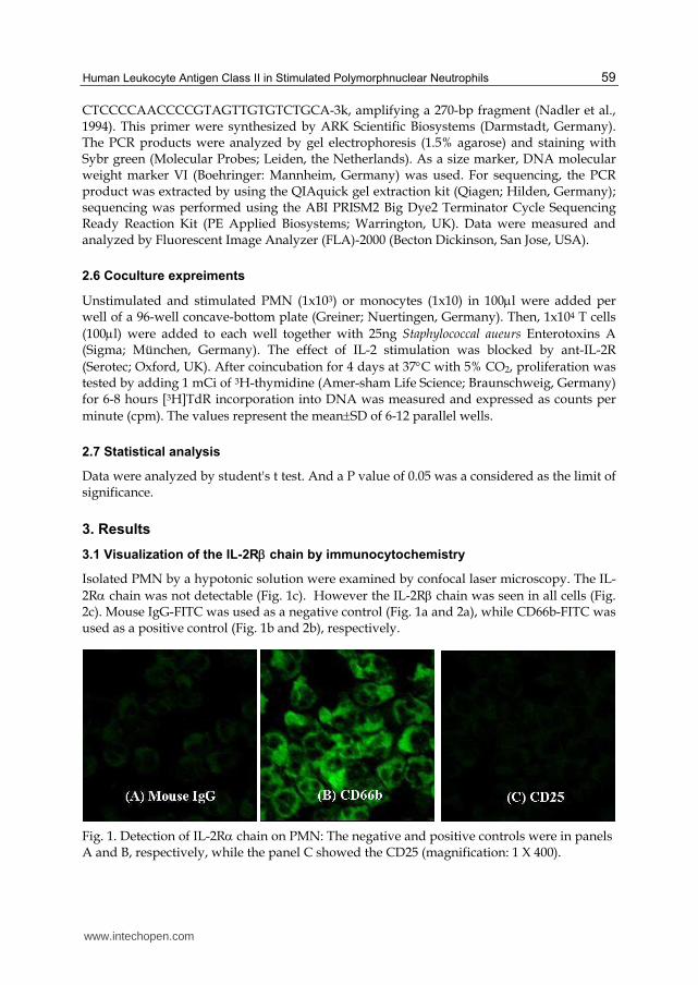
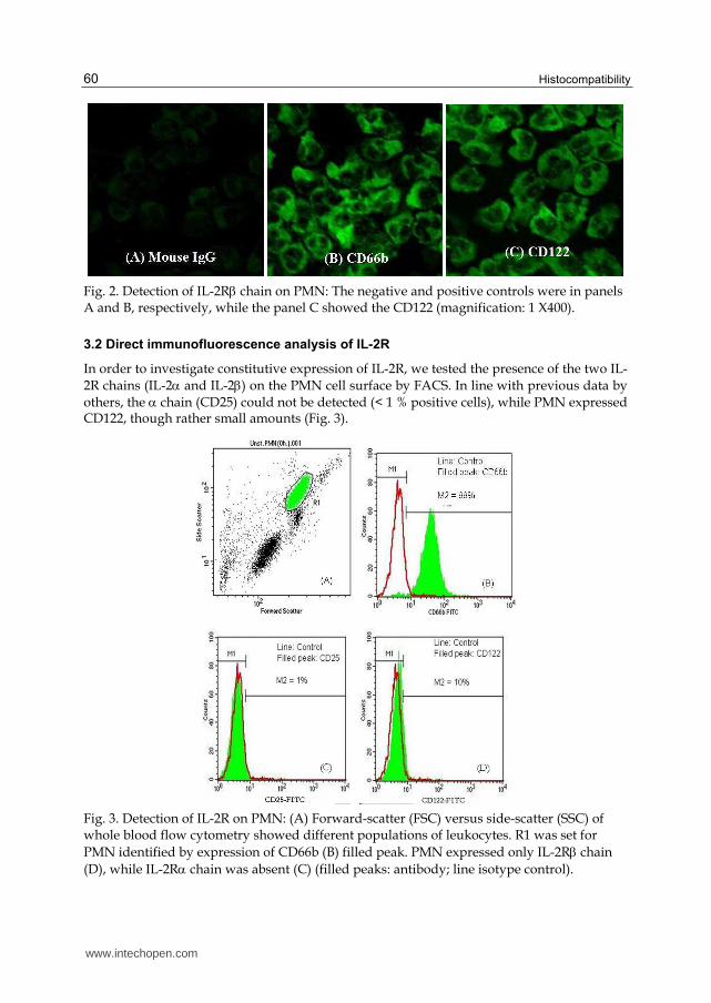
Isolated PMN by a hypotonic solution were examined by confocal laser microscopy. The IL-2R chain was not detectable (Fig. 1c). However the IL-2R chain was seen in all cells (Fig. 2c). Mouse IgG-FITC was used as a negative control (Fig. 1a and 2a), while CD66b-FITC was used as a positive control (Fig. 1b and 2b), respectively.
Fig. 1. Detection of IL-2R chain on PMN: The negative and positive controls were in panels A and B, respectively, while the panel C showed the CD25 (magnification: 1 X 400).
www.intechopen.com
Histocompatibility
60
Fig. 2. Detection of IL-2R chain on PMN: The negative and positive controls were in panels A and B, respectively, while the panel C showed the CD122 (magnification: 1 X400).
3.2 Direct immunofluorescence analysis of IL-2R
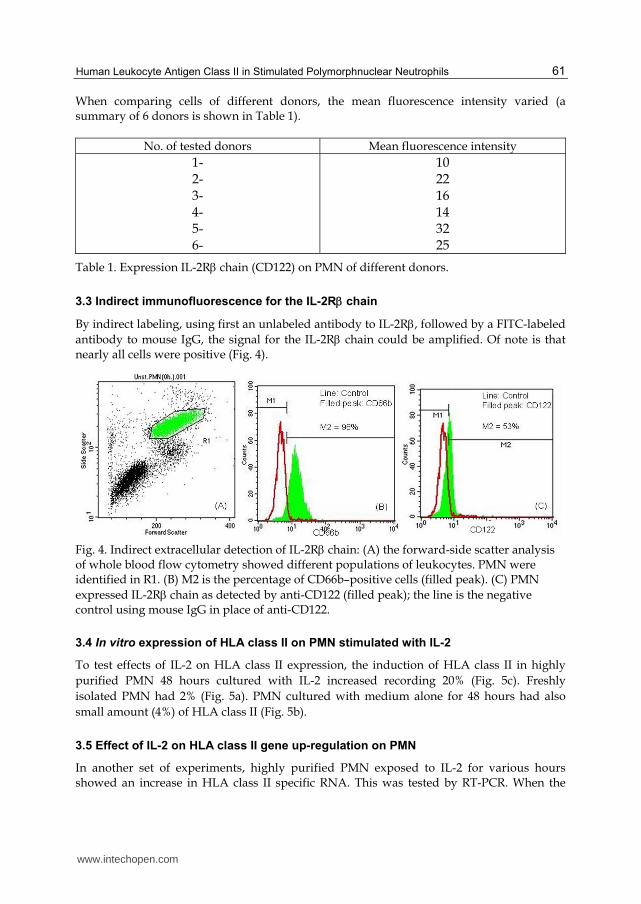
In order to investigate constitutive expression of IL-2R, we tested the presence of the two IL-2R chains (IL-2 and IL-2) on the PMN cell surface by FACS. In line with previous data by others, the chain (CD25) could not be detected (< 1 % positive cells), while PMN expressed CD122, though rather small amounts (Fig. 3).
Fig. 3. Detection of IL-2R on PMN: (A) Forward-scatter (FSC) versus side-scatter (SSC) of whole blood flow cytometry showed different populations of leukocytes. R1 was set for PMN identified by expression of CD66b (B) filled peak. PMN expressed only IL-2R chain (D), while IL-2R chain was absent (C) (filled peaks: antibody; line isotype control).
www.intechopen.com
Human Leukocyte Antigen Class II in Stimulated Polymorphnuclear Neutrophils
61
When comparing cells of different donors, the mean fluorescence intensity varied (a summary of 6 donors is shown in Table 1).
No. of tested donors Mean fluorescence intensity 1- 2- 3- 4- 5- 6-
10 22 16 14 32 25
Table 1. Expression IL-2R chain (CD122) on PMN of different donors.
3.3 Indirect immunofluorescence for the IL-2R chain
By indirect labeling, using first an unlabeled antibody to IL-2R, followed by a FITC-labeled antibody to mouse IgG, the signal for the IL-2R chain could be amplified. Of note is that nearly all cells were positive (Fig. 4).
Fig. 4. Indirect extracellular detection of IL-2R chain: (A) the forward-side scatter analysis of whole blood flow cytometry showed different populations of leukocytes. PMN were identified in R1. (B) M2 is the percentage of CD66b–positive cells (filled peak). (C) PMN expressed IL-2R chain as detected by anti-CD122 (filled peak); the line is the negative control using mouse IgG in place of anti-CD122.
3.4 In vitro expression of HLA class II on PMN stimulated with IL-2
To test effects of IL-2 on HLA class II expression, the induction of HLA class II in highly purified PMN 48 hours cultured with IL-2 increased recording 20% (Fig. 5c). Freshly isolated PMN had 2% (Fig. 5a). PMN cultured with medium alone for 48 hours had also small amount (4%) of HLA class II (Fig. 5b).
3.5 Effect of IL-2 on HLA class II gene up-regulation on PMN
In another set of experiments, highly purified PMN exposed to IL-2 for various hours showed an increase in HLA class II specific RNA. This was tested by RT-PCR. When the
www.intechopen.com
Histocompatibility
62
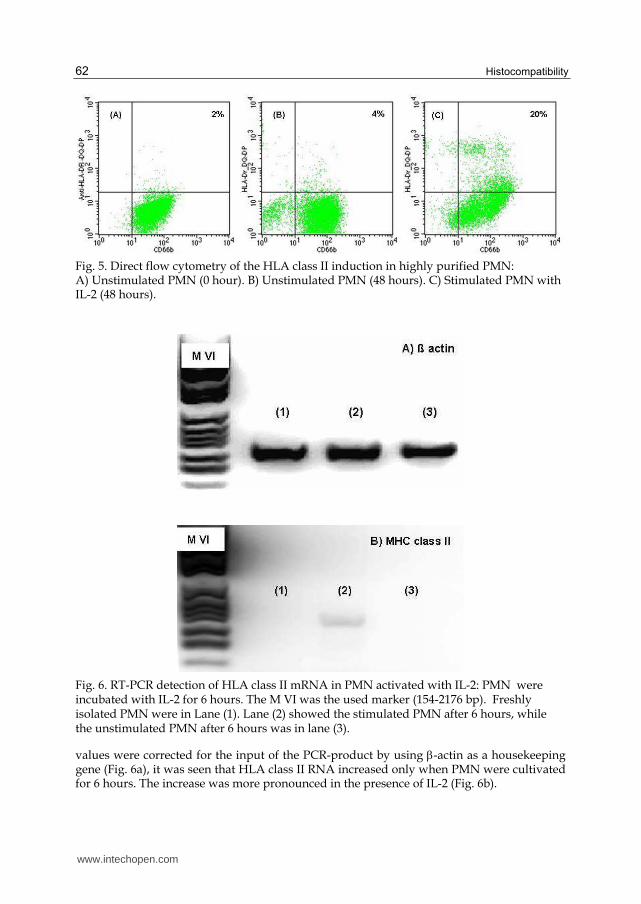
Fig. 5. Direct flow cytometry of the HLA class II induction in highly purified PMN: A) Unstimulated PMN (0 hour). B) Unstimulated PMN (48 hours). C) Stimulated PMN with IL-2 (48 hours).
Fig. 6. RT-PCR detection of HLA class II mRNA in PMN activated with IL-2: PMN were incubated with IL-2 for 6 hours. The M VI was the used marker (154-2176 bp). Freshly isolated PMN were in Lane (1). Lane (2) showed the stimulated PMN after 6 hours, while the unstimulated PMN after 6 hours was in lane (3).
values were corrected for the input of the PCR-product by using -actin as a housekeeping gene (Fig. 6a), it was seen that HLA class II RNA increased only when PMN were cultivated for 6 hours. The increase was more pronounced in the presence of IL-2 (Fig. 6b).
www.intechopen.com
Human Leukocyte Antigen Class II in Stimulated Polymorphnuclear Neutrophils
63
3.6 In vitro expression of HLA class II on PMN stimulated with IL-4
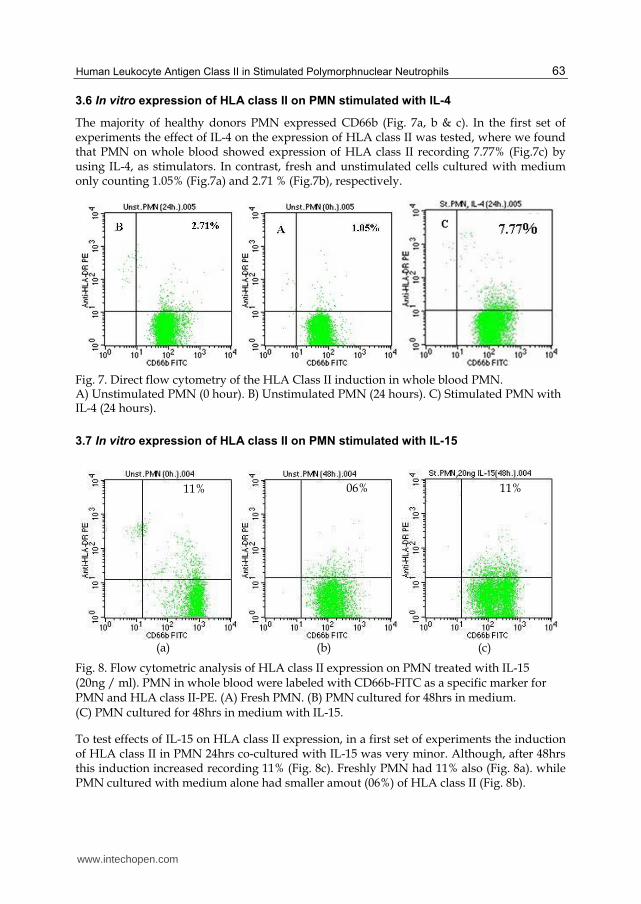
The majority of healthy donors PMN expressed CD66b (Fig. 7a, b & c). In the first set of experiments the effect of IL-4 on the expression of HLA class II was tested, where we found that PMN on whole blood showed expression of HLA class II recording 7.77% (Fig.7c) by using IL-4, as stimulators. In contrast, fresh and unstimulated cells cultured with medium only counting 1.05% (Fig.7a) and 2.71 % (Fig.7b), respectively.
Fig. 7. Direct flow cytometry of the HLA Class II induction in whole blood PMN. A) Unstimulated PMN (0 hour). B) Unstimulated PMN (24 hours). C) Stimulated PMN with IL-4 (24 hours).
3.7 In vitro expression of HLA class II on PMN stimulated with IL-15
(a) (b) (c)
Fig. 8. Flow cytometric analysis of HLA class II expression on PMN treated with IL-15 (20ng / ml). PMN in whole blood were labeled with CD66b-FITC as a specific marker for PMN and HLA class II-PE. (A) Fresh PMN. (B) PMN cultured for 48hrs in medium. (C) PMN cultured for 48hrs in medium with IL-15.
To test effects of IL-15 on HLA class II expression, in a first set of experiments the induction of HLA class II in PMN 24hrs co-cultured with IL-15 was very minor. Although, after 48hrs this induction increased recording 11% (Fig. 8c). Freshly PMN had 11% also (Fig. 8a). while PMN cultured with medium alone had smaller amout (06%) of HLA class II (Fig. 8b).
11% 06% 11%
www.intechopen.com

Histocompatibility
64
3.8 Interaction of HLA class II positive PMN with peripheral T-cells
The question was addressed, whether PMN stimulated to express HLA class II antigen, would be able to induce T-cell proliferation. For these experiments highly purified PMN were cultivated with IL-2 for 24 hours and then cocultivated with highly purified isolated T-cells in the absence or presence of Staphylococcus enterotoxin A (SEA), a well-known superantigen.
It was imperative to rule out the participation of professional antigen presenting cells, such as dendritic cell, monocytes or B-cells. Therefore great care was taken for purification of the cells, which was controlled cytofluorometry. Using this method, less than 1% contaminating cells could be detected in either the PMN or the T-cell preparation.
In a first set of experiments PMN in various numbers and for comparison monocytes were cocultivated with the T-cells and SEA and proliferation of T-cells was measured after 4 days. The experiment showed that about 10 times more PMN than monocytes were required to achieve comparable proliferation and that 100 monocytes were barely sufficient to induce T-cell proliferation above controls (T-cells alone or T-cells with SEA without PMN or monocytes, respectively) (Fig. 9).
Fig. 9. Cells were cocultured with PMN or monocytes in the numbers indicated and in the presence of SEA. After 4 days proliferation of T-cells was measured by incorporation of [3H] –thymidine, where proliferation was seen with at least 100 monocytes or 1000 PMN.
www.intechopen.com
Human Leukocyte Antigen Class II in Stimulated Polymorphnuclear Neutrophils
65
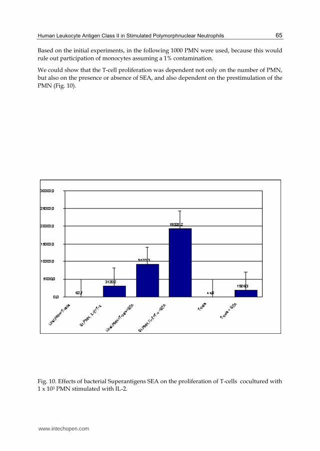
Based on the initial experiments, in the following 1000 PMN were used, because this would rule out participation of monocytes assuming a 1% contamination.
We could show that the T-cell proliferation was dependent not only on the number of PMN, but also on the presence or absence of SEA, and also dependent on the prestimulation of the PMN (Fig. 10).
Fig. 10. Effects of bacterial Superantigens SEA on the proliferation of T-cells cocultured with 1 x 10³ PMN stimulated with IL-2.
www.intechopen.com
Histocompatibility
66
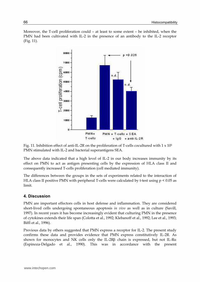
Moreover, the T-cell proliferation could – at least to some extent – be inhibited, when the PMN had been cultivated with IL-2 in the presence of an antibody to the IL-2 receptor (Fig. 11).
Fig. 11. Inhibition effect of anti-IL-2R on the proliferation of T-cells cocultured with 1 x 10³ PMN stimulated with IL-2 and bacterial superantigens SEA.
The above data indicated that a high level of IL-2 in our body increases immunity by its effect on PMN to act as antigen presenting cells by the expression of HLA class II and consequently increased T-cells proliferation (cell mediated immunity).
The differences between the groups in the sets of experiments related to the interaction of HLA class II positive PMN with peripheral T-cells were calculated by t-test using p < 0.05 as limit.
4. Discussion
PMN are important effectors cells in host defense and inflammation. They are considered short-lived cells undergoing spontaneous apoptosis in vivo as well as in culture (Savill, 1997). In recent years it has become increasingly evident that culturing PMN in the presence of cytokines extends their life span (Colotta et al., 1992; Klebanoff et al., 1992; Lee et al., 1993; Biffl et al., 1996).
Previous data by others suggested that PMN express a receptor for IL-2. The present study confirms these data and provides evidence that PMN express constitutively IL-2R. As shown for monocytes and NK cells only the IL-2R chain is expressed, but not IL-R (Espinoza-Delgado et al., 1990). This was in accordance with the present
www.intechopen.com

Human Leukocyte Antigen Class II in Stimulated Polymorphnuclear Neutrophils
67
immunofluorosence results, where we detected only Il-2R chain on resting PMN and confirmed my choosing of IL-2 as an activator of PMN.
Increasing evidence suggests that human PMN have an important role to play in regulating specific immune responses and in antigen presentation (Fanger et al., 1997; Radsak et al., 2000). In this study we have demonstrated that HLA Class II antigen was also detected, at the protein level by cytofowmetry, and also at the gene level by RT-PCR. These observations therefore agree well with the findings of others who have detected these antigens on the surface of activated neutrophils (Matsumoto et al., 1987; Radsak et al., 2000). The detection of these molecules therefore provides strong support for the hypothesis that human PMN can actively synthesize immunoregulatory molecules (Newburger et al., 2000) and have the potential to act as APCs (Fanger et al., 1997; Radsak et al., 2000). The observation that PMN may also express HLA class II antigens when appropriately stimulated (Gosselin et al., 1993 and Fanger et al., 1997), high lighted the possibility that PMN might function as accessory cells for T-cell activation. HLA class II expression was found on PMN of patients with active Wegener's disease (Haensch, et al., 1999). The close correlation of HLA class II expression to disease activity prompted the present study with the objective of investigating whether or not HLA class II-positive PMN might activate or support activation of T lymphocytes.
Induction of the above membrane-bound molecules was higher when PMN were stimulated in whole blood. The explanation of this phenomena may be due to the loss of some PMN IL-2R and IL-15R and other activities during their isolation procedure.
The absence or low detection of MHC class II on human PMN stimulated with IL-2, IL-4 and IL-15 could be due to: 1) The relatively long time (44 hrs) required for class II induction, which is beyond the period of PMN survival in most culture systems. 2) IL-2 and IL-15 receptor numbers may vary with the individual. 3) Synthetic capacity of the PMN may vary. 4) The capacities of cytokine receptors to signal may not be the same. 5) It may also be possible that the PMN themselves produce factors that inhibit class II induction. 6) Synthetic capacity of the PMN may vary. 7) The capacities of cytokine receptors to signal may not be the same. 8) It may also be possible that the PMN themselves produce factors that inhibit class II induction (Gosselin et al., 1993).
When comparing the number of monocytes and PMN required inducing T-cell proliferation, it was observed that 10 times more PMN than monocytes were necessary to yield the same extent of T-cell proliferation. However, the cell preparation used in this work never contained more than 1% of contaminating cells and certainly not the 10% of monocytes that would be required to affect the results. The observation that ten times more PMN than monocytes were required to induce a similar extent of T-cell proliferation has to be considered together with the observation that only a proportion of PMN acquired HLA class II. Thus, when only fully equipped PMN are considered, the ability to process and to present antigen is similar to that of monocytes. Whether HLA class II-positive PMN participate in the immune defense or play a role in pathophysiological events, is a matter of speculation. The fact that only a minor proportion of PMN acquire HLA class II might lead to the conclusion that a possible accessory function of PMN would be rather weak. One has, however, to bear in mind that PMN are numerous in the peripheral blood, and that even a low percentage of PMN expressing HLA class II would exceed both circulating monocytes and dendritic cells in number.
www.intechopen.com
Histocompatibility
68
Because of the notion that the presence of Staphylococcus entrotoxin coincides with relapses of Wagner's granulomatosis (Cohn-Terveart et al., 1999), we tested whether PMN was able to also present Staphylococcus enterotoxin A is a superantigen, so-called because it binds outside of the peptide-binding groove of the HLA class II and the antigen-specific domain of the T-cell receptor, and consequently activates a large portion of T-cells, preferentially those with a V beta 2 domain (Simpson et al., 1995). In accordance with previous studies (Fanger et al., 1997; Radsak et al., 2000), co-culture of PMN with T-cells and SE resulted in T-cell proliferation. Taken together, our data demonstrate that by synthesizing and expressing of HLA class II antigens, PMN acquire the capacity to present superantigens to T-cells.
5. Conclusion
In conclusion, PMN possession of IL-2R ┚ and ┛ chains that have the ability to bind with IL-2, IL-4 and IL-15 prompted us to use IL-2, IL-4 and IL-15 as stimuli for PMN. They stimulated PMN to express HLA class II which is the main antigen presenting molecules. PMN expressed HLA class II present SEA to T-cells causing their proliferation. T-cells proliferations lead to antigen destruction, B-cells activation and consequently immunoglobulin production, and activation of phagocytic cells.
6. Acknowledgement
I thank Prof. Dr. G. M. Haensch (Immunology Institute, Heidelberg, Germany) for using her laboratory in this study. I also thank her advice and encouragement throughout the work.
7. References
[1] Aversa, G., Punnonen, J., Cocks, B.G., de Waal Malefyt, R., Vega, F. Jr , Zurawski, S. M. , et al. (1993). An interleukin 4 (IL-4) mutant protein inhibits both IL-4 and IL-13-induced human immunoglobulin G4 (IgG4) and IgE synthesis and B cell proliferation: support for a common component shared by IL-4 and IL-13 receptors. J Exp Med., 178:2213-2218.
[2] Bhandari, V. (2002). Developmental differences in the role of interleukins in hyperoxic lung injury in animal models. Frontiers in Bioscience, 7: 1624-1633.
[3] Biffl, W. L., Moore, E. E., Moore, F. A., Barnett, C. C., Carl, V. S., Peterson V. M. (1996). Interleukin-6 delays neutrophil apoptosis. Arch. Surg., 131:24-30.
[4] Casatella, M.A., McDonald, P.P. (2000). Interleukin-15 its impact on neutrophil function. Curr. Opin. Hematol., 4:174-177.
[5] Cohn-Terveart, J. W., Popa, E. R., Bos, N. A. (1999). The role of superantigen in vasculitis. Curr. Opin. Rheumatol., 11:24-33.
[6] Colotta, F., Re, F., Polentarutti, N., Sozzani, S., Mantovani, A. (1992). Modulation of granulocyte survival and programmed cell death by cytokines and bacterial products. Blood, 80:2012-2020.
[7] Djeu, J. Y., Liu, J. H., Wei, S., Rui, H., Pearson, C. A., Leonard, W. J., Blanchard, D. K. (1993). Function associated with IL-2 receptor on human neutrophils: Mechanism of activation of antifungal activity against Candida alhicuns by IL-2. J. Immunol., 150:960-970.
www.intechopen.com
Human Leukocyte Antigen Class II in Stimulated Polymorphnuclear Neutrophils
69
[8] Espinoza-Delgado, I., Ortaldo, J. R., Winkler-Pickett, R., Sugamura, K., Varesio, L., Longo, D. L. (1990). Expression and role of p75 interleukin-2 receptor on human monocytes. J. Exp. Med., 171:1821-1826.
[9] Fanger, N. A., Liu, C., Guyre, P. M., Wardwell, G. K., Neil, J. O., Guo, T. L., Christian, T. P., Mudzinski, S. P., Gosselin, E. J. (1997). Activation of human T cells by major histocompatibility complex class II-expressing neutrophils: proliferation in the presence of superantigen, but not tetanus toxoid. Blood, 89:4128-4135.
[10] Gasmi, L., McLennan, A. G., Edwards, S. W. (1996). The diadenosine polyphosphates Ap3 A and Ap4 A and adenosine triphosphate interact with granulocyte-macrophage colony-stimulating factor to delay neutrophil apoptosis: implications for neutrophil-platelet interactions during inflammation. Blood, 87:3442-3449.
[11] Girard, D, Boiani, N, Beaulieu, AD. (1999). Human neutrophils express the interleukin-15 receptor alpha chain ( IL-15R alpha) but not the IL-9R alpha component. Clin.
Immunol. Immunophatol., 88: 232-240. [12] Girard, D, Paquet, ME, Paquin, R, Beaulieu, AD. (1996). Differential effects of
interleukin-15 ( IL-15) and IL-2 on human neutrophils: modulation of phagocytosis, cytoskeleton rearrangement, gene expression, and apoptosis by IL-15. Blood, 88: 3176-3184.
[13] Gosselin, E. J., Wardwell, K., Rigby, W. F., Guyre, P. M. (1993). Induction of HLA class II on human polymorphonuclear neutrophils by granulocyte/macrophage colony-stimulating factor, IFN-gamma, and IL-3. J. Immunol., 151:1482-1490.
[14] Haensch, G. M., Radsak, M., Wagner, C., Reis, B., Koch, A., Breitbart, A., Andrassy, K. (1999). Expression of major histocompatibility class II antigens on polymorphonuclear neutrophils in patients with Wegener’s granulomatosis. Kidney
Int., 55: 1811-1818. [15] Holter, W. (1997). Interleukin-4: structure and function. In: Remick DG, Friedland JS,
editors. Cytokines in health and disease. New York, NY: Marcel Dekker;. p. 53. [16] Idzerda, R. L., March, C. J., Mosley, B., Lyman, S D., Vanden Bo, s T., Gimpel, S. D. et
al. (1990). Human interleukin-4 receptor confers biological responsiveness and defines a novel receptor superfamily. J Exp Med., 171:861-873.
[17] Iking-Konert, C., Ostendorf, B., Sander, O., Jost, M., Wagner, C., Joosten, L., Schneider, M., Haensch, G. M. (2005). Transdifferentiation of polymorphonuclear neutrophils to dendritic-like cells at the site of inflammation in rheumatoid arthritis: evidence for activation by T cells. Ann. Rheum. Dis. 64:1436-1442.
[18] Iking-Konert, C., Cseko, C., Wagner, C., Stegmaier, S., Andrassy, K., Hansch, G. M. (2001a). Transdifferentiation of polymorphonuclear neutrophils: acquisition of CD83 and other functional characteristics of dendritic cells. J. Mol. Med. 79:464-474.
[19] Iking-Konert, C., Vogt, S., Radsak, M., Wagner, C., Hansch, G. M., Andrassy, K. (2001b). Polymorphonuclear neutrophils in Wegener’s granulomatosis acquire characteristics of antigen presenting cells. Kidney Int., 60:2247-2262.
[20] Iking-Konert, C., Wagner, C., Denefleh, B., Hug, F., Schneider, M., Andrassy, K., Hansch, G. M. (2002). Up-regulation of the dendritic cell marker CD83 on polymorphonuclear neutrophils (PMN): divergent expression in acute bacterial infections and chronic inflammatory disease. Clin. Exp. Immunol. 130:501-508.
www.intechopen.com

Histocompatibility
70
[21] Jupin, C., Anderson, S. Damais, C. Alouf, J. E. and Parant. M. (1988). Toxic shock syndrome toxin-1 as an inducer of human tumour necrosis factors and ┛ interferon. J. Exp. Med. 167:752-761.
[22] Klebanoff, S. J., Olszowski, S., van Voorhis, W. C., Ledbetter, J. A., Waltersdorph, A. M., Schlechte, K. G. (1992). Effects of gamma interferon on human neutrophils, protection from deterioration on storage. Blood, 80:225-234.
[23] Lagoo, A. S., Lagoo-Deenadayalan, S., Lorenz, H. M., Byrne, J., Barber, W. H., Hardy, K. J. (1994). IL-2, IL-4, and IFN-┛ gene expression versus secretion in superantigen-activated T-cells. J. Immunol. 152: 1641-1652.
[24] Lee, A., Whyte, M. B. K., Haslett, C. (1993). Inhibition of apoptosis and prolongation of neutrophil functional longevity by inflammatory mediators. J. Leukoc. Biol. 54:283-289.
[25] Lloyd, A. R., Oppenheim, J. J. (1992). Poly’s lament: The neglected role of the polymorphonuclear neutrophil in the afferent limb of the immune response. Immunol. Today, 13:169-172.
[26] Matsumoto, S., Takei, M., Moriyama, M., Imanishi, H. (1987). Enhancement of Ia like antigen expression by interferon gamma in polymorphonuclear leukocytes. Chem.
Pharm. Bull., 35:436-439. [27] Mastroianni, C. M., D ’Ettore, G., Forcina, G., Lichtner, M., Mengoni, F. D., Agostino,
C., Corpolongo, A., Massetti, A., Vullo, V. (2000). Interleukin-15 enhances neutrophil functional activity in patients with human immunodeficiency virus infection. Blood, 96: 1979-1984.
[28] Moulding, D. A., Quayle, J. A., Hart, C. A., Edwards, S. W. (1998). Mcl-1 expression in human neutrophils: regulation by cytokines and correlation with cell survival. Blood, 92:2495-2502.
[29] McDonald, P. P., Russo, M. P., Cassatella, M.A. (1998). Interleukin-15 (IL-15) induces NF-ќ B activation and IL-8 production in human neutrophils. Blood, 4828-4835.
[30] Mudzinski, S. P., Christian, T. P., Guo, T. L., Cirenza, E., Hazlett, K. R., Gosselin, E. J. (1995). Expression of HLA-DR (major histocompatibility complex class II) on neutrophils from patients treated with granulocyte macrophage colony-stimulating factor for mobilization of stem cells. Blood, 86:2452-2453.
[31] Muro, S, Taha, R. (2001). Expression of IL-15 in inflammatory pulmonary disease. J.
Allergy Clin. Immunol., 108: 970-975. [32] Nadler, S. G., Rankin, B. M., Moran-Davis, P., Cleaveland, J. S., Kiener, P. A. (1994).
Effect of interferon-c on antigen processing in human monocytes. Eur. J. Immunol., 24:3124-2130.
[33] Newburger, P. E., Subrahmanyam, Y. V. B. K., Weissman, S. M. (2000). Global analysis of neutrophil gene expression. Curr. Opin. Hematol., 7:16-20.
[34] Okuda, K., Neely, B. C., David, C. S. (1979). Expression of H-2 and Ia antigens on mouse peritoneal neutrophils. Transplantation, 28:354-356.
[35] Okuda, K., Tani, K., Ishigatsubo, Y., Yokota, S., David, C. S. (1980). Antigen-pulsed neutrophils bearing Ia antigens can induce T lymphocyte proliferate response to the syngeneic or semisyngeneic antigen-primed T lymphocytes. Transplantation, 30:368-372.
www.intechopen.com
Human Leukocyte Antigen Class II in Stimulated Polymorphnuclear Neutrophils
71
[36] Petroni, K.C., Shen, L., Guyre, P. M. (1988). Modulation of human polymorphonuclear leukocyte IgG Fc receptors and Fc receptor-mediated functions by IFN-g and glucocorticoids. J. Immunol., 140:3467-3472.
[37] Radsak, M., Iking-Konert, C., Stegmaier, S., Andrassy, K., Hänsch, G. M. (2000). Polymorphonuclear neutrophils as accessory cells for T-cell activation: major histocompatibility complex Class II restricted antigen-dependent induction of T-cell proliferation. Immunology, 101:521-530.
[38] Reali, E., Guerrini, R., Moretti, S., Spisani, S., Lanza, F., Tomatis, R., Traniello, S., Gavioli, R. (1996). Polymorphonuclear neutrophils pulsed with synthetic peptides efficiently activate memory cytotoxic T lymphocytes. J. Leukoc. Biol., 60:207-213.
[39] Reinisch, W., Tillinger, W., Lichtenberger, C., Gangl, A., Willheim, M., Scheiner, O., Steger, G. (1996). In vivo induction of HLA-DR on human neutrophils in patients treated with interferon-┛. Blood, 87:3068.
[40] Savill, J. (1997). Apoptosis in resolution of inflammation. J. Leukoc. Biol., 61:375-380. [41] See, R. H., Chow, A. W. (1992). Role of the adhesion molecule lymphocyte function
associated antigen 1 in toxic shock syndrome toxin 1-induced tumor necrosis factor alpha and interleukin-1┚ secretion by human monocytes. Infect. Immunol., 60:4957-4960.
[42] See, R. H., Kum, W. W. S., Chang, A. H., Goh, S. H., Chow, A. W. (1992). Induction of tumor necrosis factor and interleukin-1 by purified staphylo-coccal toxic shock syndrome toxin 1 requires the presence of both monocytes and T lymphocytes. Infect. Immunol., 60:2612-2618.
[43] Shauna, C., Owain, R. M., James, M. B., Iain, B. M. (2008). Murine neutrophils present Class II restricted antigen. Immunol. Lett., 15: 49-54.
[44] Shen, L., Guyre, P. M., Fanger, M. W. (1987). Polymorphonuclear leukocyte function triggered through the high affinity Fc receptor for monomeric IgG. J. Immunol., 139:534-538.
[45] Simpson, I. J., Skinner, M. A., Geursen, A. (1995). Peripheral blood T-lymphocytes in systemic vasculitis: Increased T-cell receptor V beta 2 gene usage in microscopic polyarteritis. Clin. Exp. Immunol., 101: 220-236.
[46] Smith, W. B., Guida, L., Sun, Q., Korpelainen, E. I., van den Heuvel, C., Gillis, D., Hawrylowicz, C. M., Vadas, M. A., Lopez, A. F. (1995). Neutrophils activated by granulocyte-macrophage colony-stimulating factor express receptors for interleukin-3 which mediate class II expression. Blood, 86;3938-3944.
[47] Stringer, R. E., Hart, C. A., Edwards, S. W. (1996). Sodium butyrate delays neutrophil apoptosis: role of protein biosynthesis in neutrophil survival. Br. J. Hematol., 92:169-175.
[48] Trede, N. S., Moris, T., Scholl, P. R., Geha, R. S., Chatila, T. (1994). Early activation events induced by the staphylococcal superantigen toxic shock syndrome toxin-1 in human peripheral blood monocytes. Clin. Immunol. Immunopathol., 70:137-144.
[49] Tyler, A. W., Terry, K. B., Lorne, A. B., Philip, J. G. (2006). Bovine polymorphonuclear cells passively acquire membrane lipids and integral membrane proteins from apoptotic and necrotic cells. J. Leukoc. Biol., 79:1226-1233.
www.intechopen.com
Histocompatibility
72
[50] Vachiery, N., Totte, P., Balcer, V., Martinez, D., Bensaid, A. (1999). Effect of isolation techniques, in vitro culture and IFN-┛ treatment on the constitutive expression of HLA class I and class II molecules on goat neutrophils. Vet. Immunol.
Immunopathol., 70:19-32. [51] Wagner, C., Iking-Konert, C., Hug, F., Stegmaier, S., Heppert, V., Wentzensen, A.,
Haensch G. M. (2006). Cellular inflammatory response to persistent localized Staphylococcus aureus infection: phenotypical and functional characterization of polymorphonuclear neutrophils (PMN). Clin. Exp. Immunol., 143:70-77.
[52] Waldmann, T. A. (1989). The multi-subunit interleukin-2 receptor. Ann. Rev. Biochem, 58:875-911.
[53] Zissel, G, Baumer, I. (2000). In vitro release of interleukin- 15 by bronchoalveolar lavage cells and peripheral blood mononuclear cells from patients with different lung disease. Eur. Cytokine Netw., 11: 105-112.
www.intechopen.com
HistocompatibilityEdited by Dr. Bahaa Abdel-Salam
ISBN 978-953-51-0589-3Hard cover, 188 pagesPublisher InTechPublished online 02, May, 2012Published in print edition May, 2012
InTech EuropeUniversity Campus STeP Ri Slavka Krautzeka 83/A 51000 Rijeka, Croatia Phone: +385 (51) 770 447 Fax: +385 (51) 686 166www.intechopen.com
InTech ChinaUnit 405, Office Block, Hotel Equatorial Shanghai No.65, Yan An Road (West), Shanghai, 200040, China
Phone: +86-21-62489820 Fax: +86-21-62489821
This book presents some recent researches related to histocompatibility for scientists interested in this field. Itincludes 10 chapters, in different topics, prepared by Sundararajulu Panneerchelvam and Mohd Nor Norazmi;Giada Amodio and Silvia Gregori; Adema Ribic; Bahaa K. A. Abdel-Salam; Kai-Fu Tang; Roberto Biassoni,Irene Vanni and Elisabetta Ugolotti; Wei-Cheng Yang, Lien-Siang Chou and Jer-Ming Hu; Shatrah Othman andRohana Yusof; Masahiro Hirayama, Eiichi Azuma and Yoshihiro Komada; Gustav Roder, Linda Geironson,Elna Follin, Camilla Thuring and Kajsa Paulsson.
How to referenceIn order to correctly reference this scholarly work, feel free to copy and paste the following:
Bahaa K. A. Abdel-Salam (2012). Human Leukocyte Antigen Class II in Stimulated PolymorphnuclearNeutrophils, Histocompatibility, Dr. Bahaa Abdel-Salam (Ed.), ISBN: 978-953-51-0589-3, InTech, Availablefrom: http://www.intechopen.com/books/histocompatibility/human-leukocyte-antigen-class-ii

© 2012 The Author(s). Licensee IntechOpen. This is an open access articledistributed under the terms of the Creative Commons Attribution 3.0License, which permits unrestricted use, distribution, and reproduction inany medium, provided the original work is properly cited.