Embed Size (px)
Citation preview
For Peer ReviewPTHR1 MUTATIONS ASSOCIATED WITH OLLIER DISEASE RESULT IN
RECEPTOR LOSS OF FUNCTION
Journal: Human Molecular Genetics
Manuscript ID: draft
Manuscript Type: 2 General Article - UK Office
Date Submitted by the Author:
n/a
Complete List of Authors: Couvineau, Alain; INSERMU773, Centre de Recherche Biomédicale Bichat Beaujon CRB3; Université Paris 7, UFR Médicale, 75018 Wouters, Vinciane; Laboratory of Human Molecular Genetics, de Duve Institute, Université catholique de Louvain, B-1348 Bertrand, Guylène; Service de Biochimie hormonale et génétique, AP-HP, Hôpital Bichat Claude Bernard, Université Paris 7, UFR Médicale, 75018 Rouyer, Christiane; INSERM U773, Centre de Recherche Biomédicale Bichat Beaujon CRB3; Université Paris 7, UFR Médicale, 75018 Gérard, Bénédicte; Service de Biochimie hormonale et génétique, AP-HP, Hôpital Bichat Claude Bernard, Université Paris 7, UFR Médicale, 75018 Boon, Laurence; Université catholique de Louvain, Division of Plastic Surgery Grandchamp, Bernard; Service de Biochimie hormonale et génétique, AP-HP, Hôpital Bichat Claude Bernard, Université Paris 7, UFR Médicale, 75018 Vikkula, Miikka; Lab of Human Molecular Genetics, de Duve Institute, UCL Silve, Caroline; INSERMU561, Hôpital Saint Vincent de Paul, 82 av Denfert Rochereau 75014; Service de Biochimie hormonale et génétique, AP-HP, Hôpital Bichat Claude Bernard, Université Paris 7, UFR Médicale, 75018
Key Words: PTHR1, enchondromatosis, mutation, pathophysiology
Human Molecular Genetics
For Peer Review
1
PTHR1 MUTATIONS ASSOCIATED WITH OLLIER DISEASE RESULT IN
RECEPTOR LOSS OF FUNCTION
Alain Couvineau1, Vinciane Wouters2, Guylène Bertrand3, Christiane Rouyer1, Bénédicte
Gérard3, Laurence M Boon2,4, Bernard Grandchamp3, Miikka Vikkula2, Caroline Silve1,3,5
1 INSERM U773, Centre de Recherche Biomédicale Bichat Beaujon CRB3; Université Paris
7, UFR Médicale, 75018 Paris, France
2 Laboratory of Human Molecular Genetics, de Duve Institute, Université catholique de
Louvain, B-1348 Brussels, Belgium
3 AP-HP, Hôpital Bichat Claude Bernard, Service de Biochimie hormonale et génétique;
Université Paris 7, UFR Médicale, 75018 Paris, France
4 Center for Vascular Anomalies, Division of Plastic Surgery, Cliniques universitaires St Luc,
10-1200 Brussels, Belgium
5 Present address: INSERM U561, Hôpital Saint Vincent de Paul, 84 avenue Denfert
Rochereau, 75014 Paris, France
Page 1 of 32 Human Molecular Genetics
123456789101112131415161718192021222324252627282930313233343536373839404142434445464748495051525354555657585960
For Peer Review
2
Address correspondence to :
Caroline Silve M.D., PhD
INSERM U561 et centre des maladies rares du calcium et du phosphore
Hôpital Saint Vincent de Paul
84 avenue Denfert Rochereau, 75014 Paris, France
tel : 33 1 40 48 80 17
fax : 33 1 40 48 83 40
Page 2 of 32Human Molecular Genetics
123456789101112131415161718192021222324252627282930313233343536373839404142434445464748495051525354555657585960
For Peer Review
3
Abstract
PTHR1 signalling pathway is critical for regulation of endochondral ossification. Thus,
abnormalities in genes belonging to this pathway could potentially participate in the
pathogenesis of Ollier disease / Maffucci's syndrome, two developmental disorder defined by
the presence of multiple enchondromas. In agreement, a functionally deleterious mutation in
PTHR1 (p.R150C) was identified in enchondromas from two of six unrelated patients with
enchondromatosis. However, neither the p.R150C mutation (26 tumors) nor any other
mutation in the PTHR1 gene (11 patients) could be identified in another study. To further
define the role of PTHR1 signalling pathway in Ollier disease and Maffucci syndrome, we
analysed the coding sequences of four genes (PTHR1, IHH, PTHrP and GNAS1) in
leucocyte and/or tumor DNA from 57 and 23 patients affected with Ollier disease or Maffucci
syndrome respectively. We identified three previously undescribed missense mutations in
PTHR1 in patients with Ollier disease at the heterozygous state. Two mutations (p.G121E,
p.A122T) were present only in enchondromas, and one (p.R255H) in both enchondroma and
leukocyte DNA. Assessment of receptor function demonstrated that these three mutations
impair PTHR1 function, either by reducing the affinity of the receptor for PTH or reducing
receptor expression at the cell surface. These mutations were not found in DNA from 222
controls. Including our data, PTHR1 functionally deleterious mutations have now been
identified in 5/31 enchondromas from Ollier patients. These findings provide further support
for the idea that heterozygous mutations in PTHR1 that impair receptor function participate in
the pathogenesis of Ollier disease in some patients.
Page 3 of 32 Human Molecular Genetics
123456789101112131415161718192021222324252627282930313233343536373839404142434445464748495051525354555657585960
For Peer Review
4
INTRODUCTION
Enchondromatosis (OMIM 166000) or Ollier disease (World Health Organization
terminology) (1) is a developmental disorder defined by the presence of multiple
enchondromas (2-5). Typically, these cartilaginous lesions have an asymmetric distribution,
but important variability is seen in the age of onset of the disease and the size, number,
location, and evolution of the enchondromas. Most patients have bilateral
enchondromatosis, but there is a tendency for one side of the body to be more severely
affected. The condition in which multiple enchondromatosis is associated with vascular
anomalies characterized by the presence of fusiform cells and high frequency of
mesenchymal tumors is known as Maffucci syndrome (2, 6). Enchondromas in Ollier
disease present a significant risk of malignant transformation into chondrosarcoma, which
usually occurs in young adults, and thus occurs at an earlier age than is observed in patients
with isolated chondrosarcoma (2, 7-9). The reported incidence of malignant transformation is
even higher in Maffucci's syndrome, the prognosis of which is more severe than that of Ollier
disease (2, 3). The association of Ollier disease with other tumors, particularly ovarian
juvenile granulosa cell tumors has been reported (2, 10-12).
Enchondromas are almost exclusively localized in the metaphysis of long bones and
in the small bones of the hands and feet (2, 3, 13). The tumors initially develop close to the
growth plate cartilage where endochondral bone ossification occurs and then migrate
progressively towards the diaphysis. Endochondral bone ossification is a highly regulated
process that requires the differentiation of mesenchymal cells into hypertrophic chondrocytes
and the subsequent replacement of a cartilaginous matrix by mineralized bone. It has been
postulated that enchondromas result from abnormalities in signaling pathways controlling the
proliferation and differentiation of chondrocytes, leading to the development of intraosseous
cartilaginous foci. There are few reports of cytogenetic evaluation of benign enchondromas
in Ollier disease, and no tumor-specific chromosomal abnormalities have been associated
with enchondromas, or chondrosarcomas in these diseases (14-16). Studies evaluating
Page 4 of 32Human Molecular Genetics
123456789101112131415161718192021222324252627282930313233343536373839404142434445464748495051525354555657585960
For Peer Review
5
isolated chondrosarcomas have usually revealed heterogeneous and complex changes (16),
although loss of heterozygosity at chromosomal band 9p21 has been observed in several
cases, and is associated with a loss of expression of the INK4A/p16 protein, a tumor
suppressor gene involved in control of the cell cycle (17). Expression of parathyroid related
peptide (PTHrP), its receptor PTHR1, and BCL2 may be correlated with the grade of
malignancy in chondrosarcoma (18-21).
PTHrP and Indian Hedgehog (IHH) acting on their respective receptors PTHR1 and
PTCH1 participate in a tightly coupled signalling relay that is critical for the regulation of
chondrocyte differentiation and endochondral ossification (22). Thus, abnormalities in these
genes could potentially participate in the pathogenesis of Ollier disease / Maffucci's
syndrome. In agreement with this hypothesis, a functionally deleterious mutation in PTHR1
(p.R150C) was identified in enchondromas from two of six unrelated patients with
enchondromatosis (23). However, neither the p.R150C mutation (26 tumors) nor any other
mutation in the PTHR1 gene (11 patients) could be identified in another study (24). These
findings suggest that the molecular defects associated with enchondromatosis may be
heterogeneous, but no other candidate genes, including those participating in the PTHrP /
IHH pathway, have been evaluated.
To further define the role of PTHR1 signalling pathway in Ollier disease / Maffucci
syndrome, we analysed the coding sequences of PTHR1 and three other genes (PTHrP,
IHH, Gsα) implicated in this pathway in two large cohorts of patients. In this study, we have
i) identified three new mutations in PTHR1 in patients with Ollier disease, ii) characterized
the abnormalities in receptor function resulting from these mutations, and iii) through
modelling of the N-terminal ectodomain, evaluated the impact of the PTHR1 mutations on the
receptor function.
Page 5 of 32 Human Molecular Genetics
123456789101112131415161718192021222324252627282930313233343536373839404142434445464748495051525354555657585960
For Peer Review
6
MATERIALS AND METHODS
Patients
Informed consent was obtained for all patients. The study was performed in two series of
patients. The first series comprised 46 patients (29 females, 17 males) from a French
cohort. The clinical characteristics of the patients are presented supplementary table 1.
Data were obtained by review of clinical charts, contact with physicians in charge of the
patients, and through a questionnaire sent to the patients in collaboration with the French
association for Ollier disease / Maffucci syndrome. Patient age at diagnosis ranged from 1 to
36 years (median 6.4 years). All patients were affected by at least three enchondromas.
Unilateral body involvement was present in 23 patients (52%), and bilateral involvement with
a marked asymmetry was noted in 9 patients. No patients had spinal involvement. Two
patients (HP17 and HP55) were affected by chondrosarcomas. One patient (HP17) had
Maffucci syndrome. For all patients, the diagnosis of exostosis was excluded. Sixteen
females (59%) and four (26%) had a height ≥ 50 percentile (p=0.06 by Fisher's exact test).
In all patients, serum calcium and phosphorus levels were within the normal range for the
age (data not shown). Leukocyte DNA was obtained from all patients; tumor DNA was
available for 12 patients, including the patient with Maffucci syndrome (10 enchondromas
and 2 chondrosarcomas).
The second series comprised 34 patients from a Belgium cohort, 12 patients affected
by Ollier disease (5 females, 7 males; age at the time of the study 6-38 years), and 22
patients affected by Maffucci syndrome (11 females, 11 males; age at the time of the study
15-52 years) (supplementary table 2). Leukocyte DNA was obtained from all patients except
one; tumor DNA was available for 3 patients affected with Ollier disease and 13 patients with
Maffucci syndrome (10 enchondromas, 2 chondrosarcomas, one spindle cell
hemangioendothelioma).
Genomic DNA from 222 Caucasians was used as a control group.
Page 6 of 32Human Molecular Genetics
123456789101112131415161718192021222324252627282930313233343536373839404142434445464748495051525354555657585960
For Peer Review
7
Sequence analysis
Genomic DNA was extracted from peripheral blood and/or tumors obtained at the
time of surgery using a QIAamp DNA purification Kit (Qiagen SA, Courtaboeuf, France).
Intronic, and when required, exonic primers were used to amplify all coding exons and intron-
exon junctions for the PTHR1, IHH, PTHrP and GNAS1 (Gsα exons 1-13) genes. PTHR1
gene was analysed in all leukocyte and tumor DNA samples from the French and Belgium
cohorts. IHH and PTHrP were analysed in all leukocyte and tumor DNA from the French
cohort. The GNAS1 gene (exons 1-13 of Gsa) was analysed in tumor (9 samples) and
leukocyte DNA from 4 patients from the French cohort. The sequences of primers are
available upon request. PCR products were analysed by direct sequencing (french patients
and 112 controls) or by prescreening using SSCP/heteroduplex analysis combined with
sequencing of abnormally migrating fragments (belgium patients and 110 controls).
PCR products were sequenced in both directions. Sequencing reactions were performed
using the BigDye Terminator Cycle Sequencing Ready Reaction kit (Applied biosystems,
Courtaboeuf, France), and analysed using an ABI PRISM 3100 sequencer (Applied
Biosystems) and Beckman CEQ2000 fluorescent capillary sequencer for the french and
belgium cohorts respectively.
Nucleotide positions are numbered from the ATG start codon in the cDNA (sequence
accession numbers: PTHR1, NM_000316; IHH, NM_002181; PTHrP transcript variant 1,
NM_198965; PTHrP transcript variant 2, NM_002820; PTHrP transcript variant 3,
NM_198964, GNAS1, NM_000516). All nucleotide changes (except for previously reported
polymorphisms) were verified by resequencing of a different PCR product. Only silent and
missense nucleotide changes in the coding region are reported. However, for all
polymorphisms identified in introns, it was verified that they were not predicted to affect
splicing (http://www.fruitfly.org/seq_tools/splice.html).
Site directed mutagenesis
Page 7 of 32 Human Molecular Genetics
123456789101112131415161718192021222324252627282930313233343536373839404142434445464748495051525354555657585960
For Peer Review
8
Full length wild-type (WT) complementary DNA (cDNA) encoding the human PTHR1
(PTHR1) was inserted into the pEGFP-N2 N-terminal fusion protein vector (Clontech
Laboratories, Mountain View, CA) upstream of the enhanced green fluorescent protein gene
(25). Missense mutations were introduced in the WT PTHR1 cDNA construct using the
Quick Change Site directed Mutagenesis kit (Stratagene, La Jolla, CA) and confirmed by
complete sequencing of cDNA inserts (oligonucleotide sequences available on request).
Cell culture and transient transfection
The function of all mutated PTHR1 receptors were studied following transient
expression in CHO cells. Mutant p.R150C, p.G121E, p.A122T PTHR1 were also studied
following expression in COS-7. CHO and COS-7 cells were cultured in 24 well plates as
previously described (26-28). Transfection of CHO cells were performed using 1.5 µl of
FuGENE 6 transfection reagent (Roche Diagnostics, Indianapolis, IN) with 1 µg pEGFPN2
plasmid encoding wild-type or mutant PTHR1. Transfection of COS-7 cells were performed
using lipofectamine 2000 (Invitrogen SARL, Cergy Pontoise, France) and 500 ng DNA / well
as previously described (26, 28).
[125I] PTH 1-34 binding, imunological assessment of receptor expression, and cAMP
measurement
Techniques used to assess PTH 1-34 binding, PTHR1 cell membrane expression,
and cAMP production and have been described (26-28). Brieflly, the binding properties of
wild-type and mutated hPTHR1 receptors were determined by competitive inhibition of [125I]
(Nle8,18Tyr34)human PTH (1-34) amide ([125I]-PTH1-34) (Amersham Biosciences UK Ltd,
Buckinghalmshire, England) binding to transfected cells by increasing concentrations of
unlabeled (Nle8,18Tyr34) human PTH (1-34) (Bachem UK Ltd, Merseyside, England) (PTH1-
34) as in (25, 28). The specific binding was calculated as the difference between [125I]-
peptide bound and the non-specific binding (i.e., that measured in the presence of 8x10⁻⁷M
unlabeled PTH 1-34). To determine the IC50, binding data was fitted to a sigmoid curve with
variable slope (Graphpad Prism 4, GraphPad Software Inc., San Diego, CA). Immunological
Page 8 of 32Human Molecular Genetics
123456789101112131415161718192021222324252627282930313233343536373839404142434445464748495051525354555657585960
For Peer Review
9
assessment of cell surface receptor expression was performed using a rabbit polyclonal
antibody raised against a 17 amino acid epitope present in exon E2 of the human PTHR1
(90E-S-E-E-D-K-E-A-P-T-G-S-R-Y-R-G-106R) (PTHR1 E2 antibody) and a secondary [125I]-
labelled donkey anti-rabbit immunoglobulin antibody (Amersham biosciences UK Ltd,
Buckinghalmshire, England) as previously described (29). For the determination of
intracellular cAMP production, cells were incubated without or with hPTH1-34 under
continuous agitation in 0.5 ml of culture medium containing 0.1% (w/v) bovine serum albumin
and 1 mM 3-isobutyl-1-methylxanthine (Sigma-Aldrich, Saint-Quentin Fallavier, France).
After a 20 min incubation at 37 °C, the medium was removed and cells were lysed with 1 M
perchloric acid. The cAMP present in the lysate was measured by radioimmunoassay as
described (26). Results are expressed as pmol of cAMP/well. Experiments were performed
at least three times in duplicate with two different plasmid preparations.
Confocal laser scanning microscopy
CHO cells were transiently transfected with the WT or mutant PTHR1 cDNA inserted
into the pEGFP plasmid. Forty-eight h after transfection, unpermeabilized cells were
incubated for 30 min at 4°C with the PTHR1 antibody used for the assessment of PTHR1
expression (PTHR1 E2 antibody), washed extensively with phosphate-buffered saline, and
incubated for 30 min at 4°C with a secondary rhodamine-labelled donkey anti-rabbit
immunoglobulin. Cells were fixed in 1% paraformaldehyde. Glass coverslides were mounted
and examined by confocal laser scanning microscopy (CLSM-510-META, Zeiss, Germany)
equipped with epifluorescent optics (x63 NA 1.3 oil-immersion objective). Simultaneous two-
channel recording was performed with a pinhole size of 90 µm using excitation wavelengths
of 488/588 nm, a 510/580 double dichroic mirror, and a 515-545 bandpass fluorescein
isothiocyanate filter together with a 590-nm long pass filter. Double-labelled cells were
analyzed separately to avoid spillover between channels. In all experiments, omission of the
primary antibody was confirmed to result in no detectable staining.
Page 9 of 32 Human Molecular Genetics
123456789101112131415161718192021222324252627282930313233343536373839404142434445464748495051525354555657585960
For Peer Review
10
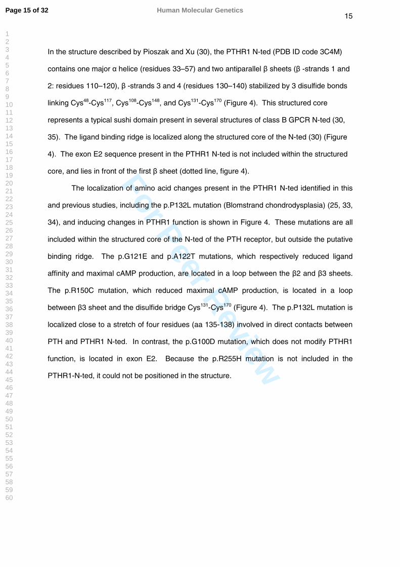
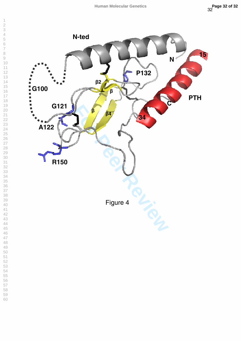
Location of mutations in the structure of the hPTHR1 N-terminal ectodomain (N-ted)
complexed to PTH.
In order to locate the spatial position of the PTHR1 mutations, we plotted each residue in the
recently published structure of the N-ted of PTHR1 (PDB ID code 3C4M) bound to PTH (15-
34) obtained by crystallization and X-ray diffraction (30).
Page 10 of 32Human Molecular Genetics
123456789101112131415161718192021222324252627282930313233343536373839404142434445464748495051525354555657585960
For Peer Review
11
RESULTS
Sequencing of candidate genes
PTHrP, IHH and Gsα were only tested in the French cohort. No variant alleles were
identified in the coding sequence of the PTHrP gene in either leukocyte or tumor DNA in the
50 patients studied whereas three previously described silent polymorphisms (rs3731878,
p.T200T; rs3731881, p.P251P; rs394452, p.T376T), and one previously undescribed
heterozygous variant (p.Y180Y / c.540T>C; HP 65) were identified in IHH. The allele
frequency of the known polymorphisms did not differ from that reported in the NCBI SNP
database (not shown). Three previously described silent polymorphisms (rs7121, p.I131I;
rs3730171, p.I186I; rs8386, p.N371N) were identified in Gsα. Again, their allele frequency
did not differ from that reported in the NCBI SNP database (not shown).
Seven heterozygous previously unreported PTHR1 SNPs, 4 silent (p.D30D, p.A72A,
p.T163T, p.V455V) and 3 non-synonymous (p.G121E, p.A122T, p.R225H), were identified
only in samples from the 80 patients with Ollier disease / Maffucci syndrome (supplementary
tables 1 and 2). The three previously undescribed non-synonymous mutations were all
found in patients with Ollier disease. Two of these mutations (p.G121E, p.A122T) were
observed in DNA from enchondromas, but not in leukocyte DNA; the R255H mutation was
detected both in enchondroma and leukocyte DNA. In addition, two known non-synonymous
polymorphisms (p.G100D, p.E546K) were identified in patients and controls (p.G100D, 1
patient and 1 control; p.E546K, 3 patients and 6 controls) (23, 31). The allele frequency of
the common synonymous PTHR1 polymorphism p.N463N (rs186987) was similar in the
patient and control populations, and to that described in the NCBI SNP database. Two
unreported heterozygous polymorphisms, one synonymous (p.S492S) and one non-
synonymous (p.P581R), were each identified in one control subject.
Functional characterization of mutant and wild-type PTHR1
Page 11 of 32 Human Molecular Genetics
123456789101112131415161718192021222324252627282930313233343536373839404142434445464748495051525354555657585960
For Peer Review
12
Functional studies evaluating the impact of all nonsynonymous SNPs identified in
patients with Ollier disease, including the three new alterations identified in this study
(p.G121E, p.A122T, p.R225H), the mutation described by Hopyan et al. (p.R150C), and the
two known polymorphisms (p.G100D, p.E546K) were performed (functional characterization
of p.E546K, but not p.G100D, has been previoulsy reported (23, 31)). In addition, The
p.H223R mutation identified in patients with Jansen's metaphyseal chondrodysplasia was
evaluated (32).
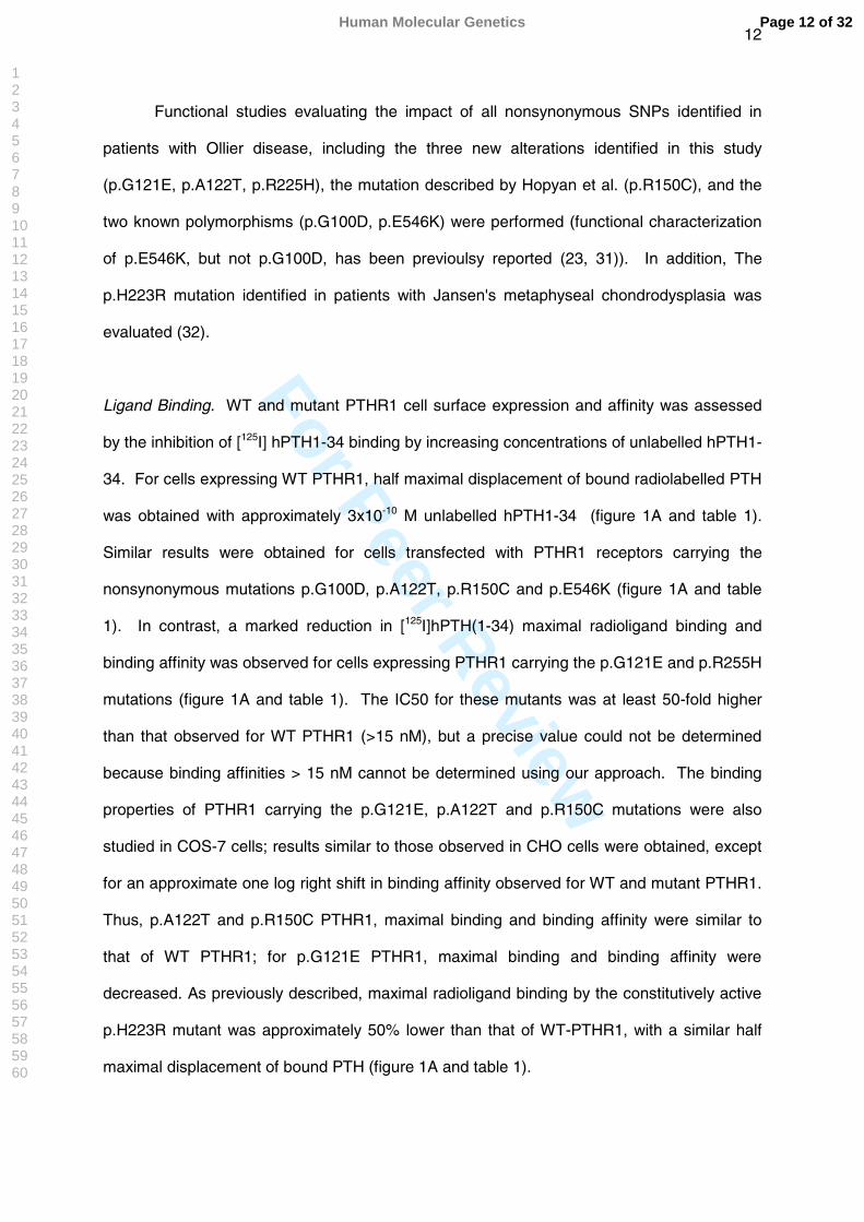
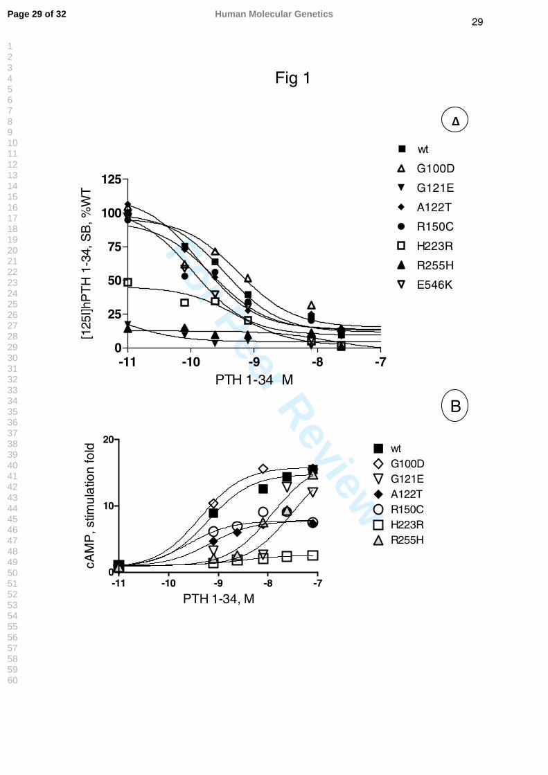
Ligand Binding. WT and mutant PTHR1 cell surface expression and affinity was assessed
by the inhibition of [125I] hPTH1-34 binding by increasing concentrations of unlabelled hPTH1-
34. For cells expressing WT PTHR1, half maximal displacement of bound radiolabelled PTH
was obtained with approximately 3x10⁻¹⁰ M unlabelled hPTH1-34 (figure 1A and table 1).
Similar results were obtained for cells transfected with PTHR1 receptors carrying the
nonsynonymous mutations p.G100D, p.A122T, p.R150C and p.E546K (figure 1A and table
1). In contrast, a marked reduction in [125I]hPTH(1-34) maximal radioligand binding and
binding affinity was observed for cells expressing PTHR1 carrying the p.G121E and p.R255H
mutations (figure 1A and table 1). The IC50 for these mutants was at least 50-fold higher
than that observed for WT PTHR1 (>15 nM), but a precise value could not be determined
because binding affinities > 15 nM cannot be determined using our approach. The binding
properties of PTHR1 carrying the p.G121E, p.A122T and p.R150C mutations were also
studied in COS-7 cells; results similar to those observed in CHO cells were obtained, except
for an approximate one log right shift in binding affinity observed for WT and mutant PTHR1.
Thus, p.A122T and p.R150C PTHR1, maximal binding and binding affinity were similar to
that of WT PTHR1; for p.G121E PTHR1, maximal binding and binding affinity were
decreased. As previously described, maximal radioligand binding by the constitutively active
p.H223R mutant was approximately 50% lower than that of WT-PTHR1, with a similar half
maximal displacement of bound PTH (figure 1A and table 1).
Page 12 of 32Human Molecular Genetics
123456789101112131415161718192021222324252627282930313233343536373839404142434445464748495051525354555657585960
For Peer Review
13
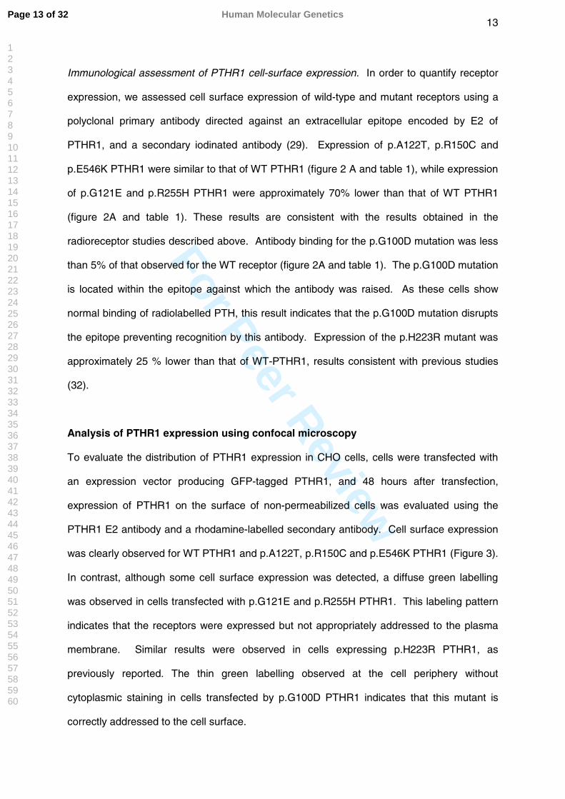
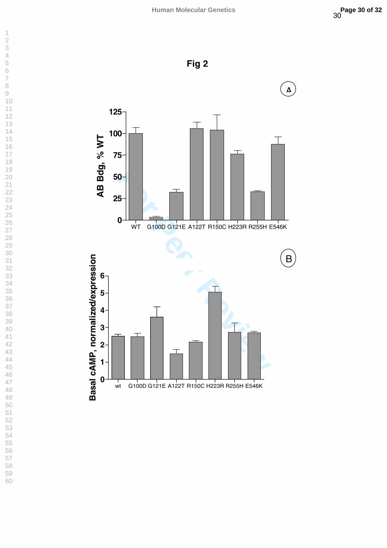
Immunological assessment of PTHR1 cell-surface expression. In order to quantify receptor
expression, we assessed cell surface expression of wild-type and mutant receptors using a
polyclonal primary antibody directed against an extracellular epitope encoded by E2 of
PTHR1, and a secondary iodinated antibody (29). Expression of p.A122T, p.R150C and
p.E546K PTHR1 were similar to that of WT PTHR1 (figure 2 A and table 1), while expression
of p.G121E and p.R255H PTHR1 were approximately 70% lower than that of WT PTHR1
(figure 2A and table 1). These results are consistent with the results obtained in the
radioreceptor studies described above. Antibody binding for the p.G100D mutation was less
than 5% of that observed for the WT receptor (figure 2A and table 1). The p.G100D mutation
is located within the epitope against which the antibody was raised. As these cells show
normal binding of radiolabelled PTH, this result indicates that the p.G100D mutation disrupts
the epitope preventing recognition by this antibody. Expression of the p.H223R mutant was
approximately 25 % lower than that of WT-PTHR1, results consistent with previous studies
(32).
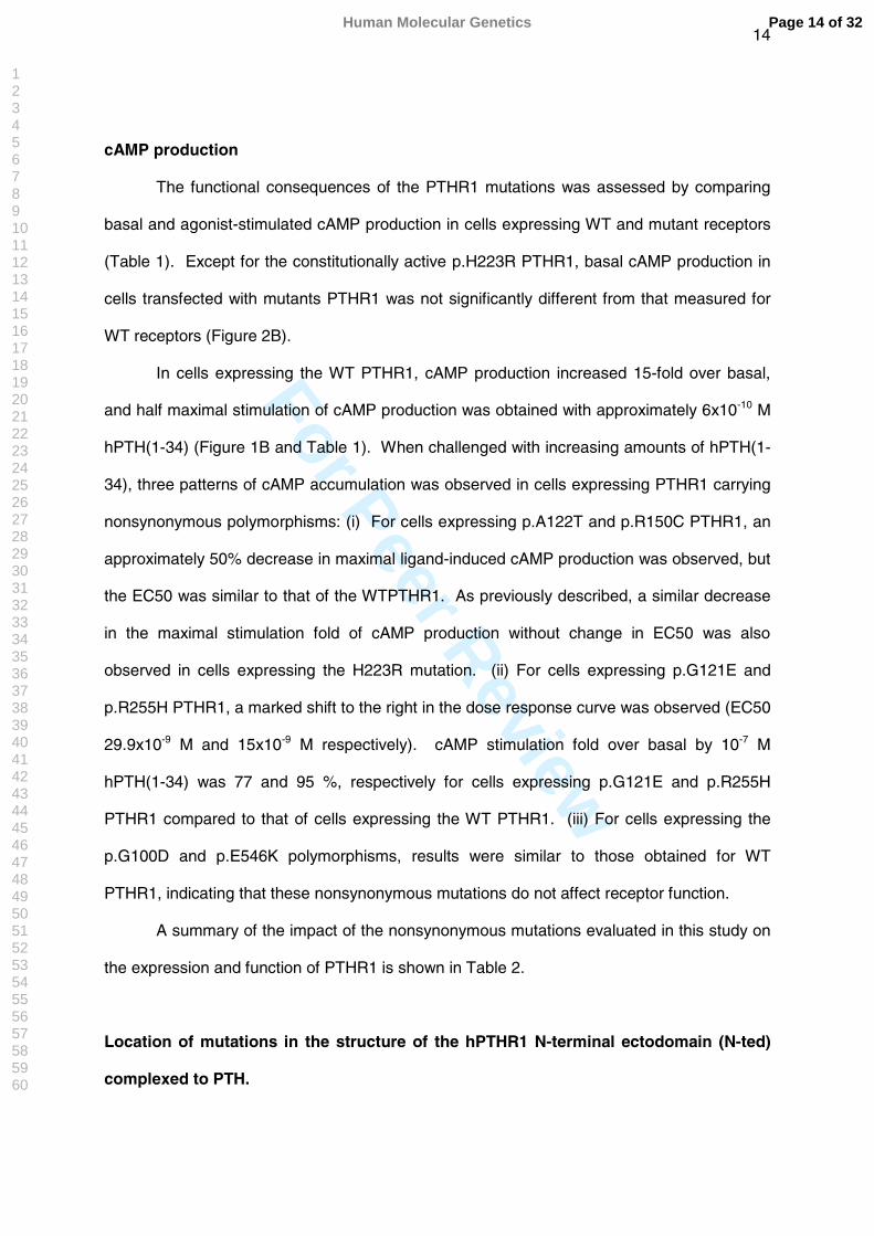
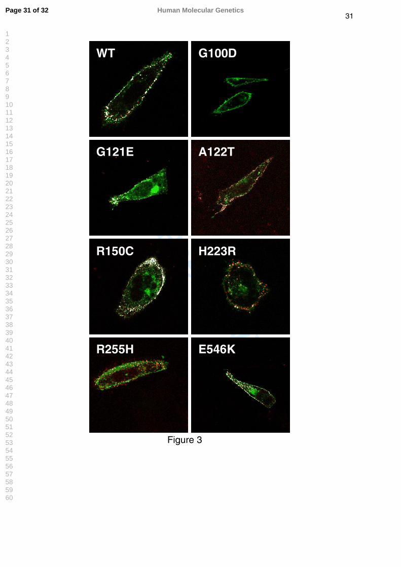
Analysis of PTHR1 expression using confocal microscopy
To evaluate the distribution of PTHR1 expression in CHO cells, cells were transfected with
an expression vector producing GFP-tagged PTHR1, and 48 hours after transfection,
expression of PTHR1 on the surface of non-permeabilized cells was evaluated using the
PTHR1 E2 antibody and a rhodamine-labelled secondary antibody. Cell surface expression
was clearly observed for WT PTHR1 and p.A122T, p.R150C and p.E546K PTHR1 (Figure 3).
In contrast, although some cell surface expression was detected, a diffuse green labelling
was observed in cells transfected with p.G121E and p.R255H PTHR1. This labeling pattern
indicates that the receptors were expressed but not appropriately addressed to the plasma
membrane. Similar results were observed in cells expressing p.H223R PTHR1, as
previously reported. The thin green labelling observed at the cell periphery without
cytoplasmic staining in cells transfected by p.G100D PTHR1 indicates that this mutant is
correctly addressed to the cell surface.
Page 13 of 32 Human Molecular Genetics
123456789101112131415161718192021222324252627282930313233343536373839404142434445464748495051525354555657585960
For Peer Review
14
cAMP production
The functional consequences of the PTHR1 mutations was assessed by comparing
basal and agonist-stimulated cAMP production in cells expressing WT and mutant receptors
(Table 1). Except for the constitutionally active p.H223R PTHR1, basal cAMP production in
cells transfected with mutants PTHR1 was not significantly different from that measured for
WT receptors (Figure 2B).
In cells expressing the WT PTHR1, cAMP production increased 15-fold over basal,
and half maximal stimulation of cAMP production was obtained with approximately 6x10⁻¹⁰ M
hPTH(1-34) (Figure 1B and Table 1). When challenged with increasing amounts of hPTH(1-
34), three patterns of cAMP accumulation was observed in cells expressing PTHR1 carrying
nonsynonymous polymorphisms: (i) For cells expressing p.A122T and p.R150C PTHR1, an
approximately 50% decrease in maximal ligand-induced cAMP production was observed, but
the EC50 was similar to that of the WTPTHR1. As previously described, a similar decrease
in the maximal stimulation fold of cAMP production without change in EC50 was also
observed in cells expressing the H223R mutation. (ii) For cells expressing p.G121E and
p.R255H PTHR1, a marked shift to the right in the dose response curve was observed (EC50
29.9x10⁻⁹ M and 15x10-9 M respectively). cAMP stimulation fold over basal by 10-7 M
hPTH(1-34) was 77 and 95 %, respectively for cells expressing p.G121E and p.R255H
PTHR1 compared to that of cells expressing the WT PTHR1. (iii) For cells expressing the
p.G100D and p.E546K polymorphisms, results were similar to those obtained for WT
PTHR1, indicating that these nonsynonymous mutations do not affect receptor function.
A summary of the impact of the nonsynonymous mutations evaluated in this study on
the expression and function of PTHR1 is shown in Table 2.
Location of mutations in the structure of the hPTHR1 N-terminal ectodomain (N-ted)
complexed to PTH.
Page 14 of 32Human Molecular Genetics
123456789101112131415161718192021222324252627282930313233343536373839404142434445464748495051525354555657585960
For Peer Review
15
In the structure described by Pioszak and Xu (30), the PTHR1 N-ted (PDB ID code 3C4M)
contains one major α helice (residues 33–57) and two antiparallel β sheets (β -strands 1 and
2: residues 110–120), β -strands 3 and 4 (residues 130–140) stabilized by 3 disulfide bonds
linking Cys48-Cys117, Cys108-Cys148, and Cys131-Cys170 (Figure 4). This structured core
represents a typical sushi domain present in several structures of class B GPCR N-ted (30,
35). The ligand binding ridge is localized along the structured core of the N-ted (30) (Figure
4). The exon E2 sequence present in the PTHR1 N-ted is not included within the structured
core, and lies in front of the first β sheet (dotted line, figure 4).
The localization of amino acid changes present in the PTHR1 N-ted identified in this
and previous studies, including the p.P132L mutation (Blomstrand chondrodysplasia) (25, 33,
34), and inducing changes in PTHR1 function is shown in Figure 4. These mutations are all
included within the structured core of the N-ted of the PTH receptor, but outside the putative
binding ridge. The p.G121E and p.A122T mutations, which respectively reduced ligand
affinity and maximal cAMP production, are located in a loop between the β2 and β3 sheets.
The p.R150C mutation, which reduced maximal cAMP production, is located in a loop
between β3 sheet and the disulfide bridge Cys131-Cys170 (Figure 4). The p.P132L mutation is
localized close to a stretch of four residues (aa 135-138) involved in direct contacts between
PTH and PTHR1 N-ted. In contrast, the p.G100D mutation, which does not modify PTHR1
function, is located in exon E2. Because the p.R255H mutation is not included in the
PTHR1-N-ted, it could not be positioned in the structure.
Page 15 of 32 Human Molecular Genetics
123456789101112131415161718192021222324252627282930313233343536373839404142434445464748495051525354555657585960
For Peer Review
16
DISCUSSION
To further define the role of PTHR1 signalling pathway in Ollier disease and Maffucci
syndrome, we have analysed the coding sequences and the intron-exon boundaries of four
genes participating in this pathway in a large cohort of patients. In this study, we identified
three previously undescribed missense mutations in PTHR1 in patients with Ollier disease.
Two of these mutations were present only in enchondromas, and one in both enchondroma
and leukocyte DNA. The assessment of receptor function demonstrated that all these
mutations impair PTHR1 function, either by reducing the affinity of the receptor for PTH or
reducing receptor expression at the cell surface. Structural modelling of PTHR1 indicated
that the deleterious mutations associated with Ollier disease and located within the N-ted all
lie within the structured core of the N-ted. Functionally deleterious PTHR1 mutations have
now been identified in 5 of 31 enchondromas from patients with Ollier disease (this study +
(23, 24), whereas such mutations have not be found in DNA from 222 control patients (this
study) evaluated by the same techniques. These findings provide further support for the idea
that heterozygous mutations in PTHR1 that impair receptor function can participate in the
pathogenesis of Ollier disease in some patients.
PTHR1 mutations and the pathogenesis of Ollier disease
It has been suggested that enchondromas result from abnormalities in signalling
pathways controlling proliferation and differentiation of chondrocytes, leading to the
development of the intraosseous cartilaginous foci. In particular, PTHR1 regulates the switch
from proliferating to hypertrophic chondrocytes, and thereby influences the number of cells
expressing Indian HedgeHog (IHH). IHH is a member of a family of morphogen proteins that
are important for embryonic patterning, and is highly expressed in the transition zone
between proliferating and hypertrophic chondrocytes. This multifunctional protein stimulates
chondrocyte proliferation, predominantly through a PTHrP-independent mechanism, and also
delays their hypertrophy by increasing PTHrP synthesis by periarticular chrondrocytes. Thus
Page 16 of 32Human Molecular Genetics
123456789101112131415161718192021222324252627282930313233343536373839404142434445464748495051525354555657585960
For Peer Review
17
PTHR1 and IHH signalling pathways are tightly coupled, although both exert functions
independently of each other. Reduced PTHR1 signalling would be expected to impair this
coupling process necessary for harmonious chondrocyte proliferation and differentiation,
thereby contributing to the development of enchondromas.
Our results and those from the literature indicate that missense PTHR1 mutations may
be involved in the development of enchondromas in a minority of patients. Three amino-acid
substitutions in PTHR1 were identified in the present study that impaired the ability of the
mutant receptors to stimulate cAMP production, due to 1) either a decrease in receptor
expression and ligand affinity (p.G121E and p.R255H), or 2) to suboptimal agonist-induced
cAMP production despite normal PTHR1 expression (p.A122T). Two of these mutations
were somatic and the third one likely germline. None of these receptors displayed
constitutional activity, whereas, as expected, increased constitutional activity was observed
for the PTHR1 expressing the p.H223R mutation identified in patients with Jansen's
metaphyseal chondrodysplasia. We also evaluated the functional properties of the p.R150C
mutation, previously identified in two patients with Ollier disease by Hopyan et al. (23).
Consistent with the results of Hopyan et al., we found that PTH-induced cAMP production
was reduced in cells transiently expressing this receptor, but in contrast to the previous
report, receptor expression was normal. Thus, the apparent increase in constitutional activity
reported by these authors following correction for receptor expression (23) was not observed
in our study. The reason why the p.R150C receptor expression was normal in our
experiments and decreased in Hopyan's studies are not clear, but are probably due to
differences in experimental conditions. Nevertheless, all mutant receptors identified
exclusively in patients with Ollier disease are characterized by impaired ligand-induced
cAMP production.
It is noteworthy that for three of the five patients with Ollier disease in whom PTHR1
mutations were identified in enchondromas, similar mutations were not detected in peripheral
blood leukocytes (this study and reference) (31). Because multiple enchondromas were
present in these patients, the findings are consistent with the hypothesis that the mutations
Page 17 of 32 Human Molecular Genetics
123456789101112131415161718192021222324252627282930313233343536373839404142434445464748495051525354555657585960
For Peer Review
18
occurred during development, resulting in genetic mosaicism in these individuals. Further
studies evaluating multiple tumors and other tissues from such patients will be required to
further support this idea. Nevertheless, sensitive screening for PTHR1 mutations in this
disease appears to require the evaluation of tumoral tissue, because mutations have now
been identified in 5/31 enchondromas, but only 2/58 samples of leukocytes DNA evaluated
by the same approach (p<0.05 by Fischer’s exact test).
In the course of these studies we also evaluated samples obtained from patients with
Maffucci syndrome (enchondromas.chondrosarcomas, n=12, blood leukocytes, n=13), but
deleterious mutaions in PTHR1 were not identified. Because of the small number of patients
studied, however, the frequency of finding PTHR1 mutations was not significantly different
comparing patients with Ollier’s disease and Maffucci syndrome (5/31 and 0/12 positive
tumors, respectively, p=0.3 by Fischer’s exact test). Further work is required to assess the
role, if any, of PTHR1 mutations in Maffucci syndrome.
Other findings are consistent with the conclusion that abnormalities in the PTHR1 /
IHH pathway can be linked to the development of endochondromas. Transgenic mice
expressing the mutant p.R150C PTHR1 under the control of the collagen type II promoter
develop tumors that are similar to those observed in human enchondromatosis (23). A
contribution of a dysregulation of IHH signalling pathway to the development of
enchondromas is also supported by the development of enchondromas in mice
overexpressing the Hedgehog (Hh) transcriptional regulator, Gli2, and the activation of a
Hedgehog-responsive Gli2-luciférase reporter construct by the p.R150C PTHR1 mutant (23).
Finally, downregulation of IHH/PTHrP signalling as a result of EXT mutation has been shown
to play a role in osteochondroma formation (35).
Even when present, it is not certain that the PTHR1 mutations identified are sufficient
alone to induce enchondromas. The p.R150C mutation has been identified both in a patient
with Ollier disease and in a parent who had other skeletal abnormalities without
enchondromas. In this regard, it will be interesting to evaluate family members of patients
expressing the p.R255H PTHR1 mutation identified in this study, but such clinical samples
Page 18 of 32Human Molecular Genetics
123456789101112131415161718192021222324252627282930313233343536373839404142434445464748495051525354555657585960
For Peer Review
19
are not currently available. Indeed, the PTHR1 loss of function mutations identified in Ollier
disease are expressed in the heterozygous state. PTHR1 dimerization has not been
documented for receptors of this family, rendering a dominant negative effect of the mutant
receptor unlikely. These findings support the hypothesis that, even when a PTHR1 mutation
is present, a combination of genetic events, germline and/or somatic, are required for the
development of enchondromas.
The nature of such putative genetic abnormalities remains to be defined.
Abnormalities involving other participants in the PTHR1 pathway are potential candidates.
For three of these genes, PTHrP, IHH and GNAS1 (Gsalpha), no missense mutation were
identified in these studies. Although these negative results do not exclude the possibility that
genetic abnormalities at these loci can contribute to the development of multiple
enchondromas, our findings suggest that such abnormalities are not a frequent cause.
Other PTHR1 polymorphisms
Currently, only two missense polymorphisms in PTHR1 have been described in
population studies, p.G100D and p.E546K. In the course of these studies, both of these
polymorphisms were identified with apparently similar frequency in controls and patients with
Ollier disease / Maffucci syndrome. We evaluated the functional properties of receptors
carrying both of these polymorphisms, but no abnormalities were identified. Amino-acid
p.G100 is located in exon E2 of the PTHR1; this exon is not present in other family II GPCR,
and its deletion has been previously shown not to affect PTHR1 function (36). Therefore the
lack of deleterious effect of the p.G100D mutation was not unexpected. The p.E546K
polymorphism has previously been extensively characterized by Schipani et al., and no
abnormalities were found (31).
3D spatial location of mutations
In order to better analyse the mechanism through which mutations may affect
receptor function, we represented the recently resolved crystal structure of the PTHR1 N-ted-
Page 19 of 32 Human Molecular Genetics
123456789101112131415161718192021222324252627282930313233343536373839404142434445464748495051525354555657585960
For Peer Review
20
PTH complex (PDB ID code 3C4M) (30), and positioned the PTHR1 mutations identified in
this and previous studies.
The observation that the functionally deleterious PTHR1 mutations (p.G121E, p.A122T,
p.R150C and p.P132L) are all located within the structured core, but outside the putative
ligand binding ridge indicates that the mutations may not directly impact receptor-ligand
interactions, but rather induce changes in the three dimensional organization of the N-ted
structured core necessary for PTH recognition and receptor activation. In support of this, the
p.P132L substitution (Blomstrand chondrodysplasia) (25, 33, 34) appears to disrupt an
interaction required to hold the structure of the N-ted and PTH–PTH1R signaling (30). An
analysis performed using a model of the VPAC1 receptor (also a class II GPCR) (37)
supports this conclusion. In this model, proline-87 in the VPAC1 receptor, which
corresponds to proline-132 in PTHR1, is also included within the structured core of the N-ted
of the VPAC1 receptor, but outside the binding ridge. The substitution of P87 by alanine
induced an important decrease in the affinity of VIP for its receptor (unpublished data). In
contrast, substitution of proline-87 by glycine, which introduces a flexible point in the
structured core, does not affect VPAC1 receptor function (38). The p.G100D mutation, which
does not modify PTHR1 function, is not located within exon E2 because the p.R255H
mutation is not included in the PTHTR1-N-ted, no structure-function correlates could be
obtained for this mutation.
In conclusion, this study provides further evidence that functionally deleterious
mutations in PTHR1 are present in a subset of patients with Ollier disease, and therefore
offers support to the idea that abnormalities in signalling pathways controlling chondrocyte
proliferation and differentiation can contribute to the pathogenesis of this disorder. Our
results emphasize that the genetic abnormalities responsible for this disease are likely to be
both heterogeneous and multi-factorial. The evaluation of additional candidate genes in
large cohorts of patients may prove rewarding.
Page 20 of 32Human Molecular Genetics
123456789101112131415161718192021222324252627282930313233343536373839404142434445464748495051525354555657585960
For Peer Review
21
Acknowledgements
We are indebted to all the family members and the Association Ollier Maffucci for their
invaluable contributions. We thank Cécile Pouzet for performing the confocal microscopy at
the Institut fédératif de recherche 02 (Inserm, Université Paris VII, CHU Xavier Bichat). We
are grateful to Cécile Jullier for sharing PTHR1 sequences from controls, and Augen Pioszak
and Eric Xu for the availability of the atomic coordinates and structure factors of the
extracellular domain of human PTH1R bound to PTH before publication in the protein data
bank. These studies were supported by grants from INSERM (RBM 0430), the réseaux
DHOS, the Fédération des Maladies Orphelines, et l'Association Ollier Maffucci (to C.S. and
B.G.), and by the Interuniversity Attraction Poles initiated by the Belgian Federal Science
Policy, network 5/25 and 6/05; Concerted Research Actions (A.R.C.) - Convention No
02/07/276 and No 07/12-005 of the Belgian French Community Ministry; the National
Institute of Health, Program Project P01 AR48564; EU FW6 Integrated project
LYMPHANGIOGENOMICS, LSHG-CT-2004-503573; and the F.N.R.S. (Fonds national de la
recherche scientifique) (to M.V., a "Maître de recherches du F.N.R.S."). Vinciane Wouters
was supported by a fellowship from F.R.I.A. (Fonds pour la formation à la recherche dans
l'industrie et dans l'agriculture), and Patrimoine UCL)
Page 21 of 32 Human Molecular Genetics
123456789101112131415161718192021222324252627282930313233343536373839404142434445464748495051525354555657585960
For Peer Review
22
LEGEND FIGURES
Figure 1. Functional evaluation of wild-type and mutant PTHR1 expressed in CHO cells.
(A). Binding of [125I] PTH 1-34 by cells incubated with radio ligand only (maximal binding)
and in the presence of increasing concentrations of unlabeled hPTH 1-34. The maximal
binding measured in cells transfected with the WT-PTHR1 was set as 100%. Results were
corrected for non-specific binding measured in the presence of 8x10⁻⁷ M PTH1-34. (B) cAMP
accumulation in response to increasing concentrations of hPTH 1-34. Results are
expressed as stimulation fold over basal. CHO cells were transfected with 1 µg plasmid DNA
coding for the WT or mutant PTHR1 and functional studies were performed 48 h later as
described in the methods. Results are the mean of at least three experiments performed in
duplicate with two plasmid preparations. SB: specific binding. WT: wild-type.
Figure 2. (A) Immunological assessment of cell-surface expression of wild-type and mutant
PTHR1 expressed in CHO cells. Transfected cells were incubated sequentially with
polyclonal rabbit anti-human PTHR1 antibody and [125I] -labeled anti-rabbit immunoglobin
antibody. Results are expressed as % [125I]-labeled anti-rabbit immunoglobin antibody bound
in cells transfected with the WT-PTHR1. p.G100D substitution disrupts the epitope
recognition by the PTHR1 antibody (see results) (B). Basal cAMP production in cells
transfected with WT or mutant PTHR1. Basal cAMP accumulation is expressed as
picomoles cAMP / well normalized to cell-surface expression as determined in (2A).
Expression of the mutant p.G100D PTHR1 was considered to be that of WT PTHR1, based
on results from radioligand binding and confocal immunofluorescence experiments (see
results). CHO cells were transfected with 1 µg plasmid DNA coding for the WT or mutant
PTHR1 and experiments performed 48 h later. Results are the mean ± SEM of at least three
experiments performed in duplicate with two plasmid preparations.
Page 22 of 32Human Molecular Genetics
123456789101112131415161718192021222324252627282930313233343536373839404142434445464748495051525354555657585960
For Peer Review
23
Figure 3. Confocal immunofluorescent microscopy analysis of wild-type and mutant PTHR1
tagged with GFP and using sequentially polyclonal rabbit anti-human PTHR1 antibody and
rhodamine-labeled anti-rabbit immunoglobin antibody, without cellular permeabilization.
Colocalisation (originally yellow, and falsy colored in white for clarity) of green GFP tagged
PTHR1 and the rhodamine labelled secondary antibody against the PTHR1 primary antibody
(directed against the E2 extracellular epitope) indicates cell surface expression. No
colocalization was observed in cells transfected with the p.G100D PTHR1, indicating that the
p.G100D mutation prevents PTHR1 recognition by the PTHR1 antibody (see results). CHO
cells were transfected with 1 µg plasmid DNA coding for the WT or mutant PTHR1, and
confocal immunofluorescent microscopy analysis was performed as described in the material
and methods.
Figure 4. Representation of the recently resolved crystal structure of the PTH–PTHR1 N-ted
complex (PDB ID code 3C4M) (30), and position the PTHR1 mutations identified in this and
previous studies. The figures shows ribbon representation of receptor N-ted. Grey: PTHR1
N-ted main chain; grey, dashed line: exon E2 (residues 57-105); yellow: β sheets; black:
disulfide bonds between residues Cys48-Cys117, Cys108-Cys148, and Cys131-Cys170); red: PTH.
The mutated amino-acids are indicated in blue. All mutated amino-acids are located within
the structured core.
Page 23 of 32 Human Molecular Genetics
123456789101112131415161718192021222324252627282930313233343536373839404142434445464748495051525354555657585960
For Peer Review
24
References
1. Fletcher, C.D.M., Unni, K. and Mertens, F. (eds.) (2002) World Health Organization
Classification of Tumors. Pathology and genetics. Tumors of Soft Tissue and Bone.
IARCPress, Lyon.
2. Maroteaux, P. and Le Merrer, M. (2002) Les maladies osseuses de l'enfant. 4ème ed.
Médecine-Sciences, Flammarion, Paris.
3. Unni, K.K. (2001) Cartilaginous lesions of bone. J Orthop Sci, 6, 457-472.
4. Whyte, M. (2003) Acquired Disorders of Cartilage and Bone. 5th edition ed. American
Society for Bone and Mineral Research, Washington DC.
5. Silve, C. and Juppner, H. (2006) Ollier disease. Orphanet J Rare Dis, 1, 37.
6. Casanova, D., Boon, L.M. and Vikkula, M. (2006) [Venous malformations: clinical
characteristics and differential diagnosis]. Ann Chir Plast Esthet, 51, 373-387.
7. Rozeman, L.B., Hogendoorn, P.C. and Bovee, J.V. (2002) Diagnosis and prognosis
of chondrosarcoma of bone. Expert Rev Mol Diagn, 2, 461-472.
8. Schaison, F., Anract, P., Coste, F., De Pinieux, G., Forest, M. and Tomeno, B. (1999)
[Chondrosarcoma secondary to multiple cartilage diseases. Study of 29 clinical cases
and review of the literature]. Rev Chir Orthop Reparatrice Appar Mot, 85, 834-845.
9. Schwartz, H.S., Zimmerman, N.B., Simon, M.A., Wroble, R.R., Millar, E.A. and
Bonfiglio, M. (1987) The malignant potential of enchondromatosis. J Bone Joint Surg
Am, 69, 269-274.
10. Vaz, R.M. and Turner, C. (1986) Ollier disease (enchondromatosis) associated with
ovarian juvenile granulosa cell tumor and precocious pseudopuberty. J Pediatr, 108,
945-947.
11. Tamimi, H.K. and Bolen, J.W. (1984) Enchondromatosis (Ollier's disease) and
ovarian juvenile granulosa cell tumor. Cancer, 53, 1605-1608.
12. Mahafza, W.S. (2004) Multiple enchondromatosis Ollier's disease with two primary
brain tumors. Saudi Med J, 25, 1261-1263.
Page 24 of 32Human Molecular Genetics
123456789101112131415161718192021222324252627282930313233343536373839404142434445464748495051525354555657585960
For Peer Review
25
13. Tiet, T.D. and Alman, B.A. (2003) Developmental pathways in musculoskeletal
neoplasia: involvement of the Indian Hedgehog-parathyroid hormone-related protein
pathway. Pediatr Res, 53, 539-543.
14. Bovee, J.V., Cleton-Jansen, A.M., Rosenberg, C., Taminiau, A.H., Cornelisse, C.J.
and Hogendoorn, P.C. (1999) Molecular genetic characterization of both components
of a dedifferentiated chondrosarcoma, with implications for its histogenesis. J Pathol,
189, 454-462.
15. Bovee, J.V., van Roggen, J.F., Cleton-Jansen, A.M., Taminiau, A.H., van der Woude,
H.J. and Hogendoorn, P.C. (2000) Malignant progression in multiple
enchondromatosis (Ollier's disease): an autopsy-based molecular genetic study. Hum
Pathol, 31, 1299-1303.
16. Sandberg, A.A. (2004) Genetics of chondrosarcoma and related tumors. Curr Opin
Oncol, 16, 342-354.
17. van Beerendonk, H.M., Rozeman, L.B., Taminiau, A.H., Sciot, R., Bovee, J.V.,
Cleton-Jansen, A.M. and Hogendoorn, P.C. (2004) Molecular analysis of the
INK4A/INK4A-ARF gene locus in conventional (central) chondrosarcomas and
enchondromas: indication of an important gene for tumour progression. J Pathol, 202,
359-366.
18. Amling, M., Posl, M., Hentz, M.W., Priemel, M. and Delling, G. (1998) PTHrP and Bcl-
2: essential regulatory molecules in chondrocyte differentiation and chondrogenic
tumors. Verh Dtsch Ges Pathol, 82, 160-169.
19. Bovee, J.V., van den Broek, L.J., Cleton-Jansen, A.M. and Hogendoorn, P.C. (2000)
Up-regulation of PTHrP and Bcl-2 expression characterizes the progression of
osteochondroma towards peripheral chondrosarcoma and is a late event in central
chondrosarcoma. Lab Invest, 80, 1925-1934.
20. Kunisada, T., Moseley, J.M., Slavin, J.L., Martin, T.J. and Choong, P.F. (2002) Co-
expression of parathyroid hormone-related protein (PTHrP) and PTH/PTHrP receptor
in cartilaginous tumours: a marker for malignancy? Pathology, 34, 133-137.
Page 25 of 32 Human Molecular Genetics
123456789101112131415161718192021222324252627282930313233343536373839404142434445464748495051525354555657585960
For Peer Review
26
21. Pateder, D.B., Gish, M.W., O'Keefe, R.J., Hicks, D.G., Teot, L.A. and Rosier, R.N.
(2002) Parathyroid hormone-related Peptide expression in cartilaginous tumors. Clin
Orthop, 198-204.
22. Kronenberg, H.M. (2003) Developmental regulation of the growth plate. Nature, 423,
332-336.
23. Hopyan, S., Gokgoz, N., Poon, R., Gensure, R.C., Yu, C., Cole, W.G., Bell, R.S.,
Juppner, H., Andrulis, I.L., Wunder, J.S. et al. (2002) A mutant PTH/PTHrP type I
receptor in enchondromatosis. Nat Genet, 30, 306-310.
24. Rozeman, L.B., Sangiorgi, L., Bruijn, I.H., Mainil-Varlet, P., Bertoni, F., Cleton-
Jansen, A.M., Hogendoorn, P.C. and Bovee, J.V. (2004) Enchondromatosis (Ollier
disease, Maffucci syndrome) is not caused by the PTHR1 mutation p.R150C. Hum
Mutat, 24, 466-473.
25. Zhang, P., Jobert, A.S., Couvineau, A. and Silve, C. (1998) A homozygous
inactivating mutation in the parathyroid hormone/parathyroid hormone-related peptide
receptor causing Blomstrand chondrodysplasia. J Clin Endocrinol Metab, 83, 3365-
3368.
26. Couvineau, A., Lacapere, J.J., Tan, Y.V., Rouyer-Fessard, C., Nicole, P. and
Laburthe, M. (2003) Identification of cytoplasmic domains of hVPAC1 receptor
required for activation of adenylyl cyclase. Crucial role of two charged amino acids
strictly conserved in class II G protein-coupled receptors. J Biol Chem, 278, 24759-
24766.
27. Jobert, A.S., Leroy, C., Butlen, D. and Silve, C. (1997) Parathyroid hormone-induced
calcium release from intracellular stores in a human kidney cell line in the absence of
stimulation of cyclic adenosine 3',5'-monophosphate production. Endocrinology, 138,
5282-5292.
28. Jobert, A.S., Zhang, P., Couvineau, A., Bonaventure, J., Roume, J., Le Merrer, M.
and Silve, C. (1998) Absence of functional receptors for parathyroid hormone and
Page 26 of 32Human Molecular Genetics
123456789101112131415161718192021222324252627282930313233343536373839404142434445464748495051525354555657585960
For Peer Review
27
parathyroid hormone-related peptide in Blomstrand chondrodysplasia. J Clin Invest,
102, 34-340.
29. Jobert, A.S., Fernandes, I., Turner, G., Coureau, C., Prie, D., Nissenson, R.A.,
Friedlander, G. and Silve, C. (1996) Expression of alternatively spliced isoforms of
the parathyroid hormone (PTH)/PTH-related peptide receptor messenger RNA in
human kidney and bone cells. Mol Endocrinol, 10, 1066-1076.
30. Pioszak, A.A. and Xu, H.E. (2008) Molecular recognition of parathyroid hormone by
its G protein-coupled receptor. Proc Natl Acad Sci U S A, 105, 5034-5039.
31. Schipani, E., Weinstein, L.S., Bergwitz, C., Iida-Klein, A., Kong, X.F., Stuhrmann, M.,
Kruse, K., Whyte, M.P., Murray, T., Schmidtke, J. et al. (1995)
Pseudohypoparathyroidism type Ib is not caused by mutations in the coding exons of
the human parathyroid hormone (PTH)/PTH-related peptide receptor gene. J Clin
Endocrinol Metab, 80, 1611-1621.
32. Schipani, E., Kruse, K. and Jüppner, H. (1995) A constitutively active mutant PTH-
PTHrP receptor in Jansen-type metaphyseal chondrodysplasia. Science, 268, 98-
100.
33. Hoogendam, J., Farih-Sips, H., Wynaendts, L.C., Lowik, C.W., Wit, J.M. and
Karperien, M. (2007) Novel mutations in the parathyroid hormone (PTH)/PTH-related
peptide receptor type 1 causing Blomstrand osteochondrodysplasia types I and II. J
Clin Endocrinol Metab, 92, 1088-1095.
34. Karaplis, A.C., Bin He, M.T., Nguyen, A., Young, I.D., Semeraro, D., Ozawa, H. and
Amizuka, N. (1998) Inactivating Mutation in the Human Parathyroid Hormone
Receptor Type 1 Gene in Blomstrand Chondrodysplasia. Endocrinology, 139, 5255-
5258.
35. Rozeman, L.B., Hameetman, L., Cleton-Jansen, A.M., Taminiau, A.H., Hogendoorn,
P.C. and Bovee, J.V. (2005) Absence of IHH and retention of PTHrP signalling in
enchondromas and central chondrosarcomas. J Pathol, 205, 476-482.
Page 27 of 32 Human Molecular Genetics
123456789101112131415161718192021222324252627282930313233343536373839404142434445464748495051525354555657585960
For Peer Review
28
36. Lee, C., Gardella, T.J., Abou-Samra, A.B., Nussbaum, S.R., Segre, G.V., Potts, J.T.,
Jr., Kronenberg, H.M. and Juppner, H. (1994) Role of the extracellular regions of the
parathyroid hormone (PTH)/PTH-related peptide receptor in hormone binding.
Endocrinology, 135, 1488-1495.
37. Ceraudo, E., Murail, S., Tan, Y.V., Lacapere, J.J., Neumann, J.M., Couvineau, A. and
Laburthe, M. (2007) The vasoactive intestinal peptide alpha helix up to C-terminus
interacts with the N-terminal ectodomain of the human VPAC1 receptor :
photoaffinity, molecular modeling and dynamics. Mol Endocrinol.
38. Couvineau, A., Gaudin, P., Maoret, J.J., Rouyer-Fessard, C., Nicole, P. and Laburthe,
M. (1995) Highly conserved aspartate 68, tryptophane 73 and glycine 109 in the N-
terminal extracellular domain of the human VIP receptor are essential for its ability to
bind VIP. Biochem Biophys Res Commun, 206, 246-252.
Page 28 of 32Human Molecular Genetics
123456789101112131415161718192021222324252627282930313233343536373839404142434445464748495051525354555657585960
For Peer Review
29
-11 -10 -9 -8 -70
10
20wtG100DG121EA122TR150CH223RR255H
PTH 1-34, M
cAM
P, s
timul
atio
n fo
ld
Tr100% of tr-ns B/T pool
-11 -10 -9 -8 -70
25
50
75
100
125
wt
G100D
G121E
A122T
R150C
H223R
R255H
E546K
PTH 1-34 M
[125
I]hP
TH
1-3
4, S
B, %
WT
A
B
Fig 1
Page 29 of 32 Human Molecular Genetics
123456789101112131415161718192021222324252627282930313233343536373839404142434445464748495051525354555657585960
For Peer Review
30
Tr% of tr2 of AB bd p 71 u773 + G100D (A)
WT G100D G121E A122T R150C H223R R255H E546K0
25
50
75
100
125A
B B
dg
, %
WT
Tr AB bdg of basal pool cru (B)
wt G100D G121E A122T R150C H223R R255H E546K0
1
2
3
4
5
6
Bas
al c
AM
P, n
orm
aliz
ed/e
xpre
ssio
n
Fig 2
A
B
Page 30 of 32Human Molecular Genetics
123456789101112131415161718192021222324252627282930313233343536373839404142434445464748495051525354555657585960
For Peer Review
31
WT
H223R
E546K
G100D
A122TG121E
R150C
R255H
Figure 3
Page 31 of 32 Human Molecular Genetics
123456789101112131415161718192021222324252627282930313233343536373839404142434445464748495051525354555657585960
For Peer Review
32
G121
A122
R150
P132
G100
N
C
34
15
PTH
β β4
β
β2
Figure 4
N-ted
Page 32 of 32Human Molecular Genetics
123456789101112131415161718192021222324252627282930313233343536373839404142434445464748495051525354555657585960