Embed Size (px)
Citation preview
Human Human Molecular Molecular GeneticsGenetics
Institute of Medical GeneticsYaoqin Gong
2003

Gene Mutation
DNA Polymorphism
Outline of this chapter

Gene MutationGene Mutation
Definition
Major Types
Mutation Detection
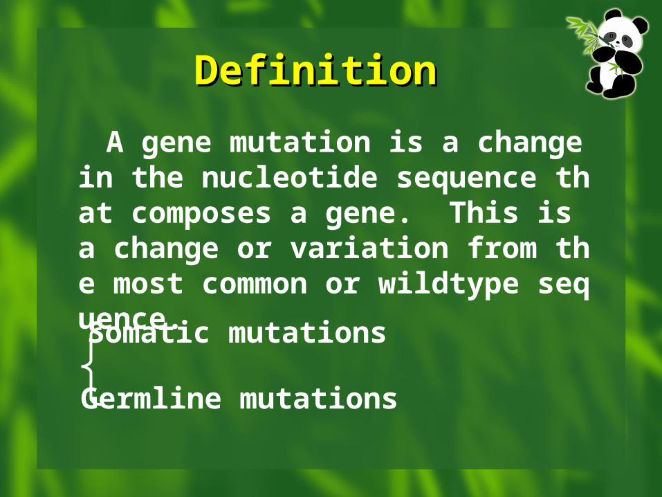
DefinitionDefinition A gene mutation is a change in the n
ucleotide sequence that composes a gene. This is a change or variation from the most common or wildtype sequence.
Somatic mutations
Germline mutations
Somatic mutationsSomatic mutations
are mutations that occur in cells of the body excluding the germline.
Affects subsequent somatic cell descendants
Limited to impact on the individual and not transmitted to offspring
Germline mutations
are mutations that occur in the germline cells
Possibility of transmission to offspring
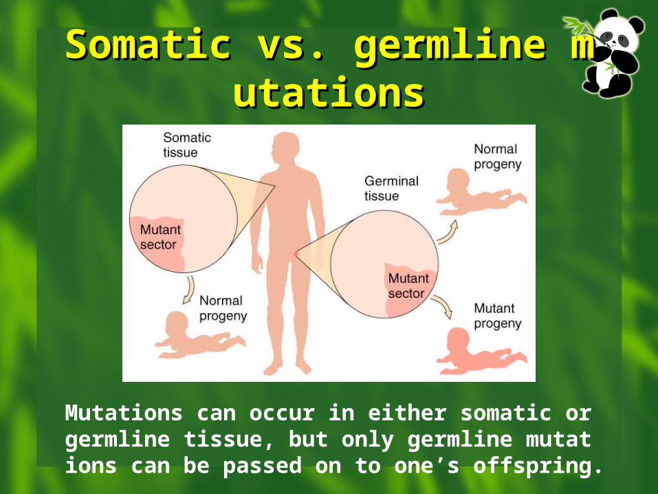
Somatic vs. germline mutaSomatic vs. germline mutationstions
Mutations can occur in either somatic or germline tissue, but only germline mutations can be passed on to one’s offspring.
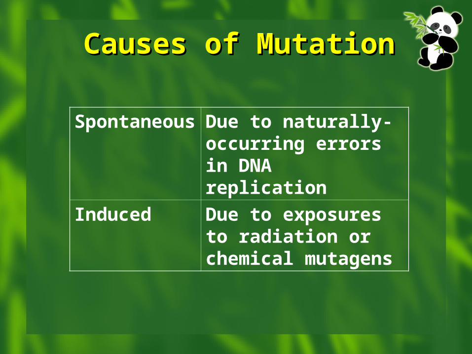
Causes of MutationCauses of Mutation
Spontaneous Due to naturally-occurring errors in DNA replication
Induced Due to exposures to radiation or chemical mutagens
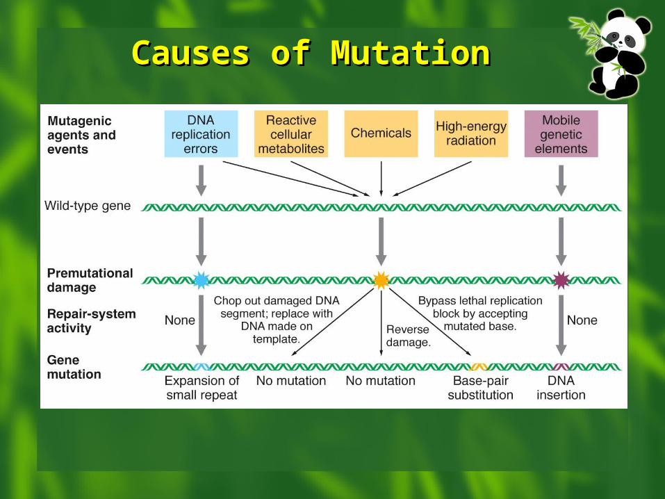
Causes of MutationCauses of Mutation
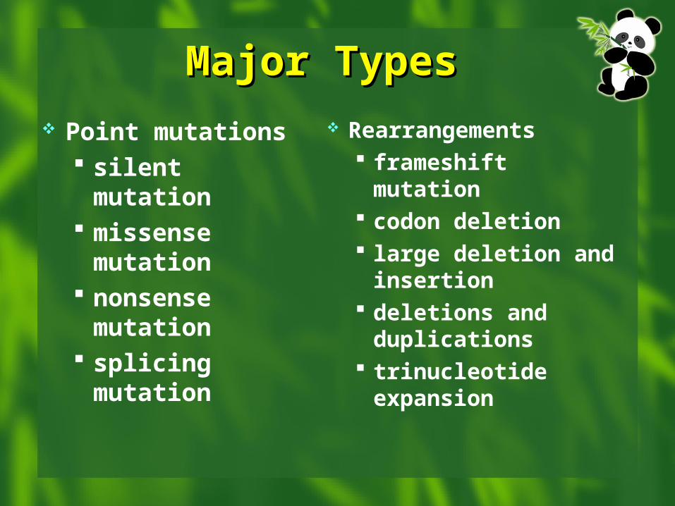
Major TypesMajor Types Point mutations
silent mutation missense
mutation nonsense
mutation splicing
mutation
Rearrangements frameshift mutation codon deletion large deletion and
insertion deletions and
duplications trinucleotide
expansion
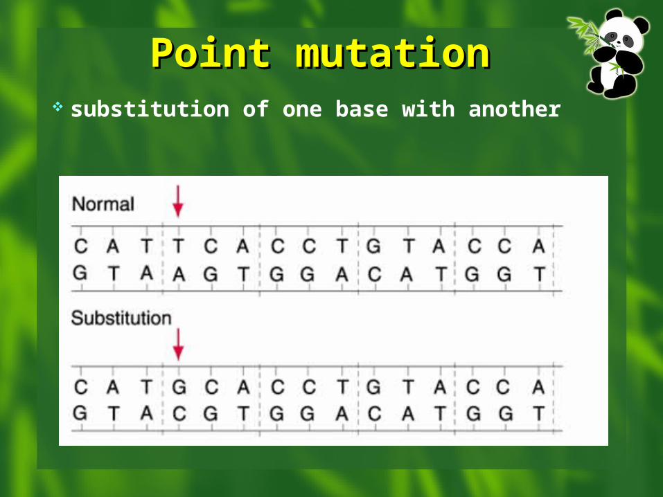
Point mutationPoint mutation substitution of one base with another
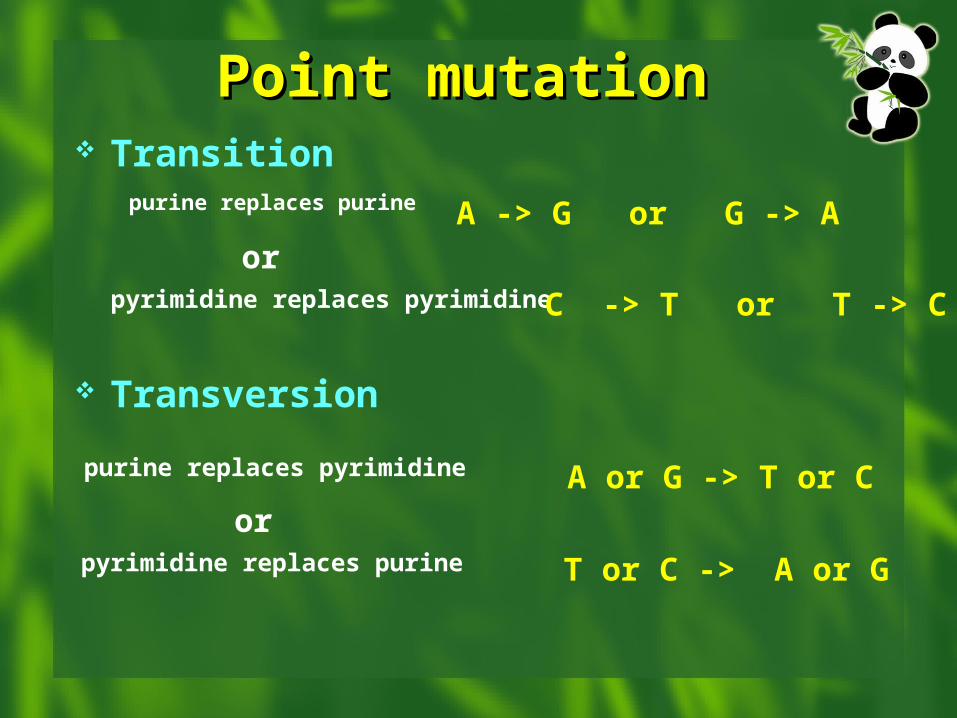
Point mutationPoint mutation Transition
Transversion
purine replaces purine
pyrimidine replaces pyrimidine
orA -> G or G -> A
C -> T or T -> C
purine replaces pyrimidine
pyrimidine replaces purine
orA or G -> T or C
T or C -> A or G
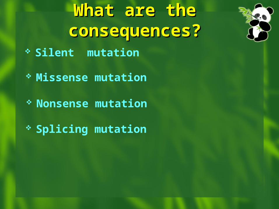
What are the What are the consequences?consequences?
Silent mutation
Missense mutation
Nonsense mutation
Splicing mutation
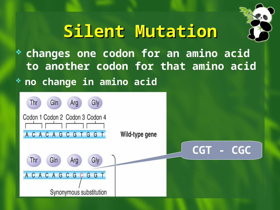
Silent MutationSilent Mutation changes one codon for an amino acid to an
other codon for that amino acid no change in amino acid
CGT - CGC
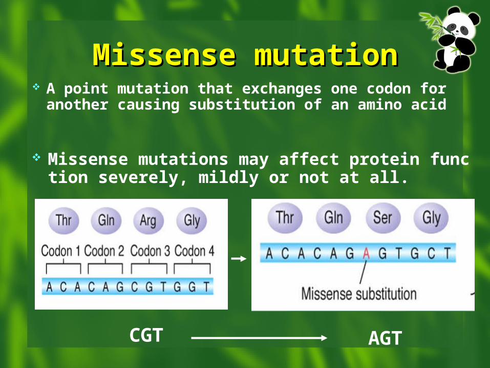
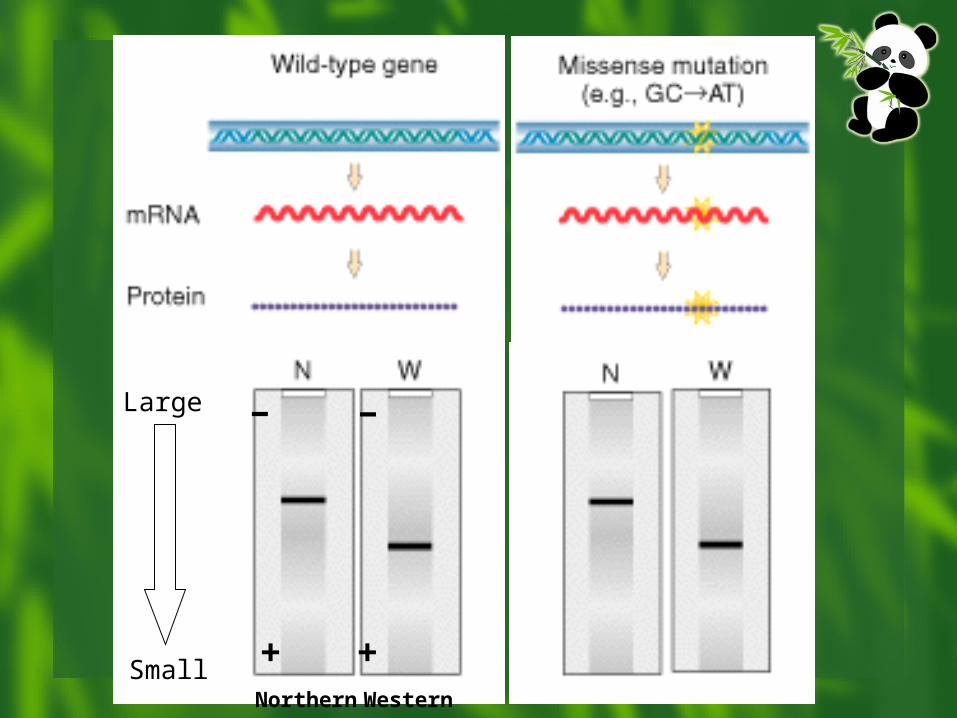
Missense mutationMissense mutation A point mutation that exchanges one codon for
another causing substitution of an amino acid
Missense mutations may affect protein function severely, mildly or not at all.
CGT AGT
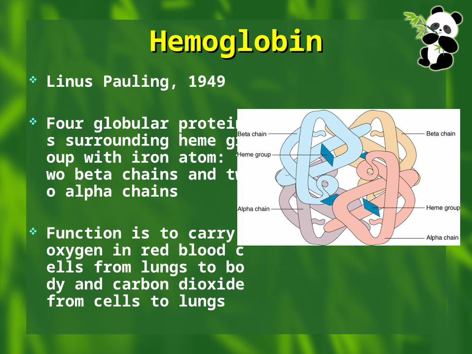
HemoglobinHemoglobin Linus Pauling, 1949
Four globular proteins surrounding heme group with iron atom: two beta chains and two alpha chains
Function is to carry oxygen in red blood cells from lungs to body and carbon dioxide from cells to lungs
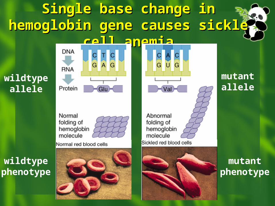
Single base change in Single base change in hemoglobin gene causes hemoglobin gene causes
sickle cell anemiasickle cell anemia
wildtypeallele
mutantallele
wildtypephenotype
mutantphenotype
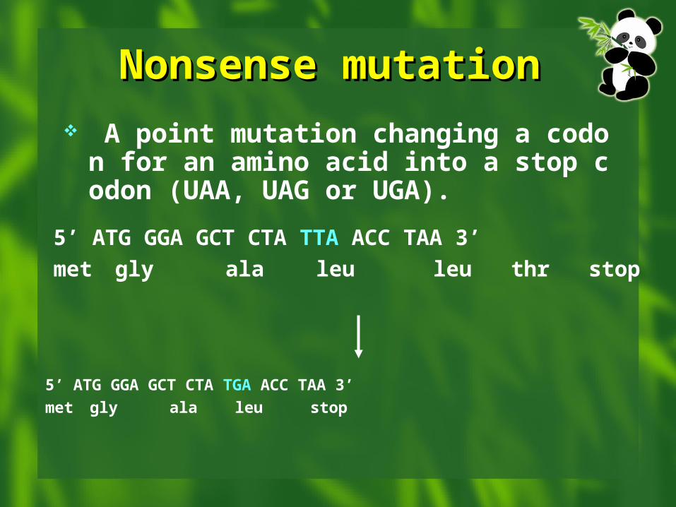
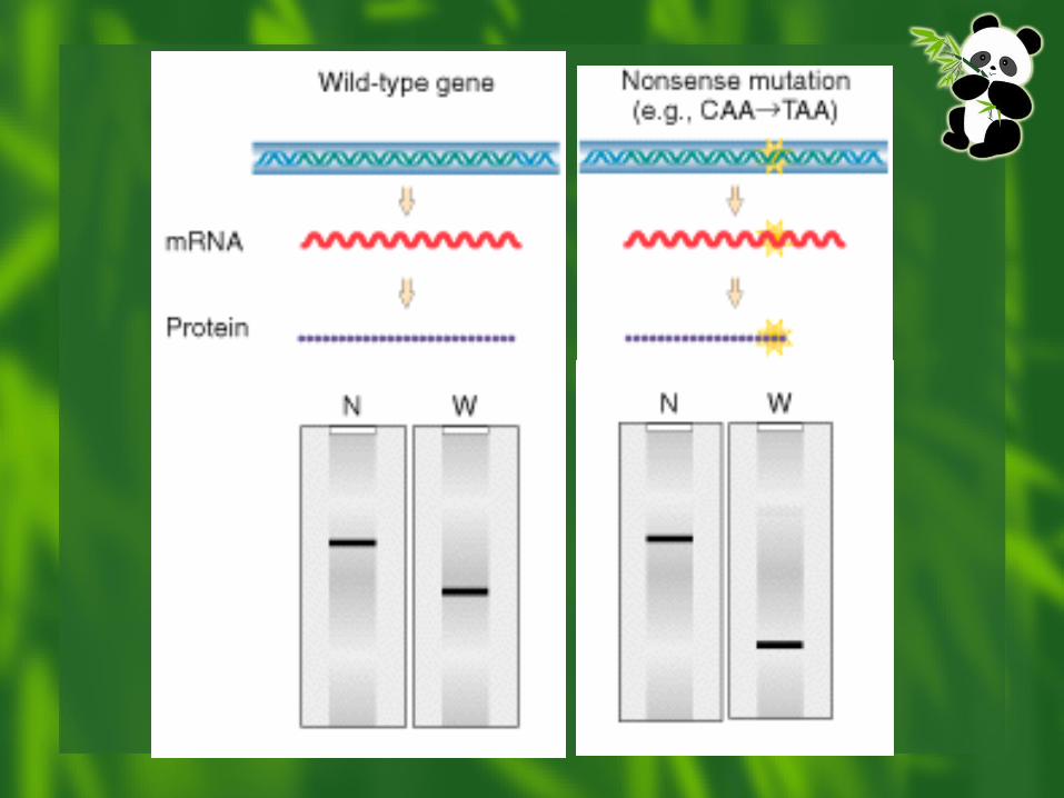
Nonsense mutationNonsense mutation A point mutation changing a codon for an
amino acid into a stop codon (UAA, UAG or UGA).
5’ ATG GGA GCT CTA TTA ACC TAA 3’
met gly ala leu leu thr stop
5’ ATG GGA GCT CTA TGA ACC TAA 3’
met gly ala leu stop
Nonsense mutationNonsense mutation
Premature stop codons create truncated proteins.
Truncated proteins are often nonfunctional.
Some truncations have dominant effects due to interference with normal functions.
Splicing MutationsSplicing Mutations
“GT/AG rule” Disruption of existing splice sites
intron is not removed from mRNA Creation of novel splice sites in exons
HbE: missense mutation and splice error features of hemoglobinopathy and thalassemi
a
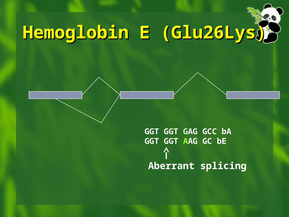
Hemoglobin E Hemoglobin E (Glu26Lys)(Glu26Lys)
GGT GGT GAG GCC bAGGT GGT AAG GC bE
Aberrant splicing
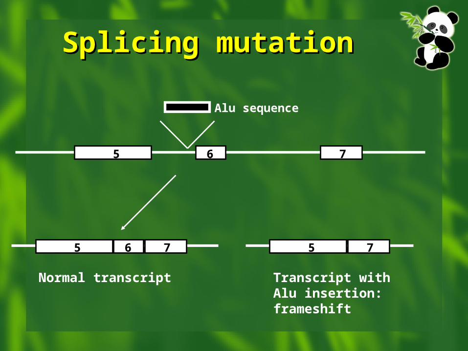
Splicing mutationSplicing mutation
6 7
Alu sequence
5
6 75 75
Normal transcript Transcript with Alu insertion: frameshift
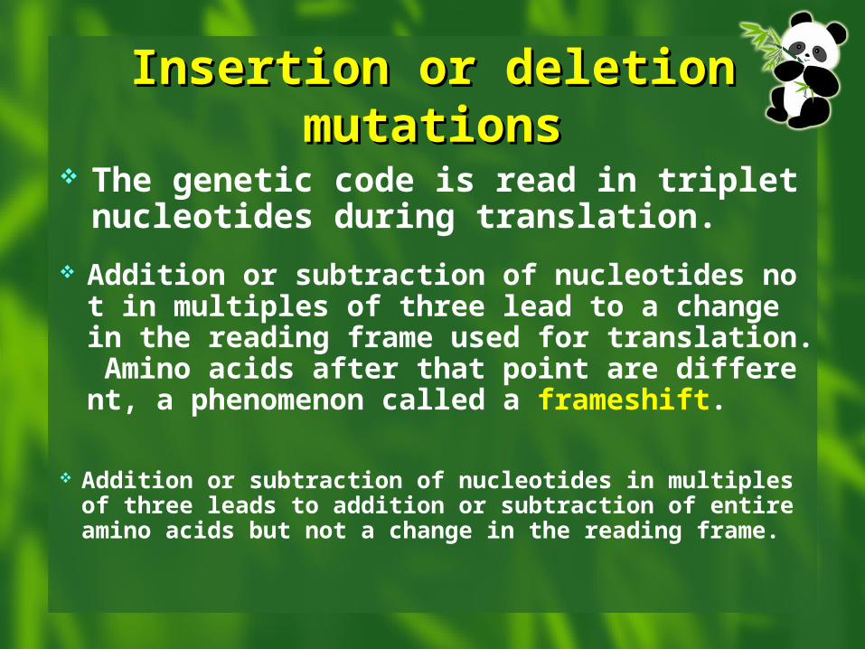
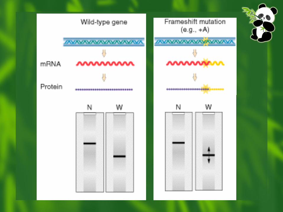
Insertion or deletion Insertion or deletion mutationsmutations
The genetic code is read in triplet nucleotides during translation.
Addition or subtraction of nucleotides not in multiples of three lead to a change in the reading frame used for translation. Amino acids after that point are different, a phenomenon called a frameshift.
Addition or subtraction of nucleotides in multiples of three leads to addition or subtraction of entire amino acids but not a change in the reading frame.
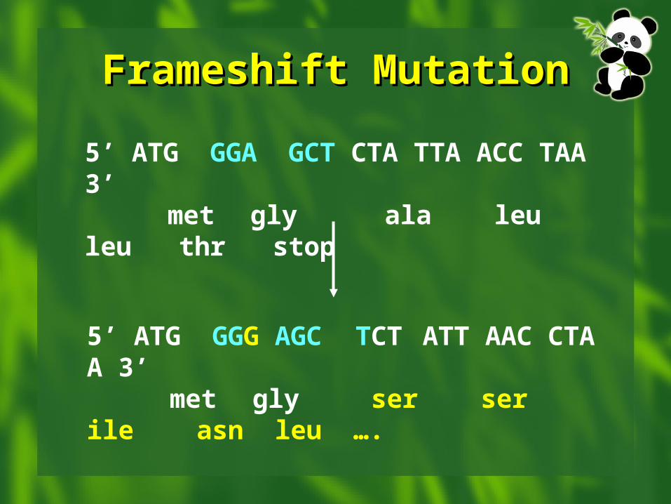
Frameshift MutationFrameshift Mutation
5’ ATG GGA GCT CTA TTA ACC TAA 3’ met gly ala leu leu thr stop
5’ ATG GGG AGC TCT ATT AAC CTA A 3’
met gly ser ser ile asn leu ….
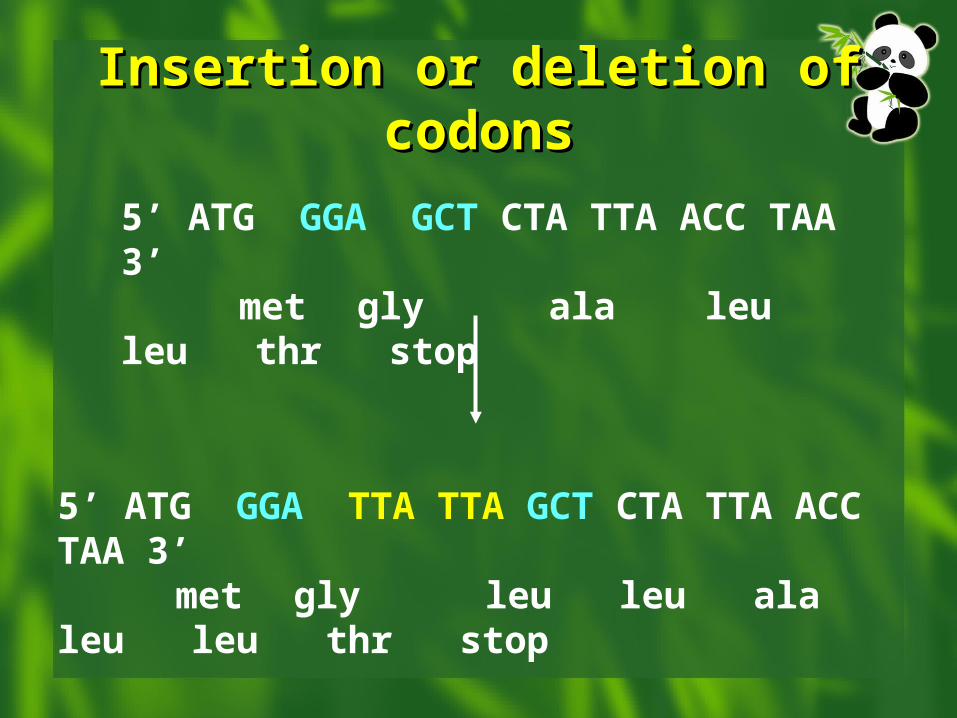
Insertion or deletion of codonsInsertion or deletion of codons
5’ ATG GGA GCT CTA TTA ACC TAA 3’ met gly ala leu leu thr stop
5’ ATG GGA TTA TTA GCT CTA TTA ACC TAA 3’ met gly leu leu ala leu leu thr
stop
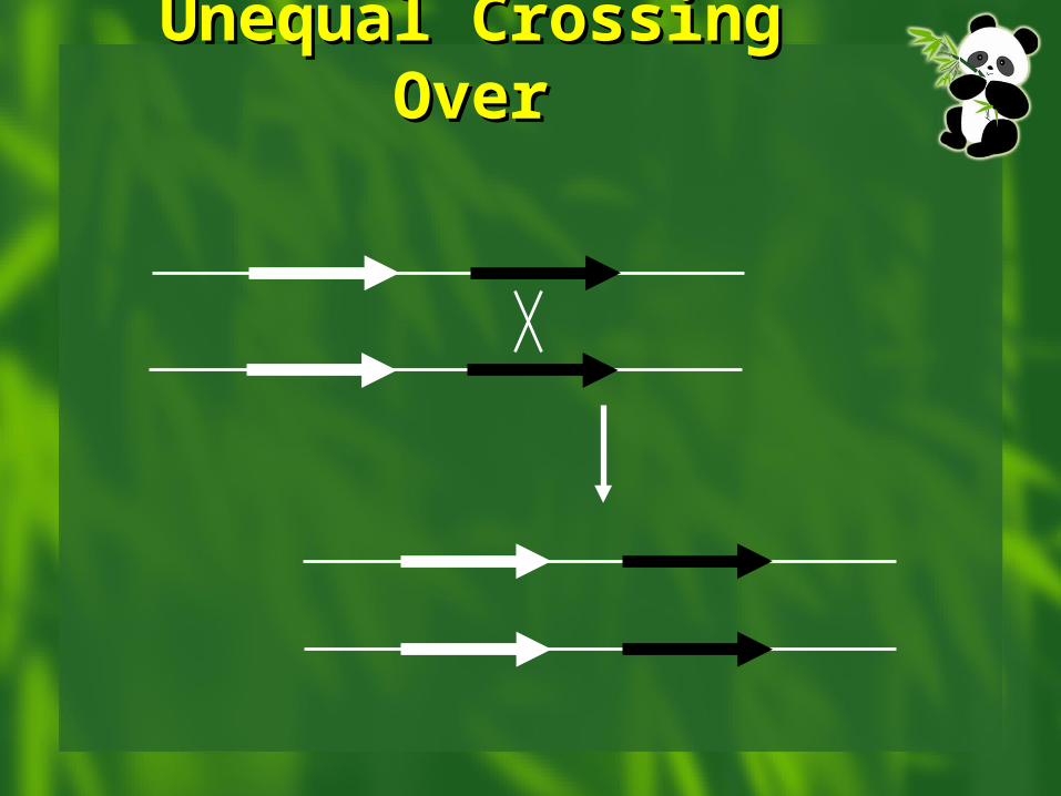
Unequal Crossing Unequal Crossing OverOver
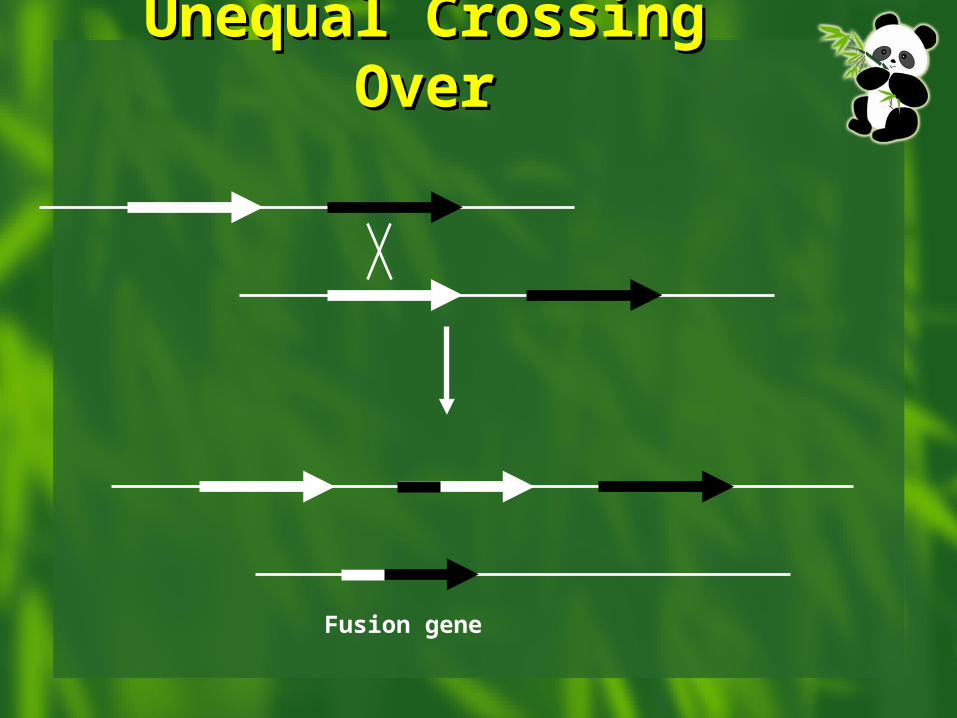
Unequal Crossing Unequal Crossing OverOver
Fusion gene
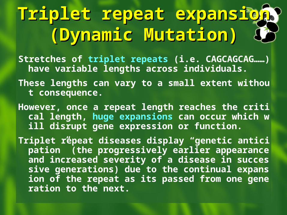
Triplet repeat expansion Triplet repeat expansion (Dynamic Mutation)(Dynamic Mutation)
Stretches of triplet repeats (i.e. CAGCAGCAG……) have variable lengths across individuals.
These lengths can vary to a small extent without consequence.
However, once a repeat length reaches the critical length, huge expansions can occur which will disrupt gene expression or function.
Triplet repeat diseases display “genetic anticipation” (the progressively earlier appearance and increased severity of a disease in successive generations) due to the continual expansion of the repeat as its passed from one generation to the next.
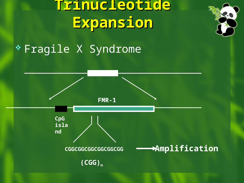
Trinucleotide Trinucleotide ExpansionExpansion
Fragile X Syndrome
CGGCGGCGGCGGCGGCGG Amplification
CpG island
FMR-1
(CGG)n
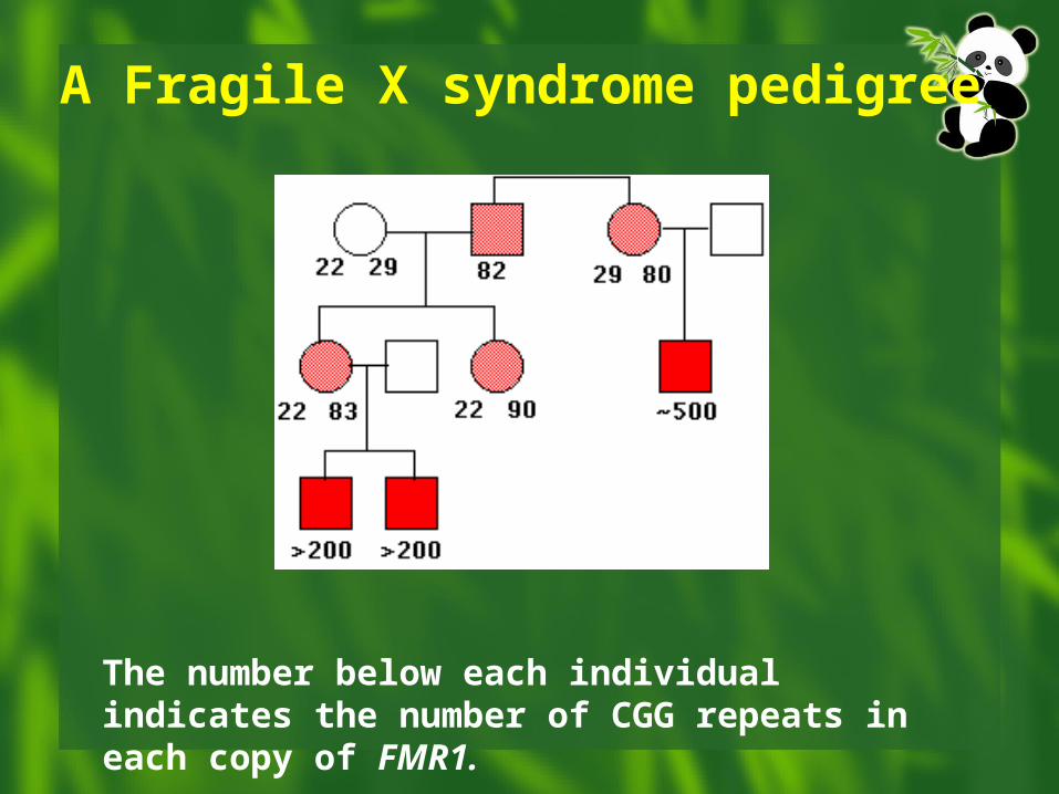
A Fragile X syndrome pedigree
The number below each individual indicates the number of CGG repeats in each copy of FMR1.
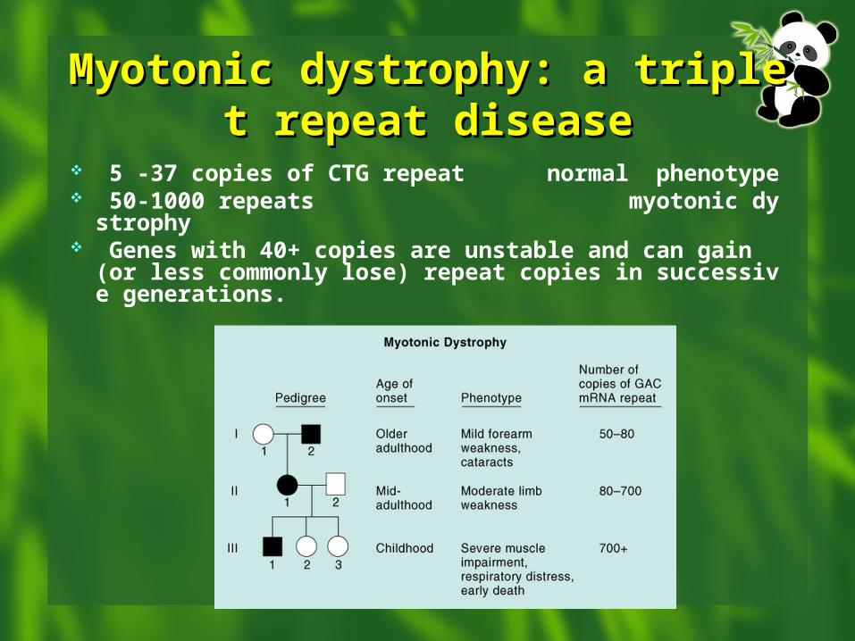
Myotonic dystrophy: a triplet repeMyotonic dystrophy: a triplet repeat diseaseat disease
5 -37 copies of CTG repeat normal phenotype 50-1000 repeats myotonic dystrophy Genes with 40+ copies are unstable and can gain
(or less commonly lose) repeat copies in successive generations.
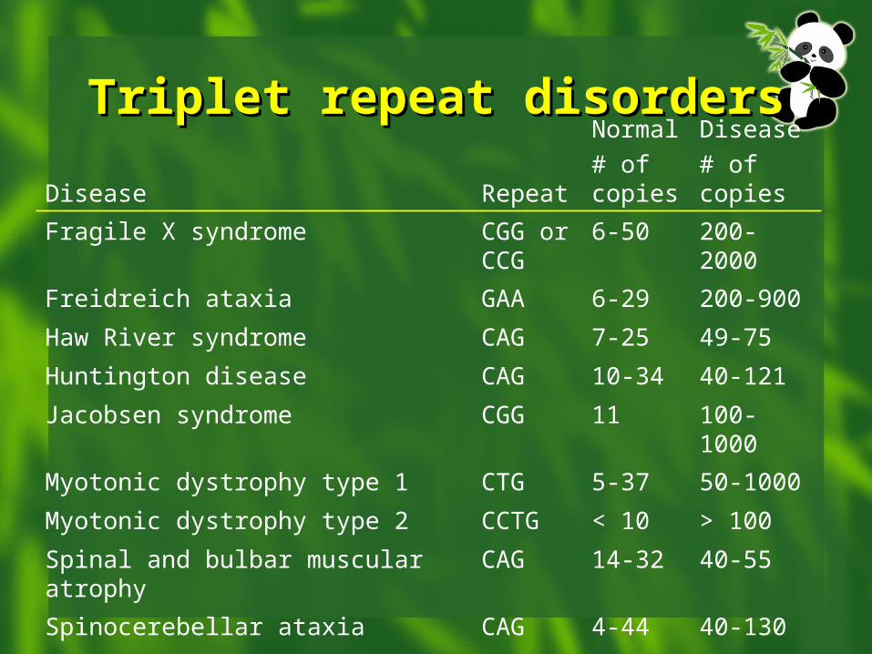
Triplet repeat disordersTriplet repeat disorders
Disease Repeat
Normal
# of copies
Disease
# of copies
Fragile X syndrome CGG or CCG
6-50 200-2000
Freidreich ataxia GAA 6-29 200-900
Haw River syndrome CAG 7-25 49-75
Huntington disease CAG 10-34 40-121
Jacobsen syndrome CGG 11 100-1000
Myotonic dystrophy type 1 CTG 5-37 50-1000
Myotonic dystrophy type 2 CCTG < 10 > 100
Spinal and bulbar muscular atrophy CAG 14-32 40-55
Spinocerebellar ataxia CAG 4-44 40-130
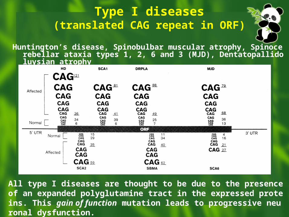
Type I diseases(translated CAG repeat in ORF)
Huntington’s disease, Spinobulbar muscular atrophy, Spinocerebellar ataxia types 1, 2, 6 and 3 (MJD), Dentatopallidoluysian atrophy
All type I diseases are thought to be due to the presence of an expanded polyglutamine tract in the expressed proteins. This gain of function mutation leads to progressive neuronal dysfunction.
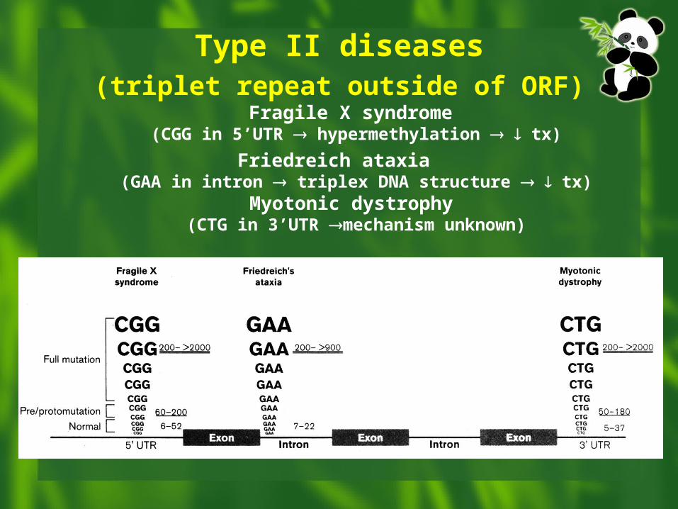
Type II diseases(triplet repeat outside of ORF)
Fragile X syndrome (CGG in 5’UTR hypermethylation tx)
Friedreich ataxia (GAA in intron triplex DNA structure tx)
Myotonic dystrophy (CTG in 3’UTR mechanism unknown)
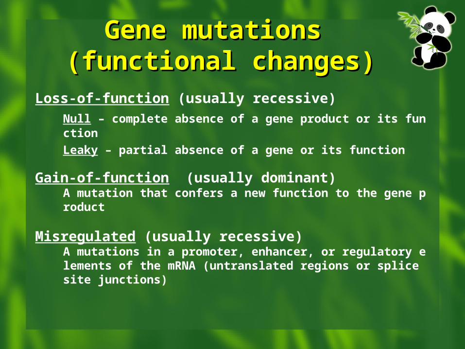
Gene mutations Gene mutations (functional changes)(functional changes)
Loss-of-function (usually recessive)
Null – complete absence of a gene product or its function
Leaky – partial absence of a gene or its function
Gain-of-function (usually dominant)A mutation that confers a new function to the gene product
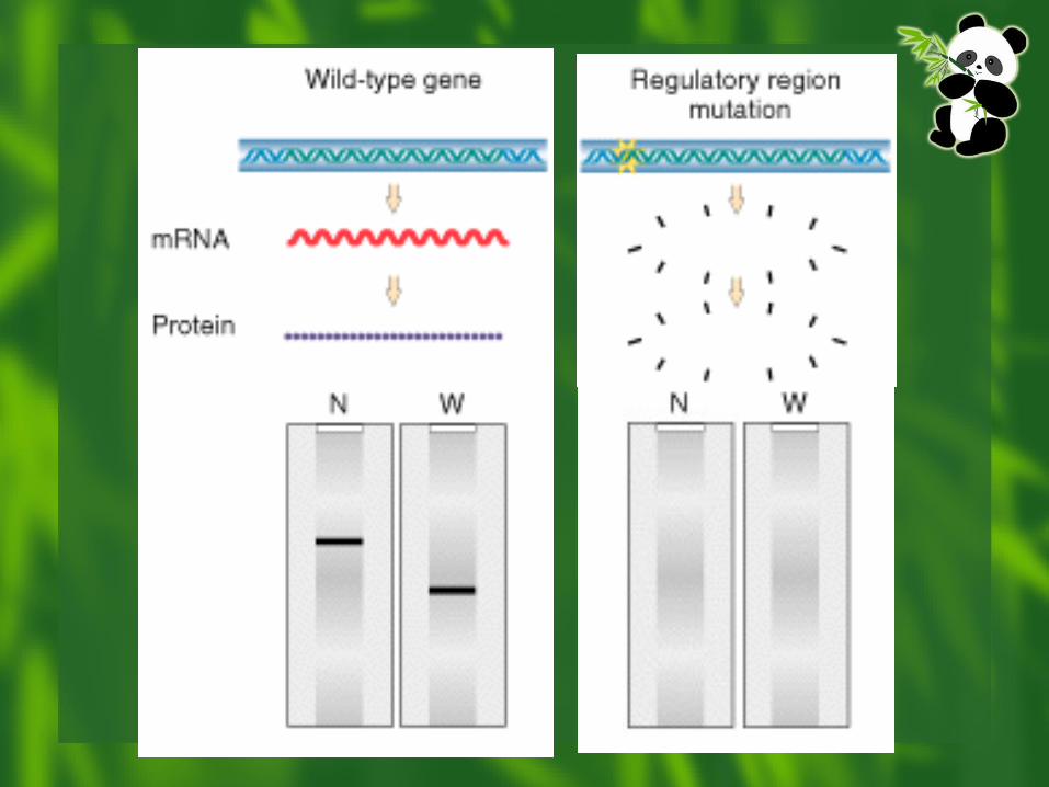
Misregulated (usually recessive)A mutations in a promoter, enhancer, or regulatory elements of the mRNA (untranslated regions or splice site junctions)
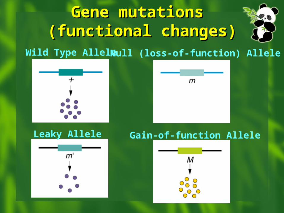
Wild Type Allele Null (loss-of-function) Allele
Leaky Allele Gain-of-function Allele
Gene mutations Gene mutations (functional changes)(functional changes)
Large
Small
- -
++Northern
Western
Mutation DetectionMutation Detection
Sequencing is “gold standard” Methods to detect known mutations Methods to detect unknown mutations
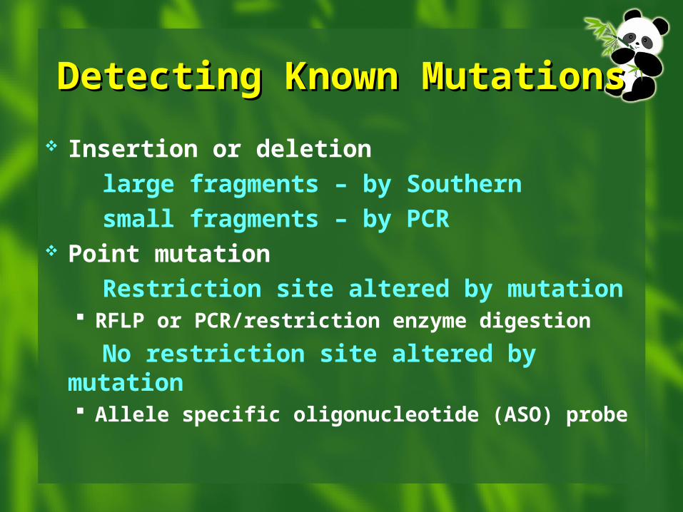
Detecting Known Detecting Known MutationsMutations
Insertion or deletion
large fragments – by Southern
small fragments – by PCR Point mutation
Restriction site altered by mutation RFLP or PCR/restriction enzyme digestion
No restriction site altered by mutation Allele specific oligonucleotide (ASO) probe
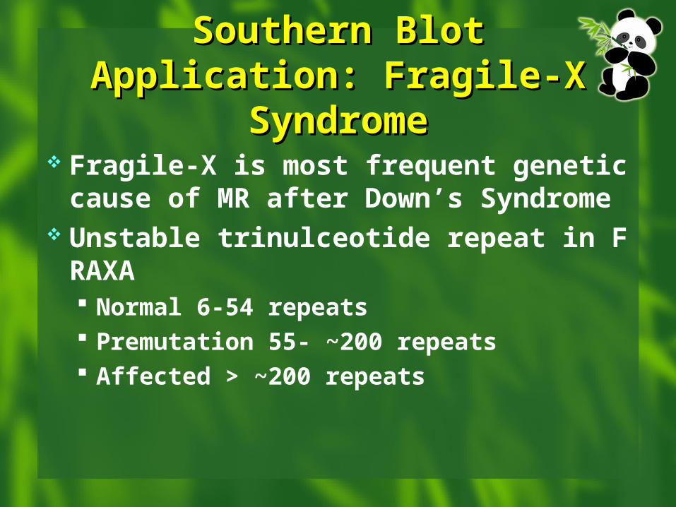
Southern Blot Application: Southern Blot Application: Fragile-X SyndromeFragile-X Syndrome
Fragile-X is most frequent genetic cause of MR after Down’s Syndrome
Unstable trinulceotide repeat in FRAXA Normal 6-54 repeats Premutation 55- ~200 repeats Affected > ~200 repeats
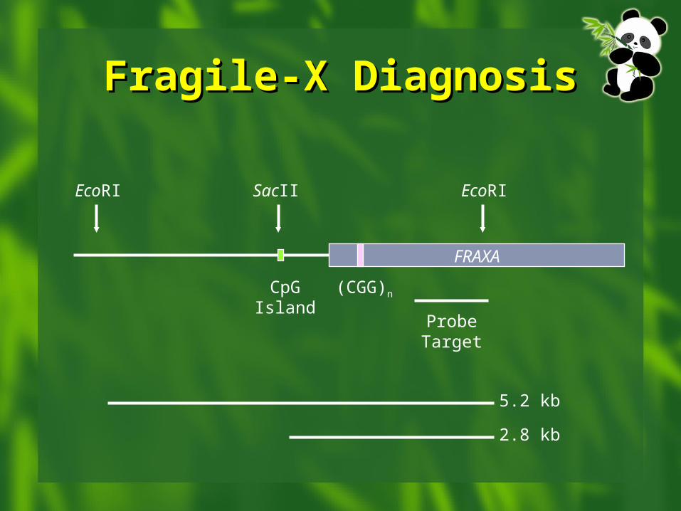
FRAXA
(CGG)n
EcoRI EcoRISacII
CpGIsland
ProbeTarget
5.2 kb
2.8 kb
Fragile-X DiagnosisFragile-X Diagnosis
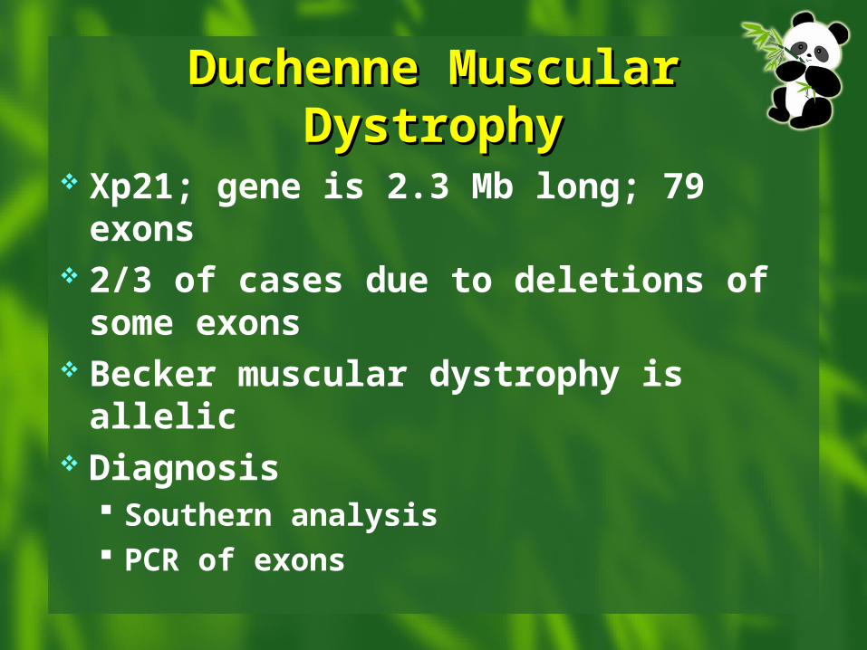
Duchenne Muscular Duchenne Muscular DystrophyDystrophy
Xp21; gene is 2.3 Mb long; 79 exons 2/3 of cases due to deletions of some
exons Becker muscular dystrophy is allelic Diagnosis
Southern analysis PCR of exons
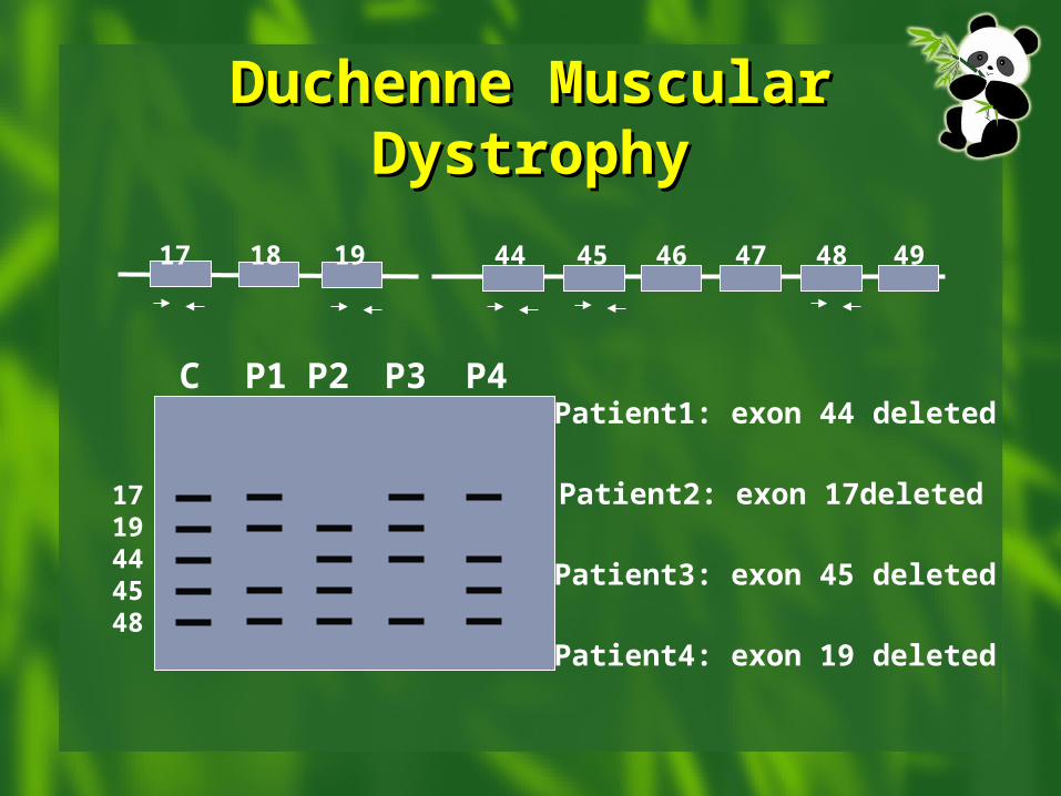
Duchenne Muscular Duchenne Muscular DystrophyDystrophy
17 18 19 44 45 46 47 48 49
1719444548
C P1 P2 P3 P4Patient1: exon 44 deleted
Patient2: exon 17deleted
Patient4: exon 19 deleted
Patient3: exon 45 deleted
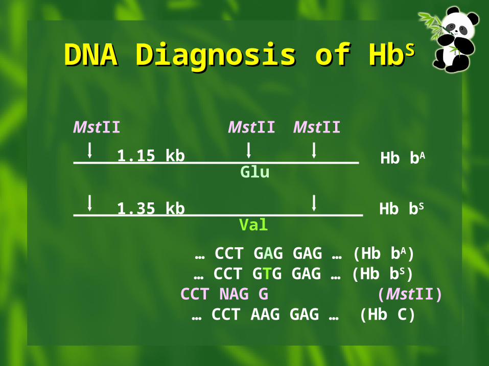
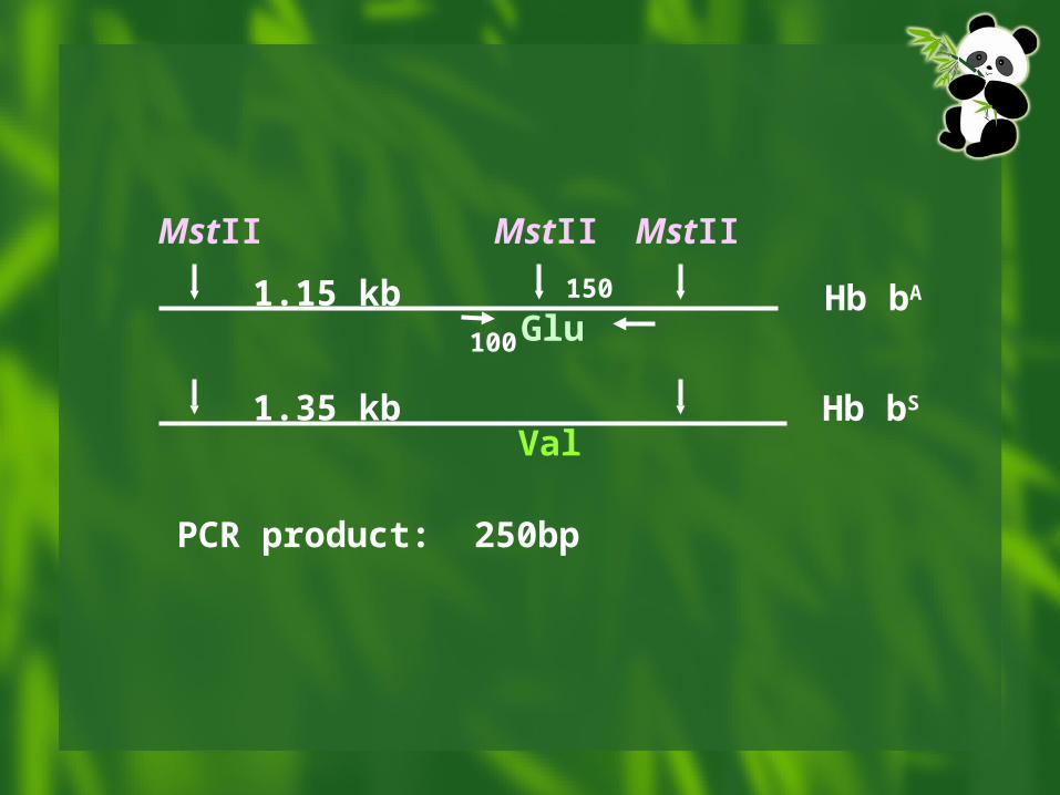
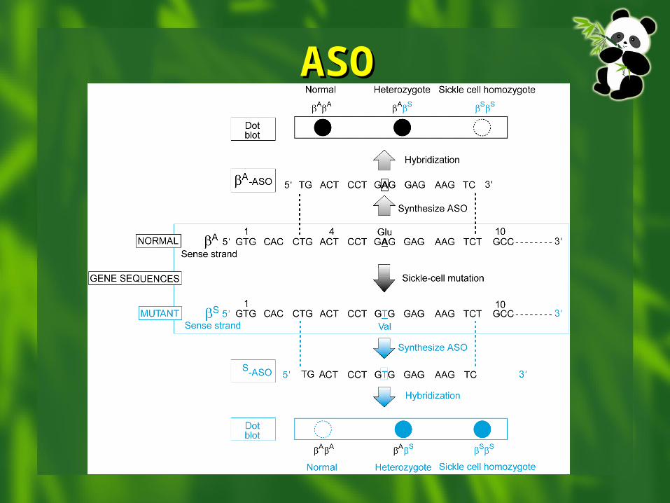
DNA Diagnosis of HbDNA Diagnosis of HbS S
Hb bA
Hb bS
MstII MstII MstII
Glu
Val
… CCT GAG GAG … (Hb bA)… CCT GTG GAG … (Hb bS)
CCT NAG G (MstII)… CCT AAG GAG … (Hb C)
1.15 kb
1.35 kb
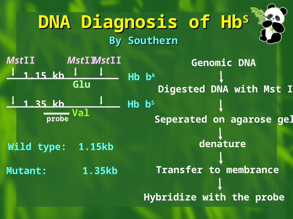
DNA Diagnosis of HbDNA Diagnosis of HbS S
By SouthernBy Southern
Genomic DNA
Digested DNA with Mst II
Seperated on agarose gel
denature
Transfer to membrance
Hybridize with the probe
Hb bA
Hb bS
MstII MstII MstII
Glu
Val
1.15 kb
1.35 kb
probe
Wild type: 1.15kb
Mutant: 1.35kb
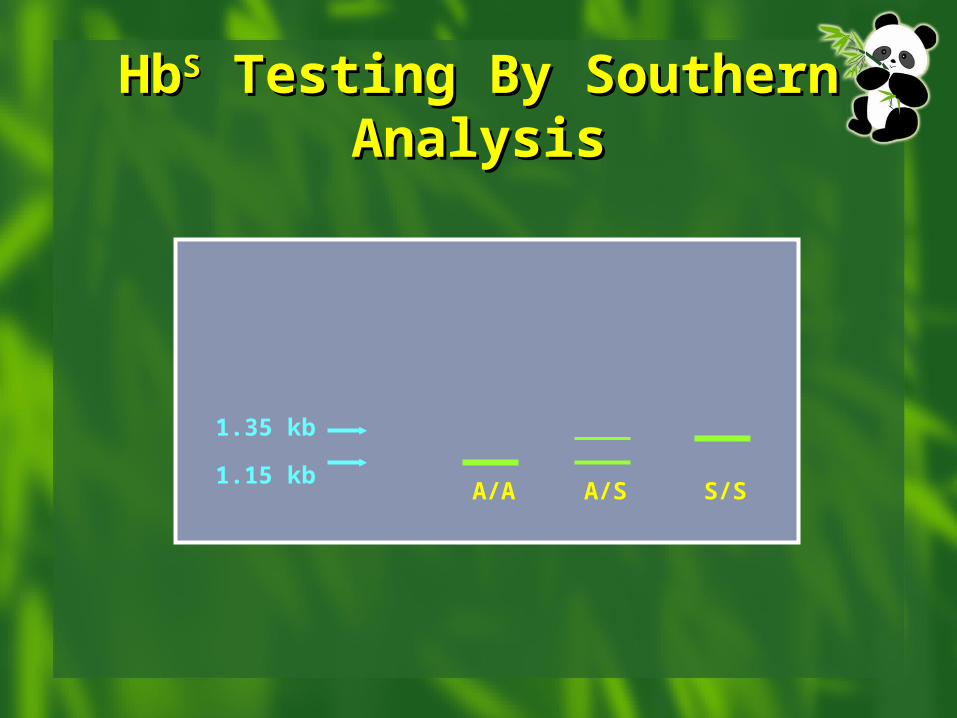
HbHbSS Testing By Southern Testing By Southern AnalysisAnalysis
A/A A/S S/S
1.35 kb
1.15 kb
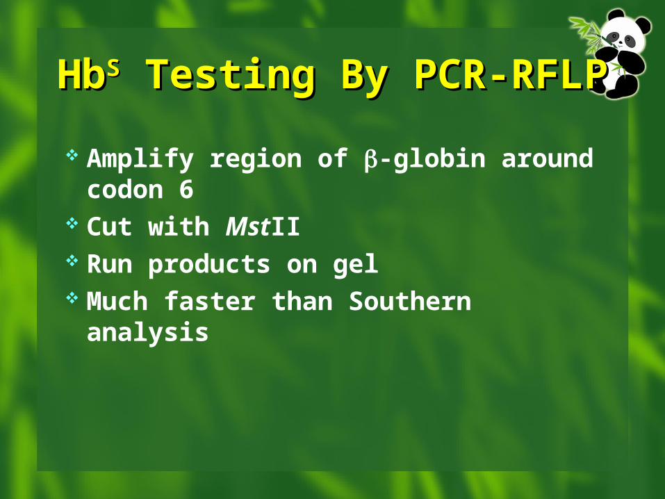
HbHbSS Testing By PCR-RFLP Testing By PCR-RFLP
Amplify region of -globin around codon 6
Cut with MstII Run products on gel Much faster than Southern analysis
Hb bA
Hb bS
MstII MstII MstII
Glu
Val
1.15 kb
1.35 kb
100
150
PCR product: 250bp
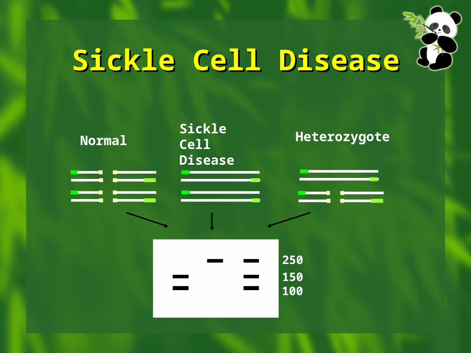
Sickle Cell DiseaseSickle Cell Disease
Sickle Cell Disease
HeterozygoteNormal
250
150100
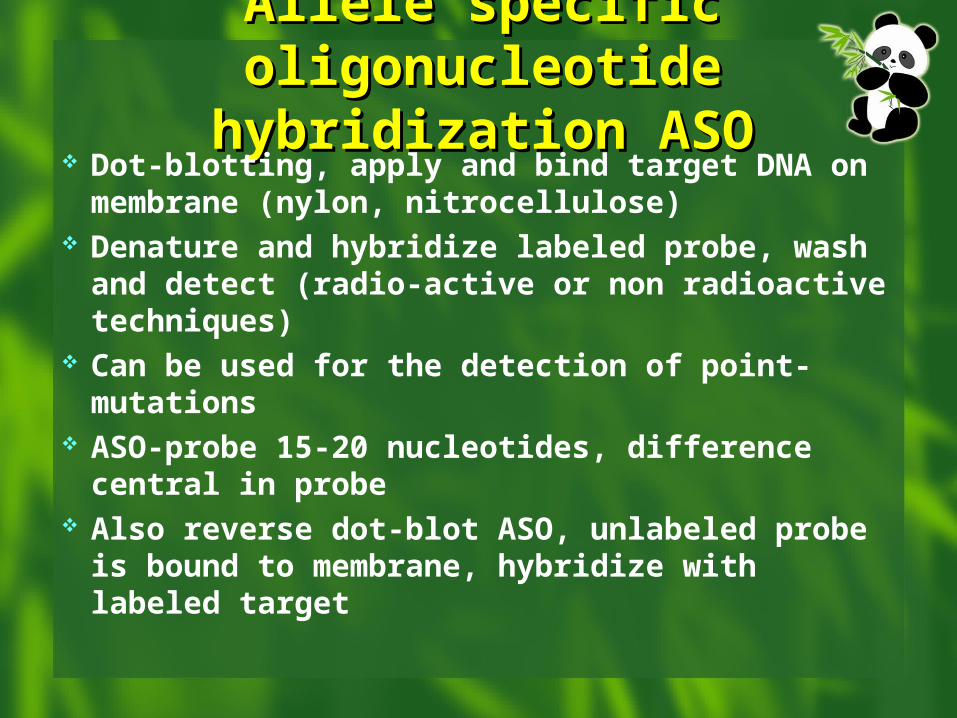
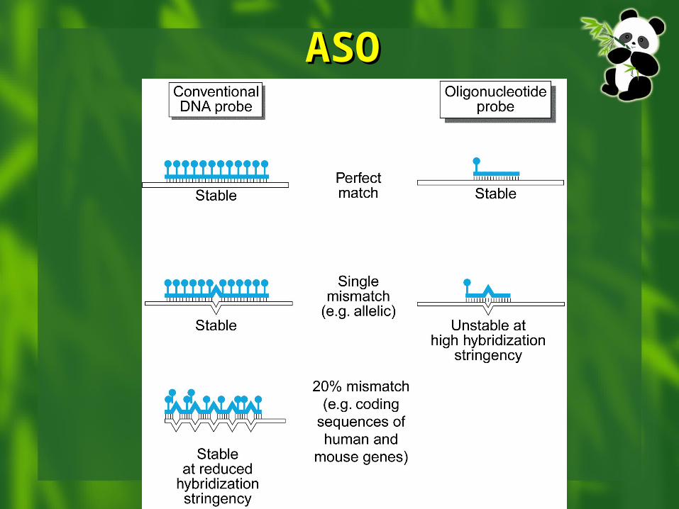
Allele specific Allele specific oligonucleotide hybridization oligonucleotide hybridization
ASOASO Dot-blotting, apply and bind target DNA on
membrane (nylon, nitrocellulose) Denature and hybridize labeled probe, wash and
detect (radio-active or non radioactive techniques)
Can be used for the detection of point-mutations ASO-probe 15-20 nucleotides, difference central
in probe Also reverse dot-blot ASO, unlabeled probe is
bound to membrane, hybridize with labeled target
ASOASO
ASOASO
Detecting Unknown Detecting Unknown MutationsMutations
Mutation screening
- Chemical cleavage of mismatch
- SSCP
- Heteroduplex formation
- Southern analysis Mutation confirming
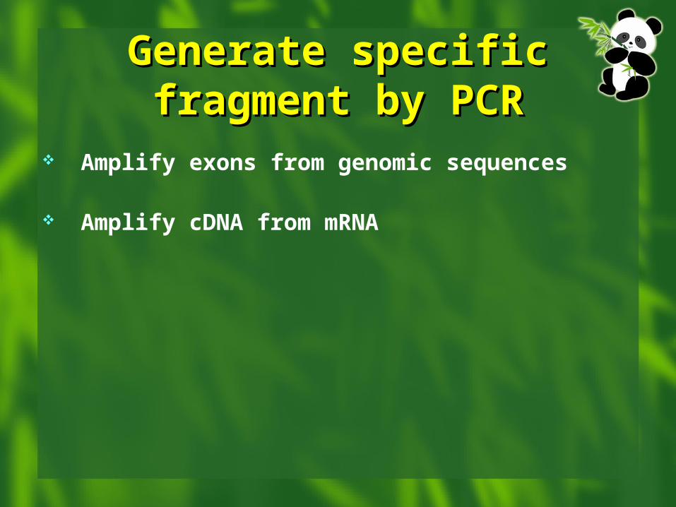
Generate specific Generate specific fragment by PCRfragment by PCR
Amplify exons from genomic sequences Amplify cDNA from mRNA
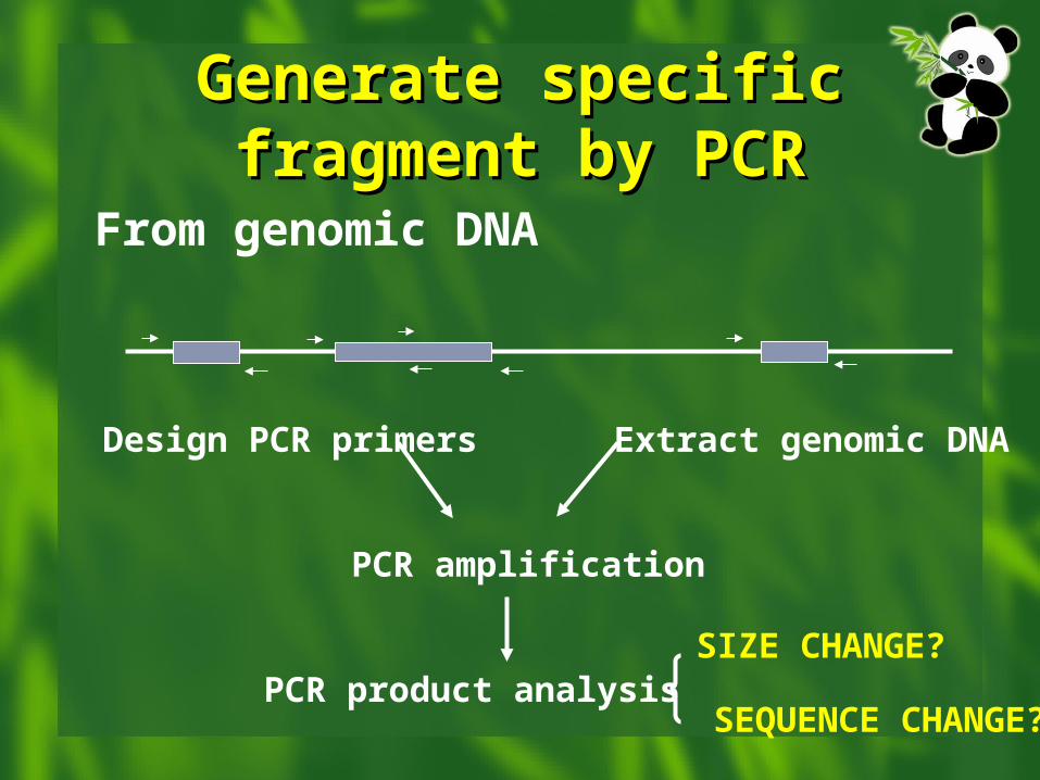
Generate specific Generate specific fragment by PCRfragment by PCR
From genomic DNA
PCR amplification
PCR product analysis
Design PCR primers Extract genomic DNA
SIZE CHANGE?
SEQUENCE CHANGE?
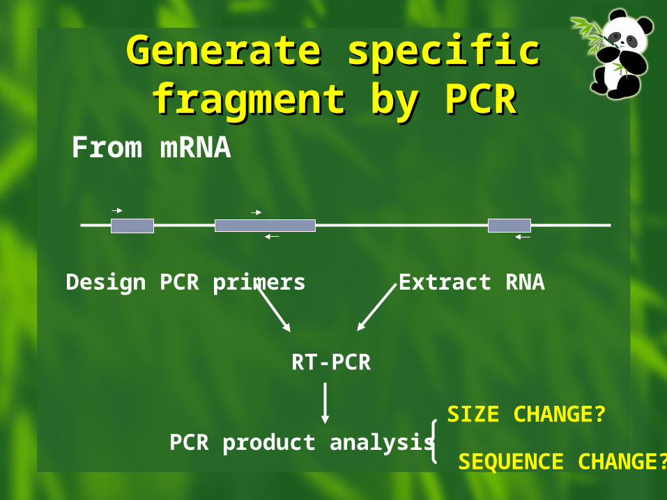
Generate specific Generate specific fragment by PCRfragment by PCR
From mRNA
RT-PCR
PCR product analysis
Design PCR primers Extract RNA
SIZE CHANGE?
SEQUENCE CHANGE?
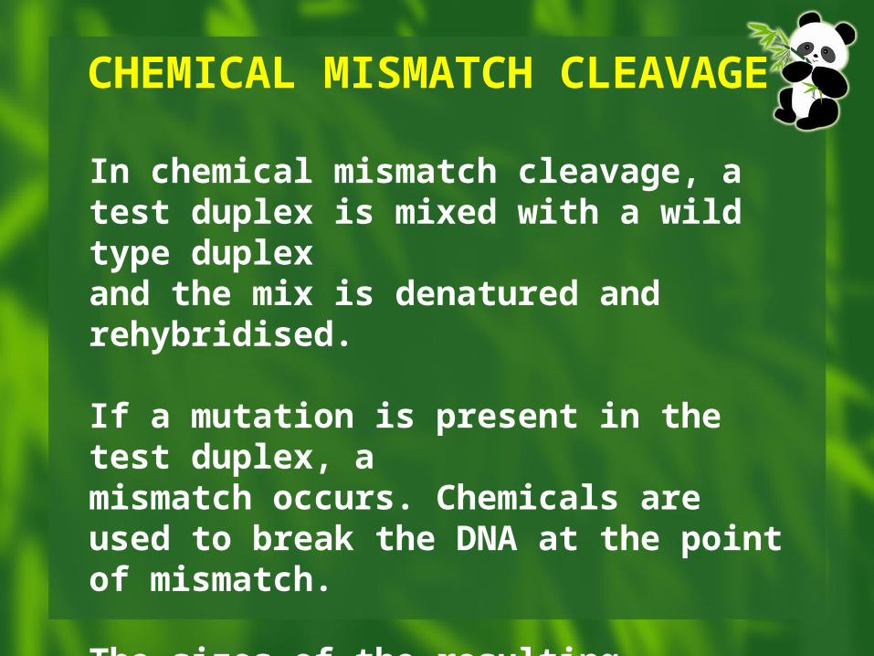
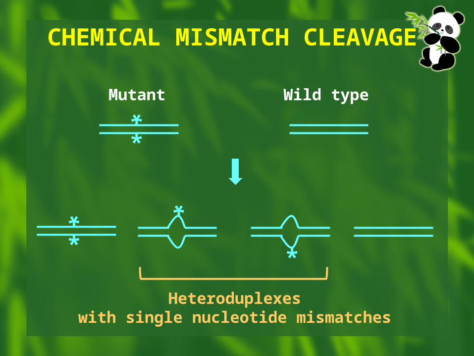
In chemical mismatch cleavage, a test duplex is mixed with a wild type duplexand the mix is denatured and rehybridised.
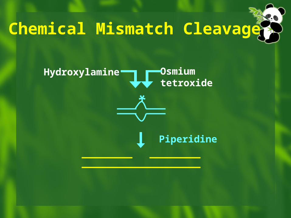
If a mutation is present in the test duplex, amismatch occurs. Chemicals are used to break the DNA at the point of mismatch.
The sizes of the resulting fragments give anindication of the position of the mutationin the duplex (see next 3 slides).
CHEMICAL MISMATCH CLEAVAGE
*
*
CHEMICAL MISMATCH CLEAVAGE
Mutant Wild type
*
*
Heteroduplexeswith single nucleotide mismatches
*
*
*
Hydroxylamine Osmiumtetroxide
Piperidine
Chemical Mismatch Cleavage
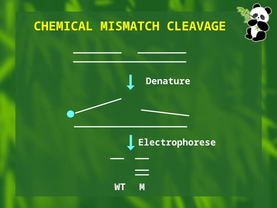
CHEMICAL MISMATCH CLEAVAGE
Electrophorese
Denature
WT M
CHEMICAL MISMATCH CLEAVAGE
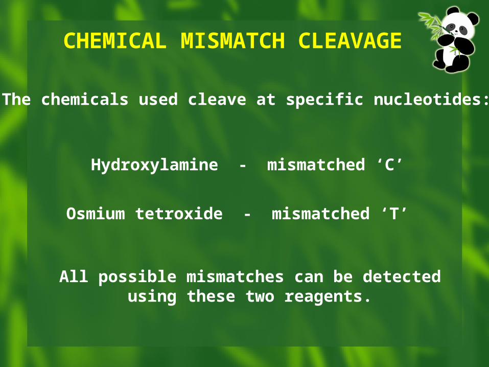
Hydroxylamine - mismatched ‘C’
Osmium tetroxide - mismatched ‘T’
The chemicals used cleave at specific nucleotides:
All possible mismatches can be detectedusing these two reagents.
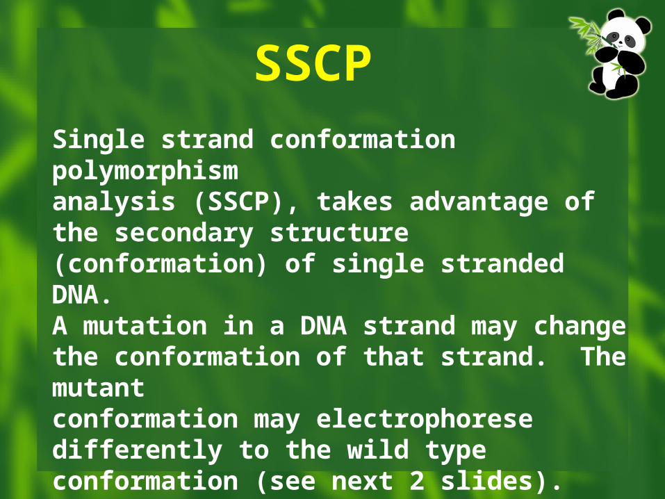
Single strand conformation polymorphismanalysis (SSCP), takes advantage of the secondary structure (conformation) of single stranded DNA.A mutation in a DNA strand may changethe conformation of that strand. The mutantconformation may electrophorese differently to the wild type conformation (see next 2 slides).
SSCP
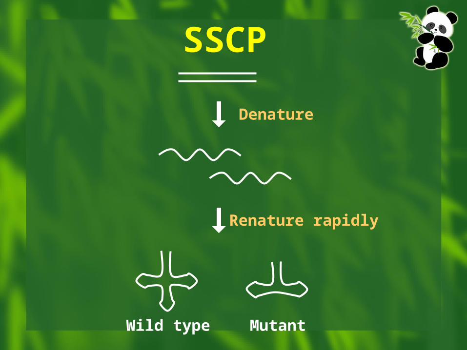
Denature
Renature rapidly
Wild type Mutant
SSCP
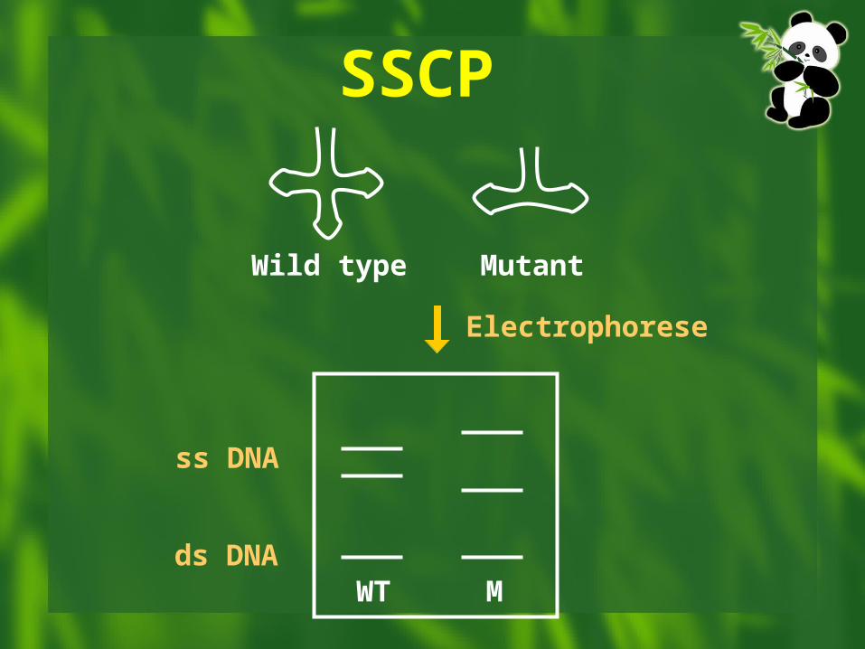
Electrophorese
ss DNA
ds DNAWT M
Wild type Mutant
SSCP
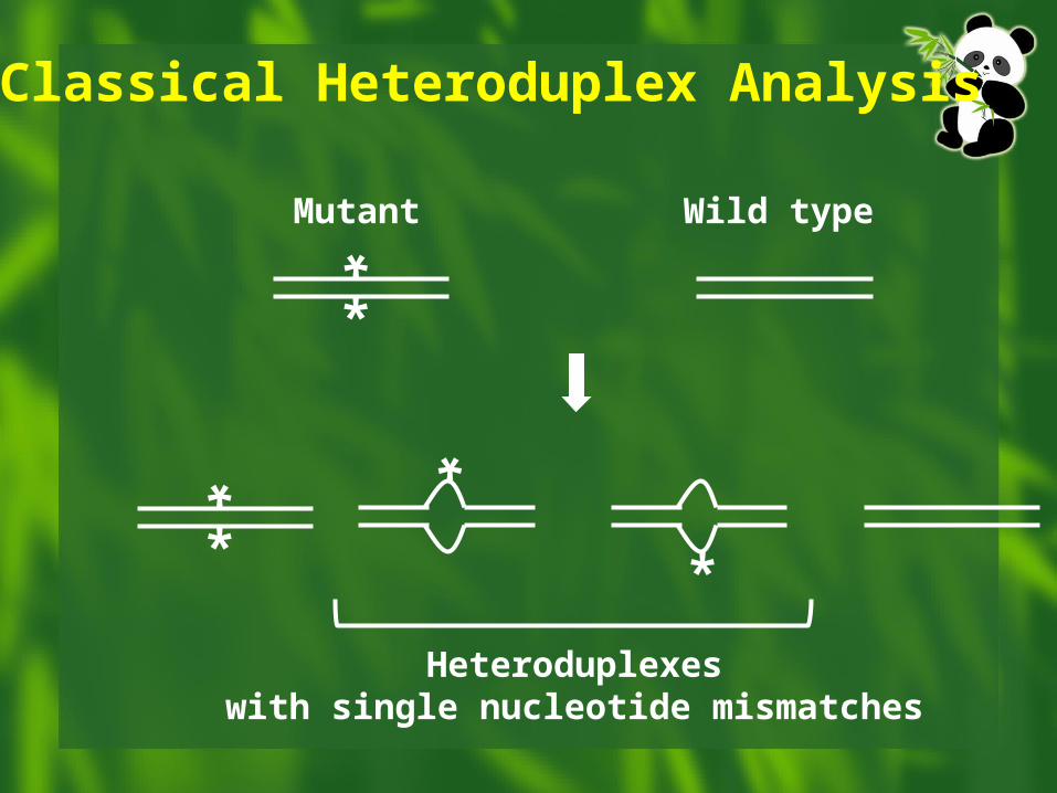
In classical heteroduplex analysis, test DNA is mixed with wild type DNA, the mix is denatured and allowed to rehybridise to give heteroduplexes. If a mutation is present, the heteroduplexes contain a mismatch which affects their electrophoretic mobility in certain gel systems (see next 2 slides).
Heteroduplex Analysis
*
*
Classical Heteroduplex Analysis
Mutant Wild type
*
*
Heteroduplexeswith single nucleotide mismatches
*
*
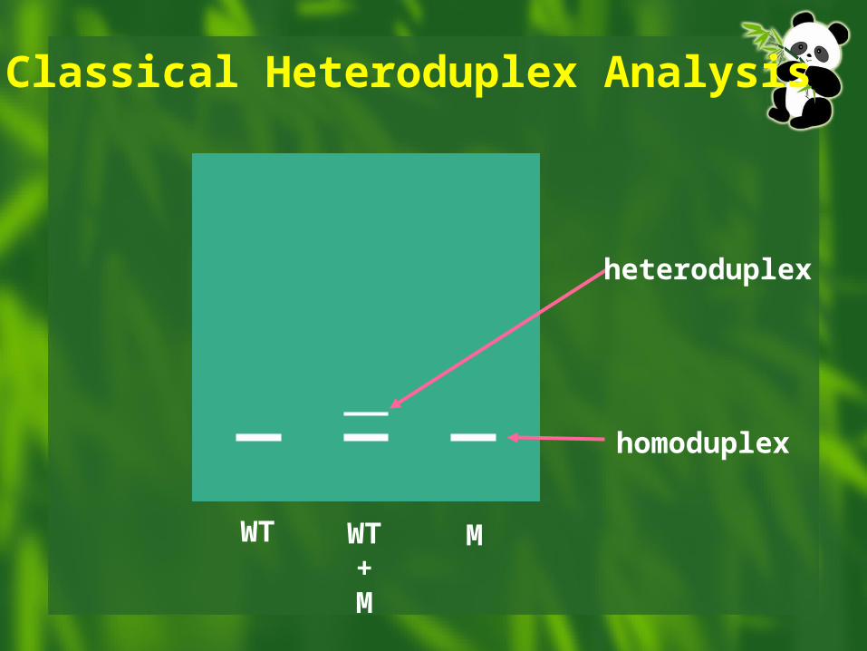
Classical Heteroduplex Analysis
WT MWT+M
heteroduplex
homoduplex
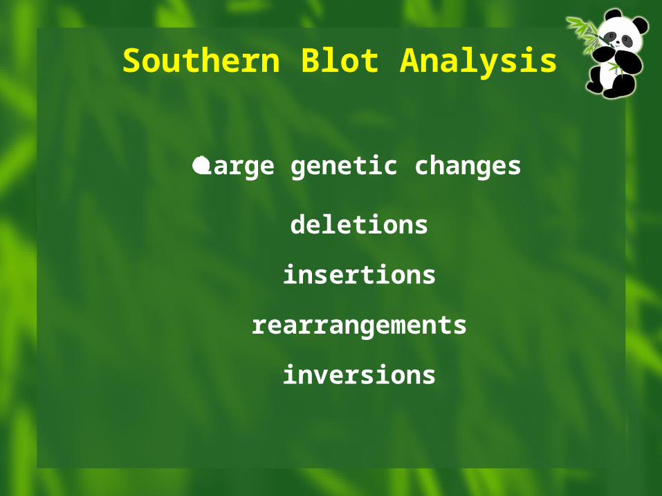
Large genetic changes
deletions
insertions
rearrangements
inversions
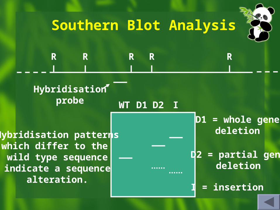
Southern Blot Analysis
Southern Blot Analysis
R R R RR
WT D1 D2 I
Hybridisationprobe
D1 = whole genedeletion
D2 = partial genedeletion
I = insertion
Hybridisation patternswhich differ to the wild type sequenceindicate a sequence
alteration.
Polymorphisms
Definition Major types Application
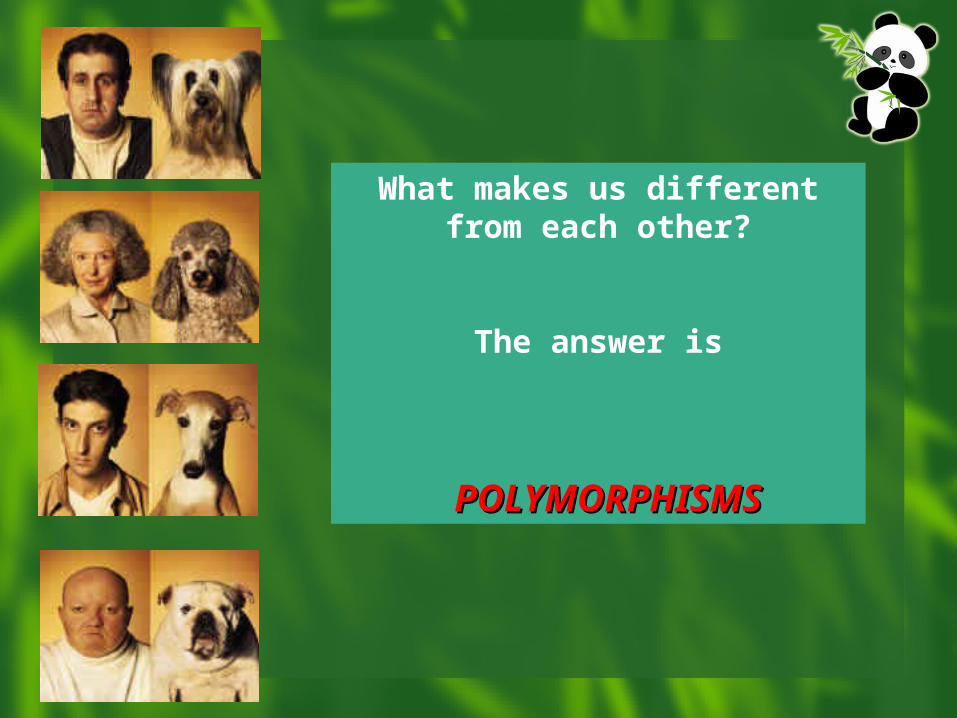
What makes us different from each other?
The answer is
POLYMORPHISMSPOLYMORPHISMS
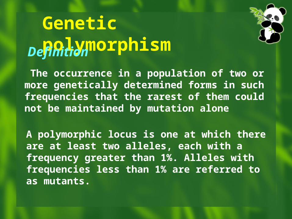
Genetic polymorphism
The occurrence in a population of two or more genetically determined forms in such frequencies that the rarest of them could not be maintained by mutation alone
Definition
A polymorphic locus is one at which there are at least two alleles, each with a frequency greater than 1%. Alleles with frequencies less than 1% are referred to as mutants.
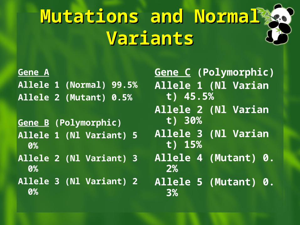
Mutations and Normal Mutations and Normal VariantsVariants
Gene A
Allele 1 (Normal) 99.5%
Allele 2 (Mutant) 0.5%
Gene B (Polymorphic)
Allele 1 (Nl Variant) 50%
Allele 2 (Nl Variant) 30%
Allele 3 (Nl Variant) 20%
Gene C (Polymorphic)Allele 1 (Nl Variant) 45.5%Allele 2 (Nl Variant) 30%Allele 3 (Nl Variant) 15%Allele 4 (Mutant) 0.2%Allele 5 (Mutant) 0.3%
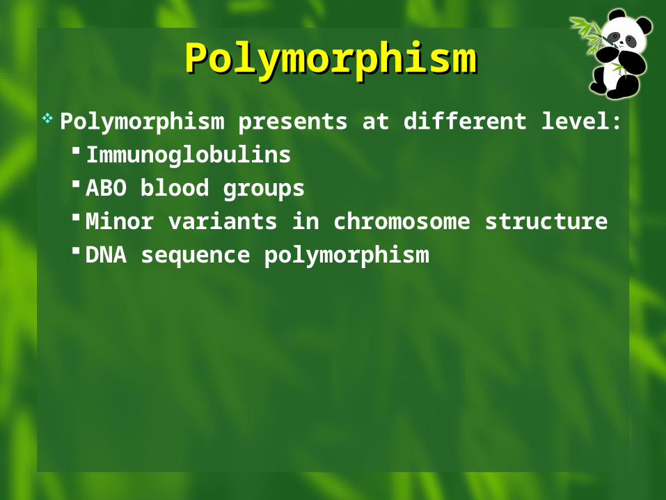
PolymorphismPolymorphism Polymorphism presents at different level:
Immunoglobulins ABO blood groups Minor variants in chromosome structure DNA sequence polymorphism
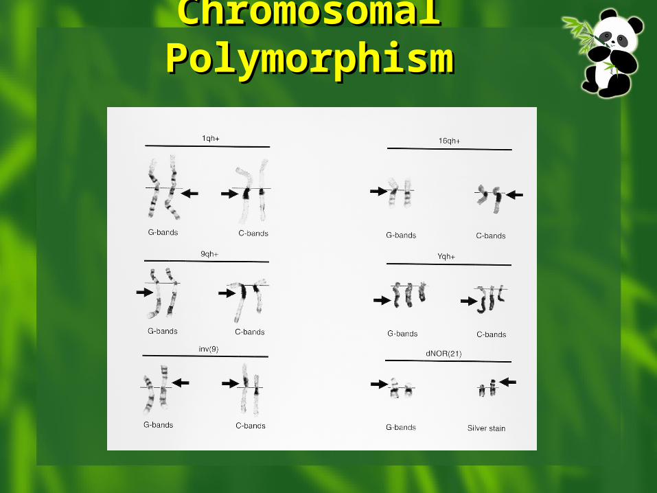
Chromosomal Chromosomal PolymorphismPolymorphism
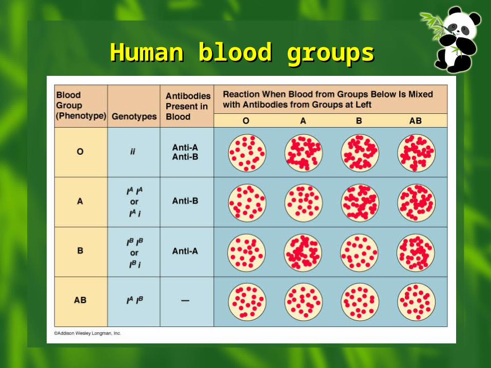
Human blood groupsHuman blood groups
It is variation of DNA sequence that is common in the general population (>1%)
Most are neutral, but some confer susceptibility or resistance to disease
It may be a single nucleotide change or variation in copy number of a repetitive sequence
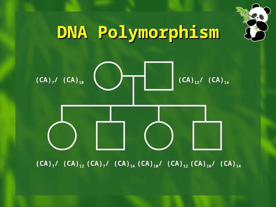
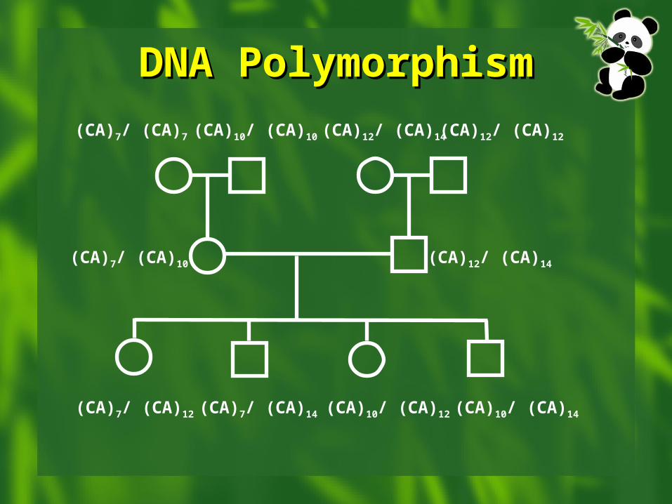
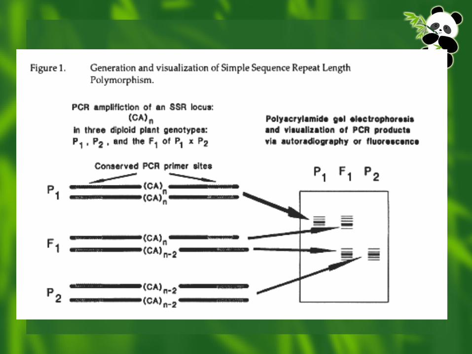
DNA PolymorphismDNA Polymorphism
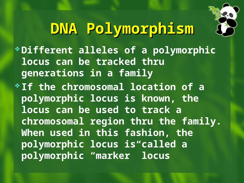
DNA PolymorphismDNA PolymorphismDifferent alleles of a polymorphic locus
can be tracked thru generations in a family
If the chromosomal location of a polymorphic locus is known, the locus can be used to track a chromosomal region thru the family. When used in this fashion, the polymorphic locus is called a polymorphic “marker” locus
DNA PolymorphismDNA Polymorphism
(CA)7/ (CA)10 (CA)12/ (CA)14
(CA)7/ (CA)12 (CA)7/ (CA)14 (CA)10/ (CA)12 (CA)10/ (CA)14
DNA PolymorphismDNA Polymorphism
(CA)7/ (CA)10 (CA)12/ (CA)14
(CA)7/ (CA)12 (CA)7/ (CA)14 (CA)10/ (CA)12 (CA)10/ (CA)14
(CA)7/ (CA)7 (CA)10/ (CA)10 (CA)12/ (CA)14 (CA)12/ (CA)12
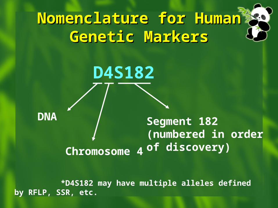
Nomenclature for Human Nomenclature for Human Genetic MarkersGenetic Markers
D4S182
Segment 182 (numbered in order of discovery)Chromosome 4
DNA
*D4S182 may have multiple alleles defined by RFLP, SSR, etc.
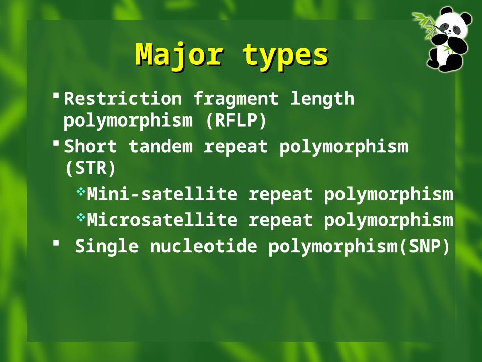
Major types Major types Restriction fragment length polymorphism
(RFLP) Short tandem repeat polymorphism (STR)
Mini-satellite repeat polymorphismMicrosatellite repeat polymorphism
Single nucleotide polymorphism(SNP)
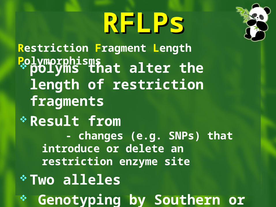
RFLPsRFLPs polyms that alter the length
of restriction fragments Result from
- changes (e.g. SNPs) that introduce or delete an restriction enzyme site
Two alleles Genotyping by Southern or PCR-
RFLP
Restriction Fragment Length Polymorphisms
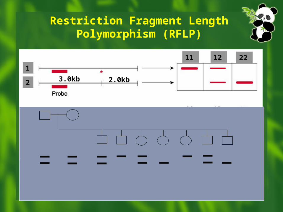
Restriction Fragment Length Polymorphism (RFLP)
3.0kb 2.0kb
1
2
11 12 22
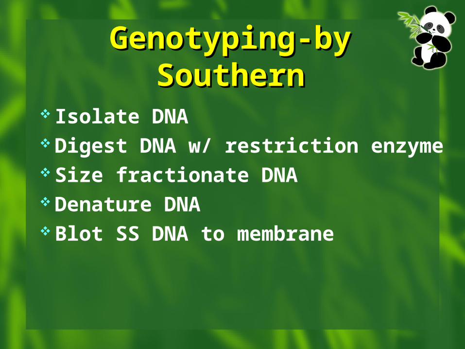
Genotyping-by Genotyping-by SouthernSouthern
Isolate DNA Digest DNA w/ restriction enzyme Size fractionate DNA Denature DNA Blot SS DNA to membrane
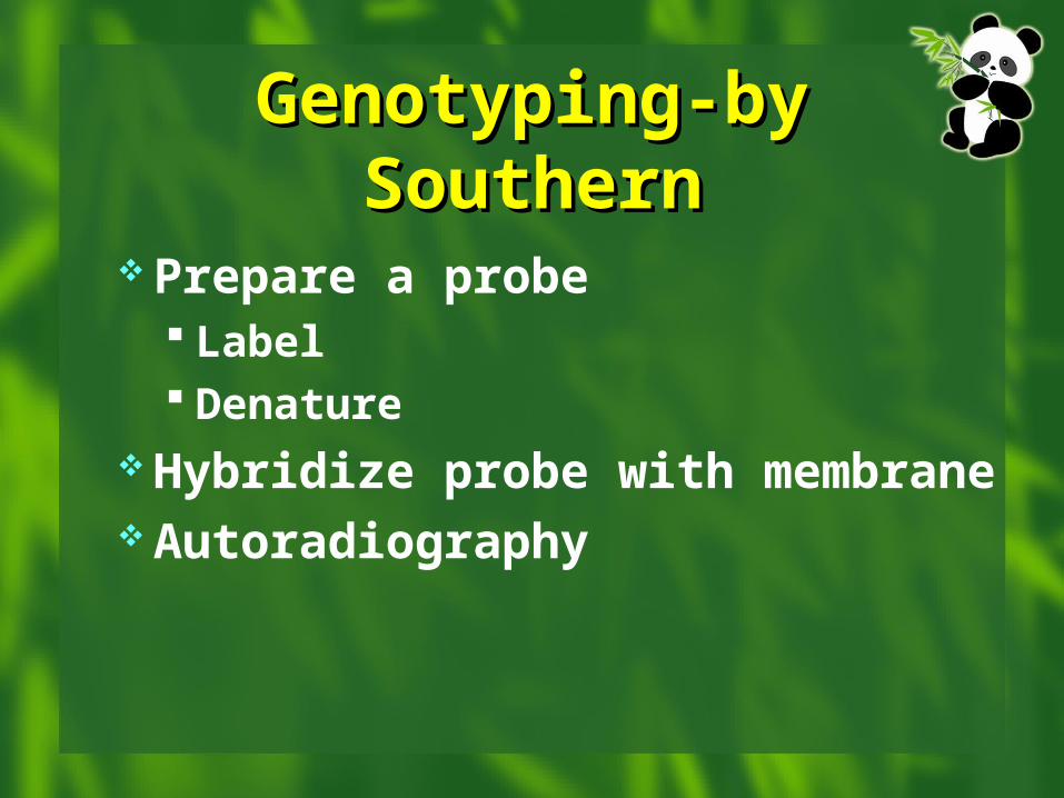
Genotyping-by Genotyping-by SouthernSouthern
Prepare a probe Label Denature
Hybridize probe with membrane Autoradiography
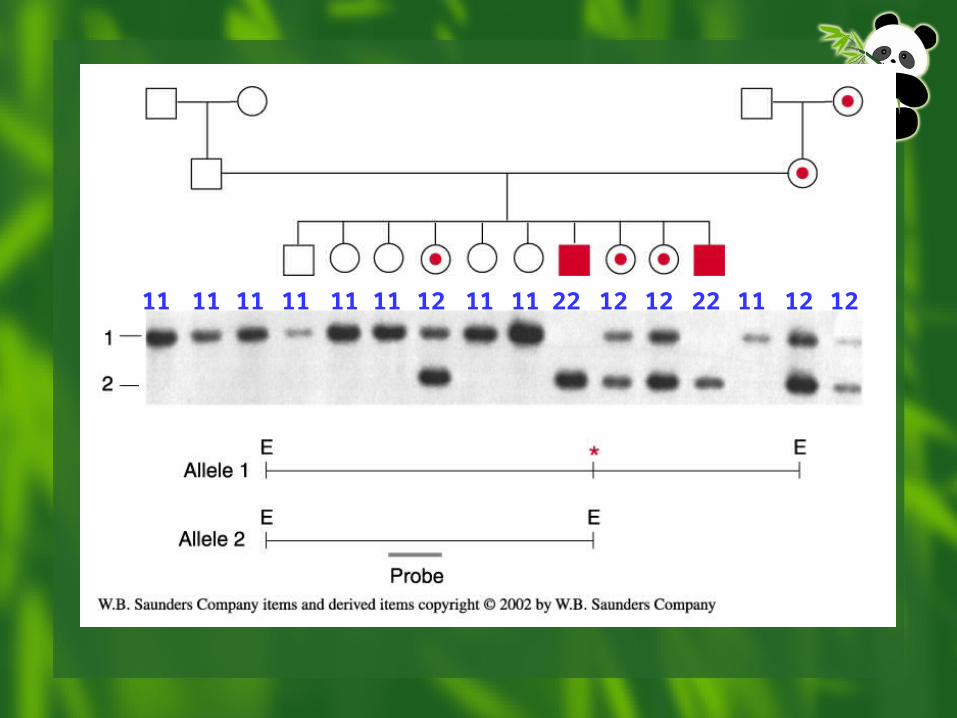
11 11 11 11 11 11 12 11 11 22 12 12 22 11 12 12
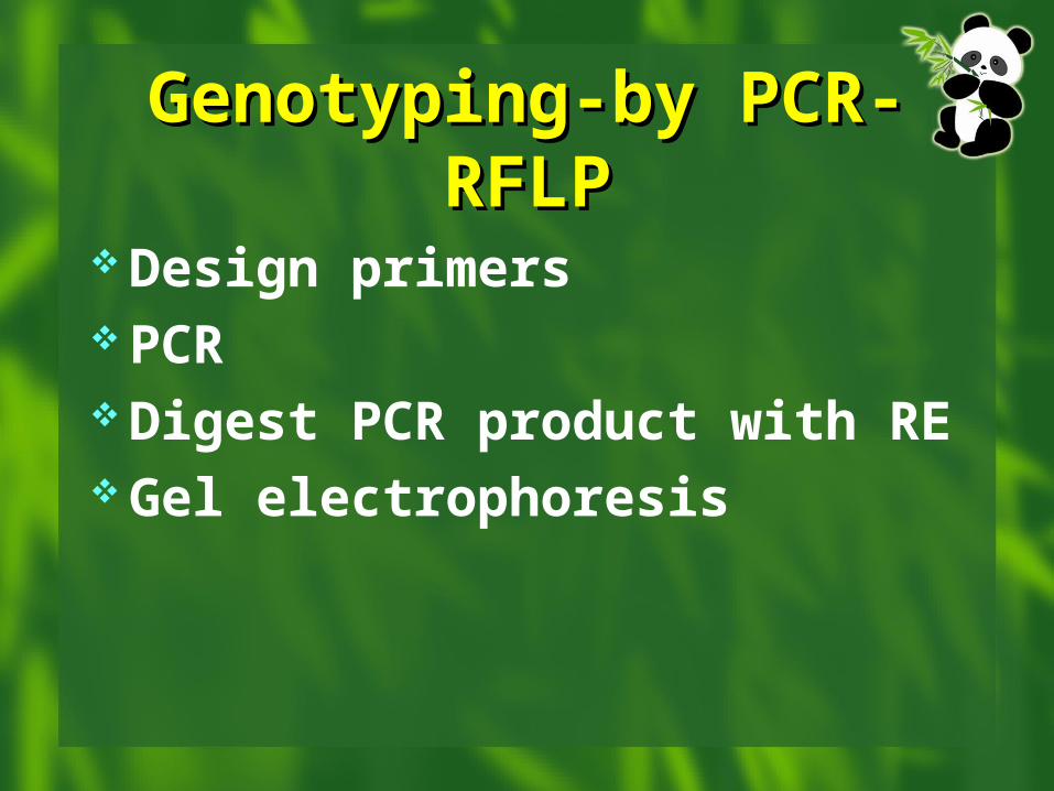
Genotyping-by PCR-Genotyping-by PCR-RFLPRFLP
Design primers PCR Digest PCR product with RE Gel electrophoresis
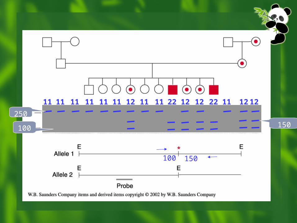
100 150
250
100 150
11 11 11 11 11 11 12 11 11 22 12 12 22 11 12 12
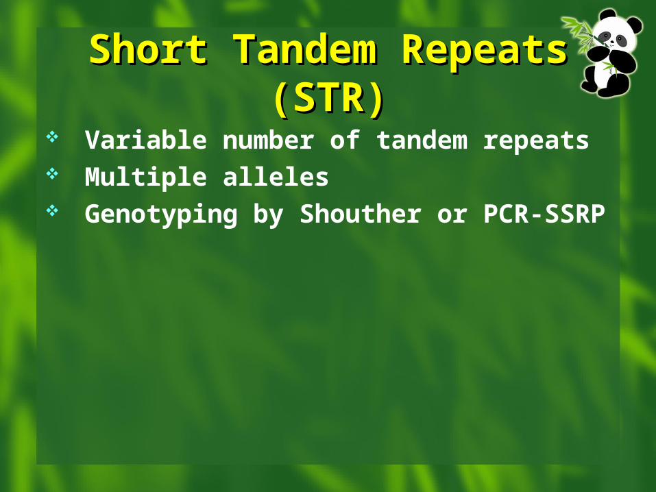
Short Tandem Repeats Short Tandem Repeats (STR)(STR)
Variable number of tandem repeats Multiple alleles Genotyping by Shouther or PCR-SSRP
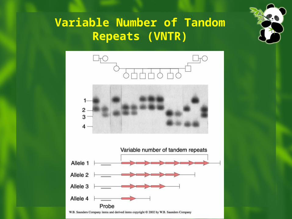
Variable Number of Tandom Repeats (VNTR)
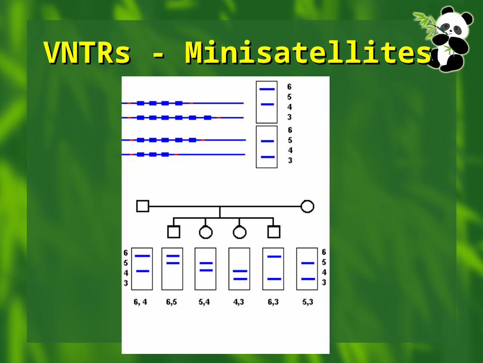
VNTRs - MinisatellitesVNTRs - Minisatellites
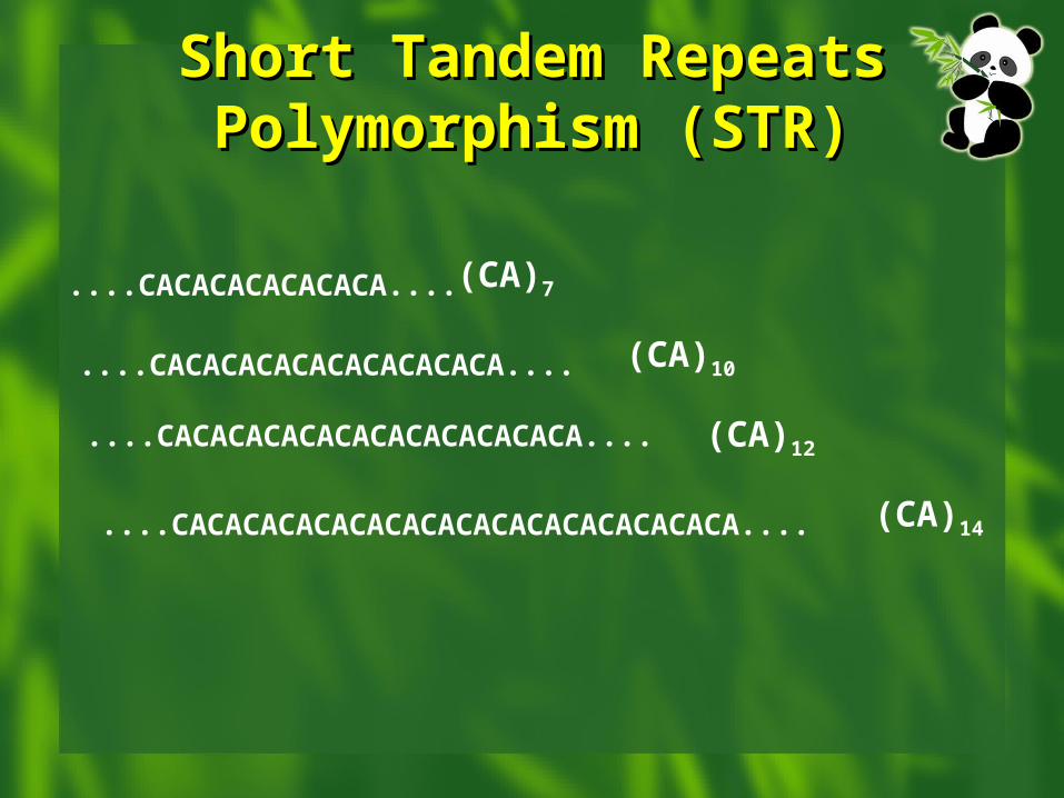
Short Tandem Repeats Short Tandem Repeats Polymorphism (STR)Polymorphism (STR)
....CACACACACACACA....
....CACACACACACACACACACA....
....CACACACACACACACACACACACA....
....CACACACACACACACACACACACACACACACA....
(CA)7
(CA)10
(CA)12
(CA)14
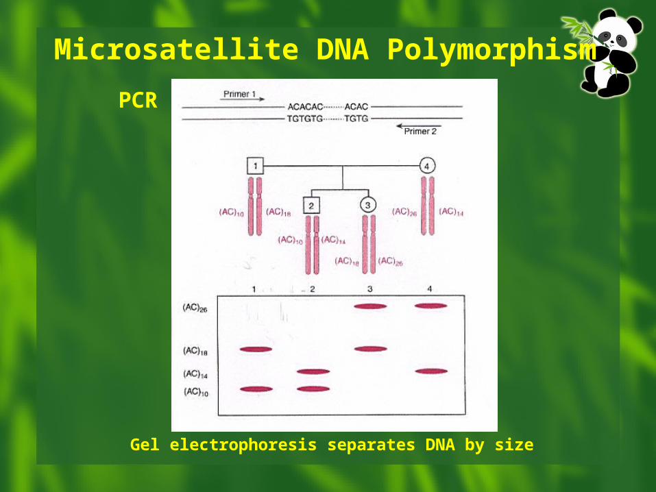
Microsatellite DNA PolymorphismPCR
Gel electrophoresis separates DNA by size
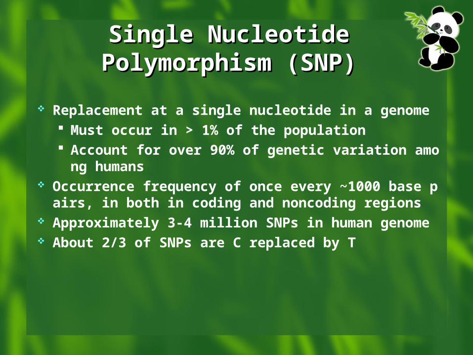
Single Nucleotide Single Nucleotide Polymorphism (SNP)Polymorphism (SNP)
Replacement at a single nucleotide in a genome Must occur in > 1% of the population Account for over 90% of genetic variation amon
g humans Occurrence frequency of once every ~1000 base pa
irs, in both in coding and noncoding regions Approximately 3-4 million SNPs in human genome About 2/3 of SNPs are C replaced by T
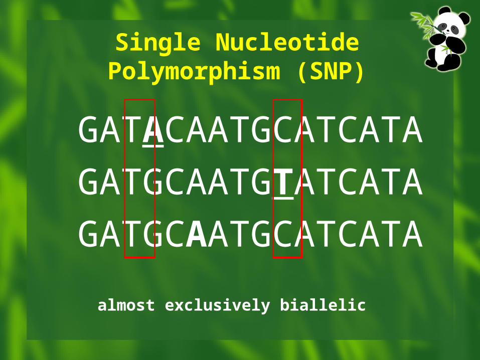
GATACAATGCATCATAGATGCAATGTATCATAGATGCAATGCATCATA
Single Nucleotide Polymorphism (SNP)
almost exclusively biallelic
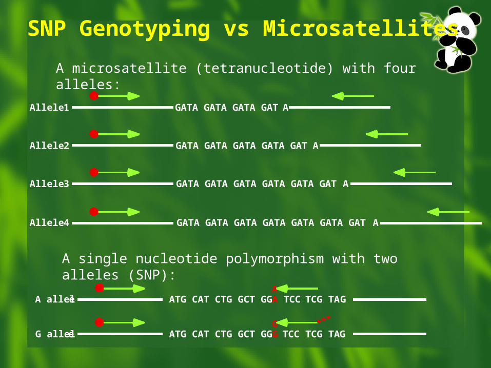
SNP Genotyping vs Microsatellites
A microsatellite (tetranucleotide) with four alleles:
GATA GATA GATA GATA
GATA GATA GATA GATA GATA
GATA GATA GATA GATA GATA GATA
GATA GATA GATA GATA GATA GATA GATA
Allele 1
Allele 2
Allele 3
Allele 4
A single nucleotide polymorphism with two alleles (SNP):
ATG CAT CTG GCT GGA TCC TCG TAGA alleleA
ATG CAT CTG GCT GGG TCC TCG TAGG alleleG ***
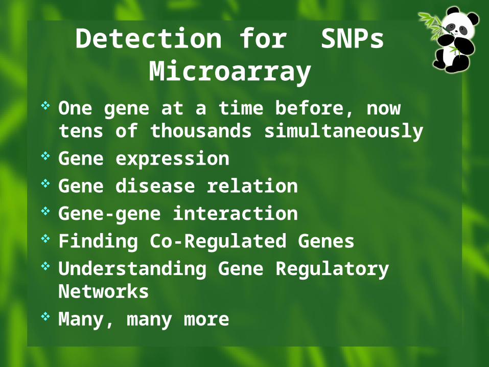
Detection for SNPsMicroarray
One gene at a time before, now tens of thousands simultaneously
Gene expression Gene disease relation Gene-gene interaction Finding Co-Regulated Genes Understanding Gene Regulatory Networks Many, many more
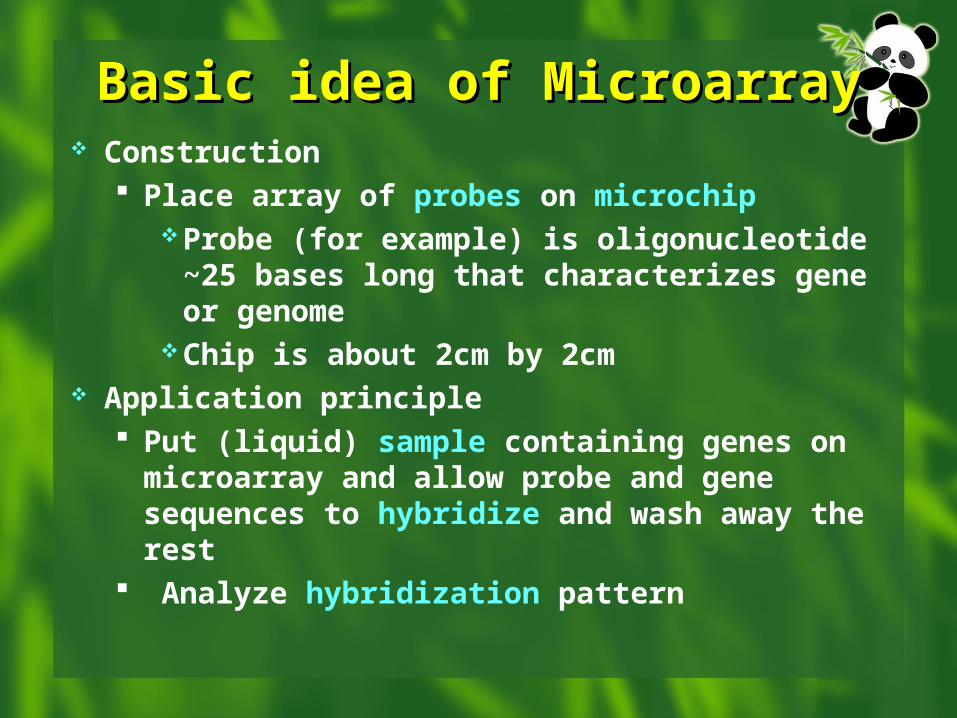
Basic idea of MicroarrayBasic idea of Microarray Construction
Place array of probes on microchipProbe (for example) is oligonucleotide ~25
bases long that characterizes gene or genome
Chip is about 2cm by 2cm Application principle
Put (liquid) sample containing genes on microarray and allow probe and gene sequences to hybridize and wash away the rest
Analyze hybridization pattern
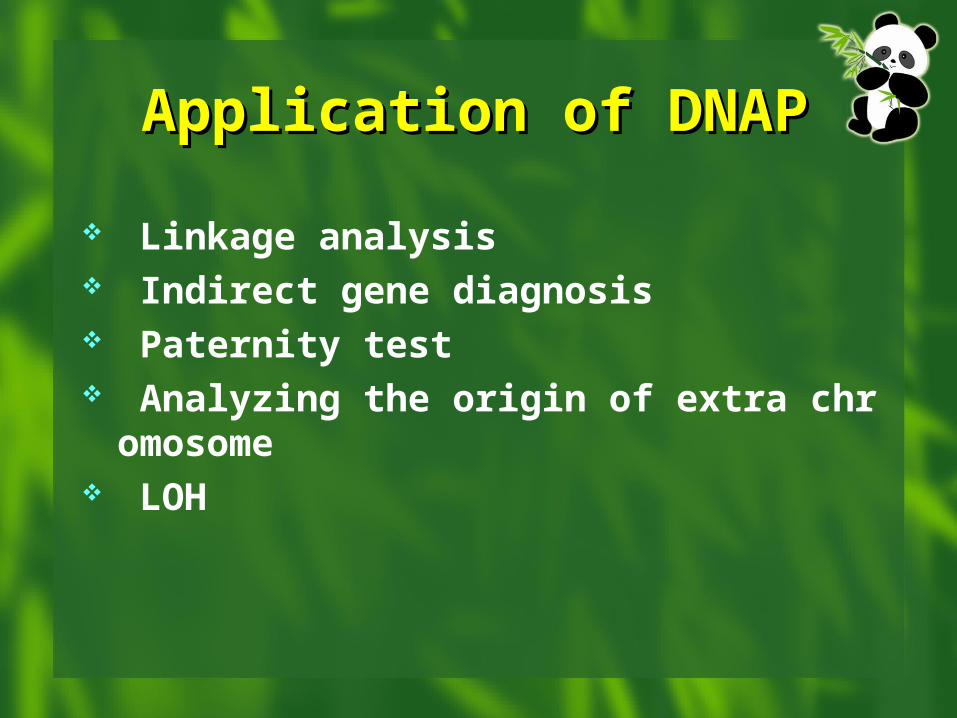
Application of DNAPApplication of DNAP
Linkage analysis Indirect gene diagnosis Paternity test Analyzing the origin of extra chromosome LOH
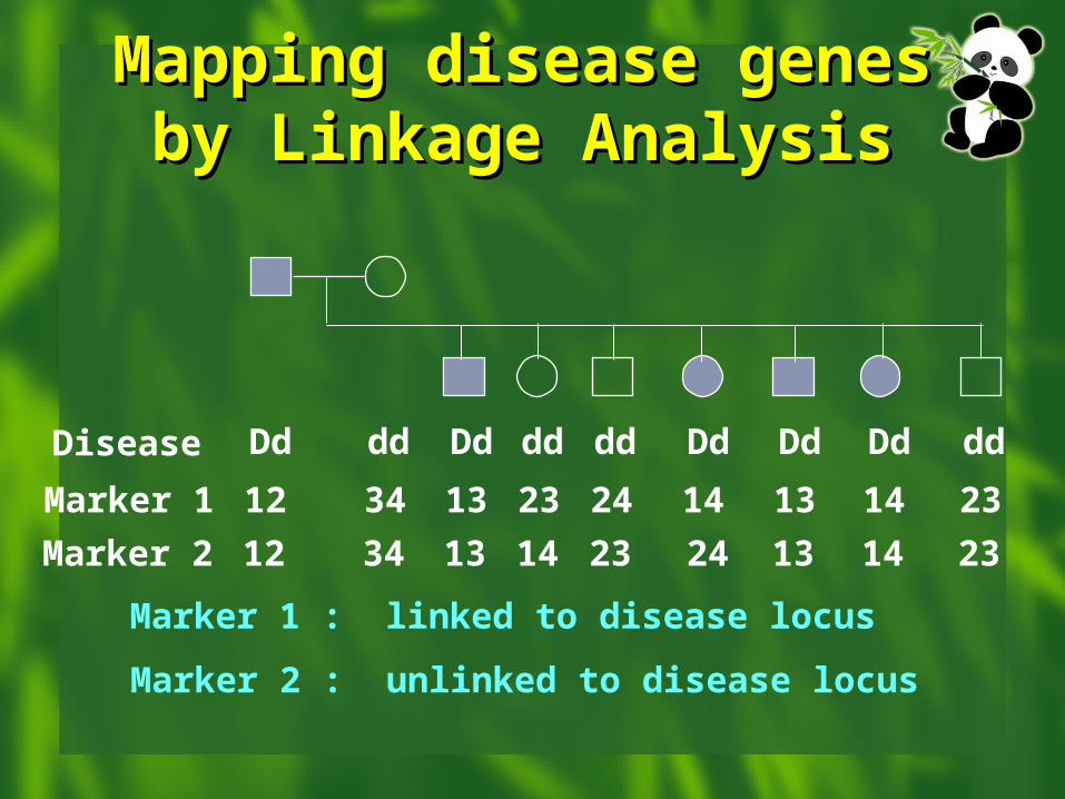
Mapping disease genes by LMapping disease genes by Linkage Analysisinkage Analysis
Disease Dd dd Dd dd dd Dd Dd Dd dd
Marker 1 12 34 13 23 24 14 13 14 23
Marker 2 12 34 13 14 23 24 13 14 23
Marker 1 : linked to disease locus
Marker 2 : unlinked to disease locus
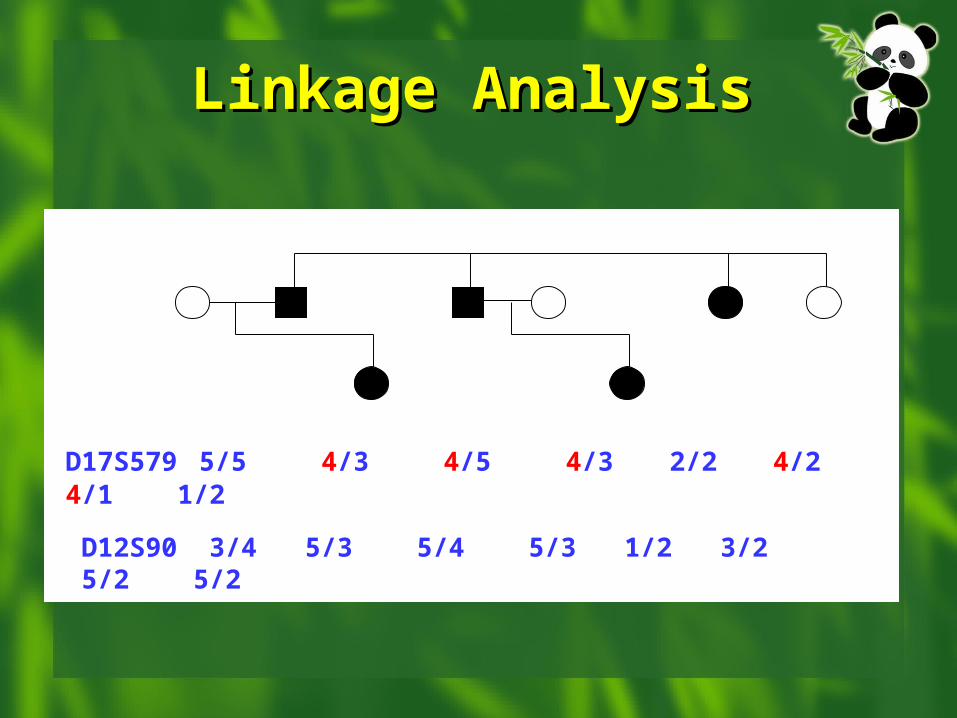
Linkage AnalysisLinkage Analysis
D17S579 5/5 4/3 4/5 4/3 2/2 4/2 4/1 1/2
3 65421
1 2
II
III
D12S90 3/4 5/3 5/4 5/3 1/2 3/2 5/2 5/2
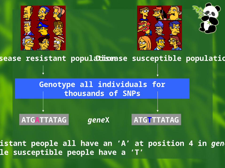
Disease resistant population Disease susceptible population
Genotype all individuals for thousands of SNPs
ATGATTATAG ATGTTTATAG
Resistant people all have an ‘A’ at position 4 in geneX, while susceptible people have a ‘T’
geneX
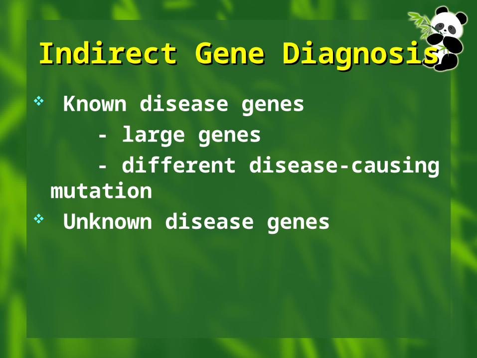
Indirect Gene DiagnosisIndirect Gene Diagnosis
Known disease genes
- large genes
- different disease-causing mutation Unknown disease genes
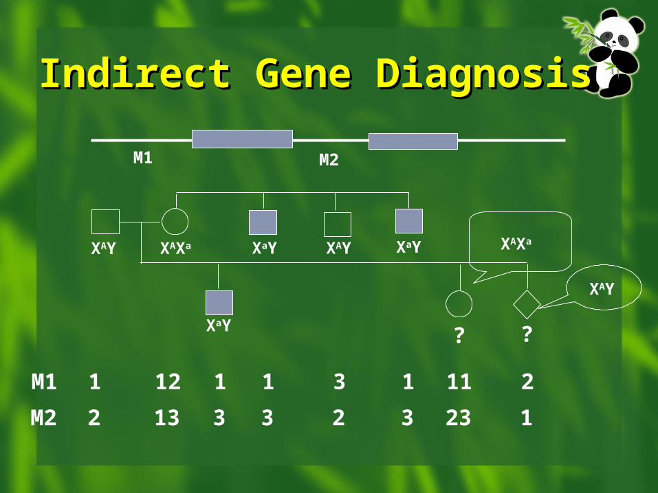
Indirect Gene DiagnosisIndirect Gene Diagnosis
M1 M2
M1 1 12 1 1 3 1 11 2
M2 2 13 3 3 2 3 23 1
XAY XAXa
XaY
XaY XAY XaY
? ?
XAXa
XAY
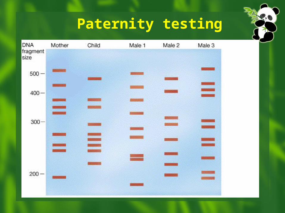
Paternity testing
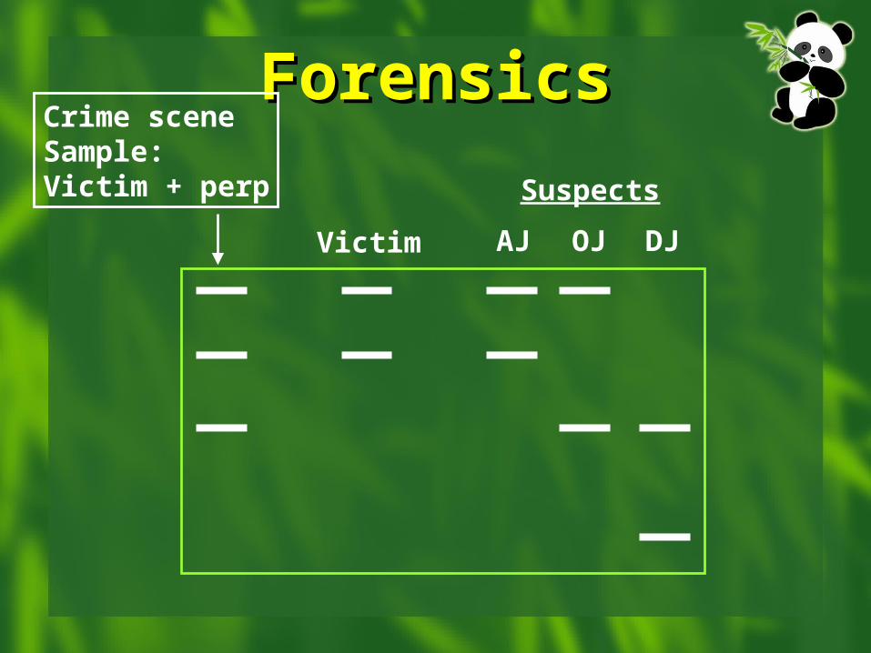
ForensicsForensicsCrime sceneSample:Victim + perp
Victim AJ OJ DJ
Suspects
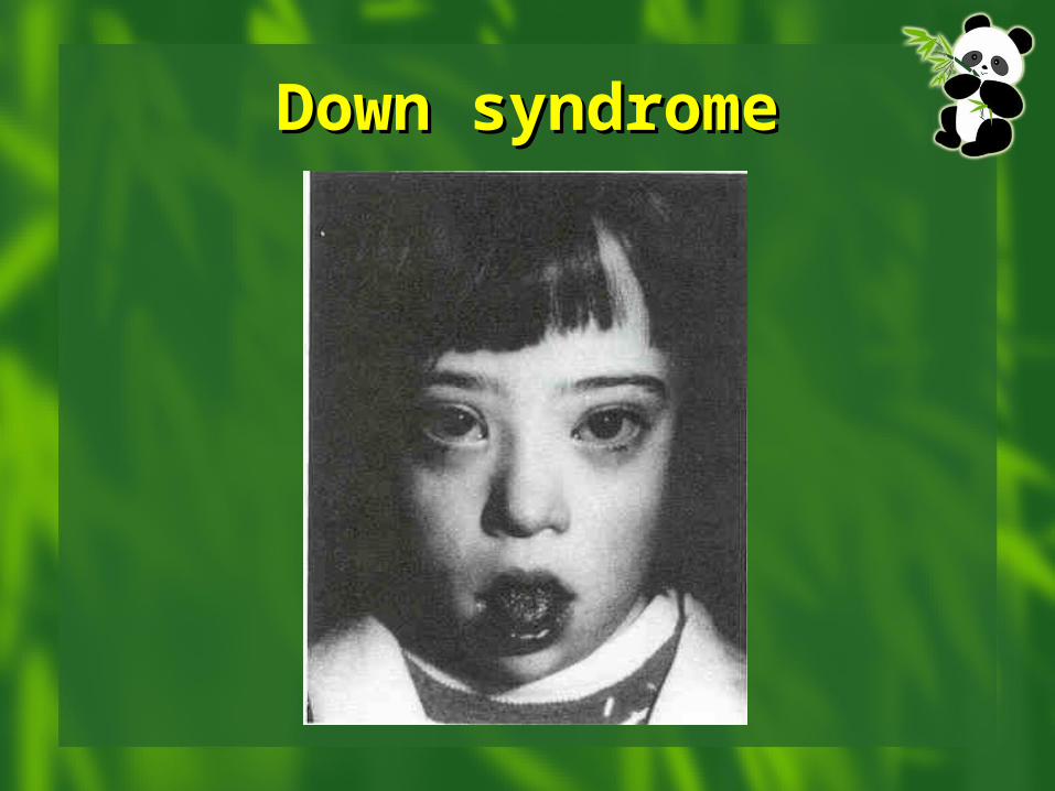
Down syndromeDown syndrome
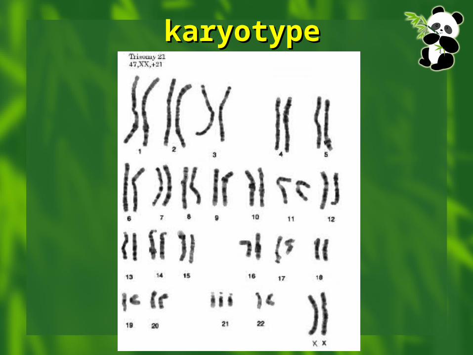
karyotypekaryotype
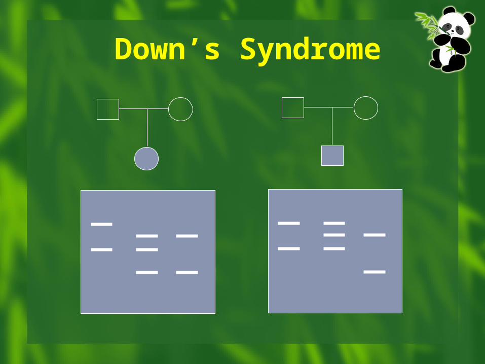
Down’s Syndrome
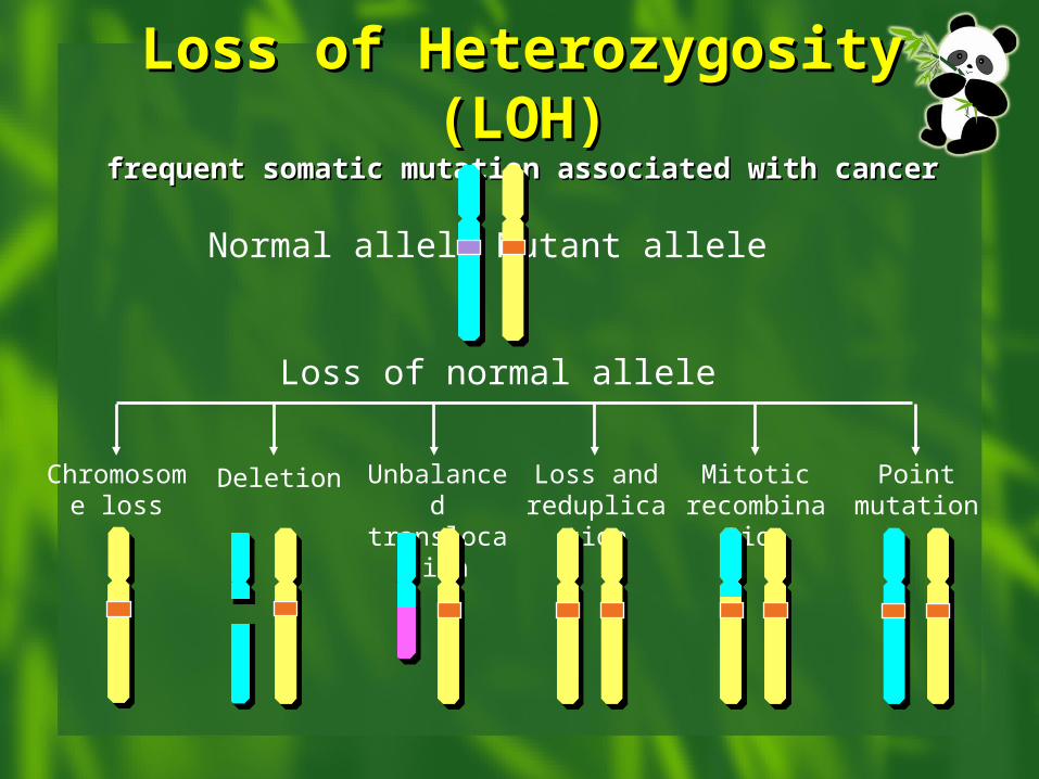
Loss of Heterozygosity Loss of Heterozygosity (LOH)(LOH)
frequent somatic mutation associated with cancerfrequent somatic mutation associated with cancer
Normal allele Mutant allele
Chromosome loss
Deletion Unbalanced translocation
Loss and reduplication
Mitotic recombination
Point mutation
Loss of normal allele
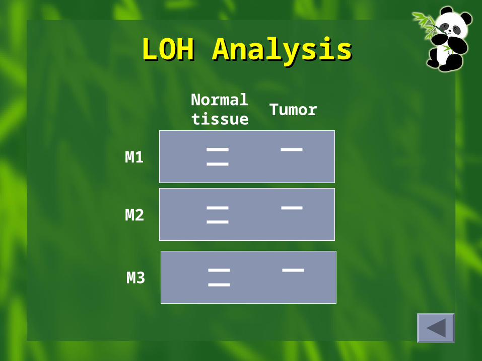
LOH AnalysisLOH Analysis
Normal tissues
Tumor
M1
M2
M3