Embed Size (px)
Citation preview

1
1
Running Title: Bone Regeneration with Perivascular Stem Cells and NELL-1
Human Perivascular Stem Cells Show Enhanced Osteogenesis and Vasculogenesis with
NELL-1 Protein
Asal Askarinam1,*Aaron W. James, M.D.
1,2,3,* Janette N. Zara, M.D.
2, Raghav Goyal
1, Mirko
Corselli, Ph.D.2, Angel Pan
1, Pei Liang Ph.D.
2, Le Chang
1, Todd Rackohn
1, David Stoker, M.D.
4,
Xinli Zhang M.D., Ph.D.1, Kang Ting D.M.D., D.Med.Sc.
1,2, Bruno Péault, Ph.D.
2,, and Chia
Soo, M.D., F.A.C.S.2
1
Dental and Craniofacial Research Institute and Section of Orthodontics, School of Dentistry,
UCLA; 2
UCLA and Orthopaedic Hospital Department of Orthopaedic Surgery and the
Orthopaedic Hospital Research Center, University of California, Los Angeles, Los Angeles, CA
90095; 3 Department of Pathology and Laboratory Medicine, University of California, Los
Angeles, David Geffen School of Medicine, 4
Private practice, Marina del Rey, California and
the Division of Plastic and Reconstructive Surgery, University of Southern California.
*A. Askarinam and A.W. James are co-first authors
Corresponding author: Chia Soo, MD
Department of Orthopaedic Surgery
University of California, Los Angeles
675 Charles E Young Dr South, MRL 2641A
Los Angeles, CA 90095-1579
Tel: 310-794-5479
Fax: 310-206-7783
KEY words: MSC, osteogenesis, vascularization, differentiation, NELL-1, pericytes, adventitial
cells
Page 1 of 45
Tis
sue
Eng
inee
ring
Par
t AH
uman
Per
ivas
cula
r St
em C
ells
Sho
w E
nhan
ced
Ost
eoge
nesi
s an
d V
ascu
loge
nesi
s w
ith N
EL
L-1
Pro
tein
(do
i: 10
.108
9/te
n.T
EA
.201
2.03
67)
Thi
s ar
ticle
has
bee
n pe
er-r
evie
wed
and
acc
epte
d fo
r pu
blic
atio
n, b
ut h
as y
et to
und
ergo
cop
yedi
ting
and
proo
f co
rrec
tion.
The
fin
al p
ublis
hed
vers
ion
may
dif
fer
from
this
pro
of.

2
2
CONTACT INFORMATION:
Author Address Telephone Fax Email Asal Askarinam*
UCLA Dental and Craniofacial
Research Institute
10833 Le Conte Ave, CHS 73-060
Los Angeles, CA 90095
310-825-
3750
310-206-
7783
asalaskarinam@gm
ail.com
Aaron W. James* Department of Pathology and Laboratory
Medicine
10833 Le Conte Ave, CHS A3-251
Los Angeles, CA 90095
310-825-
3750
310-206-
7783
aaronwjames1@gm
ail.com
Janette N. Zara UCLA Dental and Craniofacial
Research Institute
10833 Le Conte Ave, CHS 73-060
Los Angeles, CA 90095
310-825-
3750
310-206-
7783
Raghav Goyal UCLA Dental and Craniofacial
Research Institute
10833 Le Conte Ave, CHS 73-060
Los Angeles, CA 90095
310-825-
3750
310-206-
7783
Raghav.goyal@gma
il.com
Mirko Corselli UCLA Orthopaedic Hospital Research Center
615 Charles E. Young Dr. South, OHRC 410
Los Angeles, CA 90095-7358
310-825-
3750
310-206-
7783
mcorselli@mednet.
ucla.edu
Angel Pan UCLA Dental and Craniofacial
Research Institute
10833 Le Conte Ave, CHS 73-060
Los Angeles, CA 90095
310-825-
3750
310-206-
7783
Pei Liang UCLA Orthopaedic Hospital Research Center
615 Charles E. Young Dr. South, OHRC 410
Los Angeles, CA 90095-7358
310-825-
3750
310-206-
7783
peiliang1999@yaho
o.com.cn
Le Chang UCLA Dental and Craniofacial
Research Institute
10833 Le Conte Ave, CHS 73-060
Los Angeles, CA 90095
310-825-
3750
310-206-
7783
le.leslie.chang@gm
ail.com
Todd Rackohn UCLA Dental and Craniofacial
Research Institute
10833 Le Conte Ave, CHS 73-060
Los Angeles, CA 90095
310-825-
3750
310-206-
7783
David Stoker 4640 Admiralty Way #101 Marina Del Rey, CA
90292
(310) 300-
1779
unavailabl
e
davestoker@hotmai
l.com
Xinli Zhang UCLA Dental and Craniofacial
Research Institute
10833 Le Conte Ave, CHS 73-060
Los Angeles, CA 90095
310-794-
5479
310-206-
7783
cla.edu
Kang Ting UCLA School of Dentistry
Box 951668, CHS 30-113
Los Angeles, CA 90095-1668
310-206-
6305
310-206-
7783
a.edu
Bruno Péault
UCLA Orthopaedic Hospital Research Center
615 Charles E. Young Dr. South, OHRC 410
Los Angeles, CA 90095-7358
310-794-1339 unavailabl
e
Chia Soo
UCLA Department of
Orthopaedic Surgery
675 Charles E Young Dr South, MRL 2641A
Los Angeles, CA 90095-1579
310-794-
5479
310-206-
7783
Page 2 of 45
Tis
sue
Eng
inee
ring
Par
t AH
uman
Per
ivas
cula
r St
em C
ells
Sho
w E
nhan
ced
Ost
eoge
nesi
s an
d V
ascu
loge
nesi
s w
ith N
EL
L-1
Pro
tein
(do
i: 10
.108
9/te
n.T
EA
.201
2.03
67)
Thi
s ar
ticle
has
bee
n pe
er-r
evie
wed
and
acc
epte
d fo
r pu
blic
atio
n, b
ut h
as y
et to
und
ergo
cop
yedi
ting
and
proo
f co
rrec
tion.
The
fin
al p
ublis
hed
vers
ion
may
dif
fer
from
this
pro
of.
3
3
ABSTRACT: An ideal mesenchymal stem cell (MSC) source for bone tissue engineering has
yet to be identified. Such an MSC population would be easily harvested in abundance, with
minimal morbidity and with high purity. Our laboratories have identified perivascular stem cells
(PSCs) as a candidate cell source. PSCs are readily isolatable through fluorescent activated cell
sorting from adipose tissue and have been previously shown to be indistinguishable from MSCs
in phenotype and differentiation potential. PSCs consist of two distinct cell populations: [1]
pericytes (CD146+, CD34-, CD45-), which surround capillaries and microvessels, and [2]
adventitial cells (CD146-, CD34+, CD45-), found within the tunica adventitia of large arteries
and veins. We previously demonstrated the osteogenic potential of pericytes by examining
pericytes derived from human fetal pancreas, and illustrated their in vivo trophic and angiogenic
effects. In the present study, we used an intramuscular ectopic bone model to develop the
translational potential of our original findings using PSCs (as a combination of pericytes and
adventitial cells) from human white adipose tissue. We evaluated human PSC (hPSC)-mediated
bone formation and vascularization in vivo. We also examined the effects of hPSCs when
combined with the novel craniosynostosis-associated protein, NELL-1 (Nel-like Molecule I).
Implants consisting of demineralized bone matrix putty combined with either NELL-1 (3µg/µL),
hPSC (2.5 x 10⁵ cells), or hPSC+NELL-1 were inserted in the bicep femoris of SCID mice.
Bone growth was evaluated using micro computed tomography, histology and
immunohistochemistry over 4 weeks. Results demonstrated the osteogenic potential of hPSCs
and the additive effect of hPSC+NELL-1 on bone formation and vasculogenesis. Comparable
osteogenesis was observed with NELL-1 as compared to the more commonly used Bone
Morphogenetic Protein (BMP)-2. Next, hPSCs induced greater implant vascularization than
unsorted stromal vascular fraction (SVF) from patient-matched samples. Finally, we observed
Page 3 of 45
Tis
sue
Eng
inee
ring
Par
t AH
uman
Per
ivas
cula
r St
em C
ells
Sho
w E
nhan
ced
Ost
eoge
nesi
s an
d V
ascu
loge
nesi
s w
ith N
EL
L-1
Pro
tein
(do
i: 10
.108
9/te
n.T
EA
.201
2.03
67)
Thi
s ar
ticle
has
bee
n pe
er-r
evie
wed
and
acc
epte
d fo
r pu
blic
atio
n, b
ut h
as y
et to
und
ergo
cop
yedi
ting
and
proo
f co
rrec
tion.
The
fin
al p
ublis
hed
vers
ion
may
dif
fer
from
this
pro
of.
4
4
an additive effect on implant vascularization with hPSC+NELL-1 by histomorphometry and
immunohistochemistry, accompanied by in vitro elaboration of vasculogenic growth factors.
These findings hold significant implications for the cell/protein combination therapy
hPSC+NELL-1 in the development of strategies for vascularized bone regeneration.
INTRODUCTION:
Bone grafts are the current standard for treating non-healing skeletal defects [1]. However, the
disadvantages associated with bone graft harvests are significant, including limited endogenous
supply, prolonged surgical times, and harvest complications [1-4]. Cell sources such as bone
marrow mesenchymal stem cells (BMSCs) and adipose-derived stem cells (ASCs) have
generated significant interest for their tissue engineering potential [5-7]. However, the
limitations associated with these conventional stem cell sources are also significant. BMSCs, for
example, are difficult to harvest, are also in limited supply, and show reduced stem cell activity
in aged and osteoporotic patients [8, 9]. Furthermore, BMSCs and ASCs require in vitro
expansion, which precludes the use of autologous cells in an emergency situation, and also
introduces the risk of immunogenicity, infection, and genetic instability [10]. To avoid these
shortcomings, studies have also investigated the use of uncultured stromal vascular fraction
(SVF) of adipose tissue. However, a side-by-side comparison reported lower bone formation
among uncultured SVF relative to cultured ASCs [11]. Moreover, SVF is a heterogeneous cell
population which contains numerous non-stem cell types, such as inflammatory cells,
hematopoietic cells, and endothelial cells, among others [12-14]. Heterogeneity complicates
characterization of cell composition and introduces variability across samples. Such
Page 4 of 45
Tis
sue
Eng
inee
ring
Par
t AH
uman
Per
ivas
cula
r St
em C
ells
Sho
w E
nhan
ced
Ost
eoge
nesi
s an
d V
ascu
loge
nesi
s w
ith N
EL
L-1
Pro
tein
(do
i: 10
.108
9/te
n.T
EA
.201
2.03
67)
Thi
s ar
ticle
has
bee
n pe
er-r
evie
wed
and
acc
epte
d fo
r pu
blic
atio
n, b
ut h
as y
et to
und
ergo
cop
yedi
ting
and
proo
f co
rrec
tion.
The
fin
al p
ublis
hed
vers
ion
may
dif
fer
from
this
pro
of.

5
5
inconsistencies hamper efforts toward a reliable and standardized cell-based therapeutic for bone
regeneration.
As an alternative stem cell source, our laboratories first identified and then prospectively isolated
a perivascular mesenchymal stem cell (MSC)-like population. This MSC population, termed
perivascular stem cells (PSCs), is found in all vascularized tissue [15, 16] and consists of: [1]
CD146+CD34-CD45- pericytes [15, 17] , which line capillaries and microvessels, and [2]
CD146- CD34+CD45- adventitial cells [18], found in the tunica adventitia of large arteries and
veins. Pericytes were the first perivascular population studied, and were recognized for their
expression of characteristic MSC markers (including CD10, CD13, CD44, CD73, CD90, CD105)
[15]. The similarity of pericytes to MSCs was confirmed by demonstrating their ability to
differentiate into multiple mesodermal lineages in vitro [15]. Clonal analysis of pericytes also
showed expression of MSC markers and trilineage potential (differentiation into osteoblasts,
chondrocytes, and adipocytes) [15]. Likewise, subsequent studies demonstrated that adventitial
cells, although located in a different location with respect to blood vessels, also expressed MSC
markers and also displayed clonal multilineage potential in culture [18]. Furthermore, pericytes
and adventitial cells each comprise a significant portion of SVF of human adipose tissue (17.1%
and 22.5% respectively), with a combined average of 39.6% [19]. From a tissue engineering
perspective, the isolation of the maximum MSC number per unit autologous tissue would be
highly advantageous. To this end, in the present study, we combined the two PSC subpopulations
(which constitute nearly 40% total SVF) and characterized their bone forming potential. This
approach maximizes the total number of MSCs collected per harvest, thus reducing harvest
morbidity.
Page 5 of 45
Tis
sue
Eng
inee
ring
Par
t AH
uman
Per
ivas
cula
r St
em C
ells
Sho
w E
nhan
ced
Ost
eoge
nesi
s an
d V
ascu
loge
nesi
s w
ith N
EL
L-1
Pro
tein
(do
i: 10
.108
9/te
n.T
EA
.201
2.03
67)
Thi
s ar
ticle
has
bee
n pe
er-r
evie
wed
and
acc
epte
d fo
r pu
blic
atio
n, b
ut h
as y
et to
und
ergo
cop
yedi
ting
and
proo
f co
rrec
tion.
The
fin
al p
ublis
hed
vers
ion
may
dif
fer
from
this
pro
of.

6
6
There exist significant advantages to using FACS-purified PSCs for tissue engineering purposes.
First, purity ensures the specific and consistent use of only those cells known to participate in
bone differentiation. This eliminates extraneous components of SVF, such as osteogenic
differentiation-inhibiting endothelial cells, that may interfere with bone formation [14].
Recently we reported that hPSCs form significantly more bone in vivo in comparison to an equal
number of unpurified SVF cells of human adipose tissue [19]. To verify that this significant
difference was due to the superior bone formation of purified hPSCs and not to a dilution effect
resulting from the fewer number of MSCs found within the heterogeneous population of SVF
cells, we also added hSVF cells at a ratio of 10:1 to hPSCs in an intramuscular ectopic bone
model. More bone was still observed with hPSC than with hSVF treatment, confirming the
robust osteogenic potential of PSCs [19]. Furthermore, in terms of a future cell-based
therapeutic, a purified stem cell source also guarantees precise product characterization. This is
important for any treatment seeking FDA (Food and Drug Administration) approval, as
variability may raise concerns regarding consistency, safety, and efficacy. Finally, because PSCs
are found in virtually all vascularized tissue, they are abundant and easily harvestable [15].
Given its superficial location and relative abundance, adipose tissue is an ideal tissue source for
PSCs.
While purity, availability, and abundance make hPSCs an attractive stem cell source, their
potential trophic effects on vascularization are also significant. Bone receives nutrients, oxygen,
osteoinductive factors, and stem cells through the blood [20]. Therefore, an appropriate stem
Page 6 of 45
Tis
sue
Eng
inee
ring
Par
t AH
uman
Per
ivas
cula
r St
em C
ells
Sho
w E
nhan
ced
Ost
eoge
nesi
s an
d V
ascu
loge
nesi
s w
ith N
EL
L-1
Pro
tein
(do
i: 10
.108
9/te
n.T
EA
.201
2.03
67)
Thi
s ar
ticle
has
bee
n pe
er-r
evie
wed
and
acc
epte
d fo
r pu
blic
atio
n, b
ut h
as y
et to
und
ergo
cop
yedi
ting
and
proo
f co
rrec
tion.
The
fin
al p
ublis
hed
vers
ion
may
dif
fer
from
this
pro
of.

7
7
cell population for bone regeneration would also have pro-angiogenic effects [20, 21]. Pericytes
are known to maintain endothelial cell function [22, 23], and available studies have also shown
that these cells secrete more angiogenic factors, such as vascular endothelial growth factor
(VEGF) and fibroblast growth factor-2 (FGF-2), than do ASCs [24, 25]. In our previous
publication [24], our laboratory examined pericytes derived from human fetal pancreas, finding
significant pro-osteogenic and pro-vasculogenic effects. In the present study, we used an
intramuscular ectopic bone model to develop the translational potential of our original findings
now using PSCs from adult, human white adipose tissue. Therefore, our objectives were to: [1]
demonstrate the osteogenic and [2] vasculogenic potential of hPSCs (as a combination of
pericytes and adventitial cells) and [3] pursue NELL-1 as a growth factor capable of enhancing
hPSC osteogenesis and vascularization.
METHODS:
Isolation of Stromal Vascular Fraction (SVF) from Human Lipoaspirate
Human lipoaspirate donated from n=2 healthy 31 and 40 year-old female patients was obtained
after standard liposuction procedures. Lipoaspirate was collected from back, hip, flanks, and
medial thigh. Human Stromal Vascular Fraction (hSVF) was prepared by digesting lipoaspirate
with collagenase as previously described [18]. Briefly, liposaspirate was washed in an equal
volume of PBS (phosphate buffered saline) and centrifuged. The tissue layer was removed and
mixed with digestion solution (RPMI, 3.5% BSA, 10ng/mL DNAse, 1mg/mL Collagenase II)
and incubated for 70 minutes at 37°C. The filtered solution was then centrifuged. The pellet
was removed and washed twice in PBS 5mM EDTA before red blood cell lysis buffer (155mM
Page 7 of 45
Tis
sue
Eng
inee
ring
Par
t AH
uman
Per
ivas
cula
r St
em C
ells
Sho
w E
nhan
ced
Ost
eoge
nesi
s an
d V
ascu
loge
nesi
s w
ith N
EL
L-1
Pro
tein
(do
i: 10
.108
9/te
n.T
EA
.201
2.03
67)
Thi
s ar
ticle
has
bee
n pe
er-r
evie
wed
and
acc
epte
d fo
r pu
blic
atio
n, b
ut h
as y
et to
und
ergo
cop
yedi
ting
and
proo
f co
rrec
tion.
The
fin
al p
ublis
hed
vers
ion
may
dif
fer
from
this
pro
of.

8
8
NH4Cl, 10 mM KHCO3, and 0.1 mM EDTA) was added. After three additional PBS washes
and centrifugations, trypan blue staining was used to count the number of viable hSVF cells.
These hSVF cells were then either implanted or processed for hPSC purification. Cells isolated
from different donors were not pooled, but rather used individually in study.
Purification of Perivascular Stem Cells (PSCs) from Stromal Vascular Fraction
hPSCs were isolated from each donor’s hSVF by fluorescence activated cell sorting (FACS) per
previously published protocol [18]. A portion of the isolated hSVF was centrifuged and the
resulting pellet was incubated with a solution of PBS, anti-CD146, FITC (Abd Serotec; 1:100),
anti-CD45 APC-cy7 (BD Bioscience; 1:100), and anti-CD34 APC (BD Bioscience; 1:100).
After 15 minutes of incubation at 4°C and a wash in PBS, pellet was suspended in RPMI (1
mL/106) and DAPI (Invitrogen; 1:1000) in order to identify and then eliminate dead cells prior to
cell sorting. Solution was then filtered using a 70µM cell filter and then run on the FACSAria
cell sorter in order to first selectively isolate and to then combine the pericytes (CD146+,CD34-
,CD45-), and adventitial cells (CD146-,CD34+,CD45-) that make up the population of PSCs. A
portion of these cells were either immediately used in vivo or plated for in vitro studies. In select
experiments, cells were labeled with a red fluorescent dye for cell tracking purposes (PKH26
fluorescent cell linker, Sigma-Aldrich).
Cell Culture
Page 8 of 45
Tis
sue
Eng
inee
ring
Par
t AH
uman
Per
ivas
cula
r St
em C
ells
Sho
w E
nhan
ced
Ost
eoge
nesi
s an
d V
ascu
loge
nesi
s w
ith N
EL
L-1
Pro
tein
(do
i: 10
.108
9/te
n.T
EA
.201
2.03
67)
Thi
s ar
ticle
has
bee
n pe
er-r
evie
wed
and
acc
epte
d fo
r pu
blic
atio
n, b
ut h
as y
et to
und
ergo
cop
yedi
ting
and
proo
f co
rrec
tion.
The
fin
al p
ublis
hed
vers
ion
may
dif
fer
from
this
pro
of.

9
9
Cells were expanded in Dulbecco’s Modified Eagle’s Medium (DMEM), 20% fetal bovine
serum (FBS), and 1% penicillin/streptomyocin. Osteogenic differentiation of cells followed
previously published protocol [26, 27]. Medium was supplemented with 300-600 ng/mL of
NELL-1, as previously described [28]. Osteogenic differentiation was assessed using Alkaline
Phosphatase and Alizarin Red staining at 5 and 10 days of osteogenic differentiation,
respectively (with or without 300 ng/mL NELL-1). Semi-quantitative analysis of representative
38.4x Alkaline Phosphatase and Alizarin Red staining (n=3) images was performed using the
magic wand tool of Adobe Photoshop CS5. RNA isolation and quantitative RT-PCR for
osteogenic genes was performed as previously described after seven days osteogenic
differentiation with or without NELL-1 (300 ng/mL), including Osteopontin (Opn) and
Osteocalcin (Oc) [24]. Next, quantification of growth factor secretion was performed using PSCs
with or without NELL-1 (300-600 ng/mL). Briefly, cells were seeded at 80% confluency in 1%
serum conditions, after attachment medium was supplemented with NELL-1, and supernatant
was harvested after 48 hours treatment. ELISA assays for VEGF (Vascular Endothelial Growth
Factor) and FGF (Fibroblast Growth Factor)-2 were performed per manufacturers instructions
(Invitrogen, Cat No. KHG0111, HKG0021). Groups were initially unlabeled for photographic
documentation and later revealed for quantification.
Implant Preparation
Ovine demineralized bone matrix (DBX) Putty was used (100 µL, Musculoskeletal Transplant
Foundation, Edison, NJ) and combined with either 0 or 300 µg of NELL-1 lyophilized onto 15
mg β-TCP (tricalcium phosphate) particles in order to ensure prolonged NELL-1 release [29]. In
Page 9 of 45
Tis
sue
Eng
inee
ring
Par
t AH
uman
Per
ivas
cula
r St
em C
ells
Sho
w E
nhan
ced
Ost
eoge
nesi
s an
d V
ascu
loge
nesi
s w
ith N
EL
L-1
Pro
tein
(do
i: 10
.108
9/te
n.T
EA
.201
2.03
67)
Thi
s ar
ticle
has
bee
n pe
er-r
evie
wed
and
acc
epte
d fo
r pu
blic
atio
n, b
ut h
as y
et to
und
ergo
cop
yedi
ting
and
proo
f co
rrec
tion.
The
fin
al p
ublis
hed
vers
ion
may
dif
fer
from
this
pro
of.

10
10
selected experiments, 2.5 x 10⁵ of either hPSCs or hSVF cells, suspended in 20 µL of PBS, were
added. In addition, as a comparison group, select implants were treated with Bone
Morphogenetic Protein (BMP)-2 (3.75 ug) was likewise lyophilized onto β-TCP. The
components of the implant were mechanically mixed before use. A detailed summary of each of
the implant constituents is presented in Supplemental Table 1. The DBX / β-TCP implant was
chosen as it is a moldable putty with osteoinductive properties and reliable protein release
kinetics, making it readily translatable to bone defect models.
Surgical Procedures
8-week old, male SCID mice were anesthesized by isofluorane inhalation (5% induction; 2%
maintenance) and medicated with 0.05 mg/kg buprenorphine. As previously shown, bilateral
incisions were made in the hind limbs and pockets were created in the biceps femoris along the
long axis of the muscle fiber, so that one implant may be inserted in each limb with ease and
without lying in contiguity with the femoral bone (so as to not induce an osteoblastic response in
the host femoral periosteum) [30]. 5-0 Vicryl sutures were used to suture the overlying fasciae
and skin. Postoperative care included buprenorphine for 48 hours to manage pain and
trimethoprim / sulfamethoxazole for 10 days to prevent infection. All surgical procedures were
consistent with the regulations put forth by the UCLA Chancellor’s Animal Research Committee.
Radiographic Analysis
Page 10 of 45
Tis
sue
Eng
inee
ring
Par
t AH
uman
Per
ivas
cula
r St
em C
ells
Sho
w E
nhan
ced
Ost
eoge
nesi
s an
d V
ascu
loge
nesi
s w
ith N
EL
L-1
Pro
tein
(do
i: 10
.108
9/te
n.T
EA
.201
2.03
67)
Thi
s ar
ticle
has
bee
n pe
er-r
evie
wed
and
acc
epte
d fo
r pu
blic
atio
n, b
ut h
as y
et to
und
ergo
cop
yedi
ting
and
proo
f co
rrec
tion.
The
fin
al p
ublis
hed
vers
ion
may
dif
fer
from
this
pro
of.

11
11
Muscle pouch samples were harvested 4 weeks after surgery, fixed in formalin, and imaged
using high-resolution microCT (Skyscan 1172F, Skyscan, Belgium). Images were analyzed in
CTAn to determine bone volume (BV, n=8) and Bone Mineral Density (BMD, n=8). Specific
threshold values included in figure legends.
Histology and Immunohistochemistry Analysis
Samples were decalcified in 19% EDTA, embedded in paraffin, sectioned, and stained with
Hematoxylin and Eosin (H&E). Semi-quantitative analysis of representative 200x H&E images
was performed using the magic wand tool in Adobe Photoshop in order to evaluate the level of
osteogenesis by such parameters as: Percent Bone Area (% B. Ar., n=10) , Relative Bone Area
(B. Ar., n=10), osteocyte lacunar density (n=15), and percentage of filled lacunae (n=15) [31].
Immunohistochemistry was performed for BMP-2, (Bone Morphogenetic Protein 2, Santa Cruz
Biotechnology, Inc, Santa Cruz, CA), BMP-7 (Bone Morphogenetic Protein 7, Santa Cruz
Biotechnology Inc), OCN (Osteocalcin, Santa Cruz Biotechnology Inc), VEGF (Vascular
Endothelial Growth Factor, Santa Cruz Biotechnology), vWF (Von Willebrand Factor, US
Biological), Human β-2 Microglobulin (Abcam), and PCNA (Proliferating Cell Nuclear Antigen,
Dako). For immunohistochemistry, staining without primary antibody was performed as a
negative control throughout. Briefly, paraffin slices were deparaffinized, dehydrated, rinsed, and
incubated with 3% H2O2 for 20 minutes and then blocked with 0.1% bovine serum albumin in
phosphate-buffered saline (PBS) for one hour. Primary antibodies at a dilution of 1:100 was
added to each section and incubated at 37C for 1 h and at 4C overnight. ABC complex (Vector
Laboratories, Burlingame, CA) was applied to the sections following the incubation with
Page 11 of 45
Tis
sue
Eng
inee
ring
Par
t AH
uman
Per
ivas
cula
r St
em C
ells
Sho
w E
nhan
ced
Ost
eoge
nesi
s an
d V
ascu
loge
nesi
s w
ith N
EL
L-1
Pro
tein
(do
i: 10
.108
9/te
n.T
EA
.201
2.03
67)
Thi
s ar
ticle
has
bee
n pe
er-r
evie
wed
and
acc
epte
d fo
r pu
blic
atio
n, b
ut h
as y
et to
und
ergo
cop
yedi
ting
and
proo
f co
rrec
tion.
The
fin
al p
ublis
hed
vers
ion
may
dif
fer
from
this
pro
of.

12
12
biotinylated secondary antibody (Dako Corporation, Carpinteria, CA). AEC plus substrate in red
color (Dako, Carpinteria, CA) was used as a chromagen, and the sections were counterstained
with light Hematoxylin. Immunofluorescent staining was performed on frozen sections for CD31
(Santa Cruz) and PCNA (Dako), using Aflexa 488 Streptavidin as a secondary antibody and a
Hoescht 33324 counterstain (Sigma-Aldrich). Histomorphometric semi-quantitative analysis was
again performed using the magic wand tool in Adobe Photoshop on respective 400x images
(OCN, n=5; BMP-2, n=8; BMP-7, n=8; PCNA, n=10) in order to quantify the relative stain
intensity across groups. To eliminate bias, slides were initially unlabeled for photographic
documentation and later revealed for quantification. Specific Tolerance levels for Photoshop
quantification are listed in figure legends.
Statistics
A one-way ANOVA used when more than two groups compared. This was followed by a post
hoc Tukey test to specifically compare the difference between two specific groups (as in Figure
1J&K, 2B, 3B-E, 4D-F, 6B-D). In addition, a Student t-test was used to test significance when
only two groups were tested (as in Figure 1F-I, 5C-E, Figure S1C). *p ≤ 0.05 were significant.
RESULTS:
Isolation and In Vitro Differentiation of hPSC
FACS sorting was used to isolate hPSCs from the SVF of adipose tissue according to our
previously published protocol [18]. This MSC population consists of pericytes (CD146+,CD34-,
Page 12 of 45
Tis
sue
Eng
inee
ring
Par
t AH
uman
Per
ivas
cula
r St
em C
ells
Sho
w E
nhan
ced
Ost
eoge
nesi
s an
d V
ascu
loge
nesi
s w
ith N
EL
L-1
Pro
tein
(do
i: 10
.108
9/te
n.T
EA
.201
2.03
67)
Thi
s ar
ticle
has
bee
n pe
er-r
evie
wed
and
acc
epte
d fo
r pu
blic
atio
n, b
ut h
as y
et to
und
ergo
cop
yedi
ting
and
proo
f co
rrec
tion.
The
fin
al p
ublis
hed
vers
ion
may
dif
fer
from
this
pro
of.

13
13
CD45-) [15] and adventitial cells (CD146-,CD34+,CD45-) [18]. After exclusion of DAPI+ dead
cells and CD45+ hematopoietic cells (Fig. 1A,B), adventitial cells and pericytes were detected
based on the differential expression of CD34 and CD146. CD34+CD146- adventitial cells and
CD146+CD34- pericytes were then collected together as hPSCs for further in vitro and in vivo
studies.
First, we evaluated hPSCs for in vitro osteogenic differentiation. hPSCs showed robust alkaline
phosphatase (ALP) staining after 5 days in osteogenic differentiation medium (Fig. 1D) and
robust alizarin red (AR) staining at 10 days of osteogenic differentiation (Fig. 1E). Consistent
with published reports in other cell types [28, 32], hPSCs treated with NELL-1 protein (300
ng/mL), showed an increase in both ALP and AR staining at 5 and 10 days of osteogenic
differentiation, respectively (Fig. 1D,E). Semi-quantitative analysis of representative images
confirmed a significant increase in ALP and AR staining when hPSCs were treated with NELL-1
(Fig. 1F,G). Next, osteogenic gene expression was evaluated in hPSCs, with or without NELL-1
treatment for 7 days differentiation, including Osteopontin (Opn) and Osteocalcin (Oc) (Fig.
1H,I). NELL-1 resulted in an approximate 62 – 88% increase in Opn and Oc expression,
respectively. Finally, we have previously shown that NELL-1 induces Vegf expression in human
pericytes [24]. We sought to examine whether NELL-1 could likewise induce secretion of
vasculogenic growth factors in hPSCs, including VEGF and FGF (Fibroblast Growth Factor-2)
(Fig. 1J,K). Results showed a striking dose-dependent increase in VEGF protein secretion in
hPSCs with NELL-1 treatment (Fig. 1J). Likewise, NELL-1 induced FGF-2 protein secretion in
hPSC across concentrations examined (300-600 ng/mL, Fig. 1K). These data demonstrate the
osteogenic potential of hPSCs in vitro and the ability of NELL-1 to promote hPSC osteogenic
differentiation and trophic factor production.
Page 13 of 45
Tis
sue
Eng
inee
ring
Par
t AH
uman
Per
ivas
cula
r St
em C
ells
Sho
w E
nhan
ced
Ost
eoge
nesi
s an
d V
ascu
loge
nesi
s w
ith N
EL
L-1
Pro
tein
(do
i: 10
.108
9/te
n.T
EA
.201
2.03
67)
Thi
s ar
ticle
has
bee
n pe
er-r
evie
wed
and
acc
epte
d fo
r pu
blic
atio
n, b
ut h
as y
et to
und
ergo
cop
yedi
ting
and
proo
f co
rrec
tion.
The
fin
al p
ublis
hed
vers
ion
may
dif
fer
from
this
pro
of.

14
14
hPSCs Form Significantly More Bone In Vivo When Combined with NELL-1
Next, an intramuscular ectopic bone model was used to evaluate the in vivo osteogenic potential
of hPSCs in combination with NELL-1. Demineralized bone matrix (DBX Putty ®) was
selected as the osteoinductive carrier. Implants consisted of DBX alone, or DBX with hPSCs,
NELL-1 protein, or hPSCs+NELL-1 in combination. In addition, a BMP2 (Bone Morphogenetic
Protein-2) comparison group was used (hPSCs+BMP2). Implants were inserted intramuscularly
in SCID mice in order to assess ectopic bone formation. See Supplemental Table 1 for
treatment group specifics. Mice were harvested 4 weeks after surgery for evaluation of bone
growth. 3-dimensional micro computed tomography (microCT) showed an increase in bone
formation in NELL-1-treated and PSC-treated samples (Fig. 2A). Notably, the combined
hPSCs+NELL-1 treatment showed more bone formation than either cell or cytokine treatment
alone. This trend was reflected in the microCT quantification of bone mineral density (BMD)
(Fig. 2B). NELL-1 was also compared to the more commonly studied cytokine BMP2 (Bone
Morphogenetic Protein 2). Results showed BMP2 induced significant ossification among PSC-
treated samples, and induced a significantly higher BMD as compared to other treatment groups.
Following microCT imaging and analysis, samples were evaluated histologically by routine
H&E staining. Representative images of H&E stained slides for hPSC-treated samples showed
larger bone chips and reduced spacing between bone chips, as compared with DBX control
samples (Fig. 3A). These qualities were more apparent in samples with combined hPSCs and
NELL-1 treatment, suggesting an additive effect of hPSC and NELL-1 on ectopic bone
Page 14 of 45
Tis
sue
Eng
inee
ring
Par
t AH
uman
Per
ivas
cula
r St
em C
ells
Sho
w E
nhan
ced
Ost
eoge
nesi
s an
d V
ascu
loge
nesi
s w
ith N
EL
L-1
Pro
tein
(do
i: 10
.108
9/te
n.T
EA
.201
2.03
67)
Thi
s ar
ticle
has
bee
n pe
er-r
evie
wed
and
acc
epte
d fo
r pu
blic
atio
n, b
ut h
as y
et to
und
ergo
cop
yedi
ting
and
proo
f co
rrec
tion.
The
fin
al p
ublis
hed
vers
ion
may
dif
fer
from
this
pro
of.

15
15
formation (Fig. 3A). In addition, hPSCs+NELL-1-treated samples also showed evidence of
endochondral ossification (see black arrow, Fig. 3A). In comparison, hPSC+BMP2 treated
samples showed robust new bone formation, but the spaces between bone chips were filled with
notable lipid accumulation (see asterisks, Fig. 3A).
These H&E observations were next semi-quantitatively confirmed with histomorphometric
analysis of serial sections. Histomorphometric quantification of bone area (Fig. 3B) and percent
bone area (Fig. 3C) per random high-powered field confirmed our initial observations. Both
hPSC-treated samples and NELL-1-treated samples were observed to have significantly more
bone than DBX control, while samples treated with the combination hPSCs+NELL-1 showed
significantly more bone than those treated with either cells or protein alone (Fig. 3B,C). This
finding demonstrated the osteoinductive property of NELL-1, especially when combined with
hPSCs. Notably, Bone Area in hPSC+NELL-1 samples was observed to rival that of
hPSC+BMP2 samples. Next, osteocyte lacunar density was assessed, which measures the
number of osteocytes per unit of bone area [33]. In this situation, osteocyte lacunar density
indicated both new bone formation and/or re-population of DBX particle lacunae. To calculate
osteocyte lacunar density, random high-powered images of H&E stained slides for each group
were used to determine the number of osteocytes per mm2 of bone area. Again, a significant
additive effect was observed with combined hPSC and NELL-1 treatment (Fig. 3D).
Furthermore, the percentage of filled lacunae was calculated in order to determine the percentage
of DBX particle lacunae filled with osteocytes. Again, combined hPSC and NELL-1 treatment
had a significant additive effect (Fig. 3E). In summary, these data suggested that NELL-1 had a
positive effect on hPSC osteoblastogenesis.
Page 15 of 45
Tis
sue
Eng
inee
ring
Par
t AH
uman
Per
ivas
cula
r St
em C
ells
Sho
w E
nhan
ced
Ost
eoge
nesi
s an
d V
ascu
loge
nesi
s w
ith N
EL
L-1
Pro
tein
(do
i: 10
.108
9/te
n.T
EA
.201
2.03
67)
Thi
s ar
ticle
has
bee
n pe
er-r
evie
wed
and
acc
epte
d fo
r pu
blic
atio
n, b
ut h
as y
et to
und
ergo
cop
yedi
ting
and
proo
f co
rrec
tion.
The
fin
al p
ublis
hed
vers
ion
may
dif
fer
from
this
pro
of.

16
16
Immunohistochemical Analysis of Ectopic Bone Formation
Immunohistochemistry was next performed on the same experimental samples as in Fig. 2&3.
Specific stains included the bone matrix protein, osteocalcin (OCN, Fig. 4A), and the bone
cytokines of BMP-2 (Fig. 4B), and bone morphogenetic protein-7 (BMP-7, Fig. 4C). OCN
staining across all groups was located primarily within DBX chips and newly-formed bone.
hPSC-treated groups showed greater staining relative to NELL-1-treated groups, while the
combination hPSCs and NELL-1-treatment showed the highest staining intensity (Fig. 4A). This
observation was confirmed by semi-quantitative assessment by three blinded reviewers using
Adobe Photoshop (Fig. 4D). Next, BMP-2 immunohistochemistry and semi-quantification was
performed (Fig. 4B,E). Across all samples, BMP-2 staining was primarily observed among the
cells surrounding bone particles. As with OCN, the combined treatment of hPSCs and NELL-1
resulted in significantly higher staining of BMP-2 than either cells or cytokine alone. Finally,
BMP-7 immunohistochemistry and quantification was performed (Fig. 4C,F). A similar overall
distribution of BMP-7 was noted in comparison to BMP-2. Again, hPSCs and NELL-1
combination treatment induced significantly higher expression of BMP-7 when compared to
either cell or cytokine treatment alone. Thus, the immunohistochemistry for the bone matrix
protein OCN and bone cytokines, BMP-2 and BMP-7, revealed an additive effect of hPSCs and
NELL-1 on markers of bone formation.
Persistence and Proliferation of Human PSCs
Page 16 of 45
Tis
sue
Eng
inee
ring
Par
t AH
uman
Per
ivas
cula
r St
em C
ells
Sho
w E
nhan
ced
Ost
eoge
nesi
s an
d V
ascu
loge
nesi
s w
ith N
EL
L-1
Pro
tein
(do
i: 10
.108
9/te
n.T
EA
.201
2.03
67)
Thi
s ar
ticle
has
bee
n pe
er-r
evie
wed
and
acc
epte
d fo
r pu
blic
atio
n, b
ut h
as y
et to
und
ergo
cop
yedi
ting
and
proo
f co
rrec
tion.
The
fin
al p
ublis
hed
vers
ion
may
dif
fer
from
this
pro
of.

17
17
To confirm that those cells within the implant site were in fact the implanted human cells,
immunohistochemistry was performed for human β-2 microglobulin and proliferating cell
nuclear antigen (PCNA). Both hPSC-treated, and hPSC+NELL-1-treated samples showed
distinct β-2 microglobulin staining between DBX chips at 4 weeks post-implantation, indicating
the persistence of human cells (Supplemental Figure 1A). In addition, PCNA immunostaining
showed active cell proliferation within the implant site (Supplemental Figure 1B). Moreover,
semi-quantification of PCNA staining using Adobe Photoshop showed a significant increase in
samples treated with hPSC+NELL-1, in comparison to hPSCs alone (Supplemental Figure 1C).
In order to test whether the PCNA+ cells were of human (PSC) or mouse (host) origin, we next
performed immunofluoroscent staining (Supplemental Figure 1D). Here, PCNA+ cells appear
green, while implanted cells were pre-labeled with a red fluorochrome. A significant number of
PCNA+ cells were found to be implanted cells. Overall, this reflects proliferative effect of
NELL-1 on PSCs, in agreement with previous reports by our research group [24].
hPSC-treated Implants Show an Increase in Vascularization
As mentioned, new bone formation and neovascularization are intimately connected. We next
turned our attention to the effects of hPSC on blood vessel formation. For this comparison, an
equal number of patient-matched unsorted hSVF cells or purified hPSCs were implanted. In
order to visualize and quantify the size and number of blood vessels, slides were first stained
with H&E. Representative H&E images of hSVF-treated and hPSC-treated samples showed a
notable difference in the size and number of blood vessels (Fig. 5A,B). Semi-quantitative
histomorphometric analysis of H&E-stained images showed a significant increase in both blood
Page 17 of 45
Tis
sue
Eng
inee
ring
Par
t AH
uman
Per
ivas
cula
r St
em C
ells
Sho
w E
nhan
ced
Ost
eoge
nesi
s an
d V
ascu
loge
nesi
s w
ith N
EL
L-1
Pro
tein
(do
i: 10
.108
9/te
n.T
EA
.201
2.03
67)
Thi
s ar
ticle
has
bee
n pe
er-r
evie
wed
and
acc
epte
d fo
r pu
blic
atio
n, b
ut h
as y
et to
und
ergo
cop
yedi
ting
and
proo
f co
rrec
tion.
The
fin
al p
ublis
hed
vers
ion
may
dif
fer
from
this
pro
of.

18
18
vessel size (Fig. 5C) and number (Fig. 5D) in hPSC-treated samples. To further confirm these
differences, immunohistochemistry was performed for vascular endothelial growth factor
(VEGF). VEGF is a major angiogenic factor which promotes vascularization by regulating
endothelial cell proliferation and migration [20]. Samples treated with hPSCs showed greater
VEGF staining in comparison to those treated with hSVF (Fig. 5A,B). This was quantified,
showing a significant increase in VEGF expression in hPSC-treated samples in comparison to
hSVF (Fig. 5E). Next, hSVF-treated and hPSC-treated implants were examined for von
Willebrand factor (vWF) immunohistochemistry. Representative images showed greater vWF
staining in hPSC-treated samples than in hSVF-treated samples (Fig. 5A). Finally, CD31
immunoflourescent staining (a marker of endothelial cells) was performed in samples implanted
with red fluorescently tagged cells (Fig. 5F). We found that red-labeled PSCs frequently were
found in close association with CD31+ endothelial cells within the vasculature. In contrast red-
labeled SVF cells showed are more sporadic and less close association with blood vessels. These
studies collectively confirmed the significantly larger effect that hPSCs have on vascularization
in comparison to unsorted hSVF.
NELL-1 Enhances hPSC-mediated Effect on Vascularization
Finally, the effect of NELL-1 on hPSC-mediated vascularization was examined. NELL-1 has
previously been reported to have pro-angiogenic effects [24]. We returned to intramuscular
samples from Fig. 2&3, examining the effects of NELL-1 on hPSC-mediated vascularization.
Samples were first stained with H&E (Fig. 6A). H&E-stained images and corresponding semi-
quantitative histomorphometric analysis revealed an increase in blood vessel number (Fig. 6B)
Page 18 of 45
Tis
sue
Eng
inee
ring
Par
t AH
uman
Per
ivas
cula
r St
em C
ells
Sho
w E
nhan
ced
Ost
eoge
nesi
s an
d V
ascu
loge
nesi
s w
ith N
EL
L-1
Pro
tein
(do
i: 10
.108
9/te
n.T
EA
.201
2.03
67)
Thi
s ar
ticle
has
bee
n pe
er-r
evie
wed
and
acc
epte
d fo
r pu
blic
atio
n, b
ut h
as y
et to
und
ergo
cop
yedi
ting
and
proo
f co
rrec
tion.
The
fin
al p
ublis
hed
vers
ion
may
dif
fer
from
this
pro
of.

19
19
and size (Fig. 6C) in NELL-1-treated and hPSC-treated samples relative to DBX control.
hPSC+NELL-1-treated samples showed the highest blood vessel number and greatest blood
vessel size relative to either cell or cytokine treatment alone. This finding is reflected in VEGF
staining and quantification (Fig. 6D,E). Semi-quantification of staining images showed a
significant increase in VEGF staining in groups treated with both hPSCs and NELL-1 as
compared to groups with each treatment alone, consistent with our previous in vitro observations
(see again Fig. 1J). These studies demonstrated the ability of NELL-1 to enhance hPSC-
mediated vascularization.
DISCUSSION:
In summary, perivascular stem cells consist of two distinct cell populations known to exhibit
characteristic MSC properties, and to share similar proliferation and bone forming potential [19].
These include pericytes, which surround capillaries and microvessels, and adventitial cells, found
in the tunica adventitia of large arteries and veins. As mentioned, PSCs can be isolated based on
their differential expression of CD34 and CD146. In the present study, we expanded upon our
original findings to explore the translational potential of PSCs as a population of pericytes and
adventitial cells by examining both their osteogenic and vasculogenic properties. We
demonstrated that PSCs undergo both in vitro and in vivo osteogenic differentiation; and that
treatment with the craniosynostosis-associated osteoinductive protein, NELL-1, significantly
augmented hPSC-mediated bone formation and vascularization.
Page 19 of 45
Tis
sue
Eng
inee
ring
Par
t AH
uman
Per
ivas
cula
r St
em C
ells
Sho
w E
nhan
ced
Ost
eoge
nesi
s an
d V
ascu
loge
nesi
s w
ith N
EL
L-1
Pro
tein
(do
i: 10
.108
9/te
n.T
EA
.201
2.03
67)
Thi
s ar
ticle
has
bee
n pe
er-r
evie
wed
and
acc
epte
d fo
r pu
blic
atio
n, b
ut h
as y
et to
und
ergo
cop
yedi
ting
and
proo
f co
rrec
tion.
The
fin
al p
ublis
hed
vers
ion
may
dif
fer
from
this
pro
of.

20
20
hPSCs offer many advantages over conventional stem cell sources. PSCs are an abundant and
purified osteoprogenitor cell population, and can be used immediately after sorting, bypassing
culture expansion. This allows for direct implantation of cells from the FACS sorter to the
patient, eliminating both the extra time and possible risks associated with ex vivo expansion.
Furthermore, hPSCs demonstrated a positive effect on vascularization, secreting significantly
more VEGF than an equal number of SVF cells. hPSC treatment also resulted in significant
increases in blood vessel size and number. Interestingly we found that implanted PSCs were
often intimately associated with CD31+ endothelial cells, and in effect recapitulate their native
perivascular origins. However, PSCs did not transdifferentiate into CD31+ endothelial cells,
suggesting that their pro-vasculogenic effect was primarily a supportive / trophic one. In
summary, these results suggest that hPSC-mediated vascularization holds potential in treating
defects with poor blood flow, such as diabetic wounds and chronic bone defects [34, 35].
On the other hand, NELL-1 has a promising role in future bone regeneration therapies, owing to
its osteoinductive and vasculogenic properties. A detailed review of NELL-1 signaling and its
known effects can be found in a recent review article [36]. NELL-1 was first identified as
osteoinductive via its overexpression in cranial sutures [37]. Studies involving over- and under-
expression of NELL-1 in mice have yielded craniosynostotic and craniodysplastic defects,
respectively [38, 39]. Significant steps have already been made towards the use of recombinant
NELL-1 for bone tissue engineering purposes. For example, prior studies in rat intertransverse
lumbar spine fusion [29] and sheep intrabody lumbar spine fusion [40] have shown significant
efficacy in the rate of fusion and degree of bone formation. Though NELL-1 is still in its clinical
infancy and a human study has yet to be performed, its success in multiple small and large
Page 20 of 45
Tis
sue
Eng
inee
ring
Par
t AH
uman
Per
ivas
cula
r St
em C
ells
Sho
w E
nhan
ced
Ost
eoge
nesi
s an
d V
ascu
loge
nesi
s w
ith N
EL
L-1
Pro
tein
(do
i: 10
.108
9/te
n.T
EA
.201
2.03
67)
Thi
s ar
ticle
has
bee
n pe
er-r
evie
wed
and
acc
epte
d fo
r pu
blic
atio
n, b
ut h
as y
et to
und
ergo
cop
yedi
ting
and
proo
f co
rrec
tion.
The
fin
al p
ublis
hed
vers
ion
may
dif
fer
from
this
pro
of.

21
21
animal models suggests its promise as a candidate growth factor for future regenerative efforts
[19, 29, 32, 40-43]. In fact, unlike the commonly used bone morphogenetic proteins (BMPs),
NELL-1 is highly specific to the osteochondral lineage and does not carry with it the undesirable
pleiotropic effects observed with BMP-2 [19, 36]. For example, BMP-2 has been observed to
induce a significant adipogenic response in various models, whereas NELL-1-treatment
represses adipogenesis [19, 28, 41]. In the current study we directly compared the effect so
BMP2 and NELL-1 in PSC-mediated ectopic bone formation. Significantly different qualities in
bone regenerate were found: while BMP2 induced higher density of bone, this was also
intermixed with lipid accumulation. NELL-1’s specificity to bone was confirmed in previous
studies comparing the nonskeletal abnormalities observed in BMP-2-deficient mice to the
restricted skeletal defects present in NELL-1-deficient mice [36, 37, 39, 44]. From a bone tissue
engineering standpoint, osteogenic specificity is essential for reducing the potential for adverse
clinical effects of a differentiation factor [36, 45]. This is a significant concern as the dose of
BMP-2 required for successful osteogenesis in humans is associated with life-threatening
cervical swelling, severe inflammation, increased adipogenesis, osseous overgrowth, and other
complications [36, 46-49].
In addition, NELL-1 has significant effects on vascularization [50]. NELL-1’s essential role in
vascularization was confirmed with the observation that NELL-1 deficient embryos show
reduced vasculogenesis during mid-gestation [24]. NELL-1’s pro-angiogenic effect appears to
be related to VEGF expression and function. Previous in vitro studies have demonstrated an
increase in VEGF expression among human pericytes grown in NELL-1-supplemented
osteogenic medium [24]. Here, we demonstrate that the combined population of hPSCs treated
Page 21 of 45
Tis
sue
Eng
inee
ring
Par
t AH
uman
Per
ivas
cula
r St
em C
ells
Sho
w E
nhan
ced
Ost
eoge
nesi
s an
d V
ascu
loge
nesi
s w
ith N
EL
L-1
Pro
tein
(do
i: 10
.108
9/te
n.T
EA
.201
2.03
67)
Thi
s ar
ticle
has
bee
n pe
er-r
evie
wed
and
acc
epte
d fo
r pu
blic
atio
n, b
ut h
as y
et to
und
ergo
cop
yedi
ting
and
proo
f co
rrec
tion.
The
fin
al p
ublis
hed
vers
ion
may
dif
fer
from
this
pro
of.

22
22
with NELL-1 also showed significantly enhanced VEGF expression in vivo. Interestingly,
VEGF shares additional similarities with NELL-1 and also participates in bone formation. Just
as with NELL-1, for example, osteoblasts also secrete and respond to VEGF [20], suggesting a
role for VEGF in regulating the migration and differentiation of osteoblasts [20, 51, 52]. Little is
known, however, about the degree to which NELL-1’s pro-vasculogenic effects are mediated by
VEGF signaling.
In summary, the combination hPSCs+NELL-1 product holds promise as a future cell-and-
growth-factor-based therapeutic. PSCs are easily harvestable by FACS and represent a ready-to-
use purified osteoprogenitor cell population. Meanwhile, NELL-1 selectively induces osteogenic
differentiation and enhances vascularization. Future studies will aim to further develop the
translational potential of the hPSC+NELL-1 combination product by extending these findings to
skeletal defect healing models for bone regeneration.
ACKNOWLEDGEMENTS:
This work was supported by the CIRM Early Translational II Research Award TR2-01821,
NIH/NIDCR (grants R21 DE0177711 and RO1 DE01607), UC Discovery Grant 07-10677, T32
training fellowship to AWJ (5T32DE007296-14), and CIRM Training Grant Research
Fellowship to MC and JNZ (TG-01169).
AUTHOR DISCLOSURE STATEMENT:
Page 22 of 45
Tis
sue
Eng
inee
ring
Par
t AH
uman
Per
ivas
cula
r St
em C
ells
Sho
w E
nhan
ced
Ost
eoge
nesi
s an
d V
ascu
loge
nesi
s w
ith N
EL
L-1
Pro
tein
(do
i: 10
.108
9/te
n.T
EA
.201
2.03
67)
Thi
s ar
ticle
has
bee
n pe
er-r
evie
wed
and
acc
epte
d fo
r pu
blic
atio
n, b
ut h
as y
et to
und
ergo
cop
yedi
ting
and
proo
f co
rrec
tion.
The
fin
al p
ublis
hed
vers
ion
may
dif
fer
from
this
pro
of.
23
23
Drs. X.Z, K.T, and C.S. are inventors of Nell-1 related patents and K.T, B.P., and C.S. are
inventors of perivascular stem cell-related patents filed from UCLA. Drs. X.Z, K.T, and C.S are
founders of Bone Biologics Inc. which sublicenses Nell-1 patents from the UC Regents and Drs.
K.T, and C.S. are founders of Scarless Laboratories Inc. which sublicenses perivascular stem
cell-related patents from the UC Regents. Dr. Chia Soo is also an officer of Scarless
Laboratories, Inc.
REFERENCES:
1. Giannoudis, PV, Dinopoulos, H and Tsiridis, E. (2005). Bone substitutes: an update.
Injury 36 Suppl 3:S20-27.
2. Laurencin, CT, Ambrosio, AM, Borden, MD and Cooper, JA, Jr. (1999). Tissue
engineering: orthopedic applications. Annual review of biomedical engineering 1:19-46.
3. Laurie, SW, Kaban, LB, Mulliken, JB and Murray, JE. (1984). Donor-site morbidity after
harvesting rib and iliac bone. Plast Reconstr Surg 73:933-938.
4. Frodel, JL, Jr., Marentette, LJ, Quatela, VC and Weinstein, GS. (1993). Calvarial bone
graft harvest. Techniques, considerations, and morbidity. Arch Otolaryngol Head Neck Surg
119:17-23.
5. Cancedda, R, Mastrogiacomo, M, Bianchi, G, Derubeis, A, Muraglia, A and Quarto, R.
(2003). Bone marrow stromal cells and their use in regenerating bone. Novartis Found Symp
249:133-143; discussion 143-137, 170-134, 239-141.
6. Derubeis, AR and Cancedda, R. (2004). Bone marrow stromal cells (BMSCs) in bone
engineering: limitations and recent advances. Ann Biomed Eng 32:160-165.
Page 23 of 45
Tis
sue
Eng
inee
ring
Par
t AH
uman
Per
ivas
cula
r St
em C
ells
Sho
w E
nhan
ced
Ost
eoge
nesi
s an
d V
ascu
loge
nesi
s w
ith N
EL
L-1
Pro
tein
(do
i: 10
.108
9/te
n.T
EA
.201
2.03
67)
Thi
s ar
ticle
has
bee
n pe
er-r
evie
wed
and
acc
epte
d fo
r pu
blic
atio
n, b
ut h
as y
et to
und
ergo
cop
yedi
ting
and
proo
f co
rrec
tion.
The
fin
al p
ublis
hed
vers
ion
may
dif
fer
from
this
pro
of.

24
24
7. Zuk, PA, Zhu, M, Ashjian, P, De Ugarte, DA, Huang, JI, Mizuno, H, Alfonso, ZC, Fraser,
JK, Benhaim, P and Hedrick, MH. (2002). Human adipose tissue is a source of multipotent stem
cells. Molecular biology of the cell 13:4279-4295.
8. Moerman, EJ, Teng, K, Lipschitz, DA and Lecka-Czernik, B. (2004). Aging activates
adipogenic and suppresses osteogenic programs in mesenchymal marrow stroma/stem cells: the
role of PPAR-gamma2 transcription factor and TGF-beta/BMP signaling pathways. Aging cell
3:379-389.
9. Giannoudis, P, Tzioupis, C, Almalki, T and Buckley, R. (2007). Fracture healing in
osteoporotic fractures: is it really different? A basic science perspective. Injury 38 Suppl 1:S90-
99.
10. Dahl, JA, Duggal, S, Coulston, N, Millar, D, Melki, J, Shahdadfar, A, Brinchmann, JE
and Collas, P. (2008). Genetic and epigenetic instability of human bone marrow mesenchymal
stem cells expanded in autologous serum or fetal bovine serum. The International journal of
developmental biology 52:1033-1042.
11. Cheung, WK, Working, DM, Galuppo, LD and Leach, JK. (2010). Osteogenic
comparison of expanded and uncultured adipose stromal cells. Cytotherapy 12:554-562.
12. Paredes, B, Santana, A, Arribas, MI, Vicente-Salar, N, de Aza, PN, Roche, E, Such, J and
Reig, JA. (2011). Phenotypic differences during the osteogenic differentiation of single cell-
derived clones isolated from human lipoaspirates. J Tissue Eng Regen Med 5:589-599.
13. Rajashekhar, G, Traktuev, DO, Roell, WC, Johnstone, BH, Merfeld-Clauss, S, Van Natta,
B, Rosen, ED, March, KL and Clauss, M. (2008). IFATS collection: Adipose stromal cell
differentiation is reduced by endothelial cell contact and paracrine communication: role of
canonical Wnt signaling. Stem Cells 26:2674-2681.
Page 24 of 45
Tis
sue
Eng
inee
ring
Par
t AH
uman
Per
ivas
cula
r St
em C
ells
Sho
w E
nhan
ced
Ost
eoge
nesi
s an
d V
ascu
loge
nesi
s w
ith N
EL
L-1
Pro
tein
(do
i: 10
.108
9/te
n.T
EA
.201
2.03
67)
Thi
s ar
ticle
has
bee
n pe
er-r
evie
wed
and
acc
epte
d fo
r pu
blic
atio
n, b
ut h
as y
et to
und
ergo
cop
yedi
ting
and
proo
f co
rrec
tion.
The
fin
al p
ublis
hed
vers
ion
may
dif
fer
from
this
pro
of.

25
25
14. Meury, T, Verrier, S and Alini, M. (2006). Human endothelial cells inhibit BMSC
differentiation into mature osteoblasts in vitro by interfering with osterix expression. J Cell
Biochem 98:992-1006.
15. Crisan, M, Yap, S, Casteilla, L, Chen, CW, Corselli, M, Park, TS, Andriolo, G, Sun, B,
Zheng, B, Zhang, L, Norotte, C, Teng, PN, Traas, J, Schugar, R, Deasy, BM, Badylak, S,
Buhring, HJ, Giacobino, JP, Lazzari, L, Huard, J and Peault, B. (2008). A perivascular origin for
mesenchymal stem cells in multiple human organs. Cell Stem Cell 3:301-313.
16. Crisan, M, Corselli, M, Chen, CW and Peault, B. (2011). Multilineage stem cells in the
adult: a perivascular legacy? Organogenesis 7:101-104.
17. Crisan, M, Chen, CW, Corselli, M, Andriolo, G, Lazzari, L and Peault, B. (2009).
Perivascular multipotent progenitor cells in human organs. Annals of the New York Academy of
Sciences 1176:118-123.
18. Corselli, M, Chen, CW, Sun, B, Yap, S, Rubin, JP and Peault, B. (2011). The Tunica
Adventitia of Human Arteries and Veins as a Source of Mesenchymal Stem Cells. Stem Cells
Dev
19. James, AW, Zara, JN, Zhang, X, Askarinam, A, Goyal, R, Chiang, M, Yuan, W, Chang,
L, Corselli, M, Shen, J, Pang, S, Stoker, D, Ting, K, Peault, B and Soo, C. (Epub ahead of print).
Perivascular Stem Cells: A prospectively purified mesenchymal stem cell population for bone
tissue engineering. Stem Cells Translational Medicine.
20. Hsiong, SX and Mooney, DJ. (2006). Regeneration of vascularized bone. Periodontol
2000 41:109-122.
21. Carmeliet, P and Jain, RK. (2000). Angiogenesis in cancer and other diseases. Nature
407:249-257.
Page 25 of 45
Tis
sue
Eng
inee
ring
Par
t AH
uman
Per
ivas
cula
r St
em C
ells
Sho
w E
nhan
ced
Ost
eoge
nesi
s an
d V
ascu
loge
nesi
s w
ith N
EL
L-1
Pro
tein
(do
i: 10
.108
9/te
n.T
EA
.201
2.03
67)
Thi
s ar
ticle
has
bee
n pe
er-r
evie
wed
and
acc
epte
d fo
r pu
blic
atio
n, b
ut h
as y
et to
und
ergo
cop
yedi
ting
and
proo
f co
rrec
tion.
The
fin
al p
ublis
hed
vers
ion
may
dif
fer
from
this
pro
of.

26
26
22. Schor, AM, Allen, TD, Canfield, AE, Sloan, P and Schor, SL. (1990). Pericytes derived
from the retinal microvasculature undergo calcification in vitro. J Cell Sci 97 ( Pt 3):449-461.
23. Canfield, AE, Doherty, MJ, Wood, AC, Farrington, C, Ashton, B, Begum, N, Harvey, B,
Poole, A, Grant, ME and Boot-Handford, RP. (2000). Role of pericytes in vascular calcification:
a review. Z Kardiol 89 Suppl 2:20-27.
24. Zhang, X, Peault, B, Chen, W, Li, W, Corselli, M, James, AW, Lee, M, Siu, RK, Shen, P,
Zheng, Z, Shen, J, Kwak, J, Zara, JN, Chen, F, Zhang, H, Yin, Z, Wu, B, Ting, K and Soo, C.
(2011). The Nell-1 growth factor stimulates bone formation by purified human perivascular cells.
Tissue Eng Part A 17:2497-2509.
25. Chen, CW, Montelatici, E, Crisan, M, Corselli, M, Huard, J, Lazzari, L and Peault, B.
(2009). Perivascular multi-lineage progenitor cells in human organs: regenerative units, cytokine
sources or both? Cytokine Growth Factor Rev 20:429-434.
26. Levi, B, James, AW, Nelson, ER, Vistnes, D, Wu, B, Lee, M, Gupta, A and Longaker,
MT. (2010). Human adipose derived stromal cells heal critical size mouse calvarial defects.
PLoS ONE 5:e11177.
27. James, AW, Levi, B, Nelson, ER, Peng, M, Commons, GW, Lee, M, Wu, B and
Longaker, MT. (2011). Deleterious effects of freezing on osteogenic differentiation of human
adipose-derived stromal cells in vitro and in vivo. Stem cells and development 20:427-439.
28. James, AW, Pan, A, Chiang, M, Zara, JN, Zhang, X, Ting, K and Soo, C. (2011). A new
function of Nell-1 protein in repressing adipogenic differentiation. Biochem Biophys Res
Commun 411:126-131.
Page 26 of 45
Tis
sue
Eng
inee
ring
Par
t AH
uman
Per
ivas
cula
r St
em C
ells
Sho
w E
nhan
ced
Ost
eoge
nesi
s an
d V
ascu
loge
nesi
s w
ith N
EL
L-1
Pro
tein
(do
i: 10
.108
9/te
n.T
EA
.201
2.03
67)
Thi
s ar
ticle
has
bee
n pe
er-r
evie
wed
and
acc
epte
d fo
r pu
blic
atio
n, b
ut h
as y
et to
und
ergo
cop
yedi
ting
and
proo
f co
rrec
tion.
The
fin
al p
ublis
hed
vers
ion
may
dif
fer
from
this
pro
of.

27
27
29. Li, W, Lee, M, Whang, J, Siu, RK, Zhang, X, Liu, C, Wu, BM, Wang, JC, Ting, K and
Soo, C. (2010). Delivery of lyophilized Nell-1 in a rat spinal fusion model. Tissue Eng Part A
16:2861-2870.
30. James, AW, Zara, JN, M., C, M., C, W., Y, V., N, A., A, R., G, R.K., S, V., S, M., L, K.,
T, B., P, C., S and (Submitted). Use of Human Perivascular Stem Cells for Bone Regeneration. J.
Vis. Exp
31. James, AW, Theologis, AA, Brugmann, SA, Xu, Y, Carre, AL, Leucht, P, Hamilton, K,
Korach, KS and Longaker, MT. (2009). Estrogen/estrogen receptor alpha signaling in mouse
posterofrontal cranial suture fusion. PLoS ONE 4:e7120.
32. Aghaloo, T, Jiang, X, Soo, C, Zhang, Z, Zhang, X, Hu, J, Pan, H, Hsu, T, Wu, B and
Ting, K. (2007). A study of the role of nell-1 gene modified goat bone marrow stromal cells in
promoting new bone formation. Mol Ther 15:1872-1880.
33. Zarrinkalam, MR, Mulaibrahimovic, A, Atkins, GJ and Moore, RJ. (2012). Changes in
osteocyte density correspond with changes in osteoblast and osteoclast activity in an osteoporotic
sheep model. Osteoporos Int 23:1329-1336.
34. Bauer, SM, Bauer, RJ and Velazquez, OC. (2005). Angiogenesis, vasculogenesis, and
induction of healing in chronic wounds. Vasc Endovascular Surg 39:293-306.
35. Kanczler, JM and Oreffo, RO. (2008). Osteogenesis and angiogenesis: the potential for
engineering bone. Eur Cell Mater 15:100-114.
36. Zhang, X, Zara, J, Siu, RK, Ting, K and Soo, C. (2010). The role of NELL-1, a growth
factor associated with craniosynostosis, in promoting bone regeneration. Journal of dental
research 89:865-878.
Page 27 of 45
Tis
sue
Eng
inee
ring
Par
t AH
uman
Per
ivas
cula
r St
em C
ells
Sho
w E
nhan
ced
Ost
eoge
nesi
s an
d V
ascu
loge
nesi
s w
ith N
EL
L-1
Pro
tein
(do
i: 10
.108
9/te
n.T
EA
.201
2.03
67)
Thi
s ar
ticle
has
bee
n pe
er-r
evie
wed
and
acc
epte
d fo
r pu
blic
atio
n, b
ut h
as y
et to
und
ergo
cop
yedi
ting
and
proo
f co
rrec
tion.
The
fin
al p
ublis
hed
vers
ion
may
dif
fer
from
this
pro
of.

28
28
37. Ting, K, Vastardis, H, Mulliken, JB, Soo, C, Tieu, A, Do, H, Kwong, E, Bertolami, CN,
Kawamoto, H, Kuroda, S and Longaker, MT. (1999). Human NELL-1 expressed in unilateral
coronal synostosis. J Bone Miner Res 14:80-89.
38. Zhang, X, Ting, K, Pathmanathan, D, Ko, T, Chen, W, Chen, F, Lee, H, James, AW, Siu,
RK, Shen, J, Culiat, CT and Soo, C. (2012). Calvarial cleidocraniodysplasia-like defects with
ENU-induced Nell-1 deficiency. J Craniofac Surg 23:61-66.
39. Zhang, X, Kuroda, S, Carpenter, D, Nishimura, I, Soo, C, Moats, R, Iida, K, Wisner, E,
Hu, FY, Miao, S, Beanes, S, Dang, C, Vastardis, H, Longaker, M, Tanizawa, K, Kanayama, N,
Saito, N and Ting, K. (2002). Craniosynostosis in transgenic mice overexpressing Nell-1. The
Journal of clinical investigation 110:861-870.
40. Siu, RK, Lu, SS, Li, W, Whang, J, McNeill, G, Zhang, X, Wu, BM, Turner, AS, Seim,
HB, Hoang, P, Wang, JC, Gertzman, AA, Ting, K and Soo, C. (2011). Nell-1 protein promotes
bone formation in a sheep spinal fusion model. Tissue Eng Part A 17:1123-1135.
41. Yuan, W, James, AW, Zara, JN, Kim, TM, Pang, S, Siu, RK, Zhou, AM, Chiang, M,
Soofer, D, Askarinam, A, Tian, HJ, Zhang, X, Wang, CJ, Ting, K, Dong, J and Soo, C. (2012).
Assessment of High Doses of BMP-2 and NELL-1 in an Athymic Rat Posterolateral Spine
Fusion Model. The Spine Journal
42. Li, W, Zara, JN, Siu, RK, Lee, M, Aghaloo, T, Zhang, X, Wu, BM, Gertzman, AA, Ting,
K and Soo, C. (2011). Nell-1 enhances bone regeneration in a rat critical-sized femoral
segmental defect model. Plast Reconstr Surg 127:580-587.
43. Lu, SS, Zhang, X, Soo, C, Hsu, T, Napoli, A, Aghaloo, T, Wu, BM, Tsou, P, Ting, K and
Wang, JC. (2007). The osteoinductive properties of Nell-1 in a rat spinal fusion model. Spine J
7:50-60.
Page 28 of 45
Tis
sue
Eng
inee
ring
Par
t AH
uman
Per
ivas
cula
r St
em C
ells
Sho
w E
nhan
ced
Ost
eoge
nesi
s an
d V
ascu
loge
nesi
s w
ith N
EL
L-1
Pro
tein
(do
i: 10
.108
9/te
n.T
EA
.201
2.03
67)
Thi
s ar
ticle
has
bee
n pe
er-r
evie
wed
and
acc
epte
d fo
r pu
blic
atio
n, b
ut h
as y
et to
und
ergo
cop
yedi
ting
and
proo
f co
rrec
tion.
The
fin
al p
ublis
hed
vers
ion
may
dif
fer
from
this
pro
of.

29
29
44. Zhang, H and Bradley, A. (1996). Mice deficient for BMP2 are nonviable and have
defects in amnion/chorion and cardiac development. Development 122:2977-2986.
45. Walker, DH and Wright, NM. (2002). Bone morphogenetic proteins and spinal fusion.
Neurosurg Focus 13:e3.
46. Carragee, EJ, Hurwitz, EL and Weiner, BK. (2011). A critical review of recombinant
human bone morphogenetic protein-2 trials in spinal surgery: emerging safety concerns and
lessons learned. Spine J 11:471-491.
47. Riew, KD, Wright, NM, Cheng, S, Avioli, LV and Lou, J. (1998). Induction of bone
formation using a recombinant adenoviral vector carrying the human BMP-2 gene in a rabbit
spinal fusion model. Calcif Tissue Int 63:357-360.
48. Poynton, AR and Lane, JM. (2002). Safety profile for the clinical use of bone
morphogenetic proteins in the spine. Spine (Phila Pa 1976) 27:S40-48.
49. Haid, RW, Jr., Branch, CL, Jr., Alexander, JT and Burkus, JK. (2004). Posterior lumbar
interbody fusion using recombinant human bone morphogenetic protein type 2 with cylindrical
interbody cages. Spine J 4:527-538; discussion 538-529.
50. Zhang, X, Peault, B, Chen, W, Li, W, Corselli, M, James, AW, Lee, M, Siu, RK, Pang, S,
Zheng, Z, Shen, J, Kwak, J, Zara, J, Chen, F, Zhang, H, Yin, W, Wu, B, Ting, K and Soo, C.
(2011). The Nell-1 growth factor stimulates bone formation by purified human perivascular cells.
Tissue Eng Part A
51. Mayr-Wohlfart, U, Waltenberger, J, Hausser, H, Kessler, S, Gunther, KP, Dehio, C, Puhl,
W and Brenner, RE. (2002). Vascular endothelial growth factor stimulates chemotactic migration
of primary human osteoblasts. Bone 30:472-477.
Page 29 of 45
Tis
sue
Eng
inee
ring
Par
t AH
uman
Per
ivas
cula
r St
em C
ells
Sho
w E
nhan
ced
Ost
eoge
nesi
s an
d V
ascu
loge
nesi
s w
ith N
EL
L-1
Pro
tein
(do
i: 10
.108
9/te
n.T
EA
.201
2.03
67)
Thi
s ar
ticle
has
bee
n pe
er-r
evie
wed
and
acc
epte
d fo
r pu
blic
atio
n, b
ut h
as y
et to
und
ergo
cop
yedi
ting
and
proo
f co
rrec
tion.
The
fin
al p
ublis
hed
vers
ion
may
dif
fer
from
this
pro
of.

30
30
52. Midy, V and Plouet, J. (1994). Vasculotropin/vascular endothelial growth factor induces
differentiation in cultured osteoblasts. Biochem Biophys Res Commun 199:380-386.
Reprint author: Chia Soo, MD
Department of Orthopaedic Surgery
University of California, Los Angeles
675 Charles E Young Dr South, MRL 2641A
Los Angeles, CA 90095-1579
Tel: 310-794-5479
Fax: 310-206-7783
FIGURE LEGENDS:
Page 30 of 45
Tis
sue
Eng
inee
ring
Par
t AH
uman
Per
ivas
cula
r St
em C
ells
Sho
w E
nhan
ced
Ost
eoge
nesi
s an
d V
ascu
loge
nesi
s w
ith N
EL
L-1
Pro
tein
(do
i: 10
.108
9/te
n.T
EA
.201
2.03
67)
Thi
s ar
ticle
has
bee
n pe
er-r
evie
wed
and
acc
epte
d fo
r pu
blic
atio
n, b
ut h
as y
et to
und
ergo
cop
yedi
ting
and
proo
f co
rrec
tion.
The
fin
al p
ublis
hed
vers
ion
may
dif
fer
from
this
pro
of.

31
31
Page 31 of 45
Tis
sue
Eng
inee
ring
Par
t AH
uman
Per
ivas
cula
r St
em C
ells
Sho
w E
nhan
ced
Ost
eoge
nesi
s an
d V
ascu
loge
nesi
s w
ith N
EL
L-1
Pro
tein
(do
i: 10
.108
9/te
n.T
EA
.201
2.03
67)
Thi
s ar
ticle
has
bee
n pe
er-r
evie
wed
and
acc
epte
d fo
r pu
blic
atio
n, b
ut h
as y
et to
und
ergo
cop
yedi
ting
and
proo
f co
rrec
tion.
The
fin
al p
ublis
hed
vers
ion
may
dif
fer
from
this
pro
of.
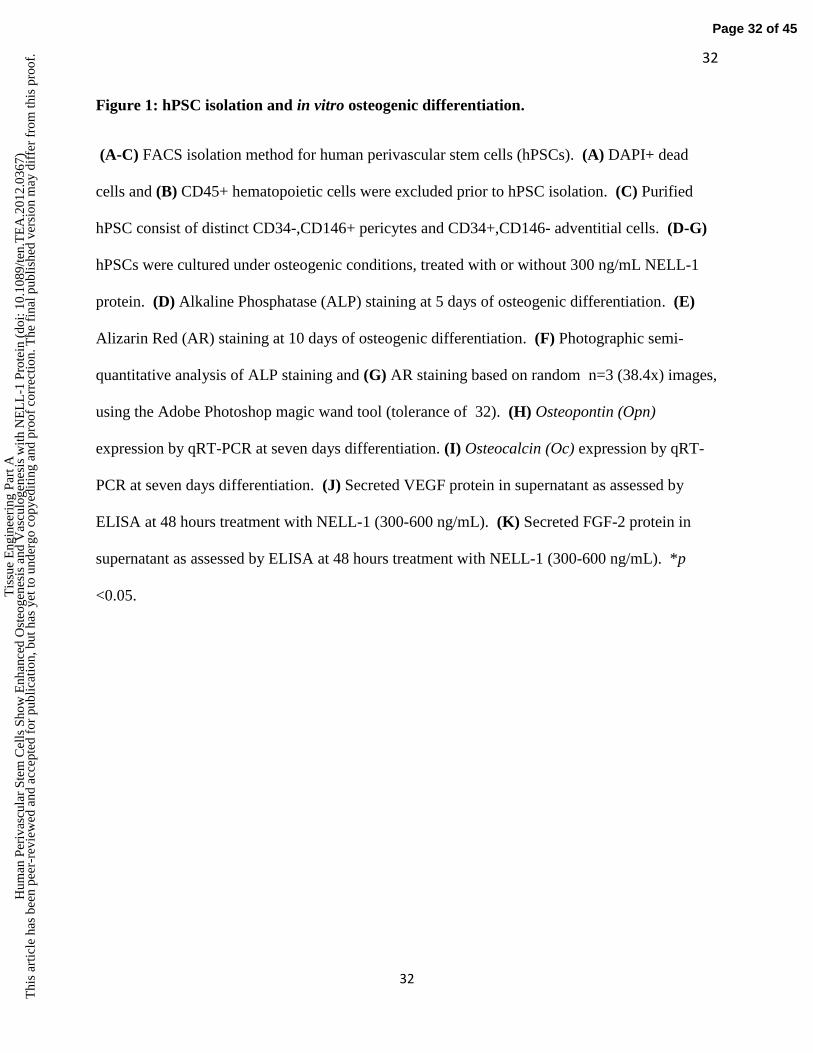
32
32
Figure 1: hPSC isolation and in vitro osteogenic differentiation.
(A-C) FACS isolation method for human perivascular stem cells (hPSCs). (A) DAPI+ dead
cells and (B) CD45+ hematopoietic cells were excluded prior to hPSC isolation. (C) Purified
hPSC consist of distinct CD34-,CD146+ pericytes and CD34+,CD146- adventitial cells. (D-G)
hPSCs were cultured under osteogenic conditions, treated with or without 300 ng/mL NELL-1
protein. (D) Alkaline Phosphatase (ALP) staining at 5 days of osteogenic differentiation. (E)
Alizarin Red (AR) staining at 10 days of osteogenic differentiation. (F) Photographic semi-
quantitative analysis of ALP staining and (G) AR staining based on random n=3 (38.4x) images,
using the Adobe Photoshop magic wand tool (tolerance of 32). (H) Osteopontin (Opn)
expression by qRT-PCR at seven days differentiation. (I) Osteocalcin (Oc) expression by qRT-
PCR at seven days differentiation. (J) Secreted VEGF protein in supernatant as assessed by
ELISA at 48 hours treatment with NELL-1 (300-600 ng/mL). (K) Secreted FGF-2 protein in
supernatant as assessed by ELISA at 48 hours treatment with NELL-1 (300-600 ng/mL). *p
<0.05.
Page 32 of 45
Tis
sue
Eng
inee
ring
Par
t AH
uman
Per
ivas
cula
r St
em C
ells
Sho
w E
nhan
ced
Ost
eoge
nesi
s an
d V
ascu
loge
nesi
s w
ith N
EL
L-1
Pro
tein
(do
i: 10
.108
9/te
n.T
EA
.201
2.03
67)
Thi
s ar
ticle
has
bee
n pe
er-r
evie
wed
and
acc
epte
d fo
r pu
blic
atio
n, b
ut h
as y
et to
und
ergo
cop
yedi
ting
and
proo
f co
rrec
tion.
The
fin
al p
ublis
hed
vers
ion
may
dif
fer
from
this
pro
of.

33
33
Page 33 of 45
Tis
sue
Eng
inee
ring
Par
t AH
uman
Per
ivas
cula
r St
em C
ells
Sho
w E
nhan
ced
Ost
eoge
nesi
s an
d V
ascu
loge
nesi
s w
ith N
EL
L-1
Pro
tein
(do
i: 10
.108
9/te
n.T
EA
.201
2.03
67)
Thi
s ar
ticle
has
bee
n pe
er-r
evie
wed
and
acc
epte
d fo
r pu
blic
atio
n, b
ut h
as y
et to
und
ergo
cop
yedi
ting
and
proo
f co
rrec
tion.
The
fin
al p
ublis
hed
vers
ion
may
dif
fer
from
this
pro
of.

34
34
Figure 2: hPSCs form significantly more bone in vivo when combined with NELL-1.
Implants consisting of DBX or DBX treated with hPSC (2.5 x 10⁵ cells), NELL-1 (3µg/µL), or
both hPSC and NELL-1 were inserted intramuscularly in SCID mice. BMP2 protein was used as
a comparison to NELL-1. See Supplemental Table 1 for treatment group specifics. Mice were
harvested 4 weeks post-implantation and evaluated for bone formation. (A) Representative 3-
dimensional microCT images at Th120, and corresponding analysis of (B) mean Bone Mineral
Density (BMD, n=8) (Th50-120 used for analysis). *p<0.05, **p<0.01.
Page 34 of 45
Tis
sue
Eng
inee
ring
Par
t AH
uman
Per
ivas
cula
r St
em C
ells
Sho
w E
nhan
ced
Ost
eoge
nesi
s an
d V
ascu
loge
nesi
s w
ith N
EL
L-1
Pro
tein
(do
i: 10
.108
9/te
n.T
EA
.201
2.03
67)
Thi
s ar
ticle
has
bee
n pe
er-r
evie
wed
and
acc
epte
d fo
r pu
blic
atio
n, b
ut h
as y
et to
und
ergo
cop
yedi
ting
and
proo
f co
rrec
tion.
The
fin
al p
ublis
hed
vers
ion
may
dif
fer
from
this
pro
of.

35
35
Page 35 of 45
Tis
sue
Eng
inee
ring
Par
t AH
uman
Per
ivas
cula
r St
em C
ells
Sho
w E
nhan
ced
Ost
eoge
nesi
s an
d V
ascu
loge
nesi
s w
ith N
EL
L-1
Pro
tein
(do
i: 10
.108
9/te
n.T
EA
.201
2.03
67)
Thi
s ar
ticle
has
bee
n pe
er-r
evie
wed
and
acc
epte
d fo
r pu
blic
atio
n, b
ut h
as y
et to
und
ergo
cop
yedi
ting
and
proo
f co
rrec
tion.
The
fin
al p
ublis
hed
vers
ion
may
dif
fer
from
this
pro
of.
36
36
Figure 3: Histological Evidence of Enhanced Osteogenic Potential of Combined hPSC-
NELL-1-Treatment
Hematoxylin and Eosin (H&E) staining was performed on the five previously mentioned
experimental groups (DBX, DBX+NELL-1, DBX+hPSC, DBX+hPSC+NELL-1, and
DBX+hPSC+BMP2). (A) Representative H&E images. (B-E) Histomorphometric semi-
quantitative analysis of random 200x H&E images: (B) Bone area (B. Ar), n=10 images per
group, tolerance=50, (C) Percent Bone Area (% B. Ar.), n=10 images per group, tolerance=50,
(D) Number Osteocyte/Bone Area, (N. Oc/B. Ar.), n=15 images per group, tolerance=20 and (E)
% filled lacunae, n=15 images per group, Tolerance=20. *p<0.05, **p<0.01. Black arrow
indicates endochondral ossification. Black asterisk indicated lipid accumulation.
Page 36 of 45
Tis
sue
Eng
inee
ring
Par
t AH
uman
Per
ivas
cula
r St
em C
ells
Sho
w E
nhan
ced
Ost
eoge
nesi
s an
d V
ascu
loge
nesi
s w
ith N
EL
L-1
Pro
tein
(do
i: 10
.108
9/te
n.T
EA
.201
2.03
67)
Thi
s ar
ticle
has
bee
n pe
er-r
evie
wed
and
acc
epte
d fo
r pu
blic
atio
n, b
ut h
as y
et to
und
ergo
cop
yedi
ting
and
proo
f co
rrec
tion.
The
fin
al p
ublis
hed
vers
ion
may
dif
fer
from
this
pro
of.
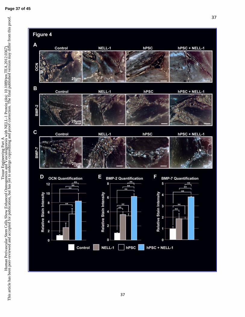
37
37
Page 37 of 45
Tis
sue
Eng
inee
ring
Par
t AH
uman
Per
ivas
cula
r St
em C
ells
Sho
w E
nhan
ced
Ost
eoge
nesi
s an
d V
ascu
loge
nesi
s w
ith N
EL
L-1
Pro
tein
(do
i: 10
.108
9/te
n.T
EA
.201
2.03
67)
Thi
s ar
ticle
has
bee
n pe
er-r
evie
wed
and
acc
epte
d fo
r pu
blic
atio
n, b
ut h
as y
et to
und
ergo
cop
yedi
ting
and
proo
f co
rrec
tion.
The
fin
al p
ublis
hed
vers
ion
may
dif
fer
from
this
pro
of.

38
38
Figure 4: Immunohistochemical analysis of ectopic bone formation.
(A-C) Immunohistochemistry was performed on the four previously mentioned experimental
groups (DBX, DBX+NELL-1, DBX+hPSC, and DBX+hPSC+NELL-1). Semi-quantitative
analysis of relative stain intensity was determined, using the magic wand tool of Adobe
Photoshop on random 400x fields. (A) Representative images of Osteocalcin (OCN)
immunohistochemistry and corresponding relative stain quantification (D), based on n=5 images
per group and tolerance of 30. (B) Representative images of Bone Morphogenetic Protein-2
(BMP-2) immunohistochemistry and corresponding relative stain quantification (E), based on
n=8 images per group and tolerance of 30. (C) Representative images of Bone Morphogenetic
Protein-7 (BMP-7) immunohistochemistry and corresponding relative stain quantification (F),
based on n=8 images per group and tolerance of 30. **p<0.01.
Page 38 of 45
Tis
sue
Eng
inee
ring
Par
t AH
uman
Per
ivas
cula
r St
em C
ells
Sho
w E
nhan
ced
Ost
eoge
nesi
s an
d V
ascu
loge
nesi
s w
ith N
EL
L-1
Pro
tein
(do
i: 10
.108
9/te
n.T
EA
.201
2.03
67)
Thi
s ar
ticle
has
bee
n pe
er-r
evie
wed
and
acc
epte
d fo
r pu
blic
atio
n, b
ut h
as y
et to
und
ergo
cop
yedi
ting
and
proo
f co
rrec
tion.
The
fin
al p
ublis
hed
vers
ion
may
dif
fer
from
this
pro
of.

39
39
Page 39 of 45
Tis
sue
Eng
inee
ring
Par
t AH
uman
Per
ivas
cula
r St
em C
ells
Sho
w E
nhan
ced
Ost
eoge
nesi
s an
d V
ascu
loge
nesi
s w
ith N
EL
L-1
Pro
tein
(do
i: 10
.108
9/te
n.T
EA
.201
2.03
67)
Thi
s ar
ticle
has
bee
n pe
er-r
evie
wed
and
acc
epte
d fo
r pu
blic
atio
n, b
ut h
as y
et to
und
ergo
cop
yedi
ting
and
proo
f co
rrec
tion.
The
fin
al p
ublis
hed
vers
ion
may
dif
fer
from
this
pro
of.

40
40
Supplemental Figure 1: Persistence and proliferation of implanted hPSCs.
(A-C) Immunohistochemistry for human β-2 Microglobulin immunohistochemistry and human
Proliferating Cell Nuclear Antigen (PCNA) was performed on hPSC and hPSC+NELL-1 treated
samples. (A) Representative images of β-2 Microglobulin immunohistochemistry. (B)
Representative images of PCNA immunohistochemistry. (C) Corresponding PCNA relative
stain quantification, based on n=10 (400x) images per group and a magic wand tolerance of 40.
*p<0.05. (D) Representative images of PCNA immunofluorescent staining, appearing green.
Implanted cells were pre-labeled with a red fluorochrome to allow in vivo tracking.
Page 40 of 45
Tis
sue
Eng
inee
ring
Par
t AH
uman
Per
ivas
cula
r St
em C
ells
Sho
w E
nhan
ced
Ost
eoge
nesi
s an
d V
ascu
loge
nesi
s w
ith N
EL
L-1
Pro
tein
(do
i: 10
.108
9/te
n.T
EA
.201
2.03
67)
Thi
s ar
ticle
has
bee
n pe
er-r
evie
wed
and
acc
epte
d fo
r pu
blic
atio
n, b
ut h
as y
et to
und
ergo
cop
yedi
ting
and
proo
f co
rrec
tion.
The
fin
al p
ublis
hed
vers
ion
may
dif
fer
from
this
pro
of.

41
41
Page 41 of 45
Tis
sue
Eng
inee
ring
Par
t AH
uman
Per
ivas
cula
r St
em C
ells
Sho
w E
nhan
ced
Ost
eoge
nesi
s an
d V
ascu
loge
nesi
s w
ith N
EL
L-1
Pro
tein
(do
i: 10
.108
9/te
n.T
EA
.201
2.03
67)
Thi
s ar
ticle
has
bee
n pe
er-r
evie
wed
and
acc
epte
d fo
r pu
blic
atio
n, b
ut h
as y
et to
und
ergo
cop
yedi
ting
and
proo
f co
rrec
tion.
The
fin
al p
ublis
hed
vers
ion
may
dif
fer
from
this
pro
of.

42
42
Figure 5: hPSC-treated implants show increased vascularization as compared to hSVF.
Equal numbers (2.5 x 10⁵) of either hPSC or hSVF cells loaded onto DBX putty were implanted
intramuscularly. Samples were harvested 4 weeks post-implantation. Specimens were stained
with H&E and for Vascular Endothelial Growth Factor (VEGF) and Von Willebrand Factor
(vWF). Histomorphometric semi-quantitative analysis of respective 400x images followed using
the Adobe Photoshop magic wand tool. (A) H&E, VEGF and vWF staining among hSVF-
treated implants. (B) H&E, VEGF and vWF staining among hPSC-treated implants. (C) Blood
Vessel Number, based on n=10 H&E images per group. (D) Mean Blood Vessel Area, based on
n=10 H&E images per group at a tolerance of 30. (E) VEGF Quantification, based on n=5
images per group and tolerance of 30. (F) CD31 immunofluorescent staining, appearing green
with red-fluorescent labeled cells. DAPI is used as a nuclear counterstain, appearing blue. White
arrow indicates close association of PSCs with a blood vessel. *p<0.05.
Page 42 of 45
Tis
sue
Eng
inee
ring
Par
t AH
uman
Per
ivas
cula
r St
em C
ells
Sho
w E
nhan
ced
Ost
eoge
nesi
s an
d V
ascu
loge
nesi
s w
ith N
EL
L-1
Pro
tein
(do
i: 10
.108
9/te
n.T
EA
.201
2.03
67)
Thi
s ar
ticle
has
bee
n pe
er-r
evie
wed
and
acc
epte
d fo
r pu
blic
atio
n, b
ut h
as y
et to
und
ergo
cop
yedi
ting
and
proo
f co
rrec
tion.
The
fin
al p
ublis
hed
vers
ion
may
dif
fer
from
this
pro
of.

43
43
Page 43 of 45
Tis
sue
Eng
inee
ring
Par
t AH
uman
Per
ivas
cula
r St
em C
ells
Sho
w E
nhan
ced
Ost
eoge
nesi
s an
d V
ascu
loge
nesi
s w
ith N
EL
L-1
Pro
tein
(do
i: 10
.108
9/te
n.T
EA
.201
2.03
67)
Thi
s ar
ticle
has
bee
n pe
er-r
evie
wed
and
acc
epte
d fo
r pu
blic
atio
n, b
ut h
as y
et to
und
ergo
cop
yedi
ting
and
proo
f co
rrec
tion.
The
fin
al p
ublis
hed
vers
ion
may
dif
fer
from
this
pro
of.

44
44
Figure 6: NELL-1 enhances hPSC-mediated vascularization in vivo.
H&E staining and VEGF immunohistochemistry were performed on the four previously
mentioned experimental groups (DBX, DBX+NELL-1, DBX+hPSC, and DBX+hPSC+NELL-1).
Histomorphometric semi-quantitative analysis of respective 200x images followed using the
Adobe Photoshop magic wand tool. (A) Representative H&E images. (B) Blood Vessel
Number, based on n=10 H&E images per group. (C) Mean Blood Vessel Area, based on n=10
H&E images per group. (D) VEGF Stain Quantification, based on n=7 images per group at a
tolerance of 30. (E) Representative image of VEGF immunohistochemistry. *p<0.05.
Supplemental Table 1: Implant contents and experimental numbers.
DBX, demineralized bone matrix; NELL-1, Nel-like molecule, type 1; hPSC, human perivascular stem
cells; hSVF, human stromal vascular fraction; BMP2, bone morphogenetic protein 2.
Experimental
Group
DBX
Volume (μL)
Protein
Concentration
(μg/μL)
Protein Total
Dose (μg)
Absolute Cell
Number
N
Control 100 0 0 0 6
NELL-1 100 3 300 0 7
hPSC 100 0 0 2.5 x 10⁵ 10
hPSC + NELL-1 100 3 300 2.5 x 10⁵ 9
hSVF 100 0 0 2.5 x 10⁵ 10
hPSC + BMP2 100 0.0375 3.75 2.5 x 10⁵ 2
Page 44 of 45
Tis
sue
Eng
inee
ring
Par
t AH
uman
Per
ivas
cula
r St
em C
ells
Sho
w E
nhan
ced
Ost
eoge
nesi
s an
d V
ascu
loge
nesi
s w
ith N
EL
L-1
Pro
tein
(do
i: 10
.108
9/te
n.T
EA
.201
2.03
67)
Thi
s ar
ticle
has
bee
n pe
er-r
evie
wed
and
acc
epte
d fo
r pu
blic
atio
n, b
ut h
as y
et to
und
ergo
cop
yedi
ting
and
proo
f co
rrec
tion.
The
fin
al p
ublis
hed
vers
ion
may
dif
fer
from
this
pro
of.

45
45
Page 45 of 45
Tis
sue
Eng
inee
ring
Par
t AH
uman
Per
ivas
cula
r St
em C
ells
Sho
w E
nhan
ced
Ost
eoge
nesi
s an
d V
ascu
loge
nesi
s w
ith N
EL
L-1
Pro
tein
(do
i: 10
.108
9/te
n.T
EA
.201
2.03
67)
Thi
s ar
ticle
has
bee
n pe
er-r
evie
wed
and
acc
epte
d fo
r pu
blic
atio
n, b
ut h
as y
et to
und
ergo
cop
yedi
ting
and
proo
f co
rrec
tion.
The
fin
al p
ublis
hed
vers
ion
may
dif
fer
from
this
pro
of.