Embed Size (px)
Citation preview
Human Physiology/The Muscular System 1
Human Physiology/The Muscular System← Senses — Human Physiology — Blood physiology →
Homeostasis — Cells — Integumentary — Nervous — Senses — Muscular — Blood — Cardiovascular — Immune — Urinary — Respiratory
— Gastrointestinal — Nutrition — Endocrine — Reproduction (male) — Reproduction (female) — Pregnancy — Genetics — Development —Answers
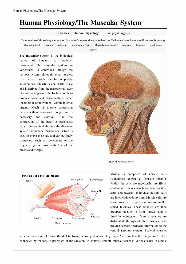
Head and Neck Muscles
The muscular system is the biologicalsystem of humans that producesmovement. The muscular system, invertebrates, is controlled through thenervous system, although some muscles,like cardiac muscle, can be completelyautonomous. Muscle is contractile tissueand is derived from the mesodermal layerof embryonic germ cells. Its function is toproduce force and cause motion, eitherlocomotion or movement within internalorgans. Much of muscle contractionoccurs without conscious thought and isnecessary for survival, like thecontraction of the heart or peristalsis,which pushes food through the digestivesystem. Voluntary muscle contraction isused to move the body and can be finelycontrolled, such as movements of thefinger or gross movements that of thebiceps and triceps.
Muscle structure
Muscle is composed of muscle cells(sometimes known as "muscle fibers").Within the cells are myofibrils; myofibrilscontain sarcomeres which are composed ofactin and myosin. Individual muscle cellsare lined with endomysium. Muscle cells arebound together by perimysium into bundlescalled fascicles. These bundles are thengrouped together to form muscle, and islined by epimysium. Muscle spindles aredistributed throughout the muscles, andprovide sensory feedback information to thecentral nervous system. Skeletal muscle,
which involves muscles from the skeletal tissue, is arranged in discrete groups. An example is the biceps brachii. It isconnected by tendons to processes of the skeleton. In contrast, smooth muscle occurs at various scales in almost
Human Physiology/The Muscular System 2
every organ, from the skin (in which it controls erection of body hair) to the blood vessels and digestive tract (inwhich it controls the caliber of a lumen and peristalsis, respectively).
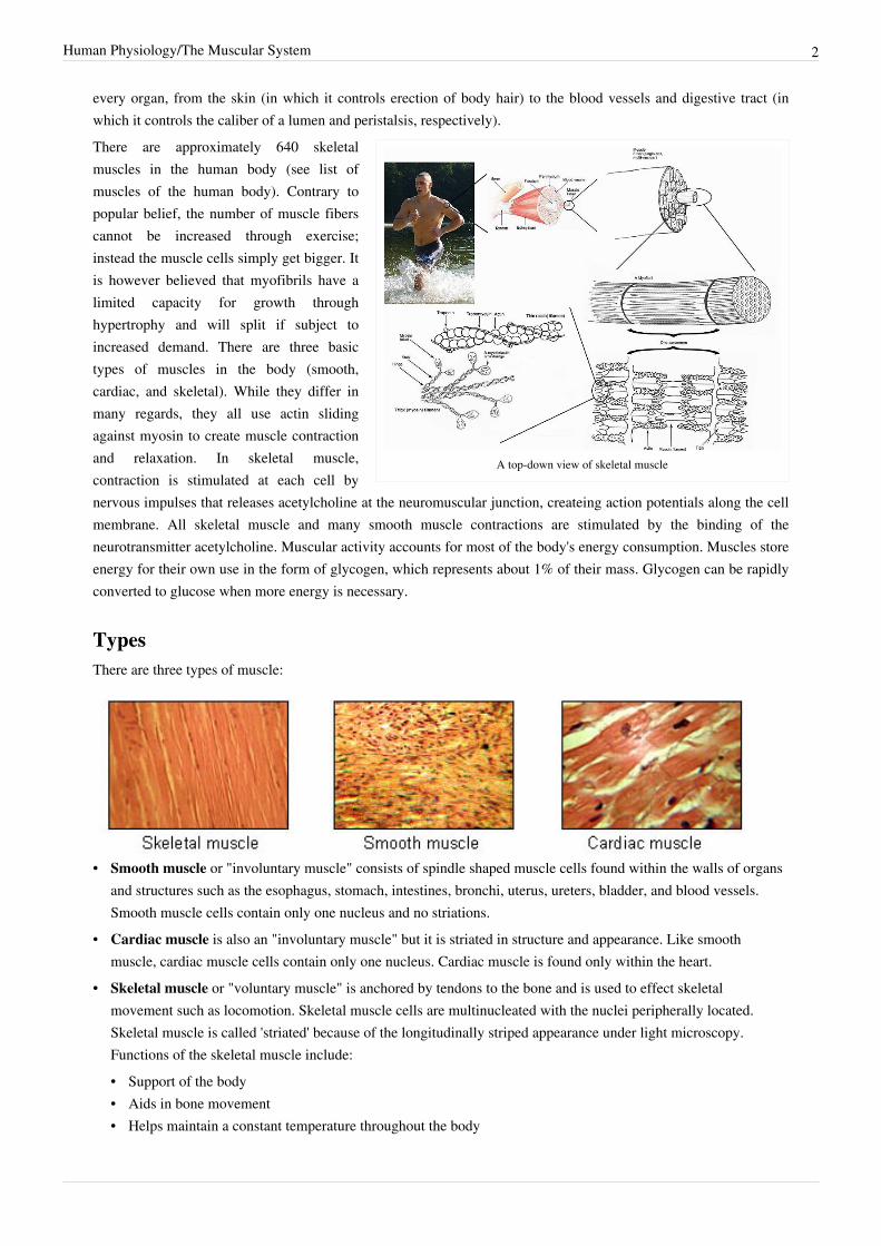
A top-down view of skeletal muscle
There are approximately 640 skeletalmuscles in the human body (see list ofmuscles of the human body). Contrary topopular belief, the number of muscle fiberscannot be increased through exercise;instead the muscle cells simply get bigger. Itis however believed that myofibrils have alimited capacity for growth throughhypertrophy and will split if subject toincreased demand. There are three basictypes of muscles in the body (smooth,cardiac, and skeletal). While they differ inmany regards, they all use actin slidingagainst myosin to create muscle contractionand relaxation. In skeletal muscle,contraction is stimulated at each cell bynervous impulses that releases acetylcholine at the neuromuscular junction, createing action potentials along the cellmembrane. All skeletal muscle and many smooth muscle contractions are stimulated by the binding of theneurotransmitter acetylcholine. Muscular activity accounts for most of the body's energy consumption. Muscles storeenergy for their own use in the form of glycogen, which represents about 1% of their mass. Glycogen can be rapidlyconverted to glucose when more energy is necessary.
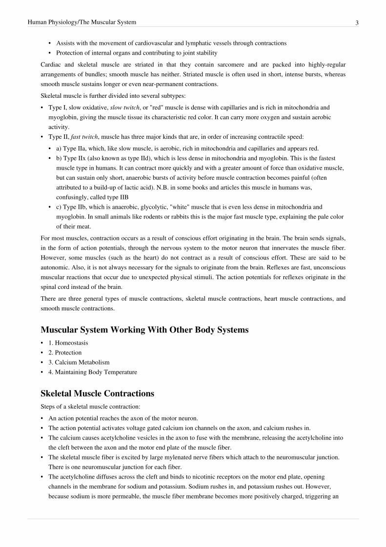
TypesThere are three types of muscle:
• Smooth muscle or "involuntary muscle" consists of spindle shaped muscle cells found within the walls of organsand structures such as the esophagus, stomach, intestines, bronchi, uterus, ureters, bladder, and blood vessels.Smooth muscle cells contain only one nucleus and no striations.
• Cardiac muscle is also an "involuntary muscle" but it is striated in structure and appearance. Like smoothmuscle, cardiac muscle cells contain only one nucleus. Cardiac muscle is found only within the heart.
• Skeletal muscle or "voluntary muscle" is anchored by tendons to the bone and is used to effect skeletalmovement such as locomotion. Skeletal muscle cells are multinucleated with the nuclei peripherally located.Skeletal muscle is called 'striated' because of the longitudinally striped appearance under light microscopy.Functions of the skeletal muscle include:• Support of the body• Aids in bone movement• Helps maintain a constant temperature throughout the body
Human Physiology/The Muscular System 3
• Assists with the movement of cardiovascular and lymphatic vessels through contractions• Protection of internal organs and contributing to joint stability
Cardiac and skeletal muscle are striated in that they contain sarcomere and are packed into highly-regulararrangements of bundles; smooth muscle has neither. Striated muscle is often used in short, intense bursts, whereassmooth muscle sustains longer or even near-permanent contractions.Skeletal muscle is further divided into several subtypes:• Type I, slow oxidative, slow twitch, or "red" muscle is dense with capillaries and is rich in mitochondria and
myoglobin, giving the muscle tissue its characteristic red color. It can carry more oxygen and sustain aerobicactivity.
• Type II, fast twitch, muscle has three major kinds that are, in order of increasing contractile speed:• a) Type IIa, which, like slow muscle, is aerobic, rich in mitochondria and capillaries and appears red.• b) Type IIx (also known as type IId), which is less dense in mitochondria and myoglobin. This is the fastest
muscle type in humans. It can contract more quickly and with a greater amount of force than oxidative muscle,but can sustain only short, anaerobic bursts of activity before muscle contraction becomes painful (oftenattributed to a build-up of lactic acid). N.B. in some books and articles this muscle in humans was,confusingly, called type IIB
• c) Type IIb, which is anaerobic, glycolytic, "white" muscle that is even less dense in mitochondria andmyoglobin. In small animals like rodents or rabbits this is the major fast muscle type, explaining the pale colorof their meat.
For most muscles, contraction occurs as a result of conscious effort originating in the brain. The brain sends signals,in the form of action potentials, through the nervous system to the motor neuron that innervates the muscle fiber.However, some muscles (such as the heart) do not contract as a result of conscious effort. These are said to beautonomic. Also, it is not always necessary for the signals to originate from the brain. Reflexes are fast, unconsciousmuscular reactions that occur due to unexpected physical stimuli. The action potentials for reflexes originate in thespinal cord instead of the brain.There are three general types of muscle contractions, skeletal muscle contractions, heart muscle contractions, andsmooth muscle contractions.
Muscular System Working With Other Body Systems• 1. Homeostasis• 2. Protection• 3. Calcium Metabolism• 4. Maintaining Body Temperature
Skeletal Muscle ContractionsSteps of a skeletal muscle contraction:• An action potential reaches the axon of the motor neuron.• The action potential activates voltage gated calcium ion channels on the axon, and calcium rushes in.• The calcium causes acetylcholine vesicles in the axon to fuse with the membrane, releasing the acetylcholine into
the cleft between the axon and the motor end plate of the muscle fiber.• The skeletal muscle fiber is excited by large mylenated nerve fibers which attach to the neuromuscular junction.
There is one neuromuscular junction for each fiber.• The acetylcholine diffuses across the cleft and binds to nicotinic receptors on the motor end plate, opening
channels in the membrane for sodium and potassium. Sodium rushes in, and potassium rushes out. However,because sodium is more permeable, the muscle fiber membrane becomes more positively charged, triggering an
Human Physiology/The Muscular System 4
action potential.• The action potential on the muscle fiber causes the sarcoplasmic reticulum to release calcium ions(Ca++).• The calcium binds to the troponin present on the thin filaments of the myofibrils. The troponin then allosterically
modulates the tropomyosin. Normally the tropomyosin physically obstructs binding sites for cross-bridge; oncecalcium binds to the troponin, the troponin forces the tropomyosin to move out of the way, unblocking thebinding sites.
• The cross-bridge (which is already in a ready-state) binds to the newly uncovered binding sites. It then delivers apower stroke.
• ATP binds the cross-bridge, forcing it to conform in such a way as to break the actin-myosin bond. Another ATPis split to energize the cross bridge again.
• Steps 7 and 8 repeat as long as calcium is present on thin filament.• Throughout this process, the calcium is actively pumped back into the sarcoplasmic reticulum. When no longer
present on the thin filament, the tropomyosin changes back to its previous state, so as to block the binding sitesagain. The cross-bridge then ceases binding to the thin filament, and the contractions cease as well.
• Muscle contraction remains as long as Ca++ is abundant in sarcoplasm.Types of Contractions:• Isometric contraction--muscle does not shorten during contraction and does not require the sliding of myofibrils
but muscles are stiff.• Isotonic contraction--inertia is used to move or work. More energy is used by the muscle and contraction lasts
longer than isometric contraction. Isotonic muscle contraction is divided into two categories: concentric, wherethe muscle fibers shorten as the muscle contracts (ie. biceps brachialis on the up phase of a biceps curl); andeccentric, where the muscle fibers lengthen as they contract (ie. biceps brachialis on the down phase of a bicepscurl).
• Twitch--exciting the nerve to a muscle or by passing electrical stimulus through muscle itself. Some fiberscontract quickly while others contract slowly.
• Tonic -maintaining postural tone against the force of gravity.The Efficiency of Muscle Contraction:• Only about 20% of input energy converts into muscular work. The rest of the energy is heat.• 50% of energy from food is used in ATP formation.• If a muscle contraction is slow or without movement, energy is lost as maintenance heat.• If muscle contraction is rapid, energy is used to overcome friction.Summation of Muscle Contraction: It is the adding together of individual muscle twitches to make strong musclemovements.• Multiple motor unit summation--increasing number of motor units contracting simultaneously.• Wave summation--increasing rapidity of contraction of individual motor units.• Tetanization--higher frequency successive contractions fuse together and cannot be distinguished from one
another.
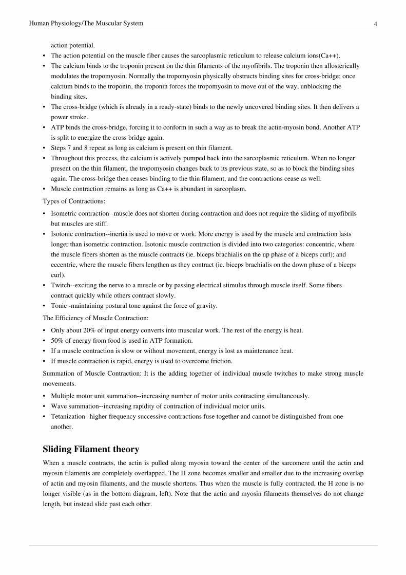
Sliding Filament theoryWhen a muscle contracts, the actin is pulled along myosin toward the center of the sarcomere until the actin andmyosin filaments are completely overlapped. The H zone becomes smaller and smaller due to the increasing overlapof actin and myosin filaments, and the muscle shortens. Thus when the muscle is fully contracted, the H zone is nolonger visible (as in the bottom diagram, left). Note that the actin and myosin filaments themselves do not changelength, but instead slide past each other.
Human Physiology/The Muscular System 5
Cellular Action of Skeletal Muscles
During cellular respiration the mitochondria, within skeletal musclecells, convert glucose from the blood to carbon dioxide and water inthe process of producing ATP (see cell physiology). ATP is needed forall muscular movement. When the need of ATP in the muscle is higherthan the cells can produce with aerobic respiration, the cells willproduce extra ATP in a process called anaerobic respiration. The firststep of aerobic respiration(glycolysis) produces two ATP per glucosemolecule. When the rest of the aerobic respiration pathway is occupiedthe pyruvate molecule can be converted to lactic acid. This method
produces much less ATP than the aerobic method, but it does it faster and allows the muscles to do a bit more than ifthey relied solely on ATP production from aerobic respiration. The drawback to this method is that lactic acidaccumulates and causes the muscles to fatigue. They will eventually stop contracting until the breakdown of lacticacid is sufficient to allow for movement once again. People experience this most noticeably when they repeatedly liftheavy things such as weights or sprint for a long distance. Muscle soreness sometimes occurs after vigorous activity,and is often misunderstood by the general public to be the result of lactic acid buildup. This is a misconceptionbecause the muscle does fatigue from lactic acid buildup, but it does not stay in the muscle tissue long enough tocause tissue breakdown or soreness. During heavy breathing, following exercise, the cells are converting the lacticacid either back into glucose or converting it to pyruvate and sending it through the additional steps of aerobicrespiration. By the time a person is breathing normally again the lactic acid has been removed. The soreness isactually from small tears in the fibers themselves. After the fibers heal they will increase in size. The number ofmitochondria will also increase if there is continued demand for additional ATP. Hence, through exercise themuscles can increase in both strength and endurance.
Another misconception is that as the muscle increases in size it also gains more fibers. This is not true. The fibersthemselves increase in size rather than in quantity. The same holds true for adipose tissue--fat cells do not increase innumber, but rather the amount of lipids (oil) in the cells increase.Muscle fibers are also genetically programmed to reach a certain size and stop growing from there, so after awhileeven the hardest working weightlifter will only reach a certain level of strength and endurance. Some people will getaround this by taking steroids. The artificial steroids cause all sorts of trouble for the person. They can cause theadrenal glands to stop producing corticosteroids and glucosteroids. This leads to the atrophy of the gland's medullaand causes permanent loss of the production of these hormones. The testicles may also atrophy in response tosteroids. Eventually the testes will stop making testosterone and sperm, rendering the male infertile.One of the more serious problems associated with abnormal gain of muscle mass is heart failure. While for mostpeople gaining muscle and losing fat is desirable, a body builder is at risk of producing more muscle mass than theheart can handle. One pound of fat contains about 3.5 miles of blood vessels, but one pound of muscle has about 6.5miles. Hence, additional muscle causes the heart to pump more blood. Some people that have too much muscle willbe very strong but will not have a healthy aerobic endurance, in part because of the difficulty of providingoxygenated blood to so much tissue.Sliding filament theory [1]
This link shows the animation of the sliding filament theory.explanation and image of sliding filament theory [2]
this link gives a better demonstration of the theory with the explanation.
Human Physiology/The Muscular System 6
Involuntary Muscle MovementSpasmsWhen Smooth and skeletal muscles go through multiple spasms it is referred either as seizure or convulsion.CrampsStrenuous activities can cause painful spasms that are long, this is referred to as cramps.
InjurySprainA injury to a joint that involves a stretched or torn ligament.Muscle StrainA strain occurs when a muscle or the tendon that attaches it to the bone is overstretched or torn. Muscle strains arealso called pulled muscles. Who gets it?Anyone can strain a muscle. However, people involved in sports or other forms of strenuous exercise are more likelyto strain a muscle. What causes it?
Muscles are bunches of fibers that can contract. Muscle strains usually occur during activities that require the muscleto tighten forcefully. The muscle is strained either because it is not properly stretched, or warmed up, before theactivity; it is too weak; or because the muscle is already injured and not allowed time to recover. So, many musclestrains occur during exercise or sports activities. They can also occur when lifting heavy objects. What are thesymptoms?When a muscle is strained, it hurts and is difficult to move. You may also feel a burning sensation in the area of theinjured muscle, or feel as though something has "popped." Sometimes the area of the strained muscle looks bruisedor swells. A strained muscle might spasm, which means it contracts suddenly and involuntarily, causing severe pain.How is it diagnosed?To diagnose a muscle strain, your doctor will examine the painful area, and ask how and when the injury happened.He or she may order other diagnostic tests, such as x-rays, to rule out any injury to the bone.What is the treatment?
Muscle strains are treated with rest, ice, compression, and elevation, or RICE. You will be told to rest the injuredarea to reduce pain and swelling. If the strain is in the leg or foot area, you may need to use crutches. Ice packs arerecommended at regular intervals (as recommended by your doctor) over the first few days after the injury. Icecauses the blood vessels to constrict, which reduces inflammation and pain. Anti-inflammatory medications mightalso be used to relieve pain. Compression and elevation help to reduce swelling. Your doctor may also recommendphysical therapy to speed your recovery. You should avoid the type of activity that caused the injury until the muscleis completely healed. Self-care tipsYou can prevent muscle strains by warming up for at least 10 minutes before participating in any strenuous exerciseor heavy lifting. When you warm up, you increase the blood circulation to the muscle and prepare it for exercise.When starting any new exercise program or sport, it's important to begin gradually so your muscles are conditionedfor the activity.
Human Physiology/The Muscular System 7
SteroidsAnabolic steroids, which are synthetic versions of the primary male sex hormone testosterone, can be injected, takenorally, or used transdermally. These drugs are Controlled Substances that can be prescribed to treat conditions suchas body wasting in patients with AIDS, and other diseases that occur when the body produces abnormally lowamounts of testosterone. However, the doses prescribed to treat these medical conditions are 10 to 100 times lowerthan the doses that are used for performance enhancement.Let me be clear:- while anabolic steroids can enhance certain types of performance or appearance,they are dangerousdrugs, and when used inappropriately, they can cause a host of severe, long-lasting, and often irreversible negativehealth consequences. These drugs can stunt the height of growing adolescents, masculinize women, and alter sexcharacteristics of men. Anabolic steroids can lead to premature heart attacks, strokes, liver tumors, kidney failure andserious psychiatric problems. In addition, because steroids are often injected, users risk contracting or transmittingHIV or hepatitis.Abuse of anabolic steroids differs from the abuse of other illicit substances because the initial use of anabolicsteroids is not driven by the immediate euphoria that accompanies most drugs of abuse, such as cocaine, heroin, andmarijuana, but by the desire of the user to change their appearance and performance, characteristics of greatimportance to adolescents. These effects of steroids can boost confidence and strength leading the user to overlookthe potential serious long-term damage that these substances can cause.Government agencies such as NIDA support research that increases our understanding of the impact of steroid useand improves our ability to prevent abuse of these drugs. For example, NIDA funding led to the development of twohighly effective programs that not only prevent anabolic steroid abuse among male and female high school athletes,but also promote other healthy behaviors and attitudes. The ATLAS (targeting male athletes) and ATHENA(targeting female athletes) programs have been adopted by schools in 29 states and Puerto Rico. Both Congress andthe Substance Abuse and Mental Health Services Administration have endorsed ATLAS and ATHENA as modelprevention programs, which could and should be implemented in more communities throughout the country.In addition to these prevention programs and other research efforts, also has invested in public education efforts toincrease awareness about the dangers of steroid abuse. We have material on our website about steroid abuse atwww.steroidabuse.gov and in April 2005 we again will distribute a "Game Plan" public service announcementdesigned to bring attention to abuse of anabolic steroids.Research has shown that the inappropriate use of anabolic steroids can have catastrophic medical, psychiatric andbehavioral consequences.I hope that students, parents, teachers, coaches and others will take advantage of the information on our websiteabout anabolic steroids abuse and join us in our prevention and education efforts. Participating in sports offers manybenefits, but young people and adults shouldn't take unnecessary health risks in an effort to win.(Nora D. Volkow,M.D.)-Human-made substances related to male sex hormones. Some athletes abuse anabolic steroids to enhanceperformance. Abuse of anabolic steroids can lead to serious health problems, some of which are irreversible.Major side effects can include liver tumors and cancer, jaundice, high blood pressure, kidney tumors, severe acne,and trembling. In males, side effects may include shrinking of the testicles and breast development. In females, sideeffects may include growth of facial hair, menstrual changes, and deepened voice. In teenagers, growth may behalted prematurely and permanently.The therapeutic use of steroids can be realized by patients and their doctors by using them in a manner that isbeneficial to the person.
Human Physiology/The Muscular System 8
Smooth Muscle Contraction• Contractions are initiated by an influx of calcium which binds to calmodulin.• The calcium-calmodulin complex binds to and activates myosin light-chain kinase.• Myosin light-chain kinase phosphorylates myosin light-chains using ATP, causing them to interact with actin
filaments.• Powerstroke.• Calcium is actively pumped out of the cell by receptor regulated channels. A second messanger, IP3, causes the
release.• As calcium is removed the calcium-calmodulin complex breaks away from the myosin light-chain kinase,
stopping phosphorylation.• Myosin phophatase dephosphorylates the myosin. If the myosin was bound to an actin molecule, the release is
slow, this is called a latch state. In this manner, smooth muscle is able to stay contracted for some time withoutthe use of much ATP. If the myosin was not bound to an actin chain it loses its affinity for actin.
It should be noted that ATP is still needed for crossbridge cycling, and that there is no reserve, such as creatinephosphate, available. Most ATP is created from aerobic metabolism, however anaerobic production may take placein times of low oxygen concentrations.
Cardiac MuscleCardiac muscle is found in the heart and lungs of humans
ATP in the Human BodyMuscles cells, like all cells, use ATP as an energy source. The total quantity of ATP in the human body at any onetime is about 0.1 Mole. The energy used by human cells requires the hydrolysis of 200 to 300 moles of ATP daily.This means that each ATP molecule is recycled 2000 to 3000 times during a single day. ATP cannot be stored, henceits consumption must closely follow its synthesis. On a per-hour basis, 1 kilogram of ATP is created, processed andthen recycled in the body. Looking at it another way, a single cell uses about 10 million ATP molecules per secondto meet its metabolic needs, and recycles all of its ATP molecules about every 20-30 seconds.
Lactic AcidCatabolized carbohydrates is known as glycolysis. The end product of glycolysis, pyruvate can go into differentdirections depending on aerobic or anaerobic conditions. In aerobic it goes through the Krebs cycle and in anaerobicit goes through the Cori cycle. In the Cori cycle pyruvate is converted to lactate, this forms lactic acid, lactic acidcauses muscle fatigue. In the aerobic conditions pyruvate goes through the Krebs cycle. For more about Krebs cyclerefer to chapter 2 Cell Physiology.
Human Physiology/The Muscular System 9
Muscle Disorders
Dermatomyositis and PolymyositisDermatomyositis and polymyositis cause inflammation of the muscles. They are rare disorders, affecting only aboutone in 100,000 people per year. More women than men are affected. Although the peak age of onset is in the 50s, thedisorders can occur at any age.Signs and symptoms — Patients complain of muscle weakness that usually worsens over several months, though insome cases symptoms come on suddenly. The affected muscles are close to the trunk (as opposed to in the wrists orfeet), involving for example the hip, shoulder, or neck muscles. Muscles on both sides of the body are equallyaffected. In some cases, muscles are sore or tender. Some patients have involvement of the muscles of the pharynx(throat) or the esophagus (the tube leading from the throat to the stomach), causing problems with swallowing. Insome cases, this leads to food being misdirected from the esophagus to the lungs, causing severe pneumonia.In dermatomyositis, there is a rash, though sometimes the rash resolves before muscle problems occur. A number ofdifferent types of rash can occur, including rashes on the fingers, the chest and shoulders, or on the upper eyelids(show picture 1-3). In rare cases, the rash of dermatomyositis appears but myopathy never develops.Other problems sometimes associated with these diseases include fever, weight loss, arthritis, cold-induced colorchanges in the fingers or toes (Raynaud phenomenon), and heart or lung problems.
Muscle AtrophyAlternative names : Atrophy of the muscles, Muscle wasting, WastingThe majority of muscle atrophy in the general population results from disuse. People with sedentary jobs and seniorcitizens with decreased activity can lose muscle tone and develop significant atrophy. This type of atrophy isreversible with vigorous exercise. Bed-ridden people can undergo significant muscle wasting. Astronauts, free of thegravitational pull of Earth, can develop decreased muscle tone and loss of calcium from their bones following just afew days of weightlessness.Muscle atrophy resulting from disease rather than disuse is generally one of two types, that resulting from damage tothe nerves that supply the muscles, and disease of the muscle itself. Examples of diseases affecting the nerves thatcontrol muscles would be poliomyelitis, amyotrophic lateral sclerosis (ALS or Lou Gehrig's disease), andGuillain-Barre syndrome. Examples of diseases affecting primarily the muscles would include muscular dystrophy,myotonia congenita, and myotonic dystrophy as well as other congenital, inflammatory or metabolic myopathies.Even minor muscle atrophy usually results in some loss of mobility or power.Common Causes• some atrophy that occurs normally with aging• cerebrovascular accident (stroke)• spinal cord injury• peripheral nerve injury (peripheral neuropathy)• other injury• prolonged immobilization• osteoarthritis• rheumatoid arthritis• prolonged corticosteroid therapy• diabetes (diabetic neuropathy)• burns• poliomyelitis• amyotrophic lateral sclerosis (ALS or Lou Gehrig's disease)
Human Physiology/The Muscular System 10
• Guillain-Barre syndrome• muscular dystrophy• myotonia congenita• myotonic dystrophy• myopathy
Muscular DystrophyMuscular dystrophy (MD) is a group of rare inherited muscle diseases in which muscle fibers are unusuallysusceptible to damage. Muscles, primarily voluntary muscles, become progressively weaker. In the late stages ofmuscular dystrophy, fat and connective tissue are often replaced by muscle fibers. In some types of musculardystrophy, heart muscles, other involuntary muscles and other organs are affected.The most common types of muscular dystrophy appear to be due to a genetic deficiency of the muscle proteindystrophin. There's no cure for muscular dystrophy, but medications and therapy can slow the course of the disease.
Medical Mysteries
Sleep Twitches
The twitching phenomenon that happens in the early stage of sleep is called a hypnagogic massive jerk, or simply ahypnic jerk. It has also been referred to as a sleep start. There has been little research on this topic, but there havebeen some theories put forth. When the body drifts off into sleep, it undergoes physiological changes related to bodytemperature, breathing rate and muscular tone. Hypnic jerks may be the result of muscle changes. Another theorysuggests that the transition from the waking to the sleeping state signals the body to relax. But the brain mayinterpret the relaxation as a sign of falling and then signal the arms and legs to wake up. Electroencephalogramstudies have shown sleep starts affect almost 10 percent of the population regularly, 80 percent occasionally, andanother 10 percent rarely.Muscle movement or twitching also may take place during the Rapid Eye Movement, or REM, phase of sleep. Thisalso is the time when dreams occur. During the REM phase, all voluntary muscular activity stops with a drop inmuscle tone, but some individuals may experience slight eyelid or ear twitching or slight jerks. Some people withREM behavioral disorder, or RBD, may experience more violent muscular twitching and full-fledged activity duringsleep. This is because they do not achieve muscle paralysis, and as a result, act out their dreams. Researchers thinkthat people with RBD lack neurological barriers that define the different stages of sleep. New research done by theMayo Clinic and published in the July 2003 issue of Sleep Medicine shows that melatonin can help lessen RBDsymptoms.Resources:
Sleep twitches, or myoclonic jerks, as they are sometimes called, are explained in easily understood languageon this website.Learn more about REM Behavior Disorder, or RBD, and treatment for sufferers.View information about various sleep disorders such as insomnia, apnea, and narcolepsy.
Human Physiology/The Muscular System 11
MicrobiologyClostridium tetani
TetanusNormally a nerve impulse initiates contraction of a muscle. At the same time, an opposing muscle receives thesignal to relax so as not to oppose the contraction. Tetanus toxin blocks the relaxation, so both sets of musclecontract. The usual cause of tetany is lack of calcium, but excess of phosphate (high phosphate-to-calciumratio) can also trigger the spasms.
Clostridium botulinum
Infant botulism (floppy baby syndrome) the most common form of botulism in the U.S. of the four forms ofbotulism.If ingested, the toxin is absorbed in the intestine, goes to the blood, and on to the nervous system. It acts on theperipheral nervous system by blocking the impulse that is normally passes along to the nervous system. Byclocking the impulse that is normally passed along to motor end plates so the muscle contraction can bereleased, resulting in paralysis.
GlossaryActin
A protien that forms a long polymer rods called microfilaments; Interacts with myosin to cause movement inmuscles.
ATP"Adenosine Triphosphate" is a nucleotide that comes from adenosine that takes place in muscle tissue: Thisprovides a large source of energy for cellular reactions.
Cardiac muscleis also an "involuntary muscle" but it's a specialized kind of muscle found only within the heart.
Clostridium botulinumA pathogen that causes botulism, gram stain positive, morphology is rod shaped, grows in anaerobicconditions, and produces spores.
Clostridium tetaniA pathogen that causes lock jaw, gram stain positive, morphology is tennis racket shaped rod, grows inanaerobic conditions, and produces spores.
Cori cycleIn anaerobic conditions produces lactic acid.
CrampA localized muscle spasm that happens after strenuous activity.
GlycogenGlucose that has been converted for energy storage. Muscles store energy for their own use in this form.
Lactic acidCauses muscle fatigue.
MuscleContractile tissue that is derived from the mesodermal layer of embryonic germ cells.
Muscular Dystrophy
Human Physiology/The Muscular System 12
A hereditary disease characterized by progressive atrophy of muscle fibersMyosin
The fibrous motor protein that uses ATP to drive movements along actin filaments.Sarcoplasmic Reticulum
Smooth-surfaced tubules forming a plexus around each myofibril that function as a storage and release area forcalcium ions (CA+2).
Skeletal musclethis "voluntary muscle" is anchored by tendons to the bone and is used to affect skeletal movement such aslocomotion.
Smooth musclethis "involuntary muscle" is found within the walls of organs and structures such as the esophagus, stomach,intestines, bronchi, uterus, ureters, bladder, and blood vessels.
SprainInjuries that involves a stretched or torn ligament.
StrainA injury to the muscle or tendon attachment
References• Van De Graaff (2002) Human Anatomy 6th ed. McGraw-Hill Higher Education• Windmaier, P.W. Raff, H. Strang, T.S. (2004) Vander, Sherman, & Luciano's Human Physiology, the
Mechanisms of Body Function 9th ed. Mcgraw-Hill
References[1] http:/ / 3dotstudio. com/ zz. html[2] http:/ / www. ucl. ac. uk/ ~sjjgsca/ muscleSlidingFilament. html
Article Sources and Contributors 13
Article Sources and ContributorsHuman Physiology/The Muscular System Source: http://en.wikibooks.org/w/index.php?oldid=2010390 Contributors: Adrignola, AmWengert, BrendaJohnson, Brentwaldrop, Cde grey,Danseyffert, Danzman, Daynaclegg, ElizabethDurham, FatBoySlim011, Fransanfan, Frj1947, Gartoly, Gayad, Harrissc, Herbythyme, JenVan, Jomegat, Jusjih, Kmarv84, Krstnplmr, Linsey8,Linsey88, Mike.lifeguard, Nikkjeppson, Provophys, Pwoodson, QuiteUnusual, Recent Runes, Reece, Scottholmes, Shakah, Stephanie greenwood, Sterlingsilver, Sundance Raphael, Trevan5,Trisha83, Whiteknight, Xania, Xxagile, 98 anonymous edits
Image Sources, Licenses and ContributorsImage:Lateral head anatomy.jpg Source: http://en.wikibooks.org/w/index.php?title=File:Lateral_head_anatomy.jpg License: Attribution Contributors: Patrick J. Lynch, medical illustratorImage:Illu muscle structure.jpg Source: http://en.wikibooks.org/w/index.php?title=File:Illu_muscle_structure.jpg License: Public Domain Contributors: Arcadian, NevitImage:Skeletal muscle.jpg Source: http://en.wikibooks.org/w/index.php?title=File:Skeletal_muscle.jpg License: GNU Free Documentation License Contributors: Deadstar, Noca2plus, Rama,Raul654, Splette, 2 anonymous editsImage:Illu_muscle_tissues.jpg Source: http://en.wikibooks.org/w/index.php?title=File:Illu_muscle_tissues.jpg License: Public Domain Contributors: Arcadian, Fvasconcellos, Pieter Kuiper, 5anonymous editsImage:SlidingMyofibril-775px.png Source: http://en.wikibooks.org/w/index.php?title=File:SlidingMyofibril-775px.png License: GNU Free Documentation License Contributors: Provophys
LicenseCreative Commons Attribution-Share Alike 3.0 Unportedhttp:/ / creativecommons. org/ licenses/ by-sa/ 3. 0/