Embed Size (px)
Citation preview
Garcia et al. Virology Journal 2013, 10:305http://www.virologyj.com/content/10/1/305
RESEARCH Open Access
Human rhinoviruses and enteroviruses ininfluenza-like illness in Latin AmericaJosefina Garcia1,2*, Victoria Espejo1, Martha Nelson2, Merly Sovero1, Manuel V Villaran1, Jorge Gomez3,Melvin Barrantes4, Felix Sanchez5, Guillermo Comach6, Ana E Arango7, Nicolas Aguayo8, Ivette L de Rivera9,Wilson Chicaiza10, Mirna Jimenez11, Washington Aleman12, Francisco Rodriguez13, Marina S Gonzales14,Tadeusz J Kochel15 and Eric S Halsey1
Abstract
Background: Human rhinoviruses (HRVs) belong to the Picornaviridae family with high similarity to humanenteroviruses (HEVs). Limited data is available from Latin America regarding the clinical presentation and strains ofthese viruses in respiratory disease.
Methods: We collected nasopharyngeal swabs at clinics located in eight Latin American countries from 3,375subjects aged 25 years or younger who presented with influenza-like illness.
Results: Our subjects had a median age of 3 years and a 1.2:1.0 male:female ratio. HRV was identified in 16% andHEV was identified in 3%. HRVs accounted for a higher frequency of isolates in those of younger age, in particularchildren < 1 years old. HRV-C accounted for 38% of all HRVs detected. Phylogenetic analysis revealed a highproportion of recombinant strains between HRV-A/HRV-C and between HEV-A/HEV-B. In addition, both EV-D68 andEV-A71 were identified.
Conclusions: In Latin America as in other regions, HRVs and HEVs account for a substantial proportion ofrespiratory viruses identified in young people with ILI, a finding that provides additional support for thedevelopment of pharmaceuticals and vaccines targeting these pathogens.
BackgroundAcute respiratory infections (ARIs) are a leading cause ofacute illness worldwide and remain the most importantcause of pediatric mortality [1]. Lower respiratory tractinfections (LRTIs) are among the leading causes ofhospitalization and death in children less than 5 years oldworldwide, particularly in resource-poor countries [2].Human rhinoviruses (HRVs) and enteroviruses (HEVs)
belong to the Picornaviridae family and are prominentcauses of respiratory disease [3]. They share identicalgenomic organization and high sequence homology [4].Their genome is divided into three sections: a 5’untrans-lated region (5’UTR), an open reading frame of thepolyprotein that codes for all four capsid proteins (VP1-4)and the non-structural genes, and a 3’untranslated region [5].
* Correspondence: [email protected] Naval Medical Research Unit 6, Lima, Peru2Fogarty International Center, National Institutes of Health, Bethesda, MD,USAFull list of author information is available at the end of the article
© 2013 Garcia et al.; licensee BioMed Central LCommons Attribution License (http://creativecreproduction in any medium, provided the or
Although many reports link HRV primarily to illnessin children [6,7], disease in other populations such asmilitary recruits [8] and nursing home residents havebeen reported [9]. HRV infection often results in mildupper respiratory disease like the common cold, but itmay also cause more serious disease by exacerbatingasthma or other pre-existing respiratory disorders. Incontrast, HEVs infect primarily the gastrointestinal tractand can spread to other sites, but some HEVs displayspecific tropism for the respiratory tract [10,11].There are more than 100 different serotypes of HRVs
taxonomically grouped into two species HRV-A andHRV-B, according to the alignment of nucleotide frag-ments of the VP1 gene, the VP4/VP2 gene, and, morerecently, the whole genome sequence [4,12]. A differentspecies, HRV-C, that shares 53 - 57% homology at theamino acid level with HRV-A and HRV-B, was identifiedin 2006 in patients with acute LRTIs in Africa, Asia,Australia, Europe and North America [13-16]. Since its
td. This is an open access article distributed under the terms of the Creativeommons.org/licenses/by/2.0), which permits unrestricted use, distribution, andiginal work is properly cited.
Garcia et al. Virology Journal 2013, 10:305 Page 2 of 12http://www.virologyj.com/content/10/1/305
detection, HRV-C has been reported to be a prominentrespiratory pathogen in children, causing up to 5% ofLRTIs [17] and found in 42% of children with influenza-like infection (ILI) without identification of anotherpathogen by conventional means [18]. HRV-C has alsobeen implicated as a frequent cause of asthma exacer-bation in children [15]. Recent studies indicate thatHRVs, as well as HEVs, show great genetic diversity byrecombination [17,19], as has been increasingly repor-ted for the HRV-A/HRV-C recombinants [20].There are few reports about HRVs and HEVs as causes
of respiratory disease in Latin America [21-25], inclu-ding one that describes HRV antibodies in Amazontribes [26], but no large-scale genetic characterization ofHRVs and HEVs has been performed in the region todate. In this study, we investigated the recent circulationof HRV and HEV with emphasis on recombinant strainsin children and young adults in Latin America.
ResultsHRV and HEV in Central and South AmericaWe collected 3,375 nasopharyngeal swabs from subjectswith ILI symptoms from eight countries throughoutCentral and South America. We performed direct RT-PCR for HRVs and HEVs and sequenced all positivesamples (n = 632) (Figure 1). Our subjects had a medianage of 3 years, ranging from less than 1 month to25 years, an interquartile range of 1 to 8 years, and amale/female ratio of 1.2:1.Overall, HRVs and HEVs were identified in 16% (548
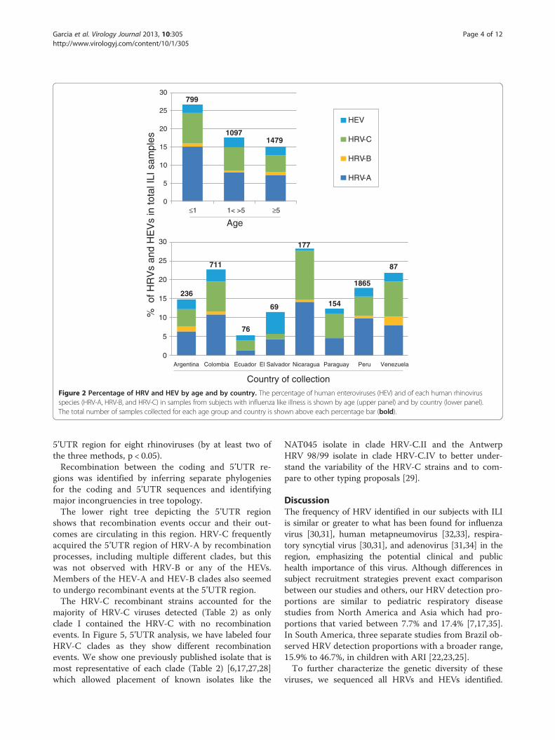
samples) and 3% (84 samples) of the ILI cases, respec-tively. Among the HRVs, HRV-A was the most repre-sented species (9% of ILI cases), followed by HRV-C(6%) and HRV-B (1%). Although the number of ILIsamples collected among countries varied considerably(Figure 2, lower panel) we found no statiscally signifi-cant geographic differences in the proportions of HEV,HRV-A, HRV-B, and HRV-C.HRVs were identified significantly more frequently in
children younger than 1 year (24%) compared to thosebetween 1 and 5 years of age (15%) or older than fiveyears (13%) (Figure 2, upper panel), a statistically sig-nificant finding (p <0.05 for both). However, using thetwo proportion z-test, we noted no difference in propor-tions of specific HRV species or HEVs per total ILI casesamong different age groups. Nevertheless, the risk ofdetecting HRV-C in children younger than 5 years withILI was 1.59 (C.I. 1.17-2.17; p <0.05) compared to olderchildren and young adults (5–25 years).Certain pre-existing respiratory conditions, such as
rhinitis or chronic bronchitis, were found more often inthose with HRV isolated (O.R. = 1.14 [95% C.I. 1.09 –1.81]; ×2 = 7.82; p-value < 0.05). Furthermore, the presenceof these pre-existing conditions specifically increased the
risk for detection of HRV-C (O.R. = 1.71 [95% C.I. 1.18 –2.45]; ×2 = 9.17; p < 0.05); asthma was a condition thatdoubled the risk of HRV-C detection (O.R. = 2.07 [95%C.I. 1.08 – 3.79]; ×2 = 6.19; p <0.05).Coxsackieviruses comprised the majority of the HEV
group (65% of the HEVs identified), showing a variety oftypes: 9 for coxsackievirus A and 5 for coxsackievirus B(Figure 3). In addition, we detected multiple HEV sero-types, including EV-D68, EV-C99, EV-C104, EV-C109,and EV-B110. We also identified EV-A71 in two par-ticipants, both from the department of Tumbes, Peru,although from different cities. The first subject, a oneyear-old girl from the city of Tumbes (same name as thedepartment), presented on April 27, 2011, with fever,rhinorrhea, cough, and erythema on pharyngeal exami-nation. The second, a two year-old girl from the city ofZarumilla, presented on May 10, 2011, with fever,malaise, rhinorrhea, cough, and weight loss. Neithersubject had rash, gastrointestinal manifestations, convul-sions, change in consciousness, or other neurologicaldeficits. Lastly, two polioviruses were detected and wererelated to the Sabin-1 polio vaccine strain.By cell culture/immunofluorescence (as well as real
time PCR for influenza viruses), we detected other res-piratory viruses in 11% of the HRV-positive samples and11% of the HEV-positive samples. The most commonlydetected viruses were adenovirus, influenza virus A andparainfluenza virus 1 (Figure 4).
Clinical manifestations of HRV and HEV infectionsAlthough we found a higher frequency of cough, rhi-norrhea, and dyspnea in subjects with HRV-C comparedto those with etiologies other than HRV and HEV, thisfinding was not significantly more common when com-paring HRV-C with HRV-A or HRV-B. HEV showed asignificantly lower frequency of cough than HRV-C andthe non-HRV/non-HEV group. Finally, there was no sta-tistically significant difference in the rate of hospitalizationfor HRV-C (20%) compared to all other viruses. Otherfrequencies and comparisons are on Table 1.
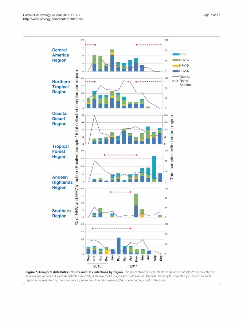
Temporal distribution of HRV and HEVWe analyzed the temporal distribution of the three HRVspecies and HEV across six regions, grouped by similarclimatic and geographical characteristics. Figure 1 showsthe collection sites that were selected and grouped intoregions for this analysis. Figure 5 depicts the percentageof each HRV species and HEV per total ILI samplescollected and shows that HRV was present in tropicalregions (Central America, northern and tropical forestregions) all year long. In no region was HRV or HEVactivity detected more often in the rainy seasonor the higher temperature season. However, HRV-Caccounted for a higher percentage (in most cases > 5%)
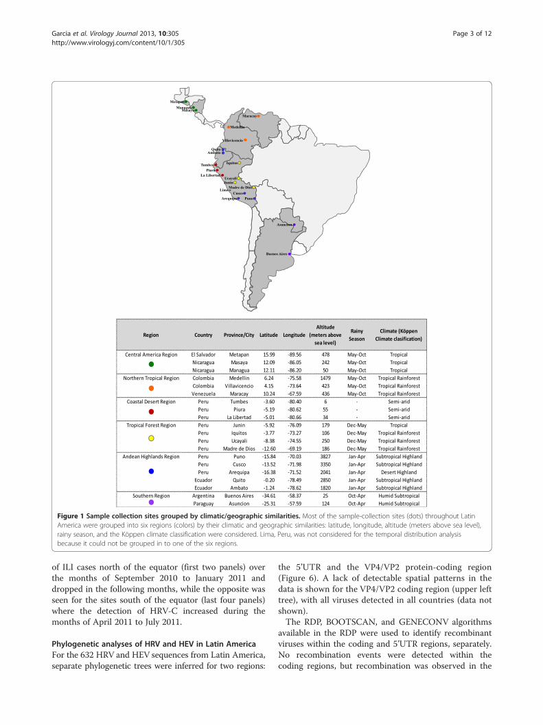
Figure 1 Sample collection sites grouped by climatic/geographic similarities. Most of the sample-collection sites (dots) throughout LatinAmerica were grouped into six regions (colors) by their climatic and geographic similarities: latitude, longitude, altitude (meters above sea level),rainy season, and the Köppen climate classification were considered. Lima, Peru, was not considered for the temporal distribution analysisbecause it could not be grouped in to one of the six regions.
Garcia et al. Virology Journal 2013, 10:305 Page 3 of 12http://www.virologyj.com/content/10/1/305
of ILI cases north of the equator (first two panels) overthe months of September 2010 to January 2011 anddropped in the following months, while the opposite wasseen for the sites south of the equator (last four panels)where the detection of HRV-C increased during themonths of April 2011 to July 2011.
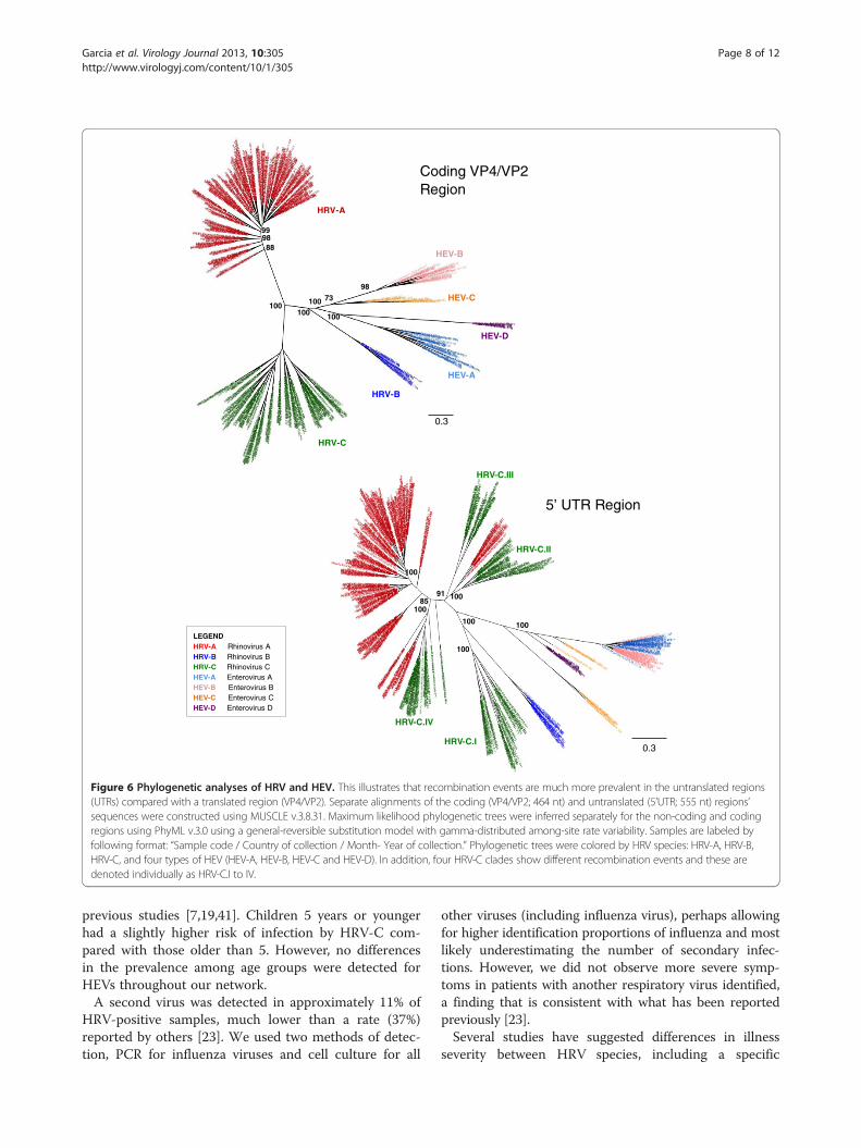
Phylogenetic analyses of HRV and HEV in Latin AmericaFor the 632 HRV and HEV sequences from Latin America,separate phylogenetic trees were inferred for two regions:
the 5’UTR and the VP4/VP2 protein-coding region(Figure 6). A lack of detectable spatial patterns in thedata is shown for the VP4/VP2 coding region (upper lefttree), with all viruses detected in all countries (data notshown).The RDP, BOOTSCAN, and GENECONV algorithms
available in the RDP were used to identify recombinantviruses within the coding and 5’UTR regions, separately.No recombination events were detected within thecoding regions, but recombination was observed in the
HEV
HRV-C
HRV-B
HRV-A
0
5
10
15
20
25
30
≤1 1< >5 ≥5
0
5
10
15
20
25
30
Argentina Colombia Ecuador El Salvador Nicaragua Paraguay Peru Venezuela
Country of collection
% o
f HR
Vs
and
HE
Vs
in to
tal I
LI s
ampl
es
Age
236
711
76
69
177
154
1865
87
799
10971479
Figure 2 Percentage of HRV and HEV by age and by country. The percentage of human enteroviruses (HEV) and of each human rhinovirusspecies (HRV-A, HRV-B, and HRV-C) in samples from subjects with influenza like illness is shown by age (upper panel) and by country (lower panel).The total number of samples collected for each age group and country is shown above each percentage bar (bold).
Garcia et al. Virology Journal 2013, 10:305 Page 4 of 12http://www.virologyj.com/content/10/1/305
5’UTR region for eight rhinoviruses (by at least two ofthe three methods, p < 0.05).Recombination between the coding and 5’UTR re-
gions was identified by inferring separate phylogeniesfor the coding and 5’UTR sequences and identifyingmajor incongruencies in tree topology.The lower right tree depicting the 5’UTR region
shows that recombination events occur and their out-comes are circulating in this region. HRV-C frequentlyacquired the 5’UTR region of HRV-A by recombinationprocesses, including multiple different clades, but thiswas not observed with HRV-B or any of the HEVs.Members of the HEV-A and HEV-B clades also seemedto undergo recombinant events at the 5’UTR region.The HRV-C recombinant strains accounted for the
majority of HRV-C viruses detected (Table 2) as onlyclade I contained the HRV-C with no recombinationevents. In Figure 5, 5’UTR analysis, we have labeled fourHRV-C clades as they show different recombinationevents. We show one previously published isolate that ismost representative of each clade (Table 2) [6,17,27,28]which allowed placement of known isolates like the
NAT045 isolate in clade HRV-C.II and the AntwerpHRV 98/99 isolate in clade HRV-C.IV to better under-stand the variability of the HRV-C strains and to com-pare to other typing proposals [29].
DiscussionThe frequency of HRV identified in our subjects with ILIis similar or greater to what has been found for influenzavirus [30,31], human metapneumovirus [32,33], respira-tory syncytial virus [30,31], and adenovirus [31,34] in theregion, emphasizing the potential clinical and publichealth importance of this virus. Although differences insubject recruitment strategies prevent exact comparisonbetween our studies and others, our HRV detection pro-portions are similar to pediatric respiratory diseasestudies from North America and Asia which had pro-portions that varied between 7.7% and 17.4% [7,17,35].In South America, three separate studies from Brazil ob-served HRV detection proportions with a broader range,15.9% to 46.7%, in children with ARI [22,23,25].To further characterize the genetic diversity of these
viruses, we sequenced all HRVs and HEVs identified.
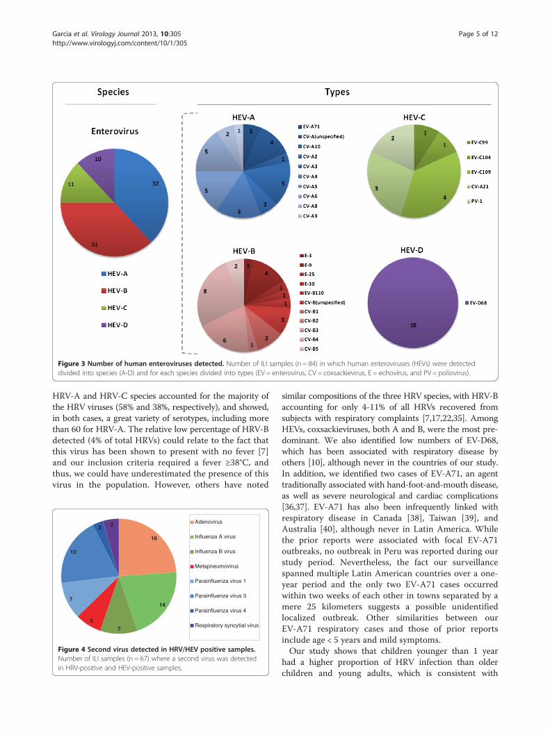
Figure 3 Number of human enteroviruses detected. Number of ILI samples (n = 84) in which human enteroviruses (HEVs) were detecteddivided into species (A-D) and for each species divided into types (EV = enterovirus, CV = coxsackievirus, E = echovirus, and PV = poliovirus).
Garcia et al. Virology Journal 2013, 10:305 Page 5 of 12http://www.virologyj.com/content/10/1/305
HRV-A and HRV-C species accounted for the majority ofthe HRV viruses (58% and 38%, respectively), and showed,in both cases, a great variety of serotypes, including morethan 60 for HRV-A. The relative low percentage of HRV-Bdetected (4% of total HRVs) could relate to the fact thatthis virus has been shown to present with no fever [7]and our inclusion criteria required a fever ≥38°C, andthus, we could have underestimated the presence of thisvirus in the population. However, others have noted
16
14
7
5
7
13
2 3 Adenovirus
Influenza A virus
Influenza B virus
Metapneumovirus
Parainfluenza virus 1
Parainfluenza virus 3
Parainfluenza virus 4
Respiratory syncytial virus
Figure 4 Second virus detected in HRV/HEV positive samples.Number of ILI samples (n = 67) where a second virus was detectedin HRV-positive and HEV-positive samples.
similar compositions of the three HRV species, with HRV-Baccounting for only 4-11% of all HRVs recovered fromsubjects with respiratory complaints [7,17,22,35]. AmongHEVs, coxsackieviruses, both A and B, were the most pre-dominant. We also identified low numbers of EV-D68,which has been associated with respiratory disease byothers [10], although never in the countries of our study.In addition, we identified two cases of EV-A71, an agenttraditionally associated with hand-foot-and-mouth disease,as well as severe neurological and cardiac complications[36,37]. EV-A71 has also been infrequently linked withrespiratory disease in Canada [38], Taiwan [39], andAustralia [40], although never in Latin America. Whilethe prior reports were associated with focal EV-A71outbreaks, no outbreak in Peru was reported during ourstudy period. Nevertheless, the fact our surveillancespanned multiple Latin American countries over a one-year period and the only two EV-A71 cases occurredwithin two weeks of each other in towns separated by amere 25 kilometers suggests a possible unidentifiedlocalized outbreak. Other similarities between ourEV-A71 respiratory cases and those of prior reportsinclude age < 5 years and mild symptoms.Our study shows that children younger than 1 year
had a higher proportion of HRV infection than olderchildren and young adults, which is consistent with
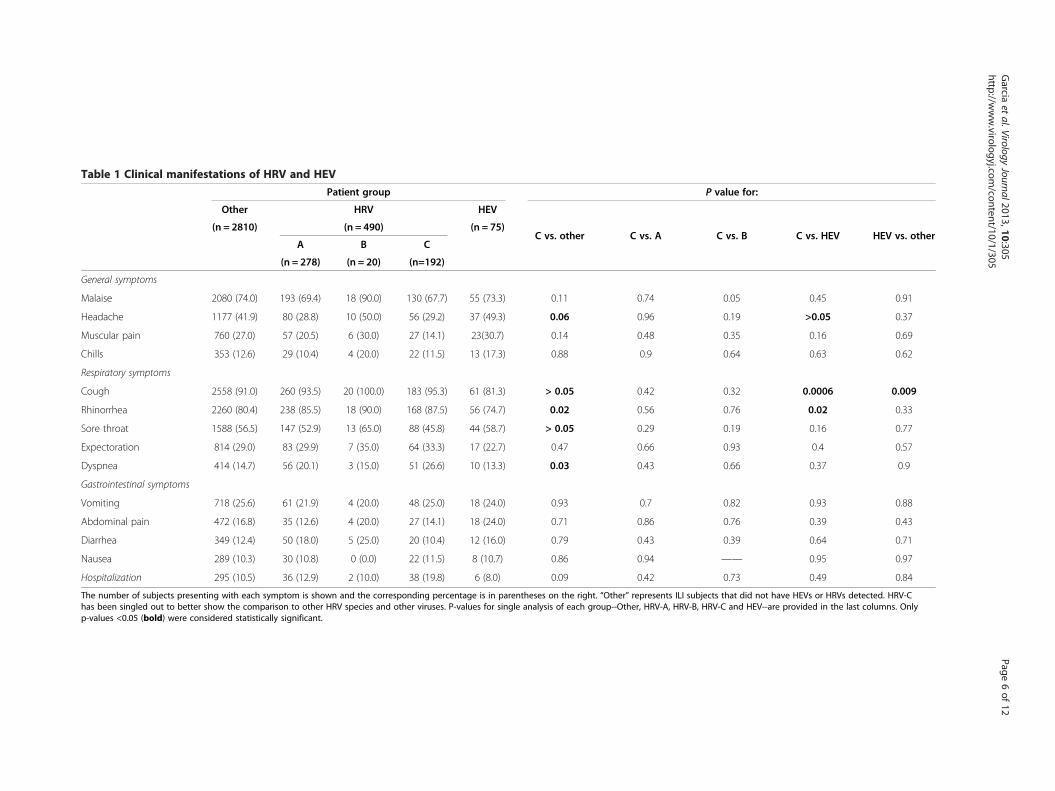
Table 1 Clinical manifestations of HRV and HEV
Patient group P value for:
Other HRV HEV
C vs. other C vs. A C vs. B C vs. HEV HEV vs. other(n = 2810) (n = 490) (n = 75)
A B C
(n = 278) (n = 20) (n=192)
General symptoms
Malaise 2080 (74.0) 193 (69.4) 18 (90.0) 130 (67.7) 55 (73.3) 0.11 0.74 0.05 0.45 0.91
Headache 1177 (41.9) 80 (28.8) 10 (50.0) 56 (29.2) 37 (49.3) 0.06 0.96 0.19 >0.05 0.37
Muscular pain 760 (27.0) 57 (20.5) 6 (30.0) 27 (14.1) 23(30.7) 0.14 0.48 0.35 0.16 0.69
Chills 353 (12.6) 29 (10.4) 4 (20.0) 22 (11.5) 13 (17.3) 0.88 0.9 0.64 0.63 0.62
Respiratory symptoms
Cough 2558 (91.0) 260 (93.5) 20 (100.0) 183 (95.3) 61 (81.3) > 0.05 0.42 0.32 0.0006 0.009
Rhinorrhea 2260 (80.4) 238 (85.5) 18 (90.0) 168 (87.5) 56 (74.7) 0.02 0.56 0.76 0.02 0.33
Sore throat 1588 (56.5) 147 (52.9) 13 (65.0) 88 (45.8) 44 (58.7) > 0.05 0.29 0.19 0.16 0.77
Expectoration 814 (29.0) 83 (29.9) 7 (35.0) 64 (33.3) 17 (22.7) 0.47 0.66 0.93 0.4 0.57
Dyspnea 414 (14.7) 56 (20.1) 3 (15.0) 51 (26.6) 10 (13.3) 0.03 0.43 0.66 0.37 0.9
Gastrointestinal symptoms
Vomiting 718 (25.6) 61 (21.9) 4 (20.0) 48 (25.0) 18 (24.0) 0.93 0.7 0.82 0.93 0.88
Abdominal pain 472 (16.8) 35 (12.6) 4 (20.0) 27 (14.1) 18 (24.0) 0.71 0.86 0.76 0.39 0.43
Diarrhea 349 (12.4) 50 (18.0) 5 (25.0) 20 (10.4) 12 (16.0) 0.79 0.43 0.39 0.64 0.71
Nausea 289 (10.3) 30 (10.8) 0 (0.0) 22 (11.5) 8 (10.7) 0.86 0.94 —— 0.95 0.97
Hospitalization 295 (10.5) 36 (12.9) 2 (10.0) 38 (19.8) 6 (8.0) 0.09 0.42 0.73 0.49 0.84
The number of subjects presenting with each symptom is shown and the corresponding percentage is in parentheses on the right. “Other” represents ILI subjects that did not have HEVs or HRVs detected. HRV-Chas been singled out to better show the comparison to other HRV species and other viruses. P-values for single analysis of each group--Other, HRV-A, HRV-B, HRV-C and HEV--are provided in the last columns. Onlyp-values <0.05 (bold) were considered statistically significant.
Garcia
etal.Virology
Journal2013,10:305Page
6of
12http://w
ww.virologyj.com
/content/10/1/305
% o
f HR
V a
nd H
EV
infe
ctio
n (P
ositi
ve s
ampl
e / t
otal
col
lect
ed s
ampl
es p
er r
egio
n)
Tot
al s
ampl
es c
olle
cted
per
reg
ion
Central America Region
Northern Tropical Region
Coastal Desert Region
Tropical ForestRegion
Andean HighlandsRegion
Southern Region
0
40
80
120
0
15
30
45
60
0
40
80
120
0
15
30
45
60
0
40
80
120
0
15
30
45
60
0
40
80
120
0
15
30
45
60
0
40
80
120
0
15
30
45
60
2010 2011
HEV
HRV-C
HRV-B
HRV-A
Total ILIRainy Season
Sep
Oct
No
v
Dec Ja
n
Feb
Mar
Ap
r
May Ju
n
Jul
Au
g
Sep
Figure 5 Temporal distribution of HRV and HEV infections by region. The percentage of viral infections (positive samples/total collected ILIsamples per region in Figure 4) detected monthly is shown for HEV and each HRV species. The total ILI samples collected per month in eachregion is represented by the continuous purple line. The rainy season (RS) is depicted by a red dotted line.
Garcia et al. Virology Journal 2013, 10:305 Page 7 of 12http://www.virologyj.com/content/10/1/305
HRV-C
HRV-A
HRV-B
HEV-A
HEV-D
HEV-C
HEV-B
0.3
Coding VP4/VP2Region
73100
98
100100
889899
100
5’ UTR Region
0.3
HRV-C.II
HRV-C.III
100
HRV-C.I
HRV-C.IV
91
100100
100
10085
100
LEGENDHRV-A Rhinovirus AHRV-B Rhinovirus BHRV-C Rhinovirus CHEV-A Enterovirus AHEV-B Enterovirus BHEV-C Enterovirus CHEV-D Enterovirus D
Figure 6 Phylogenetic analyses of HRV and HEV. This illustrates that recombination events are much more prevalent in the untranslated regions(UTRs) compared with a translated region (VP4/VP2). Separate alignments of the coding (VP4/VP2; 464 nt) and untranslated (5’UTR; 555 nt) regions’sequences were constructed using MUSCLE v.3.8.31. Maximum likelihood phylogenetic trees were inferred separately for the non-coding and codingregions using PhyML v.3.0 using a general-reversible substitution model with gamma-distributed among-site rate variability. Samples are labeled byfollowing format: “Sample code / Country of collection / Month- Year of collection.” Phylogenetic trees were colored by HRV species: HRV-A, HRV-B,HRV-C, and four types of HEV (HEV-A, HEV-B, HEV-C and HEV-D). In addition, four HRV-C clades show different recombination events and these aredenoted individually as HRV-C.I to IV.
Garcia et al. Virology Journal 2013, 10:305 Page 8 of 12http://www.virologyj.com/content/10/1/305
previous studies [7,19,41]. Children 5 years or youngerhad a slightly higher risk of infection by HRV-C com-pared with those older than 5. However, no differencesin the prevalence among age groups were detected forHEVs throughout our network.A second virus was detected in approximately 11% of
HRV-positive samples, much lower than a rate (37%)reported by others [23]. We used two methods of detec-tion, PCR for influenza viruses and cell culture for all
other viruses (including influenza virus), perhaps allowingfor higher identification proportions of influenza and mostlikely underestimating the number of secondary infec-tions. However, we did not observe more severe symp-toms in patients with another respiratory virus identified,a finding that is consistent with what has been reportedpreviously [23].Several studies have suggested differences in illness
severity between HRV species, including a specific
Table 2 HRV-C clades 5’UTR phylogenetic analysis
Clade % of Total HRV Csamples
Genbank accessionnumber
Country ofcollection
Year ofcollection
Strainidentifyer
Reference
I 29 GQ223228 China 2007 N10 Huang et al. (2009) [17]
II 24 EF077280 USA 2003 NAT045 Kistler et al. (2007) [27]
III 18 AB683895 Phillipines 2011 Fuji et al. (2011) [6]
IV 29 JN990702 USA 2009 26 Lau et al. (2007) [28]
The 5’UTR phylogenetic tree enable us to define four clades of HRV-C serotype (in Figure 6). The percentage of HRV-C samples included in each clade is shown.For each clade, we included one previously published isolate that is most representative of each clade, which allowed placement of known isolates like theNAT045 isolate in clade HRV-C.II and the Antwerp HRV 98/99 isolate in clade HRV-C.IV to better understand the variability of the HRV-C strains and to compare toother typing proposals [29]. Each GenBank isolate accession number, country of collection and year of collection, strain identifier, and the reference manuscriptare shown.
Garcia et al. Virology Journal 2013, 10:305 Page 9 of 12http://www.virologyj.com/content/10/1/305
association of HRV-C with wheezing and asthma exacer-bation [6,7,35]. In agreement with these studies, ourresults showed a higher frequency of dyspnea in subjectswith HRV-C compared with other viruses; in addition,we noted an association of HRV-C detection with pre-existing respiratory diseases, in particular with asthma.Our phylogenetic analyses showed the presence of
almost all known serotypes of HRV-A and HRV-B, aswell as the presence of HRV-C. HRV-C variants are notyet formally classified into types, although there havebeen some proposed typing methods [29]. We show thatrecombinant events occur at the 5’UTR [8] consistentwith others who reported picornaviruses having recom-bination breakpoints restricted to non-structural regionsof their genome [42]. We showed that the circulatingHRV-C recombinants accounted for the majority of allHRV-C species, illustrating how much this virus variesover a very short period of time.Limitations of our study include a lack of uniformity
in samples collected among countries and the fact thatwe collected samples over a period of only one year,preventing definitive characterization of the seasonalityof HRV and HEV in Latin America. Although an analysisspanning three to five years would have been ideal, weanalyzed the temporal distribution of the three HRV spe-cies and HEVs by aggregating collection sites by theirclimatic and geographic similarities. As has been pre-viously described in other continents [13,43], HRVs andHEVs showed a similar “year-long” temporal distributionthroughout Central and South America during the one-year period of collection. We did not detect any sea-sonality for specific HRV-A or HRV-B species, althoughHRV-C species seemed to possess opposite seasonaltrends on either side of the equator. Others have notedseasonal differences among the individual HRV species,including high rates of HRV-A in April in the U.S. [35]and low rates in summer in China [41], high rates ofHRV-B during winter in Australia [40] and China [41],and high rates of HRV-C in October in the U.S. [35] andwinter in China [17]; like ours, these studies collectedsamples for only a one or two-year time span. Anotherimportant limitation of our study was that the
identification of HRV and HEV did not unequivocallyimply causality, a factor that would have been obviatedwith the inclusion of age-matched asymptomatic controls.Although we are not the first to demonstrate a high
prevalence of HRV in children with ILI, our results pro-vide new perspectives into this virus’s global reach andadditional insight into its clinical and phylogenetic charac-teristics in this underreported area of the world. Whileproven preventive and treatment strategies exist for othercommon respiratory viruses, including respiratory syn-cytial virus (pharmacotherapy, passive immunization),adenovirus (active immunization), and influenza virus(pharmacotherapy, active immunization), no HRV vaccineor antiviral currently exists [44,45]. Our high prevalenceof HRV and HEV identification in young Latin Americansindicates that pharmacotherapy against influenza virus orthe common bacterial etiologies of respiratory diseaseshould not be presumptive, but guided by diagnostic con-firmation when available in this population. Despite thecurrent limitations of rapid diagnosis and clinical manage-ment of HRV or HEV respiratory disease, we hope thatsurveillance such as ours will provide further impetus forthe development of rapid diagnostic methods, vaccines,and antivirals, as well as further elucidation of what popu-lations are most at risk and what types of HRV and HEVspecies are most worth targeting.
MethodsEthicsThis protocol was approved as less than minimal risk re-search by the Naval Medical Research Center (NMRC),Silver Spring, Maryland. Institutional Review Board (IRB;Protocol NMRCD.2002.0019) in compliance with all ap-plicable federal regulations governing the protection ofhuman subjects. Authorization was given to perform thestudy using an information sheet approved and stampedby the IRB. As this was part of clinical care and routinesurveillance benefiting the ministries of health, verbal con-sent was obtained from all participants. This method ofconsent was accepted by the NMRC IRB as well as byeach of the institutions involved.
Garcia et al. Virology Journal 2013, 10:305 Page 10 of 12http://www.virologyj.com/content/10/1/305
Specimen and data collectionFrom September 2010 to September 2011, in colla-boration with eight Central and South American coun-tries’ ILI passive surveillance networks (Nicaragua, ElSalvador, Venezuela, Colombia, Ecuador, Peru, Paraguayand Argentina; Figure 1), nasopharyngeal swabs were col-lected from 3,375 subjects with ILI presenting to out-patient medical clinics in 21 cities, as described previously[46-49]. All participants were 25 years old or younger, hada fever (≥38°C), and either a cough or sore throat [50].Once collected, swabs were placed in viral transport mediaand stored at −80°C until they were transported on dry iceto the Naval Medical Research Unit No. 6 (NAMRU-6)facilities in Lima, Peru. Every site used an identical casereport form. This form collected basic epidemiologicinformation and symptoms prior to presentation.
DNA extraction and PCRWe extracted and amplified viral RNA from 140 μl of theviral transport media using a viral RNA kit (QIAamp,Qiagen®). This was performed in 22 μl of reaction mixtureconsisting of 2.2 μl of nuclease-free water, 3 μl of MgSO4,
(5.0 ×), 15 μl of 2× reaction mix (Super Script III One-Step RT-PCR System) with platinum (Taq High Fidelitykit), 0.6 μl 20 μM concentrations of the sense and anti-sense primers, 0.6 μl Enzyme Mix, and 8 μl of template.HRV and HEV detection was performed by semi-nestedreverse transcription–PCR (RT-PCR) targeting the 5’UTRand a partial sequence of the VP4/VP2 genes includingthe nucleotides 165–1079 of the genomic RNA. We usedP1-1 HRV (CAAGCACTTCTGTYWCCCC) and 9565_RHRV (GCATCNGGYARYTTCCACCACCAICC) [12].The amplification was carried out in a thermocycler7700 (Applied Bioystems, Foster City, CA). Cyclingconditions included a reverse transcription step at 50°Cfor 30 min and 95°C for 15 min followed by 45 PCRcycles: 94°C for 30 seconds; 55°C for 30 seconds; 72°C for90 sec; and final incubation for 72°C for 10 min. The amp-lified products of 915 bp were analyzed by electrophoresison an agarose 2% gel. PCR products were purified withCentri-Seps Columns (Princeton Separations). Purifiedproducts were directly used for sequencing of viral nucleicacids from clinical specimens.
Sequencing and phylogenetic analysesThe 5’UTR and VP4/VP2 coding region of all HRV andHEV positive samples obtained (n = 632) were sequencedand included in the following phylogenetic analyses.These regions were selected for sequencing becauseVP4/VP2 is the most commonly studied region of HRVand the 5’UTR regions have been shown to recombinefrequently. For direct sequencing of viral nucleic acidsfrom clinical specimens, gene fragments were amplifiedand sequenced with the use of a Big Dye terminator cycle
sequencing kit (version 3.1, Applied Biosystems) and in-ternal primers Generic F HRV (AGCCTGCGTGGCKGCC) and NCR2 HRV (ACTACTTTGGGTGTCCGTGTTTC) on a Genetic Analyser system (version 3130xL,Applied Biosystems).Separate alignments of the coding (VP4/VP2; 464 nt)
and non-coding (555 nt) regions’ sequences were con-structed using MUSCLE v.3.8.31 [51]. Maximum like-lihood phylogenetic trees were inferred separately forthe non-coding and coding regions using PhyML v.3.0using a general-reversible substitution model with gamma-distributed among-site rate variability [52]. Phylogenetictrees were visualized in FigTree v1.3.1 and colored byHRV species: HRV-A, HRV-B, HRV-C, and four clades ofHEV (HEV-A, HEV-B, HEV-C, and HEV-D). Completesequences (≈1,000 nt) were assembled, aligned, and editedusing Sequencer (version 4.8 – Gene Codes Corporation,Ann Arbor, MI, USA) and BioEdit (version 7.0.0 -IsisPharmaceuticals, Inc., Dublin, Ireland) software. Phyloge-netic trees were generated with CLUSTAL X version 2.0.1and MEGA version 3.1 software [53]. Fifty-eight HRV and34 HEV sequences, representing all different clades foundduring the analysis were submitted to GenBank and theiraccession numbers are: JX129393 - JX129484. The RDP,BOOTSCAN, and GENECONV algorithms available inthe Recombination Detection Program (RDP3, available athttp://web.cbio.uct.ac.za/~darren/rdp.html) were used toidentify recombinant viruses within the coding and 5’UTRregions, separately.
Detection of other respiratory virusesAll HRV- and HEV-positive samples underwent viralisolation by inoculation into four cell lines: MDCK,Vero76, VeroE6 and LLCMK2 cells (ATCC, Manassas,VA 20108), as previously reported [54]. After ten days ofculture, the cells were spotted onto microscope slides.Cell suspensions were dried and fixed in chilled acetonefor 15 minutes. Immunofluorescence assay was per-formed using the Respiratory Virus Screening and Iden-tification Kit (D3 DFA Respiratory Virus DiagnosticHybrids; Athens, OH) for the identification of adeno-viruses, influenza A and B viruses, parainfluenza viruses(types 1, 2, 3 and 4), and respiratory syncytial virus.In addition, real time-PCR for influenza viruses was
performed on all HRV- and HEV-positive samples as pre-viously described [49]. This was performed as part of theroutine sample processing of the respiratory surveillancenetwork at NAMRU-6.
Statistical analysesData was entered into a database using Microsoft Accessand analyzed using Stata/SE 10.0 for Windows (StataCorpLP, College Station, TX). Two proportion z-tests wereused to compare proportions; p-values ≤0.05 were
Garcia et al. Virology Journal 2013, 10:305 Page 11 of 12http://www.virologyj.com/content/10/1/305
considered statistically significant; 95% confidence inter-vals (C.I.) were calculated for each odd ratio (O.R.), andassociations were assessed using Pearson’s chi-square (×2)or Fisher’s tests.
Competing interestsNone of the authors has a financial or personal conflict of interest related tothis study. The corresponding author had full access to all data in the studyand final responsibility for the decision to submit this publication.
Authors’ contributionsJG and TJK contributed in the design and conception of the study. VE, MSand MVV performed the analysis of the data. JG, ESH and MN performed theanalysis and interpretation of data. JG drafted and submitted the manuscript.JG, MB, FS, GC, AEA, NA, ILR, WC, MJ, WA, FR and MG contributed with theacquisition of data. All authors contributed with the critical revision and finalapproval of manuscript.
Copyright statementAuthors Tadeusz J. Kochel and Eric S. Halsey are military service membersand Josefina Garcia, Manuel Villaran, and Victoria Espejo are employees ofthe U.S. Government. This work was prepared as part of their official duties.Title 17 U.S.C. § 105 provides that ‘Copyright protection under this title is notavailable for any work of the United States Government’. Title 17 U.S.C. § 101defines a U.S. Government work as a work prepared by a military servicemembers or employees of the U.S. Government as part of those person’sofficial duties.
DisclaimerThe views expressed in this article are those of the authors and do notnecessarily reflect the official policy or position of the Department of theNavy, Department of Defense, nor the U.S. Government.
FundingThis study was funded by the United States Department of Defense GlobalEmerging Infections Systems Research Program, WORK UNIT NUMBER:847705.82000.25GB.B0016.
Author details1US Naval Medical Research Unit 6, Lima, Peru. 2Fogarty International Center,National Institutes of Health, Bethesda, MD, USA. 3Dirección General deEpidemiología, Ministerio de Salud, Lima, Perú. 4Hospital Solano, BuenosAires, Argentina. 5Hospital Infantil Manuel de Jesus Rivera, Managua,Nicaragua. 6LARDIDEV-Biomed-UC, Maracay, Venezuela. 7Universidad deAntioquia, Medellín, Colombia. 8ONG Rayos de Sol, Asuncion, Paraguay.9Universidad Nacional Autónoma de Honduras, Tegucigalpa, Honduras.10Hospital Vozandes and Universidad de las Americas, Quito, Ecuador.11Hospital Nacional de Metapan, Metapan, El Salvador. 12Clinica Alcivar andHospital Vernaza, Guayaquil, Ecuador. 13Hospital Departamental HumbertoAlvarado de Masaya, Masaya, Managua, Nicaragua. 14LaboratorioDepartamental, Secretaria Seccional de Salud del Meta, Villavicencio,Colombia. 15US Naval Medical Research Center, Silver Spring, MD, USA.
Received: 22 March 2013 Accepted: 31 July 2013Published: 11 October 2013
References1. Lozano R, Naghavi M, Foreman K, Lim S, Shibuya K, Aboyans V, Abraham J,
Adair T, Aggarwal R, Ahn SY, et al: Global and regional mortality from 235causes of death for 20 age groups in 1990 and 2010: a systematicanalysis for the Global Burden of Disease Study 2010. Lancet 2012,380:2095–2128.
2. Bryce J, Boschi-Pinto C, Shibuya K, Black RE: WHO estimates of the causesof death in children. Lancet 2005, 365:1147–1152.
3. Hayden FG: Rhinovirus and the lower respiratory tract. Rev Med Virol 2004,14:17–31.
4. Tapparel C, Junier T, Gerlach D, Cordey S, Van Belle S, Perrin L, Zdobnov EM,Kaiser L: New complete genome sequences of human rhinoviruses shedlight on their phylogeny and genomic features. BMC Genomics 2007,8:224.
5. Lewis-Rogers N, Crandall KA: Evolution of Picornaviridae: an examinationof phylogenetic relationships and cophylogeny. Mol Phylogenet Evol 2010,54:995–1005.
6. Fuji N, Suzuki A, Lupisan S, Sombrero L, Galang H, Kamigaki T, Tamaki R,Saito M, Aniceto R, Olveda R, Oshitani H: Detection of human rhinovirus Cviral genome in blood among children with severe respiratory infectionsin the Philippines. PLoS One 2011, 6:e27247.
7. Iwane MK, Prill MM, Lu X, Miller EK, Edwards KM, Hall CB, Griffin MR,Staat MA, Anderson LJ, Williams JV, et al: Human rhinovirus speciesassociated with hospitalizations for acute respiratory illness in young USchildren. J Infect Dis 2011, 204:1702–1710.
8. Savolainen-Kopra C, Blomqvist S, Smura T, Roivainen M, Hovi T, Kiang D,Kalra I, Yagi S, Louie JK, Boushey H, et al: 5’ noncoding region alone doesnot unequivocally determine genetic type of human rhinovirus strains.J Clin Microbiol 2009, 47:1278–1280.
9. Hicks LA, Shepard CW, Britz PH, Erdman DD, Fischer M, Flannery BL, Peck AJ,Lu X, Thacker WL, Benson RF, et al: Two outbreaks of severe respiratorydisease in nursing homes associated with rhinovirus. J Am Geriatr Soc2006, 54:284–289.
10. Linsuwanon P, Puenpa J, Suwannakarn K, Auksornkitti V, Vichiwattana P,Korkong S, Theamboonlers A, Poovorawan Y: Molecular epidemiology andevolution of human enterovirus serotype 68 in Thailand, 2006–2011.PLoS One 2012, 7:e35190.
11. Oberste MS, Maher K, Schnurr D, Flemister MR, Lovchik JC, Peters H,Sessions W, Kirk C, Chatterjee N, Fuller S, et al: Enterovirus 68 is associatedwith respiratory illness and shares biological features with both theenteroviruses and the rhinoviruses. J Gen Virol 2004, 85:2577–2584.
12. Laine P, Savolainen C, Blomqvist S, Hovi T: Phylogenetic analysis of humanrhinovirus capsid protein VP1 and 2A protease coding sequencesconfirms shared genus-like relationships with human enteroviruses.J Gen Virol 2005, 86:697–706.
13. Arden KE, McErlean P, Nissen MD, Sloots TP, Mackay IM: Frequentdetection of human rhinoviruses, paramyxoviruses, coronaviruses, andbocavirus during acute respiratory tract infections. J Med Virol 2006,78:1232–1240.
14. Briese T, Renwick N, Venter M, Jarman RG, Ghosh D, Kondgen S,Shrestha SK, Hoegh AM, Casas I, Adjogoua EV, et al: Global distribution ofnovel rhinovirus genotype. Emerg Infect Dis 2008, 14:944–947.
15. Khetsuriani N, Lu X, Teague WG, Kazerouni N, Anderson LJ, Erdman DD:Novel human rhinoviruses and exacerbation of asthma in children.Emerg Infect Dis 2008, 14:1793–1796.
16. Lamson D, Renwick N, Kapoor V, Liu Z, Palacios G, Ju J, Dean A, St George K,Briese T, Lipkin WI: MassTag polymerase-chain-reaction detection ofrespiratory pathogens, including a new rhinovirus genotype, that causedinfluenza-like illness in New York State during 2004–2005. J Infect Dis2006, 194:1398–1402.
17. Huang T, Wang W, Bessaud M, Ren P, Sheng J, Yan H, Zhang J, Lin X,Wang Y, Delpeyroux F, Deubel V: Evidence of recombination and geneticdiversity in human rhinoviruses in children with acute respiratoryinfection. PLoS One 2009, 4:e6355.
18. Renwick N, Schweiger B, Kapoor V, Liu Z, Villari J, Bullmann R, Miething R,Briese T, Lipkin WI: A recently identified rhinovirus genotype is associatedwith severe respiratory-tract infection in children in Germany. J Infect Dis2007, 196:1754–1760.
19. Tapparel C, Junier T, Gerlach D, Van-Belle S, Turin L, Cordey S, Muhlemann K,Regamey N, Aubert JD, Soccal PM, et al: New respiratory enterovirus andrecombinant rhinoviruses among circulating picornaviruses. Emerg InfectDis 2009, 15:719–726.
20. Wisdom A, Kutkowska AE, McWilliam Leitch EC, Gaunt E, Templeton K,Harvala H, Simmonds P: Genetics, recombination and clinical features ofhuman rhinovirus species C (HRV-C) infections; interactions of HRV-Cwith other respiratory viruses. PLoS One 2009, 4:e8518.
21. Monto AS: A community study of respiratory infections in the tropics. 3.Introduction and transmission of infections within families.Am J Epidemiol 1968, 88:69–79.
22. Moreira LP, Kamikawa J, Watanabe AS, Carraro E, Leal E, Arruda E,Granato CF, Bellei NC: Frequency of human rhinovirus species inoutpatient children with acute respiratory infections at primary carelevel in Brazil. Pediatr Infect Dis J 2011, 30:612–614.
23. Paula NT, Carneiro BM, Yokosawa J, Freitas GR, Oliveira TF, Costa LF,Silveira HL, Queiroz DA: Human rhinovirus in the lower respiratory tract
Garcia et al. Virology Journal 2013, 10:305 Page 12 of 12http://www.virologyj.com/content/10/1/305
infections of young children and the possible involvement of asecondary respiratory viral agent. Mem Inst Oswaldo Cruz 2011,106:316–321.
24. Pitrez PM, Stein RT, Stuermer L, Macedo IS, Schmitt VM, Jones MH, Arruda E:[Rhinovirus and acute bronchiolitis in young infants]. J Pediatr (Rio J)2005, 81:417–420.
25. Souza LS, Ramos EA, Carvalho FM, Guedes VM, Rocha CM, Soares AB,Velloso Lde F, Macedo IS, Moura FE, Siqueira M, et al: Viral respiratoryinfections in young children attending day care in urban NortheastBrazil. Pediatr Pulmonol 2003, 35:184–191.
26. Thwing CJ, Arruda E, Vieira Filho JP, Castelo Filho A, Gwaltney JM Jr:Rhinovirus antibodies in an isolated Amazon Indian tribe. Am J Trop MedHyg 1993, 48:771–775.
27. Kistler AL, Webster DR, Rouskin S, Magrini V, Credle JJ, Schnurr DP,Boushey HA, Mardis ER, Li H, DeRisi JL: Genome-wide diversity andselective pressure in the human rhinovirus. Virol J 2007, 4:40.
28. Lau SK, Yip CC, Tsoi HW, Lee RA, So LY, Lau YL, Chan KH, Woo PC, Yuen KY:Clinical features and complete genome characterization of a distincthuman rhinovirus (HRV) genetic cluster, probably representing apreviously undetected HRV species, HRV-C, associated with acuterespiratory illness in children. J Clin Microbiol 2007, 45:3655–3664.
29. Simmonds P, McIntyre C, Savolainen-Kopra C, Tapparel C, Mackay IM, Hovi T:Proposals for the classification of human rhinovirus species C intogenotypically assigned types. J Gen Virol 2010, 91:2409–2419.
30. Laguna-Torres VA, Gomez J, Aguilar PV, Ampuero JS, Munayco C, Ocana V,Perez J, Gamero ME, Arrasco JC, Paz I, et al: Changes in the viraldistribution pattern after the appearance of the novel influenza A H1N1(pH1N1) virus in influenza-like illness patients in Peru. PLoS One 2010,5:e11719.
31. Valero N, Larreal Y, Arocha F, Gotera J, Mavarez A, Bermudez J, Moran M,Maldonado M, Marina Espina L: [Viral etiology of acute respiratoryinfections]. Invest Clin 2009, 50:359–368.
32. Galiano M, Videla C, Puch SS, Martinez A, Echavarria M, Carballal G:Evidence of human metapneumovirus in children in Argentina. J MedVirol 2004, 72:299–303.
33. Gray GC, Capuano AW, Setterquist SF, Sanchez JL, Neville JS, Olson J,Lebeck MG, McCarthy T, Abed Y, Boivin G: Human metapneumovirus,Peru. Emerg Infect Dis 2006, 12:347–350.
34. Herrera-Rodriguez DH, de la Hoz F, Marino C, Ramirez E, Lopez JD, Velez C:[Adenovirus in children under five years of age. Circulation patterns andclinical and epidemiological characteristics in Colombia, 1997–2003].Rev Salud Publica (Bogota) 2007, 9:420–429.
35. Miller EK, Edwards KM, Weinberg GA, Iwane MK, Griffin MR, Hall CB,Zhu Y, Szilagyi PG, Morin LL, Heil LH: A novel group of rhinoviruses isassociated with asthma hospitalizations. J Allergy Clin Immunol 2009,123:98–104 e101.
36. Huang WC, Huang LM, Kao CL, Lu CY, Shao PL, Cheng AL, Fan TY, Chi H,Chang LY: Seroprevalence of enterovirus 71 and no evidence ofcrossprotection of enterovirus 71 antibody against the otherenteroviruses in kindergarten children in Taipei city. J Microbiol ImmunolInfect 2012, 45:96–101.
37. Kim KH: Enterovirus 71 infection: An experience in Korea, 2009. Korean JPediatr 2010, 53:616–622.
38. Merovitz L, Demers AM, Newby D, McDonald J: Enterovirus 71 infectionsat a Canadian center. Pediatr Infect Dis J 2000, 19:755–757.
39. Tsai HP, Kuo PH, Liu CC, Wang JR: Respiratory viral infections amongpediatric inpatients and outpatients in Taiwan from 1997 to 1999.J Clin Microbiol 2001, 39:111–118.
40. Kennett ML, Birch CJ, Lewis FA, Yung AP, Locarnini SA, Gust ID: Enterovirustype 71 infection in Melbourne. Bull World Health Organ 1974, 51:609–615.
41. Jin Y, Yuan XH, Xie ZP, Gao HC, Song JR, Zhang RF, Xu ZQ, Zheng LS,Hou YD, Duan ZJ: Prevalence and clinical characterization of a newlyidentified human rhinovirus C species in children with acute respiratorytract infections. J Clin Microbiol 2009, 47:2895–2900.
42. Lukashev AN: Role of recombination in evolution of enteroviruses.Rev Med Virol 2005, 15:157–167.
43. Monto AS: Epidemiology of viral respiratory infections. Am J Med 2002,112(Suppl 6A):4S–12S.
44. Laconi S, Madeddu MA, Pompei R: Study of the biological activity of novelsynthetic compounds with antiviral properties against humanrhinoviruses. Molecules 2011, 16:3479–3487.
45. Rohde GG: Rhinovirus vaccination: the case in favour. Eur Respir J 2011, 37:3–4.46. Comach G, Teneza-Mora N, Kochel TJ, Espino C, Sierra G, Camacho DE,
Laguna-Torres VA, Garcia J, Chauca G, Gamero ME, et al: Sentinelsurveillance of influenza-like illness in two hospitals in Maracay,Venezuela: 2006–2010. PLoS One 2012, 7:e44511.
47. Douce RW, Aleman W, Chicaiza-Ayala W, Madrid C, Sovero M, Delgado F,Rodas M, Ampuero J, Chauca G, Perez J, et al: Sentinel surveillance ofinfluenza-like-illness in two cities of the tropical country of Ecuador:2006–2010. PLoS One 2011, 6:e22206.
48. Laguna-Torres VA, Gomez J, Ocana V, Aguilar P, Saldarriaga T, Chavez E,Perez J, Zamalloa H, Forshey B, Paz I, et al: Influenza-like illness sentinelsurveillance in Peru. PLoS One 2009, 4:e6118.
49. Laguna-Torres VA, Sanchez-Largaespada JF, Lorenzana I, Forshey B,Aguilar P, Jimenez M, Parrales E, Rodriguez F, Garcia J, Jimenez I, et al:Influenza and other respiratory viruses in three Central Americancountries. Influenza Other Respi Viruses 2011, 5:123–134.
50. Ortiz JR, Sotomayor V, Uez OC, Oliva O, Bettels D, McCarron M, Bresee JS,Mounts AW: Strategy to enhance influenza surveillance worldwide.Emerg Infect Dis 2009, 15:1271–1278.
51. Edgar RC: MUSCLE: multiple sequence alignment with high accuracy andhigh throughput. Nucleic Acids Res 2004, 32:1792–1797.
52. Guindon S, Dufayard JF, Lefort V, Anisimova M, Hordijk W, Gascuel O:New algorithms and methods to estimate maximum-likelihoodphylogenies: assessing the performance of PhyML 3.0. Syst Biol 2010,59:307–321.
53. Kumar S, Tamura K, Nei M: MEGA3: Integrated software for MolecularEvolutionary Genetics Analysis and sequence alignment. Brief Bioinform2004, 5:150–163.
54. Sovero M, Garcia J, Kochel T, Laguna-Torres VA, Gomez J, Chicaiza W,Barrantes M, Sanchez F, Jimenez M, Comach G, et al: Circulating strains ofhuman respiratory syncytial virus in Central and South America.PLoS One 2011, 6:e22111.
doi:10.1186/1743-422X-10-305Cite this article as: Garcia et al.: Human rhinoviruses and enterovirusesin influenza-like illness in Latin America. Virology Journal 2013 10:305.
Submit your next manuscript to BioMed Centraland take full advantage of:
• Convenient online submission
• Thorough peer review
• No space constraints or color figure charges
• Immediate publication on acceptance
• Inclusion in PubMed, CAS, Scopus and Google Scholar
• Research which is freely available for redistribution
Submit your manuscript at www.biomedcentral.com/submit