Embed Size (px)
Citation preview

| INVESTIGATION
Human SOD1 ALS Mutations in a Drosophila Knock-InModel Cause Severe Phenotypes and Reveal
Dosage-Sensitive Gain- andLoss-of-Function Components
Aslı Sxahin,* Aaron Held,* Kirsten Bredvik,* Paxton Major,* Toni-Marie Achilli,† Abigail G. Kerson,*
Kristi Wharton,* Geoff Stilwell,† and Robert Reenan*,1
*Department of Molecular Biology, Cellular Biology, and Biochemistry, Brown University, Providence, Rhode Island 02912, and†Department of Biology, Rhode Island College, Providence, Rhode Island 02908
ABSTRACT Amyotrophic Lateral Sclerosis (ALS) is the most common adult-onset motor neuron disease and familial forms can becaused by numerous dominant mutations of the copper-zinc superoxide dismutase 1 (SOD1) gene. Substantial efforts have beeninvested in studying SOD1-ALS transgenic animal models; yet, the molecular mechanisms by which ALS-mutant SOD1 protein acquirestoxicity are not well understood. ALS-like phenotypes in animal models are highly dependent on transgene dosage. Thus, issues ofwhether the ALS-like phenotypes of these models stem from overexpression of mutant alleles or from aspects of the SOD1 mutationitself are not easily deconvolved. To address concerns about levels of mutant SOD1 in disease pathogenesis, we have geneticallyengineered four human ALS-causing SOD1 point mutations (G37R, H48R, H71Y, and G85R) into the endogenous locus of DrosophilaSOD1 (dsod) via ends-out homologous recombination and analyzed the resulting molecular, biochemical, and behavioral phenotypes.Contrary to previous transgenic models, we have recapitulated ALS-like phenotypes without overexpression of the mutant protein.Drosophila carrying homozygous mutations rendering SOD1 protein enzymatically inactive (G85R, H48R, and H71Y) exhibited neuro-degeneration, locomotor deficits, and shortened life span. The mutation retaining enzymatic activity (G37R) was phenotypicallyindistinguishable from controls. While the observed mutant dsod phenotypes were recessive, a gain-of-function component wasuncovered through dosage studies and comparisons with age-matched dsod null animals, which failed to show severe locomotordefects or nerve degeneration. We conclude that the Drosophila knock-in model captures important aspects of human SOD1-basedALS and provides a powerful and useful tool for further genetic studies.
KEYWORDS ALS; SOD1; Drosophila; motor neuron
AMYOTROPHIC Lateral Sclerosis (ALS) is characterizedby progressive loss of upper and lower motor neurons
(MNs) leading to paralysis, and death in afflicted individualswithin 3–5 years after diagnosis, on average. The mecha-nisms of disease pathogenesis leading to the exclusive demiseof MNs remain unclear and there is no effective therapy. Over50 genes have been linked to ALS, suggesting large genetic
heterogeneity of the disease mirroring a diverse range ofclinical presentations (Abel et al. 2012; Sreedharan andBrown 2013; Leblond et al. 2014). The first ALS-associatedmutations were found in the superoxide dismutase 1 (SOD1)gene (Rosen et al. 1993). SOD1 encodes a small protein of153 amino acids (16 kDa), constitutes �1% of the cytoplas-mic protein, and is expressed ubiquitously (Pardo et al.1995). Functional SOD1 is a homodimer and catalyzes theconversion of superoxide radicals to hydrogen peroxide. Re-cent data suggest SOD1 also acts as a transcription factor andupregulates genes involved in oxidative stress response (Huet al. 2009; Tsang et al. 2014; Bunton-Stasyshyn et al. 2015).Strong evidence indicates that SOD1mutations produce toxicgain-of-function properties at the protein level. The vastmajority of the .150 SOD1 mutations identified in patients
Copyright © 2017 by the Genetics Society of Americadoi: 10.1534/genetics.116.190850Manuscript received April 29, 2016; accepted for publication November 13, 2016;published Early Online December 13, 2016.Supplemental material is available online at www.genetics.org/lookup/suppl/doi:10.1534/genetics.116.190850/-/DC1.1Corresponding author: Department of Molecular Biology, Cellular Biology, andBiochemistry, Brown University, Sidney Frank Hall Life Science Bldg., Box G-L372,185 Meeting St., Providence, RI 02912. E-mail: [email protected]
Genetics, Vol. 205, 707–723 February 2017 707

show dominant inheritance patterns, and disease severitycorrelates with aggregation potential of mutant proteinrather than loss of enzymatic activity (Abel et al. 2012;Saccon et al. 2013).
Animalmodels containingmutantALS-associated transgeneshave been important tools for understanding disease pathogen-esis. To date, most transgenic models generated in multiplespecies express mutant human SOD1 (hSOD1) in a geneticbackground containing the endogenous wild-type SOD1 genein rodents (Gurney et al. 1994; Trotti et al. 1999; Kato 2008), inDrosophila (Watson et al. 2008; Bahadorani et al. 2013) and inothermodel organisms (Joyce et al.2011). Themajority of thesemodels recapitulate characteristics of ALS, including progressivemotor deficits, paralysis, MN degeneration, and early lethality(McGoldrick et al. 2013). However, ALS-like phenotypes inthese animals are highly dependent on transgene expressionlevels and severity of phenotypes correlate with level of proteinoverexpression (Gurney et al. 1994; Alexander et al. 2004;Wang et al. 2009a). Furthermore, overexpression of wild-typehSOD1 (hSOD1wt) is sufficient to recapitulate some ALS phe-notypes such as mitochondrial dysfunction, axonal degenera-tion, and premature MN death (Jaarsma et al. 2000; Ezziet al. 2007; Graffmo et al. 2013). These results suggest mutantphenotypes are sensitive to gene dose and/or SOD1 proteinlevels. Extrapolating information gained from these diseasemodels to human ALS is therefore difficult.
While mutations in other genes cause ALS, the pathwaysleading to SOD1-mediated toxicity are still poorly under-stood and it is unclear to what extent pathogenic mecha-nisms are shared between the various forms of familial ALS(fALS). Intriguingly, cytoplasmic SOD1 inclusionshavebeenreported in ALS patients irrespective of SOD1 mutations(Gruzman et al. 2007; Bosco et al. 2010; Forsberg et al.2010), strengthening the hypothesis that there may be acommonmechanism for neurodegeneration in ALS and em-phasizing a critical role for SOD1 regarding the generalpathogenesis of the disease. Uncovering how mutations inSOD1 ultimately lead to the dysfunction and the ultimatedeath of MNs may shed light on how ALS develops andprogresses in all patients, regardless of sporadic or familialmodes of transmission.
To create an ALS model in which mutant SOD1 protein isexpressed at endogenous levels, we have used ends-out ho-mologous recombination (HR) as a gene replacement strategyto introduce relevant ALS-associated mutations at conservedresidues of the Drosophila sod1 (dsod) gene. Similar strategieshave been used in flies to successfully model other diseases(Sun et al. 2012; Schutte et al. 2014). In this study, weshow dsod G85R, H71Y, and H48R mutants exhibit recessiveALS-associated phenotypes including a shortened life span,progressive paralysis, and degeneration of MNs. We also dem-onstrate that aspects of these phenotypes represent a gainof function through comparisons with SOD1 null (dsodnull)alleles. Using Drosophila as a genetically tractable model or-ganism, we have developed an additional tool which can beused to uncover relevant ALS-related cellular responses.
Materials and Methods
Drosophila strains
Drosophila were raised at a constant 25�, on standard corn-meal molasses food and under 12 hr day/night cycles. ThedsodX-39 and dsodG51S lines (n1 or n108) were ordered fromBloomington Drosophila Stock Center (BDSC) (stock num-bers 24492 and 24490, respectively). The names of the dSODmutations are based on the human SOD1 amino acid num-bering system throughout this work. The dsodX-16 line was akind gift from W. C. Orr from Wayne State University.
HR and CRISPR
We performed ends-out HR to create three independent lines ofdsodWTLoxP, dsodG37R, dsodH48R, dsodH71Y, and dsodG85R using pre-viously reported methods (Staber et al. 2011). Briefly, we usedthe ends-out targeting vector p[w25.2] that contains the miniwhitemarker (white+), a selectable red eye color, flanked by LoxPsites for subsequent removal by Cre recombinase (Figure 1). Ho-mology arm 1, corresponding to exon 1 of dsod; and homologyarm 2, corresponding to exon 2 of dsod; were cloned and se-quenced in pTOPO (Life Technologies) and then shuttled intothe multiple cloning sites of the vector to generate p[w25-dsod],which was then introduced into the Drosophila genome bystandard P-element transgenic methods (Genetic Services). Fulltargeting region coordinates were chromosome 3L: 11,103,794–11,108,715 (4922 nt). All targeting was done in a w1118 back-ground, as previously described, and performed in a wild-typechromosome 3 background (Gell and Reenan 2013). A full list ofcloning, mutagenic, and sequencing primers can be found inSupplemental Material, Table S3. Targeting was performed togenerate multiple independent targeting events that incorporate(G37R, H48R, H71Y, and G85R) or exclude engineered muta-tions (WTLoxP). All targeted animals have a LoxP “scar” of 72 nt.
After generating the transgenic flies, white or mosaic-eyedfemales were collected from heat-shocked vials and thencrossed with yw;ey-Flp,nocsco/CyO males and only red-eyedfemale progeny were selected for additional validation. Tar-geted alleles were validated by PCR amplification, using pri-mers outside the region of targeting and primers specific to thewhite+marker. Following recombination, thewhite+minigeneselection cassette was removed via genetic crosses, which in-troducedCre recombinase. All targeted alleleswere sequencedto verify that no unintended mutations were introduced. It isimportant to note here that all of the targeted alleles contain anatural polymorphism at the site 1013 having a C instead of anA, which leads to the N98K missense mutation in humanamino acid numbering system (N96K in Drosophila dSODnumbering system), which is also referred as dsod fast allelein the literature (Phillips et al. 1995). The dsodG51S allele that isgenerated through EMS mutagenesis does not contain thispolymorphism, and it is the dsodslow allele. dsod fast and dsodslow
alleles do not result in a phenotypic change inDrosophila, theyare named after their differential mobility on a native poly-acrylamide gel (Lee et al. 1981; Hudson et al. 1994; Phillipset al. 1995). All three knock-in lines from each line were
708 A. Sxahin et al.

backcrossed to w1118, a white-eyed genetic background, forfive generations. Drosophila stocks were kept as heterozygotesusing third chromosome balancers. Heterozygous dsod allelesthat were used in the experiments were generated by crossingmutant lines to the wild-type line WTLoxP.
To create dsodnull alleles, CRISPR/Cas9 was used to removethe complete dsod ORF (Genetivision, Houston, TX). Primersused to create target sequences were 59CTTCGACGAATTCGCAAGTAGAAT and 59AAACATTCTACTTGCGAATTCGTC for guideRNA 1 (gRNA1); and 59CTTCGGCATTTATTGGGGAATTCC and59AAACGGAATTCCCCAATAAATGCC for gRNA2. Two indepen-dent lines were analyzed and reported herein.
Eclosion assay
For the eclosion percentages, we set up at least 12 vials ofcohorts of 2–3 heterozygous males and virgin females at 25�on standard cornmeal molasses food and under 12 hr day/night cycles. The parents were transferred to a new vial everyday. The heterozygous dsod alleles were balanced over a thirdchromosome balancer: TM3,GFP,ser,w+ (BDSC stock number4534). When the progeny started eclosing, heterozygousprogeny having red eyes due to the balancer were countedand homozygous progeny having white eyes due to lack ofthe balancer which eclosed or stuck in the pupal case werecounted. The progeny carrying homozygous balancers diedearly in development due to a recessive lethal marker on thebalancer chromosome. The progeny from each vial werecounted until the number of homozygous mutants reached200. Then, the eclosion percentages were calculated by (totalnumber of eclosed homozygous adults)/(total number of ho-mozygous pupal cases)3100. The results of 12 vials wereaveraged and Fisher’s exact test was performed.
Survival assay
For the survival assay, parental flies were raised at 25� onstandard cornmeal molasses food and under 12 hr day/night
cycles. The parental flies were collected from population-density-controlled broods (2–3 males and females in eachvial) to avoid any confounding effects due to overcrowding.The parents were allowed to mate and lay eggs for 2 daysbefore being transferred to fresh food twice. Three trials ofsurvival analysis were performed for each genotype. For theheterozygous genotypes, to eliminate maternal effects on thedsod mutant allele, in trial 1 the mutant allele was passedfrom the mother and in trial 2 the mutant allele was passedfrom the father. The third trial was either identical to eitherthe trial 1 cross or the trial 2 cross. The offspring from theseparents were collected over a period of 24 hr and sorted bysex. A total of 12 males and 12 females were kept in vialscontaining standard cornmeal molasses food. For each geno-type, multiple replicate vials were set up so the total samplesize was 200–300 for each sex. Flies were transferred ontofresh food three times a week by blinded undergraduate re-searchers. The number of deaths was recorded. Survival as-says were either performed at 18 or 25�. Once all the flieswere dead, log-rank tests were performed for statisticalanalysis.
H2O2 feeding assay
All chemicals were delivered to Drosophila in instant food(Nutri-Fly Instant, Genesee, 66–118). Instant food is pre-pared based on the manufacturer recipe. For each vial, 2 ginstant food was dissolved in 5 ml Milli-Q water or the chem-ical solution. For each bottle, 21 g instant food was dissolvedin 50 ml Milli-Q water or the chemical solution.
A 2% concentration of hydrogen peroxide (H2O2) was di-luted from 30% stock (Fisher Chemical H325-100) and de-livered to 1-day-posteclosion Drosophila in vials. Each vialcontained 50 males or 50 females. Three trials of H2O2 sur-vival analysis were performed for each genotype and sex. Asin survival analysis, for the heterozygous genotypes to elim-inate the maternal effects on the dsod mutant allele, in trial
Figure 1 ALS-associated mutations introduced into dsod. Mutations G37R, H48R, H71Y, and G85R were introduced into dsod by ends-out HR. (A)Alignment of hSOD1 and dSOD show amino acid changes were at conserved residues. Black bars are mutations linked to familial ALS, orange residuesare conserved between species, gray residues are similarly charged domains between organisms. Asterisk (*) shows the targeted residues. (B) Sequenc-ing chromatograms confirm the presence of targeted mutations. Orange, G37R (GGC/CGC); blue, H48R (CAC/CGC); green, H71Y (CAT/TAC);red, G85R (GGC/CGC). (C) Targeted mutations and the intronic LoxP site did not interfere with alternative splicing of dsod messenger RNA. dsod full-length �500-bp and Gapdh gene-specific primers were used to amplify the region of interest from total RNA isolates.
Drosophila Knock-In Model of ALS 709

one the mutant allele is passed from the mother and in trialtwo the mutant allele is passed from the father. Trial threewas identical either to the trial one cross or the trial two cross.The number of deaths was recorded every 8 hr by blindedundergraduate researchers. Once all the flies were dead, log-rank test was performed for statistical analysis.
Larval motility assay (manual version)
ThelarvalmotilityassaywasperformedaspreviouslydescribedinBatlevi et al. (2010). Larvaewere selected during the wanderingthird instar stage. They were washed in 13 PBS and placed on a1% agarose plate made with 0.5% TBE (100- 3 15-mm petridish) andwere allowed 1min to acclimate. The platewas placedon a1-31-mmsquare grid and the larvaewere allowed to crawlfor 2 min. The total number of squares a larva crossed wascounted by blinded undergraduate researchers. We considereda square to be crossed when the larvae’s posterior end crossed aline and the total number of squares crossed was counted. Dun-nett’s test following one-way ANOVA was used to compare theexperimental group to the wild type.
Larval motility assay (computational version)
In the computational larval motility assay, five larvae wereallowed to crawl on a 22-cm diameter dish filled with 1%agarose in H2O. The larvae were videotaped for 1.5 min andvideos were analyzed using a Matlab program that calculatestotal distance traveled for each larva. A total of .30 larvaewere used per group. If two larvae collided, the data werediscarded in case it altered behavior. Experiments used eithermidthird instar larvae or wandering third instar larvae. Wan-dering third instars were identified as fully grown larva thathad exited the food but not inverted their anterior spiracles.Midthird instars were pulled from the food �24 hr beforewandering. Tukey’s honest significant difference (HSD) testwas used to calculate significance.
Adult climbing assay (negative geotaxis assays)
A total of 10 adult male or female flies at the appropriate agewere placed into a vial without food and negative geotaxisassays were conducted in three trials with each group con-sisting of 20 vials for each sex and genotype. The flies weregently tapped to the bottom of a vial and allowed to climb for5min. The number of flies reaching above a 75%mark (5 cm)of the total cylinder length in 5 min was recorded and theaverage calculated. Dunnett’s test following one-way ANOVAwas used to compare the experimental group to thewild type.
Western blotting
For all the denaturing gels that are shown in the figures,protein samples were homogenized in Leammli buffer (Bio-Rad, Hercules, CA) and b-mercaptoethanol, and run out on a4–20% gradient gel (Bio-Rad Mini-Protean-TGX, 456-1093).For the mutant tissue that shows trace amounts of dSODprotein on denaturing gels (dSODG85R/G85R, dSODH71Y/H71Y,dSODG51S/G51S), various protein extraction solutions wereassayed, e.g., radioimmunoprecipitation assay buffer in com-
bination with proteinase inhibitor cocktails, but no significantimprovements were observed. For adult tissue the abdomenregion was discarded and for larval tissue intestines were re-moved. Unless otherwise specified, one adult fly or one larvawas homogenized in 50 ml sample buffer and a 10 ml samplewas run on 100 V. Samples were transferred to nitrocellulosemembrane for 1 hr at 4�. For developmental time-point West-ern blots, the total protein amount was equalized betweendifferent samples via Coomassie Protein Assay using the man-ufacturer instructions (Thermo Scientific, 1856209). Rabbitpolyclonal anti-dSOD antibody (a kind gift from W. C. Orrfrom Wayne State University) was used at 1:3000, and anti-actin (Millipore, Bedford, MA) was used at 1:50,000. Horse-radish peroxidase (HRP)-conjugated goat anti-mouse secondaryantibody (Abcamab5930)wasusedat1:5000andHRP-conjugatedgoat anti-rabbit secondary antibody (Jackson Immuno 111-0350144) was used at 1:10,000. Immunoreactive bands werevisualized by ECL detection reagent (Genesee, Amersham ECLReagent, 84-817). Nonsaturated bands were quantified onImageJ (National Institutes of Health) and expressed as a ratioin relation to the internal reference actin. At least three biologicalreplicates were quantified.
For high-salt buffer protein extraction experiments, the fol-lowing buffers have been tried: (i) 750 mM NaCl, 50 mM Tris-HCl (pH 7.5), 10 mM NaF, 5 mM EDTA; (ii) 750 mM NaCl,50 mM Tris-HCl (pH 7.5), 5 mM EDTA, proteinase inhibitorcocktail (Roche), 0.1% Triton X-100; (iii) 5% SDS, 50 mM Tris-HCl (pH 7.5), 5mMEDTA, 175mMNaCl; (iv) 5% SDS, 50mMTris-HCl (pH 7.5), 5 mM EDTA, 175 mM NaCl, 8 M urea; (v)750 mM NaCl, 50 mM Tris-HCl (pH 8.8), 10 mM NaF, 5 mMEDTA; (vi)750mMNaCl, 50mMTris, 10mMNaF,5mMEDTA;and (vi) Bio-Rad ReadyPrep Protein Extraction Kit (Soluble/Insoluble, 163-2083). The extraction conditions were similar tothe methodology that was reported previously (Watson et al.2008). Briefly, five male adults (abdomen discarded) were ho-mogenized in 100 ml high-salt buffer. Then, 30 ml of the proteinsample was removed and run as “total” protein sample. The restof the protein sample was spun at 10,000 g for 30 min in 4�.Then, 30 ml of the protein sample was removed and run as“supernatant” protein sample. The pellet was resuspended in100 ml of high-salt buffer and spun at 10,000 g for 30 min at 4�twice. Finally, the pellet was resuspended in 50 ml high-saltbuffer and run as “pellet” sample.
The semi-denaturingdetergent agarosegel electrophoresisassay was performed as described before in Halfmann andLindquist (2008) and Cushman-Nick et al. (2013).
Native PAGE
For nondenaturing gels, protein samples from two adult flies(abdomens discarded) were homogenized in 50 ml NativeSample Buffer (Bio-Rad, 161-0738) and run out on a 10%gradient gel (Bio-RadMini-Protean-TGX, 456-1033) for 5–8 hron 100 V at 4�. The following protein standards were used:prestained Isoelectric Focusing Standards (Bio-Rad, 161-0310) and nonstained Novex NativeMARKUnstained ProteinStandard (Fisher Scientific, LC0725). As a positive control,
710 A. Sxahin et al.

human (ENZO, 80‐1642) or bovine (ENZO, ALX-202-022-UT50) SOD1 protein isolates were diluted in native samplebuffer and 50 units were loaded to the well. The gel wasblotted with 1:3000 dSOD antibody and the transfer condi-tions were the same as described in the previous Westernblotting section. In addition, the same samples were run inSDS-PAGE denaturing gels, after adding b-mercaptoethanol.
SOD activity assay
ForSODactivityassay, the samesampleswithnativePAGEwereused.Briefly, after running thegel for5–8hr, thegelwaswashedthree times in distilled water. The gel was incubated for 15minin 10mgNitro Blue Tetrazolium (NBT) (SigmaN5514-10TAB)and 4 mg riboflavin (Fisher BP167-50) solution. Then, the gelwas incubated for 15 min in TEMED-water solution (10 mlTEMED in 10 ml distilled water in the dark). Finally, the gelwas washed three times in distilled water and imaged on awhite light box until the desired contrast was reached.
Leg muscle atrophy and nerve structure analyses
For bright field leg images, each leg was removed withtweezers on a silicon (Dow Corning, Sylgard 184 siliconeelastomer kit) plate. The legs were incubated in Vectashield(Fisher NC9532821) overnight at 4�. The images were takenwith a Carl Zeiss (Thornwood, NY) AX10 Imager M1.dsodG85R/G85R full-leg lengths were calculated by tracingalong themidline of the femur on ImageJ. The nerve integrityof 2-week-old dsodH71Y/H71Y legs was evaluated either basedon whether they maintained nerve branches or not. Addi-tional parameters including continuity of nerve bundle, nervesize, number of leg kinks, and lower leg morphology werescored blinded for genotypes on a scale of one, being theworst, to five, being the best.
For muscle atrophy imaging, relevant dsod mutants werecombined with a myosin heavy chain (mhc)-tau-GFP reporter(BDSC: 38460): dsodWTLoxP/WTLoxP, mhc-tau-GFP; dsodG85R/G85R,mhc-tau-GFP; dsodH71Y/H71Y lines were generated. Legs were dis-sected and fixed as described previously (Soler et al. 2004).Briefly, the full animal was fixed in 4% paraformaldehyde for5 hr at room temperature. Then, the legs were dissected andcontinued to fix more overnight at 4�. Images were taken at theconfocal microscopy as described in the microscopy section.
Tunnel assay, immunohistochemistry, and microscopy
Adult or larval brains were dissected in PBS with 0.1% Tween20 (PBTX) on a silicon (Dow Corning, Sylgard 184 siliconeelastomer kit) plate. The tissue was fixed with 4% paraformal-dehyde for 20 min at room temperature on the nuator. Afterthree roundsof quickwashes in PBTX, the tissuewas blocked inPBTXwith5%normal goat serum.Theprimaryantibodiesusedin this study were anti-Elav and anti-Repo (DevelopmentalStudies Hybridoma Bank), both used at 1:200; Alexa Fluor488 and 564 secondary antibodies were used at 1:200. DAPI(Invitrogen, Carlsbad, CA) was used at 1:1000.
For the terminal deoxynucleotidyl transferase (TdT) dUTPnick end labeling (TUNEL) assay, the CF-488 TUNEL kit
(Biotium) was used according to manufacturer instructions.After Elav and Repo staining performed as described above,the tissuewas refixedwith4%paraformaldehyde for20minatroom temperature on the nuator. After three rounds of quickwashes in PBTX, the tissue was reblocked in PBTX with 5%normal goat serum. Each genotype was incubated in 10 mlTUNEL equilibrium buffer for 5 min. Then, the buffer wasreplaced with the enzyme solution (2 ml TdT enzyme in100 ml TUNEL reaction buffer) and incubated for 2 hr at37� in a humidifying chamber. Finally, the tissue was washedthree times with PBST.
All tissuewasmounted inVectashield(FisherNC9532821).All confocal Z-series images were obtained by LSM510 con-focal microscope. Images were contrast enhanced in AdobeIllustrator. Each image shown is a representative example ofn $ 5 unless otherwise reported.
Data availability
The full list of Drosophila lines used in this study is found inTable S2. The authors state that all data necessary forconfirming the conclusions presented in the article are rep-resented fully within the article .
Results
Generation of dsod mutant Drosophila alleles via HR
To develop Drosophila models of fALS, we knocked in G37R,H48R, H71Y, and G85R point mutations into the endogenousdsod locus via HR. SOD1 is highly conserved between organ-isms; hSOD1 and dSOD differ at 49/153 residues, and 22 ofthese different residues possess a similar side-chain chemistry(Figure 1, A and B) (Bertini et al. 1998). The mutations weselected are highly conserved across species and have varyingsignificance for the tertiary protein structure and enzymaticactivity of the SOD1 protein in humans, and the patientscarrying these mutations have different disease onsets anddisease progression (Juneja et al. 1997; Rabe et al. 2010;Weis et al. 2011; Özoguz et al. 2015) (Table S1). The target-ing and targeted mutations had no effect on dsod transcriptlevels or splicing (Figure 1C). Because the HR techniqueleaves a 72-bp LoxP site within the dsod intron, dsodWTLoxP
lines were analyzed as controls in these studies.For mutant and control lines, we generated at least three
independent targeting events. To remove any potential con-founding genetic background differences, one line for eachmutation was backcrossed to w1118 for five generations. Inaddition to the alleles we created by HR, we analyzeddsodG51S, a previously reported line that carries a point mu-tation as a result of EMS mutagenesis (Campbell et al. 1986;Phillips et al. 1995).
dsodG85R, dsodH48R, and dsodH71Y mutants showreduced viability and fertility
All three independent dsodG85R lines generated by HR arehomozygous lethal as pharate adults and the vast majority
Drosophila Knock-In Model of ALS 711

of these animals die with their heads everted from the pupalcase and a characteristic shortening of the legs, in particularthe metathoracic legs (Figure 2A and File S1). We observedrare (�1/1000) dsodG85R/G85R homozygotes that eclosed.These escapers live less than an hour and exhibit severe para-paresis, uncoordinated locomotion, leg muscle twitching,extreme proboscis pulsing, and seizures (File S2). Similareclosion phenotypes were seen in dsodH48R/H48R homozygouslines but slightly more adults eclose (2.3%), but they diewithin 24 hr. Homozygous dsodH71Y/H71Ymutants exhibit lesssevere eclosion abnormalities than dsodG85R/G85R homozy-gous adults (eclosion rate: 33.33 6 6.00%) (Figure 2C)and the majority displayed an obvious wrinkled-wing pheno-type (Figure 2B). Interestingly, after 2 weeks, dsodH71Y/H71Y
exhibit a locomotor uncoordination phenotype similar to thatof the dsodG85R/G85R escaper flies (File S3). The dsodG51S allelethat was generated via EMSmutagenesis, was also reported todisplay eclosion defects similar to those of dsodH71Y/H71Y
(Staveley et al. 1991; Parkes et al. 1998). Here, we confirmthat dsodG51S/G51S animals exhibit lower eclosion rates
(48.48 6 10.00%) (Figure 2C). We tested trans-heterozygotedsodG85R/H71Y flies and found eclosion rates intermediate be-tween the severe dsodG85R/G85R and dsodH71Y/H71Y alleles(15.00 6 7.00%) (Figure 2C). Moreover, the severity of theeclosion defects for dsodH71Y/H71Y and dsodG85R/G85R flies weredosage dependent (Figure 2, D and E). Reducing the mutantallele copy number by 50% by placing ALS-mutant alleles overa previously characterized null mutation partially rescued theeclosion phenotype (37.14 6 16.01% for dsodG85R/X-16 and42.29 6 4.65% for dsodH71Y/X-16). However, eclosed adultsexhibit an uncoordinated behavior and early death (File S4).These data suggest that both the G85R and H71Y mutationsconfer at least some part of their effects via a dosage-sensitivetoxic gain of function.
Eclosed dsodH71Y/H71Y adults exhibit a severely reducedlife span (12.376 0.27 days), similar to that of dsodG51S/G51S
homozygotes (12.70 6 0.97 days) (Figure 2, F and G). Theviable adults of dsodH71Y/H71Y are also infertile. Heterozygouscombinations of mutant dsod alleles with dsodWTloxP as well asdsodG37R/G37R homozygotes showed no life-span reduction
Figure 2 Targeted mutations cause eclosion defects and shorten life span. (A) A representative image of dsodG85R/G85R. (B) The wrinkled-wingphenotype of eclosed adult dsodH71Y/H71Y flies. (C) Homozygous dsodG51S/G51S, dsodH71Y/H71Y, dsodH48R/H48R, and dsodG85R/G85R flies do not eclosein expected Mendelian ratios. The eclosion rates of heterozygote flies and dsodG37R/G37R flies are normal. Error bars are SEM. *** P , 0.0001, Fisher’sexact test. Eclosion defects are dosage sensitive for (D) dsodG85R and (E) dsodH71Y mutants. Single-copy dsod mutants were analyzed as trans-heterozygotes with dsodX-16, an allele that deletes the first exon and promoter region. Bars are average eclosion rates from 12 different vials withat least 200 homozygous mutant progeny. Error bars are SEM. *** P , 0.0001, Fisher’s exact test. (F) Life spans were analyzed for dsod mutants andcontrol dsodWTloxP/WTLoxP flies and survivorship was plotted over time. Survival curves represent an average of three life-span trials. *** P , 0.0001, log-rank test. (G) Statistical comparisons of life-span analysis show eclosed dsodH71Y/H71Y flies have a severely shortened life span, similar to dsodG51S/G51S.dsodG85R/G85R flies do not survive to adulthood, thus are excluded from this experiment. All heterozygous mutants we analyzed have a normal life span.
712 A. Sxahin et al.

under standard laboratory conditions (Figure 2, F and G, andFigure S1).
Because SOD1 functions to convert superoxide radicals toH2O2 and water, we treated dsod mutants with H2O2 andassessed survival to monitor response to oxidative stress(Fridovich 1986). Treating the heterozygous adults(dsodH71Y/WTLoxP and dsodG85R/WTLoxP) with a superoxide rad-ical (O22) generator did not alter survival when compared towild-type dsodWTLoxP/WTLoxP controls (Figure S2). However,homozygous dsodH71Y/H71Y flies were extremely sensitive tooxidative stress, consistent with previous results regardingthe dsodG51S/G51S homozygous allele (Parkes et al. 1998;Kirby et al. 2008) (Figure S2, A and B). Finally, dsodG37R/G37R
homozygotes respond similarly to dsodWTLoxP/WTLoxP controls(Figure S2, C and D).
Severe locomotion defects in dsodG85R and dsodH71Y
mutants
Motordysfunction is oneof thefirst apparent symptoms inALSpatients (Pasinelli and Brown 2006). We therefore analyzedlocomotion in dsod mutants by examining crawling behaviorin larvae and climbing behavior in adults. During late thirdinstar larval stages, dsodH71Y/H71Y, dsodG85R/G85R, anddsodG51S/G51S homozygotes crawled at almost half the rate ofwild-type larvae (Figure 3A). When we reduced the mutantcopy number by half, the crawling defect was suppressedcompletely; again supporting the notion of dominantgain-of-function toxicity (Figure 3, B and C). HomozygousdsodG37R/G37R were indistinguishable from dsodWTLoxP/WTLoxP
controls (Figure 3A). In keeping with a gain of function, het-erozygous dsodH71Y/WTLoxP and dsodG85R/WTLoxP larvae showedcrawling defects relative to wild type; indicating a dominantcomponent for this phenotype at this life stage (Figure 3A).Because dsodH71Y/H71Y and dsodG51S/G51S adults survive forreasonable lengths of time, we assessed locomotion in adultsthrough a negative geotaxis climbing assay. HomozygousdsodH71Y/H71Y and dsodG51S/G51S lines show an almost-complete loss of climbing ability within 1 week of eclosion(Figure 3D for males and Figure S3A for females). All het-erozygous combinations with dsodWTLoxP/WTLoxP as well asdsodG37R/G37R homozygotes were indistinguishable fromdsodWTLoxP/WTLoxP flies throughout course of the assay (Figure 3and Figure S3). Thus, in the adult stage, heterozygotes for thesevere alleles appear to be recessive for locomotor defects.
Muscle atrophy and denervation in dsodG85R anddsodH71Y mutants
Denervation of muscles by MNs in the limbs of patients is acharacteristic symptom of ALS. At the terminal stages,dsodG85R/G85R animals struggle in the pupal case while usu-ally failing to eclose (File S1). These flies look morphologi-cally wild type except for the fact that their legs are shorterwhen compared to wild-type flies (Figure 4, A and B, andFigure S4). In addition, it was clear from visual inspectionof the few escaper flies that dsodG85R/G85R and 2-week-olddsodH71Y/H71Y animals drag their metathoracic legs (leg 3)
while walking (File S2 and File S3). We determined the over-all health scores of legs from dsodG85R/G85R flies based onnerve health, lower leg structure, and kink severity of thefemur based on a scale of 1–5 (worst to best) (Materialsand Methods, and Figure S5). In dsodG85R/G85R flies, leg 3 wasthemost severely affected, however leg 2 and leg 1were not ashealthy as a wild-type control legs. Wild-type legs scored
Figure 3 Locomotion defects are apparent in dsod larvae and adults. (A)Larval crawling ability is measured manually in single-blinded assayscounting the total number of squares each third larva traveled in2 min. Error bars are SEM. dsod mutant larvae display crawling defectin a dosage-dependent manner. N . 40, Dunnett’s test, *** P , 0.001.Computational measure of (B) dsodG85R/G85R and (C) dsodH71Y/H71Y larvalcrawling behavior in a dosage-dependent manner. Tukey’s HSD test,*** P , 0.001, N . 33. (D) Adult climbing ability is significantly reducedin homozygous dsodG51S/G51S and dsodH71Y/H71Y flies within a week ofeclosion compared to dsodWTLoxP/WTLoxP. Heterozygous dsodWTLoxP/G85R
and dsodWTLoxP/H71Y flies show normal climbing ability when assessedwithin a 7-week period after eclosion. In all adult climbing tests in thisfigure, male flies were used. Error bars are SEM. *** P , 0.001, one-wayANOVA followed by Dunnett’s post hoc test.
Drosophila Knock-In Model of ALS 713

higher than 4.5, whereas dsodG85R/G85R scoreswere 2.87 for leg1, 2.5 for leg 2, and 1.40 for leg 3.
Next, we used anmhc-tau-GFP intrinsic fluorescent musclereporter to investigate the muscle structure and condition ofdsod mutants (Figure 4C and Figure S4). In dsodG85R/G85R
flies, the muscle appeared to be undergoing severe atrophyand theMNs surrounding the leg appear degenerated (Figure4A and Figure S4). In dsodH71Y/H71Y, themuscle structure andleg length were normal at eclosion (Figure 4, C and D). How-ever, leg nerves lost efferent branches by 2 weeks in 13 out of15 legs when these flies were near the terminal stage of theirlife span (Figure 4D).
MNs of dsod mutants do not undergo cell deathand gliosis
SinceMNdeath is a featureofALS,weexaminedwhetherMNsdied in ourmutant flies (Figure 5). To visualizeMNs, we useda nuclear GFP protein (UAS-nlsGFP) that was expressed un-der an MN-specific driver (ok371-GAL4) as well as stainingnuclear DNAwith DAPI. We examined both third instar larvalbrains and adult ventral nerve cord (specifically theT1/T2 regionof the thoracic ganglia) of dsodG85R/G85R and dsodH71Y/H71Y
and did not observe any MN cell body loss (Figure 5). An-other signature of ALS, gliosis in the neuronal samples, wasalso not seen in larval central nervous system, adult brain, oradult ventral nerve cord as observed by staining for the glialcell marker, Repo, between mutants and controls (Figure S6).We also found no evidence for an increase in TUNEL staining
in mutants, further strengthening the argument for the ab-sence of MN death in third instar larval brains and adultventral nerve cord of ALS-mutant Drosophila at the stagesassessed (Figure S6).
Protein levels and dismutase activity of mutantdsod animals
In ALS patients, hSOD1 missense mutations variously alterprotein folding, protein stability, enzymatic activity, andmetal-binding properties; while others maintain wild-typeSOD1-like properties (Valentine et al. 2005). hSOD1G85R
lacks enzymatic activity while hSOD1G37R displayed 150%activity of wild-type SOD1 protein based on previous studies,while enzymatic activity has not been characterized for H48Ror H71Y mutations (Borchelt et al. 1994) (Table S1). Withour allelic series, we asked whether the ALS-like phenotypeseverity correlates with the possible loss of superoxide scav-enging. We measured the state of dSOD dimerization andenzymatic activity via a native gel-based SOD activityassay (Figure 6). Based on the native gel, homozygousdsodH71Y/H71Y, dsodG85/G85R, dsodH48R/H48R, dsodG51/G51S,and heteroallelic dsodX16/X39 control adults exhibited no de-tectable SOD1 activity while dsodG37/G37R, and all heterozy-gotes, were indistinguishable from wild-type Canton-S anddsodWTLoxP/WTLoxP (Figure 6).
To determine dSOD protein levels in dsodmutants, we useddenaturing and native polyacrylamide gels to quantify proteinamounts based on immunoreactivity with a dSOD antibody.
Figure 4 dsod mutant adults show progressive degeneration of the leg MNs. (A) Bright-field image of the MN bundle in the femur of the metathoracic(leg 3) leg exhibit deformation in dsodG85R/G85R. N = 10. Bar, 0.2 mm. (B) Average leg length quantification of dsodWTLoxP/WTLoxP, dsodH71Y/H71Y, anddsodG85R/G85R legs in microns. All dsodG85R/G85R legs are significantly shorter than wild type. N = 10. Error bars are SEM. *** P , 0.001, one-tailedstudent’s t-test. (C) Leg degeneration is progressive in dsodH71Y/H71Y adults. Bright-field and fluorescent-microscope images of the same metathoracicleg of 1-day-old and 14-day-old adults:mhc-tau-GFP; dsodWTLoxP/WTLoxP andmhc-tau-GFP; dsodH71Y/H71Y. N = 10. Bar, 0.2 mm. (D) Quantification of legsshowing MN degeneration based on bright-field images. Leg 1, prothoracic; leg 2, mesothoracic; leg 3, metathoracic. N .10. Error bars are SEM. *** P,0.001, one-tailed student’s t-test.
714 A. Sxahin et al.

Similar to SOD1 enzymatic assays, the null dsodX-16/X-39,dsodH71Y/H71Y, dsodG85R/G85R, dsodH48R/H48R, and dsodG51S/G51S
homozygotes also did not reveal any SOD1 protein immuno-reactivity by SDS-polyacrylamide denaturing gel (Figure S7and Figure S8A). These data are consistent with reports ofreduced SOD1 protein amount in the nontransgenic mousemodel harboring the mSOD1D83G/D83G mutation (Joyce et al.2015). Because mutant SOD1 forms insoluble aggregates inALS patients and model animals, we then examined if theundetectable dSOD protein on SDS-PAGE gel is in insolubleprotein fractions by extracting protein using six different
high-salt or urea protein extraction solutions. All of these ex-tractionmethods failed to reveal detectable dSODprotein in theinsoluble fraction via SDS-PAGE (a representative gel is shownin Figure S8). Semidenaturing detergent agarose gel electro-phoresis also failed to reveal highermolecular size SOD1-positiveinclusions (a representative gel is shown in Figure S9).
Because the SOD1 activity assay is based on native gelelectrophoresis, we performed native gel Western blot anal-ysis to assess whether immunoreactive dSOD dimers arepresent. Surprisingly, the native polyacrylamide gel resultsrevealed that dSOD dimers are present in all mutants on anondenaturing gel at levels approaching the wild-type con-trols (Figure 7). Moreover, in the native gel immunoblot,dSOD positive protein species with altered motilities weredetected in homozygous mutants and heterozygotes. Theseadditional protein bands could represent misfolded dSOD ordestabilized dimers yielding monomeric dSOD (Rakhit et al.2007; Auclair et al. 2010; Liu et al. 2012). It is an intriguingobservation that the faster migrating species of dSOD immu-noreactivity seen in homozygotes, which comprises substan-tial amounts of dSOD immunoreactivity, is greatly diminishedin heterozygotes. This observation hints at potential mutant:wild-type subunit interactions to form dimeric complexes.
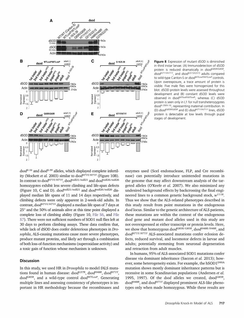
Next, we attempted to discern whether the lack of detect-able dSOD on an SDS-PAGE denaturing gel is a generaltechnical artifact, or if it is related to the progression of diseasepathology. To assess if ALS mutations affect dSOD proteinstability or accessibility to extraction in earlier stages asdramatically as in adults, wemeasured protein levels of dSODduring the adult stage from all mutant lines by SDS-PAGE(Figure 8A). As previously observed in the endogenouspoint-mutant mSOD1D83R/D83R mouse, dsodH71Y/H71Y anddsodG85R/G85R exhibited a significant reduction in amounts ofdSOD protein. Intriguingly, in the previously designated “nullor dsod2/2” allele dsodG51S (other names: n108 or n1 allele),
Figure 5 Absence of MN cell body loss in dsod G85R/G85R mutants. (A)The nuclear GFP protein (UAS-nlsGFP) is expressed under the ok371MN-specific driver (ok371-GAL4). The MN regions that are shown inthe following pictures are highlighted in red squares. DAPI is used to labelall nuclei. MNs and DAPI staining in dsodG85R/G85R (B) third instar larvalbrains (N = 5) and (C) adult ventral nerve chord (N = 5) do not exhibit MNloss. Bar, 50 mm.
Figure 6 Superoxide dismutase activity is diminished in severe dsod mu-tants. (A) SOD1 dismutase activity was assessed by NBT after native-PAGE. Homoyzgotes dsodG85R/G85R, dsodH71Y/H71Y, dsodG51S/G51S,dsodH48R/H48R show dramatically reduced enzyme activity compared todsodG37R/G37R, heterozygous combinations, and dsodWTloxP/WTloxP con-trols. Purified bovine SOD1 protein was used as a positive control. (B)Coomassie Blue staining of the same samples analyzed above electro-phoresed on native-PAGE gel.
Drosophila Knock-In Model of ALS 715

we observed protein expression, suggesting that the dsodG51S
point mutation did not result in a protein null expression aspreviously reported (Campbell et al. 1986; Staveley et al.1991; Phillips et al. 1995; Parkes et al. 1998; Kirby et al.2008; O’Keefe et al. 2011; Mishra et al. 2014).
Next, we asked whether lack of immunoreactivity in SDS-PAGEgels is consistent throughout development orwhether itis stage specific. Detection of dSOD protein from homozygousdsodH71Y/H71Y and dsodG85R/G85R is found in L1 larvae anddeclines gradually during development, indicating possiblematernal contribution which reduces during larval develop-ment (Figure 8, B, D, and E). Maternally derived dSOD pro-tein is only detectable in L1 larvae, albeit at reduced levelscompared to wild-type L1 larvae (Figure 8, B and C).
Toxicity associated with the dsodG85R allele isdosage sensitive
To determine to what extent mutant dsod alleles producedosage-sensitive phenotypes, we increased dsod copy numberand assessed phenotypes. We used the original HR cassettecontaining the entire wild-type dsod locus to increase genedose. Transformed lines containing P[dsodWTLoxP] show dSODprotein levels are comparable to the endogenous dsod locus byWestern blot analysis (Figure 9, A and B). Flies carrying fourcopies of dSODwt (P[dsodWTLoxP]/P[dsodWTLoxP]; dsodWTLoxP
/dsodWTLoxP) showed a reduced life span compared to two copydSODwt controls (Figure 9, C and D; Figure S10, A and B;38.91 6 0.55 days for males and 49.80 6 1.25 for femalesvs. 52.396 0.74 days formales and 61.956 0.85 for females).
We further assessed the effect on phenotype of altering thecopy number of mutant dsod relative to wild-type dsod. Ani-mals containing either one or two extra doses of wild-typedsod (P[dsodWTLoxP]/P[dsodWTLoxP]; dsodG85R/dsodG85R orP[dsodWTLoxP]/+; dsodG85R/dsodG85R) showed dose-dependentsuppression of dsodG85R/dsodG85R lethality at eclosion (FigureS10C). One dose of dsodwt partially rescued viability to �70%,while two doses rescued eclosion to wild-type levels. However,flies carrying two extra wild-type dsod alleles in a dsodG85R
background (P[dsodWTLoxP]/P[dsodWTLoxP]; dsodG85R/dsodG85R)showed a 40% decrease in life span relative to controls carryingfour wild-type dsod alleles (28.98 6 0.61 days for males and39.376 1.32 days for females, compared to 52.396 0.74 daysfor males and 61.956 0.85 days in females) (Figure 9, C and D;Figure S10, A and B). In all cases, flies carrying four copies ofdsod failed to show eclosion or progressive locomotor defectsregardless of whether four wild-type copies or two wild-typeand two mutant copies were present (Figure S10, C, D, andE). Nevertheless, there was a general rapid loss of locomotorfunction in the days preceding death for P[dsodWTLoxP]/P[dsodWTLoxP]; dsodG85R/dsodG85R flies (File S5).
Complete loss-of-function mutations at dsod support again-of-function toxicity component for phenotypicALS alleles
Because mutant dsod alleles exhibit predominantly recessivephenotypes and lack enzymatic activity by native gel analysis,
we compared our dsodmutants with sodnull lines to determinewhether any components of the phenotype might be due toloss of function. To analyze sodnull, we assessed phenotypesusing a heteroallelic combination of previously reported nullmutations (X-16/X-39) (Staveley et al. 1991; Parkes et al.1998; Missirlis et al. 2003). A heteroalleleic combinationwas used because homozygous sodX-16/X-16 or sodX-39/X-39 al-leles showed that numerous downward-modifying mutationshad clearly accumulated in the stock, severely reducing eclo-sion rates and viability. Extensive backcrossing of these stocksdid not alleviate this problem. To overcome this obstacle, wegenerated new sodnull mutations via CRISPR/cas9 that re-moved the dsod ORF (see Materials and Methods) (Figure10A). Two lines were characterized, dsodnull21 and dsodnull26,which had similar eclosion ratios as originally reported for
Figure 7 Altered conformations of dSOD mutant protein. (A) Analysis bynative-PAGE dSOD blot and (B) the same gel over exposed show bandsconsistent with dSOD dimer present on the gel in all genotypes exceptnull. Additional immunoreactive species were present, representing pos-sibly misfolded or monomeric dSOD (*). (C) Coomassie staining of thenative gel showing total protein loaded on the gel.
716 A. Sxahin et al.
dsodX-16 and dsodX-39 alleles, which displayed complete infertil-ity (Mockett et al. 2003) similar to dsodH71Y/H71Y (Figure 10B).In contrast to dsodH71Y/H71Y, dsodnull21/null21 and dsodnull26/null26
homozygotes exhibit less severe climbing and life-span defects(Figure 10, C and D). dsodnull21/null21 and dsodnull26/null26 dis-played median life spans of 11 and 14 days respectively, andclimbing defects were only apparent in 2-week-old adults. Incontrast, dsodH71Y/H71Y displayed amedian life span of 7 days at25� and the 50% of animals alive at this time point displayed acomplete loss of climbing ability (Figure 10, File S6, and FileS7). There were not sufficient numbers of SOD1 null flies left at30 days to perform climbing assays. These data confirm that,while lack of dSOD does confer deleterious phenotypes in Dro-sophila, ALS-causing mutations cause more severe phenotypes,produce mutant proteins, and likely act through a combinationof both loss-of-functionmechanisms (superoxidase activity) anda toxic gain of function whose mechanism is unknown.
Discussion
In this study, we used HR in Drosophila to model fALS muta-tions found in human disease: dsodG37R, dsodH48R, dsodH71Y,dsodG85R, and a wild-type control dsodWTLoxP. Generatingmultiple lines and assessing consistency of phenotypes is im-portant in HR methodology because the recombinases and
enzymes used (SceI endonuclease, FLP, and Cre recombi-nase) can potentially introduce unintended mutations inthe genome that may affect downstream analysis of the tar-geted alleles (O’Keefe et al. 2007). We also minimized anyundesired background effects by backcrossing the final engi-neered lines to a common genetic background stock, w1118.Thus we show that the ALS-related phenotypes described inthis study result from point mutations in the endogenousdsod locus. Similar to the genetic architecture of ALS patients,these mutations are within the context of the endogenousdsod gene and mutant dsod alleles used in this study arenot overexpressed at either transcript or protein levels. Here,we show that homozygous dsodG85R/G85R, dsodH48R/H48R, anddsodH71Y/H71Y ALS-associated mutations confer eclosion de-fects, reduced survival, and locomotor defects in larvae andadults; potentially stemming from neuronal degenerationand retraction from adult muscles.
In humans, 95% of ALS-associated SOD1mutations conferdisease via dominant inheritance (Saccon et al. 2013); how-ever, some heterogeneity exists. For example, the hSOD1D90A
mutation shows mostly dominant inheritance patterns but isrecessive in some Scandinavian populations (Andersen et al.1995, 1997). Of the dsod alleles we created, dsodG85R,dsodH48R, and dsodH71Y displayed prominent ALS-like pheno-types only when made homozygous. While these results are
Figure 8 Expression of mutant dSOD is diminishedin third instar larvae. (A) Immunodetection of dSODprotein is reduced dramatically in dsodG85R/G85R,dsodH71Y/H71Y, and dsodG51S/G51S adults comparedto wild-type Canton-S or dsodWTLoxP/WTLoxP controls.Upon overexposure, a trace amount of protein isvisible. Five male flies were homogenized for thisblot. dSOD protein levels were assessed throughoutdevelopment and (B) constant dSOD levels wereobserved in dsodWTloxP/WTloxP; whereas (C) dSODprotein is seen only in L1 for null transheterozygotesdsodX-39/X-16, representing maternal contribution. In(D) dsodG85R/G85R and (E) dsodH71Y/H71Y lines, dSODprotein is detectable at low levels through pupalstages of development.
Drosophila Knock-In Model of ALS 717

discrepant with the genetic pattern of inheritance for humanfALS in general, mice carrying an endogenous mSOD1D83G
point mutation (Joyce et al. 2015) created by N-ethyl-N-nitrosourea (ENU)mutagenesis display predominantly reces-sive phenotypes. These phenotypes include progressivemotor and behavioral deficits and loss of muscle force dueto degeneration of lower and upper MNs (Joyce et al. 2015).Interestingly, SOD1null mice do not display this phenotype,providing strong evidence that ALS-mutant SOD1 can conferboth loss of SOD1 enzymatic activity, and a toxic gain of func-tion (Joyce et al. 2015). We observed very similar results indsod mutant flies. Most phenotypes we observed were reces-sive yet clearly more severe than sodnull/null mutants. Theseendogenous SOD1 mutants in mice and flies demonstratethat ALS-like phenotypes are achievable in model organismswithout the necessity for transgenic overexpression of themutant allele. In addition, 73% of reported cases of SOD1mutant canines develop degenerative myelopathy (Zenget al. 2014) and exhibit ALS-like symptoms only when mu-tant alleles were homozygous.
There are several possible explanations for the apparentrecessive nature for our observed phenotypes. First, given theimportance of mutant dSOD1 protein dosage in acceleratingthe ALS-like phenotype in transgenic SOD1-mediated ALSmodels (Acevedo-Arozena et al. 2011), it is possible that
heterozygotes may develop symptoms later in life, or wouldonly display symptoms if they lived well beyond their normallife span. Mice, dogs, and fruit flies have relatively short lifespans compared to humans. Since ALS is a late-onset diseasein humans (�55 years of age in average), we speculate thatextending the life span of heterozygous fruit flies and non-transgenic SOD1 animal models beyond their normal lifespan might be required to cause more dramatic, late-onsetALS-like phenotypes. Alternatively, there may be some sec-ond site modifier or genetic background differences presentin model organisms that in wild-type form partially suppresspathogenesis, similar to what is proposed in Scandinavianpopulations.
A second explanation for lack of dominant inheritance inanimal models involves dosage of mutant dSOD protein. Theamount of disease-causingmutant proteinmay be insufficientto cause cellular toxicity in one dose, or one dose of wild-typedSOD confers a novel protective effect in the short life span ofanimal models. We addressed this possibility in several ways.We observed that lowering mutant dsodG85R or dsodH71Y genecopy number by half in dsodG85R/X-16 mutants rescued eclo-sion (Figure 2, D and E) and larval locomotion defect (Figure3, B and C). However, adult dsodG85R/X-16 flies still exhibitedvery uncoordinated behavior (File S4). Therefore, expressingonly one copy of dsodG85R is clearly less severe than the two
Figure 9 dsodG85R allele causes survival defects through gain of toxic function in a dosage-sensitive manner. (A) dSOD protein levels are gene dosedependent for dsodWTloxP alleles but not for mutant dsodG85R or (B) dsodH71Y by denaturing SDS-PAGE analysis. (C) Average life span of three trials ofmales of transgenic lines expressing extra dsod on the second chromosome and controls. dsodG85R/G85R flies die in the pupal case and do not survive toadulthood. (D) Summary statistics for life-span analysis. log-rank test was used. dsodG85R/dsodWTLoxP and dsodWTLoxP /dsodWTLoxP are not significantlydifferent from each other. P, 0.0001 P[dsodWTLoxP]/P[dsodWTLoxP]; dsodG85R/dsodG85R, P[dsodWTLoxP]/1;dsodG85R/dsodG85R and P[dsodWTLoxP ]/P[dsodWTLoxP];dsodWTLoxP/dsodWTLoxP life spans compared to wild type (dsodWTLoxP/dsodWTLoxP).
718 A. Sxahin et al.

doses of a homozygote, yet confers much of the severity of theALS-like phenotype. As such, the phenotypes we see do notresult strictly from loss of dSOD activity, but from some toxicgain of function which wild-type dSOD can counter via aprotective effect, potentially by dimerization with mutantdSOD.
While the majority of the mutant phenotypes we observedwere recessive,heterozygousdsodG85R/WTLoxPanddsodH71Y/WTLoxP
late third instar larvae show locomotor defects. This transientphenotype might stem from the differential gene expressionmilieu or neuronal physiology between the larval stage andthe adult stages. The molecular mechanisms underlying thisdiscrepancy remain to be determined. It is also possible thatfurther experiments with altered environmental conditionssuch as altered diet, stress levels, or pharmacological inter-ventions could reveal an ALS-like phenotype for Drosophilaheterozygous dsod mutant flies.
Another observation suggesting that our ALS-like symp-toms are likely due to a toxic gain of function of mutant dSODproteins are provided by the transgenic expression of wild-type dsod from an engineered locus expressing normal dSODlevels. In this case, doses of wild-type dSOD protein only
partially rescued dsodG85R/G85R phenotypes (Figure 9), lead-ing to shortened life span. Similar to human patients devel-oping late-onset ALS, as well as exhibiting a short progressionquickly followed by death, P[dsodWTLoxP]/P[dsodWTLoxP];dsodG85R/dsodG85R flies exhibited locomotor deficits for abouta day before dying early (55 days posteclosion) (File S5),which is comparable to ALS patients losing locomotor func-tion over a 1–2 year span before the need for respiratorysupport (Benditt and Boitano 2008). Thus, we observe thatin flies that are “effective” heterozygotes, albeit with twodoses each of wild-type and G85R dsod, a shortened life spanand apparent precipitous loss of locomotor function occurs;arguing again for a toxic gain of function. These flies withfour copies of dsod (two wild-type copies and two G85R mu-tant copies) may be most similar to modeling the heterozy-gote state found in ALS patients with SOD1 mutations.
Even excess dosage of wild-type dSOD appeared to beslightly deleterious in our study. Four copies of wild-type dsod(P[dsodWTLoxP]/P[dsodWTLoxP]; dsodWTLoxP/dsodWTLoxP) exhibiteda shortened life span compared to two-dosewild-type dsodWTLoxP
/dsodWTLoxP flies. This shortening of life span was milder com-pared to when two G85R alleles were expressed. Previous
Figure 10 Phenotype comparison between dsodH71Y/H71Y and additional dsodnull alleles supports gain-of-function toxicity for ALS-associated dsod pointmutations. (A) Two new dsod null lines were generated by removing the dsod ORF using CRISPR/cas9, dsodnull21 and dsodnull26, resulting in no dSODprotein product on SDS-PAGE. (B) dsodnull21/null21 and dsodnull26/null26 lines had similar eclosion ratios as dsodH71Y. Error bars are SEM. *** P , 0.0001,Fisher’s exact test. (C) dsodnull21 and dsodnull26 lines still retain climbing ability by 2 weeks compared to dsodH71Y/H71Y. Error bars are SEM. *** P ,0.001, one-way ANOVA followed by Dunnett’s post hoc test. (D) Life span of dsodnull21/null21 and dsodnull26/null26 lines are significantly shorter thandsodH71Y/H71Y. Survival curves represent an average of three life-span trials. *** P , 0.0001, log-rank test. (E) Summary statistics for life-span analysis.
Drosophila Knock-In Model of ALS 719
studies agreewith this finding and underscore the importance ofSOD1 protein dosage. Upon overexpression of wild-type hSOD1in mice, wild-type SOD1 can acquire an abnormal conformationand lead to ALS-like symptoms such as mitochondrial dysfunc-tion, axon degeneration, premature MN death, and SOD1 ag-gregation (Jaarsma et al. 2000; Graffmo et al. 2013). Theoverexpression of hSOD1 also caused locomotion deficits inDro-sophila (Watson et al. 2008). So, while copies of wild-type dSODmight be protective through dimerizationwith themutant dSODor through the restoration of cellular damage induced by oxida-tive stress due to loss of enzymatic activity, our study agrees withothers that elevated levels of normal SOD1 can be deleterious;emphasizing the need to use models with near physiologicallevels of SOD1 protein.
A consensus has not been reached regarding the nature ofdisrupted cellular pathways in ALS pathogenesis. Althoughmost of the SOD1 transgenic animals exhibit similar cellularresponses such as apoptosis andglia activation, it is not knownwhat proportion of these responses may stem from overex-pression of the mutant protein. As one hSOD1G85R transgenicmouse model suggests, in later stages there may be SOD1protein aggregates in MNs and glial cells (Wang et al.2009b). Homozygous SOD1 mutant dogs also exhibited mis-folded SOD1 species (Awano et al. 2009; Wininger et al.2011; Crisp et al. 2013; Zeng et al. 2014). On the other hand,in SOD1 mutant patient induced pluripotent stem cell mod-els, insoluble SOD1 species were detected only after inhibit-ing the proteasome (Kiskinis et al. 2014) or usingultrasensitive methods such as immunogold staining fol-lowed by electron microscopy analysis (Chen et al. 2014).Homozygous mSOD1D83G/D83G mice lack SOD1-positive pro-teinaceous inclusions, retain no detectable dismutase activity,and show very little detectable SOD1 protein by denaturingSDS-PAGE (Joyce et al. 2015). A lack of protein aggregationin endogenous SOD1 mouse mutants suggests that the SOD1protein aggregation is not required for ALS pathogenesis;however, we have not determined the state of aggregationin dsod mutants.
Previously published Drosophila ALS models recapitulatesome ALS-like phenotypes such as locomotion deficits, glio-sis, reduced synaptic transmission, SOD1 protein aggrega-tion, and mitochondrial defects; however, these models arebased on overexpression of hSOD1 (Watson et al. 2008;Bahadorani et al. 2013). Our data in HR-generated dsodmu-tants agrees with some aspects of this previously publishedwork including a lack of MN loss, the presence of climbingdeficits, and a sensitivity to oxidative stress (Watson et al.2008; Bahadorani et al. 2013). Our preliminary results sug-gest that the cell bodies of MNs remain intact even at theterminal stages of dsodG85R/G85R. Lack of MN death despiteneuromuscular deficits is not surprising because ALS is con-sidered a “dying back” disorder in which muscle denervationprecedes the death of theMN cell body (Dadon-Nachum et al.2011). This phenomenon of neuromuscular-junction degen-eration preceding MN cell body death was observed in trans-genic mutant hSOD1-expressing mouse models (Kato 2008),
a nontransgenic mouse model (Joyce et al. 2015), and anearly autopsy of an ALS patient who unexpectedly died fromunrelated causes (Fischer et al. 2004). Neurodegenerativeprocesses do appear to come into play in the later stages ofa dsod mutant approaching adulthood in Drosophila. ThedsodG85R/G85R pharate adults fail to eclose and show short-ened leg phenotypes (Figure 2 and Figure 4). Accompanyingthis phenotype, the leg nerves appear degenerated, withleg 3 exhibiting a distinctly more severe phenotype thanleg 2 and leg 1 (File S1 and File S2). In dsodH71Y/H71Y, all legslook wild type upon eclosion. However, by day 14, 87% ofleg 3 nerves have lost most of the side branches divergingfrom the main nerve (Figure 4 and File S3). In short, thereappears to be a degenerative process involving at least MNsthat correlates with phenotypic severity and is progressive,and yet, does not appear to be the result of MN cell death.
Our study also addresses loss-of-function and gain-of-toxic-function questions at a protein level. The little or nodetectable dismutase activity in homozygous dsodG85R/G85R,dsodH71Y/H71Y, dsodH48R/H48R, and dsodG51S/G51S mutant linessuggests a loss of SOD1 enzymatic activity in these mutants.While we cannot rule out low levels of enzymatic activitybelow levels of detection, SOD1 activity seen within thesemutants is clearly diminished compared to either heterozy-gous or homozygous dsodWTLoxP/WTLoxP. To assess protein lev-els of mutant dSOD proteins, we used standard SDS-PAGEfollowed by immunoblot with antibodies to dSOD. For phe-notypic mutants (G85R, H48R, H71Y, and G51S) we ob-served very little dSOD protein (Figure 7 and Figure S7).dsodG37R flies showed normal levels of dSOD protein in addi-tion to being active enzymatically (Figure 6 and Figure 7). Inparallel with our findings, SOD1 mutants show dramaticallyreduced protein amounts on denaturing SDS-PAGE gels inmice homozygous for an endogenous mSOD1D83G point mu-tation (�10% of wild type in homozygotes) (Joyce et al.2015) and patient-induced pluripotent stem cell-derivedMNs (Chen et al. 2014; Kiskinis et al. 2014). These resultshave been explained by the instability ofmutant SOD1 and itssubsequent degradation (Kabuta et al. 2006). It has beenpreviously reported that mutant SOD1, especially SOD1G85R,has a decreased half-life when compared to wild-type SOD1(Borchelt et al. 1994; Farr et al. 2011). This is in agreementwith current literature suggesting that hSOD1G85R monomersare unable to form dimers with each other or withmSOD1 andhSOD1 (Wang et al. 2009b).
Given the results of all attempts to detect dSOD mutantproteins from animals carrying phenotypic mutations, wewere thus surprised by our immunoblot results using nativegel electrophoresis. In all cases, mutant dSOD proteins weredetected in homozygous animals. In fact, levels of protein onthe blots were similar to wild-type controls, and much of theprotein was found at the size of the normal dSOD dimer(Figure 7). However, in homozygous mutants, there alsoappeared a faster migrating species that we infer is mono-meric dSOD, and this species represented a substantialamount of dSOD immunoreactivity in homozygotes. In
720 A. Sxahin et al.

heterozygotes, again, roughly normal amounts of dSOD pro-teins are seen, but the anomalous fast migrating band is lessintense. We interpret this as dimer formation between mutantand wild-type dSOD proteins. Further experiments will be nec-essary to confirm this speculation. Nevertheless, the datastrongly suggest that all of the phenotypic mutants character-ized in this study produce substantial amounts of dSOD pro-tein, which is a prerequisite for generating a toxic gain offunction. In addition, the data suggest a potential mechanism(dimerization) for alleviating phenotypes caused by homozy-gosity of toxic mutations. Recent research has shown thatsmall, soluble monomers of SOD1, rather than insoluble ag-gregates, are likely to be the cytotoxic species causing neuro-degeneration (Rakhit et al. 2007; Auclair et al. 2010; Liu et al.2012).
Lastly, our newly generated dsodnull mutations confirmresults from other studies in a conclusive manner (Reaumeet al. 1996; Huang et al. 1997; Ho et al. 1998; Matzuk et al.1998; Yoshida et al. 2000; Saccon et al. 2013). dsodnull mu-tants display eclosion defects, shortened life spans and infer-tility (Figure 10). However, their life span is not nearly asshort as dsodH71Y/H71Y homozygotes, and they do not showany locomotor deficits until much later in life. All of ourphenotypic mutants (H48R, G85R, and H71Y) as homozy-gotes show undetectable levels of dSOD activity in gel assays,yet have muchmore severe phenotypes than the homozygotedsodnull/null mutants. We conclude from this that the pheno-types we observe in animals are a combination of both loss-and gain-of-function components. Recently, it has beenstrongly suggested that dominantly inherited human SOD1-based ALS is modified additionally by the loss of function ofSOD1 activity, as most patients with mutations demonstratean in vivo loss of activity (Saccon et al. 2013). Further studieswill be necessary to untangle the loss- and gain-of-functioncontributions to disease phenotype, but our studies raise aninteresting conjecture that the mutation G37R, which showsSOD1 activity but no phenotype as homozygotes, may bedominated by the gain-of-function component of the pro-posed mechanism. Thus, these mutations may be useful ifunder appropriate conditions or treatments, a phenotypecan be revealed in heterozygotes.
Our Drosophila model provides a rich, fast, and efficientsystem complementary to rodent model organisms foraddressing mechanisms associated with human SOD1 muta-tions causing ALS and for elucidating the dosage-sensitiveresults of SOD1-mediated ALS, as well as unraveling the con-tributions of loss and gain of function to mutant phenotypes.Using Drosophila to model ALS provides a unique system inwhich to assess effects on conserved molecular pathwaysfundamental to neuronal function and dysfunction in ALS.Moreover, the severe phenotypic consequences of some ofthese mutations provide an excellent motivation for unbiasedforward genetic suppressor screens to identify genes that,when mutated, can reverse the effects of these human dis-ease-causingmutations; potentially serving as the foundationfor novel therapeutic approaches acting through such genes.
Acknowledgments
We thank Cynthia Staber for critical reading of the manu-script. We are grateful to William C. Orr for the gift ofantibody raised against dSOD. This work was supported bythe National Institutes of Health (GM-068118 to K.W., T32DK-060415 to A.H.), the Judith and Jean Pape AdamsFoundation to K.W., the Institutional Development AwardNetwork for Biomedical Research Excellence from theNational Institute of General Medical Sciences of theNational Institutes of Health under grant number P20GM-103430 to G.S., and the Amyotrophic Lateral SclerosisAssociation #2279 to R.R. Sequencing analysis was con-ducted at a Rhode Island National Science FoundationExperimental Program to Stimulate Competitive Research(EPSCoR) facility, the Genomics and Sequencing Center,supported in part by the National Science FoundationEPSCoR cooperative agreement #EPS-1004057. Stocksobtained from the Bloomington Drosophila Stock Center(National Institutes of Health P40 OD-018537) were usedin this study.
Literature Cited
Abel, O., J. F. Powell, P. M. Andersen, and A. Al-Chalabi,2012 ALSoD: a user-friendly online bioinformatics tool foramyotrophic lateral sclerosis genetics. Hum. Mutat. 33: 1345–1351.
Acevedo-Arozena, A., B. Kalmar, S. Essa, T. Ricketts, P. Joyce et al.,2011 A comprehensive assessment of the SOD1G93A low-copy transgenic mouse, which models human amyotrophic lat-eral sclerosis. Dis. Model. Mech. 4: 686–700.
Alexander, G. M., K. L. Erwin, N. Byers, J. S. Deitch, B. J. Augelliet al., 2004 Effect of transgene copy number on survival in theG93A SOD1 transgenic mouse model of ALS. Brain Res. Mol.Brain Res. 130: 7–15.
Andersen, P. M., P. Nilsson, V. Ala-Hurula, M. L. Keränen, I. Tarvainenet al., 1995 Amyotrophic lateral sclerosis associated with homo-zygosity for an Asp90Ala mutation in CuZn-superoxide dismutase.Nat. Genet. 10: 61–66.
Andersen, P. M., P. Nilsson, M. L. Keränen, L. Forsgren, J. Hägglundet al., 1997 Phenotypic heterogeneity in motor neuron diseasepatients with CuZn-superoxide dismutase mutations in Scandi-navia. Brain 120: 1723–1737.
Auclair, J. R., K. J. Boggio, G. A. Petsko, D. Ringe, and J. N. Agar,2010 Strategies for stabilizing superoxide dismutase (SOD1),the protein destabilized in the most common form of familialamyotrophic lateral sclerosis. Proc. Natl. Acad. Sci. USA 107:21394–21399.
Awano, T., G. S. Johnson, C. M. Wade, M. L. Katz, G. C. Johnsonet al., 2009 Genome-wide association analysis reveals a SOD1mutation in canine degenerative myelopathy that resemblesamyotrophic lateral sclerosis. Proc. Natl. Acad. Sci. USA 106:2794–2799.
Bahadorani, S., S. T. Mukai, J. Rabie, J. S. Beckman, J. P. Phillipset al., 2013 Expression of zinc-deficient human superoxide dis-mutase in Drosophila neurons produces a locomotor defectlinked to mitochondrial dysfunction. Neurobiol. Aging 34:2322–2330.
Batlevi, Y., D. N. Martin, U. B. Pandey, C. R. Simon, C. M. Powerset al., 2010 Dynein light chain 1 is required for autophagy,protein clearance, and cell death in Drosophila. Proc. Natl. Acad.Sci. USA 107: 742–747.
Drosophila Knock-In Model of ALS 721

Benditt, J. O., and L. Boitano, 2008 Respiratory treatment ofamyotrophic lateral sclerosis. Phys. Med. Rehabil. Clin. N. Am.19: 559–572.
Bertini, I., S. Manganl, and M. S. Viezzoli, 1998 Structure andproperties of copper-zinc superoxide dismutases. Adv. Inorg.Chem. 45: 127–250.
Borchelt, D. R., M. K. Lee, H. S. Slunt, M. Guarnieri, Z. S. Xu et al.,1994 Superoxide dismutase 1 with mutations linked to famil-ial amyotrophic lateral sclerosis possesses significant activity.Proc. Natl. Acad. Sci. USA 91: 8292–8296.
Bosco, D. A., G. Morfini, N. M. Karabacak, Y. Song, F. Gros-Louiset al., 2010 Wild-type and mutant SOD1 share an aberrantconformation and a common pathogenic pathway in ALS. Nat.Neurosci. 13: 1396–1403.
Bunton-Stasyshyn, R. K. A., R. A. Saccon, P. Fratta, and E. M. C.Fisher, 2015 SOD1 function and its implications for amyotro-phic lateral sclerosis pathology: new and renascent themes.Neuroscientist 21: 519–529.
Campbell, S. D., A. J. Hilliker, and J. P. Phillips, 1986 Cytogeneticanalysis of the cSOD microregion in Drosophila melanogaster.Genetics 112: 205–215.
Chen, H., K. Qian, Z. Du, J. Cao, A. Petersen et al., 2014 ModelingALS with iPSCs reveals that mutant SOD1 misregulates neuro-filament balance in motor neurons. Cell Stem Cell 14: 796–809.
Crisp, M. J., J. Beckett, J. R. Coates, and T. M. Miller,2013 Canine degenerative myelopathy: biochemical charac-terization of superoxide dismutase 1 in the first naturally oc-curring non-human amyotrophic lateral sclerosis model. Exp.Neurol. 248: 1–9.
Cushman-Nick, M., N. M. Bonini, and J. Shorter, 2013 Hsp104suppresses polyglutamine-induced degeneration post onset ina drosophila MJD/SCA3 model. PLoS Genet. 9: e1003781.
Dadon-Nachum, M., E. Melamed, and D. Offen, 2011 The “dying-back” phenomenon of motor neurons in ALS. J. Mol. Neurosci.43: 470–477.
Ezzi, S. A., M. Urushitani, and J.-P. Julien, 2007 Wild-type super-oxide dismutase acquires binding and toxic properties of ALS-linked mutant forms through oxidation. J. Neurochem. 102:170–178.
Farr, G. W., Z. Ying, W. A. Fenton, and A. L. Horwich,2011 Hydrogen-deuterium exchange in vivo to measure turn-over of an ALS-associated mutant SOD1 protein in spinal cord ofmice. Protein Sci. 20: 1692–1696.
Fischer, L. R., D. G. Culver, P. Tennant, A. A. Davis, M. Wang et al.,2004 Amyotrophic lateral sclerosis is a distal axonopathy: ev-idence in mice and man. Exp. Neurol. 185: 232–240.
Forsberg, K., P. A. Jonsson, P. M. Andersen, D. Bergemalm, K. S.Graffmo et al., 2010 Novel antibodies reveal inclusions con-taining non-native SOD1 in sporadic ALS patients. PLoS One 5:e11552.
Fridovich, I., 1986 Superoxide dismutases. Adv. Enzymol. Relat.Areas Mol. Biol. 58: 61–97.
Gell, S. L., and R. A. Reenan, 2013 Mutations to the piRNA path-way component Aubergine enhance meiotic drive of segregationdistorter in Drosophila melanogaster. Genetics 193: 771–784.
Graffmo, K. S., K. Forsberg, J. Bergh, A. Birve, P. Zetterström et al.,2013 Expression of wild-type human superoxide dismutase-1in mice causes amyotrophic lateral sclerosis. Hum. Mol. Genet.22: 51–60.
Gruzman, A., W. L. Wood, E. Alpert, M. D. Prasad, R. G. Milleret al., 2007 Common molecular signature in SOD1 for bothsporadic and familial amyotrophic lateral sclerosis. Proc. Natl.Acad. Sci. USA 104: 12524–12529.
Gurney, M. E., H. Pu, A. Y. Chiu, M. C. Dal Canto, and C. Y. Polchowet al., 1994 Motor neuron degeneration in mice that express ahuman Cu,Zn superoxide dismutase mutation. Science 264:1772–1775.
Halfmann, R., and S. Lindquist, 2008 Screening for amyloid ag-gregation by semi-denaturing detergent-agarose gel electropho-resis. J. Vis. Exp., 838.
Ho, Y. S., M. Gargano, J. Cao, R. T. Bronson, I. Heimler et al.,1998 Reduced fertility in female mice lacking copper-zinc su-peroxide dismutase. J. Biol. Chem. 273: 7765–7769.
Hu, S., Z. Xie, A. Onishi, X. Yu, L. Jiang et al., 2009 Profiling thehuman protein-DNA interactome reveals ERK2 as a transcrip-tional repressor of interferon signaling. Cell 139: 610–622.
Huang, T. T., M. Yasunami, E. J. Carlson, A. M. Gillespie, A. G.Reaume et al., 1997 Superoxide-mediated cytotoxicity in su-peroxide dismutase-deficient fetal fibroblasts. Arch. Biochem.Biophys. 344: 424–432.
Hudson, R. R., K. Bailey, D. Skarecky, J. Kwiatowski, and F. J. Ayala,1994 Evidence for positive selection in the superoxide dismu-tase (Sod) region of Drosophila melanogaster. Genetics 136:1329–1340.
Jaarsma, D., E. D. Haasdijk, J. A. Grashorn, R. Hawkins, W. vanDuijn et al., 2000 Human Cu/Zn superoxide dismutase(SOD1) overexpression in mice causes mitochondrial vacuoliza-tion, axonal degeneration, and premature motoneuron deathand accelerates motoneuron disease in mice expressing a famil-ial amyotrophic lateral sclerosis mutant SOD1. Neurobiol. Dis.7: 623–643.
Joyce, P. I., P. Fratta, E. M. C. Fisher, and A. Acevedo-Arozena,2011 SOD1 and TDP-43 animal models of amyotrophic lateralsclerosis: recent advances in understanding disease toward thedevelopment of clinical treatments. Mamm. Genome 22: 420–448.
Joyce, P. I., P. Mcgoldrick, R. A. Saccon, W. Weber, P. Fratta et al.,2015 A novel SOD1-ALS mutation separates central and pe-ripheral effects of mutant SOD1 toxicity. Hum. Mol. Genet. 24:1883–1897.
Juneja, T., M. A. Pericak-Vance, N. G. Laing, S. Dave, and T. Siddique,1997 Prognosis in familial amyotrophic lateral sclerosis: progres-sion and survival in patients with glu100gly and ala4val mutationsin Cu,Zn superoxide dismutase. Neurology 48: 55–57.
Kabuta, T., Y. Suzuki, and K. Wada, 2006 Degradation of amyo-trophic lateral sclerosis-linked mutant Cu,Zn-superoxide dismu-tase proteins by macroautophagy and the proteasome. J. Biol.Chem. 281: 30524–30533.
Kato, S., 2008 Amyotrophic lateral sclerosis models and humanneuropathology: similarities and differences. Acta Neuropathol.115: 97–114.
Kirby, K., L. T. Jensen, J. Binnington, A. J. Hilliker, J. Ulloa et al.,2008 Instability of superoxide dismutase 1 of Drosophila inmutants deficient for its cognate copper chaperone. J. Biol.Chem. 283: 35393–35401.
Kiskinis, E., J. Sandoe, L. A. Williams, G. L. Boulting, R. Mocciaet al., 2014 Pathways disrupted in human ALS motor neuronsidentified through genetic correction of mutant SOD1. Cell StemCell 14: 781–795.
Leblond, C. S., H. M. Kaneb, P. A. Dion, and G. A. Rouleau,2014 Dissection of genetic factors associated with amyotro-phic lateral sclerosis. Exp. Neurol. 262, Part B: 91–101.
Lee, Y. M., H. P. Misra, and F. J. Ayala, 1981 Superoxide dismu-tase in Drosophila melanogaster: biochemical and structuralcharacterization of allozyme variants. Proc. Natl. Acad. Sci.USA 78: 7052–7055.
Liu, H.-N., S. Tjostheim, K. Dasilva, D. Taylor, B. Zhao et al.,2012 Targeting of monomer/misfolded SOD1 as a therapeuticstrategy for amyotrophic lateral sclerosis. J. Neurosci. 32: 8791–8799.
Matzuk, M. M., L. Dionne, Q. Guo, T. R. Kumar, and R. M. Lebovitz,1998 Ovarian function in superoxide dismutase 1 and 2 knock-out mice. Endocrinology 139: 4008–4011.
722 A. Sxahin et al.

McGoldrick, P., P. I. Joyce, E. M. C. Fisher, and L. Greensmith,2013 Rodent models of amyotrophic lateral sclerosis. Biochim.Biophys. Acta 1832: 1421–1436.
Mishra, M., A. Sharma, A. K. Shukla, R. Kumar, U. N. Dwivedi et al.,2014 Genotoxicity of dichlorvos in strains of Drosophila mel-anogaster defective in DNA repair. Mutat. Res. Genet. Toxicol.Environ. Mutagen. 766: 35–41.
Missirlis, F., J. Hu, K. Kirby, A. J. Hilliker, T. A. Rouault et al.,2003 Compartment-specific protection of iron-sulfur proteinsby superoxide dismutase. J. Biol. Chem. 278: 47365–47369.
Mockett, R. J., S. N. Radyuk, J. J. Benes, W. C. Orr, and R. S. Sohal,2003 Phenotypic effects of familial amyotrophic lateral sclero-sis mutant Sod alleles in transgenic Drosophila. Proc. Natl. Acad.Sci. USA 100: 301–306.
O’Keefe, L. V., P. Smibert, A. Colella, T. K. Chataway, R. Saint et al.,2007 Know thy fly. Trends Genet. 23: 238–242.
O’Keefe, L. V., A. Colella, S. Dayan, Q. Chen, A. Choo et al.,2011 Drosophila orthologue of WWOX, the chromosomal frag-ile site FRA16D tumour suppressor gene, functions in aerobicmetabolism and regulates reactive oxygen species. Hum. Mol.Genet. 20: 497–509.
Özoguz, A., Ö. Uyan, G. Birdal, C. Iskender, E. Kartal et al.,2015 The distinct genetic pattern of ALS in Turkey and novelmutations. Neurobiol. Aging 36: 1764.e9–1764.e18.
Pardo, C. A., Z. Xu, D. R. Borchelt, D. L. Price, S. S. Sisodia et al.,1995 Superoxide dismutase is an abundant component in cellbodies, dendrites, and axons of motor neurons and in a subset ofother neurons. Proc. Natl. Acad. Sci. USA 92: 954–958.
Parkes, T. L., K. Kirby, J. P. Phillips, and A. J. Hilliker,1998 Transgenic analysis of the cSOD-null phenotypic syn-drome in Drosophila. Genome 41: 642–651.
Pasinelli, P., and R. H. Brown, 2006 Molecular biology of amyo-trophic lateral sclerosis: insights from genetics. Nat. Rev. Neuro-sci. 7: 710–723.
Phillips, J. P., J. A. Tainer, E. D. Getzoff, G. L. Boulianne, K. Kirbyet al., 1995 Subunit-destabilizing mutations in Drosophila cop-per/zinc superoxide dismutase: neuropathology and a model ofdimer dysequilibrium. Proc. Natl. Acad. Sci. USA 92: 8574–8578.
Rabe, M., A. Felbecker, S. Waibel, P. Steinbach, P. Winter et al.,2010 The epidemiology of CuZn-SOD mutations in Germany:a study of 217 families. J. Neurol. 257: 1298–1302.
Rakhit, R., J. Robertson, C. Vande Velde, P. Horne, D. M. Ruthet al., 2007 An immunological epitope selective for patholog-ical monomer-misfolded SOD1 in ALS. Nat. Med. 13: 754–759.
Reaume, A. G., J. L. Elliott, E. K. Hoffman, N. W. Kowall, R. J.Ferrante et al., 1996 Motor neurons in Cu/Zn superoxide dis-mutase-deficient mice develop normally but exhibit enhancedcell death after axonal injury. Nat. Genet. 13: 43–47.
Rosen, D. R., T. Siddique, D. Patterson, D. A. Figlewicz, P. Sappet al., 1993 Mutations in Cu/Zn superoxide dismutase geneare associated with familial amyotrophic lateral sclerosis. Na-ture 362: 59–62.
Saccon, R. A., R. K. A. Bunton-Stasyshyn, E. M. C. Fisher, and P.Fratta, 2013 Is SOD1 loss of function involved in amyotrophiclateral sclerosis? Brain 136: 2342–2358.
Schutte, R. J., S. S. Schutte, J. Algara, E. V. Barragan, J. Gilliganet al., 2014 Knock-in model of Dravet syndrome reveals a con-
stitutive and conditional reduction in sodium current.J. Neurophysiol. 112: 903–912.
Soler, C., M. Daczewska, J. P. Da Ponte, B. Dastugue, and K. Jagla,2004 Coordinated development of muscles and tendons of theDrosophila leg. Development 131: 6041–6051.
Sreedharan, J., and R. H. Brown, 2013 Amyotrophic lateral scle-rosis: problems and prospects. Ann. Neurol. 74: 309–316.
Staber, C. J., S. Gell, J. E. C. Jepson, and R. A. Reenan,2011 Perturbing A-to-I RNA editing using genetics and homol-ogous recombination. Methods Mol. Biol. 718: 41–73.
Staveley, B. E., A. J. Hilliker, and J. P. Phillips, 1991 Genetic or-ganization of the cSOD microregion of Drosophila melanogaster.Genome 34: 279–282.
Sun, L., J. Gilligan, C. Staber, R. J. Schutte, V. Nguyen et al.,2012 A knock-in model of human epilepsy in Drosophila re-veals a novel cellular mechanism associated with heat-inducedseizure. J. Neurosci. 32: 14145–14155.
Trotti, D., A. Rolfs, N. C. Danbolt, R. H. Brown, and M. A. Hediger,1999 SOD1 mutants linked to amyotrophic lateral sclerosisselectively inactivate a glial glutamate transporter. Nat. Neuro-sci. 2: 427–433.
Tsang, C. K., Y. Liu, J. Thomas, Y. Zhang, and X. F. S. Zheng,2014 Superoxide dismutase 1 acts as a nuclear transcriptionfactor to regulate oxidative stress resistance. Nat. Commun. 5:3446.
Valentine, J. S., P. A. Doucette, and S. Zittin Potter, 2005 Copper-zinc superoxide dismutase and amyotrophic lateral sclerosis.Annu. Rev. Biochem. 74: 563–593.
Wang, J., G. W. Farr, D. H. Hall, F. Li, K. Furtak et al., 2009a AnALS-linked mutant SOD1 produces a locomotor defect associ-ated with aggregation and synaptic dysfunction when ex-pressed in neurons of Caenorhabditis elegans. PLoS Genet. 5:e1000350.
Wang, J., G. W. Farr, C. J. Zeiss, D. J. Rodriguez-Gil, J. H. Wilsonet al., 2009b Progressive aggregation despite chaperone asso-ciations of a mutant SOD1-YFP in transgenic mice that developALS. Proc. Natl. Acad. Sci. USA 106: 1392–1397.
Watson, M. R., R. D. Lagow, K. Xu, B. Zhang, and N. M. Bonini,2008 A Drosophila model for amyotrophic lateral sclerosis re-veals motor neuron damage by human SOD1. J. Biol. Chem.283: 24972–24981.
Weis, J., I. Katona, G. Müller-Newen, C. Sommer, G. Necula et al.,2011 Small-fiber neuropathy in patients with ALS. Neurology76: 2024–2029.
Wininger, F. A., R. Zeng, G. S. Johnson, M. L. Katz, G. C. Johnsonet al., 2011 Degenerative myelopathy in a bernese mountaindog with a novel SOD1 missense mutation. J. Vet. Intern. Med.25: 1166–1170.
Yoshida, T., N. Maulik, R. M. Engelman, Y. S. Ho, and D. K. Das,2000 Targeted disruption of the mouse Sod I gene makes thehearts vulnerable to ischemic reperfusion injury. Circ. Res. 86:264–269.
Zeng, R., J. R. Coates, G. C. Johnson, L. Hansen, T. Awano et al.,2014 Breed distribution of SOD1 alleles previously associatedwith canine degenerative myelopathy. J. Vet. Intern. Med. 28:515–521.
Communicating editor: H. J. Bellen
Drosophila Knock-In Model of ALS 723