Embed Size (px)
Citation preview
Hindawi Publishing CorporationCritical Care Research and PracticeVolume 2012, Article ID 204314, 8 pagesdoi:10.1155/2012/204314
Research Article
Human versus Computer Controlled Selection of VentilatorSettings: An Evaluation of Adaptive Support Ventilation andMid-Frequency Ventilation
Eduardo Mireles-Cabodevila,1, 2 Enrique Diaz-Guzman,3
Alejandro C. Arroliga,4 and Robert L. Chatburn2
1 Department of Pulmonary and Critical Care Medicine, University of Arkansas for Medical Sciences, 4301 West Markham Street,Slot 555, Little Rock, AR 77205, USA
2 Respiratory Institute, Cleveland Clinic, 9500 Euclid Avenue, A90, Cleveland, OH 44195, USA3 Department of Pulmonary and Critical Care, University of Kentucky, Lexington, KY 40536-0284, USA4 Department of Medicine, Scott and White and Texas A and M Health Science Center College of Medicine, 2401 South 31st Street,Temple, TX 76508, USA
Correspondence should be addressed to Eduardo Mireles-Cabodevila, [email protected]
Received 24 May 2012; Accepted 7 September 2012
Academic Editor: Samir Sakka
Copyright © 2012 Eduardo Mireles-Cabodevila et al. This is an open access article distributed under the Creative CommonsAttribution License, which permits unrestricted use, distribution, and reproduction in any medium, provided the original work isproperly cited.
Background. There are modes of mechanical ventilation that can select ventilator settings with computer controlled algorithms(targeting schemes). Two examples are adaptive support ventilation (ASV) and mid-frequency ventilation (MFV). We studiedhow different clinician-chosen ventilator settings are from these computer algorithms under different scenarios. Methods. A surveyof critical care clinicians provided reference ventilator settings for a 70 kg paralyzed patient in five clinical/physiological scenarios.The survey-derived values for minute ventilation and minute alveolar ventilation were used as goals for ASV and MFV, respectively.A lung simulator programmed with each scenario’s respiratory system characteristics was ventilated using the clinician, ASV, andMFV settings. Results. Tidal volumes ranged from 6.1 to 8.3 mL/kg for the clinician, 6.7 to 11.9 mL/kg for ASV, and 3.5 to 9.9 mL/kgfor MFV. Inspiratory pressures were lower for ASV and MFV. Clinician-selected tidal volumes were similar to the ASV settings forall scenarios except for asthma, in which the tidal volumes were larger for ASV and MFV. MFV delivered the same alveolar minuteventilation with higher end expiratory and lower end inspiratory volumes. Conclusions. There are differences and similaritiesamong initial ventilator settings selected by humans and computers for various clinical scenarios. The ventilation outcomes arethe result of the lung physiological characteristics and their interaction with the targeting scheme.
1. Introduction
The evolution of the computerized control of mechanicalventilators has reached the level where the ventilator canselect some (previously human selected) settings based oncomputer controlled targeting schemes [1–4]. One of thesecontrol algorithms is called an “optimum targeting scheme”for which the only commercially available mode is adaptivesupport ventilation (ASV). “Optimum”, in this contextmeans to minimize the work rate of breathing a patientwould have to do if breathing unassisted with the ventilatorselected tidal volume and frequency [5, 6]. These settings
are based on the ventilator’s assessment of respiratory systemcharacteristics (i.e., alveolar minute ventilation requirement,estimated dead space volume, and expiratory time constant).Although ASV has embedded rules that attempt to preventhypoventilation, air trapping, and volutrauma, the primarygoal is not the prevention of lung injury. ASV has beenreported to choose ventilator settings that provide adequateventilation in patients with a variety of clinical conditions [7–9].
We developed mid-frequency ventilation (MFV) [10], amode of ventilation using an optimum targeting scheme withthe goal of maximizing alveolar ventilation and minimizing
2 Critical Care Research and Practice
tidal volume to promote lung protection. The theoreticalbasis for MFV has been described elsewhere [10]. In brief,MFV uses a mathematical model for pressure controlventilation where patient characteristics (alveolar minuteventilation requirement, dead space ratio, and inspiratory,and expiratory time constants) are used to calculate optimalfrequency and tidal volume settings. In this case, optimum isdefined as the frequency and tidal volume that produce themaximum alveolar minute ventilation for a given inspiratorypressure setting (above PEEP). MFV results in higher ventila-tor frequencies delivering the lowest tidal volume possible fora given target minute ventilation and inspiratory pressure,while using a conventional ventilator.
In order to allow a computer to choose ventilator set-tings, the clinician, must trust the process by which thesesettings are determined. Although several studies have beenpublished with ASV, [1, 8, 9, 11, 12] none compared themdirectly to human performance. The purpose of this studywas to compare the initial ventilator settings selected byhuman operators with those selected by two computeralgorithms (ASV and MFV) in five hypothetical clinicalscenarios.
2. Materials and Methods
The study was divided into 2 steps. The first step wasto determine the clinician-selected ventilator settings. Asurvey was made available to all the medical and surgicalcritical care physicians, fellows, and respiratory therapistsat the Cleveland Clinic. The survey asked for proposedventilator settings for five hypothetical patient scenarios. Thesecond step was to evaluate the ventilation outcomes (tidalvolume, lung volumes, and airway pressures). We used alung simulator programmed with the scenarios’ respiratorysystem characteristics ventilated with the clinician-selectedsettings, ASV, and MFV.
2.1. First Step: Electronic Survey. The Institutional ReviewBoard approved the survey. An electronic survey (http://www.surveymonkey.com/, Portland, OR) was sent by email tofaculty and fellows and posted in the respiratory therapywebsite from December 1 to 31, 2007. The survey pre-sented five clinical scenarios (Table 1). All the scenariosused the same baseline parameters: a 70 kg predicted bodyweight, male, paralyzed. The scenarios included a patientwith normal lungs, two patients with restrictive disorders(ARDS and morbid obesity), and two with obstructivelung disease (COPD and status asthmaticus). The scenar-ios were hypothetical, and included arterial blood gases(validity confirmed by the Henderson-Hasselbalch formula[13]); ventilator settings in volume controlled continuousmandatory ventilation and previously published values forlung resistance and compliance for each condition (Table 1).The survey asked what ventilator settings the clinician wouldchoose for each scenario. The options were tidal volume goal,respiratory rate, I : E ratio, and PEEP.
We only used the results from surveys that had allanswers completed. The survey results were used to calculate
the clinician goal for minute ventilation (respiratory ratemultiplied by tidal volume) and alveolar minute ventilation(tidal volume minus dead space volume (estimated as2.2 mL/kg) multiplied by respiratory rate). A fixed dead spacevolume was used in all clinical scenarios to fully appreciatethe effects of the settings.
2.2. Second Step: Lung Simulator. We used a lung simulator(Ingmar ASL 5000, IngMar Medical Ltd., Pittsburgh, PA) torecreate the clinical scenarios in the survey. The simulatorwas set up as a passive respiratory system composed of asingle linear constant resistance and single constant com-pliance. The respiratory system compliances and resistancesused in the survey were programmed for each clinicalscenario (Table 1). The parameters were constant duringthe experiments. Data from the simulator were recorded ina high-resolution file (500 Hz sampling frequency). Tidalvolumes and end inspiratory and end expiratory volumeswere measured as the excursion of the piston inside the lungsimulator.
Two mechanical ventilators were used: a HamiltonGalileo, (Hamilton Medical AG, Bonaduz, Switzerland) todeliver clinician settings (with pressure control ventilation)and ASV and a Drager Evita XL (Drager Medical AG &Co., Lubeck, Germany) to deliver MFV. The change inventilator to deliver the MFV was due to our previousexperience [10] that showed the Drager ventilator generatingthe sharply rectangular pressure waveform necessary forefficient MFV. The ventilators were connected to the lungsimulator using a conventional circuit (70 inches long) withseparate inspiratory and expiratory limbs (Airlife; CardinalHealth, McGaw Park, IL) without a humidifier chamber. Allexperiments were conducted using room air (FIO2 = 0.21)and reported as measured. The ventilators were calibratedand tested for leaks prior to the experiments.
2.3. Experimental Protocol
2.3.1. Clinician Settings. For each of the clinical scenarios weobtained the average tidal volume, respiratory rate, I : E ratio,and PEEP selected in the survey. The ventilator was set withthese values.
To maintain comparability with MFV and ASV (bothpressure controlled modes), clinician ventilator settings weredelivered with pressure controlled continuous mandatoryventilation (i.e., all breaths were time triggered, pressure lim-ited, and time cycled). Inspiratory pressure was determinedin a preliminary run on the simulator to achieve the targettidal volume. Pressure rise time was set to the minimumavailable on each ventilator (Hamilton 50 ms, Drager 0 ms).
2.3.2. Adaptive Support Ventilation. The ventilator was pro-grammed for ventilation on an adult male patient. Theheight was set at 174 cm, which represents 70 kg of predictedbody weight. For each case scenario, the percent minuteventilation was set to achieve the target minute ventilationobtained from the clinician survey. ASV was maintained
Critical Care Research and Practice 3
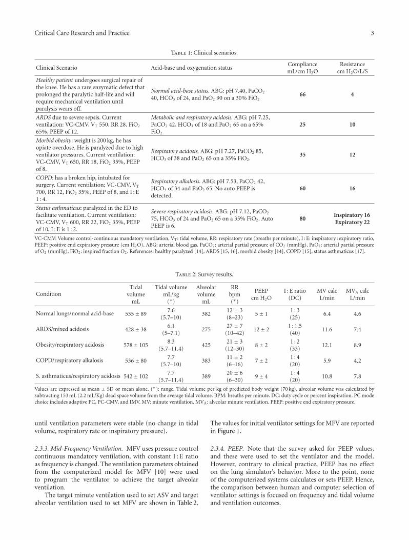
Table 1: Clinical scenarios.
Clinical Scenario Acid-base and oxygenation statusCompliancemL/cm H2O
Resistancecm H2O/L/S
Healthy patient undergoes surgical repair ofthe knee. He has a rare enzymatic defect thatprolonged the paralytic half-life and willrequire mechanical ventilation untilparalysis wears off.
Normal acid-base status. ABG: pH 7.40, PaCO2
40, HCO3 of 24, and PaO2 90 on a 30% FiO266 4
ARDS due to severe sepsis. Currentventilation: VC-CMV, VT 550, RR 28, FiO2
65%, PEEP of 12.
Metabolic and respiratory acidosis. ABG: pH 7.25,PaCO2 42, HCO3 of 18 and PaO2 65 on a 65%FiO2
25 10
Morbid obesity: weight is 200 kg, he hasopiate overdose. He is paralyzed due to highventilator pressures. Current ventilation:VC-CMV, VT 650, RR 18, FiO2 35%, PEEPof 8.
Respiratory acidosis. ABG: pH 7.27, PaCO2 85,HCO3 of 38 and PaO2 65 on a 35% FiO2.
35 12
COPD: has a broken hip, intubated forsurgery. Current ventilation: VC-CMV, VT
700, RR 12, FiO2 35%, PEEP of 8, and I : E1 : 4.
Respiratory alkalosis. ABG: pH 7.53, PaCO2 42,HCO3 of 34 and PaO2 65. No auto PEEP isdetected.
60 16
Status asthmaticus: paralyzed in the ED tofacilitate ventilation. Current ventilation:VC-CMV, VT 600, RR 22, FiO2 35%, PEEPof 10, I : E is 1 : 2.
Severe respiratory acidosis. ABG: pH 7.12, PaCO2
75, HCO3 of 24 and PaO2 65 on a 35% FiO2. AutoPEEP is 6.
80Inspiratory 16Expiratory 22
VC-CMV: Volume control-continuous mandatory ventilation, VT: tidal volume, RR: respiratory rate (breaths per minute), I : E: inspiratory : expiratory ratio,PEEP: positive end expiratory pressure (cm H2O), ABG: arterial blood gas. PaCO2: arterial partial pressure of CO2 (mmHg), PaO2: arterial partial pressureof O2 (mmHg), FiO2: inspired fraction O2. References: healthy paralyzed [14], ARDS [15, 16], morbid obesity [14], COPD [15], status asthmaticus [17].
Table 2: Survey results.
ConditionTidal
volumemL
Tidal volumemL/kg
(∗)
Alveolarvolume
mL
RRbpm(∗)
PEEPcm H2O
I : E ratio(DC)
MV calcL/min
MVA calcL/min
Normal lungs/normal acid-base 535± 897.6
(5.7–10)382
12 ± 3(8–23)
5± 11 : 3(25)
6.4 4.6
ARDS/mixed acidosis 428± 386.1
(5–7.1)275
27 ± 7(10–42)
12± 21 : 1.5(40)
11.6 7.4
Obesity/respiratory acidosis 578± 1058.3
(5.7–11.4)425
21 ± 3(12–30)
8± 21 : 2(33)
12.1 8.9
COPD/respiratory alkalosis 536± 807.7
(5.7–10)383
11 ± 2(6–16)
7± 21 : 4(20)
5.9 4.2
S. asthmaticus/respiratory acidosis 542± 1027.7
(5.7–11.4)389
20 ± 6(6–30)
9± 41 : 4(20)
10.8 7.8
Values are expressed as mean ± SD or mean alone. (∗): range. Tidal volume per kg of predicted body weight (70 kg), alveolar volume was calculated bysubtracting 153 mL (2.2 mL/Kg) dead space volume from the average tidal volume. BPM: breaths per minute. DC: duty cycle or percent inspiration. PC modechoice includes adaptive PC, PC-CMV, and IMV. MV: minute ventilation. MVA: alveolar minute ventilation. PEEP: positive end expiratory pressure.
until ventilation parameters were stable (no change in tidalvolume, respiratory rate or inspiratory pressure).
2.3.3. Mid-Frequency Ventilation. MFV uses pressure controlcontinuous mandatory ventilation, with constant I : E ratioas frequency is changed. The ventilation parameters obtainedfrom the computerized model for MFV [10] were usedto program the ventilator to achieve the target alveolarventilation.
The target minute ventilation used to set ASV and targetalveolar ventilation used to set MFV are shown in Table 2.
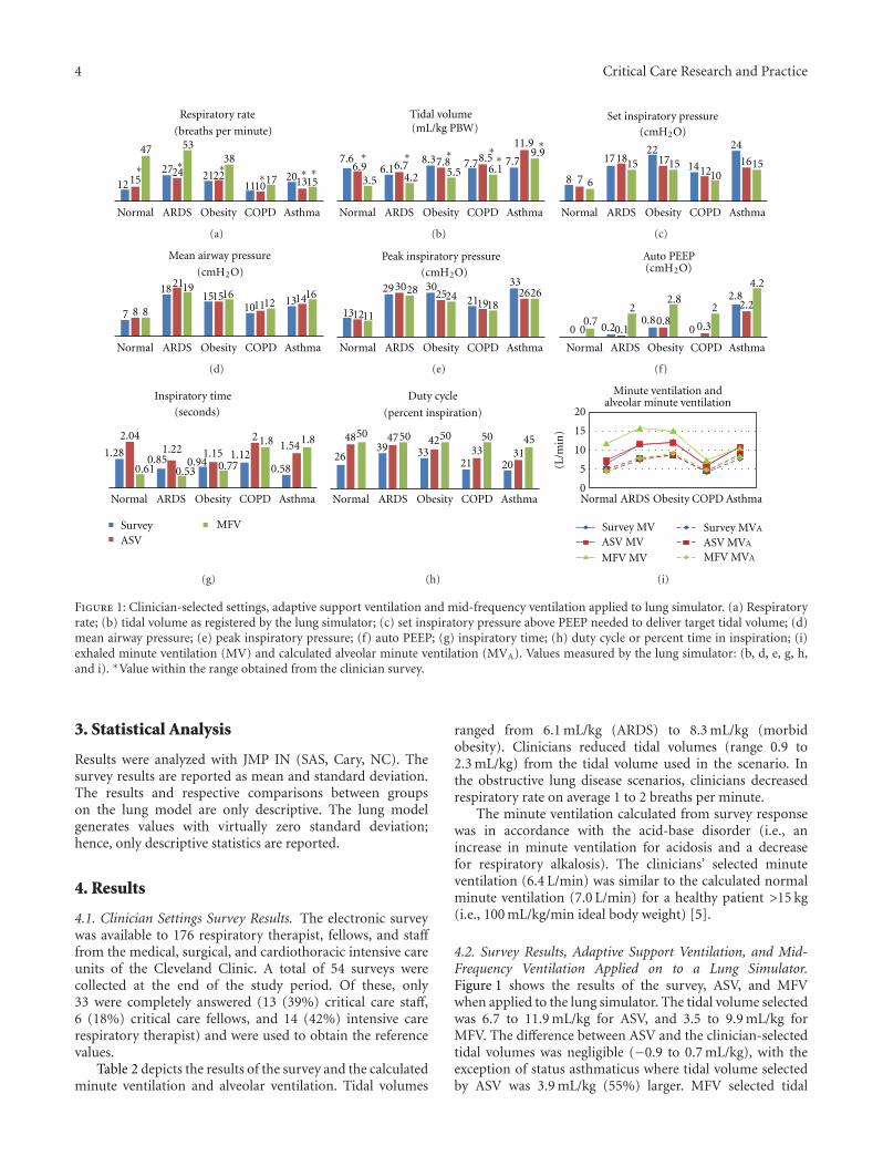
The values for initial ventilator settings for MFV are reportedin Figure 1.
2.3.4. PEEP. Note that the survey asked for PEEP values,and these were used to set the ventilator and the model.However, contrary to clinical practice, PEEP has no effecton the lung simulator’s behavior. More to the point, noneof the computerized systems calculates or sets PEEP. Hence,the comparison between human and computer selection ofventilator settings is focused on frequency and tidal volumeand ventilation outcomes.
4 Critical Care Research and Practice
1227
2111
201524 22
10 13
47 5338
17 15
Normal ARDS Obesity COPD Asthma
Respiratory rate
(breaths per minute)
∗ ∗ ∗∗ ∗ ∗
(a)
7.66.1
8.3 7.7 7.76.9 6.7 7.8 8.511.9
3.5 4.2 5.5 6.1
9.9
Normal ARDS Obesity COPD Asthma
Tidal volume(mL/kg PBW)
∗ ∗ ∗ ∗∗
∗
(b)
8
1722
14
24
7
18 1712
16
6
15 1510
15
Normal ARDS Obesity COPD Asthma
Set inspiratory pressure
(cmH2O)
(c)
7
1815
1013
8
2115
1114
8
1916
1216
Normal ARDS Obesity COPD Asthma
Mean airway pressure
(cmH2O)
(d)
13
29 3021
33
12
3025
1926
11
28 2418
26
Normal ARDS Obesity COPD Asthma
Peak inspiratory pressure
(cmH2O)
(e)
0 0.20.8
0
2.8
0 0.10.8 0.3
2.2
0.72
2.82
4.2
Normal ARDS Obesity COPD Asthma
Auto PEEP(cmH2O)
(f)
1.280.85 0.94
1.120.58
2.041.22 1.15
21.54
0.61 0.53 0.77
1.8 1.8
Normal ARDS Obesity COPD Asthma
Inspiratory time
(seconds)
SurveyASV
MFV
(g)
Normal ARDS Obesity COPD
Duty cycle
(percent inspiration)
Asthma
26
485039
4750
334250
2133
50
2031
45
(h)
0
5
10
15
20
Normal ARDS Obesity COPD Asthma
(L/m
in)
Minute ventilation and alveolar minute ventilation
Survey MVASV MV
MFV MV
Survey MVA
ASV MVA
MFV MVA
(i)
Figure 1: Clinician-selected settings, adaptive support ventilation and mid-frequency ventilation applied to lung simulator. (a) Respiratoryrate; (b) tidal volume as registered by the lung simulator; (c) set inspiratory pressure above PEEP needed to deliver target tidal volume; (d)mean airway pressure; (e) peak inspiratory pressure; (f) auto PEEP; (g) inspiratory time; (h) duty cycle or percent time in inspiration; (i)exhaled minute ventilation (MV) and calculated alveolar minute ventilation (MVA). Values measured by the lung simulator: (b, d, e, g, h,and i). ∗Value within the range obtained from the clinician survey.
3. Statistical Analysis
Results were analyzed with JMP IN (SAS, Cary, NC). Thesurvey results are reported as mean and standard deviation.The results and respective comparisons between groupson the lung model are only descriptive. The lung modelgenerates values with virtually zero standard deviation;hence, only descriptive statistics are reported.
4. Results
4.1. Clinician Settings Survey Results. The electronic surveywas available to 176 respiratory therapist, fellows, and stafffrom the medical, surgical, and cardiothoracic intensive careunits of the Cleveland Clinic. A total of 54 surveys werecollected at the end of the study period. Of these, only33 were completely answered (13 (39%) critical care staff,6 (18%) critical care fellows, and 14 (42%) intensive carerespiratory therapist) and were used to obtain the referencevalues.
Table 2 depicts the results of the survey and the calculatedminute ventilation and alveolar ventilation. Tidal volumes
ranged from 6.1 mL/kg (ARDS) to 8.3 mL/kg (morbidobesity). Clinicians reduced tidal volumes (range 0.9 to2.3 mL/kg) from the tidal volume used in the scenario. Inthe obstructive lung disease scenarios, clinicians decreasedrespiratory rate on average 1 to 2 breaths per minute.
The minute ventilation calculated from survey responsewas in accordance with the acid-base disorder (i.e., anincrease in minute ventilation for acidosis and a decreasefor respiratory alkalosis). The clinicians’ selected minuteventilation (6.4 L/min) was similar to the calculated normalminute ventilation (7.0 L/min) for a healthy patient >15 kg(i.e., 100 mL/kg/min ideal body weight) [5].
4.2. Survey Results, Adaptive Support Ventilation, and Mid-Frequency Ventilation Applied on to a Lung Simulator.Figure 1 shows the results of the survey, ASV, and MFVwhen applied to the lung simulator. The tidal volume selectedwas 6.7 to 11.9 mL/kg for ASV, and 3.5 to 9.9 mL/kg forMFV. The difference between ASV and the clinician-selectedtidal volumes was negligible (−0.9 to 0.7 mL/kg), with theexception of status asthmaticus where tidal volume selectedby ASV was 3.9 mL/kg (55%) larger. MFV selected tidal
Critical Care Research and Practice 5
End inspiratory volume
Tid
al v
olu
me
End expiratory volume
Baseline (0 mL)
750700650600550500450400350300250200150100
50
End inspiratory volume (mL/Kg)
Survey ASV MFV
Normal 12.1 11.5 8.5ARDS 10.3 10.9 9.1 5
Obesity 12.3 11.9 10.9COPD 13.8 14.6 13.7 6Asthma 21.2 24.4 24.7 15 17
Tidal volume (mL/Kg)
Survey ASV MFV
Normal 7.6 6.9 3.5ARDS 6.1 6.7 4.2 10
Obesity 8.3 7.8 5.5
COPD 7.7 8.5 6.1 10Asthma 7.7 11.9 9.9 55 29
End expiratory volume (mL)
Survey ASV MFV
Normal 4.5 4.6 4.8 2 6ARDS 4.6 4.3 4.9 8
Obesity 4.3 4.3 5.4 0 26
COPD 6.2 6.2 7.6 0 24Asthma 13.5 12.8 15.1 12
Lung volumes
−5 −30
−4 −12
−11
−1
−9 −54
−6 −34
−31
−21
−7
−5
%diff1 %diff2
%diff1 %diff2
%diff1 %diff2
%diff1: percent difference between survey and ASV%diff2: percent difference between survey and MFV
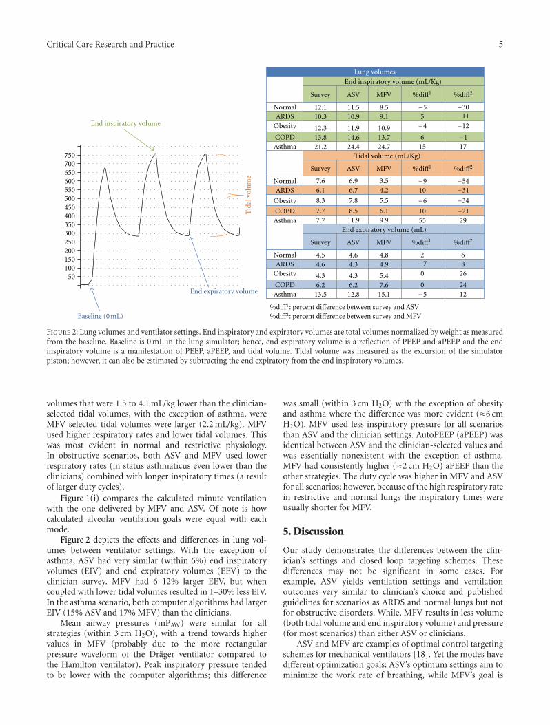
Figure 2: Lung volumes and ventilator settings. End inspiratory and expiratory volumes are total volumes normalized by weight as measuredfrom the baseline. Baseline is 0 mL in the lung simulator; hence, end expiratory volume is a reflection of PEEP and aPEEP and the endinspiratory volume is a manifestation of PEEP, aPEEP, and tidal volume. Tidal volume was measured as the excursion of the simulatorpiston; however, it can also be estimated by subtracting the end expiratory from the end inspiratory volumes.
volumes that were 1.5 to 4.1 mL/kg lower than the clinician-selected tidal volumes, with the exception of asthma, wereMFV selected tidal volumes were larger (2.2 mL/kg). MFVused higher respiratory rates and lower tidal volumes. Thiswas most evident in normal and restrictive physiology.In obstructive scenarios, both ASV and MFV used lowerrespiratory rates (in status asthmaticus even lower than theclinicians) combined with longer inspiratory times (a resultof larger duty cycles).
Figure 1(i) compares the calculated minute ventilationwith the one delivered by MFV and ASV. Of note is howcalculated alveolar ventilation goals were equal with eachmode.
Figure 2 depicts the effects and differences in lung vol-umes between ventilator settings. With the exception ofasthma, ASV had very similar (within 6%) end inspiratoryvolumes (EIV) and end expiratory volumes (EEV) to theclinician survey. MFV had 6–12% larger EEV, but whencoupled with lower tidal volumes resulted in 1–30% less EIV.In the asthma scenario, both computer algorithms had largerEIV (15% ASV and 17% MFV) than the clinicians.
Mean airway pressures (mPAW) were similar for allstrategies (within 3 cm H2O), with a trend towards highervalues in MFV (probably due to the more rectangularpressure waveform of the Drager ventilator compared tothe Hamilton ventilator). Peak inspiratory pressure tendedto be lower with the computer algorithms; this difference
was small (within 3 cm H2O) with the exception of obesityand asthma where the difference was more evident (≈6 cmH2O). MFV used less inspiratory pressure for all scenariosthan ASV and the clinician settings. AutoPEEP (aPEEP) wasidentical between ASV and the clinician-selected values andwas essentially nonexistent with the exception of asthma.MFV had consistently higher (≈2 cm H2O) aPEEP than theother strategies. The duty cycle was higher in MFV and ASVfor all scenarios; however, because of the high respiratory ratein restrictive and normal lungs the inspiratory times wereusually shorter for MFV.
5. Discussion
Our study demonstrates the differences between the clin-ician’s settings and closed loop targeting schemes. Thesedifferences may not be significant in some cases. Forexample, ASV yields ventilation settings and ventilationoutcomes very similar to clinician’s choice and publishedguidelines for scenarios as ARDS and normal lungs but notfor obstructive disorders. While, MFV results in less volume(both tidal volume and end inspiratory volume) and pressure(for most scenarios) than either ASV or clinicians.
ASV and MFV are examples of optimal control targetingschemes for mechanical ventilators [18]. Yet the modes havedifferent optimization goals: ASV’s optimum settings aim tominimize the work rate of breathing, while MFV’s goal is
6 Critical Care Research and Practice
to maximize alveolar ventilation and minimize tidal volume.MFV is not currently available as a mode on ventilators, sothere are no published studies of clinical outcomes. Studieshave evaluated ASV as the sole mode of ventilation [9], or inpatients with stable gas exchange (without reporting baselineventilator settings) [8], or had a specific protocols to set thecomparator ventilator settings (fixed tidal volume, SIMV)regardless of lung disease or mechanics [19–21] or wheredone with ASV prototypes [1, 11]. Our study eliminatedvariability by utilizing the minute ventilation goals chosenby clinicians to set two optimal control modes. These yieldedinformation on the effects of current ventilation strategiesand those of computerized models.
In normal lung physiology, ASV chosen ventilator set-tings were similar to the clinician’s choice and tidal volume(6.9 mL/kg) was within the range considered to be lung pro-tective. MFV used 54% less tidal volume (3.5 mL/kg) whichwas associated with a 30% reduction in end inspiratoryvolume. Interestingly, there was minimal difference in aPEEP,PIP, and mPAW amongst the three settings.
In ARDS, the tidal volume used by ASV was 0.6 mL/kg(10%) higher than the clinician’s choice, well within rangeconsidered to be lung protective [22, 23]. The ASV algorithmuses >1 respiratory time constant to set the inspiratorytime [5] which led to longer inspiratory times and thuscontributed to a higher mPAW compared to the clinicians.In the obesity scenario, where compliance and resistancewere higher, ASV used lower tidal volume (0.5 mL/kg) withslightly longer inspiratory time resulting in the similar airwaypressure. In comparison, in both restrictive disorders, MFVused higher than normal respiratory rates to deliver 31–34% lower tidal volumes than clinicians, resulting in anEEV 8–26% higher (recruitment) and 11-12% lower EIV(stretching). The combination of low EIV and high EEV,especially in restrictive lung disorders, are in concordancewith MFV goal to maximize lung protection, that is,preventing atelectrauma and alveolar stretching.
In obstructive disorders, clinicians used different patternsof ventilation for COPD (low tidal volume/low respiratoryrate) and status asthmaticus (low tidal volume/high respi-ratory rate) while the computerized models used the samepattern (large tidal volume/low respiratory rates) for bothscenarios. Our results are in concordance with the ventilatorsetting patterns found by Arnal et al. [9] and Belliato etal. [8] in COPD (they did not report patients with statusasthmaticus). The discrepancy in the clinician’s ventilationpattern choice for obstructive disease can be explained bythree situations. First, the COPD scenario depicted a patientwith respiratory alkalosis due to overventilation, whichintuitively required less minute ventilation, compared withsevere respiratory acidosis in status asthmaticus (requiringan improvement not only in MV but also in gas exchange).Second, clinicians are used to managing respiratory failuredue to COPD, not status asthmaticus. The reduction incases of status asthmaticus requiring mechanical ventilation[24] may have led clinicians to become less familiar withthe management of this condition. The goal of ventilatormanagement in status asthmatics has been to prolong theexpiratory phase (i.e., decreasing the I : E by low respiratory
rate, high flows, and short inspiratory time) while toleratinghypercapnia and acidosis [25, 26]. This “lack of practice”may also explain the high level of PEEP selected in aparalyzed patient where no trigger asynchrony could occurand where, although controversial, it could worsen airtrapping [27, 28]. Lastly, low tidal volume ventilation is beingapplied to everyone [29]. Although the trend in patient withstatus asthmaticus [24] was present prior to the ARDS netseminal article [23] it was likely enhanced by it. As a matterof fact, the tidal volume recommended in review articlesthrough time has decreased (1980’s: 10–12 [26, 30] 1990’s:8–10 [26, 31] 2000’s: 8–10 [32], 6–8 [28, 33] 5–7 [34, 35]mL/kg) in the absence of any new clinical observations sincethose of Darioli and Perret [25] and Tuxen et al. [26, 36].
There are limitations to our study. First, the surveysampled critical care physicians and respiratory therapistsin a single large academic institution. The poor responserate may have been due to the time of the year (Decemberholidays) and inadequate delivery of the survey (i.e., tosome departments the survey was made available througha website rather than direct email). While recognizing thatthe sample is small and obtained during a holiday period,the objective of the survey was to obtain a measure ofhow clinicians react to ventilation scenarios. The surveymay not represent regional or national practices; however, itrepresents a snap shot of mechanical ventilation in a largeacademic institution during a given moment in time. Itcan be argued that “expert” clinicians would do better thanwhat the survey revealed, or that guidelines indicate differentcourses of action. We concede that setting the ventilatoris a complex process where changes to parameters shouldbe made in response to airway pressure measurements andclinical findings. Further, the initial settings are changedaccording to response, sometimes immediately. Yet, thesurvey results do demonstrate that clinicians sometimesfail to follow guidelines, protocols, or physiology. “Expert”clinicians are not available at the bedside all the time. Thevariability in settings chosen by clinicians, some againstcommon teachings, is the strength of the study. Thisvariablity represents differences in humans experience, levelsof education, and propensity to follow protocols. For betteror worse, computers assure adherence to protocols.
Another limitation is the reliance on a relatively simpleversion of the equation of motion as the basis for themathematical models used by the computer algorithms andthe lung simulator. The equation considers the lung as asingle alveolar unit with constant compliance and resistance,which is an oversimplification of the heterogeneous natureof the lung, particularly in disease states. However, twofactors support its application in this study. First, theequation of motion has been used in commercially availableventilator targeting schemes (Proportional Assist VentilationPlus, Proportional Pressure Support, and Adaptive SupportVentilation) [9, 37]. Second, and most important to ourstudy, Belliato et al. [8] demonstrated that in passiveconditions, the lung simulator we used, when programmedwith the measured patient lung resistance and compliancebehaved identically to the patients studied (same pressures,and volumes). The simulator allowed us to obtain data which
Critical Care Research and Practice 7
would have been impossible to obtain in “real life conditions”since the same patients could not be sensibly placed onthree ventilator settings without changing the clinical andrespiratory system status.
Another limitation is the lack of respiratory effort duringthis study. The absence of respiratory effort in clinicalpractice is the exception rather than the rule. For example,Arnal et al. [9] showed that in spontaneously breathingpatients, the ventilator settings chosen by ASV were similarregardless of physiology, and were only different in extremerestrictive and obstructive lung disease. ASV uses adaptivepressure targeting in spontaneously breathing patients. Thatis, the patient decides the tidal volume and respiratoryrate, thus the observed breathing pattern is less dependenton the ventilator settings and more dependent on thepatient respiratory drive. It is still to be determined whatthe behavior and role of MFV would be in spontaneouslybreathing patients.
Finally, the fact that ASV and MFV use a closed loopcontrol to find the settings to achieve the target minuteventilation means that the initial settings chosen by thedevice are adjusted over the next minutes to achieve thetarget goal. This would inherently bias the results towardsASV and MFV, as the clinician did not have a chanceto optimize its settings based on ventilation outcomes.However, the goal of the study was to demonstrate thedifferences in choices, and given that this was a staticmodel, the settings chosen by the closed loop algorithms hadminimal variation.
6. Conclusions
Computer controlled targeting schemes may result in similarventilator settings to those chosen by a clinician (e.g., ASV innormal lung physiology) or very different settings (e.g., MFVin ARDS physiology delivering less volume and pressure) forthe same minute ventilation goal. The targeting scheme’s goaland its interaction with the lung physiological characteristicsexplain these differences.
Conflict of Interests
The authors declare no conflict of interests.
Acknowledgment
No source of funding to report. Portions of this study werepresented as an abstract/oral presentation at the CHESTmeeting in Philadelphia, USA, in 2008.
References
[1] T. P. Laubscher, A. Frutiger, S. Fanconi, and J. X. Brunner,“The automatic selection of ventilation parameters duringthe initial phase of mechanical ventilation,” Intensive CareMedicine, vol. 22, no. 3, pp. 199–207, 1996.
[2] F. Lellouche, J. Mancebo, P. Jolliet et al., “A multicenter ran-domized trial of computer-driven protocolized weaning from
mechanical ventilation,” American Journal of Respiratory andCritical Care Medicine, vol. 174, no. 8, pp. 894–900, 2006.
[3] E. Kondili, G. Prinianakis, C. Alexopoulou, E. Vakouti, M.Klimathianaki, and D. Georgopoulos, “Respiratory load com-pensation during mechanical ventilation—proportional assistventilation with load-adjustable gain factors versus pressuresupport,” Intensive Care Medicine, vol. 32, no. 5, pp. 692–699,2006.
[4] R. L. Chatburn and E. Mireles-Cabodevila, “Closed-loop con-trol of mechanical ventilation: description and classification oftargeting schemes,” Respiratory Care, vol. 56, no. 1, pp. 85–98,2011.
[5] R. S. Campbell, R. D. Branson, and J. A. Johannigman, “Adapt-ive support ventilation,” Respiratory Care Clinics of NorthAmerica, vol. 7, no. 3, pp. 425–440, 2001.
[6] F. T. Tehrani, “Automatic control of mechanical ventilation.Part 1: theory and history of the technology,” Journal ofClinical Monitoring and Computing, vol. 22, no. 6, pp. 409–415, 2008.
[7] D. M. Linton, G. Renov, J. Lafair, L. Vasiliev, and G. Friedman,“Adaptive Support Ventilation as the sole mode of ventilatorysupport in chronically ventilated patients,” Critical Care andResuscitation, vol. 8, no. 1, pp. 11–14, 2006.
[8] M. Belliato, A. Palo, D. Pasero, G. A. Iotti, F. Mojoli, andA. Braschi, “Evaluation of adaptive support ventilation inparalysed patients and in a physical lung model,” InternationalJournal of Artificial Organs, vol. 27, no. 8, pp. 709–716, 2004.
[9] J. M. Arnal, M. Wysocki, C. Nafati et al., “Automatic selectionof breathing pattern using adaptive support ventilation,”Intensive Care Medicine, vol. 34, no. 1, pp. 75–81, 2008.
[10] E. Mireles-Cabodevila and R. L. Chatburn, “Mid-frequencyventilation: unconventional use of conventional mechanicalventilation as a lung-protection strategy,” Respiratory Care,vol. 53, no. 12, pp. 1669–1677, 2008.
[11] T. P. Laubscher, A. Frutiger, S. Fanconi, H. Jutzi, and J. X.Brunner, “Automatic selection of tidal volume, respiratoryfrequency and minute ventilation in intubated ICU patientsas startup procedure for closed-loop controlled ventilation,”International Journal of Clinical Monitoring and Computing,vol. 11, no. 1, pp. 19–30, 1994.
[12] D. Tassaux, E. Dalmas, P. Gratadour, and P. Jolliet, “Patient-ventilator interactions during partial ventilatory support: apreliminary study comparing the effects of adaptive supportventilation with synchronized intermittent mandatory ventila-tion plus inspiratory pressure support,” Critical Care Medicine,vol. 30, no. 4, pp. 801–807, 2002.
[13] R. W. Schrier, Renal and Electrolyte Disorders, R.W. Schrier2010, Lipincott Williams & Wilkins, Philadelphia, Pa, USA,6th edition.
[14] P. Pelosi, M. Croci, I. Ravagnan, P. Vicardi, and L. Gattinoni,“Total respiratory system, lung, and chest wall mechanicsin sedated-paralyzed postoperative morbidly obese patients,”Chest, vol. 109, no. 1, pp. 144–151, 1996.
[15] C. Broseghini, R. Brandolese, R. Poggi et al., “Respiratorymechanics during the first day of mechanical ventilation inpatients with pulmonary edema and chronic airway obstruc-tion,” American Review of Respiratory Disease, vol. 138, no. 2,pp. 355–361, 1988.
[16] K. P. Steinberg, L. D. Hudson, R. B. Goodman et al., “Efficacyand safety of corticosteroids for persistent acute respiratorydistress syndrome,” The New England Journal of Medicine, vol.354, no. 16, pp. 1671–1684, 2006.
8 Critical Care Research and Practice
[17] M. I. Gold and M. Helrich, “Pulmonary mechanics duringgeneral anesthesia: V. Status asthmaticus,” Anesthesiology, vol.32, no. 5, pp. 422–428, 1970.
[18] R. L. Chatburn, “Classification of ventilator modes: updateand proposal for implementation,” Respiratory Care, vol. 52,no. 3, pp. 301–323, 2007.
[19] P. C. Gruber, C. D. Gomersall, P. Leung et al., “Randomizedcontrolled trial comparing adaptive-support ventilation withpressure-regulated volume-controlled ventilation with auto-mode in weaning patients after cardiac surgery,” Anesthesiol-ogy, vol. 109, no. 1, pp. 81–87, 2008.
[20] C. F. Sulzer, R. Chiolero, P. G. Chassot, X. M. Mueller, andJ. P. Revelly, “Adaptive support ventilation for fast trachealextubation after cardiac surgery: a randomized controlledstudy,” Anesthesiology, vol. 95, no. 6, pp. 1339–1345, 2001.
[21] A. H. Petter, R. L. Chiolero, T. Cassina, P. G. Chassot, X. M.Muller, and J. P. Revelly, “Automatic “Respirator/Weaning”with adaptive support ventilation: the effect on duration ofendotracheal intubation and patient management,” Anesthesiaand Analgesia, vol. 97, no. 6, pp. 1743–1750, 2003.
[22] V. R. Ramnath, D. R. Hess, and B. T. Thompson, “Conven-tional mechanical ventilation in acute lung injury and acuterespiratory distress syndrome,” Clinics in Chest Medicine, vol.27, no. 4, pp. 601–613, 2006.
[23] “Ventilation with lower tidal volumes as compared withtraditional tidal volumes for acute lung injury and the acuterespiratory distress syndrome,” The New England Journal ofMedicine, vol. 342, no. 18, pp. 1301–1308, 2000.
[24] C. C. Kao, S. Jain, K. K. Guntupalli, and V. Bandi, “Mechanicalventilation for asthma: a 10-year experience,” Journal ofAsthma, vol. 45, no. 7, pp. 552–556, 2008.
[25] R. Darioli and C. Perret, “Mechanical controlled hypoventi-lation in status asthmaticus,” American Review of RespiratoryDisease, vol. 129, no. 3, pp. 385–387, 1984.
[26] D. V. Tuxen and S. Lane, “The effects of ventilatory pattern onhyperinflation, airway pressures, and circulation in mechan-ical ventilation of patients with severe air-flow obstruction,”American Review of Respiratory Disease, vol. 136, no. 4, pp.872–879, 1987.
[27] M. P. Caramez, J. B. Borges, M. R. Tucci et al., “Paradoxicalresponses to positive end-expiratory pressure in patients withairway obstruction during controlled ventilation,” CriticalCare Medicine, vol. 33, no. 7, pp. 1519–1528, 2005.
[28] B. D. Medoff, “Invasive and noninvasive ventilation in patientswith asthma,” Respiratory Care, vol. 53, no. 6, pp. 740–748,2008.
[29] O. Gajic, S. I. Dara, J. L. Mendez et al., “Ventilator-associatedlung injury in patients without acute lung injury at the onsetof mechanical ventilation,” Critical Care Medicine, vol. 32, no.9, pp. 1817–1824, 2004.
[30] P. K. Franklin, “Review of acute severe asthma,” WesternJournal of Medicine, vol. 150, no. 5, pp. 552–556, 1989.
[31] B. D. Levy, B. Kitch, and C. H. Fanta, “Medical and ventilatorymanagement of status asthmaticus,” Intensive Care Medicine,vol. 24, no. 2, pp. 105–117, 1998.
[32] M. Oddo, F. Feihl, M. D. Schaller, and C. Perret, “Managementof mechanical ventilation in acute severe asthma: practicalaspects,” Intensive Care Medicine, vol. 32, no. 4, pp. 501–510,2006.
[33] D. R. Stather and T. E. Stewart, “Clinical review: mechanicalventilation in severe asthma,” Critical Care, vol. 9, no. 6, pp.581–587, 2005.
[34] C. S. Barbas, V. Pinheiro Bdo, A. Vianna et al., “Mechanicalventilation in acute asthma crisis,” Jornal Brasileiro de Pneu-mologia, vol. 33, supplement 2, pp. S106–S110, 2007.
[35] P. Phipps and C. S. Garrard, “The pulmonary physician incritical care• 12: acute severe asthma in the intensive careunit,” Thorax, vol. 58, no. 1, pp. 81–88, 2003.
[36] D. V. Tuxen, T. J. Williams, C. D. Scheinkestel, D. Czarny, andG. Bowes, “Use of a measurement of pulmonary hyperinfla-tion to control the level of mechanical ventilation in patientswith acute severe asthma,” American Review of RespiratoryDisease, vol. 146, no. 5, pp. 1136–1142, 1992.
[37] M. Younes, “Proportional assist ventilation, a new approachto ventilatory support: theory,” American Review of RespiratoryDisease, vol. 145, no. 1, pp. 114–120, 1992.

Submit your manuscripts athttp://www.hindawi.com
Stem CellsInternational
Hindawi Publishing Corporationhttp://www.hindawi.com Volume 2014
Hindawi Publishing Corporationhttp://www.hindawi.com Volume 2014
MEDIATORSINFLAMMATION
of
Hindawi Publishing Corporationhttp://www.hindawi.com Volume 2014
Behavioural Neurology
EndocrinologyInternational Journal of
Hindawi Publishing Corporationhttp://www.hindawi.com Volume 2014
Hindawi Publishing Corporationhttp://www.hindawi.com Volume 2014
Disease Markers
Hindawi Publishing Corporationhttp://www.hindawi.com Volume 2014
BioMed Research International
OncologyJournal of
Hindawi Publishing Corporationhttp://www.hindawi.com Volume 2014
Hindawi Publishing Corporationhttp://www.hindawi.com Volume 2014
Oxidative Medicine and Cellular Longevity
Hindawi Publishing Corporationhttp://www.hindawi.com Volume 2014
PPAR Research
The Scientific World JournalHindawi Publishing Corporation http://www.hindawi.com Volume 2014
Immunology ResearchHindawi Publishing Corporationhttp://www.hindawi.com Volume 2014
Journal of
ObesityJournal of
Hindawi Publishing Corporationhttp://www.hindawi.com Volume 2014
Hindawi Publishing Corporationhttp://www.hindawi.com Volume 2014
Computational and Mathematical Methods in Medicine
OphthalmologyJournal of
Hindawi Publishing Corporationhttp://www.hindawi.com Volume 2014
Diabetes ResearchJournal of
Hindawi Publishing Corporationhttp://www.hindawi.com Volume 2014
Hindawi Publishing Corporationhttp://www.hindawi.com Volume 2014
Research and TreatmentAIDS
Hindawi Publishing Corporationhttp://www.hindawi.com Volume 2014
Gastroenterology Research and Practice
Hindawi Publishing Corporationhttp://www.hindawi.com Volume 2014
Parkinson’s Disease
Evidence-Based Complementary and Alternative Medicine
Volume 2014Hindawi Publishing Corporationhttp://www.hindawi.com


![Jamie Roberts, MA, CCRP, MPH[c] Senior Clinical Project ... · Jamie Roberts, MA, CCRP, MPH[c] Senior Clinical Project Manager, CTTI March 1, 2016. ... aspirin or clot busting drugs6-3%](https://img.pdfslide.net/doc/110x75/5b3da7f87f8b9a28308c0959/jamie-roberts-ma-ccrp-mphc-senior-clinical-project-jamie-roberts-ma.jpg)