Embed Size (px)
Citation preview
Hyaluronan Anchored to Activated CD44 on Central NervousSystem Vascular Endothelial Cells Promotes LymphocyteExtravasation in Experimental AutoimmuneEncephalomyelitis*□S
Received for publication, February 24, 2012, and in revised form, July 4, 2012 Published, JBC Papers in Press, August 3, 2012, DOI 10.1074/jbc.M112.356287
Clayton W. Winkler‡, Scott C. Foster‡, Steven G. Matsumoto‡§, Marnie A. Preston‡, Rubing Xing‡, Bruce F. Bebo‡,Fatima Banine‡, Michelle A. Berny-Lang¶, Asako Itakura¶, Owen J. T. McCarty¶, and Larry S. Sherman‡1
From the ‡Division of Neuroscience, Oregon National Primate Research Center, Oregon Health & Science University, Beaverton,Oregon 97006, the ¶Integrative Biosciences Department, School of Dentistry, Oregon Health and Science University, Portland,Oregon 97239, and the §Department of Biomedical Engineering, School of Medicine, Oregon Health & Science University,Portland, Oregon 97239
Background:Multiple sclerosis (MS) is a demyelinating disease involving lymphocyte infiltration into the central nervoussystem (CNS).Results:The glycosaminoglycanhyaluronan (HA), anchored to brain blood vessels via theCD44 receptor, facilitates lymphocytebinding to vessels and CNS infiltration.Conclusion: HA-CD44 interactions on brain endothelial cells facilitate the initiation of inflammatory demyelinating disease.Significance: Findings elucidate mechanisms promoting lymphocyte rolling in inflammatory CNS diseases.
The extravasation of lymphocytes across central nervous sys-tem (CNS) vascular endothelium is a key step in inflammatorydemyelinating diseases including multiple sclerosis (MS) andexperimental autoimmune encephalomyelitis (EAE). The glyco-saminoglycan hyaluronan (HA) and its receptor, CD44, havebeen implicated in this process but their precise roles areunclear. We find that CD44�/� mice have a delayed onset ofEAEcomparedwithwild type animals.Using an in vitro lympho-cyte rolling assay, we find that fewer slow rolling (<1�m/s)wildtype (WT) activated lymphocytes interact with CD44�/� brainvascular endothelial cells (ECs) than withWTECs.We also findthat CD44�/� ECs fail to anchor HA to their surfaces, and thatslow rolling lymphocyte interactions withWTECs are inhibitedwhen the ECs are treated with a pegylated form of the PH20hyaluronidase (PEG-PH20). Subcutaneous injection of PEG-PH20 delays the onset of EAE symptoms by �1 day and tran-siently ameliorates symptoms for 2 days following disease onset.These improved symptoms correspond histologically to degra-dation of HA in the lumen of CNS blood vessels, decreaseddemyelination, and impaired CD4� T-cell extravasation. Col-lectively these data suggest that HA tethered to CD44 on CNSECs is critical for the extravasation of activated T cells into theCNS providing new insight into the mechanisms promotinginflammatory demyelinating disease.
Multiple sclerosis (MS)2 is a central nervous system (CNS)disorder characterized by the extravasation of pathogenic lym-phocytes into the brain and spinal cord. Lymphocyte recruit-ment into the CNS during MS attacks results in inflammation,demyelination, and axonopathy, leading to neurological dis-ability (1, 2). Experimental autoimmune encephalomyelitis(EAE) is an animal model of inflammatory demyelinating dis-ease that recapitulates many of the pathological and clinicaltraits of MS. The molecular events that contribute to lympho-cyte extravasation inMS and EAE include interactions betweenadhesion molecules on the surface of both activated lympho-cytes and CNS vascular endothelial cells (ECs). These interac-tions lead to intracellular signaling events that enhance cell-celladhesion and promote the crossing of lymphocytes across CNSvascular endothelium (3). In most tissues, this process is initi-ated by L-selectin on lymphocytes and P- and E-selectins onECs binding to their transmembrane glycoprotein ligands (4).These transient interactions result in lymphocyte rolling alongthe endothelial cell surface, enabling signaling that inducesexpression and activation of integrins that mediate firm adhe-sion (5). However, the involvement of selectins in EAE andMSpathogenesis is a contentious issue. Although there is evidencethat P- and E-selectins are expressed by superficial blood ves-sels of the brain and that they can mediate rolling (6–8), evi-dence from both antibody blocking experiments and experi-ments with knock-out and transgenic mouse models suggestselectins and their ligands are not essential for the developmentof EAE (9, 10). These findings indicate the involvement of otheradhesive molecules in lymphocyte rolling on vessels within theCNS.
* This work was supported, in whole or in part, by National Institutes of HealthGrants P51 RR000163 and P30 NS061800, Halozyme Therapeutics Inc., theLaura Fund for Multiple Sclerosis Research, National Multiple SclerosisSociety Grant CA1055-A-3, and a Vertex Pharmaceuticals scholar award.
□S This article contains supplemental Figs. S1–S3.1 To whom correspondence should be addressed: 505 N.W. 185th Ave.,
Beaverton, OR 97006. Tel.: 503-690-5217; Fax: 503-690-5384; E-mail:[email protected].
2 The abbreviations used are: MS, multiple sclerosis; EC, endothelial cell; EAE,experimental autoimmune encephalomyelitis; HA, hyaluronan; HABP,biotinylated HA-binding protein; TLR, Toll-like receptor; MOG, myelin oli-godendrocyte glycoprotein; qRT, quantitative reverse transcription.
THE JOURNAL OF BIOLOGICAL CHEMISTRY VOL. 287, NO. 40, pp. 33237–33251, September 28, 2012© 2012 by The American Society for Biochemistry and Molecular Biology, Inc. Published in the U.S.A.
SEPTEMBER 28, 2012 • VOLUME 287 • NUMBER 40 JOURNAL OF BIOLOGICAL CHEMISTRY 33237
by guest on April 3, 2019
http://ww
w.jbc.org/
Dow
nloaded from

CD44 is a single-pass transmembrane glycoprotein widelyexpressed on a number of cell types including lymphocytes andECs (11–14). CD44 functions as a receptor for hyaluronan(HA), a glycosaminoglycan that is synthesized in awide range ofsizes up to �10 � 107 Da. HA is composed of repetitive disac-charide units ofN-acetyl-D-glucosamine and D-glucuronic acid.It is synthesized at the inner plasma membrane by one of threeHA synthases (HAS1–3) that extrude HA into the extracellularmatrix of many cell types including ECs (15, 16). Onceextruded, HA can be tethered to the surface of ECs by HAsynthases or CD44 (17). CD44 and HA have been implicated inregulating cell-cell adhesion, proliferation, migration, and dif-ferentiation (18).CD44 expression andHA synthesis are increased in response
to proinflammatory signals and numerous studies have impli-cated CD44 and HA in lymphocyte-endothelial cell interac-tions as well as the regulation of inflammatory responses (19).Proinflammatory stimulation of lymphocytes and ECs facili-tates post-translationmodification of CD44 inducingHAbind-ing activity (17–19). Disruption of HA-CD44 interactions byanti-CD44 antibodies is sufficient to impair activated T-celladhesion to endothelial cells in vitro (20). Additionally,CD44-HA interactions are required for superantigen-stimu-lated T-cells to efficiently home to sites of inflammation in theperitoneal cavity (21).In the context of inflammatory CNS disease, blocking anti-
bodies against CD44 delay EAE onset and decrease diseaseseverity coincident with fewer lymphocytes present in the CNS(22–24). Similarly, one report found that EAE induced inCD44�/� mice is significantly attenuated (25). However, incontrast to the studies utilizing CD44 blocking antibodies, thisstudy attributed the decrease in EAE disease severity to a phe-notypic shift in the activated lymphocyte population throughan HA-independent mechanism (25). It is unclear, therefore,whether the contribution of CD44 to EAE andMS disease pro-gression is linked to lymphocyte extravasation or alterations inlymphocyte phenotypes. The requirement for HA in EAE onsetand progression is also not clear.To elucidate the role of CD44 and HA in lymphocyte-EC
interactions during EAE pathogenesis, we utilized CD44�/�
mice and a pegylated form of recombinant human PH20 (PEG-PH20) to degrade HA in the lumen of CNS blood vessels. Wefind that HA is tethered by CD44 to the luminal surface ofTNF� stimulated ECs isolated from the brain and that slowrolling lymphocyte interactions are disrupted on ECs lackingCD44. In contrast, CD3/CD28-stimulated CD44�/� lympho-cytes interact normally with wild type brain ECs. Removal ofHA from ECs with PEG-PH20 treatment also results inimpaired lymphocyte rolling. In vivo PEG-PH20 treatmentdelays the onset of EAE and reduces the number of T-cell infil-trates early in disease. These data indicate that HA tethered toCD44 on ECs promotes lymphocyte rolling during EAEpathogenesis.
EXPERIMENTAL PROCEDURES
Induction of EAE—EAEwas induced usingmousemyelin oli-godendrocyte glycoprotein, peptides 35–55 (MOG35–55), syn-thesized artificially by Peptides International. MOG35–55 was
combinedwith complete Freund’s adjuvant containing heat-inac-tivatedmycobacterium tuberculosis as previously described (26).EAEScoring—Beginning the day following EAE induction, an
experimenter, blinded to genotype or treatment condition,assigned a clinical disease score daily until days 13 or 21. Thefollowing clinical disease scoring scale was used: 0, no symp-toms; 1, tail weakness (completely flaccid); 2, hindlimb weak-ness (animal can be easily flipped radially onto its back whengrasped at base of tail); 3, animal walks with hind limbs splayedoutwards; 4, one hindlimb partially or substantially paralyzed;5, both hindlimbs completely paralyzed, no spastic movement;6,moribund (animal is euthanized immediately). Increments of0.5 were used for disease severity between the indicated scores.Hyaluronidase Administration in Mice—PEG-PH20 was
provided by Halyozyme Therapeutics Inc. An aliquot of a PEG-PH20 stock solution was prepared in advance each day thatinjections were to take place. Aliquots were diluted in PBS andpassed through a 0.22-�m low protein binding syringe filter tosterilize the solution. Mice were randomly assigned to twogroups to receive injections every other day of either 50 �l ofsubcutaneous sterile PBS (vehicle control) or 50 �l of subcuta-neous PEG-PH20 (50 units/kg) into hind flanks. Injections con-tinued until the experiment was terminated on days 13 or 21postinduction of EAE or 6 days after beginning the injections inthe case of naive animals.Splenocyte Culture and Isolation—Splenocytes from WT
C57BL/6 mice were cultured in T75 flasks coated with anti-CD3 and anti-CD28 (eBioscience) antibodies for 72 h to induceT-cell-specific activation and clonal expansion as previouslydescribed (27). Cultures were harvested using a Lympholyte�(Sigma) gradient according to themanufacturer’s protocol. Thelymphocyte layer was removed using a sterile Pasteur pipetteand pelleted by centrifugation. The resulting pellet was washedand suspended in RPMImedium supplemented with 1% FBS, 2mM L-glutamine, 50 �M 2-mercaptoethanol, and 1 mM sodiumpyruvate at a concentration of 1� 107 splenocytes/ml. Cultureswere maintained in a humidified 5% CO2, 95% air atmosphereat 37 °C.Murine Primary Brain Endothelial Cell Culture—Primary
brain endothelial cells were isolated and grown as previouslydescribed (28). Briefly, forebrains from 8-week-old WT orCD44�/� mice were isolated, minced, then digested with 1mg/ml of collagenase CLS2 (Worthington Biochemical) inDul-becco’s modified Eagle’s medium (DMEM; Sigma) containing50 �g/ml of gentamycin and 2mM glutamine in a shaker for 1 hat 37 °C. The cell pellet was separated from white matter andother cellular debris by centrifugation in DMEM with 20%bovine serum albumin (1000 � g, 20 min). The microvesselsobtained in the pellet were further digested with 1 mg/ml ofcollagenase/dispase (Roche Applied Science) in DMEM for 45min at 37 °C. Microvessel fragments were separated on a 33%continuous Percoll gradient (1000 � g, 10 min), collected, andwashed twice inDMEMbefore plating on 35-mmplastic dishesor glass coverlips coated with rat tail collagen and humanfibronectin (Sigma) for parallel plate assays or immunocyto-chemistry, respectively. Cultures were maintained in DMEMsupplemented with 20% plasma-derived bovine serum (AtlasBiologicals), 1 ng/ml of fibroblast growth factor-2 (R&D Sys-
HA Anchored to CD44 on ECs Mediates Lymphocyte Extravasation
33238 JOURNAL OF BIOLOGICAL CHEMISTRY VOLUME 287 • NUMBER 40 • SEPTEMBER 28, 2012
by guest on April 3, 2019
http://ww
w.jbc.org/
Dow
nloaded from
tems), and 4 �g/ml of puromycin (Sigma) in a humidified 5%CO2, 95% air atmosphere at 37 °C. Four days following plating,EC cultures visually contained continuous monolayers ofadherent spindle-shaped cells consistent with previously pub-lished findings (28) (supplemental Fig. S1A). EC cultures con-tained strong CD31 (an endothelial marker) labeling thatclearly delimited the cell membrane of most Hoechst� nuclei(supplemental Fig. S1B). No GFAP� astrocyte contaminationwas observed in these cultures (supplemental Fig. S1B, bottompanel).Parallel Plate Assay—Lymphocyte adhesion and rolling
along brain ECs was quantified under flow conditions using aparallel-plate flow chamber. A 35-mm dish with an EC mono-layer was assembled to a flow chamber (150-�mchannel depth,1.26-mm channel width) and mounted on the stage of a ZeissAxiovert 200M microscope (Zeiss) and maintained at 37 °C inan air curtain incubator. The exit port was connected to aninfuse and withdraw syringe pump (Harvard Apparatus) tocontrol flow rate through the chamber. The microscope wasequippedwith aCCDcamera (AxiocamMRm,Zeiss) and imag-ing software (Stallion SlideBook version 5.0.0.10, IntelligentImaging Innovations, Inc.) for monitoring cell movement.EC monolayers were stimulated with 10 ng/ml of TNF-� for
4 h prior to flow chamber assembly to increase CD44 surfaceexpression and HA binding activity (e.g. Fig. 3B). In selectedexperiments, EC monolayers were incubated with eithersterile PBS as vehicle control or 100 units/ml of PEG-PH20(Halozyme, Inc.) in PBS for 1 h at 37 °C prior to use in the flowchamber assembly. After monolayers were washed with Hanks’balanced salt solution, 0.6ml ofCD3/CD28 stimulated lympho-cytes (at 1 � 106 total cells/ml in Hanks’ balanced salt solution)were superfused through the chamber for 7 min at 0.5 dyn/cm2
thereby mimicking the fluid mechanical environment of CNSpostcapillary venules.Interactions between lymphocytes and brain ECs were visu-
alized in real-time by phase-contrast digital video microscopy.A single field of view (�10; 0.55 mm2) was monitored duringeach trial. The number of total interacting cells and the averagerolling velocity of each interacting cell were analyzed for eachexperiment. Interacting lymphocyteswere defined as those thatinteracted with the EC monolayer for at least 1 s. Average roll-ing speedwas determined using the particle tracking features ofthe Stallion imaging software. Criteria were set with the auto-mated particle tracking function to exclude mask objects thatwere: 1) not phase bright, 2) less than 8 �m in diameter, and 3)weremoving faster than 100�m/s. Each particle pathwasman-ually examined and in some cases, manual particle tracking wasused to correct inaccuracies in the automated path.CD44 Exon-specific RT-PCR Analysis—Total RNA was iso-
lated from WT and CD44�/� EC cultures and CD3/CD28-stimulated lymphocytes using TRIzol reagent (Invitrogen).cDNAs were synthesized using MultiScribeTM reverse tran-scriptase (Applied Biosystems) according to themanufacturer’sprotocol and a CD44 primer from the 3� nonvariant portion ofmouse CD44 (exon 19, 5�-tag gca cta cac ccc aat ctt ca-3�).cDNA products were amplified using Phusion� Hot Start IIDNA Polymerase (Finnzymes) and a primer from the 5� non-variant region of mouse CD44 (exon 1, 5�-tcc ctc cgt ttc atc cag
cac-3�) and another primer straddling two nonvariant 3� exons(exon 16–17, 5�-ggt tcg cac ttg agt gtc ca-3�). The PCR wasperformed using a Mastercycler thermocycler (Eppendorf)with the following protocol: 40 cycles of 10 s at 98 °C, 30 s at64 °C, and 1.5 min at 72 °C, followed by incubation at 72 °Cfor 5 min. This reaction was stopped at the end of the 25thcycle and 2 �l was removed for CD44 variant-specific nestedprimer analysis. The thermocycler protocol was subse-quently re-started at the 25th cycle and the reaction wascontinued to completion.The 2 �l from the above reaction was amplified using the
same reagents and protocols listed above with the exceptionthat variant exons 6 (5�-tgg ttt cag aac gga tgg cag g-3�), 7 (5�-ccacaa caa cca tcc aag tca aa-3�), and 10 (5�-tct tcc cac aga tac aactac tt-3�) specific nested 5� primers were added. All reactionproducts were analyzed by electrophoresis in 1.5% agarose andvisualized by ethidium bromide staining.SYBRGreen I Real-time RT-PCRAnalysis (qRT-PCR)—Total
RNA from EC cultures was obtained as above and single-stranded cDNAs were synthesized using the ImProm-IIReverse Transcriptase synthesis kit (Promega Corporation)according to themanufacturer’s protocol. The primer sets usedwere designed using Primer Express� software version 3.0(Applied Biosystems) and synthesized by Integrated DNATechnologies. The primer sequences were: HAS1 forward,5�-gcg agc act cag gat cat ctt-3� and reverse, 5�-cca gga gtc catagc gat ctg-3�; HAS2 forward, 5�-aaa ggg acc tgg tga gac aga a-3�and reverse, 5�-ccc att ttt gca tga tgc aa-3�; HAS3 forward,5�-gcg cat tgc ctt tcc aaa-3� and reverse, 5�-tgc cac cca gca cctcat-3�. The 18 S ribosomal RNAwas used as a normalizing unitfor each reaction. Primer sets were purchased as a kit (TaqManRibosomal RNA Control Reagents Kit; Applied Biosystems).The qPCR assays were carried out with Platinum SYBR GreenqPCR Supermix-UDG (Invitrogen) in a 7500 Fast TaqManinstrument (Applied Biosystems) using a default thermocyclingprogram. Assays were performed in triplicate. The normalizedexpression of the target genewith respect to 18 Swas computedfor all samples using the ��CT method in Microsoft Excel.HA Quantification—HA was quantified from culture me-
dium supernatant or cellular lysates of WT and CD44�/� ECscultured to confluence in 35-mm dishes. Cell lysates wereobtained by incubation of EC monolayers in 200 �l of buffercontaining 20mMTris-HCl, 150mMNaCl, 1mMEDTA, and 1%Triton X-100. Samples were applied to an enzyme-linkedimmunosorbent assay (ELISA)-based assay (Echelon Biosci-ences) according to the manufacturer’s instructions. Mediumsupernatant samples were diluted 1:4 and cell lysates 1:2 to afinal volume of 350�l in the kit diluent buffer. Triplicate 100-�lfractions were transferred into the ELISA plate. At the end ofthe assay, absorbances were read at 450 nm on a 96-well platereader (Molecular Devices).Immunohistochemistry—At appropriate EAE time points,
micewere euthanized using isoflurane (NovaPlus) andperfusedtranscardially with heparin saline followed by 4% paraformal-dehyde. Lumbar spinal cords were removed, freeze-embedded,and serially sectioned at a thickness of 10 �m on a cryostat(Leica) and placed on glass slides. Sections were washed 3 � 5min with PBS and blocked in PBS with 5% BSA with 0.05%
HA Anchored to CD44 on ECs Mediates Lymphocyte Extravasation
SEPTEMBER 28, 2012 • VOLUME 287 • NUMBER 40 JOURNAL OF BIOLOGICAL CHEMISTRY 33239
by guest on April 3, 2019
http://ww
w.jbc.org/
Dow
nloaded from
Triton X-100 (blocking buffer) for 1 h at room temperature.Sections were then incubated with primary antibodies againstCD4 (1:300, BD Pharmigen), CD31 (1:50, Ab cam), neurofila-ment-L (1:1000,Millipore), and/orHAS1 (1:25, SantaCruzBio-technology) in PBS overnight at 4 °C. Biotinylated HA-bindingprotein (bHABP, 1:250, Calbiochem) was used in place of aprimary antibody to visualize HA. Negative controls includedomitting the primary antibody. Sectionswerewashed 3� 5minwith PBS the next day and then incubated with the appropriatesecondary antibody (goat anti-rat Alexa 488 and goat anti-rab-bit Alexa 633, 1:1000, Molecular Probes) or Cy3 streptavidin(1:2000, Jackson Labs) in place of secondary to visualize HA for2 h at room temperature. Sections were washed in PBS, andthen incubated in FluoroMyelin (1:300, Invitrogen) for 20 minat room temperature to visualize myelin and Hoechst 33342(1:5000, Invitrogen) for 10 min at room temperature to labelnuclei. Sectionswerewashed in PBS andmountedwith ProlongGold mounting media (Invitrogen), then imaged using a ZeissAxioskop 40 fluorescence microscope (Zeiss) or an invertedLeica SP5 AOBS spectral confocal system (Leica).Immunocytochemistry—EC cultures on coverslips were fixed
in 4% paraformaldehyde in PBS at room temperature for 15min, rinsed with PBS, and treated for 1 h with blocking buffer(see above). Cultures were incubated overnight at 4 °C with theprimary antibody diluted in PBS. Cells were stained with anti-bodies against CD44 (IM7 hybridoma, 1:40, ATCC), CD31 (seeabove), and glial fibrillary acidic protein (GFAP, 1:500, Dako).bHABP (see above) was used to visualize HA. Subsequently,cells were rinsed 3 � 5 min in PBS and incubated with therelevant secondary antibodies or Cy3 streptavidin as above for2 h at room temperature. Cultureswere rinsed 3� 5min in PBSand incubated in Hoechst for 10 min, mounted, and examinedas above.Stereology—Following immunohistochemical labeling, 12
sections of the lumbar spinal cord were analyzed from eachanimal (n � 8 animals per group). Digital photomicroscopicimageswere captured on a Zeiss Axiovert 200M (Zeiss) fluores-cence microscope interfaced with a Marianas Digital Micros-copyWork station (Intelligent Imaging Innovation Inc.). Mon-tages of the entire lumbar cordwere created and analyzed using
SlideBookTM software. Using FluoroMyelin labeling, a maskdelimiting the spinal cord white matter was created for eachsection and a grid was generated within the mask area for anal-ysis. The grid consisted of 10-�m boxes spaced 50 �m apart inthe xy axis with each box representing a 2500-�m square area.Based on FluoroMyelin labeling, boxes that fell entirely withinthe lesion areas were counted and divided by the total numberof boxes within the grid and multiplied by 100 to determine apercent lesion area.Infiltrating T-cell counts were performed by setting a thresh-
old value for CD4 immunolabeling that resolved single cells.Subsequently, a mask was applied within the CD4 channel torecord each cell’s position in the section. The software was thenused to overlay the CD4 mask onto the mask generated for thelesion analysis and to record the number of CD4� cells withinthe white matter in each section.Statistical Analysis—Differences between treatment groups
in HA ELISA and parallel plate assays were analyzed by a Stu-dent’s t test. Differences in mean EAE disease score betweengroups were analyzed by a repeated measures analysis of vari-ance. Statistical significance was defined as p � 0.05 for allanalyses.
RESULTS
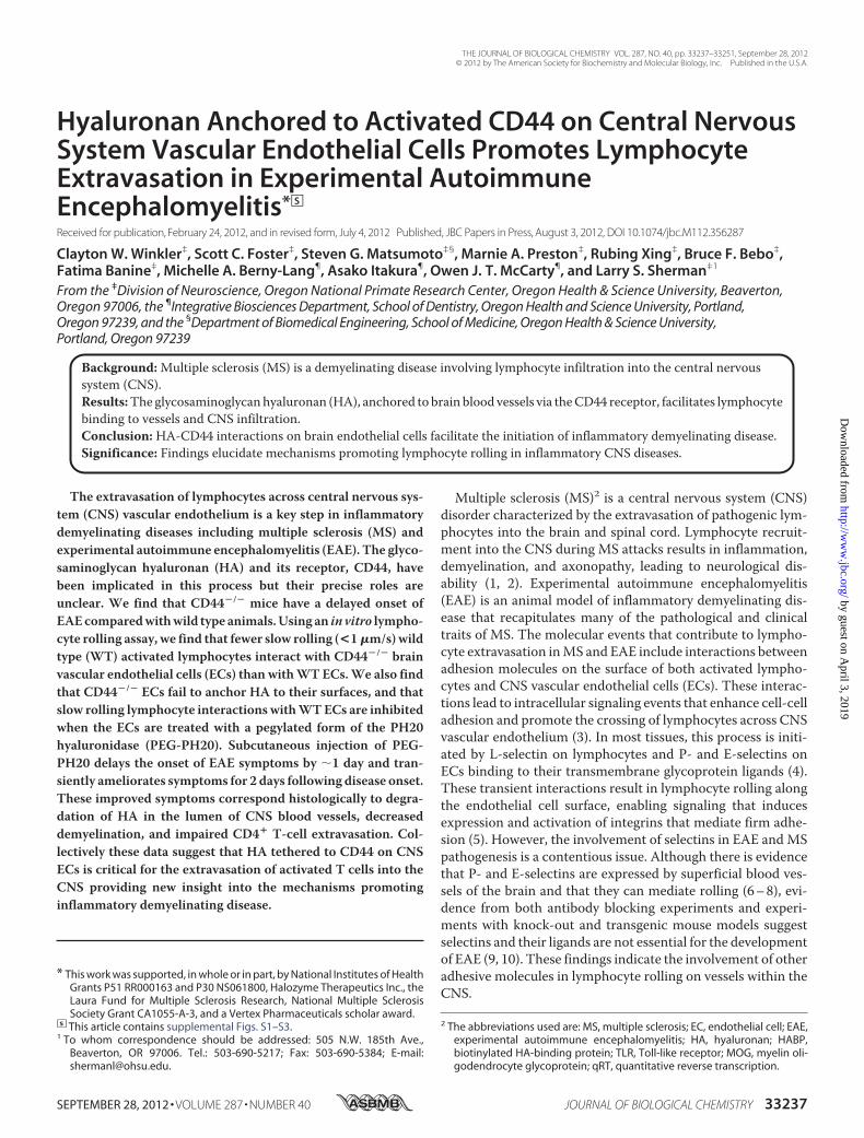
EAEOnset Is Delayed in CD44�/� Mice—Inhibition of CD44activity using neutralizing antibodies (22–24) or deletion of theCD44 gene (25) are reported to attenuate EAE onset and pro-gression. To confirm that genetic ablation of CD44 is sufficientto ameliorate EAE symptoms, we tested how EAE progresses inCD44�/� mice that develop severe disease. Clinical diseasesigns were observed over a 25-day period in 8-week-old femaleWT and CD44�/� mice on a C57B6;129 background, activelyimmunized with MOG35–55. Disease was evident beginning atday 10 postinoculation inWT animals with peak scores occur-ring by day 14 (Fig. 1A). Consistent with the findings of Guanand co-workers (25), CD44�/� mice manifested disease with adelayed onset, no earlier than day 14 postinoculation and as lateas day 18 with an average peak disease on day 16 (Fig. 1B).However, in contrast to the previous study (25), we found thatCD44�/� mice developed similar levels of disability. For both
FIGURE 1. EAE disease onset is delayed in CD44�/� animals. Disease was induced by inoculation of female WT (A) or CD44�/� (B) C57/B6/S129 backgroundmice with MOG35–55 peptide emulsified in CFA. Disease symptoms manifest on or near day 10 postinoculation in WT animals but 3– 8 days later in CD44�/�. represents animals being euthanized due to severe disease.
HA Anchored to CD44 on ECs Mediates Lymphocyte Extravasation
33240 JOURNAL OF BIOLOGICAL CHEMISTRY VOLUME 287 • NUMBER 40 • SEPTEMBER 28, 2012
by guest on April 3, 2019
http://ww
w.jbc.org/
Dow
nloaded from
groups, scores remained elevated to at least a score of 2.5 after20 days postinoculation. These data demonstrate that CD44contributes to the initiation of EAE onset but is not necessaryfor disease progression.CD44 on Brain ECs but Not Lymphocytes Is Critical for Lym-
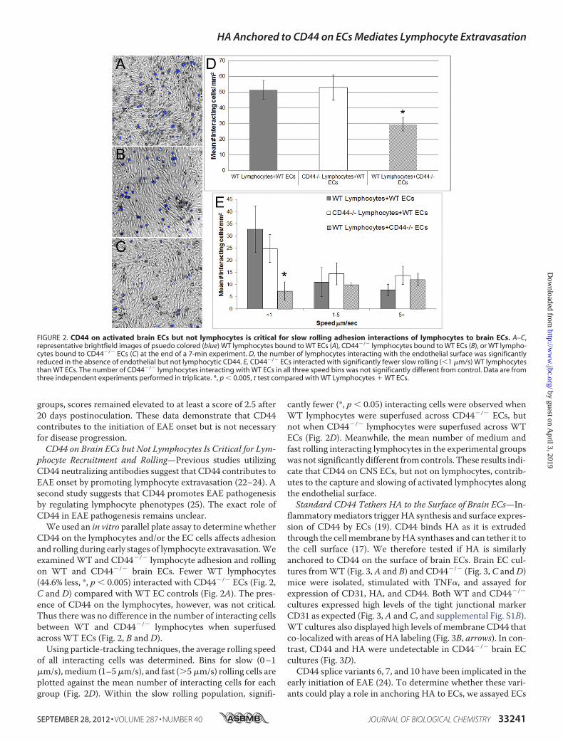
phocyte Recruitment and Rolling—Previous studies utilizingCD44 neutralizing antibodies suggest that CD44 contributes toEAE onset by promoting lymphocyte extravasation (22–24). Asecond study suggests that CD44 promotes EAE pathogenesisby regulating lymphocyte phenotypes (25). The exact role ofCD44 in EAE pathogenesis remains unclear.We used an in vitro parallel plate assay to determine whether
CD44 on the lymphocytes and/or the EC cells affects adhesionand rolling during early stages of lymphocyte extravasation.Weexamined WT and CD44�/� lymphocyte adhesion and rollingon WT and CD44�/� brain ECs. Fewer WT lymphocytes(44.6% less, *, p � 0.005) interacted with CD44�/� ECs (Fig. 2,C and D) compared with WT EC controls (Fig. 2A). The pres-ence of CD44 on the lymphocytes, however, was not critical.Thus there was no difference in the number of interacting cellsbetween WT and CD44�/� lymphocytes when superfusedacross WT ECs (Fig. 2, B and D).Using particle-tracking techniques, the average rolling speed
of all interacting cells was determined. Bins for slow (0–1�m/s),medium (1–5�m/s), and fast (5�m/s) rolling cells areplotted against the mean number of interacting cells for eachgroup (Fig. 2D). Within the slow rolling population, signifi-
cantly fewer (*, p � 0.05) interacting cells were observed whenWT lymphocytes were superfused across CD44�/� ECs, butnot when CD44�/� lymphocytes were superfused across WTECs (Fig. 2D). Meanwhile, the mean number of medium andfast rolling interacting lymphocytes in the experimental groupswas not significantly different fromcontrols. These results indi-cate that CD44 on CNS ECs, but not on lymphocytes, contrib-utes to the capture and slowing of activated lymphocytes alongthe endothelial surface.Standard CD44 Tethers HA to the Surface of Brain ECs—In-
flammatorymediators triggerHA synthesis and surface expres-sion of CD44 by ECs (19). CD44 binds HA as it is extrudedthrough the cellmembrane byHA synthases and can tether it tothe cell surface (17). We therefore tested if HA is similarlyanchored to CD44 on the surface of brain ECs. Brain EC cul-tures fromWT (Fig. 3, A and B) and CD44�/� (Fig. 3, C andD)mice were isolated, stimulated with TNF�, and assayed forexpression of CD31, HA, and CD44. Both WT and CD44�/�
cultures expressed high levels of the tight junctional markerCD31 as expected (Fig. 3, A and C, and supplemental Fig. S1B).WT cultures also displayed high levels of membrane CD44 thatco-localized with areas of HA labeling (Fig. 3B, arrows). In con-trast, CD44 and HA were undetectable in CD44�/� brain ECcultures (Fig. 3D).CD44 splice variants 6, 7, and 10 have been implicated in the
early initiation of EAE (24). To determine whether these vari-ants could play a role in anchoring HA to ECs, we assayed ECs
FIGURE 2. CD44 on activated brain ECs but not lymphocytes is critical for slow rolling adhesion interactions of lymphocytes to brain ECs. A–C,representative brightfield images of psuedo colored (blue) WT lymphocytes bound to WT ECs (A), CD44�/� lymphocytes bound to WT ECs (B), or WT lympho-cytes bound to CD44�/� ECs (C) at the end of a 7-min experiment. D, the number of lymphocytes interacting with the endothelial surface was significantlyreduced in the absence of endothelial but not lymphocytic CD44. E, CD44�/� ECs interacted with significantly fewer slow rolling (�1 �m/s) WT lymphocytesthan WT ECs. The number of CD44�/� lymphocytes interacting with WT ECs in all three speed bins was not significantly different from control. Data are fromthree independent experiments performed in triplicate. *, p � 0.005, t test compared with WT Lymphocytes � WT ECs.
HA Anchored to CD44 on ECs Mediates Lymphocyte Extravasation
SEPTEMBER 28, 2012 • VOLUME 287 • NUMBER 40 JOURNAL OF BIOLOGICAL CHEMISTRY 33241
by guest on April 3, 2019
http://ww
w.jbc.org/
Dow
nloaded from
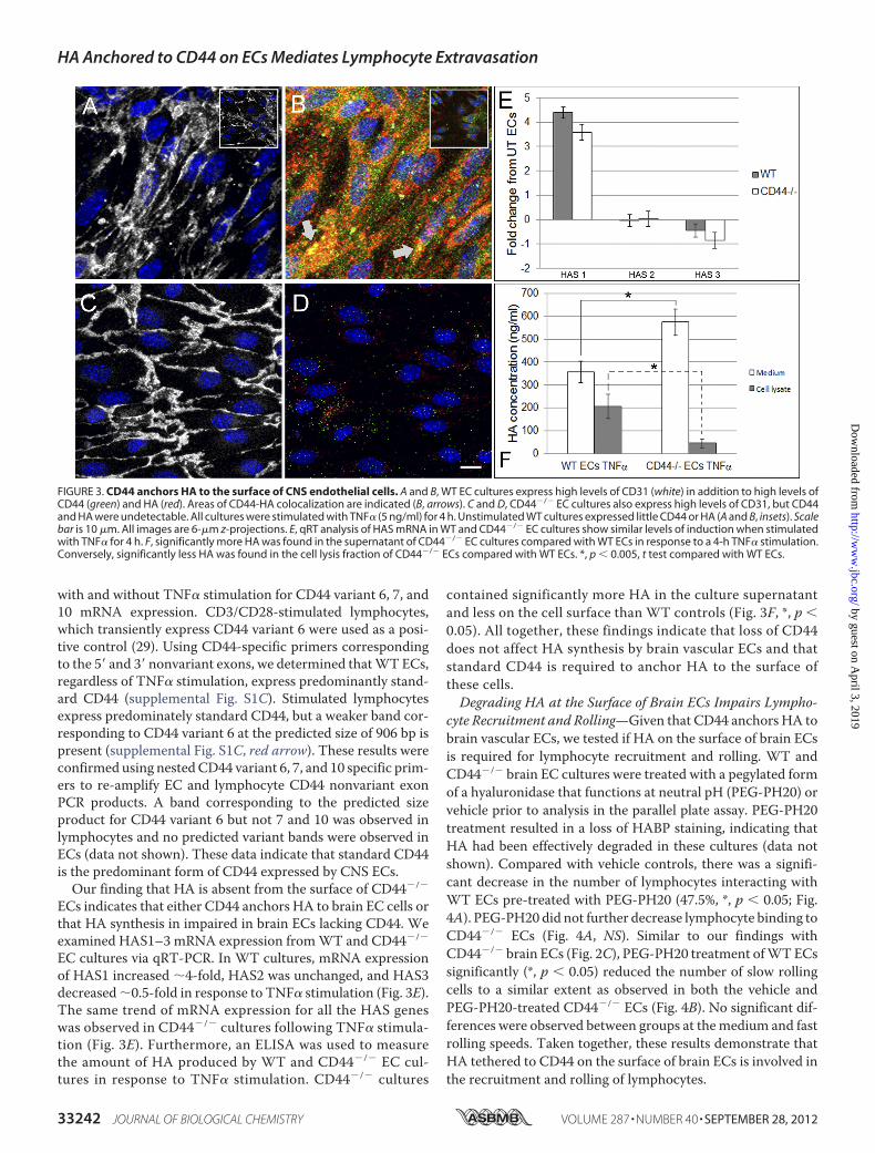
with and without TNF� stimulation for CD44 variant 6, 7, and10 mRNA expression. CD3/CD28-stimulated lymphocytes,which transiently express CD44 variant 6 were used as a posi-tive control (29). Using CD44-specific primers correspondingto the 5� and 3� nonvariant exons, we determined thatWTECs,regardless of TNF� stimulation, express predominantly stand-ard CD44 (supplemental Fig. S1C). Stimulated lymphocytesexpress predominately standard CD44, but a weaker band cor-responding to CD44 variant 6 at the predicted size of 906 bp ispresent (supplemental Fig. S1C, red arrow). These results wereconfirmed using nestedCD44 variant 6, 7, and 10 specific prim-ers to re-amplify EC and lymphocyte CD44 nonvariant exonPCR products. A band corresponding to the predicted sizeproduct for CD44 variant 6 but not 7 and 10 was observed inlymphocytes and no predicted variant bands were observed inECs (data not shown). These data indicate that standard CD44is the predominant form of CD44 expressed by CNS ECs.Our finding that HA is absent from the surface of CD44�/�
ECs indicates that either CD44 anchors HA to brain EC cells orthat HA synthesis in impaired in brain ECs lacking CD44. Weexamined HAS1–3 mRNA expression fromWT and CD44�/�
EC cultures via qRT-PCR. In WT cultures, mRNA expressionof HAS1 increased �4-fold, HAS2 was unchanged, and HAS3decreased�0.5-fold in response to TNF� stimulation (Fig. 3E).The same trend of mRNA expression for all the HAS geneswas observed in CD44�/� cultures following TNF� stimula-tion (Fig. 3E). Furthermore, an ELISA was used to measurethe amount of HA produced by WT and CD44�/� EC cul-tures in response to TNF� stimulation. CD44�/� cultures
contained significantly more HA in the culture supernatantand less on the cell surface than WT controls (Fig. 3F, *, p �0.05). All together, these findings indicate that loss of CD44does not affect HA synthesis by brain vascular ECs and thatstandard CD44 is required to anchor HA to the surface ofthese cells.Degrading HA at the Surface of Brain ECs Impairs Lympho-
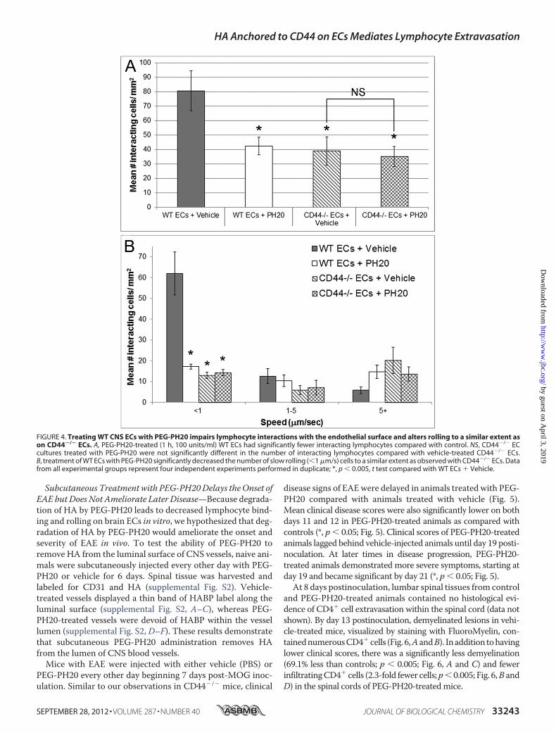
cyte Recruitment and Rolling—Given that CD44 anchors HA tobrain vascular ECs, we tested if HA on the surface of brain ECsis required for lymphocyte recruitment and rolling. WT andCD44�/� brain EC cultures were treated with a pegylated formof a hyaluronidase that functions at neutral pH (PEG-PH20) orvehicle prior to analysis in the parallel plate assay. PEG-PH20treatment resulted in a loss of HABP staining, indicating thatHA had been effectively degraded in these cultures (data notshown). Compared with vehicle controls, there was a signifi-cant decrease in the number of lymphocytes interacting withWT ECs pre-treated with PEG-PH20 (47.5%, *, p � 0.05; Fig.4A). PEG-PH20 did not further decrease lymphocyte binding toCD44�/� ECs (Fig. 4A, NS). Similar to our findings withCD44�/� brain ECs (Fig. 2C), PEG-PH20 treatment ofWTECssignificantly (*, p � 0.05) reduced the number of slow rollingcells to a similar extent as observed in both the vehicle andPEG-PH20-treated CD44�/� ECs (Fig. 4B). No significant dif-ferences were observed between groups at themedium and fastrolling speeds. Taken together, these results demonstrate thatHA tethered to CD44 on the surface of brain ECs is involved inthe recruitment and rolling of lymphocytes.
FIGURE 3. CD44 anchors HA to the surface of CNS endothelial cells. A and B, WT EC cultures express high levels of CD31 (white) in addition to high levels ofCD44 (green) and HA (red). Areas of CD44-HA colocalization are indicated (B, arrows). C and D, CD44�/� EC cultures also express high levels of CD31, but CD44and HA were undetectable. All cultures were stimulated with TNF� (5 ng/ml) for 4 h. Unstimulated WT cultures expressed little CD44 or HA (A and B, insets). Scalebar is 10 �m. All images are 6-�m z-projections. E, qRT analysis of HAS mRNA in WT and CD44�/� EC cultures show similar levels of induction when stimulatedwith TNF� for 4 h. F, significantly more HA was found in the supernatant of CD44�/� EC cultures compared with WT ECs in response to a 4-h TNF� stimulation.Conversely, significantly less HA was found in the cell lysis fraction of CD44�/� ECs compared with WT ECs. *, p � 0.005, t test compared with WT ECs.
HA Anchored to CD44 on ECs Mediates Lymphocyte Extravasation
33242 JOURNAL OF BIOLOGICAL CHEMISTRY VOLUME 287 • NUMBER 40 • SEPTEMBER 28, 2012
by guest on April 3, 2019
http://ww
w.jbc.org/
Dow
nloaded from
Subcutaneous Treatment with PEG-PH20Delays theOnset ofEAE but Does Not Ameliorate Later Disease—Because degrada-tion of HA by PEG-PH20 leads to decreased lymphocyte bind-ing and rolling on brain ECs in vitro, we hypothesized that deg-radation of HA by PEG-PH20 would ameliorate the onset andseverity of EAE in vivo. To test the ability of PEG-PH20 toremove HA from the luminal surface of CNS vessels, naive ani-mals were subcutaneously injected every other day with PEG-PH20 or vehicle for 6 days. Spinal tissue was harvested andlabeled for CD31 and HA (supplemental Fig. S2). Vehicle-treated vessels displayed a thin band of HABP label along theluminal surface (supplemental Fig. S2, A–C), whereas PEG-PH20-treated vessels were devoid of HABP within the vessellumen (supplemental Fig. S2, D–F). These results demonstratethat subcutaneous PEG-PH20 administration removes HAfrom the lumen of CNS blood vessels.Mice with EAE were injected with either vehicle (PBS) or
PEG-PH20 every other day beginning 7 days post-MOG inoc-ulation. Similar to our observations in CD44�/� mice, clinical
disease signs of EAEwere delayed in animals treated with PEG-PH20 compared with animals treated with vehicle (Fig. 5).Mean clinical disease scores were also significantly lower on bothdays 11 and 12 in PEG-PH20-treated animals as compared withcontrols (*, p � 0.05; Fig. 5). Clinical scores of PEG-PH20-treatedanimals lagged behind vehicle-injected animals until day 19 posti-noculation. At later times in disease progression, PEG-PH20-treated animals demonstrated more severe symptoms, starting atday 19 and became significant by day 21 (*, p � 0.05; Fig. 5).At 8 days postinoculation, lumbar spinal tissues from control
and PEG-PH20-treated animals contained no histological evi-dence of CD4� cell extravasation within the spinal cord (data notshown). By day 13 postinoculation, demyelinated lesions in vehi-cle-treated mice, visualized by staining with FluoroMyelin, con-tainednumerousCD4�cells (Fig. 6,AandB). Inaddition tohavinglower clinical scores, there was a significantly less demyelination(69.1% less than controls; p � 0.005; Fig. 6, A and C) and fewerinfiltratingCD4� cells (2.3-fold fewer cells;p� 0.005; Fig. 6,B andD) in the spinal cords of PEG-PH20-treatedmice.
FIGURE 4. Treating WT CNS ECs with PEG-PH20 impairs lymphocyte interactions with the endothelial surface and alters rolling to a similar extent ason CD44�/� ECs. A, PEG-PH20-treated (1 h, 100 units/ml) WT ECs had significantly fewer interacting lymphocytes compared with control. NS, CD44�/� ECcultures treated with PEG-PH20 were not significantly different in the number of interacting lymphocytes compared with vehicle-treated CD44�/� ECs.B, treatment of WT ECs with PEG-PH20 significantly decreased the number of slow rolling (�1 �m/s) cells to a similar extent as observed with CD44�/� ECs. Datafrom all experimental groups represent four independent experiments performed in duplicate; *, p � 0.005, t test compared with WT ECs � Vehicle.
HA Anchored to CD44 on ECs Mediates Lymphocyte Extravasation
SEPTEMBER 28, 2012 • VOLUME 287 • NUMBER 40 JOURNAL OF BIOLOGICAL CHEMISTRY 33243
by guest on April 3, 2019
http://ww
w.jbc.org/
Dow
nloaded from

To address the possibility that delayed disease onset in PEG-PH20-treated animals is related to reduced axonopathy, spinalcords from naive, day 13 control and PEG-PH20-treated ani-
mals were labeled with neurofilament-L. Representative, highmagnification images of similar sized lesion areas are illustratedin supplemental Fig. S3. PEG-PH20-treated animals had fewerdysmorphic axons than controls (supplemental Fig. S3, F and E,red arrows). Additionally, more denuded axons were evident inPEG-PH20-treated lesions than controls (supplemental Fig. S3,H and I, white arrows).
Stereologic analysis of spinal cords from mice with EAE 21days postinoculation revealed that PEG-PH20 treatment con-tinued to significantly reduce the degree of demyelination albeitto a lesser extent than at day 13 (24.4% smaller lesion volumethan controls; Fig. 7, A versus C and E). However, consistentwith the elevated disease scores at this time point, these animalsdemonstrated significantly increased numbers of infiltratingT-cells (1.9-fold more than controls; Fig. 7, C versus D and F).Additionally, the number of neurofilament-L positive axonsproximal to and within areas of demyelination was visuallyindistinguishable between treatment groups (supplemental Fig.S3, J and K).
Overall, these data suggest that PEG-PH20 treatmenteffectively delays disease onset by impairing T-cell extravasa-tion resulting in less CNS inflammation, demyelination, andaxonopathy early in disease. However, prolonged administra-tion of PEG-PH20 increases CNS inflammation resulting inmore severe clinical disease and negates/diminishes, respec-tively, its benefit to axon survival/demyelination observed inearly disease.
FIGURE 5. Subcutaneously injected PEG-PH20 delays the clinical onset ofactive EAE but does not ameliorate disease severity later in disease. EAEwas induced in 48 C57BL/6 female mice. Half (24) of the animals were ran-domly selected to receive subcutaneous PBS (vehicle) injections (50 �l) andthe other half, PEG-PH20 (1000 units in 50 �l of PBS). Injections were given inthe hind flank every other day beginning 7 days postinoculation (lowerarrows). Mice were scored by blinded experiment. PEG-PH20-treated animalshad delayed onset of disease by 2 days, but by day 21 had higher diseasescores than controls (*). n � 8 animals were randomly selected from eachgroup and euthanized 8, 13, and 21 days postinoculation for histology (upperarrows). *, p � 0.05, repeated measures analysis of variance.
FIGURE 6. PEG-PH20-treated EAE animals have less demyelination and fewer CD4� infiltrating T-cells than PBS sham controls 13 days postinocula-tion. Representative ventral funicular lesions of lumbar spinal cord from PBS (vehicle, A and B) and PEG-PH20-treated (C and D) EAE animals taken on day 13 areillustrated. Myelin FluoroMyelin is shown as red, CD4� T-cells are green, and cell nuclei are stained blue (Hoechst). Vehicle-treated animals have more demy-elination than PEG-PH20-treated animals as quantified by stereology in E. Also, fewer CD4� cells are present in PEG-PH20-treated sections (F) by stereologicquantification demonstrating delayed infiltration. Scale bar � 100 �m. *, p � 0.005, t test.
HA Anchored to CD44 on ECs Mediates Lymphocyte Extravasation
33244 JOURNAL OF BIOLOGICAL CHEMISTRY VOLUME 287 • NUMBER 40 • SEPTEMBER 28, 2012
by guest on April 3, 2019
http://ww
w.jbc.org/
Dow
nloaded from
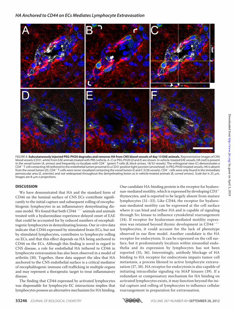
HA Is Removed from CNS Blood Vessels at EAE Day 13 byPEG-PH20 but Is Re-expressed at EAEDay 21—Toconfirm thatPEG-PH20 treatment leads to chronic reductions in HA in thelumen of CNS vessels, lumbar spinal tissue from animals withEAE was harvested from PEG-PH20-treated and vehicle-treated mice at 13 days postinoculation and labeled for ECs,T-cells, andHA (Fig. 8,A–F). A representative image of a bloodvessel from a vehicle-treated animal shows HA labeling withinthe EC lumen adjacent to CD31� tight junctions (Fig. 8A).Additionally, CD4� cells were often observed (18/32 vessels)co-localizing with HA within the lumen of the vessel (Fig. 8, Band C, arrow and arrowhead). CD4� cells are also observed inthe perivascular space co-localized with high levels of HA, as istypical of active EAE lesions (Fig. 8, B and C, curved arrows). Incontrast, HA is undetectable within the luminal area of bloodvessels from PEG-PH20-treated animals (Fig. 8D). No CD4�
cells are observed within the lumen of PEG-PH20-treated ves-sels (0/26 vessels), however, some CD4� cells are present inHA-rich regions within the perivascular area (Fig. 8, E and F,asterisks).A possible explanation for increased T-cell infiltration into
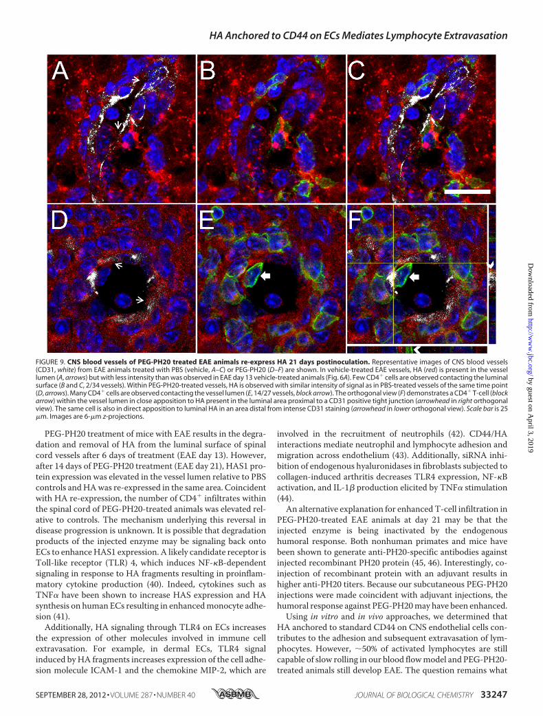
the CNS of mice with EAE following prolonged PEG-PH20treatment is that HA is re-expressed in the lumen of CNS ves-sels at later times of disease progression. To address this possi-bility, CNS vessels from day 21 EAE animals treated with vehi-cle or PEG-PH20 were examined for HA expression. We findthat HA is present along the lumen surface (arrows) in bothvehicle- and PEG-PH20-treated EAE day 21 vessels (Fig. 9, A
and D), but that the intensity of staining in both cases is signif-icantly less than is observed in vessels from day 13 vehicle-treated EAE animals. Consistent with the low intensity HAstaining, few vessels in vehicle-treated day 21 EAE mice con-tained CD4� cells interacting with the luminal surface (Fig. 9,Band C, 2/34 vessels). However, despite the relatively weak HAlabel, vessels from PEG-PH20-treated EAE day 21 mice oftenhad CD4� cells interacting with the luminal surface (14/27 ves-sels, Fig. 9, E and F, block arrow and arrowheads).
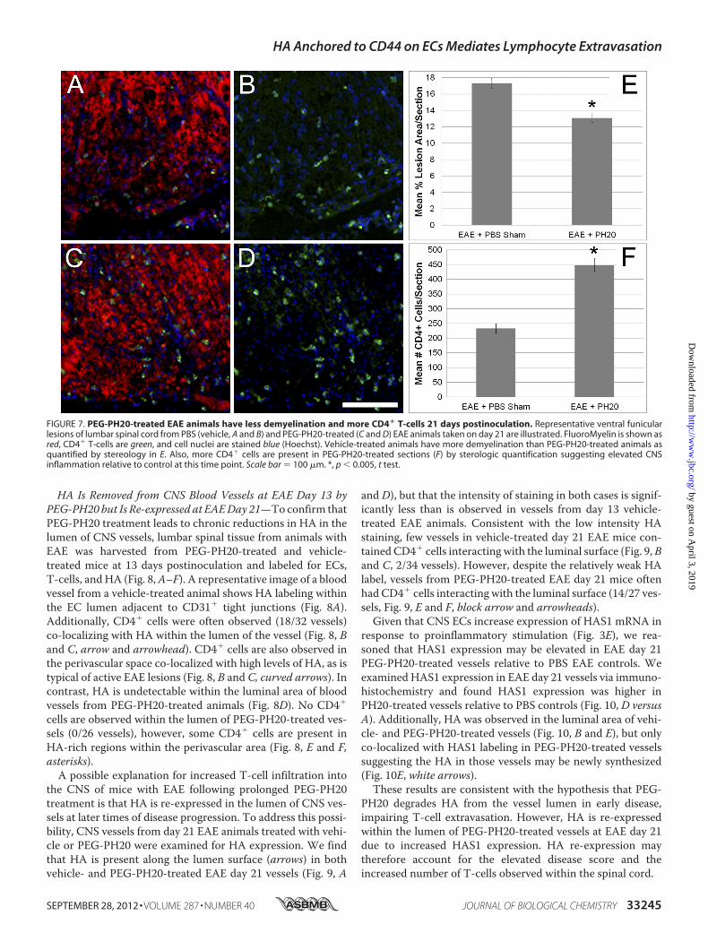
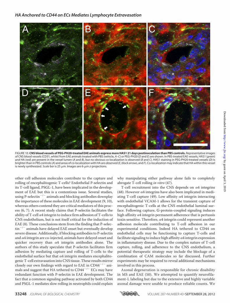
Given that CNS ECs increase expression of HAS1 mRNA inresponse to proinflammatory stimulation (Fig. 3E), we rea-soned that HAS1 expression may be elevated in EAE day 21PEG-PH20-treated vessels relative to PBS EAE controls. Weexamined HAS1 expression in EAE day 21 vessels via immuno-histochemistry and found HAS1 expression was higher inPH20-treated vessels relative to PBS controls (Fig. 10, D versusA). Additionally, HA was observed in the luminal area of vehi-cle- and PEG-PH20-treated vessels (Fig. 10, B and E), but onlyco-localized with HAS1 labeling in PEG-PH20-treated vesselssuggesting the HA in those vessels may be newly synthesized(Fig. 10E, white arrows).These results are consistent with the hypothesis that PEG-
PH20 degrades HA from the vessel lumen in early disease,impairing T-cell extravasation. However, HA is re-expressedwithin the lumen of PEG-PH20-treated vessels at EAE day 21due to increased HAS1 expression. HA re-expression maytherefore account for the elevated disease score and theincreased number of T-cells observed within the spinal cord.
FIGURE 7. PEG-PH20-treated EAE animals have less demyelination and more CD4� T-cells 21 days postinoculation. Representative ventral funicularlesions of lumbar spinal cord from PBS (vehicle, A and B) and PEG-PH20-treated (C and D) EAE animals taken on day 21 are illustrated. FluoroMyelin is shown asred, CD4� T-cells are green, and cell nuclei are stained blue (Hoechst). Vehicle-treated animals have more demyelination than PEG-PH20-treated animals asquantified by stereology in E. Also, more CD4� cells are present in PEG-PH20-treated sections (F) by sterologic quantification suggesting elevated CNSinflammation relative to control at this time point. Scale bar � 100 �m. *, p � 0.005, t test.
HA Anchored to CD44 on ECs Mediates Lymphocyte Extravasation
SEPTEMBER 28, 2012 • VOLUME 287 • NUMBER 40 JOURNAL OF BIOLOGICAL CHEMISTRY 33245
by guest on April 3, 2019
http://ww
w.jbc.org/
Dow
nloaded from
DISCUSSION
We have demonstrated that HA and the standard form ofCD44 on the luminal surface of CNS ECs contribute signifi-cantly to the initial capture and subsequent rolling of encepha-litogenic lymphocytes in an inflammatory demyelinating dis-ease model.We found that both CD44�/� animals and animalstreated with a hyaluronidase experience delayed onset of EAEthat could be accounted for by reduced numbers of encephali-togenic lymphocytes in demyelinating lesions. Our in vitro dataindicate that CD44 expressed by stimulated brain ECs, but notby stimulated lymphocytes, contributes to lymphocyte rollingon ECs, and that this effect depends on HA being anchored toCD44 on the ECs. Although this finding is novel in regard toCNS disease, a role for endothelial HA tethered to CD44 inlymphocyte extravasation has also been observed in a model ofarthritis (30). Together, these data support the idea that HAanchored to the CNS endothelial surface is a critical mediatorof encephalitogenic immune cell trafficking in multiple organsand may represent a therapeutic target to treat inflammatorydisease.The finding that CD44 expressed by activated lymphocytes
was dispensable for lymphocyte-EC interactions implies thatlymphocytes possess an alternativemechanism forHAbinding.
One candidate HA-binding protein is the receptor for hyaluro-nan-mediatedmotility, which is expressed by developingCD3�
thymocytes, and is reported to be largely absent from maturelymphocytes (31–33). Like CD44, the receptor for hyaluro-nan-mediated motility can be expressed at the cell surfacewhere it can bind and tether HA and is capable of signalingthrough Src kinase to influence cytoskeletal rearrangement(34). If receptor for hyaluronan-mediated motility expres-sion was retained beyond thymic development in CD44�/�
lymphocytes, it could account for the lack of phenotypeobserved in our flow model. Another candidate is the HAreceptor for endocytosis. It can be expressed on the cell sur-face, but it predominately localizes within sinusodial endo-thelia and its expression by lymphocytes has not beenreported (35, 36). Interestingly, antibody blockage of HAbinding to HA receptor for endocytosis impairs tumor cellmetastasis, a process likened to active lymphocyte extrava-sation (37, 38). HA receptor for endocytosis is also capable ofinitiating intracellular signaling via MAP kinases (39). If aredundant or compensatory mechanism for HA binding onactivated lymphocytes exists, it may function beyond the ini-tial capture and rolling of lymphocytes to influence cellularrearrangement in preparation for extravasation.
FIGURE 8. Subcutaneously injected PEG-PH20 degrades and removes HA from CNS blood vessels of day 13 EAE animals. Representative images of CNSblood vessels (CD31, white) from EAE animals treated with PBS (vehicle, A–C) or PEG-PH20 (D and E) are shown. In vehicle-treated EAE vessels, HA (red) is presentin the vessel lumen (A, arrows) and frequently co-localizes with CD4� (green) T-cells (B, block arrows, 18/32 vessels). The orthogonal view (C) demonstrates aCD4� T-cell contacting HA tethered to the endothelial lumen proximal to a CD31 positive tight junction (arrowhead). In PEG-PH20-treated vessels, HA is absentfrom the vessel lumen (D). CD4� T-cells were never visualized contacting the vessel lumen (E and F, 0/26 vessels). CD4� cells were only found in the immediateperivascular area (E, asterisks) and not widespread throughout the demyelinating lesion as in vehicle-treated animals (B, curved arrows). Scale bar is 25 �m.Images are 8-�m z-projections.
HA Anchored to CD44 on ECs Mediates Lymphocyte Extravasation
33246 JOURNAL OF BIOLOGICAL CHEMISTRY VOLUME 287 • NUMBER 40 • SEPTEMBER 28, 2012
by guest on April 3, 2019
http://ww
w.jbc.org/
Dow
nloaded from
PEG-PH20 treatment of mice with EAE results in the degra-dation and removal of HA from the luminal surface of spinalcord vessels after 6 days of treatment (EAE day 13). However,after 14 days of PEG-PH20 treatment (EAE day 21), HAS1 pro-tein expression was elevated in the vessel lumen relative to PBScontrols andHAwas re-expressed in the same area. Coincidentwith HA re-expression, the number of CD4� infiltrates withinthe spinal cord of PEG-PH20-treated animals was elevated rel-ative to controls. The mechanism underlying this reversal indisease progression is unknown. It is possible that degradationproducts of the injected enzyme may be signaling back ontoECs to enhanceHAS1 expression. A likely candidate receptor isToll-like receptor (TLR) 4, which induces NF-�B-dependentsignaling in response to HA fragments resulting in proinflam-matory cytokine production (40). Indeed, cytokines such asTNF� have been shown to increase HAS expression and HAsynthesis on human ECs resulting in enhancedmonocyte adhe-sion (41).Additionally, HA signaling through TLR4 on ECs increases
the expression of other molecules involved in immune cellextravasation. For example, in dermal ECs, TLR4 signalinduced byHA fragments increases expression of the cell adhe-sion molecule ICAM-1 and the chemokine MIP-2, which are
involved in the recruitment of neutrophils (42). CD44/HAinteractions mediate neutrophil and lymphocyte adhesion andmigration across endothelium (43). Additionally, siRNA inhi-bition of endogenous hyaluronidases in fibroblasts subjected tocollagen-induced arthritis decreases TLR4 expression, NF-�Bactivation, and IL-1� production elicited by TNF� stimulation(44).An alternative explanation for enhanced T-cell infiltration in
PEG-PH20-treated EAE animals at day 21 may be that theinjected enzyme is being inactivated by the endogenoushumoral response. Both nonhuman primates and mice havebeen shown to generate anti-PH20-specific antibodies againstinjected recombinant PH20 protein (45, 46). Interestingly, co-injection of recombinant protein with an adjuvant results inhigher anti-PH20 titers. Because our subcutaneous PEG-PH20injections were made coincident with adjuvant injections, thehumoral response against PEG-PH20may have been enhanced.Using in vitro and in vivo approaches, we determined that
HA anchored to standard CD44 on CNS endothelial cells con-tributes to the adhesion and subsequent extravasation of lym-phocytes. However, �50% of activated lymphocytes are stillcapable of slow rolling in our blood flowmodel and PEG-PH20-treated animals still develop EAE. The question remains what
FIGURE 9. CNS blood vessels of PEG-PH20 treated EAE animals re-express HA 21 days postinoculation. Representative images of CNS blood vessels(CD31, white) from EAE animals treated with PBS (vehicle, A–C) or PEG-PH20 (D–F) are shown. In vehicle-treated EAE vessels, HA (red) is present in the vessellumen (A, arrows) but with less intensity than was observed in EAE day 13 vehicle-treated animals (Fig. 6A). Few CD4� cells are observed contacting the luminalsurface (B and C, 2/34 vessels). Within PEG-PH20-treated vessels, HA is observed with similar intensity of signal as in PBS-treated vessels of the same time point(D, arrows). Many CD4� cells are observed contacting the vessel lumen (E, 14/27 vessels, block arrow). The orthogonal view (F) demonstrates a CD4� T-cell (blockarrow) within the vessel lumen in close apposition to HA present in the luminal area proximal to a CD31 positive tight junction (arrowhead in right orthogonalview). The same cell is also in direct apposition to luminal HA in an area distal from intense CD31 staining (arrowhead in lower orthogonal view). Scale bar is 25�m. Images are 6-�m z-projections.
HA Anchored to CD44 on ECs Mediates Lymphocyte Extravasation
SEPTEMBER 28, 2012 • VOLUME 287 • NUMBER 40 JOURNAL OF BIOLOGICAL CHEMISTRY 33247
by guest on April 3, 2019
http://ww
w.jbc.org/
Dow
nloaded from
other cell adhesion molecules contribute to the capture androlling of encephalitogenic T-cells? Endothelial P-selectin andits T-cell ligand, PSGL-1, have been implicated in the develop-ment of EAE but this is a contentious issue. Several studies,using P-selectin�/� animals and blocking antibodies downplaythe importance of these molecules in EAE development (9, 10),whereas others contend they are critical mediators of this proc-ess (6, 7). A recent study claims that P-selectin facilitates theability of T-cell�4 integrin to induce firm adhesion of T-cells toCNS endothelium, but is not itself critical for the induction ofEAE (8). These conclusions stem from the finding that P-selec-tin�/� animals have delayed EAE onset but eventually developsevere disease. Additionally, if blocking antibodies to P-selectinand�4 integrin are co-injected, animals have delayed onset andquicker recovery than �4 integrin antibodies alone. Theauthors of this study speculate that P-selectin facilitates firmadhesion by mediating capture and rolling of T-cells to theendothelial surface but that �4 integrin mediates encephalito-genic T-cell extravasation intoCNS tissue. These resultsmirrorclosely our own findings with regard to EAE in CD44�/� ani-mals and suggest that HA tethered to CD44�/� ECs may haveredundant function with P-selectin in EAE development. Thefact that a common signaling pathway activated by both CD44and PSGL-1 mediates slow rolling in neutrophils could explain
why manipulating either pathway alone fails to completelyabrogate T-cell rolling in vitro (47).T-cell recruitment into the CNS depends on �4 integrins
(48). However �4 integrins have also been implicated in medi-ating T-cell capture (49). Low affinity �4 integrin interactingwith endothelial VCAM-1 allows for the transient capture ofencephalitogenic T-cells at the CNS endothelial luminal sur-face. Following capture, G-protein-coupled signaling induceshigh affinity �4 integrin permanent adherence that is pertussistoxin sensitive. Therefore, �4 integrin could represent anotheradhesion molecule contributing to T-cell adhesion in ourexperimental conditions. Indeed HA tethered to CD44 onendothelial cells may be functioning to capture T-cells andfacilitate signaling to induce high affinity�4 integrin expressionin inflammatory disease. Due to the complex nature of T-cellcapture, rolling, and adherence to the CNS endothelium, apotential therapeutic strategy may include the blockage of acombination of CAM molecules so far discussed. Furtherexperiments may be required to reveal additional mechanismsinvolved in this process.Axonal degeneration is responsible for chronic disability
in MS and EAE (50). We attempted to quantify neurofila-ment-L labeling but due to the extensive and highly variableaxonal damage were unable to produce reliable counts. We
FIGURE 10. CNS blood vessels of PEG-PH20-treated EAE animals express more HAS1 21 days postinoculation than PBS controls. Representative imagesof CNS blood vessels (CD31, white) from EAE animals treated with PBS (vehicle, A–C) or PEG-PH20 (D and E) are shown. In PBS-treated EAE vessels, HAS1 (green)and HA (red) are present in the vessel lumen (A and B), but no obvious co-localization is observed (B and C). HAS1 staining in PEG-PH20-treated vessels (D) isbrighter than in PBS controls (A) and areas of co-localization with HA are observed (E, block arrows, and F). Co-localization may indicate that HA within this vesselis newly synthesized. Scale bar is 25 �m. Images are 6-�m z-projections.
HA Anchored to CD44 on ECs Mediates Lymphocyte Extravasation
33248 JOURNAL OF BIOLOGICAL CHEMISTRY VOLUME 287 • NUMBER 40 • SEPTEMBER 28, 2012
by guest on April 3, 2019
http://ww
w.jbc.org/
Dow
nloaded from
determined that the quality and number of axons in andaround lesions was increased with PEG-PH20 treatment onday 13 post-inoculation. However, the causative relationshipbetween PEG-PH20 treatment and reduced axonal damageis unclear due to fewer CD4� cells in the spinal cord at thistime point. A recent EAE study demonstrates foci of infil-trating immune cells correlate with locally impaired axonaltransport and markers of axonal damage at disease onset(51). It is likely the increase in spared axons is related to lowerlevels of inflammation within demyelinating lesions and notdirectly attributable to the activity of PEG-PH20. This idea is sup-ported by the fact that the quality and number of axons in andaround lesionswas indistinguishable between treatment groups at21 days postinoculation. These findings are reminiscent of otherstudies inhibiting immune cell infiltrationwhere axonopathy con-tinues despite decreased CNS inflammation (52).Our experiments showed that both CD44�/� and PEG-
PH20-treated mice eventually became symptomatic andattained disease scores equivalent to controls. A recent study(25) reported a delay in onset of disease, which the authors didnot discuss further, coupled with prolonged attenuation ofsymptoms when EAE was induced in CD44�/� animals. Theauthors conclude that signaling through CD44 in encephalito-genic CD4� T-cells results in epigenetic changes that drive dif-ferentiation of the cell toward a Th1/Th17 proinflammatoryphenotype. Although such phenotypic shifts may contribute toEAE progression, results from our group and others (22–24)indicate that CD44 also plays a significant role in the initiationof T cell rolling on ECs and, therefore, the extravasation ofencephalitogenic lymphocytes into the CNS.It is possible that the difference between our studies is related
to differences in the severity of EAE that is being induced. Guanet al. (25) reported a maximal mean disease score of �3.25 inWT controls and �1.15 for CD44�/�, whereas our animalsreached a maximal mean score of 4.33 for controls and 4.40 forCD44�/�. Recently it was shown that the monosaccharideN-acetylglucosamine, a component of HA, effectively inhibitedT-cell activation and amelioratedMOG35–55 induced C57BL/6EAE. However, treatment is clinically ineffective if EAE isinduced in 2D2 T-cell receptor transgenic mice, which displaymore robust disease (53). The severity of EAE disease is knownto vary depending on the population of cellular infiltrates.Other studies have shown that adoptive transfer ofmyelin reac-tive CD8� T-cells induces more severe disease than active dis-ease induction alone (54, 55). Although we did not directlyassay CD8� cells, our ability to induce more severe diseaseimplies a greater contribution of the CD8� phenotype to dis-ease pathogenicity in our model. As such, the contribution ofdeficient CD44 signaling to CD4� encephalitogenic T-cell phe-notype described by Guan et al. (25) may be masked in ourfindings. Because myelin reactive CD8� cells are more preva-lent within MS lesions than CD4� cells (56), our findings maybetter reflect the potential therapeutic outcomes of manipulat-ing CD44 or HA.In conclusion we have demonstrated that standard CD44 on
CNSECs but not activated lymphocytes contributes to lympho-cyte rolling on the endothelial surface. These findings expandthe knowledge of mechanisms promoting inflammatory demy-
elinating CNS disease and suggest that HA anchored to ECCD44 represents a therapeutic target to reduce immune cellinfiltration into the brain. Future studies aimed at testingwhether transient HA degradation during early stage diseasemight have the potential to enhance other therapeutic agentsthat limitMS attacks will reveal the potential of targetingHA asa means of limiting disease severity.
Acknowledgments—We thank the Achievement Awards for CollegeScientists (ARCS) society of Portland for supporting ClaytonWinkler.We also thank Dr. Weping Su and Dr. Anda Cornea for technicalassistance. Recombinant human PEG-PH20 was provided for thisstudy by a grant from Halozyme Therapeutics Inc.
REFERENCES1. McFarland, H. F., andMartin, R. (2007) Multiple sclerosis. A complicated
picture of autoimmunity. Nat. Immunol. 8, 913–9192. Holmøy, T. (2007) Immunopathogenesis of multiple sclerosis. Concepts
and controversies. Acta Neurol. Scand. Suppl. 187, 39–453. Greenwood, J., Heasman, S. J., Alvarez, J. I., Prat, A., Lyck, R., and Engel-
hardt, B. (2011) Review, leucocyte-endothelial cell cross-talk at the blood-brain barrier. A prerequisite for successful immune cell entry to the brain.Neuropathol. Appl. Neurobiol. 37, 24–39
4. Ley, K., Laudanna, C., Cybulsky, M. I., and Nourshargh, S. (2007) Gettingto the site of inflammation. The leukocyte adhesion cascade updated.Nat.Rev. Immunol. 7, 678–689
5. Kinashi, T. (2005) Intracellular signaling controlling integrin activation inlymphocytes. Nat. Rev. Immunol. 5, 546–559
6. Piccio, L., Rossi, B., Scarpini, E., Laudanna, C., Giagulli, C., Issekutz, A. C.,Vestweber, D., Butcher, E. C., and Constantin, G. (2002)Molecular mech-anisms involved in lymphocyte recruitment in inflamed brain microves-sels. Critical roles for P-selectin glycoprotein ligand-1 and heterotrimericGi-linked receptors. J. Immunol. 168, 1940–1949
7. Kerfoot, S.M., andKubes, P. (2002)Overlapping roles of P-selectin and�4integrin to recruit leukocytes to the central nervous system in experimen-tal autoimmune encephalomyelitis. J. Immunol. 169, 1000–1006
8. Kerfoot, S.M., Norman,M.U., Lapointe, B.M., Bonder, C. S., Zbytnuik, L.,and Kubes, P. (2006) Re-evaluation of P-selectin and �4 integrin as targetsfor the treatment of experimental autoimmune encephalomyelitis. J. Im-munol. 176, 6225–6234
9. Döring, A., Wild, M., Vestweber, D., Deutsch, U., and Engelhardt, B.(2007) E- and P-selectin are not required for the development of experi-mental autoimmune encephalomyelitis in C57BL/6 and SJL mice. J. Im-munol. 179, 8470–8479
10. Engelhardt, B., Vestweber, D., Hallmann, R., and Schulz, M. (1997) E- andP-selectin are not involved in the recruitment of inflammatory cells acrossthe blood-brain barrier in experimental autoimmune encephalomyelitis.Blood 90, 4459–4472
11. Siegelman, M. H., DeGrendele, H. C., and Estess, P. (1999) Activation andinteraction of CD44 and hyaluronan in immunological systems. J. Leuko-cyte Biol. 66, 315–321
12. Puré, E., and Cuff, C. A. (2001) A crucial role for CD44 in inflammation.Trends Mol. Med. 7, 213–221
13. Rampon, C., Weiss, N., Deboux, C., Chaverot, N., Miller, F., Buchet, D.,Tricoire-Leignel, H., Cazaubon, S., Baron-Van Evercooren, A., andCouraud, P.O. (2008)Molecularmechanismof systemic delivery of neuralprecursor cells to the brain. Assembly of brain endothelial apical cups andcontrol of transmigration by CD44. Stem Cells 26, 1673–1682
14. Jong, A., Wu, C. H., Shackleford, G. M., Kwon-Chung, K. J., Chang, Y. C.,Chen, H. M., Ouyang, Y., and Huang, S. H. (2008) Involvement of humanCD44 during Cryptococcus neoformans infection of brain microvascularendothelial cells. Cell. Microbiol. 10, 1313–1326
15. Girish, K. S., and Kemparaju, K. (2007) Themagic glue hyaluronan and itseraser hyaluronidase. A biological overview. Life Sci. 80, 1921–1943
16. Mohamadzadeh, M., DeGrendele, H., Arizpe, H., Estess, P., and Siegel-
HA Anchored to CD44 on ECs Mediates Lymphocyte Extravasation
SEPTEMBER 28, 2012 • VOLUME 287 • NUMBER 40 JOURNAL OF BIOLOGICAL CHEMISTRY 33249
by guest on April 3, 2019
http://ww
w.jbc.org/
Dow
nloaded from
man, M. (1998) Proinflammatory stimuli regulate endothelial hyaluronanexpression and CD44/HA-dependent primary adhesion. J. Clin. Investig.101, 97–108
17. Nandi, A., Estess, P., and Siegelman, M. H. (2000) Hyaluronan anchoringand regulation on the surface of vascular endothelial cells is mediatedthrough the functionally active form of CD44. J. Biol. Chem. 275,14939–14948
18. Ponta, H., Sherman, L., and Herrlich, P. A. (2003) CD44, from adhesionmolecules to signalling regulators. Nat. Rev. Mol. Cell Biol. 4, 33–45
19. Johnson, P., and Ruffell, B. (2009) CD44 and its role in inflammation andinflammatory diseases. Inflamm. Allergy Drug Targets 8, 208–220
20. DeGrendele, H. C., Estess, P., Picker, L. J., and Siegelman, M. H. (1996)CD44 and its ligand hyaluronatemediate rolling under physiologic flow. Anovel lymphocyte-endothelial cell primary adhesion pathway. J. Exp.Med.183, 1119–1130
21. DeGrendele, H. C., Estess, P., and Siegelman, M. H. (1997) Requirementfor CD44 in activated T-cell extravasation into an inflammatory site. Sci-ence 278, 672–675
22. Brocke, S., Piercy, C., Steinman, L., Weissman, I. L., and Veromaa, T.(1999) Antibodies to CD44 and integrin �4, but not L-selectin, preventcentral nervous system inflammation and experimental encephalomyeli-tis by blocking secondary leukocyte recruitment. Proc. Natl. Acad. Sci.U.S.A. 96, 6896–6901
23. Brennan, F. R., O’Neill, J. K., Allen, S. J., Butter, C., Nuki, G., and Baker, D.(1999) CD44 is involved in selective leucocyte extravasation during in-flammatory central nervous system disease. Immunology 98, 427–435
24. Laman, J. D., Maassen, C. B., Schellekens, M. M., Visser, L., Kap, M., deJong, E., van Puijenbroek,M., van Stipdonk,M. J., vanMeurs, M., Schwär-zler, C., and Günthert, U. (1998) Therapy with antibodies against CD40L(CD154) and CD44-variant isoforms reduces experimental autoimmuneencephalomyelitis induced by a proteolipid protein peptide.Multiple Scle-rosis 4, 147–153
25. Guan, H., Nagarkatti, P. S., and Nagarkatti, M. (2011) CD44 Recipro-cally regulates the differentiation of encephalitogenic Th1/Th17 andTh2/regulatory T cells through epigenetic modulation involving DNAmethylation of cytokine gene promoters, thereby controlling the de-velopment of experimental autoimmune encephalomyelitis. J. Immu-nol. 186, 6955–6964
26. Tuohy, T.M.,Wallingford,N., Liu, Y., Chan, F.H., Rizvi, T., Xing, R., Bebo,B., Rao, M. S., and Sherman, L. S. (2004) CD44 overexpression by oligo-dendrocytes. A novel mouse model of inflammation-independent demy-elination and dysmyelination. Glia 47, 335–345
27. Sugie, K., Jeon, M. S., and Grey, H. M. (2004) Activation of naive CD4 Tcells by anti-CD3 reveals an important role for Fyn in Lck-mediated sig-naling. Proc. Natl. Acad. Sci. U.S.A. 101, 14859–14864
28. Deli, M. A., Sakaguchi, S., Nakaoke, R., Abrahám, C. S., Takahata, H.,Kopacek, J., Shigematsu, K., Katamine, S., and Niwa, M. (2000) PrP frag-ment 106–126 is toxic to cerebral endothelial cells expressing PrP(C).Neuroreport 11, 3931–3936
29. Arch, R., Wirth, K., Hofmann, M., Ponta, H., Matzku, S., Herrlich, P., andZöller, M. (1992) Participation in normal immune responses of a metas-tasis-inducing splice variant of CD44. Science 257, 682–685
30. Mikecz, K., Brennan, F. R., Kim, J. H., and Glant, T. T. (1995) Anti-CD44treatment abrogates tissue oedema and leukocyte infiltration in murinearthritis. Nat. Med. 1, 558–563
31. Pilarski, L.M.,Miszta, H., and Turley, E. A. (1993) Regulated expression ofa receptor for hyaluronan-mediated motility on human thymocytes andT-cells. J. Immunol. 150, 4292–4302
32. Pilarski, L. M., Masellis-Smith, A., Belch, A. R., Yang, B., Savani, R. C., andTurley, E. A. (1994) RHAMM, a receptor for hyaluronan-mediated motil-ity, on normal human lymphocytes, thymocytes and malignant B cells. Amediator in B cell malignancy? Leuk. Lymphoma 14, 363–374
33. Gares, S. L., and Pilarski, L. M. (2000) Balancing thymocyte adhesion andmotility. A functional linkage between �1 integrins and the motility re-ceptor RHAMM. Dev. Immunol. 7, 209–225
34. Turley, E. A., Noble, P. W., and Bourguignon, L. Y. (2002) Signaling prop-erties of hyaluronan receptors. J. Biol. Chem. 277, 4589–4592
35. Falkowski,M., Schledzewski, K., Hansen, B., andGoerdt, S. (2003) Expres-
sion of stabilin-2, a novel fasciclin-like hyaluronan receptor protein, inmurine sinusoidal endothelia, avascular tissues, and at solid/liquid inter-faces. Histochem. Cell Biol. 120, 361–369
36. Zhou, B., Weigel, J. A., Fauss, L., andWeigel, P. H. (2000) Identification ofthe hyaluronan receptor for endocytosis (HARE). J. Biol. Chem. 275,37733–37741
37. Bockhorn, M., Jain, R. K., and Munn, L. L. (2007) Active versus passivemechanisms in metastasis. Do cancer cells crawl into vessels, or are theypushed? Lancet Oncology 8, 444–448
38. Simpson, M. A., Weigel, J. A., andWeigel, P. H. (2012) Systemic blockadeof the hyaluronan receptor for endocytosis prevents lymph node metas-tasis of prostate cancer. Intl. J. Cancer. 131, E836-E840
39. Kyosseva, S. V., Harris, E. N., and Weigel, P. H. (2008) The hyaluronanreceptor for endocytosis mediates hyaluronan-dependent signal trans-duction via extracellular signal-regulated kinases. J. Biol. Chem. 283,15047–15055
40. Shimada, M., Yanai, Y., Okazaki, T., Noma, N., Kawashima, I., Mori, T.,and Richards, J. S. (2008) Hyaluronan fragments generated by sperm-secreted hyaluronidase stimulate cytokine/chemokine production via theTLR2 and TLR4 pathway in cumulus cells of ovulated COCs, which mayenhance fertilization. Development 135, 2001–2011
41. Vigetti, D., Genasetti, A., Karousou, E., Viola, M., Moretto, P., Clerici,M., Deleonibus, S., De Luca, G., Hascall, V. C., and Passi, A. (2010)Proinflammatory cytokines induce hyaluronan synthesis and mono-cyte adhesion in human endothelial cells through hyaluronan synthase2 (HAS2) and the nuclear factor-�B (NF-�B) pathway. J. Biol. Chem.285, 24639–24645
42. Taylor, K. R., Trowbridge, J. M., Rudisill, J. A., Termeer, C. C., Simon,J. C., and Gallo, R. L. (2004) Hyaluronan fragments stimulate endothe-lial recognition of injury through TLR4. J. Biol. Chem. 279, 17079–17084
43. Khan, A. I., Kerfoot, S. M., Heit, B., Liu, L., Andonegui, G., Ruffell, B.,Johnson, P., and Kubes, P. (2004) Role of CD44 and hyaluronan in neutro-phil recruitment. J. Immunol. 173, 7594–7601
44. Campo, G. M., Avenoso, A., D’Ascola, A., Scuruchi, M., Prestipino, V.,Nastasi, G., Calatroni, A., and Campo, S. (2012) The inhibition of hyalu-ronan degradation reduced proinflammatory cytokines in mouse synovialfibroblasts subjected to collagen-induced arthritis. J. Cell. Biochem. 113,1852–1867
45. Deng, X., Meyers, S. A., Tollner, T. L., Yudin, A. I., Primakoff, P. D., He,D. N., and Overstreet, J. W. (2002) Immunological response of femalemacaques to the PH-20 sperm protein following injection of recombi-nant proteins or synthesized peptides. J. Reprod. Immunol. 54, 93–115
46. Hardy, C.M., Clydesdale, G.,Mobbs, K. J., Pekin, J., Lloyd,M. L., Sweet, C.,Shellam, G. R., and Lawson, M. A. (2004) Assessment of contraceptivevaccines based on recombinantmouse spermprotein PH20.Reproduction127, 325–334
47. Yago, T., Shao, B., Miner, J. J., Yao, L., Klopocki, A. G., Maeda, K., Cogge-shall, K. M., and McEver, R. P. (2010) E-selectin engages PSGL-1 andCD44 through a common signaling pathway to induce integrin �1�2-mediated slow leukocyte rolling. Blood 116, 485–494
48. Yednock, T. A., Cannon, C., Fritz, L. C., Sanchez-Madrid, F., Steinman, L.,and Karin, N. (1992) Prevention of experimental autoimmune encephalo-myelitis by antibodies against �4�1 integrin. Nature 356, 63–66
49. Vajkoczy, P., Laschinger, M., and Engelhardt, B. (2001) �4-Integrin-VCAM-1 binding mediates G protein-independent capture of encephali-togenic T cell blasts to CNS white matter microvessels. J. Clin. Investig.108, 557–565
50. Wujek, J. R., Bjartmar, C., Richer, E., Ransohoff, R. M., Yu, M., Tuohy,V. K., and Trapp, B. D. (2002) Axon loss in the spinal cord determinespermanent neurological disability in an animal model of multiple sclero-sis. J. Neuropathol. Exp. Neurol. 61, 23–32
51. Soulika, A. M., Lee, E., McCauley, E., Miers, L., Bannerman, P., and Plea-sure, D. (2009) Initiation and progression of axonopathy in experimentalautoimmune encephalomyelitis. J. Neurosci. 29, 14965–14979
52. Trapp, B. D., and Nave, K. A. (2008) Multiple sclerosis. An immune orneurodegenerative disorder? Annu. Rev. Neurosci. 31, 247–269
HA Anchored to CD44 on ECs Mediates Lymphocyte Extravasation
33250 JOURNAL OF BIOLOGICAL CHEMISTRY VOLUME 287 • NUMBER 40 • SEPTEMBER 28, 2012
by guest on April 3, 2019
http://ww
w.jbc.org/
Dow
nloaded from
53. Grigorian, A., Araujo, L., Naidu, N. N., Place, D. J., Choudhury, B., andDemetriou, M. (2011)N-Acetylglucosamine inhibits T-helper 1 (Th1)/T-helper 17 (Th17) cell responses and treats experimental autoimmune en-cephalomyelitis. J. Biol. Chem. 286, 40133–40141
54. Huseby, E. S., Liggitt, D., Brabb, T., Schnabel, B., Ohlén, C., and Gover-man, J. (2001) A pathogenic role for myelin-specific CD8� T cells in amodel for multiple sclerosis. J. Exp. Med. 194, 669–676
55. Sun, D., Whitaker, J. N., Huang, Z., Liu, D., Coleclough, C., Wekerle, H.,and Raine, C. S. (2001) Myelin antigen-specific CD8� T cells are enceph-alitogenic and produce severe disease in C57BL/6 mice. J. Immunol. 166,7579–7587
56. Hauser, S. L., Bhan, A. K., Gilles, F., Kemp, M., Kerr, C., andWeiner, H. L.(1986) Immunohistochemical analysis of the cellular infiltrate in multiplesclerosis lesions. Ann. Neurol. 19, 578–587
HA Anchored to CD44 on ECs Mediates Lymphocyte Extravasation
SEPTEMBER 28, 2012 • VOLUME 287 • NUMBER 40 JOURNAL OF BIOLOGICAL CHEMISTRY 33251
by guest on April 3, 2019
http://ww
w.jbc.org/
Dow
nloaded from

T. McCarty and Larry S. ShermanXing, Bruce F. Bebo, Fatima Banine, Michelle A. Berny-Lang, Asako Itakura, Owen J. Clayton W. Winkler, Scott C. Foster, Steven G. Matsumoto, Marnie A. Preston, Rubing
Autoimmune EncephalomyelitisEndothelial Cells Promotes Lymphocyte Extravasation in Experimental
Hyaluronan Anchored to Activated CD44 on Central Nervous System Vascular
doi: 10.1074/jbc.M112.356287 originally published online August 3, 20122012, 287:33237-33251.J. Biol. Chem.
10.1074/jbc.M112.356287Access the most updated version of this article at doi:
Alerts:
When a correction for this article is posted•
When this article is cited•
to choose from all of JBC's e-mail alertsClick here
Supplemental material:
http://www.jbc.org/content/suppl/2012/08/03/M112.356287.DC1
http://www.jbc.org/content/287/40/33237.full.html#ref-list-1
This article cites 56 references, 26 of which can be accessed free at
by guest on April 3, 2019
http://ww
w.jbc.org/
Dow
nloaded from