Embed Size (px)
Citation preview

Hyaluronan cisplatin conjugate in five dogs with soft tissue sarcomas
Rachel O. Venable, DVM; Deanna Worley, DVM; Daniel Gustafson, Ryan Hansen, E. J. Ehrhart, DVM; Daniel Aires MD; Shuang Cai, PhD; Mark Cohen, MD; Laird Forrest, PhD From the Department of Clinical Sciences and Biomedical Sciences College of Veterinary Medicine, Colorado State University, Fort Collins, CO 80523 (Venable, Worley, Gustafson, and Hansen); the Division of Dermatology, University of Kansas Medical Center, Kansas City, KS 66160 (Aires); the Department of Surgery and Pharmacology, University of Kansas Medical Center, Kansas City, KS 66160 (Cohen); and the Department of Pharmaceutical Chemistry, University of Kansas, Lawrence, KS 66047 (Cai and Forrest) Presented in abstract form at the 2011 Annual Conference of Veterinary Cancer Society, Albuquerque, New Mexico, November 5, 2011. Support in part by the National Institutes of Health, and the American Cancer Society, and NanoPharm LLC. Address correspondence to Dr. Rachel Venable at [email protected]
Abbreviations
HA hyaluronan
H&E haematoxylin-eosin

Objective-This study investigates intratumoral delivery of a novel hyaluronan-cisplatin
nanocarrier with goals of reduced systemic toxicity and enhanced tumor and lymphatic
chemotherapeutic penetration.
Animals-5 dogs with spontaneously-occurring soft tissue sarcomas (STSs) were enrolled.
Procedure-Approximately 1.5 mls of nanocarrier with 20 mg cisplatin were injected into one
external STS per animal. Blood for pharmacokinetics was collected at ½hr, 1hr, 2hrs, 3hrs, 4hrs,
24hrs, and 96hrs. Urinalysis was performed at initiation and 96hrs. Each STS and its draining
sentinel lymph node(s) were removed at 96hrs. Platinum levels were measured in blood, tumor,
and lymph nodes via ICP-MS analysis.
Results-There were no observed tissue reactions 96 hours following injection into the tumor.
Average AUC for unbound platinum was 661.5 +/- (228.4) ng/ml, and total platinum was 2355.5
+/- (897.7) ng/ml. Cmax of unbound platinum was 56.47 +/- (20.9) ng/ml, and total platinum was
81.6 +/- (40.4) ng/ml. The t1/2 were 2.49 and 42.9 hours respectively for unbound and total
platinum. Average platinum concentrations ranged from 3324.5 ng/g to 8228.8 ng/g in STSs, and
from 129.5 ng/g to 6066.0 ng/g in lymph nodes.
Conclusions-Hyaluronan-cisplatin nanocarrier was well tolerated following intratumoral
injection. Systemic platinum exposure appeared to be reduced versus traditional intravenous
delivery. Hyaluronan-cisplatin nanocarrier demonstrated up to 1000-fold higher levels in tumors
versus systemic circulation, and was concentrated by local lymphatics at levels up to 100 fold
greater than hemovascular circulation. These characteristics make it a promising new
chemotherapy modality.

Introduction
Hyaluronan is a natural polysaccharide with alternating D-glucuronic acid and N-acetyl-D-
glucosamine units. Hyaluronan and its breakdown products are cleared by the lymphatics via
receptor-mediated endocytosis and lysosomal degradation. 1-4 Hyaluronan is part of the
extracellular matrix, and found in synovial fluid, cartilage, and vitreous humor of the eye, and
dermis. 3,5 It is involved in multiple important processes such as cell adhesion, organization of
the extracellular matrix, growth, migration, tumor formation, and metastasis.3,5,6 Hyaluronan is
nonimmunogenic which makes it an ideal nanocarrier for many different drugs such as cisplatin,
paclitaxel, doxorubicin, and mitomycin.1,5,7
Cisplatin (cis-diamminedichloroplatinum or CDDP) is a DNA damaging agent that inhibits
protein and rRNA synthesis.8,9 In-vitro causes of cisplatin cytoxicity include platinum binding to
DNA, creation of interstrand cross-links, and formation of intrastrand bidentate N-7 adducts at
d(GpG) and d(ApG).10 It is cell cycle phase nonspecific and is renally cleared.9 It is used to
treat many human solid tumors including head and neck squamous sarcomas, lymphomas, small
cell and nonsmall cell lung, testicular, ovarian, gastric, esophageal, and pancreatic cancers. It is
also used in treatment of companion animal solid tumors including osteosarcomas, carcinomas,
and sarcomas.9,11 Use of cisplatin has been limited due to significant side effects and laborious
administration. Common side effects include significant nephrotoxicity involving renal tubular
inflammation and necrosis, leucopenia, nausea, anemia, and chronic neurotoxicity with
ototoxicity.2,11 The cisplantin toxicities seen in people are also seen in animals which limits its
veterinary use.
Cisplatin toxicity increases with peak plasma levels, but its effectiveness does not.11 Different
treatment strategies have been proposed such as metronomic chemotherapy, and local injection

of cisplatin to affected tissues while isolating systemic circulation to decrease the peak plasma
levels and therefore decrease the rate of toxicity especially nephrotoxicity.11 These treatment
options can be lengthy and fiscally unattractive as well as requiring specialized skills and
equipment not widely available.11
Nanocarriers with HA combined with cisplatin represent a new treatment modality that may
decrease peak plasma levels while maintaining cisplatin efficacy.11 In addition to being
nonimmunogenic, HA is a ligand for CD44 receptors which are located on lymphocytes and
some cancer cells.11,12 Once bound to CD44, HA is catabolized, brought into the cell via
receptor-mediated endocytosis, degraded in lysosomes, and then sent into the lymphatic
microcirculation.11 When cisplatin is combined with HA, the nanoparticles are activated as the
nanoconjugate comes in contact with hyaluronidase. In addition to lymph nodes, hyaluronidase
is also expressed on many tumors and can in fact be a marker of tumorigenesis. After tumor
hyaluronidase or receptor -mediated endocytosis activates the HA-cisplatin, then the cisplatin is
released into the peri-tumoral microlymphatics.3
Soft tissue sarcomas (STS) in the dog are mesenchymal and arise from connective tissues. They
represent approximately 8-17% of skin and subcutaneous tumors.13,14 Theses tumors are locally
invasive with wide local excision being the treatment of choice. Recurrence following surgery is
reported to be 7 to 32%.13,15,16 Cisplatin,doxorubicin, Mitoxantrone, and paclitaxel have been
tried in a wide variety of sarcomas, adjuvant chemotherapy has not been fully evaluated in STS
and its role is relatively unknown.16 Doxorubicin has not been found to have an increased
effectiveness as adjunctive therapy in high grade STS.17 A protocol of doxorubicin in
combination with cyclophosphamide found an overall response rate of 23%.18
Dogs did not tolerate locally-delivered Cisplatin suspension well, resulting in premature
termination of the study.19 Dogs with spontaneous osteosarcoma treated with a cisplatin-
containing implant after limb sparing surgery showed a non-significant decrease in local
recurrence.20 The implants require DMSO, and the DMSO can deactivate cisplatin which could
impair efficacy.
The aims of this study are to gain experience with HA as a nanocarrier for local tissue delivery of
cisplatin in spontaneously-occurring soft tissue sarcomas in dogs; characterize pharmacokinetics
of the HA-cisplatin conjugate; and assess whether HA-cisplatin conjugate has preferential local
lymphatic penetration. Our hypothesis is that canine STS will respond to local injection of HA-
cisplatin, and that HA-delivered cisplatin will be concentrated within tumor-draining lymph
nodes as compared to intravascular concentrations. A second hypothesis is that the the plasma
concentration of cisplatin will be lower than tumor concentration, and there will be no evidence
of systemic toxicity.
Material and Methods
Animals-Five client owned dogs weighing greater than 10 kg with spontaneously occurring soft
tissue sarcomas presenting to the Animal Cancer Center at Colorado State University for surgical
treatment were enrolled. The STS pretreatment diagnosis was done by needle core biopsy or
incisional biopsy. Routine blood work including a complete blood count, chemistry panel and
urinalysis were performed. All clients signed a consent form prior to study enrollment and all of
the procedures were approved by the Institutional Animal Care and Use Committee and had
hospital clinical board approval.
Synthesis of Hyaluronan-Cisplatin conjugates-Cisplatin was conjugated to HA using a
previously reported procedure.11 An ionic complex was formed between cisplatin and HA

containing ca. 20% by weight bound platinum, which releases cisplatin over several days into the
local lymphatics. The platinum content was validated by atomic absorption spectroscopy (AAS)
(Varian SpectrAA GTA-110 with graphite furnace).
HA-Cisplatin administration-The tumor was clipped of fur and measured with calipers in
minimally two or three dimensions when possible. All dogs were sedated for intratumoral
injection pending individual dog disposition and 20 mg of HA-cisplatin conjugate in a volume
less than 2 milliliters was injected into the center of the tumor in all dogs. After 96 hours
following injection the tumor was again measured.
Regional lymphoscintigraphy –After 96 hours following HA-cisplatin conjugate
administration, the dogs returned for sentinel lymph node mapping and surgical tumor removal.
All dogs were sedated for regional lymphoscintigraphy imaging and were injected with 125
microCuries of filtered technetium sulfur colloid peritumorally in four quadrants. Single photon
emission computer tomography images were taken using a GE Millenium VG gamma camera
every five minutes until the first draining lymph node basin was visualized.
Surgical procedure-All dogs’ tumor sites were clipped including the area of the draining lymph
node basin. Intracavitary sentinel lymph nodes were not removed within the chest or abdomen
unless it was part of the planned tumor resection. Methylene blue (0.4 mL, 5 mg/mL) was
injected around the tumor in four quadrants. Tumors were excised via wide local excision per
routine clinical practice. A handheld gamma probe was used intraoperatively to identify
individual draining lymph nodes for intraoperative lymphoscintigraphy. Separate instruments
were used for lymph node extirpation. Once removed, the tumor and lymph nodes were
remeasured. The tumors were dissected into quadrants. Core samples were collected from the

center of the tumor and at approximately every 2 cm radiating outward for larger tumors, placed
in cryotubes and frozen at -80˚C for later platinum analysis. Remaining ex vivo tissue was
placed in10% neutral buffered formalin for H & E staining and image analysis to determine
percent tumor necrosis and histology. The lymph nodes were sectioned in half, and half of the
tissue was placed in cryotubes and frozen at -80˚C for later platinum analysis and the other half
was placed in 10% neutral buffered formalin for H & E staining for histology.
Pharmacokinetic Evaluation- - Approximately two mLs of whole blood in serum tubes and
heparinized blood collected in EDTA tubes were obtained to measure unbound (plasma) and
total (serum) platinum concentrations at 0 minutes, 30 minutes, 1 hr, 2 hrs, 3 hrs, and 4 hrs from
an indwelling catheter following drug administration. The dogs then returned in 24 hrs and 96
hrs for additional blood samples. Also blood for a complete blood count and chemistry panel as
well as urine for a urinalysis was done at 0 and 96 hrs. The whole blood was centrifuged and the
serum portion was collected and placed in a cryotube prior to freezing as well as the EDTA tubes
were centrifuged and the plasma collected. The plasma and serum were collected into cryotubes
and immediately frozen at -80˚C. Plasma and serum were thawed at the time of assessment. The
serum was placed in a centrifugal filter unit with 10,000 MWCO. Centrifugation was performed
at 7,500 x g for 20 minutes. The filter was then removed and volume of filtrate recorded, and
then sufficient 6% nitric acid was added to obtain a 1:10 dilution to obtain the ultrafiltration
portion. Plasma was also collected and sufficient 6% nitric acid was added to obtain a 1:10
dilution.
Platinum determination by ICP-MS- Plasma (unbound) and serum (total) were prepared as
described above and frozen tumor and lymph node sections were thawed at room temperature
and then analyzed. Preparation method was similar for all sample types. Tissue (0.5 g wet

weight) or plasma samples were placed in 15-mL centrifuge tubes, and then 0.75mL of
concentrated nitric acid was added and samples were heated to 95˚C for 6 hrs. After cooling the
samples, 0.5 ml of 30% hydrogen peroxide was added and heated to 80˚C for 30 minutes. After
the second cooling, 0.25 mL of concentrated hydrochloric acid was added and heated to 80˚C for
30 minutes. After the third cooling, the samples were brought to approximately 5 mL final
volume with purified water, and the exact volumes were determined gravimetrically. Samples
were grouped into sets of approximately 20, and each set was prepared with a blank, a spiked
blank, a standard reference material (NRC Canada's DOLT-4), a duplicate sample, and a spiked
sample. Digestates were diluted 10x in purified water for platinum analysis by ICP-MS
(PerkinElmer Sciex Elan DRC II) and were analyzed undiluted for other elements using ICP-
OES (PerkinElmer Optima 7300).
Pharmacokinetics modeling-Cmax was determined from plasma and serum concentration-time
profiles. Area under the curve (AUC) was calculated by linear trapezoidal summation from time
zero to infinity. The t1/2 was calculated using the elimination rate constant with Kel=0.693/t1/2
from 4 to 96 hours.
Histology and pharmocodynamics - The tumor was longitudinally sectioned. Sections
representing the complete longitudinal plane of the tumor were fixed in formalin for 24 to 48
hours. The sections were processed with an extended protocol, paraffin-embedded, sectioned at
4 microns, and H&E stained. All sections from a sample were completely scanned using an
AxioCam HRc Carl Zeiss camera coupled to a Carl Zeiss Axioplan 2 microscope with a
mechanical stage and utilizing Carl Zeiss Axiovision analysis software. The scanned images
were viewed simultaneously with the H&E stained sections. Total region of tumor and regions of
necrosis were outlined on the images. Regions outlined on the images were then analyzed with

the Axiovision image analysis software to determine tumor area and the area of tumor necrosis.
Total tumor and necrosis areas for a tumor were determined by adding results of all sections for
that tumor. Percent necrosis for a tumor was determined as total area of necrosis/total tumor area.
The lymph nodes were longitudinally sectioned. Sections representing the complete longitudinal
central plane of the lymph node were fixed in formalin for 24-48 hrs. The sections were
processed with an extended protocol, paraffin-embedded, sectioned at 4 microns, and
haematoxylin-eosin (H&E) stained. Histology was evaluated by a pathologist.
Results
Five client-owned dogs were enrolled in the study. Two were mixed breed, one Labrador
retriever, one Golden retriever, and one Chesapeake Bay retriever. All dogs were of similar age
with a median age of 9 years old (range 7.5 to 10 years). There were three male castrated and
two female spayed. They all had similar weight with a median weight of 38 kg (range 27.1 kg-
43.1 kg). Each dog had only one STS examined for the study, and the tumor locations were
antebrachium, stifle, ventral thorax, lateral flank, and medial hock. Four of five tumors were
greater than 5 cm in diameter and the average largest tumor diameter was 8.9 cm (4.4 cm to 13.7
cm). There was no evidence of metastasis at the time of presentation.
Tumor response-Following injection all tumors remained stable in size as determined by serial
measurements at the 96 hour time point. All tumors were completely removed via
histopathologic assessment of surgical tumor margins. The STS histologic grading system
evaluates cell differentiation, mitosis, and percent necrosis on a scale of I to III.15 The dogs in
this study had a median tumor grade of II (range I-III).

Toxicity- One dog had a grade one dermal reaction (VCOG consensus statement 2004) 21 24
hours post HA-cisplatin injection in the tumor which consisted of two areas of mild erythema
one at the injection site and the other at the distal aspect of the mass. No other dogs had any
reactions at the injection site or noted in the tumor at the 96 hour time point.
Blood values prior to injection and 96 hrs post injection were not significantly different between
the complete blood cell counts or serum chemistry. One dog had a grade one thrombocytopenia
at the 96 hour complete blood count.21 One dog was hyposthenuric prior to treatment and at 96
hrs. All renal values remained within normal reference intervals.
Two dogs with amputations performed to remove the STSs developed mild seromas ten days
post amputation. Two dogs developed dehiscence at the surgical site located on the stifle and
ventral thorax. The stifle resection developed partial dehiscence nine days postoperatively from
the distal aspect of the incision to the point of maximal tension over the joint and was
approximately 8.8 cm in length. The original tumor on the stifle was 9 cm in the largest
diameter. This was managed as an open wound and then surgically closed five days later. The
ventral thorax resection was large with the largest diameter being 7.4 cm and developed partial
dehiscence at the cranial aspect of the incision at the level of the drain approximately 2.2 cm in
diameter 22 days postoperatively. This was managed as an open wound and healed. One dog
developed a seroma fourteen days post operatively at the lateral flank that developed into an
abscess eight days later. This was lanced and treated with antibiotics.
Sentinel lymph nodes- Regional lymphoscintigraphy was performed in three out of five dogs.
The draining lymph nodes were identified with lymphoscintigraphy and gamma camera.
Sentinel lymph nodes were obtained surgically in four of the five dogs. One dog had a draining

lymph node identified by regional lymphoscintigraphy, but the lymph node could not be found at
the time of surgery with intraoperative lymphoscintigraphy and blue dye mapping. The two
other dogs not receiving regional lymphoscintigraphy had the local draining lymph nodes
identified by intraoperative methylene blue injection and hand held gamma probe guidance and
were removed. Histology of all lymph nodes was reactive hyperplasia and/or chronic
histiocytosis. No evidence of neoplasia was found in any of the lymph nodes. The tumor
locations and associated draining lymph nodes are described in Table 1.
Platinum levels in the lymph nodes, tumors and blood-The platinum concentration in the
STSs was greater than in the lymph nodes and blood. The average platinum concentrations
within excised STSs ranged from 3324.5 ng/g to 8228.8 ng/g. The platinum concentrations
within the draining lymph nodes were 129.5 ng/g to 6066 ng/g. The average AUC, t1/2, and Cmax
for the unbound and total platinum are listed in Table 2 through Table 4.
Percent tumor necrosis- The averaged percent tumor necrosis ranged from 0.25% to 6.61%.
Discussion
Platinum levels were significantly higher in the tumor and its lymph nodes than in the systemic
circulation. This has also been demonstrated in rodent studies evaluating this
nanoconjugate.4,11,12 Platinum levels remained much higher at 96 hours in lymph node and
tumor, with average platinum concentration in tumors approximately one-thousand-fold greater
than plasma and one-hundred-fold greater than serum. The lymph node platinum concentration
ranges at the 96 hour time point were ten-fold to one-hundred-fold greater than in the plasma and
serum. Hyaluronan-cisplatin achieved preferential uptake into the local regional draining lymph

nodes indicating it is a good treatment delivery system, especially for neoadjuvant or adjuvant
chemotherapy when there is a high likelihood of lymphatic spread.
Nanoconjugated HA cisplatin showed lower toxicity than cisplatin alone. Studies examining the
pharmacokinetics of HA-cisplatin conjugates administered subcutaneously and intravenously in
rodents found increased plasma AUC with decreased peak plasma levels compared to
conventional intravenous cisplatin.11 Higher concentrations in the tumor and lower
concentrations in circulation help decrease renal and other toxicity. Acute renal toxicity due to
cisplatin usually develops within the first 24 hours.10 In this study no dogs developed any
systemic toxicity up to 96 hours post injection. Mild thrombocytopenia in one dog was likely
not related to the drug exposure because the half-life of platelets is much greater than 96 hours.
In this study all five dogs had varying types of complications at the surgical site. Not
surprisingly the two dogs with amputations developed mild seromas, which are anecdotally not
unusual following limb amputation. As in the case of the dog that developed a seroma on the
lateral flank that then progressed into an abscess, seroma formation two weeks postoperative is
not uncommon following wide surgical excision in this location. It is also not uncommon for
seromas to become secondarily infected. The tumors in both of these dogs were a considerable
distance away from the amputation site making less likely that the seromas were related to the
tumor or HA-cisplatin injection site. The three other dogs developed complications at their
incision sites, which might have been due to the wide 2-3 cm tumor excision margins in
locations with little or no redundant skin. This study selected for STS greater than 2 cm which
can make complete wide-margin surgical removal and primary wound closure challenging. All
tumors were completely removed for histologic evaluation. Dehiscence in a taut, high motion
area overlying the stifle is a normal risk factor of tumor removal in routine clinical practice.

Similarly, the ventral thorax tissue is notoriously taut without redundant tissue; dehiscence at this
location is a common risk and can transpire if no undue tension is present with primary closure.
There is the possibility that the HA-cisplatin conjugate interferes with wound healing. This has
not been evaluated in rodent models.4,11,12 Other studies evaluating cisplatin administered locally
have also found high complication rates. Earlier studies evaluating OPLA-cisplatin implants in
dogs found a wound complication rate of 47.5% to 60%.20,22 Another recent study in dogs with
surgically removed STS placed a cisplatin biodegradable implant delivery system and found a
wound complication rate of 84.2%.23 This study had a wound complication rate of 3/5 dogs
(60%). Further studies evaluating wound healing with this nanoconjugate are needed to further
elucidate if the drug inhibits healing, or if complications are simply those associated with the
wide margins needed to obtain complete removal of these tumors.
This study determined the regional draining lymph nodes for each tumor by sentinel lymph node
mapping by regional lymphoscintigraphy using a gamma camera and also intraoperatively by
using methylene blue dye and a hand held gamma probe. No lymph nodes were found to have
evidence of STS metastasis in this group of dogs. Soft tissue sarcoma metastasis more
commonly occurs in the lungs as it is spread hematogenously, and it is less likely to occur in the
lymph nodes.15 Greater risk of metastasis can be associated with tumor grade.13-15 The average
STS grade in this group of dogs was grade II, which has an uncertain metastatic rate but has been
reported to be less than 15%.15 Soft tissue sarcoma is a good model for this nanocarrier in a pilot
study where it is of interest to instill HA-cisplatin into a tumor that can be completely excised
and whose draining lymph nodes can easily be determined. This tumor model assessed product
tolerability in the dog model and there was minimal morbidity associated with lymph node
excision, and no complications occurred related to lymph node extirpation.

The data demonstrate that the study drug increased uptake of platinum in the regional draining
lymph nodes; this has been previously noted in rodent HA-cisplatin studies that found increased
platinum accumulation in local regional lymph nodes and low-level sustained systemic release
that allowed for decreased peak plasma concentrations.4,11 Hyaluronan enters into the
microlymphatics from the interstitial space and is activated within the lymphatic system making
the HA-cisplatin conjugate a new modality to treat lymphatically spread and metastatic cancer.
Hyaluronan-cisplatin given intratumorally is also taken up intracellularly by tumor cells because
many tumor cells express the HA ligand CD44, as do lymph nodes.11,12 Lymph is partially
comprised of tissue interstitial fluid that enters into the lymphatic circulation via active processes
of opening lymph endothelial valves, after which the fluid travels unidirectionally through a
series of collecting lymphatic vessels and nodes.24 Hyaluronan-cisplatin enters preferentially
into the lymphatic system, and the carrier is cleaved from the conjugate by lysosomal
degradation, which activates the drug.11,12 This new treatment delivery system has been used in
murine models for breast cancer, intrapleural injection for malignant pleural mesothelioma, and
endotracheal instillation for lung cancer.2,5,12 Doxorubicin conjugated to HA for breast cancer
has also been examined in rodents.1
The tumors remained stable in size 96 hours post injection. Chemotherapy has not been
evaluated extensively in dogs with measurable STS. A response rate of 15% to 74% has been
reported and this has been with varying systemic therapies and agents.16 Another study
evaluating two doses of doxorubicin found the response rate for STS to be 22%.18 There was not
a large amount of tumor necrosis in the histologic and percent tumor necrosis evaluations.
However, significant necrosis would not be expected at this early time point.

This is the first study evaluating the pharmacokinetics of HA-Cisplatin nanoconjugate in dogs.
The AUC and Cmax in our study were less than previously described with this nanoconjugate in a
rodent model.11 This may be due to the fact that we administered HA-cisplatin intratumorally
and not subcutaneously as was done in the rodent model. Also, we gave 20 mg to each dog
which averaged to 0.56 mg/kg, while the rodents received 3.3 mg/kg. The t1/2 was greater in our
study, but this is not surprising as dogs have a slower heart rate and metabolism than rodents.
Our t ½ is within the range of multiple reported studies for t ½β in people in the second phase of
cisplatin clearance (total 30.5 hr to 130 hr) with the first phase representing the terminal half-life
of unbound platinum and the second phase being total platinum.25
There is limited data available for AUC for cisplatin in dogs. The AUC can be extrapolated from
a previous study that evaluated the effect of hyperthermia on the pharmacokinetics of cisplatin in
normal dogs, and our AUC for total and unbound is very similar with our unbound platinum
AUC within 400(ng hr)/ml difference, and our total platinum AUC was 1,629 (ng hr)/ml
greater.26 An early study evaluating intravenous cisplatin administration at 1 mg/kg in normal
dogs found much higher plasma concentrations in the first four hours, approximately one-
thousand-fold higher than seen in this study.27 (Table 5) Although a part of this much higher
systemic cisplatin exposure is no doubt due to the higher dose in that study, most of the
difference is likely due to the nature of the study drug in this study. Hyaluronan-cisplatin
nanoconjugate helps maintain lower systemic levels while achieving higher concentrations
locally in the tumor and regional lymphatic tissue. The lower systemic levels help to prevent
systemic toxicities.
Theon and coworkers 28 conducted a study of intratumorally-injected cisplatin in canines with
oral malignant melanoma using a study dose (0.25 mg/kg or ca. 5 mg/m2) much closer to the
dose we administered (0.56 mg/kg or ca. 11 mg/m2). Compared to Theon’s study, the
intratumoral HA-cisplatin demonstrated a 1.1-fold reduction in the Cmax, 50.2- and 14.9-fold
increases in the t1/2 and AUC, respectively, even though the animals in our study were treated
with a 1.2-fold higher dose of the drug. The findings suggested that our HA-cisplatin formulation
has significantly enhanced the retention and accumulation of the drug in vivo at lower doses,
which could be utilized as maintenance chemotherapy, reducing the cycle number of the
treatment. In addition, Theon et al reported the incidence of grade 1, 2, and 3 toxicities following
the intratumoral cisplatin chemotherapy. In comparison, dogs treated with HA-cisplatin were
free of drug-induced severe toxicities (grade 2 & 3) for the duration of the study.
Cisplatin has been evaluated in the dog in different delivery modalities. Cisplatin has been given
intra-arterially to dogs with osteosarcoma treated with limb spare surgery and radiation
therapy.29 Cisplatin has also been given subcutaneously and intracavitary. High toxicity was
observed in subcutaneous administration, but not in intracavitary administration.19,30
Intracavitary administration however is best for a small subset of cancer treatments: palliation of
pleural and/or abdominal effusions. Another modality studied evaluated cisplatin release from a
D, L-Polylactic acid (OPLA) implant in dogs with soft tissue sarcomas, nasal tumors, and
osteosarcoma treated with limb sparing surgery.20,22,31-33 These studies did not find any
statistically significant clinical improvement with the cisplatin implant. A study evaluating
cisplatin release from D, L-polylactic acid implant in normal beagle dogs adjacent to a cortical
allograft found a much higher AUC for both dogs with the implants and beagles given cisplatin
intravenously compared to our study. The dose used in these dogs was much higher than ours.
The beagles with implants received 54.4 mg/m2 and 81.6 mg/m2 (AUC 27,050 ug·min/ml) and
the dogs given cisplatin intravenously received 70 mg/m2 (AUC 940.3 ug·min/ml).33 In our

study the dogs’ dosages ranged from 16 mg/m2 to 22 mg/m2. It is also difficult to compare the
variable administration routes. In a study evaluating cisplatin impregnated
polymethylmethacrylate in healthy dogs dosed at 25.68 mg/m2 or 20 mg/dog, plasma
concentrations were approximately one hundred fold greater than in our study.34 The AUC was
less than our study (AUC 1.093 ug·min/ml) which can be attributed to the different delivery
routes and dosages.34 Although this study dosage was similar to ours (20 mg/dog), our HA-
cisplatin conjugate showed less systemic absorption and more sustained release from carrier.
Soft tissue sarcomas are a good tumor model because these are tumors that can be easily
measured and amenable to a drug injection directly into the tumor. Soft tissue sarcomas have a
low metastatic rate to the local lymph node and lung, yet it is straightforward determining if
there is presence of cisplatin is in the sentinel lymph node(s) which tend to be externally located
and readily accessible. Cisplatin levels were higher in the tumor and lymph nodes compared to
systemic circulation. Tumors remained stable in size and did not progress over the 96 hour time
frame. Further investigation into possible inhibition of healing at the surgical site is needed,
although in clinical practice excision soon after intratumoral chemotherapy would likely not be
standard procedure.
In conclusion this study demonstrated that HA-cisplatin nanoconjugate can be given
intratumorally to dogs resulting in higher cisplatin concentration in tumors and draining lymph
nodes with preferential regional lymphatic uptake, lower cisplatin concentration in plasma, and
no systemic toxicities noted within the first 96 hours.
References
1. Cai S, Thati S, Bagby TR, et al. Localized doxorubicin chemotherapy with a biopolymeric nanocarrier improves survival and reduces toxicity in xenografts of human breast cancer. J Control Release 2010;146:212-218. 2. Xie Y, Aillon KL, Cai S, et al. Pulmonary delivery of cisplatin-hyaluronan conjugates via endotracheal instillation for the treatment of lung cancer. Int J Pharm 2010;392:156-163. 3. Jeong YI, Kim ST, Jin SG, et al. Cisplatin-incorporated hyaluronic acid nanoparticles based on ion-complex formation. Journal of Pharmaceutical Sciences 2008;97:1268-1276. 4. Cai S, Xie YM, Bagby TR, et al. Intralymphatic chemotherapy using a hyaluronan-cisplatin conjugate. Journal of Surgical Research 2008;147:247-252. 5. Ampollini L, Sonvico F, Barocelli E, et al. Intrapleural polymeric films containing cisplatin for malignant pleural mesothelioma in a rat tumour model: a preliminary study. Eur J Cardiothorac Surg 2010;37:557-565. 6. Kamal A, Datta K. Upregulation of hyaluronan binding protein 1 (HABP1/p32/gC1qR) is associated with cisplatin induced apoptosis. Apoptosis 2006;11:861-874. 7. Chen JP, Leu YL, Fang CL, et al. Thermosensitive hydrogels composed of hyaluronic acid and gelatin as carriers for the intravesical administration of cisplatin. J Pharm Sci 2011;100:655-666. 8. Jordan P, Carmo-Fonseca M. Cisplatin inhibits synthesis of ribosomal RNA in vivo. Nucleic Acids Res 1998;26:2831-2836. 9. Chun R, Garrett L, Vail D. Cancer Chemotherapy In: Withrow S,Vail D, eds. Small Animal Clinical Oncology. 4 ed. St. Louis: Saunders, 2007;163-192. 10. Reed E, Kohn K. Platinum Analogues In: Chabner B,Collins J, eds. Cancer Chemotherapy: Principles and Practice. Philadelphia: J.B. Lippincott Company, 1990;465-484. 11. Cai S, Xie Y, Davies NM, et al. Pharmacokinetics and disposition of a localized lymphatic polymeric hyaluronan conjugate of cisplatin in rodents. J Pharm Sci 2010;99:2664-2671. 12. Cohen MS, Cai S, Xie Y, et al. A novel intralymphatic nanocarrier delivery system for cisplatin therapy in breast cancer with improved tumor efficacy and lower systemic toxicity in vivo. The American Journal of Surgery 2009;198:781-786. 13. Dennis MM, McSporran KD, Bacon NJ, et al. Prognostic factors for cutaneous and subcutaneous soft tissue sarcomas in dogs. Vet Pathol 2011;48:73-84. 14. Stefanello D, Morello E, Roccabianca P, et al. Marginal excision of low-grade spindle cell sarcoma of canine extremities: 35 dogs (1996-2006). Vet Surg 2008;37:461-465. 15. Kuntz CA, Dernell WS, Powers BE, et al. Prognostic factors for surgical treatment of soft-tissue sarcomas in dogs: 75 cases (1986-1996). J Am Vet Med Assoc 1997;211:1147-1151. 16. Dernell WS, Withrow SJ, Kuntz CA, et al. Principles of treatment for soft tissue sarcoma. Clinical Techniques in Small Animal Practice 1998;13:59-64.

17. Selting KA, Powers BE, Thompson LJ, et al. Outcome of dogs with high-grade soft tissue sarcomas treated with and without adjuvant doxorubicin chemotherapy: 39 cases (1996–2004). Journal of the American Veterinary Medical Association 2005;227:1442-1448. 18. Ogilvie GK, Reynolds HA, Richardson RC, et al. Phase-Ii Evaluation of Doxorubicin for Treatment of Various Canine Neoplasms. Journal of the American Veterinary Medical Association 1989;195:1580-1583. 19. O'Brien ME, Wigler N, Inbar M, et al. Reduced cardiotoxicity and comparable efficacy in a phase III trial of pegylated liposomal doxorubicin HCl (CAELYX/Doxil) versus conventional doxorubicin for first-line treatment of metastatic breast cancer. Ann Oncol 2004;15:440-449. 20. Withrow SJ, Liptak JM, Straw RC, et al. Biodegradable cisplatin polymer in limb-sparing surgery for canine osteosarcoma. Ann Surg Oncol 2004;11:705-713. 21. Veterinary Co-operative Oncology Group - Common Terminology Criteria for Adverse Events (VCOG-CTCAE) following chemotherapy or biological antineoplastic therapy in dogs and cats v1.0. Vet Comp Oncol 2004;2:195-213. 22. Dernell WS, Withrow SJ, Straw RC, et al. Intracavitary treatment of soft tissue sarcomas in dogs using cisplatin in a biodegradable polymer. Anticancer Res 1997;17:4499-4505. 23. Havlicek M, Straw RS, Langova V, et al. Intra-operative cisplatin for the treatment of canine extremity soft tissue sarcomas. Vet Comp Oncol 2009;7:122-129. 24. Zawieja DC. Contractile physiology of lymphatics. Lymphat Res Biol 2009;7:87-96. 25. Canal P. Platinum Compounds: Pharmacokinetics and Pharmacodynamics In: Grochow L,Ames M, eds. A Clinican's Guide to Chemotherapy Pharmacokinetics and Pharmacodynamics. Baltimore: Williams & Willkins a Waverly Company, 1998;345-373. 26. Riviere JE, Page RL, Dewhirst MW, et al. Effect of hyperthermia on cisplatin pharmacokinetics in normal dogs. Int J Hyperthermia 1986;2:351-358. 27. Litterst CL, Gram TE, Dedrick RL, et al. Distribution and disposition of platinum following intravenous administration of cis-diamminedichloroplatinum(II) (NSC 119875) to dogs. Cancer Res 1976;36:2340-2344. 28. Theon AP, Madewell BR, Ryu J, et al. Concurrent irradiation and intratumoral chemotherapy with cisplatin: a pilot study in dogs with spontaneous tumors. Int J Radiat Oncol Biol Phys 1994;29:1027-1034. 29. Withrow SJ, Thrall DE, Straw RC, et al. Intraarterial Cisplatin with or without Radiation in Limb-Sparing for Canine Osteosarcoma. Cancer 1993;71:2484-2490. 30. Moore AS, Kirk C, Cardona A. Intracavitary cisplatin chemotherapy experience with six dogs. J Vet Intern Med 1991;5:227-231. 31. Lana SE, Dernell WS, Lafferty MH, et al. Use of radiation and a slow-release cisplatin formulation for treatment of canine nasal tumors. Veterinary Radiology & Ultrasound 2004;45:577-581. 32. Mehl ML, Seguin B, Dernell WS, et al. Survival analysis of one versus two treatments of local delivery cisplatin in a biodegradable polymer for canine osteosarcoma. Vet Comp Oncol 2005;3:81-86. 33. Straw RC, Withrow SJ, Douple EB, et al. Effects of cis-diamminedichloroplatinum II released from D,L-polylactic acid implanted adjacent to cortical allografts in dogs. J Orthop Res 1994;12:871-877.

34. Buss MS, Henry CJ, Tyler JW, et al. Systemic and tissue chamber fluid platinum concentrations released from cis-diamminedichloroplatinum II-impregnated polymethylmethacrylate in healthy dogs. American Journal of Veterinary Research 1999;60:280-283. 35. Ogawara K, Un K, Minato K, et al. Determinants for in vivo anti-tumor effects of PEG liposomal doxorubicin: importance of vascular permeability within tumors. Int J Pharm 2008;359:234-240. 36. Freireich EJ, Gehan EA, Rall DP, et al. Quantitative comparison of toxicity of anticancer agents in mouse, rat, hamster, dog, monkey, and man. Cancer Chemother Rep 1966;50:219-244.
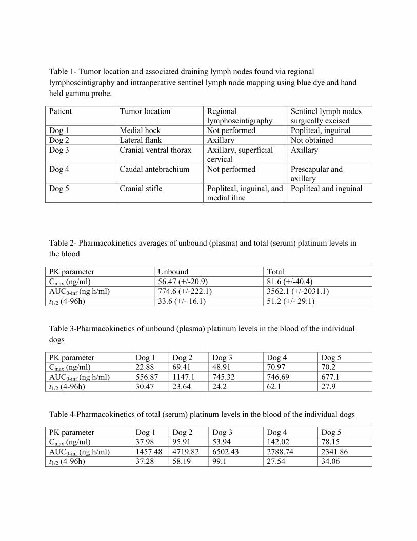
Table 1- Tumor location and associated draining lymph nodes found via regional lymphoscintigraphy and intraoperative sentinel lymph node mapping using blue dye and hand held gamma probe.
Patient Tumor location Regional lymphoscintigraphy
Sentinel lymph nodes surgically excised
Dog 1 Medial hock Not performed Popliteal, inguinal Dog 2 Lateral flank Axillary Not obtained Dog 3 Cranial ventral thorax Axillary, superficial
cervical Axillary
Dog 4 Caudal antebrachium Not performed Prescapular and axillary
Dog 5 Cranial stifle Popliteal, inguinal, and medial iliac
Popliteal and inguinal
Table 2- Pharmacokinetics averages of unbound (plasma) and total (serum) platinum levels in the blood
PK parameter Unbound Total Cmax (ng/ml) 56.47 (+/-20.9) 81.6 (+/-40.4) AUC0-inf (ng h/ml) 774.6 (+/-222.1) 3562.1 (+/-2031.1) t1/2 (4-96h) 33.6 (+/- 16.1) 51.2 (+/- 29.1)
Table 3-Pharmacokinetics of unbound (plasma) platinum levels in the blood of the individual dogs
PK parameter Dog 1 Dog 2 Dog 3 Dog 4 Dog 5 Cmax (ng/ml) 22.88 69.41 48.91 70.97 70.2 AUC0-inf (ng h/ml) 556.87 1147.1 745.32 746.69 677.1 t1/2 (4-96h) 30.47 23.64 24.2 62.1 27.9
Table 4-Pharmacokinetics of total (serum) platinum levels in the blood of the individual dogs
PK parameter Dog 1 Dog 2 Dog 3 Dog 4 Dog 5 Cmax (ng/ml) 37.98 95.91 53.94 142.02 78.15 AUC0-inf (ng h/ml) 1457.48 4719.82 6502.43 2788.74 2341.86 t1/2 (4-96h) 37.28 58.19 99.1 27.54 34.06

Table 5- Comparative pharmacokinetics of HA-cisplatin with other routes
Intratumoral HA-cisplatin
Intravenous cisplatin35
Intratumoral cisplatin19
Intratumoral cisplatin 28
Dose, mg/m2 11≈ 10 70 5≈ Cmax, ng/mL (serum)
81.6 ± 40.4 294 ± 60 770* 170* ǂ
t1/2, hrs 51.2 ± 29.1 29.1 ± 15.7 96* ǂ 1* ǂ AUC0-inf, ng·h/mL (serum)
3562.1 ± 2031.1 663 ± 52 58800* 224* ǂ
Tumor concentration, ng/g
5650.0 (3324.5 - 8228.8) ×
- - -
LN concentration, ng/g
2485.0 (129.5 - 6066.0) ×
- - -
Bioavailability 488% 100% 1270% 116%
* Standard deviation not reported by authors
ǂ Determined from concentration-time curve provided by authors
× Tissue concentrations measured at 96 h
≈ Converted from mg/kg dose based on Freireich et al 36
Notes: