Embed Size (px)
Citation preview
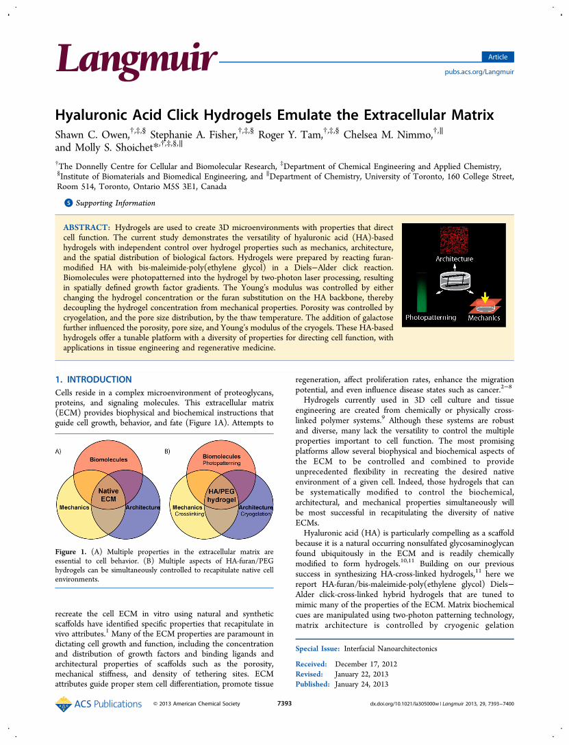
Hyaluronic Acid Click Hydrogels Emulate the Extracellular MatrixShawn C. Owen,†,‡,§ Stephanie A. Fisher,†,‡,§ Roger Y. Tam,†,‡,§ Chelsea M. Nimmo,†,∥
and Molly S. Shoichet*,†,‡,§,∥
†The Donnelly Centre for Cellular and Biomolecular Research, ‡Department of Chemical Engineering and Applied Chemistry,§Institute of Biomaterials and Biomedical Engineering, and ∥Department of Chemistry, University of Toronto, 160 College Street,Room 514, Toronto, Ontario M5S 3E1, Canada
*S Supporting Information
ABSTRACT: Hydrogels are used to create 3D microenvironments with properties that directcell function. The current study demonstrates the versatility of hyaluronic acid (HA)-basedhydrogels with independent control over hydrogel properties such as mechanics, architecture,and the spatial distribution of biological factors. Hydrogels were prepared by reacting furan-modified HA with bis-maleimide-poly(ethylene glycol) in a Diels−Alder click reaction.Biomolecules were photopatterned into the hydrogel by two-photon laser processing, resultingin spatially defined growth factor gradients. The Young’s modulus was controlled by eitherchanging the hydrogel concentration or the furan substitution on the HA backbone, therebydecoupling the hydrogel concentration from mechanical properties. Porosity was controlled bycryogelation, and the pore size distribution, by the thaw temperature. The addition of galactosefurther influenced the porosity, pore size, and Young’s modulus of the cryogels. These HA-basedhydrogels offer a tunable platform with a diversity of properties for directing cell function, withapplications in tissue engineering and regenerative medicine.
1. INTRODUCTIONCells reside in a complex microenvironment of proteoglycans,proteins, and signaling molecules. This extracellular matrix(ECM) provides biophysical and biochemical instructions thatguide cell growth, behavior, and fate (Figure 1A). Attempts to
recreate the cell ECM in vitro using natural and syntheticscaffolds have identified specific properties that recapitulate invivo attributes.1 Many of the ECM properties are paramount indictating cell growth and function, including the concentrationand distribution of growth factors and binding ligands andarchitectural properties of scaffolds such as the porosity,mechanical stiffness, and density of tethering sites. ECMattributes guide proper stem cell differentiation, promote tissue
regeneration, affect proliferation rates, enhance the migrationpotential, and even influence disease states such as cancer.2−8
Hydrogels currently used in 3D cell culture and tissueengineering are created from chemically or physically cross-linked polymer systems.9 Although these systems are robustand diverse, many lack the versatility to control the multipleproperties important to cell function. The most promisingplatforms allow several biophysical and biochemical aspects ofthe ECM to be controlled and combined to provideunprecedented flexibility in recreating the desired nativeenvironment of a given cell. Indeed, those hydrogels that canbe systematically modified to control the biochemical,architectural, and mechanical properties simultaneously willbe most successful in recapitulating the diversity of nativeECMs.Hyaluronic acid (HA) is particularly compelling as a scaffold
because it is a natural occurring nonsulfated glycosaminoglycanfound ubiquitously in the ECM and is readily chemicallymodified to form hydrogels.10,11 Building on our previoussuccess in synthesizing HA-cross-linked hydrogels,11 here wereport HA-furan/bis-maleimide-poly(ethylene glycol) Diels−Alder click-cross-linked hybrid hydrogels that are tuned tomimic many of the properties of the ECM. Matrix biochemicalcues are manipulated using two-photon patterning technology,matrix architecture is controlled by cryogenic gelation
Special Issue: Interfacial Nanoarchitectonics
Received: December 17, 2012Revised: January 22, 2013Published: January 24, 2013
Figure 1. (A) Multiple properties in the extracellular matrix areessential to cell behavior. (B) Multiple aspects of HA-furan/PEGhydrogels can be simultaneously controlled to recapitulate native cellenvironments.
Article
pubs.acs.org/Langmuir
© 2013 American Chemical Society 7393 dx.doi.org/10.1021/la305000w | Langmuir 2013, 29, 7393−7400
conditions, and matrix mechanical properties are tuned by thematrix density and degree of cross-linking (Figure 1B).
2. EXPERIMENTAL SECTION2.1. Materials. Dried sodium hyaluronate (HA) (2.34 × 10⁵ amu),
was purchased from Lifecore Biomedical (Chaska, MN, USA). Bis-(N-ethylmaleimide)-poly(ethylene glycol) (PEG-(mal)2) (3.0 × 10³ amu)was purchased from RAPP Polymere GmbH (Germany). 4-(4,6-Dimethoxy-1,3,5-triazin-2-yl)-4-methylmorpholinium chloride(DMTMM) and dimethyl sulfoxide (DMSO) were purchased fromSigma-Aldrich (St. Louis, MO, USA). Furfurylamine was purchasedfrom Acros Organics (Belgium). 2-(N-Morpholino)-ethanesulfonicacid (MES) buffer and 2-[(2-hydroxy-1-bis[hydroxy methyl ethyl)aminoethanesulfonic acid (TES) buffer were purchased from BioshopCanada Inc. (Burlington, ON, Canada). 5-Dulbecco’s phosphate-buffered saline (PBS) was purchased from Multicell Technologies Inc.(Woonsocket, RI, USA). AlexaFluor 488-hydrazide and AlexaFluor568-hydrazide were purchased from Invitrogen (Eugene, OR, USA).Dialysis membranes were purchased from Spectrum Laboratories Inc.(Rancho Dominguez, CA, USA). Recombinant human epidermalgrowth factor (EGF) was purchased from PeproTech Inc. (Rocky Hill,NJ, USA). Sulfosuccinimidyl[4-iodoacetyl]aminobenzoate (Sulfo-SIAB) and dialysis cassettes were purchased from Thermo-Fisher(Pittsburgh, PA, USA).2.2. Synthesis and Characterization of HA-Furan and
Coumarin-HA-Furan. Furan-modified HA derivatives (HA-furan)were synthesized as previously described.11 Briefly, HA-furanderivatives were prepared by dissolving HA (0.40 g, 1.04 mmolcarboxylates) in 40 mL of MES buffer (100 mM, pH 5.5). To prepare40%-substituted HA-furan, DMTMM (0.25 g, 1.02 mmol, 1 equiv)was added to the mixture and stirred for 10 min. Furfurylamine (47.2μL, 0.50 mmol, 0.5 equiv) was then added dropwise to the solution.To prepare 55%-substituted HA-furan, DMTMM (0.56 g, 2.04 mmol,2 equiv) and furfurylamine (94.4 μL, 1.02 mmol, 1 equiv) were used.The reaction was stirred at room temperature for 24 h and thendialyzed against distilled water for 3 days (Mw cutoff 12−14 kDa).Water was removed by lyophilization to obtain HA-furan derivatives asa white powder. The degree of substitution (DS) was determined from1H NMR spectra by comparing the ratio of the areas under the protonpeaks at 6.26, 6.46, and 7.65 ppm (furan protons) to the peak at 1.9ppm (N-acetyl glucosamine protons of HA). 1H NMR spectra wererecorded in D2O on a Varian Mercury-400 MHz NMR spectrometer(Palo Alto, CA, USA).Derivatives of 6-bromo-7-hydroxycoumarin sulfide (coumarin) were
prepared according to a published procedure12 and conjugated to HA-furan carboxylates with DMTMM to form coumarin HA-furanderivatives (coumarin-HA-furan). HA-furan (0.526 g, 1.194 mmolcarboxylates) was dissolved in 50 mL of MES buffer (100 mM, pH5.5). Following complete dissolution, DMTMM (0.066 g, 0.23988mmol, 0.2 equiv) in 300 μL of MES buffer (100 mM, pH 5.5) wasadded, and the reaction mixture was stirred for 30 min. Coumarin(0.0394 g, 0.1194 mmol, 0.1 equiv) in 100 μL of DMSO was thenadded dropwise, and the reaction was stirred in the dark at roomtemperature for 24 h (Supporting Information Figure 1). The reactionsolution was then dialyzed in the dark against distilled water for 3 days(Mw cutoff 12−14 kDa) and lyophilized in the dark to obtaincoumarin-HA-furan as a fluorescent, white spongy powder (λabs = 335nm). The DS was determined from 1H NMR.2.3. General Preparation of HA-Furan/PEG Hydrogels. HA-
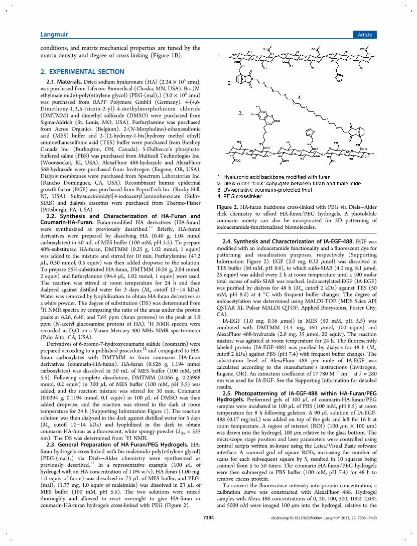
furan hydrogels cross-linked with bis-maleimido-poly(ethylene glycol)(PEG-(mal)2) via Diels−Alder chemistry were synthesized aspreviously described.11 In a representative example (100 μL ofhydrogel with an HA concentration of 1.0% w/v), HA-furan (1.00 mg,1.0 equiv of furan) was dissolved in 73 μL of MES buffer, and PEG-(mal)2 (1.37 mg, 1.0 equiv of maleimide) was dissolved in 23 μL ofMES buffer (100 mM, pH 5.5). The two solutions were mixedthoroughly and allowed to react overnight to give HA-furan orcoumarin-HA-furan hydrogels cross-linked with PEG (Figure 2).
2.4. Synthesis and Characterization of IA-EGF-488. EGF wasmodified with an iodoacetamide functionality and a fluorescent dye forpatterning and visualization purposes, respectively (SupportingInformation Figure 2). EGF (2.0 mg, 0.32 μmol) was dissolved inTES buffer (50 mM, pH 8.6), to which sulfo-SIAB (4.0 mg, 8.1 μmol,25 equiv) was added every 2 h at room temperature until a 100 molartotal excess of sulfo-SIAB was reached. Iodoacetylated-EGF (IA-EGF)was purified by dialysis for 48 h (Mw cutoff 2 kDa) against TES (50mM, pH 8.0) at 4 °C with frequent buffer changes. The degree ofiodoacetylation was determined using MALDI-TOF (MDS Sciex APIQSTAR XL Pulsar MALDI QTOF, Applied Biosystems, Foster City,CA).
IA-EGF (1.0 mg, 0.16 μmol) in MES (50 mM, pH 5.5) wascombined with DMTMM (4.4 mg, 160 μmol, 100 equiv) andAlexaFluor 488-hydrazide (2.0 mg, 35 μmol, 20 equiv). The reactionmixture was agitated at room temperature for 24 h. The fluorescentlylabeled protein (IA-EGF-488) was purified by dialysis for 48 h (Mwcutoff 2 kDa) against PBS (pH 7.4) with frequent buffer changes. Thesubstitution level of AlexaFluor 488 per mole of IA-EGF wascalculated according to the manufacturer’s instructions (Invitrogen,Eugene, OR). An extinction coefficient of 17 780 M−1 cm−1 at λ = 280nm was used for IA-EGF. See the Supporting Information for detailedresults.
2.5. Photopatterning of IA-EGF-488 within HA-Furan/PEGHydrogels. Preformed gels of 100 μL of coumarin-HA-furan/PEGsamples were incubated in 100 μL of PBS (100 mM, pH 8.5) at roomtemperature for 8 h following gelation. A 90 μL solution of IA-EGF-488 (0.17 mg/mL) was added on top of the gels and left for 16 h atroom temperature. A region of interest (ROI) (100 μm × 100 μm)was drawn into the hydrogel, 100 μm relative to the glass bottom. Themicroscope stage position and laser parameters were controlled usingcontrol scripts written in-house using the Leica/Visual Basic softwareinterface. A scanned grid of square ROIs, increasing the number ofscans for each subsequent square by 5, resulted in 10 squares beingscanned from 5 to 50 times. The coumarin-HA-furan/PEG hydrogelswere then submerged in PBS buffer (100 mM, pH 7.4) for 48 h toremove excess protein.
To convert the fluorescence intensity into protein concentration, acalibration curve was constructed with AlexaFluor 488. Hydrogelsamples with Alexa 488 concentrations of 0, 20, 100, 500, 1000, 2500,and 5000 nM were imaged 100 μm into the hydrogel, relative to the
Figure 2. HA-furan backbone cross-linked with PEG via Diels−Alderclick chemistry to afford HA-furan/PEG hydrogels. A photolabilecoumarin moiety can also be incorporated for 3D patterning ofiodoacetamide-functionalized biomolecules.
Langmuir Article
dx.doi.org/10.1021/la305000w | Langmuir 2013, 29, 7393−74007394
glass bottom. The calibration curve, along with the known number offluorophores per protein, was used to calculate the proteinconcentration.2.6. Confocal Settings for Photopatterning and Fluores-
cence Imaging. All patterns were created using a Leica TCS-SP2confocal microscope (Leica Microsystems, Wetzlar, Germany)equipped with a multiphoton Mai Tai broadband Ti-sapphire laser(Spectra-Physics) using a 20× objective (NA = 0.4) and an electronicstage. The multiphoton laser was set to 740 nm with an offset of 75%and a gain of 43% for patterning.Images were collected by confocal microscopy on an Olympus
FV1000 at 20× magnification using the following excitation andemission wavelengths: for Alexa-488, excitation at 485 nm, emission at520 nm; for Alexa-546, excitation at 560 nm, emission at 580 nm.ImageJ was used for fluorescence quantification.2.7. Preparation of HA-Furan/PEG Cryogels. HA-furan (40%
substitution, 10.00 mg, 1.0 equiv of furan) was dissolved in 0.5 mL ofMES buffer (10 mM, pH 5.5). For sugar-modified formulations, D-galactose was added (39.6 mg, 220 μmol) to the HA-furan and mixed.The viscous liquid was cooled to 4 °C and degassed for 1 min toremove any air bubbles. In another vial, PEG-(mal)2 cross-linker(13.65 mg, 1.0 equiv of maleimide) was dissolved in 0.5 mL of MESbuffer (10 mM, pH 5.5), cooled to 4 °C, and carefully mixed with thesolution containing HA-furan. The mixture was then flash frozen inliquid nitrogen and maintained at either −6 or −15 °C for 8 h. Thesolutions were then warmed to room temperature for 1 h, followed byanother identical freeze/thaw cycle. The reactions were quenched byadding 10 mg/mL N-ethylhydroxy maleimide (0.2 mL) and incubatedovernight at room temperature.2.8. Pore Size Characterization. Unreacted carboxylates of the
HA-furan polysaccharide backbone were conjugated to AlexaFluor568-hydrazide using DMTMM chemistry to enable the visualization ofthe pore size and pore wall thickness. HA-cryogels were diluted withMES buffer (100 mM, pH 5.5), followed by the addition of DMTMM(4 mg, 15 μmol) and AlexaFluor 568-hydrazide (1 mg, 1.75 μmol) andmixed overnight at room temperature in the dark. The labeled gelswere washed extensively with MES buffer, followed by distilled water.Three Z-stack sections of each gel were then imaged on an Olympusconfocal microscope at 20× magnification. Three images from each Zstack were analyzed for pore size using ImageJ software.2.9. Mechanical Compression Testing. The Young’s moduli
were determined for HA-furan/PEG hydrogels and cryogels that hadbeen preswollen in PBS for a day and formed into cylindrical sampleswith a diameter of 5 mm. Samples were placed between twoimpermeable flat platens connected to a DAQ-Nano17 forcetransducer (ATI Industrial Automation) on a Mach-1 micro-mechanical system (Biomomentum). Samples were subjected to aninitial tare force of 0.01 N to even out surface defects, and the platen-to-platen separation was taken as the initial sample height. Uniaxial,unconfined compression was performed at 37 °C at a deformation rateof 10 μm/s until an applied strain of 20% was reached. The Young’smodulus was taken as the slope of the resultant stress versus strainchart for each sample.2.10. Statistical Analysis. All statistical analyses were performed
using GraphPad Prism version 5.00 for Windows (GraphPad Software,San Diego, CA, USA, www.graphpad.com). Differences among groupsof three or more treatments were assessed by one-way ANOVA witheither Bonferroni or Newton-Keuls post hoc corrections to identifystatistical differences. Differences among two treatments were assessedusing unpaired t tests. An α level of 0.05 was set as the criterion forstatistical significance. Graphs are annotated with p values representedas * ≤ 0.05, ** ≤ 0.01, or *** ≤ 0.001. All data are presented as mean± standard deviation.
3. RESULTS AND DISCUSSION
3.1. Synthesis of HA-Furan and Coumarin-HA-Furanwith Control over the Degree of Substitution. HA-furanwas synthesized as previously reported to give a furansubstitution level of 40 or 55% by altering the ratio of HA to
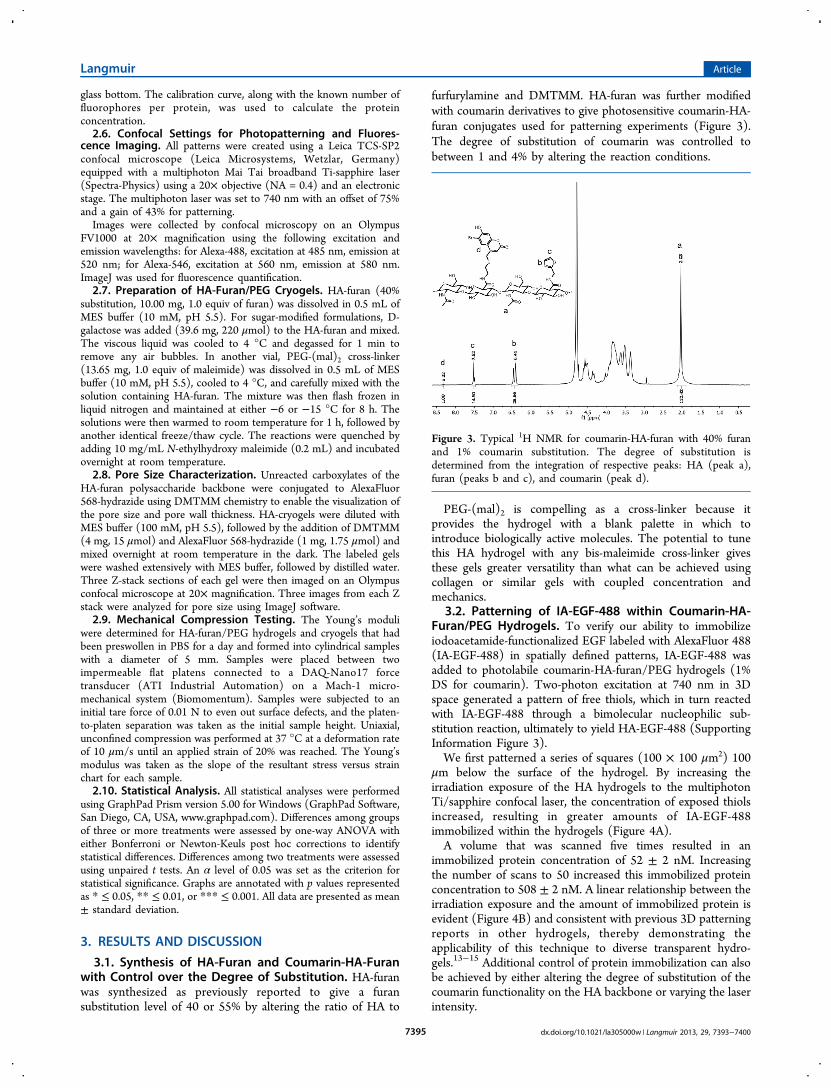
furfurylamine and DMTMM. HA-furan was further modifiedwith coumarin derivatives to give photosensitive coumarin-HA-furan conjugates used for patterning experiments (Figure 3).The degree of substitution of coumarin was controlled tobetween 1 and 4% by altering the reaction conditions.
PEG-(mal)2 is compelling as a cross-linker because itprovides the hydrogel with a blank palette in which tointroduce biologically active molecules. The potential to tunethis HA hydrogel with any bis-maleimide cross-linker givesthese gels greater versatility than what can be achieved usingcollagen or similar gels with coupled concentration andmechanics.
3.2. Patterning of IA-EGF-488 within Coumarin-HA-Furan/PEG Hydrogels. To verify our ability to immobilizeiodoacetamide-functionalized EGF labeled with AlexaFluor 488(IA-EGF-488) in spatially defined patterns, IA-EGF-488 wasadded to photolabile coumarin-HA-furan/PEG hydrogels (1%DS for coumarin). Two-photon excitation at 740 nm in 3Dspace generated a pattern of free thiols, which in turn reactedwith IA-EGF-488 through a bimolecular nucleophilic sub-stitution reaction, ultimately to yield HA-EGF-488 (SupportingInformation Figure 3).We first patterned a series of squares (100 × 100 μm2) 100
μm below the surface of the hydrogel. By increasing theirradiation exposure of the HA hydrogels to the multiphotonTi/sapphire confocal laser, the concentration of exposed thiolsincreased, resulting in greater amounts of IA-EGF-488immobilized within the hydrogels (Figure 4A).A volume that was scanned five times resulted in an
immobilized protein concentration of 52 ± 2 nM. Increasingthe number of scans to 50 increased this immobilized proteinconcentration to 508 ± 2 nM. A linear relationship between theirradiation exposure and the amount of immobilized protein isevident (Figure 4B) and consistent with previous 3D patterningreports in other hydrogels, thereby demonstrating theapplicability of this technique to diverse transparent hydro-gels.13−15 Additional control of protein immobilization can alsobe achieved by either altering the degree of substitution of thecoumarin functionality on the HA backbone or varying the laserintensity.
Figure 3. Typical 1H NMR for coumarin-HA-furan with 40% furanand 1% coumarin substitution. The degree of substitution isdetermined from the integration of respective peaks: HA (peak a),furan (peaks b and c), and coumarin (peak d).
Langmuir Article
dx.doi.org/10.1021/la305000w | Langmuir 2013, 29, 7393−74007395
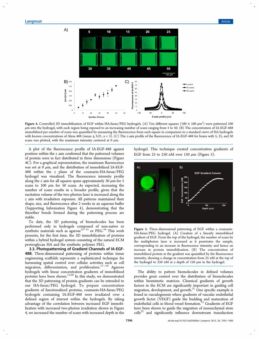
A plot of the fluorescence profile of IA-EGF-488 againstposition within the z axis confirmed that the patterned volumesof protein were in fact distributed in three dimensions (Figure4C). For a graphical representation, the maximum fluorescencewas set at 0 μm, and the distribution of immobilized IA-EGF-488 within the z plane of the coumarin-HA-furan/PEGhydrogel was visualized. The fluorescence intensity profilealong the z axis for all squares spans approximately 30 μm for 5scans to 100 μm for 50 scans. As expected, increasing thenumber of scans results in a broader profile, given that theexcitation volume of the two-photon laser is increased along thez axis with irradiation exposure. All patterns maintained theirshape, size, and fluorescence after 2 weeks in an aqueous buffer(Supporting Information Figure 4), demonstrating that thethioether bonds formed during the patterning process arestable.To date, the 3D patterning of biomolecules has been
performed only in hydrogels composed of non-native orsynthetic materials such as agarose12−15 or PEG.16 This workpresents, for the first time, the 3D immobilization of proteinswithin a hybrid hydrogel system consisting of the natural ECMproteoglycan HA and the synthetic polymer PEG.3.3. Photopatterning of Gradient Channels of IA-EGF-
488. Three-dimensional patterning of proteins within tissueengineering scaffolds represents a sophisticated technique forharnessing spatial control over cellular activities such as cellmigration, differentiation, and proliferation.17−19 Agarosehydrogels with linear concentration gradients of immobilizedproteins have been shown.13,20 In this study, we demonstratedthat the 3D patterning of protein gradients can be extended toour HA-furan/PEG hydrogel. To prepare concentrationgradients of functionalized proteins, coumarin-HA-furan/PEGhydrogels containing IA-EGF-488 were irradiated over adefined region of interest within the hydrogels. By takingadvantage of the correlation between increased EGF immobi-lization with increased two-photon irradiation shown in Figure4, we increased the number of scans with increased depth in the
hydrogel. This technique created concentration gradients ofEGF from 25 to 250 nM over 150 μm (Figure 5).
The ability to pattern biomolecules in defined volumesprovides great control over the distribution of biomoleculeswithin biomimetic matrices. Chemical gradients of growthfactors in the ECM are significantly important in guiding cellmigration, development, and growth.21 One specific example isfound in vasculogenesis where gradients of vascular endothelialgrowth factor (VEGF) guide the budding and maturation ofendothelial cells in blood vessel formation.13 Gradients of EGFhave been shown to guide the migration of mesenchymal stemcells22 and significantly influence downstream transduction
Figure 4. Controlled 3D immobilization of EGF within HA-furan/PEG hydrogels. (A) Ten different squares (100 × 100 μm2) were patterned 100μm into the hydrogel, with each region being exposed to an increasing number of scans ranging from 5 to 50. (B) The concentration of IA-EGF-488immobilized per number of scans was quantified by measuring the fluorescence from each square in comparison to a standard curve of HA hydrogelswith known concentrations of Alexa 488 (mean ± S.D., n = 3). (C) The z-axis profile of the fluorescence of IA-EGF-488 for boxes with 5, 25, and 50scans was plotted, with the maximum intensity centered at 0 μm.
Figure 5. Three-dimensional patterning of EGF within a coumarin-HA-furan/PEG hydrogel. (A) Creation of a linearly immobilizedgradient of EGF. From the top of the hydrogel, the number of scans bythe multiphoton laser is increased as it penetrates the sample,corresponding to an increase in fluorescence intensity and hence anincrease in protein immobilization. (B) The concentration ofimmobilized protein in the gradient was quantified by the fluorescenceintensity, showing a change in concentration from 25 nM at the top ofthe hydrogel to 250 nM at a depth of 150 μm in the hydrogel.
Langmuir Article
dx.doi.org/10.1021/la305000w | Langmuir 2013, 29, 7393−74007396
events through extracellular signal-related kinase (ERK)activation.23 Chemical gradients of EGF are also important inthe progression of many diseases, including cancer, where theyinfluence the metastatic potential and cell migration.24,25
3.4. Macroporous HA-Furan/PEG Cryogels. The hydro-gel pore size has been shown to influence cell behavior such asmotility, attachment, and growth.26,27 Therefore, we sought todevelop a method to control the pore sizes formed in the HA-furan/PEG gels. Macroporous HA-furan/PEG gels wereformed using a cryogelation technique previously describedfor poly(vinyl alcohol) hydrogels.28,29 HA-furan and PEG-(mal)2 were mixed together and immediately flash frozen toform ice crystals, concentrating solutes into the intergranularspace between ice crystals. The presence of ice crystals acts toform the pores, resulting in macroporous hydrogels. Consistentwith the properties of macroporous cryogels,29 the HA-furan/PEG cryogels were spongy hydrogels with interconnected porenetworks.The temperature and presence of carbohydrates have been
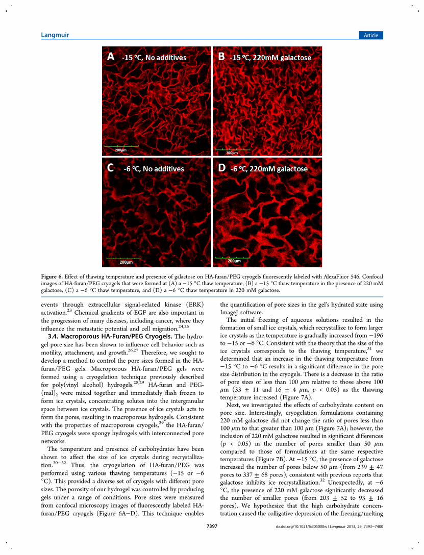
shown to affect the size of ice crystals during recrystalliza-tion.30−32 Thus, the cryogelation of HA-furan/PEG wasperformed using various thawing temperatures (−15 or −6°C). This provided a diverse set of cryogels with different poresizes. The porosity of our hydrogel was controlled by producinggels under a range of conditions. Pore sizes were measuredfrom confocal microscopy images of fluorescently labeled HA-furan/PEG cryogels (Figure 6A−D). This technique enables
the quantification of pore sizes in the gel’s hydrated state usingImageJ software.The initial freezing of aqueous solutions resulted in the
formation of small ice crystals, which recrystallize to form largerice crystals as the temperature is gradually increased from −196to −15 or −6 °C. Consistent with the theory that the size of theice crystals corresponds to the thawing temperature,31 wedetermined that an increase in the thawing temperature from−15 °C to −6 °C results in a significant difference in the poresize distribution in the cryogels. There is a decrease in the ratioof pore sizes of less than 100 μm relative to those above 100μm (33 ± 11 and 16 ± 4 μm, p < 0.05) as the thawingtemperature increased (Figure 7A).Next, we investigated the effects of carbohydrate content on
pore size. Interestingly, cryogelation formulations containing220 mM galactose did not change the ratio of pores less than100 μm to that greater than 100 μm (Figure 7A); however, theinclusion of 220 mM galactose resulted in significant differences(p < 0.05) in the number of pores smaller than 50 μmcompared to those of formulations at the same respectivetemperatures (Figure 7B). At −15 °C, the presence of galactoseincreased the number of pores below 50 μm (from 239 ± 47pores to 337 ± 68 pores), consistent with previous reports thatgalactose inhibits ice recrystallization.32 Unexpectedly, at −6°C, the presence of 220 mM galactose significantly decreasedthe number of smaller pores (from 203 ± 52 to 93 ± 16pores). We hypothesize that the high carbohydrate concen-tration caused the colligative depression of the freezing/melting
Figure 6. Effect of thawing temperature and presence of galactose on HA-furan/PEG cryogels fluorescently labeled with AlexaFluor 546. Confocalimages of HA-furan/PEG cryogels that were formed at (A) a −15 °C thaw temperature, (B) a −15 °C thaw temperature in the presence of 220 mMgalactose, (C) a −6 °C thaw temperature, and (D) a −6 °C thaw temperature in 220 mM galactose.
Langmuir Article
dx.doi.org/10.1021/la305000w | Langmuir 2013, 29, 7393−74007397
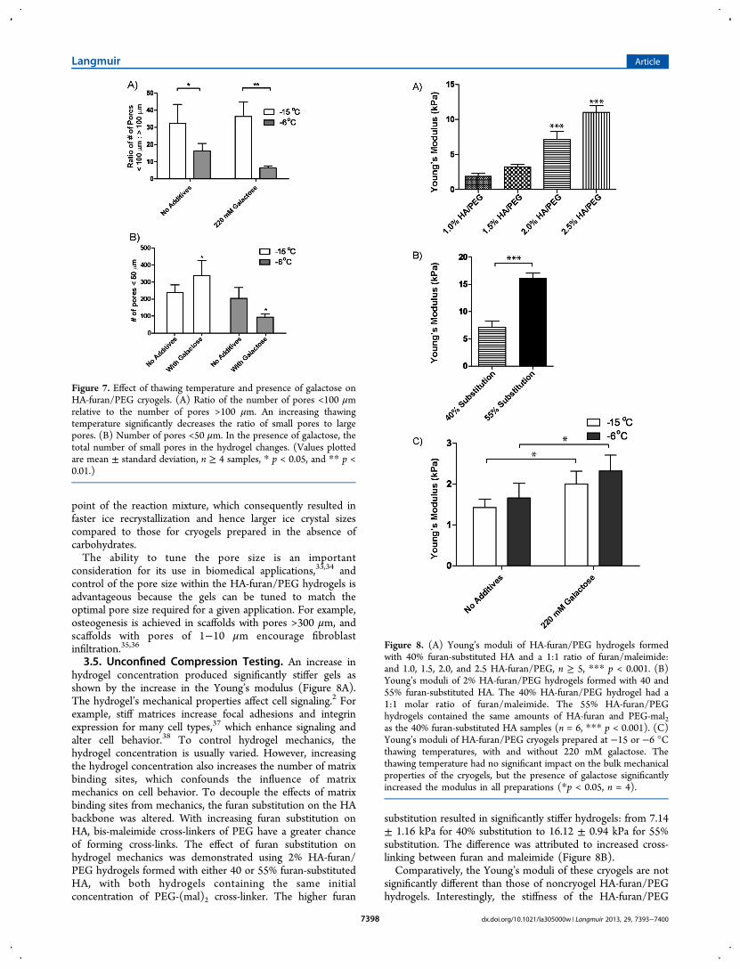
point of the reaction mixture, which consequently resulted infaster ice recrystallization and hence larger ice crystal sizescompared to those for cryogels prepared in the absence ofcarbohydrates.The ability to tune the pore size is an important
consideration for its use in biomedical applications,33,34 andcontrol of the pore size within the HA-furan/PEG hydrogels isadvantageous because the gels can be tuned to match theoptimal pore size required for a given application. For example,osteogenesis is achieved in scaffolds with pores >300 μm, andscaffolds with pores of 1−10 μm encourage fibroblastinfiltration.35,36
3.5. Unconfined Compression Testing. An increase inhydrogel concentration produced significantly stiffer gels asshown by the increase in the Young’s modulus (Figure 8A).The hydrogel’s mechanical properties affect cell signaling.2 Forexample, stiff matrices increase focal adhesions and integrinexpression for many cell types,37 which enhance signaling andalter cell behavior.38 To control hydrogel mechanics, thehydrogel concentration is usually varied. However, increasingthe hydrogel concentration also increases the number of matrixbinding sites, which confounds the influence of matrixmechanics on cell behavior. To decouple the effects of matrixbinding sites from mechanics, the furan substitution on the HAbackbone was altered. With increasing furan substitution onHA, bis-maleimide cross-linkers of PEG have a greater chanceof forming cross-links. The effect of furan substitution onhydrogel mechanics was demonstrated using 2% HA-furan/PEG hydrogels formed with either 40 or 55% furan-substitutedHA, with both hydrogels containing the same initialconcentration of PEG-(mal)2 cross-linker. The higher furan
substitution resulted in significantly stiffer hydrogels: from 7.14± 1.16 kPa for 40% substitution to 16.12 ± 0.94 kPa for 55%substitution. The difference was attributed to increased cross-linking between furan and maleimide (Figure 8B).Comparatively, the Young’s moduli of these cryogels are not
significantly different than those of noncryogel HA-furan/PEGhydrogels. Interestingly, the stiffness of the HA-furan/PEG
Figure 7. Effect of thawing temperature and presence of galactose onHA-furan/PEG cryogels. (A) Ratio of the number of pores <100 μmrelative to the number of pores >100 μm. An increasing thawingtemperature significantly decreases the ratio of small pores to largepores. (B) Number of pores <50 μm. In the presence of galactose, thetotal number of small pores in the hydrogel changes. (Values plottedare mean ± standard deviation, n ≥ 4 samples, * p < 0.05, and ** p <0.01.)
Figure 8. (A) Young’s moduli of HA-furan/PEG hydrogels formedwith 40% furan-substituted HA and a 1:1 ratio of furan/maleimide:and 1.0, 1.5, 2.0, and 2.5 HA-furan/PEG, n ≥ 5, *** p < 0.001. (B)Young’s moduli of 2% HA-furan/PEG hydrogels formed with 40 and55% furan-substituted HA. The 40% HA-furan/PEG hydrogel had a1:1 molar ratio of furan/maleimide. The 55% HA-furan/PEGhydrogels contained the same amounts of HA-furan and PEG-mal2as the 40% furan-substituted HA samples (n = 6, *** p < 0.001). (C)Young’s moduli of HA-furan/PEG cryogels prepared at −15 or −6 °Cthawing temperatures, with and without 220 mM galactose. Thethawing temperature had no significant impact on the bulk mechanicalproperties of the cryogels, but the presence of galactose significantlyincreased the modulus in all preparations (*p < 0.05, n = 4).
Langmuir Article
dx.doi.org/10.1021/la305000w | Langmuir 2013, 29, 7393−74007398
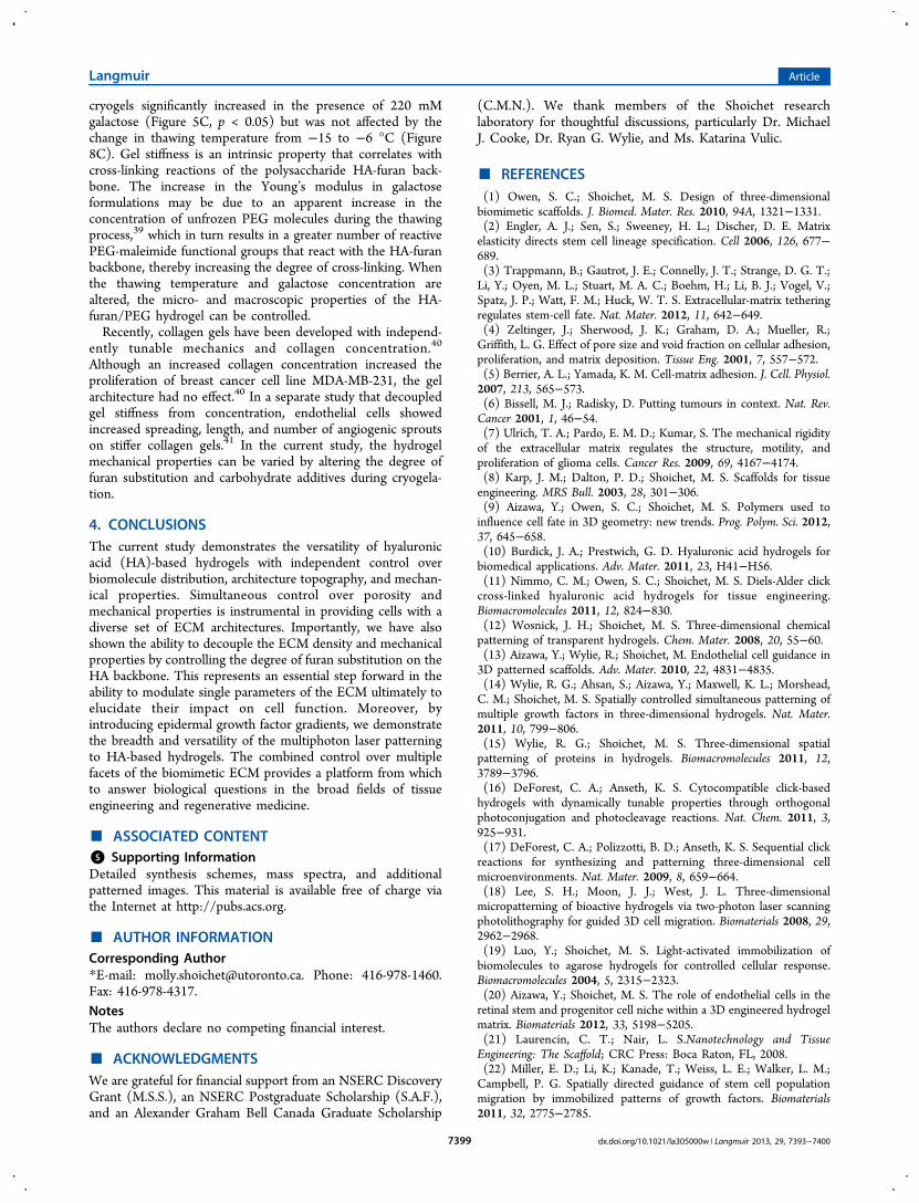
cryogels significantly increased in the presence of 220 mMgalactose (Figure 5C, p < 0.05) but was not affected by thechange in thawing temperature from −15 to −6 °C (Figure8C). Gel stiffness is an intrinsic property that correlates withcross-linking reactions of the polysaccharide HA-furan back-bone. The increase in the Young’s modulus in galactoseformulations may be due to an apparent increase in theconcentration of unfrozen PEG molecules during the thawingprocess,39 which in turn results in a greater number of reactivePEG-maleimide functional groups that react with the HA-furanbackbone, thereby increasing the degree of cross-linking. Whenthe thawing temperature and galactose concentration arealtered, the micro- and macroscopic properties of the HA-furan/PEG hydrogel can be controlled.Recently, collagen gels have been developed with independ-
ently tunable mechanics and collagen concentration.40
Although an increased collagen concentration increased theproliferation of breast cancer cell line MDA-MB-231, the gelarchitecture had no effect.40 In a separate study that decoupledgel stiffness from concentration, endothelial cells showedincreased spreading, length, and number of angiogenic sproutson stiffer collagen gels.41 In the current study, the hydrogelmechanical properties can be varied by altering the degree offuran substitution and carbohydrate additives during cryogela-tion.
4. CONCLUSIONSThe current study demonstrates the versatility of hyaluronicacid (HA)-based hydrogels with independent control overbiomolecule distribution, architecture topography, and mechan-ical properties. Simultaneous control over porosity andmechanical properties is instrumental in providing cells with adiverse set of ECM architectures. Importantly, we have alsoshown the ability to decouple the ECM density and mechanicalproperties by controlling the degree of furan substitution on theHA backbone. This represents an essential step forward in theability to modulate single parameters of the ECM ultimately toelucidate their impact on cell function. Moreover, byintroducing epidermal growth factor gradients, we demonstratethe breadth and versatility of the multiphoton laser patterningto HA-based hydrogels. The combined control over multiplefacets of the biomimetic ECM provides a platform from whichto answer biological questions in the broad fields of tissueengineering and regenerative medicine.
■ ASSOCIATED CONTENT*S Supporting InformationDetailed synthesis schemes, mass spectra, and additionalpatterned images. This material is available free of charge viathe Internet at http://pubs.acs.org.
■ AUTHOR INFORMATIONCorresponding Author*E-mail: [email protected]. Phone: 416-978-1460.Fax: 416-978-4317.NotesThe authors declare no competing financial interest.
■ ACKNOWLEDGMENTSWe are grateful for financial support from an NSERC DiscoveryGrant (M.S.S.), an NSERC Postgraduate Scholarship (S.A.F.),and an Alexander Graham Bell Canada Graduate Scholarship
(C.M.N.). We thank members of the Shoichet researchlaboratory for thoughtful discussions, particularly Dr. MichaelJ. Cooke, Dr. Ryan G. Wylie, and Ms. Katarina Vulic.
■ REFERENCES(1) Owen, S. C.; Shoichet, M. S. Design of three-dimensionalbiomimetic scaffolds. J. Biomed. Mater. Res. 2010, 94A, 1321−1331.(2) Engler, A. J.; Sen, S.; Sweeney, H. L.; Discher, D. E. Matrixelasticity directs stem cell lineage specification. Cell 2006, 126, 677−689.(3) Trappmann, B.; Gautrot, J. E.; Connelly, J. T.; Strange, D. G. T.;Li, Y.; Oyen, M. L.; Stuart, M. A. C.; Boehm, H.; Li, B. J.; Vogel, V.;Spatz, J. P.; Watt, F. M.; Huck, W. T. S. Extracellular-matrix tetheringregulates stem-cell fate. Nat. Mater. 2012, 11, 642−649.(4) Zeltinger, J.; Sherwood, J. K.; Graham, D. A.; Mueller, R.;Griffith, L. G. Effect of pore size and void fraction on cellular adhesion,proliferation, and matrix deposition. Tissue Eng. 2001, 7, 557−572.(5) Berrier, A. L.; Yamada, K. M. Cell-matrix adhesion. J. Cell. Physiol.2007, 213, 565−573.(6) Bissell, M. J.; Radisky, D. Putting tumours in context. Nat. Rev.Cancer 2001, 1, 46−54.(7) Ulrich, T. A.; Pardo, E. M. D.; Kumar, S. The mechanical rigidityof the extracellular matrix regulates the structure, motility, andproliferation of glioma cells. Cancer Res. 2009, 69, 4167−4174.(8) Karp, J. M.; Dalton, P. D.; Shoichet, M. S. Scaffolds for tissueengineering. MRS Bull. 2003, 28, 301−306.(9) Aizawa, Y.; Owen, S. C.; Shoichet, M. S. Polymers used toinfluence cell fate in 3D geometry: new trends. Prog. Polym. Sci. 2012,37, 645−658.(10) Burdick, J. A.; Prestwich, G. D. Hyaluronic acid hydrogels forbiomedical applications. Adv. Mater. 2011, 23, H41−H56.(11) Nimmo, C. M.; Owen, S. C.; Shoichet, M. S. Diels-Alder clickcross-linked hyaluronic acid hydrogels for tissue engineering.Biomacromolecules 2011, 12, 824−830.(12) Wosnick, J. H.; Shoichet, M. S. Three-dimensional chemicalpatterning of transparent hydrogels. Chem. Mater. 2008, 20, 55−60.(13) Aizawa, Y.; Wylie, R.; Shoichet, M. Endothelial cell guidance in3D patterned scaffolds. Adv. Mater. 2010, 22, 4831−4835.(14) Wylie, R. G.; Ahsan, S.; Aizawa, Y.; Maxwell, K. L.; Morshead,C. M.; Shoichet, M. S. Spatially controlled simultaneous patterning ofmultiple growth factors in three-dimensional hydrogels. Nat. Mater.2011, 10, 799−806.(15) Wylie, R. G.; Shoichet, M. S. Three-dimensional spatialpatterning of proteins in hydrogels. Biomacromolecules 2011, 12,3789−3796.(16) DeForest, C. A.; Anseth, K. S. Cytocompatible click-basedhydrogels with dynamically tunable properties through orthogonalphotoconjugation and photocleavage reactions. Nat. Chem. 2011, 3,925−931.(17) DeForest, C. A.; Polizzotti, B. D.; Anseth, K. S. Sequential clickreactions for synthesizing and patterning three-dimensional cellmicroenvironments. Nat. Mater. 2009, 8, 659−664.(18) Lee, S. H.; Moon, J. J.; West, J. L. Three-dimensionalmicropatterning of bioactive hydrogels via two-photon laser scanningphotolithography for guided 3D cell migration. Biomaterials 2008, 29,2962−2968.(19) Luo, Y.; Shoichet, M. S. Light-activated immobilization ofbiomolecules to agarose hydrogels for controlled cellular response.Biomacromolecules 2004, 5, 2315−2323.(20) Aizawa, Y.; Shoichet, M. S. The role of endothelial cells in theretinal stem and progenitor cell niche within a 3D engineered hydrogelmatrix. Biomaterials 2012, 33, 5198−5205.(21) Laurencin, C. T.; Nair, L. S.Nanotechnology and TissueEngineering: The Scaffold; CRC Press: Boca Raton, FL, 2008.(22) Miller, E. D.; Li, K.; Kanade, T.; Weiss, L. E.; Walker, L. M.;Campbell, P. G. Spatially directed guidance of stem cell populationmigration by immobilized patterns of growth factors. Biomaterials2011, 32, 2775−2785.
Langmuir Article
dx.doi.org/10.1021/la305000w | Langmuir 2013, 29, 7393−74007399
(23) Kholodenko, B. N.; Hancock, J. F.; Kolch, W. Signalling ballet inspace and time. Nat. Rev. Mol. Cell Biol. 2010, 11, 414−426.(24) Nelson, C. M.; Bissell, M. J. Of extracellular matrix, scaffolds,and signaling: tissue architecture regulates development, homeostasis,and cancer. Annu. Rev. Cell Dev. Biol. 2006, 22, 287−309.(25) Fischbach, C.; Chen, R.; Matsumoto, T.; Schmelzle, T.; Brugge,J. S.; Polverini, P. J.; Mooney, D. J. Engineering tumors with 3Dscaffolds. Nat. Methods 2007, 4, 855−860.(26) Harley, B. A. C.; Kim, H. D.; Zaman, M. H.; Yannas, I. V.;Lauffenburger, D. A.; Gibson, L. J. Microarchitecture of three-dimensional scaffolds influences cell migration behavior via junctioninteractions. Biophys. J. 2008, 95, 4013−4024.(27) Ranucci, C. S.; Kumar, A.; Batra, S. P.; Moghe, P. V. Control ofhepatocyte function on collagen foams: sizing matrix pores towardselective induction of 2-D and 3-D cellular morphogenesis.Biomaterials 2000, 21, 783−793.(28) Hassan, C. M.; Peppas, N. A. Structure and Applications ofPoly(vinyl alcohol) Hydrogels Produced by Conventional Cross-linking or by Freezing/Thawing Methods. In Biopolymers, PVAHydrogels, Anionic Polymerisation, Nanocomposites; Chang, J. Y., Ed.;Springer: New York, 2000, Vol. 153, pp 37−65(29) Lozinsky, V. I.; Galaev, I. Y.; Plieva, F. M.; Savinal, I. N.;Jungvid, H.; Mattiasson, B. Polymeric cryogels as promising materialsof biotechnological interest. Trends Biotechnol. 2003, 21, 445−451.(30) Knight, C. A.; Hallett, J.; Devries, A. L. Solute effects on icerecrystallization - an assessment technique. Cryobiology 1988, 25, 55−60.(31) Budke, C.; Heggemann, C.; Koch, M.; Sewald, N.; Koop, T. Icerecrystallization kinetics in the presence of synthetic antifreezeglycoprotein analogues using the framework of LSW theory. J. Phys.Chem. B 2009, 113, 2865−2873.(32) Tam, R. Y.; Ferreira, S. S.; Czechura, P.; Chaytor, J. L.; Ben, R.N. Hydration index-a better parameter for explaining small moleculehydration in inhibition of ice recrystallization. J. Am. Chem. Soc. 2008,130, 17494−17501.(33) Slaughter, B. V.; Khurshid, S. S.; Fisher, O. Z.; Khademhosseini,A.; Peppas, N. A. Hydrogels in regenerative medicine. Adv. Mater.2009, 21, 3307−3329.(34) Mattiasson, B.; Kumar, A.; Galaev, I. Macroporous Polymers:Production Properties and Biotechnological/Biomedical Applications, CRCPress/Taylor & Francis: Boca Raton, FL, 2010.(35) Karageorgiou, V.; Kaplan, D. Porosity of 3D biomaterialscaffolds and osteogenesis. Biomaterials 2005, 26, 5474−5491.(36) Raeber, G. P.; Lutolf, M. P.; Hubbell, J. A. Molecularlyengineered PEG hydrogels: a novel model system for proteolyticallymediated cell migration. Biophys. J. 2005, 89, 1374−1388.(37) Paszek, M. J.; Zahir, N.; Johnson, K. R.; Lakins, J. N.;Rozenberg, G. I.; Gefen, A.; Reinhart-King, C. A.; Margulies, S. S.;Dembo, M.; Boettiger, D.; Hammer, D. A.; Weaver, V. M. Tensionalhomeostasis and the malignant phenotype. Cancer Cell 2005, 8, 241−254.(38) Mitra, S. K.; Schlaepfer, D. D. Integrin-regulated FAK-Srcsignaling in normal and cancer cells. Curr. Opin. Cell Biol. 2006, 18,516−523.(39) Izutsu, K.; Yoshioka, S.; Kojima, S.; Randolph, T. W.; Carpenter,J. F. Effects of sugars and polymers on crystallization of poly(ethyleneglycol) in frozen solutions: phase separation between incompatiblepolymers. Pharm. Res. 1996, 13, 1393−1400.(40) Carey, S. P.; Kraning-Rush, C. M.; Williams, R. M.; Reinhart-King, C. A. Biophysical control of invasive tumor cell behavior byextracellular matrix microarchitecture. Biomaterials 2012, 33, 4157−4165.(41) Mason, B. N.; Starchenko, A.; Williams, R. M.; Bonassar, L. J.;Reinhart-King, C. A. Tuning three-dimensional collagen matrixstiffness independently of collagen concentration modulates endothe-lial cell behavior. Acta Biomater. 2013, 9, 4635−4644.
Langmuir Article
dx.doi.org/10.1021/la305000w | Langmuir 2013, 29, 7393−74007400