Embed Size (px)
Citation preview
Thorax (1974), 29, 451.
Hydatid disease of the heartReport of five cases and review of the literature
G. CALAMAI, A. M. PERNA, and A. VENTURINI
Department of Cardiac Surgery and Experimental Cardiology, Arcispedale S.M. Nuova,Florence, Italy
Calamai, G., Perna, A. M., and Venturini, A. (1974). Thorax, 29, 451-458. Hydatiddisease of the heart: report of five cases and review of the literature. The world literatureon the surgical treatment of echinococcosis of the heart is reviewed. Few cases aresurgically treated, although the disease has been known for a long time. Localizationto the liver and lungs is the most frequent. Cardiopulmonary bypass techniques makepossible surgical treatment of hydatid cyst of the heart. The present paper is concernedwith five cases operated upon between 1959 and 1969, three males and two females,their ages ranging from 13 to 46 years. A preoperative diagnosis was made in each case.One case was operated upon under cardiopulmonary bypass. The need for cardio-pulmonary bypass on a stand-by basis is emphasized. The localization of the hydatidcyst was in the left ventricular wall (three cases), right ventricular wall (one case), andmultiple (one case). The frequency of cardiac echinococcosis ranges between 0-5% and2% according to various authors. Diagnosis is achieved with the aid of laboratory tests,radiology, and angiography; but the presence of the disease must be suspected in allpatients who come from endemic areas. Surgical therapy is mandatory. Due to thegrowth characteristics of the cyst itself, the danger of damaging the ventricular wallat operation is increased; thus it is essential to have cardiopulmonary bypass facilitiesimmediately available.
Surgical treatment of a cardiac hydatid cyst wasfirst attempted by Marten and de Crespigny(1921). The first case successfully operated uponwas described by Long (1932). D'Abreu (1950)reported another case. Muller (1957) reported26 surgically treated cases and 13 in which itwas not clearly specified whether the treatmenthad been surgical. According to Al-Naaman andAl-Omeri (1970), the total number of surgicallytreated cases was 69, including three personalcases. In a recent review of the literature Heyat,Mokhtari, Hajaliloo, and Shakibi (1971) reported118 cases of cardiac echinococcosis treated surgi-cally. An additional case was described byMurphy, Kean, Venturini, and Lillehei (1971);another case was described by Hazan, Leblanc,Aobillard, and Mathey (1970), three cases byUrquia, Perez Leon, de los Arcos, and Madurga(1972), one case by Dodek, De Mots, Antonovic,and Hodam (1972), and three cases by Romanoff(1973). Between 1959 and 1969, five cases ofhydatid cyst of the heart were successfully
operated upon at the Institute of Surgery of RomeUniversity. Our paper is concemed with thedescription of these five cases which bring to atotal of 133 the number of such cases describedin the world literature.
Hydatid cyst of the heart is an ominous diseasewhich, in the absence of surgical treatment, isusually fatal (Peters, Dexter, and Weiss, 1945).An improvement in the prognosis of these patientsfollows surgery, especially since open-heart tech-niques allow radical treatment (Larghero, 1964;Di Bello and Menendez, 1963; Dodek et al., 1972;de los Arcos et al., 1971).
CASE REPORTS
CASE 1 P.A., a 25-year-old man, was admitted on16 February 1959 to the Surgical Department of RomeUniversity. Four years before admission he had hadan episode of crushing precordial pain, radiating tothe neck and to the left shoulder and lasting only afew minutes. A few days later he presented with fever,shivering, cough, and bloodstained sputum; two
451
on July 29, 2020 by guest. Protected by copyright.
http://thorax.bmj.com
/T
horax: first published as 10.1136/thx.29.4.451 on 1 July 1974. Dow
nloaded from
G. Calamai, A. M. Perna, and A. Venturini
months later fainting and loss of consciousnessoccurred. For the next two years the patient was inno distress. In 1957 he presented with a furtherepisode of precordial pain followed by fever andshivering. A chest radiograph disclosed cardiacenlargement.
Physical examination was negative on admission.The electrocardiogram showed normal sinus rhythmwith left ventricular strain. A chest tomogram anddiagnostic pneumothorax showed a lesion thoughtto be an hydatid cyst of the pericardium. Bloodtests were normal, and the Casoni intradermal skintest was positive. The patient was explored on 25March 1959. Through a left thoracotomy in the fourthintercostal space, a hydatid cyst was found coveredby the pleura which was adherent to the pericardium.The pericardium was opened and it was found to beincorporated in the wall of the cyst and adherent tothe myocardium. The cyst was then aspirated andirrigated with iodine solution. The capsule waspartially resected, leaving a portion adhering to theunderlying myocardium.
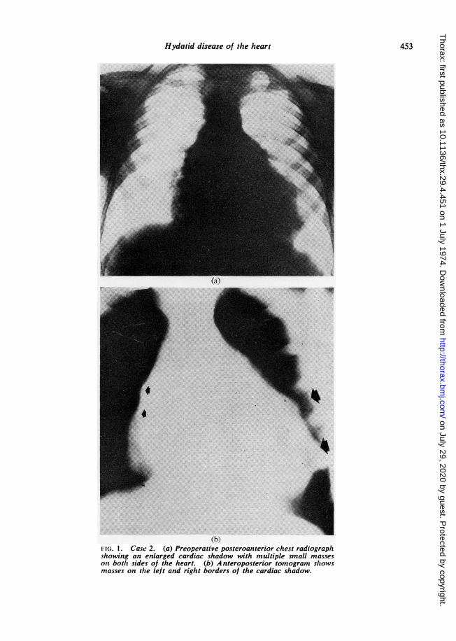
CASE 2 C.G., a 13-year-old boy, was admitted on 10June 1968. Two years before admission he had pre-sented with an episode of fever (40°C), widespreadoedema, itching, dyspnoea, precordial pain, andcough. Chest films showed an enlarged cardiac shadowwith a number of small masses along the left andright borders (Fig. la and b); on the lateral pro-jection a bilobed mass was seen anteriorly. A loudsplit second sound over the pulmonary area, and athird heart sound were audible over a wide area.Blood sugar, haemoglobin, and haematocrit values
were within no*mal limits. A Casoni intradermalskin test was positive and a Weinberg complementfixation test was positive. A full blood count wasnormal except for 13% eosinophils in the peripheralblood smear.The electrocardiogram showed normal sinus rhythm
and inverted T waves in leads II, III, aVF and theleft precordial leads; Q waves were present in leadsIII and aVF.The preoperative diagnosis was echinococcosis of
the pericardium.On 19 July 1968 an exploratory operation was
performed through an anterior bilateral thoracotomyin the fourth intercostal space with transverse sternaltransection. The pericardium was completely ad-herent to the heart. Two cysts, 3 cm in diameter,were removed from the anterior surface of the rightventricle. Smaller cysts were isolated and removedfrom the right atrium. Four infected cysts were re-moved from the mediastinal aspect of the heart fol-lowing irrigation with iodine into the cyst cavities. Thecysts were adherent to the surface of the left ventricu-lar wall. Other cysts were removed (following irriga-tion) from the left atrium. The left phrenic nerve,involved by the hydatid disease, was transected. Atthe conclusion of surgery the heart showed normal
contractability. The patient died six months afteroperation from chronic heart failure due to extensivedisease of the myocardium.
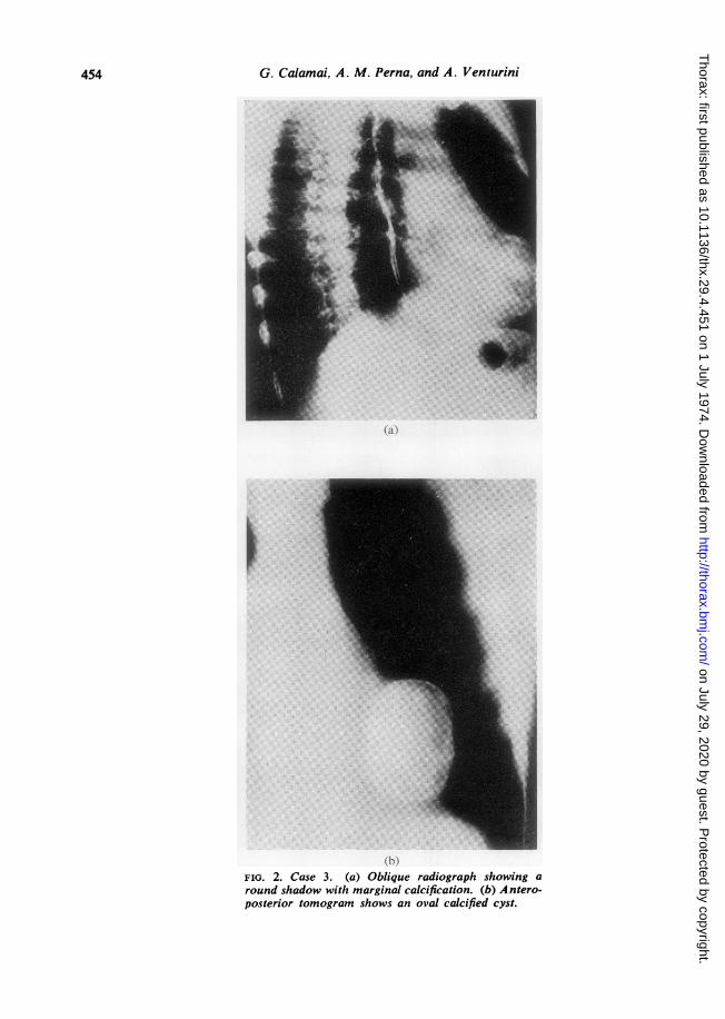
CASE 3 C.G., a 45-year-old man, was admitted inNovember 1968. His principal complaint was of pre-cordial pain for two months. A midsystolic murmur inthe second intercostal space at the right sternal edgewas found. An early diastolic murmur was audiblealong the left sternal edge. The electrocardiogramshowed normal sinus rhythm and evidence of previousanterolateral myocardial infarction. Chest filmsshowed a round shadow with marginal calcificationprojecting on the left ventricle (Fig. 2a and b). Therewere 5% eosinophils in the peripheral blood smear;all other blood tests were within normal limits.On 22 May 1969 an exploratory thoracotomy was
performed through the fourth left intercostal space.The pericardium was found to be adherent to abulging mass on the left ventricle. Aspiration of themass produced fresh blood. The patient was placedon total cardiopulmonary bypass. The mass was thenexcised. The left ventricle was widely opened, thepapillary muscles and mitral valve being left intact.The ventriculotomy was then closed with multiplemattress sutures buttressed with strips of Teflon felt.The patient regained a normal cardiac output as soonas cardiopulmonary bypass was discontinued.
CASE 4 B.G.D., a 9-year-old boy, was admitted on10 June 1969 complaining of intermittent precordialpain of short duration. In April 1969 a chest radio-graph had shown a round shadow along the left heartborder. A loud and split pulmonary sound and amidsystolic murmur in the second and third inter-costal space along the left sternal edge were audible.The electrocardiogram showed normal sinus rhythmand signs of anterolateral subepicardial myocardialischaemia.Blood tests were within normal limits.The patient was operated upon on 16 July 1969. A
hydatid cyst was found adherent to the left ventricularapex. The pericardium was packed with iodizedgauze and the cyst was aspirated. Ten millilitres oftransparent fluid was obtained. The cyst was thenopened and the cyst membrane was enucleated intactfrom the underlying myocardium. The left ventricularwall was approximated by interrupted silk sutures.
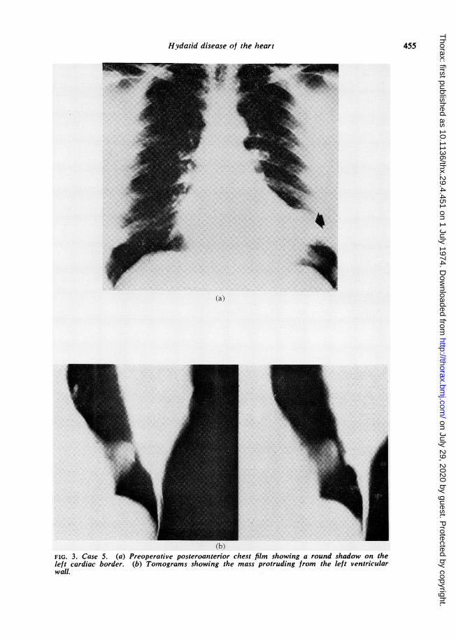
CASE 5 G.A., a 43-year-old woman, was admitted inJuly 1969. Her principal complaint was intermittentcrushing precordial pain, radiating to the leftshoulder, of approximately six years' duration. Amass in the left lower pulmonary field was first notedin November 1968 on routine pre-employment radio-graphs. A diagnosis of echinococcosis of the heartwas made following electrocardiogram, chest radio-graphs (Fig. 3a and b), a positive Weinberg comple-ment fixation test, and a positive Casoni intradermal
452
on July 29, 2020 by guest. Protected by copyright.
http://thorax.bmj.com
/T
horax: first published as 10.1136/thx.29.4.451 on 1 July 1974. Dow
nloaded from
Hydatid disease of the heart
(a)'-
(b)FIG. 1. Case 2. (a) Preoperative posteroanterior chest radiographshowing an enlarged cardiac shadow with multiple small masseson both sides of the heart. (b) Anteroposterior tomogram showsmasses on the left and right borders of the cardiac shadow.
453
on July 29, 2020 by guest. Protected by copyright.
http://thorax.bmj.com
/T
horax: first published as 10.1136/thx.29.4.451 on 1 July 1974. Dow
nloaded from
G. Calamai, A. M. Perna, and A. Venturini
(a )
r
FIG. 2. Case 3. (a) Oblique radiograph showing around shadow with marginal calcification. (b) A ntero-posterior tomogram shows an oval calcified cyst.
454
on July 29, 2020 by guest. Protected by copyright.
http://thorax.bmj.com
/T
horax: first published as 10.1136/thx.29.4.451 on 1 July 1974. Dow
nloaded from
Hydatid disease of the heart
4*
(a)
(b)FIG. 3. Case 5. (a) Preoperative posteroanterior chest film showing a round shadow on theleft cardiac border. (b) Tomograms showing the mass protruding from the left ventricularwall.
455
on July 29, 2020 by guest. Protected by copyright.
http://thorax.bmj.com
/T
horax: first published as 10.1136/thx.29.4.451 on 1 July 1974. Dow
nloaded from
G. Calamai, A. M. Perna, and A. Venturini
skin reaction. The peripheral blood smear revealed4 % eosinophils.The patient was operated upon and the mass was
excised through a left thoracotomy according to thepreviously described techniques. She made anuneventful recovery.
DISTRIBUTION OF HYDATID DISEASE OF THE HEART
Once the hexacanth embryo has embolized to acoronary artery the following distribution withinthe heart may be found. Ivanissevich and Rivas(1962), in a series of 194 cases of hydatid diseaseof the heart, reported the following distribution:in 116 (60%) cases the disease occurred in theleft ventricular myocardium, and in 33 (17%)in the right ventricle; in 18 (9%) cases in theinterventricular septum; in 16 (8%) cases in theright atrium; in 8 (4%) cases in the left atrium;and in 3 (2%) cases in the interatrial septum.
In our series of five cases the distribution wasthree in the left ventricular wall, one in the rightventricular wall, and one case with multiplelocalization.
SYMPTOMS AND SIGNS
Hydatid cyst of the myocardium can be asympto-matic; occasionally the first clinical manifestationis sudden death (Guarini and Torraco, 1962;Comakov, 1965). More often precordial pain ispresent (Di Bello and Menendez, 1963; Heyat etal., 1971).
Other possible signs are due to compressionby the cyst. Mitral stenosis can be simulated whenthe cyst lies in the left atrium obstructing thecardiac outflow.Angina is a main complaint if the cyst com-
presses a coronary artery.Electrocardiographic changes simulating myo-
cardial infarction can be caused by the hydatidcyst (Vestri and Tardio, 1968) or conductiondefects due to compression of the bundle of His(Heimann, 1928; Corkill, 1929; Ghanem andDarwish, 1951; de los Arcos et al., 1971).When the cyst is located in the right heart the
most common features are hepatomegaly,oliguria, ascites, or chronic cor pulmonale due torepeated pulmonary emboli (Aguirre et al., 1956;Al-Naaman and Al-Omeri, 1970).From rupture of a cyst into the cardiac cavity
a variety-of allergic phenomena result, rangingfrom wheals to anaphylactic shock. Acute peri-carditis may be caused by rupture of the cyst intothe pericardial sac.
DIAGNOSIS
In spite of many symptoms the diagnosis during
life may be difficult. If a patient comes from anendemic area the possibility of hydatid diseaseshould be kept in mind. A chest radiograph isoften the first clue to the diagnosis, showing anabnormality of the cardiac outline.
Fluoroscopy adds information about motilityor absence of normal pulsation, suggesting thepossibility of a hydatid cyst.
Occasionally the presence of calcification is anaid to diagnosis (Blondeau, Laupretre, andMiramond de la Roquette, 1938).Angiography must be considered the most
specific of all diagnostic procedures if a fillingdefect in the cardiac cavity is revealed.The electrocardiogram shows a thinner cardiac
wall in the area of localization of the parasite andlow-voltage R waves, QRS notching, and T-waveinversion and increase of R waves in adjacentareas, or absence of anomalous Q waves.When the cyst is in the interventricular septum
arrhythmia may be present, and the QRS appearsnotched.The laboratory tests are of little help: the
Casoni intradermal skin test and Weinberg com-plement fixation test are often negative andeosinophilia is not a constant finding.
DIFFERENTIAL DIAGNOSIS
A hydatid cyst must be differentiated from abronchial carcinoma originating close to the lunghilum, tumours of the heart, benign tumours ofthe mediastinum and pericardium, and aneurysmsof the heart.
Ventricular aneurysms are the most cimmonand can be easily differentiated by means ofangiography.According to Murphy et al. (1971), the conse-
quences of cardiac hydatid cyst are: death ofthe cyst with calcification; rupture, either intra-pericardial, causing pericarditis, or intracardiac,leading to anaphylactic shock or embolicphenomena.Cor pulmonale or metastatic pulmonary
echinococcosis, systemic emboli or systemicmetastasis of the hydatid cyst can be caused byrupture of the cyst into the right or left heart.
TREATMENT
There is no known medical treatment of cardiacechinococcosis. In view of the serious complica-tions of this disease surgical treatment ismandatory.The chest should be entered through a left,
right or bilateral thoracotomy, depending on the
456
on July 29, 2020 by guest. Protected by copyright.
http://thorax.bmj.com
/T
horax: first published as 10.1136/thx.29.4.451 on 1 July 1974. Dow
nloaded from
Hydatid disease of the heart
site of the cyst. Once the cyst has been freed fromthe surrounding structures its content should beaspirated and then irrigated with hypertonic(33%) saline in order to achieve sterilization ofthe parasite wall and viable elements (Vaglio andGuarini, 1965). When the cyst is located insidethe wall of the left ventricle, removal may bedifficult because of the thinness of the surround-ing myocardium (Guarini and Torraco, 1962).In our opinion, bypass facilities should be avail-able every time the ventricular walls are involvedby a hydatid cyst. Once the cyst has been removedthe area should again be irrigated with hypertonicsaline.The mortality of this procedure is low and the
postoperative course is usually uneventful; thepatient soon resumes normal life.
BIBLIOGRAPHY
Ab6, J. C. and Matteucci, P. (1970). Quiste hidaticodel mediastino. Torax, 19, 208.
Aguirre, C. V., Purcallas, J., Baldomir, J. M., Suzacq,C. V., Hazan, J., Horjales, C. O., Dighiero, J.,and Canabal, E. J. (1956). Coraz6n pulmonaragudo hidatico. Archivos del Instituto de Cardi-ologia de Mexico, 26, 211.
Al-Naaman, Y. D. and Al-Omeri, M. M. (1970). Car-diopericardial echinococcosis causing myocardialinsufficiency, right-sided heart failure and con-strictive pericarditis. (Ex: ) 9th Congress of theInternational Cardiovascular Society, BuenosAires, 1969. Journal of Cardiovascular Surgery,11, 303.
Bianchi, C., Rodriguez, R., Saavedra, J., and Reyes,H. (1972). Hidatidosis cardiaca. Revista Medicade Chile, 100, 46.
Blondeau, A., Laupretre, and Miramond de laRoquette, J. (1938). Kyste hydatique du coeur.Particularites de l'image radiologique. Alg&rieMedicale, 42, 299.
Comakov, M. (1965). Sudden death in a case of car-diac echinococcosis. Folia Medica (Plovdiv), 7,378.
Corkill. N. L. (1929). Hydatid cyst of the heart.British Medical Journal, 2, 622.
D'Abreu, A. L. (1950). Removal of hydatid cvst fromthe wall of the left ventricle. Thorax, 5, 362.
Del Bono, G. (1962). Cardiac echinococcosis in domes-tic animals. Zooprofilassi, 17, 5.
de los Arcos, E., Madurga, M. P., Perez Leon,J., Martinez, J. L., and Urquia, M. (1971).Hydatid cyst of the interventricular septumcausing left anterior hemiblock. British HeartJournal, 33, 623.
Dev, F. (1949). L'Echinococcose Primitive. Masson,Paris.
Dew. H. R. (1928). Hydatid Disease. AustralasianMedical Publishing Co. Ltd., Sydney.
Di Bello, R. and Menendez. H. (1963). Intracardiacrupture of hydatid cyst of the heart. Circulation.27, 366.
Dighiero, J., Canabal, E. J., Aguirre, C. V., Hazan,J., and Horjales, J. 0. (1958). Echinococcusdisease of the heart. Circulation, 17, 127.
Dodek, A., DeMots, H., Antonovic, J. A., andHodam, R. P. (1972). Echinococcus of the heart.An unusual tumor of the heart and liver. Ameri-can Journal of Cardiology, 30, 293.
Ghanem, M. H. and Darwish, A. E. (1951). Hydatidheart disease with paroxismal tachycardia. BritishHeart Journal, 13, 109.
Guarini, L. and Torraco, Q. (1962). L'echinococcosidel pericardio. Minerva Chirurgica, 17, 481.
Hazan, E., Leblanc, J., Robillard, M., and Mathey,J. (1970). Kyste hydatique du ventricule droitrevele par un accident aigu. Exerese d'urgenceavec remplacement prothetique de la valvuletricuspide. Chirurgie, 96, 257.
Heilbrunn, A., Kittle, C. F., and Dunn, M. (1963).Surgical management of echinococcus cysts ofthe heart and pericardium. Circulation, 27, 219.
Heimann, H. L. (1928). Hydatid cyst in the heart.British Medical Journal, 1, 801.
Heyat, J., Mokhtari, H., Hajaliloo, J., and Shakibi,J. G. (1971). Surgical treatment of echinococcalcyst of the heart. Journal of Thoracic and Car-diovascular Surgery, 61, 755.
Ivanissevich, 0. and Rivas, C. I. (1962). EquinococosisHidatidica. TAlleres graficos del Ministero deEducation y Justicia, Buenos Aires.
Kean, B. H. and Breslau, R. C. (1964). Parasites ofthe Human Heart. Grune and Stratton, NewYork.
Larghero, P. (1954). Tratamiento del quiste hidaticodel ventriculo izquierdo; diez observaciones enel Uruguay. Torax, 3, 263.
Long, W. J. (1932). Hydatid cyst in the left ventri-cular wall of the heart. Medical Journal ofAustralia, 2) 701.
Marten, R. H. and de Crespigny, C. T. C. (1921).Notes on a case of hydatid of the heart. MedicalJournal of A ustralia, 1, 287.
Matteucci, P., Rubio, R., and Fiandra, 0. (1970).Hidatidosis doble del corazon. Torax, 19, 204.
Muller, M. A. (1957). Le kyste hydatique du coeur.These, Universite de Nancy.
Murphy, T. E., Kean, B. H., Venturini, A., andLillehei, C. W. (1971). Echinococcus cyst of theleft ventricle. Journal of Thoracic and Cardio-vascular Surgery, 61, 443.
Papamichael, E., Ikkos, D., Milingos, M., and Yan-nacopoulos, J. (1971). Echinococcosis of theheart. Chest, 59, 280.
Peters, J. H., Dexter, L., and Weiss, S. (1945). Clinicaland theoretical considerations of involvement ofthe left side of the heart with echinococcal cysts.A merican Heart Journal, 29, 143.
457
on July 29, 2020 by guest. Protected by copyright.
http://thorax.bmj.com
/T
horax: first published as 10.1136/thx.29.4.451 on 1 July 1974. Dow
nloaded from
G. Calamai, A. M. Perna, and A. Venturini
Ramos, G., del Villar, J. L., Saing, J. L., del Busto,E. F., Gonzalez, E., and Ortega, J. (1971). Car-diac hydatidosis. Revista Clinica Espanola, 121,411.
Romanoff, H. (1973). Echinococcosis of the heart.Report of three new cases. Journal of Thoracicand Cardiovascular Surgery, 66, 29.and Milwidsky, H. (1962). Primary echinococ-
cosis of the heart cured by operation. Journal ofThoracic and Cardiovascular Surgery, 43, 677.
Urquia, M., Perez Leon, J., de los Arcos, E., andMadurga, P. (1972). Surgical treatment of cardiacechinococcosis. Journal of Cardiovascular Surgery(Torino), 13, 191.
Vaglio, L. and Guarini, L. (1965). Sudiun caso diechinococcosi del cuore. Gazzetta Internazionaledi Medicina e Chirurgia, 70, 1588.
Vestri, A. and Tardio, R. (1968). Aspetti elettrocar-diografici dell'echinococcosi del cuore. Comuni-cazione tenuta alla Societa romana di chirurgia.EMES.
Requests for reprints to: Professor AnacletoVenturini, Department of Cardiac Surgery, Arcispe-dale S.M. Nuova, Careggi, Florence, Italy.
458
on July 29, 2020 by guest. Protected by copyright.
http://thorax.bmj.com
/T
horax: first published as 10.1136/thx.29.4.451 on 1 July 1974. Dow
nloaded from


![Central Nervous System Hydatid Disease - SM Journals · Currently, hydatid disease is a global problem due to the ease of travelling [11]. Despite advances . in treatment and imaging](https://img.pdfslide.net/doc/110x75/5f2184391df5c764283375db/central-nervous-system-hydatid-disease-sm-journals-currently-hydatid-disease.jpg)