Embed Size (px)
Citation preview

Journal of Pharmacology & Clinical ResearchISSN: 2473-5574
Review ArticleVolume 5 Issue 3 - April 2018DOI: 10.19080/JPCR.2018.05.555661
J of Pharmacol & Clin ResCopyright © All rights are reserved by Shivani Chauhan
Hydrogen Sulfide (H2S ) - Poison Gas to Signaling Molecule in Regulation of Human Biology
Shivani Chauhan*1, Lubhan Singh2, Vipul Kumar2 and Chandrakala3
1Aurobindo Pharmaceuticals Ltd, India 2Department of Pharmaceutical Technology, Rameesh Institute of Vocational Studies, India3Glocal University, Saharanpur, India
Submission: March 22, 2018; Published: April 10, 2018
*Corresponding author: Shivani Chauhan, Department of MarketResearch Aurobindo Pharmaceuticals Ltd., UP, India, Tel: Email:
IntroductionHydrogen sulfide is a small gaseous and the diffusible
compound with the formula H2S that constitute a family of labile gas-transmitters together with Nitric oxide (NO) and Carbon monoxide (CO) [1,2]. Although all three gas-transmitters has the same toxicity status, H2S yet has not been awarded the degree of initial skepticism which is associated with other two NO and CO [3]. It is a colorless, flammable gas with the characteristic foul smell of rotten eggs. Often it results from the bacterial decomposition of organic matter in the absence of oxygen. It also occurs in volcanic gases, natural gas, and some well waters. Despite being as environmental pollutant and bio hazardous compound it is widely integrated in various human physiological processes and diseases [4,5]. The main mechanism behind H2S toxicity is mitochondrial damage or oxidative damage through blockade of cytochrome-c oxidase [6,7]. Recent studies suggested
that H2S acts as an important mediator in various signaling pathways of human biology. Endogenously it is produced in various parts of the body like blood vessels [8], heart [9], GIT and central nervous system [10]. The present article reviews the prominent role of H2S in human biology with special focus on current literature and clinically relevant studies.
Synthesis of Hydrogen Sulfide in Mammalian and Human Tissues
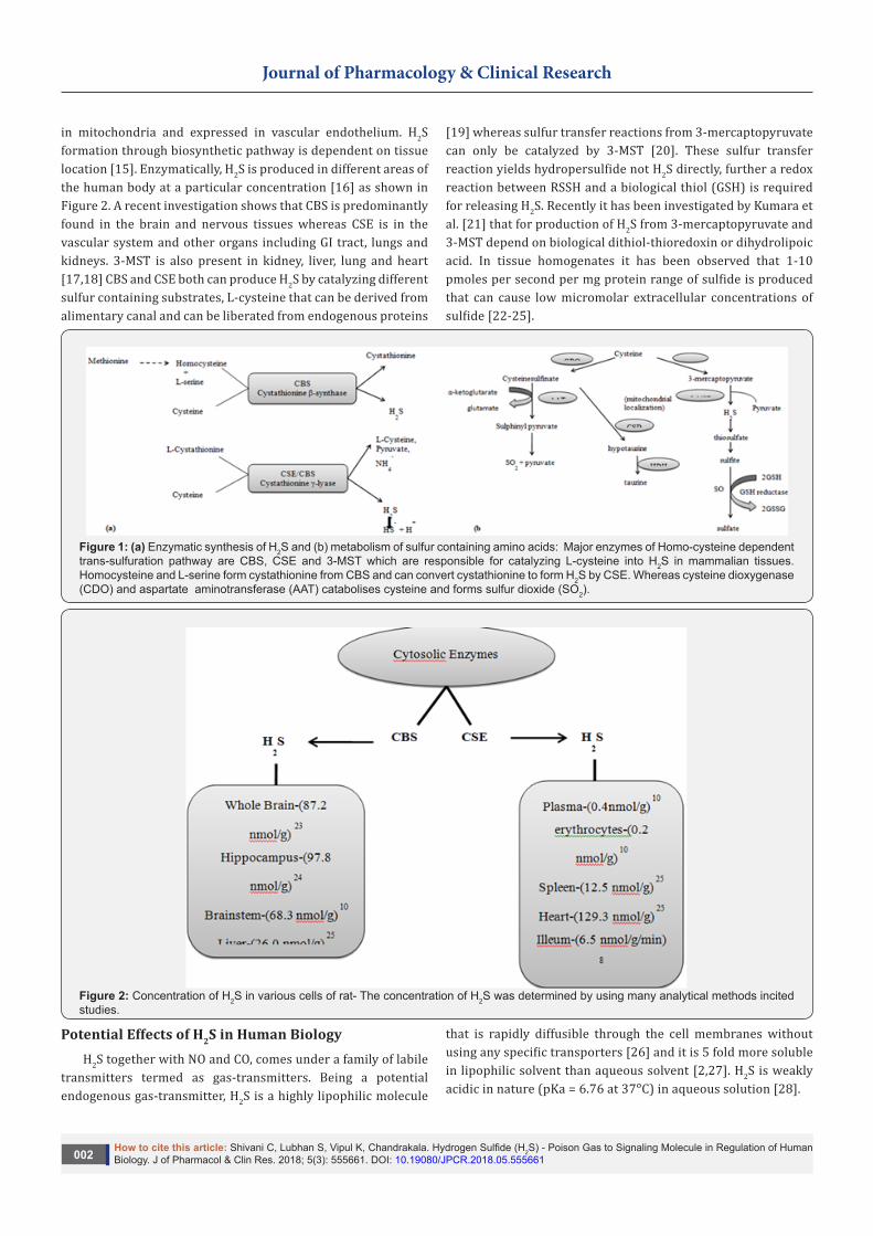
L-Cysteine (sulfur containing amino acid) is the major substrate for producing H2S in mammalian tissues via two pyridoxal-5’phosphate (PLP) dependant enzymes: cystathionine-β synthase (CBS) and cystathionine-γ lyase (CSE) as well as a PLP independent enzyme 3-mercaptopyruvate sulfurtransferase (3-MST) [11-14] as depicted in Figure 1. Both CBS and CSE exist in cytosol whereas the 3-MST present mainly
J of Pharmacol & Clin Res 5(3): JPCR.MS.ID. 555661 (2018) 001
Abstract
Hydrogen sulfide is a blossomed exploration of whiff. It is a well known poisonous gas but now serve as a gaseous transmitter which may act as an important signaling mediator in different cellular and physiological processes. It is recently discovered, that H2S is generated enzymatically from L-cysteine in various mammalian and human tissues to carry out number of signaling pathways in human biology. It has been revealed that ATP-sensitive K+ (KATP) channel is widely accepted cellular target associated with H2S which is responsible for vasorelaxation, cardioprotection, neuroprotection and also in diabetes mellitus. Evidence is accumulating to demonstrate that H2S exerts significant effects in different diseases by different mechanisms of action; also this review summarizes a detailed description of current signaling mechanism responsible for various effects in human biology.
Keywords: Hydrogen sulfide (H2S); Cystathionine-β synthase (CBS); Cystathionine-γ lyase (CSE); 3-mercaptopyruvate sulfurtransferase (3-MST); KATP channel
Abbreviations: H2S: Hydrogen Sulfide; CBS: Cystathionine-β-synthase; CSE: Cystathionine-ϒ-Lyase; CAT: Cysteine Aminotransferase; MST: 3-Mercaptopyruvate Sulfurtransferase; NO: Nitric Oxide; CO: Carbon Monoxide; PKA: Protein Kinase A; NADPH: Nicotinamide Adenine Dinucleotide Phosphate; NaHS: Sodium Hydrosulfide; MAPK: Mitogen-Activated Protein Kinase; AOA: Aminooxyacetate; ICAM-1: Intercellular Adhesion Molecule; TNF-α: Tumor Necrosis Factor-α; TRPV-1: Transient Receptor Potential Vanilloid-1; ERK: Extracellular Signal Related Kinase; EGFR: Endothelial Growth Factor Receptor; LTP: Long Term Potentiation; ROS: Reactive Oxygen Species; RNS: Reactive Nitrite Species.
Journal of Pharmacology & Clinical Research
How to cite this article: Shivani C, Lubhan S, Vipul K, Chandrakala. Hydrogen Sulfide (H2S) - Poison Gas to Signaling Molecule in Regulation of Human Biology. J of Pharmacol & Clin Res. 2018; 5(3): 555661. DOI: 10.19080/JPCR.2018.05.555661002
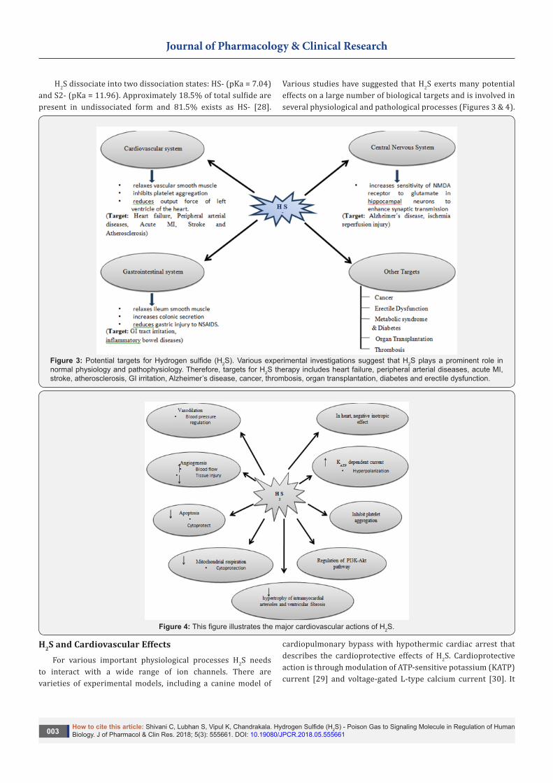
in mitochondria and expressed in vascular endothelium. H2S formation through biosynthetic pathway is dependent on tissue location [15]. Enzymatically, H2S is produced in different areas of the human body at a particular concentration [16] as shown in Figure 2. A recent investigation shows that CBS is predominantly found in the brain and nervous tissues whereas CSE is in the vascular system and other organs including GI tract, lungs and kidneys. 3-MST is also present in kidney, liver, lung and heart [17,18] CBS and CSE both can produce H2S by catalyzing different sulfur containing substrates, L-cysteine that can be derived from alimentary canal and can be liberated from endogenous proteins
[19] whereas sulfur transfer reactions from 3-mercaptopyruvate can only be catalyzed by 3-MST [20]. These sulfur transfer reaction yields hydropersulfide not H2S directly, further a redox reaction between RSSH and a biological thiol (GSH) is required for releasing H2S. Recently it has been investigated by Kumara et al. [21] that for production of H2S from 3-mercaptopyruvate and 3-MST depend on biological dithiol-thioredoxin or dihydrolipoic acid. In tissue homogenates it has been observed that 1-10 pmoles per second per mg protein range of sulfide is produced that can cause low micromolar extracellular concentrations of sulfide [22-25].
Figure 1: (a) Enzymatic synthesis of H2S and (b) metabolism of sulfur containing amino acids: Major enzymes of Homo-cysteine dependent trans-sulfuration pathway are CBS, CSE and 3-MST which are responsible for catalyzing L-cysteine into H2S in mammalian tissues. Homocysteine and L-serine form cystathionine from CBS and can convert cystathionine to form H2S by CSE. Whereas cysteine dioxygenase (CDO) and aspartate aminotransferase (AAT) catabolises cysteine and forms sulfur dioxide (SO2).
Figure 2: Concentration of H2S in various cells of rat- The concentration of H2S was determined by using many analytical methods incited studies.
Potential Effects of H2S in Human BiologyH2S together with NO and CO, comes under a family of labile
transmitters termed as gas-transmitters. Being a potential endogenous gas-transmitter, H2S is a highly lipophilic molecule
that is rapidly diffusible through the cell membranes without using any specific transporters [26] and it is 5 fold more soluble in lipophilic solvent than aqueous solvent [2,27]. H2S is weakly acidic in nature (pKa = 6.76 at 37ᵒC) in aqueous solution [28].
Journal of Pharmacology & Clinical Research
How to cite this article: Shivani C, Lubhan S, Vipul K, Chandrakala. Hydrogen Sulfide (H2S) - Poison Gas to Signaling Molecule in Regulation of Human Biology. J of Pharmacol & Clin Res. 2018; 5(3): 555661. DOI: 10.19080/JPCR.2018.05.555661003
H2S dissociate into two dissociation states: HS- (pKa = 7.04) and S2- (pKa = 11.96). Approximately 18.5% of total sulfide are present in undissociated form and 81.5% exists as HS- [28].
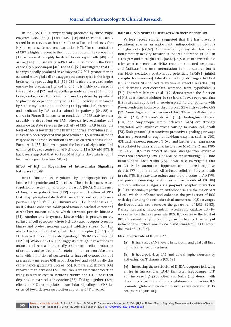
Various studies have suggested that H2S exerts many potential effects on a large number of biological targets and is involved in several physiological and pathological processes (Figures 3 & 4).
Figure 3: Potential targets for Hydrogen sulfide (H2S). Various experimental investigations suggest that H2S plays a prominent role in normal physiology and pathophysiology. Therefore, targets for H2S therapy includes heart failure, peripheral arterial diseases, acute MI, stroke, atherosclerosis, GI irritation, Alzheimer’s disease, cancer, thrombosis, organ transplantation, diabetes and erectile dysfunction.
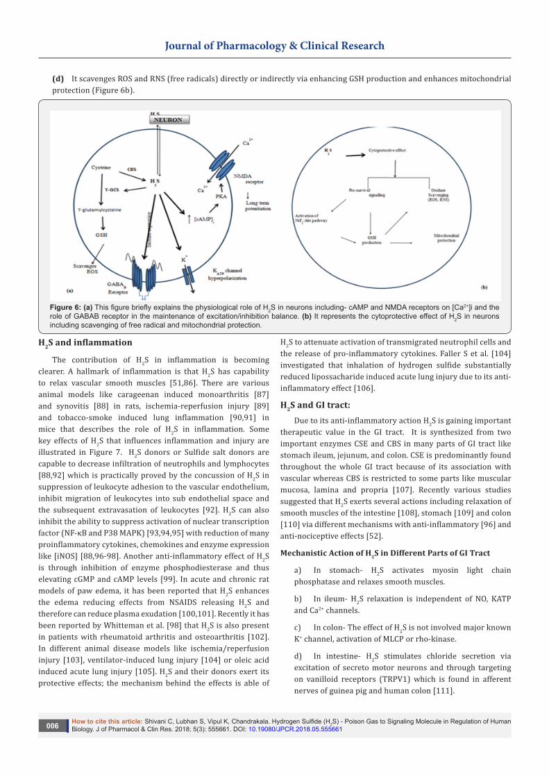
Figure 4: This figure illustrates the major cardiovascular actions of H2S.
H2S and Cardiovascular EffectsFor various important physiological processes H2S needs
to interact with a wide range of ion channels. There are varieties of experimental models, including a canine model of
cardiopulmonary bypass with hypothermic cardiac arrest that describes the cardioprotective effects of H2S. Cardioprotective action is through modulation of ATP-sensitive potassium (KATP) current [29] and voltage-gated L-type calcium current [30]. It

Journal of Pharmacology & Clinical Research
How to cite this article: Shivani C, Lubhan S, Vipul K, Chandrakala. Hydrogen Sulfide (H2S) - Poison Gas to Signaling Molecule in Regulation of Human Biology. J of Pharmacol & Clin Res. 2018; 5(3): 555661. DOI: 10.19080/JPCR.2018.05.555661004
has been seen that H2S plays a prominent role in myocardial pre and post conditioning responses [31]. H2S exerts a negative inotropic and chronotropic effects both in-vivo and in-vitro in the heart [32] and protect the heart against injury following coronary artery ligation [33] and ischemia [34]. Recent review suggested that in vasculature, H2S in combination with NO generates nitrosothiol with inotropic properties [129,130].
Mechanistic Pathways that are Implicated in the Cardioprotective Effect of H2S are
a) Involvement of ATP sensitive K+ channel and Voltage gated L-type calcium current [29,30]
b) Regulation of mitochondrial respiration [7]
c) Regulation of cytoprotective genes such as Nrf-2 [35]
d) Activation of cardiac extracellular-signal-regulated kinase (ERK) and/or phosphotidyl-inositol 3-kinase (PI3K-Akt) pathway [36].
Along with these cardioprotective effects H2S also interacts with those ion channels that are involved in the membrane action potential and may be effective in cardiac arrhythmias [37]. In addition to these effects against collagen, ADP and
aggregating agents H2S can inhibit human platelet aggregation in-vitro [38]. Furthermore, several studies on vascular tissue have been concluded that H2S is a potent vasodilator and perhaps an EDHF (endothelium derived hyperpolarizing factor) [8,35]. Experimentation in isolated rat aortic and portal vein and using a perfused rat mesenteric showed that H2S dilates only blood vessels [8,39-41] not coronary [39] and vascular beds. The Vasodilatory effect of H2S is independent of guanylyl cyclase/cGMP pathway [42]. However H2S induces vasorelaxation through involvement of cGMP-dependent protein kinase-I. Recently, it was focused on the role of H2S in chronic changes of vasculature. It has been reported that chronic treatment with NaHS can reduce hypertrophy of intramyocardial arterioles and ventricular fibrosis in hypertensive rats [43]. In addition, H2S also inhibits L-type calcium currents in rat cardiomyocytes and the study suggested that if cardiomyocytes were treated with (dithiothreitol) DTT, an H2S donor could change in cardiac function [44]. Wei H et al. [132] reported that H2S could inhibit hyperpolarization of activated inward current and delayed rectifier potassium channels in human cardiomyocytes. These effects could have a significant role in prolongation of the action potential and vasodilatory function of H2S [45].
H2S and Nervous System Biosynthesis of H2S and its Regulation in CNS
Figure 5: Production and Regulation of H2S in central nervous system: when electrical stimulation or neuronal excitation occurs, electrical signals descend to axon terminals and Ca2+ enters into the nerve terminal then interact with calmodulin which activates the CBS and the formation of H2S. An influx of Ca2+, perhaps triggered by the activation of NMDA receptors by glutamate or via separate channels, binds to calmodulin (CaM), thereby activating CBS. CBS seems to be the main H2S-forming enzyme in the CNS. SAM is an allosteric activator of CBS whereas hydroxylamine and amino-oxyacetate are inhibitors of CBS.
Journal of Pharmacology & Clinical Research
How to cite this article: Shivani C, Lubhan S, Vipul K, Chandrakala. Hydrogen Sulfide (H2S) - Poison Gas to Signaling Molecule in Regulation of Human Biology. J of Pharmacol & Clin Res. 2018; 5(3): 555661. DOI: 10.19080/JPCR.2018.05.555661005
In the CNS, H2S is enzymatically produced by three major enzymes- CBS, CSE [11] and 3-MST [46] and there it is usually stored in astrocytes as bound sulfane sulfur and releases free H2S in response to neuronal excitation [47]. The concentration of CBS is highly present in the hippocampus and the cerebellum [48] whereas it is highly localized to microglial cells [49] and astrocytes [50]. Generally, mRNA of CBS is found in the brain especially hippocampus [48]. Lee et al. [51] investigated that H2S is enzymatically produced in astrocytes 7.9 fold greater than in cultured microglial cell and suggest that astrocytes is the largest brain cell for producing H2S [51]. CSE is also the second major enzyme for producing H2S and in CNS; it is highly expressed in the spinal cord [52] and cerebellar granule neurons [53]. In the brain, endogenous H2S is formed from L-cysteine by pyridoxal 5’-phosphate dependent enzyme CBS. CBS activity is enhanced by S-adenosyl-L-methionine (SAM) and pyridoxal 5’-phosphate and mediated by Ca2+ and a calmodulin pathway [54, 55] as shown in Figure 5. Longer-term regulation of CBS activity most probably is dependent on SAM whereas hydroxylamine and amino-oxyacetate reverses the activity of CBS. In AD brains, the level of SAM is lower than the brains of normal individuals [56]. It has also been reported that production of H2S is stimulated in response to neuronal excitation as well as electrical stimulation. Furne et al. [57] has investigated the brains of eight mice and estimated free concentration of H2S around 14 ± 3.0 nM [57]. It has been suggested that 50-160µM of H2S in the brain is found for physiological function [58,59].
Effect of H2S in Regulation of Intracellular Signaling Pathways in CNS
Brain function is regulated by phosphorylation of intracellular proteins and Ca2+ release. These both processes are regulated by activation of protein kinase-A (PKA). Maintenance of long term potentiation (LTP) requires activation of PKA that may phosphorylate NMDA receptors and can enhance permeability of Ca2+ [60,61]. Kimura et al. [17] found that NaHS, an H2S donor enhances cAMP production in cerebral cortex and cerebellum neuron culture which activates protein kinase-A [62]. Another one is tyrosine kinase which is present on the surface of cell receptor; where H2S activates receptor tyrosine kinase and protect neurons against oxidative stress [63]. H2S also activates endothelial growth factor receptor (EGFR) and EGFR activation can modulate signaling of NMDA receptors and LTP [48]. Whiteman et al. [64] suggests that H2S may work as an antioxidant because it potentially inhibits intracellular nitration of proteins and oxidation of proteins in human neuroblastoma cells with inhibition of peroxynitrite induced cytotoxicity and presumably increases GSH production [64] and additionally this can enhance glutamate uptake [65]. Kimura and Kimura [66] reported that increased GSH level can increase neuroprotection using immature cortical neurons culture and HT22 cells that depends on extracellular cysteine [66]. Taking together, these effects of H2S can regulate intracellular signaling in CNS i.e. oriented towards neuroprotection and other CNS diseases.
Role of H2S in Neuronal Diseases with their Mechanism
Various recent studies suggested that H2S has played a prominent role as an antioxidant, antiapoptotic in neurons and glial cells [66,67]. Additionally, H2S may also have anti-inflammatory activity because it induces alteration in Ca2+ in astrocytes and microglial cells [68,69]. H2S seem to have multiple roles as it can enhance NMDA receptor mediated responses and facilitate long term potentiation in hippocampus but it can block excitatory postsynaptic potentials (EPSPs) (inhibit synaptic transmission). Literature findings also suggested that H2S enhances NO-induced relaxation of smooth muscles [70] and decreases corticotrophin secretion from hypothalamus [71]. Therefore Kimura et al. [17] demonstrated the function of H2S as a neuromodulator in the brain. It was reported that H2S is abundantly found in cerebrospinal fluid of patients with Down syndrome because of chromosome 21 which encodes CBS [72]. Neurodegenerative diseases of the CNS such as Alzheimer’s disease (AD), Parkinson’s disease (PD), Huntington’s disease (HD) and Amylotropic lateral sclerosis (ALS) are strongly associated with oxidative stress causing neuronal cell death [73]. Endogenous H2S can activate protective signaling pathways that are processed through antioxidant enzymes such as SOD, GSH and heme-oxygenase-1 (HO-1) and further their expression is regulated by transcriptional factors like NFe2, NrF2 and PGC-1α [74,75]. H2S may protect neuronal damage from oxidative stress via increasing levels of GSH or redistributing GSH into mitochondrial localization [76]. It was also investigated that in AD, NaSH attenuated lipposaccharide-induced cognitive defects [77] and inhibited Aβ induced cellular injury or death in rats [78]. H2S may also reduce amyloid-β plaques in AD [79], can prevent neurodegeneration in mouse models of PD [80] and can enhance analgesia via µ-opioid receptor interaction [81]. In ischemia/reperfusion, mitochondria are the major part of cell which is affected and enhances the production of ROS with depolarizing the mitochondrial membrane. H2S scavenges the free radicals and decreases the generation of ROS [82,83]. During ischemia, mitochondrial cytochrome oxidase activity was enhanced that can generate ROS. H2S decrease the level of ROS and imparting cytoprotection, also inactivates the activity of mitochondrial cytochrome oxidase and stimulate SOD to lower the level of ROS [84].
Mechanistic role of H2S in CNS –
(a) It increases cAMP levels in neuronal and glial cell lines and primary neuron cultures
(b) It hyperpolarizes CA1 and dorsal raphe neurons by activating KATP channels [85, 62]
(c) Increasing the sensitivity of NMDA receptors following a rise in intracellular cAMP facilitates hippocampal LTP and increase H2S production and NaHS (H2S donor) with direct electrical stimulation and glutamate application. H2S promotes glutamate mediated neurotransmission via NMDA receptors (Figure 6a).
Journal of Pharmacology & Clinical Research
How to cite this article: Shivani C, Lubhan S, Vipul K, Chandrakala. Hydrogen Sulfide (H2S) - Poison Gas to Signaling Molecule in Regulation of Human Biology. J of Pharmacol & Clin Res. 2018; 5(3): 555661. DOI: 10.19080/JPCR.2018.05.555661006
(d) It scavenges ROS and RNS (free radicals) directly or indirectly via enhancing GSH production and enhances mitochondrial protection (Figure 6b).
Figure 6: (a) This figure briefly explains the physiological role of H2S in neurons including- cAMP and NMDA receptors on [Ca2+]i and the role of GABAB receptor in the maintenance of excitation/inhibition balance. (b) It represents the cytoprotective effect of H2S in neurons including scavenging of free radical and mitochondrial protection.
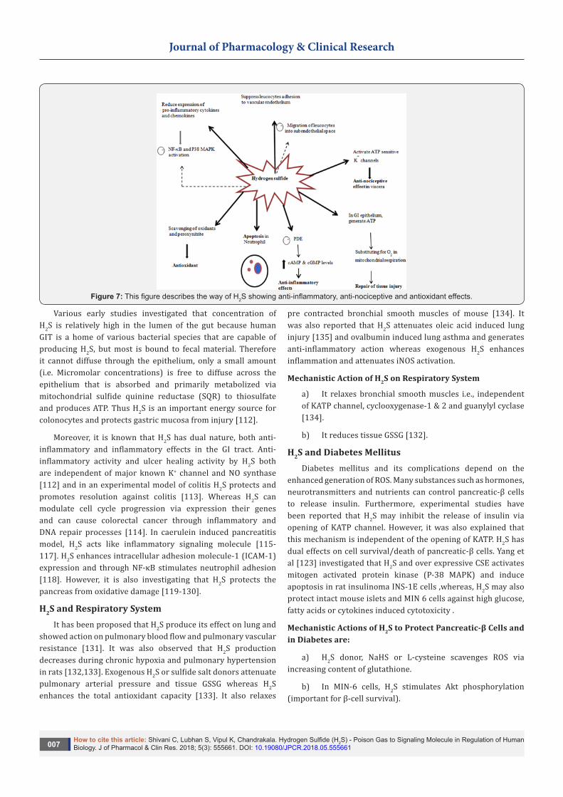
H2S and inflammation
The contribution of H2S in inflammation is becoming clearer. A hallmark of inflammation is that H2S has capability to relax vascular smooth muscles [51,86]. There are various animal models like carageenan induced monoarthritis [87] and synovitis [88] in rats, ischemia-reperfusion injury [89] and tobacco-smoke induced lung inflammation [90,91] in mice that describes the role of H2S in inflammation. Some key effects of H2S that influences inflammation and injury are illustrated in Figure 7. H2S donors or Sulfide salt donors are capable to decrease infiltration of neutrophils and lymphocytes [88,92] which is practically proved by the concussion of H2S in suppression of leukocyte adhesion to the vascular endothelium, inhibit migration of leukocytes into sub endothelial space and the subsequent extravasation of leukocytes [92]. H2S can also inhibit the ability to suppress activation of nuclear transcription factor (NF-κB and P38 MAPK) [93,94,95] with reduction of many proinflammatory cytokines, chemokines and enzyme expression like [iNOS] [88,96-98]. Another anti-inflammatory effect of H2S is through inhibition of enzyme phosphodiesterase and thus elevating cGMP and cAMP levels [99]. In acute and chronic rat models of paw edema, it has been reported that H2S enhances the edema reducing effects from NSAIDS releasing H2S and therefore can reduce plasma exudation [100,101]. Recently it has been reported by Whitteman et al. [98] that H2S is also present in patients with rheumatoid arthritis and osteoarthritis [102]. In different animal disease models like ischemia/reperfusion injury [103], ventilator-induced lung injury [104] or oleic acid induced acute lung injury [105]. H2S and their donors exert its protective effects; the mechanism behind the effects is able of
H2S to attenuate activation of transmigrated neutrophil cells and the release of pro-inflammatory cytokines. Faller S et al. [104] investigated that inhalation of hydrogen sulfide substantially reduced lipossacharide induced acute lung injury due to its anti-inflammatory effect [106].
H2S and GI tract:Due to its anti-inflammatory action H2S is gaining important
therapeutic value in the GI tract. It is synthesized from two important enzymes CSE and CBS in many parts of GI tract like stomach ileum, jejunum, and colon. CSE is predominantly found throughout the whole GI tract because of its association with vascular whereas CBS is restricted to some parts like muscular mucosa, lamina and propria [107]. Recently various studies suggested that H2S exerts several actions including relaxation of smooth muscles of the intestine [108], stomach [109] and colon [110] via different mechanisms with anti-inflammatory [96] and anti-nociceptive effects [52].
Mechanistic Action of H2S in Different Parts of GI Tract
a) In stomach- H2S activates myosin light chain phosphatase and relaxes smooth muscles.
b) In ileum- H2S relaxation is independent of NO, KATP and Ca2+ channels.
c) In colon- The effect of H2S is not involved major known K+ channel, activation of MLCP or rho-kinase.
d) In intestine- H2S stimulates chloride secretion via excitation of secreto motor neurons and through targeting on vanilloid receptors (TRPV1) which is found in afferent nerves of guinea pig and human colon [111].
Journal of Pharmacology & Clinical Research
How to cite this article: Shivani C, Lubhan S, Vipul K, Chandrakala. Hydrogen Sulfide (H2S) - Poison Gas to Signaling Molecule in Regulation of Human Biology. J of Pharmacol & Clin Res. 2018; 5(3): 555661. DOI: 10.19080/JPCR.2018.05.555661007
Figure 7: This figure describes the way of H2S showing anti-inflammatory, anti-nociceptive and antioxidant effects.
Various early studies investigated that concentration of H2S is relatively high in the lumen of the gut because human GIT is a home of various bacterial species that are capable of producing H2S, but most is bound to fecal material. Therefore it cannot diffuse through the epithelium, only a small amount (i.e. Micromolar concentrations) is free to diffuse across the epithelium that is absorbed and primarily metabolized via mitochondrial sulfide quinine reductase (SQR) to thiosulfate and produces ATP. Thus H2S is an important energy source for colonocytes and protects gastric mucosa from injury [112].
Moreover, it is known that H2S has dual nature, both anti-inflammatory and inflammatory effects in the GI tract. Anti-inflammatory activity and ulcer healing activity by H2S both are independent of major known K+ channel and NO synthase [112] and in an experimental model of colitis H2S protects and promotes resolution against colitis [113]. Whereas H2S can modulate cell cycle progression via expression their genes and can cause colorectal cancer through inflammatory and DNA repair processes [114]. In caerulein induced pancreatitis model, H2S acts like inflammatory signaling molecule [115-117]. H2S enhances intracellular adhesion molecule-1 (ICAM-1) expression and through NF-κB stimulates neutrophil adhesion [118]. However, it is also investigating that H2S protects the pancreas from oxidative damage [119-130].
H2S and Respiratory SystemIt has been proposed that H2S produce its effect on lung and
showed action on pulmonary blood flow and pulmonary vascular resistance [131]. It was also observed that H2S production decreases during chronic hypoxia and pulmonary hypertension in rats [132,133]. Exogenous H2S or sulfide salt donors attenuate pulmonary arterial pressure and tissue GSSG whereas H2S enhances the total antioxidant capacity [133]. It also relaxes
pre contracted bronchial smooth muscles of mouse [134]. It was also reported that H2S attenuates oleic acid induced lung injury [135] and ovalbumin induced lung asthma and generates anti-inflammatory action whereas exogenous H2S enhances inflammation and attenuates iNOS activation.
Mechanistic Action of H2S on Respiratory System
a) It relaxes bronchial smooth muscles i.e., independent of KATP channel, cyclooxygenase-1 & 2 and guanylyl cyclase [134].
b) It reduces tissue GSSG [132].
H2S and Diabetes MellitusDiabetes mellitus and its complications depend on the
enhanced generation of ROS. Many substances such as hormones, neurotransmitters and nutrients can control pancreatic-β cells to release insulin. Furthermore, experimental studies have been reported that H2S may inhibit the release of insulin via opening of KATP channel. However, it was also explained that this mechanism is independent of the opening of KATP. H2S has dual effects on cell survival/death of pancreatic-β cells. Yang et al [123] investigated that H2S and over expressive CSE activates mitogen activated protein kinase (P-38 MAPK) and induce apoptosis in rat insulinoma INS-1E cells ,whereas, H2S may also protect intact mouse islets and MIN 6 cells against high glucose, fatty acids or cytokines induced cytotoxicity .
Mechanistic Actions of H2S to Protect Pancreatic-β Cells and in Diabetes are:
a) H2S donor, NaHS or L-cysteine scavenges ROS via increasing content of glutathione.
b) In MIN-6 cells, H2S stimulates Akt phosphorylation (important for β-cell survival).
Journal of Pharmacology & Clinical Research
How to cite this article: Shivani C, Lubhan S, Vipul K, Chandrakala. Hydrogen Sulfide (H2S) - Poison Gas to Signaling Molecule in Regulation of Human Biology. J of Pharmacol & Clin Res. 2018; 5(3): 555661. DOI: 10.19080/JPCR.2018.05.555661008
H2S and Reproductive System/Erectile Dysfunction (ED)
It has been investigated that CBS and CSE were found in the Leydig, Sertoli and germ cells of rat testis [125] as well as in intrauterine tissues and human placenta [126]. Although H2S relaxes vas deferens smooth muscle and showed vasodilatory properties in the corpus cavernosum [127]. Therefore, H2S is an effective therapy for ED. The study explores the mechanism that H2S enhances the NO production via expression of constitutive nitric oxide synthase (NOS) isoforms i.e. endothelial NOS (eNOS) and neuronal NOS (nNOS) in rat corpus cavernosum and states that exogenously applied NaHS enhances eNOS but not nNOS. This can cure inherited erectile impairment which happens due to attenuation of endothelial NO formation in cavernosum [128]. However, per se effect of H2S in reproduction is still under investigation.
Conclusion During the past decade, several researches have been
focused on H2S. This review has centered the role of H2S in human biology. Indeed, efforts are going on to investigate the therapeutic potential of H2S in human diseases. It was discovered that H2S is continuously produced enzymatically in mammals and regulates the vast array of physiological processes. Now recently it is considered as a new gaseous signaling molecule that effect on all organ systems and in various pathological states. Accumulating evidence suggests that H2S have been implicated in neurotransmission via various pathways. It acts as a physiologic vasodilator and plays a prominent role in cardioprotection, pro and anti-inflammatory processes and several metabolic disorders such as diabetes, obesity, gout. Nevertheless, our understanding of mechanistic action is still fragmentary. Since it has a diverse biological profile to treat a number of diseases, H2S has come under a promising research to develop H2S-releasing prodrugs. It might be possible that H2S prodrugs can enhance H2S bioavailability and be efficacious in physiological processes.
References1. Mustafa A, Gadalla M, Snyder S (2009) Signaling by Gasotransmitters.
Sci Signal 2(68): re2.
2. Wang R (2002) Two’s company, three’s a crowd - Can H2S be the third endogenous gaseous transmitter?. FASEB J 16(13): 1792-1798.
3. Guidotti TL (2010) Hydrogensulfide: advances in understanding human toxicity. Int J Toxicol 29(6): 569-581.
4. Motterlini R, Otterbein L (2010) The therapeutic potential of carbon monoxide. Nat Rev Drug Discov 9(9): 728-743.
5. Li L, Hsu A, Moore P (2009) Actions and interactions of nitric oxide, carbon monoxide and hydrogen sulphide in the cardiovascular system and in inflammation- a tale of three gases!. Pharmacol Ther 123(3): 386-400
6. Khan AA, Schuler MM, Prior MG, Yong S, Coppock RW, et al. (1990) Effects of hydrogen sulfide exposure on lung mitochondrial respiratory chain enzymes in the rat. Toxicol Appl Pharmacol 103(3): 482-490.
7. Dorman DC, Moulin FJM, McManus BE, Mahle KC, James RA, et al. (2002) Cytochrome oxidase inhibition induced by acute hydrogen sulfide inhalation: correlation with tissue sulfide concentrations in the rat brain, liver, lung, and nasal epithelium. Toxicol Sci 65(1): 18-25.
8. Zhao W, Zhang J, Lu Y, Wang R (2001) The vasorelaxant effect of H2S as a novel endogenous gaseous K(ATP) channel opener. EMBO J 20(21): 6008-6016.
9. Geng B, Yang J, Qi Y, Zhao J, Pang Y, et al. (2004) H2S generated by heart in rat and its effects on cardiac function. Biochem. Biophys. Res. Commun. 313(2): 362-368.
10. Warenycia MW, Goodwin LR, Benishin CG, Reiffenstein RJ, Francom DM, et al. (1989) Acute hydrogen sulfide poisoning. Demonstration of selective uptake of sulfide by the brainstem by measurement of brain sulfide levels. Biochem Pharmacol 38(6): 973-981.
11. Stipanuk MH, Beck PW (1982) Characterization of the enzymic capacity for cysteine desulphhydration in liver and kidney of the rat. Biochem J 206(2): 267-277.
12. Griffith OW (1987) Mammalian sulfur amino acid metabolism: an overview. In: Methods in enzymology 143: 366-376.
13. Erickson PF, Maxwell IH, Su IJ, Baumann M, Glode LM (1990) Sequence of cDNA for rat cystathionine y-lyase and comparison of deduced amino acid sequence with related Escherichia coli enzymes. Biochem J 269(2): 335-340.
14. Swaroop M, Bradley K, Ohura T, Tahara T, Roper MD, et al. (1992) Rat cystathionine p-synthase. J Biol Chem 267(16): 11455-11461.
15. (2012) hydrogen sulfide: biochemistry and medicine.
16. Li L, Rose P, Moore PK (2011) Hydrogen sulfide and cell signaling. Annu Rev Pharmacol Toxicol 51: 169-187.
17. Kimura H (2010) Hydrogen sulfide: its production, release and functions. Amino Acids.
18. Nagahara N, Ito T, Kitamura H, Nishino T (1998) Tissue and subcellular distribution of mercaptopyruvate sulfurtransferase in the rat: confocal laser fluorescence and immunoelectron microscopic studies combined with biochemical analysis. Histochem Cell Biol 110(3): 243-250.
19. Hildebrandt TM, Grieshaber MK (2008) Three enzymatic activities catalyze the oxidation of sulfide to thiosulfate in mammalian and invertebrate mitochondria. FEBS J 275(13): 3352-3361.
20. Banerjee R, Kabil O (2010) Redox biochemistry of hydrogen sulfide. J Biol Chem 285(29): 21903-21907.
21. Mikami Y, Shibuya N, Kimura Y, Nagahara N, Ogasawara Y (2011) Thioredoxin and dihydrolipoic acid are required for 3-mercaptopyruvate sulfurtransferase to produce hydrogen sulfide. Biochem J 439(3): 479-485.
22. Szabo C (2007) Hydrogen sulphide and its therapeutic potential. Nat Rev Drug Discov 6(11): 917-935.
23. Goodwin LR, Francom D, Dieken FP, Taylor JD, Warenycia MW, et al. (1989) Determination of sulfide in brain tissue by gas dialysis/ion chromatography: postmortem studies and two case reports. J Analyt Toxicol 13(2): 105-109.
24. Hannestad U, Margheri S, Sorbo B (1989) A sensitive gas chromatographic method for determination of protein-associated sulfur. Anal Biochem 178(2): 394-398.
25. Ogasawara Y, Isoda S, Tanabe S (1994) Tissue and subcellular distribution of bound and acid-labile sulfur, and the enzymic capacity for sulfide production in the rat. Biol Pharm Bull 17(12): 1535-1542.
26. Mathai J C, Missner A, Kugler P, Saparov S M, Zeidel ML (2009) No facilitator required for membrane transport of hydrogen sulfide. Proc Natl Acad Sci USA 106(36): 16633-16638
Journal of Pharmacology & Clinical Research
How to cite this article: Shivani C, Lubhan S, Vipul K, Chandrakala. Hydrogen Sulfide (H2S) - Poison Gas to Signaling Molecule in Regulation of Human Biology. J of Pharmacol & Clin Res. 2018; 5(3): 555661. DOI: 10.19080/JPCR.2018.05.555661009
27. Searcy DG, Lee SH (1998) Sulfur reduction by human erythrocytes. J Exp Zool 282(3): 310-322.
28. Dombkowski RA, Russell MJ, Olson KR (2004) Hydrogen sulfide as an endogenous regulator of vascular smooth muscle tone in trout. Am J Physiol Regul Integr Comp Physiol 286(4): 678-685
29. Cheng Y, Ndisang JF, Tang G, Cao K, Wang R (2004) Hydrogen sulfide-induced relaxation of resistance mesenteric artery beds of rats. Am J Physiol Heart Circ Physiol 287(5): 2316-2323.
30. Lavu M, Bhushan S, Lefer DJ (2011) Hydrogen sulfide-mediated cardioprotection: mechanisms and therapeutic potential. Clin Sci (Lond) 120(6): 219-229.
31. Gross GJ and Fryer RM (1999) Sarcolemmal versus mitochondrial ATP-sensitive Kþ channels and myocardial preconditioning. Circ Res 84(9): 973-979.
32. Zhu YZ,Wang ZJ, Ho P, Loke YY, Zhu YC, et al. (2007) Hydrogen sulfide and its possible roles in myocardial ischemia in experimental rats. J Appl Physiol 102(1): 261-268
33. Pan TT Feng ZN, Lee SW, Moore PK, Bian JS (2006) Endogenous hydrogen sulfide contributes to the cardioprotection by metabolic inhibition preconditioning in the rat ventricular myocytes. J Mol Cell Cardiol 40(1): 119-130
34. Calvert WJ, Coetzee WA, Lefer DJ (2010) Novel insights into Hydrogen Sulfide-Mediated Cytoprotection 12(10): 1203-12017.
35. Hu Y, Chen X, Pan TT, Neo KL, Lee SW, et al. (2007) Cardioprotection induced by hydrogen sulfide preconditioning involves activation of ERK and PI3K/Akt pathways. Pflugers Arch 455(4): 607-616.
36. Tang G, Wu L, Wang R (2010) Interaction of hydrogen sulfide with ion channels. Clin Exp Pharmacol Physiol 37(7): 753-763.
37. Zagli G, Riccardo Patacchinib, Marcello Trevisanic, Rosanna Abbatea, Sandro Cinottid, et al. (2007) Hydrogen sulfide inhibits human platelet aggregation. Eur J Pharmacol 559: 65-68.
38. Johansen D, Ytrehus K, Baxter GF (2006) Exogenous hydrogen sulfide (H2S) protects against regional myocardial ischemia-reperfusion injury - evidence for a role of KATP channels. Basic Res Cardiol 101: 53-60.
39. Hosoki R, Matsuki N, Kimura H (1997) The possible relaxant effect of hydrogen sulfide as an endogenous smooth muscle relaxant in synergy with nitric oxide. Biochem Biophys Res Commun 237(3): 527-531.
40. Ali MY, CY Ping Y, YP Mok, L Ling, M Whiteman, et al. (2006) Regulation of vascular nitric oxide in vitro and in vivo; a new role for endogenous hydrogen sulphide?. Br J Pharmacol 149(6): 625-634.
41. Zhao W, Wang R (2002) H(2)S-induced vasorelaxation and underlying cellular and molecular mechanisms. Am J Physiol Heart Circ Physiol 283(2): 474-480.
42. Shi YX, Chen Y, Zhu YZ, Huang GY, Moore PK, et al. (2007) Chronic sodium hydrosulfide treatment decreases medial thickening of intramyocardial coronary arterioles, interstitial fibrosis and ROS production in SHR. Am J Physiol Heart Circ Physiol 293: 2093-2100.
43. Zhang R, Sun Y, Tsai H, Tang C, Jin H, et al. (2012) Hydrogen Sulfide Inhibits L-Type Calcium Currents Depending upon the Protein Sulfhydryl State in Rat Cardiomyocytes 7(5): 37073.
44. wei H et al (2012) Hydrogen Sulfide Suppresses Outward Rectifier Potassium Currents in Human Pluripotent Stem Cell-Derived Cardiomyocytes 7(11): 50641.
45. Shibuya N, Tanaka M, Yoshida M, Ogasawara Y, Togawa T, et al. (2009) 3-Mercaptopyruvate sulfurtransferase produces hydrogen sulfide and bound sulfane sulfur in the brain. Antioxid Redox Signal 11(4): 703-714.
46. Ishigami M, Hiraki K, Umemura K, Ogasawara Y, Ishii K, et al. (2009) A source of hydrogen sulfide and a mechanism of its release in the brain. Antioxid Redox Signal 11(2): 205-214.
47. Abe K, Kimura H (1996) The possible role of hydrogen sulfide as an endogenous neuromodulator. J Neurosci 16(3): 1066-1071.
48. Hu LF, Wong PT, Moore PK, Bian JS (2007) Hydrogen sulfide attenuates lipopolysaccharide-induced inflammation by inhibition of p38 mitogen-activated protein kinase in microglia. J Neurochem 100(4): 1121-1128.
49. Enokido Y, Suzuki E, Iwasawa K, Namekata K, Okazawa H, et al. (2005) Cystathionine beta-synthase, a key enzyme for homocysteine metabolism, is preferentially expressed in the radial glia/astrocyte lineage of developing mouse CNS. FASEB J 19(13): 1854-1856.
50. Lee M, Schwab C, Yu S, McGeer E, McGeer PL (2009) Astrocytes produce the antiinflammatory and neuroprotective agent hydrogen sulfide. Neurobiol Aging 30(10): 1523-1534.
51. Distrutti E, Sediari L, Mencarelli A, Renga B, Orlandi S, et al. (2006) Evidence that hydrogen sulfide exerts antinociceptive effects in the gastrointestinal tract by activating KATP channels. J Pharmacol Exp Ther 316(1): 325-335.
52. GarciaBereguiain MA, SamhanArias AK, MartinRomero FJ, GutierrezMerino C (2008) Hydrogen sulfide raises cytosolic calcium in neurons through activation of L-type Ca2+ channels. Antioxid Redox Signal 10(1): 31-42.
53. Eto K, Ogasawara M, Umemura K, Nagai Y, Kimura H (2002) Hydrogen sulfide is produced in response to neuronal excitation. J Neurosci 22(9): 3386-3391.
54. Finkelstein J D, Kyle W E, Martin JJ, Pick AM (1975) Activation of cystathionine synthase by adenosylmethionine and adenosylethionine. Biochem Biophys Res Commun 66(1): 81-87.
55. Morrison LD, Smith DD, Kish SJ (1996) Brain S-adenosylmethionine levels are severely decreased in Alzheimer’s disease. J Neurochem 67(3): 1328-1331.
56. Furne J, Saeed A, Levitt MD (2008) Whole tissue hydrogen sulfide concentrations are orders of magnitude lower than presently accepted values. Am J Physiol Regul Integr Comp Physiol 295(5): 1479-1489.
57. Goodwin LR, Francom D, Dieken FP, Taylor JD, Warenycia MW, et al. (1989) Determination of sulfide in brain tissue by gas dialysis/ion chromatography: postmortem studies and two case reports. J Anal Toxicol 13(2): 105-109.
58. Savage JC, Gould DH (1990) Determination of sulfides in brain tissue and rumen fluid by ion-interaction reversed-phase high-performance liquid chromatography. J Chromatogr 526: 540-545.
59. Leonard AS, Hell JW (1997) Cyclic AMP-dependent protein kinase and protein kinase C phosphorylate N-methyl-D-aspartate receptors at different sites. J Biol Chem 272(18): 12107-121015.
60. Skeberdis VA, Chevaleyre V, Lau CG, Goldberg JH, Pettit DL, et al. (2006) Protein kinase A regulates calcium permeability of NMDA receptors. Nat Neurosci 9(4): 501-510.
61. Kimura H (2000). Hydrogen sulfide induces cyclic AMP and modulates the NMDA receptor. Biochem Biophys Res Commun 267(1): 129.
62. Umemura K, Kimura H (2007) Hydrogen sulfide enhances reducing activity in neurons: neurotrophic role of H2S in the brain?. Antioxid Redox Signal 9(11): 2035.
63. Whiteman M, Armstrong JS, Chu SH, JiaLing S, Wong BS, et al. (2004) The novel neuromodulator hydrogen sulfide: an endogenous peroxynitrite ‘scavenger’?. J Neurochem 90(3): 765.
Journal of Pharmacology & Clinical Research
How to cite this article: Shivani C, Lubhan S, Vipul K, Chandrakala. Hydrogen Sulfide (H2S) - Poison Gas to Signaling Molecule in Regulation of Human Biology. J of Pharmacol & Clin Res. 2018; 5(3): 555661. DOI: 10.19080/JPCR.2018.05.5556610010
64. Lu M, Hu LF, Hu G, Bian JS (2008) Hydrogen sulfide protects astrocytes against H2O2-induced neural injury via enhancing glutamate uptake. Free Rad Biol Med 45(12): 1705-1713.
65. Kimura Y, Kimura H (2004) Hydrogen sulfide protects neurons from oxidative stress. FASEB J 18(10): 1165-1167.
66. Yin WL, He JQ, Hu B, Jiang ZS, Tang XQ (2009) Hydrogen sulfide inhibits MPP (+)-induced apoptosis in PC12 cells. Life Sci 85(7-8): 269-275.
67. Lee SW, Hu YS, Hu LF, Lu Q, Dawe GS, et al. (2006) Hydrogen sulphide regulates calcium homeostasis in microglial cells. Glia 54(2): 116-124.
68. Nagai Y, Tsugane M, Oka J, Kimura H (2004) Hydrogen sulfide induces calcium waves in astrocytes. FASEB J 18(3): 557-559.
69. Hosoki R, Matsuki N, Kimura H (1997) The possible role of hydrogen sulfide as an endogenous smooth muscle relaxant in synergy with nitric oxide. Biochem Biophys Res Commun 237(3): 527- 531.
70. Dello Russo C, Tringali G, Ragazzoni E, Maggiano N, Menini E, et al. (2000) Evidence that hydrogen sulphide can modulate hypothalamo-pituitary-adrenal axis function: In vitro and in vivo studies in the rat. J Neuroendocrinol 12(3): 225-233.
71. Kamoun P, Belardinelli MC, Chabli A, Lallouchi K, Chadefaux Vekemans B (2003) Endogenous hydrogen sulfide overproduction in Down syndrome. Am J Med Genet A 116(3): 310-311.
72. E. Trushina, CT McMurray (2007) Oxidative stress and mitochondrial dysfunction in neurodegenerative diseases. Neuroscience 145(4): 1233-1248.
73. fujita K, Yamafuji M, Nakabeppu Y, Noda M (2012) Therapeutic Approach to Neurodegenerative Diseases by Medical Gases: Focusing on Redox Signaling and Related Antioxidant Enzymes.
74. Lee M, Schwab C, Yu S, McGeer E, Mc Geer PL (2009) Astrocytes produce the anti inflammatory and neuroprotective agent hydrogen sulfide. Neurobiol Aging 30(10): 1523-1534.
75. Y Kimura, YI Goto, H Kimura (2010) Hydrogen sulfide increases glutathione production and suppresses oxidative stress in mitochondria. Antioxidants and Redox Signaling 121(1): 1-13.
76. Gong QH, Wang Q, Pan LL, Liu XH, Huang H, et al. (2010) Hydrogen sulfide attenuates lipopolysaccharide-induced cognitive impairment: a pro-inflammatory pathway in rats. Pharmacol Biochem Behav 96(1): 52-58.
77. Tang XQ, Yang CT, Chen J, Yin WL, Li Y J, et al. (2008) Effect of hydrogen sulphide on β-amyloid-induced damage in PC12 cells. Clin Exp Pharmacol Physiol 35(2): 180-186.
78. Zhang H, Gao Y, Zhao F, Dai Z, Meng T, et al. (2011) Hydrogen sulfide reduces mRNA and protein levels of beta-site amyloid precursor protein cleaving enzyme 1 in PC12 cells. Neurochem Int 58(2): 169-175.
79. Hu LF, Lu M, Tiong CX, Dawe GS, Hu G, et al. (2010) Neuroprotective effects of hydrogen sulfide on Parkinson’s disease rat models. Aging Cell 9(2): 135-146.
80. Distrutti E, Cipriani S, Renga B, Mencarelli A, Migliorati M, et al. (2010) Hydrogen sulphide induces micro opioid receptor- dependent analgesia in a rodent model of visceral pain. Mol Pain 6: 36.
81. S Jha, J WCalvert, MR Duranski, A Ramachandran, DJ Lefer (2008) Hydrogen sulfide attenuates hepatic ischemia-reperfusion injury: role of antioxidant and antiapoptotic signaling. American Journal of Physiology 295(2): 801-806.
82. Z Fu, X Liu, B Geng, L Fang, et al. (2008) Hydrogen sulfide protects rat lung from ischemia-reperfusion injury. Life Sciences 82(23-24): 1196-1202.
83. WH Sun, F Liu, Y Chen, YC Zhu (2012) Hydrogen sulfide decreases the
levels of ROS by inhibiting mitochondrial complex IV and increasing SOD activities in cardiomyocytes under ischemia/reperfusion. Biochemical and Biophysical Research Communications 421(2): 164-169.
84. K Qu, SW Lee, J S Bian, C M Low, P T H Wong (2008) Hydrogen sulfide: neurochemistry and neurobiology. Neurochemistry International 52(2): 155-165.
85. Zhong GZ, Chen RF, Cheng YQ, Tang CS, Du JB (2003) The role of hydrogen sulfide generation in the pathogenesis of hypertension in rats induced by inhibition of nitric oxide synthase. J Hypertens 21: 1879-1885.
86. Andruski B, McCafferty DM, Ignacy T, Millen B, McDougall JJ (2008) Leukocyte trafficking and pain behavioral responses to a hydrogen sulfide donor in acute monoarthritis. Am J Physiol Regul Integr Comp Physiol 295(3): 814-820.
87. Ekundi Valentim E, Santos KT, Camargo EA, Denadai Souza A, Teixeira SA (2010) Differing effects of exogenous and endogenous hydrogen sulphide in carrageenan-induced knee joint synovitis in the rat. Br J Pharmacol 159: 1463-1474.
88. Zuidema MY, Yang Y, Wang M, Kalogeris T, Liu Y, et al. (2010) Antecedent hydrogen sulfide elicits an anti-inflammatory phenotype in postischemic murine small intestine: role of BK channels. Am J Physiol Heart Circ Physiol 299(5): 1554-1567.
89. Chen YH, Wang PP, Wang XM, He YJ, Yao WZ, et al. (2011) Involvement of endogenous hydrogen sulfide in cigarette smoke-induced changes in airway responsiveness and inflammation of rat lung. Cytokine 53(3): 334-341.
90. Han W, Dong Z, Dimitropoulou C, Su Y (2011) Hydrogen sulfide ameliorates tobacco smoke-induced oxidative stress and emphysema in mice. Antioxid Redox Signal 15(8): 2121-2134.
91. Zanardo RCO, Brancaleone V, Distrutti E, Fiorucci S, Cirino G, et al. (2006) Hydrogen sulphide is an endogenous modulator of leukocyte-mediated inflammation. FASEB J 20(12): 2118-2120.
92. Levine J, Ellis CJ, Furne JK, Springfield J, Levitt MD (1998) Fecal hydrogen sulfide production in ulcerative colitis. Am J Gastroenterol 93(1): 83-87.
93. Levitt MD, Springfield J, Furne J, Koenig T, Suarez FL (2002) Physiology of sulfide in the rat colon: use of bismuth to assess colonic sulfide production. J Appl Physiol 92(4): 1655-1660,
94. Moore J, Babidge W, Millard S, Roediger W le (1998) Colonic luminal hydrogen sulfide is not elevated in ulcerative colitis. Dig Dis Sci 43(1): 162-165.
95. Fiorucci S, Mencarelli A, Caliendo G, Santagada V, Distrutti E, et al. (2007) Enhanced activity of a hydrogen-sulfide releasing mesalamine derivative (ATB- 429) in a mouse model of colitis. Br J Pharmacol 150(8): 996-1002.
96. Suarez F, Furne J, Springfield J, Levitt M (1998) Production and elimination of sulfur-containing gases in the rat colon. Am J Physiol 274(4): 727-733.
97. Whiteman M, Li L, Rose P, Tan CH, Parkinson DB (2010) The effect of hydrogen sulfide donors on lipopolysaccharide- induced formation of inflammatory mediators in macrophages. Antioxid Redox Signal 12(10): 1147-1154.
98. Bucci M, Papapetropoulos A, Vellecco V, Zhou Z, Pyriochou A (2010) Hydrogen sulfide is an endogenous inhibitor of phosphodiesterase activity. Arterioscler Thromb Vasc Biol 30(10): 1998-2004.
99. Wallace J L, Caliendo G, Santagada V, Cirino G, Fiorucci S (2007) Gastrointestinal safety and anti-inflammatory effects of a hydrogen sulfide-releasing diclofenac derivative in the rat. Gastroenterology 132(1): 261-271.
Journal of Pharmacology & Clinical Research
How to cite this article: Shivani C, Lubhan S, Vipul K, Chandrakala. Hydrogen Sulfide (H2S) - Poison Gas to Signaling Molecule in Regulation of Human Biology. J of Pharmacol & Clin Res. 2018; 5(3): 555661. DOI: 10.19080/JPCR.2018.05.5556610011
100. Wallace JL, Caliendo G, Santagada V, Cirino G (2010) Markedly reduced toxicity of a hydrogen sulfide-releasing derivative of naproxen (ATB-346). Br J Pharmacol 159(6): 1236-1246.
101. Whiteman M, Haigh R, Tarr JM, Gooding KM, Shore AC (2010) Detection of hydrogen sulfide in plasma and knee-joint synovial fluid from rheumatoid arthritis patients: relation to clinical and laboratory measures of inflammation. Ann NY Acad Sci 1203: 146-150.
102. Biermann J, Lagreze WA, Schallner N, Schwer CI, Goebel U (2011) Inhalative preconditioning with hydrogen sulfide attenuated apoptosis after retinal ischemia/reperfusion injury . Mol Vis 17: 1275-1286.
103. Faller S, Ryter SW, Choi AM, Loop T, Schmidt R (2010) Inhaled hydrogen sulfide protects against ventilator-induced lung injury.Anesthesiology 113(1): 104-115.
104. Li T, Zhao B, Wang C, Wang H, Liu Z (2008) Regulatory effects of hydrogen sulfide on IL-6, IL-8 and IL-10 levels in the plasma and pulmonary tissue of rats with acute lung injury. Exp Biol Med (Maywood ) 233(9): 1081-1087.
105. Faller S, Kornelia K Zimmermann, Karl M Strosing, Helen Engelstaedter, Hartmut Buerkle, et al. (2012) Inhaled hydrogen sulfide protects against lipopolysaccharide-induced acute lung injury in mice 2: 26
106. Martin GR, McKnight GW, Dicay MS, Coffin CS, Ferraz JG (2010) Hydrogen sulphide synthesis in the rat and mouse gastrointestinal tract. Dig Liver Dis 42(2): 103-109.
107. Nagao M, Linden DR, Duenes JA, Sarr MG (2011) Mechanisms of action of the gasotransmitter hydrogen sulfide in modulating contractile activity of longitudinal muscle of rat ileum. J Gastrointest Surg 15(1): 12-22.
108. Dhaese I, Lefebvre RA (2009) Myosin light chain phosphatase activation is involved in the hydrogen sulfide-induced relaxation in mouse gastric fundus. Eur J Pharmacol 606(1-3): 180-186.
109. Dhaese I, van Colen I, Lefebvre RA (2010) Mechanisms of action of hydrogen sulfide in relaxation of mouse distal colonic smooth muscle. Eur J Pharmacol 628(1-3): 179-186.
110. Krueger D, Foerster M, Mueller K, Zeller F, Slotta-Huspenina J, et al. (2010) Signaling mechanisms involved in the intestinal pro-secretory actions of hydrogen sulfide. Neurogastroenterol Motil 22(11): 1224-1231.
111. Wallace JL, Dicay M, McKnight W, Martin GR (2007) Hydrogen sulfide enhances ulcer healing in rats. FASEB J 21(14): 4070-4076.
112. Wallace JL, Vong L, McKnight W, Dicay M, Martin GR (2009) Endogenous and exogenous hydrogen sulfide promotes resolution of colitis in rats. Gastroenterology 137(2): 569-578.
113. Attene Ramos MS, Nava GM, Muellner MG, Wagner ED, Plewa MJ (2010) DNA damage and toxicogenomic analyses of hydrogen sulfide in human intestinal epithelial FHs 74 Int cells. Environ Mol Mutagen 51(4): 304-314.
114. Bhatia M, Wong FL, Fu D, Lau HY, Moochhala SM, Moore PK (2005) Role of hydrogen sulfide in acute pancreatitis and associated lung injury. FASEB J 19(9): 623-625.
115. Tamizhselvi R, Moore PK, Bhatia M (2007) Hydrogen sulfide acts as a mediator of inflammation in acute pancreatitis: in vitro studies using isolated mouse pancreatic acinar cells. J Cell Mol Med 11(2): 315-326.
116. Tamizhselvi R, Moore PK, Bhatia M (2008) Inhibition of hydrogen sulfide synthesis attenuates chemokine production and protects mice against acute pancreatitis and associated lung injury. Pancreas 36(4): 24-31.
117. Tamizhselvi R, Koh YH, Sun J, Zhang H, Bhatia M (2010) Hydrogen sulfide induces ICAM-1 expression and neutrophil adhesion to caerulein-treated pancreatic acinar cells through NF-kappaB and Src-family kinases pathway. Exp Cell Res 316(9): 1625-1636.
118. Taniguchi S, Kang L, Kimura T, Niki I (2010) Hydrogen sulphide protects mouse pancreatic beta-cells from cell death induced by oxidative stress, but not by endoplasmic reticulum stress. Br J Pharmacol 162(5): 1171-1178.
119. Yang W, Yang G, Jia X, Wu L, Wang R (2005) Activation of KATP channels by H2S in rat insulin-secreting cells and the underlying mechanisms. J Physiol 569(2): 519-531.
120. Ali MY, Whiteman M, Low CM, Moor PK (2007) Hydrogen sulphide reduces insulin secretion from HIT-T15 cells by a KATP channel dependent pathway. J Endocrinol 195(1): 105-112.
121. Kaneko Y, Kimura Y, Kimura H, Niki I (2006) L-cysteine inhibits insulin release from the pancreatic β-cell: possible involvement of metabolic production of hydrogen sulfide, a novel gasotransmitter. Diabetes 55(5): 1391-1397.
122. Yang G, Yang W, Wu L, Wang R (2007) H2S, endoplasmic reticulum stress, and apoptosis of insulin-secreting beta cells. J Biol Chem 282(22): 16567-16576.
123. Kaneko Y, Kimura T, Taniguchi S, Souma M, Kojima Y, et al. (2009) Glucose-induced production of hydrogen sulfide may protect the pancreatic beta-cells from apoptotic cell death by high glucose. FEBS Lett 583(2): 377-382.
124. Sugiura Y, Kashiba M, Maruyama K, Hoshikawa K, Sasaki R, et al. (2005) Cadmium exposure alters metabolomics of sulfur-containing amino acids in rat testes. Antioxid Redox Signal 7(5-6): 781-787.
125. Patel P, Vatish M, Heptinstall J, Wang R, Carson RJ (2009) The endogenous production of hydrogen sulphide in intrauterine tissues. Reprod Biol Endocrinol 7: 10.
126. Li J, Li Y, Du Y, Mou K, Sun H (2010) Endogenous hydrogen sulfide as a mediator of vas deferens smooth muscle relaxation. Fertil Steril 95(5): 1833-1835.
127. Meng J, Ganesan Adaikan P, Srilatha B (2012) Hydrogen sulfide promotes nitric oxide production in corpus cavernosum by enhancing expression of endothelial nitric oxide synthase 25(3): 89-90.
128. Yong QC, Cheong JL, Hua F, Deng LW, Khoo YM, et al. (2011) Regulation of heart functions by endogenous gaseous mediators-cross-talk between nitric oxide and hydrogen sulfide. Antioxid Redox Signal 14(11): 2081-2091.
129. Yong QC, Hu LF, Wang S, Huang D, Bian JS (2010) Hydrogen sulfide interacts with nitric oxide in the heart: possible involvement of nitroxyl. Cardiovasc Res 88(3): 482-491.
130. Shi L, Du J, Qi J, Wei B, Tang C, et al. (2003) Effects of high pulmonary blood flow on pulmonary vasculature structure and the gene expression of cystathionine-gamma-lyase. Beijing Da Xue Xue Bao 35: 566-570.
131. Wei HL, Zhang CY, Jin HF, Tang CS, Du JB (2008) Hydrogen sulfide regulates lung tissue-oxidized glutathione and total antioxidant capacity in hypoxic pulmonary hypertensive rats. Acta Pharmacol Sin 29(6): 670-679.
132. Zhang C, Du J, Bu D, Yan H, Tang X, et al. (2003) The regulatory effect of hydrogen sulfide on hypoxic pulmonary hypertension in rats. Biochem Biophys Res Commun 302(4): 810-816.
133. Kubo S, Doe I, Kurokawa Y, Kawabata (2007) A Hydrogen sulfide causes relaxation in mouse bronchial smooth muscle. J Pharm Sci 104(4): 392-396.
Journal of Pharmacology & Clinical Research
How to cite this article: Shivani C, Lubhan S, Vipul K, Chandrakala. Hydrogen Sulfide (H2S) - Poison Gas to Signaling Molecule in Regulation of Human Biology. J of Pharmacol & Clin Res. 2018; 5(3): 555661. DOI: 10.19080/JPCR.2018.05.5556610012
134. Li T, Zhao B, Wang C, Wang H, Liu Z (2008) Regulatory effects of hydrogen sulfide on IL-6, IL-8 and IL-10 levels in the plasma and pulmonary tissue of rats with acute lung injury. Exp Biol Med (Maywood) 233(9): 1081-1087.
135. Chen YH, Wu R, Geng B, Qi YF, Wang PP (2009) Endogenous hydrogen sulfide reduces airway inflammation and remodeling in a rat model of asthma. Cytokine 45(2): 117-123.
Your next submission with Juniper Publishers will reach you the below assets
• Quality Editorial service• Swift Peer Review• Reprints availability• E-prints Service• Manuscript Podcast for convenient understanding• Global attainment for your research• Manuscript accessibility in different formats
( Pdf, E-pub, Full Text, Audio) • Unceasing customer service
Track the below URL for one-step submission https://juniperpublishers.com/online-submission.php
This work is licensed under CreativeCommons Attribution 4.0 LicenseDOI: 10.19080/JPCR.2018.05.555661