Embed Size (px)
Citation preview
Hydrophilic Plus Slides: A Surface‐Modified
Microscope Slide for Improved Tissue Adhesion
Alfonso Heras1, Gretchen King1, Emin Oroudjev1, Joe Vargas1, Eduardo Luévano Flores2,
Marco Aurelio González Romo3, Alejandra Zárate Osorno4, Carlos Ortiz Hidalgo5 and Marc Key1
1BIO SB, Inc., Santa Bárbara, CA, USA, 2Hospital CIMA, Chihuahua, Chihuahua,
3Laboratorio de Patología, Villahermosa, Tabasco, 4Hospital Español, México, DF
and 5Hospital ABC, México, DF.
Abstract:
Adherence of tissue sections to glass microscope slides for immunohistochemical analysis may be compromised
due to treatment with heat‐induced antigen retrieval and subsequent application of multiple reagents. The use of
positively‐charged slides or adhesive‐coated slides my improve tissue retention but tissue lifting and detachment
may still occur. In the present study we evaluated a new type of glass microscope slide with a novel positively
charged surface (Hydrophilic Plus), and compared its ability to retain tissues following HIER treatment to untreated
microscopes slides and to other commercially available positively‐charged microscope slides. These results showed
that 1) the Hydrophilic Plus glass slides carried approximately three‐times the number of positive charges
compared to the other commercially available positively‐charged slides, 2) where strongly hydrophilic, and 3)
displayed improved tissue‐adhesion characteristics compared to selected commercially available slides.
Introduction:
Tissue loss during immunohistochemical (IHC) staining procedures may occur due to weak interaction of tissues
with glass surfaces. Histological methods for preparing tissue sections for microscopic examination commonly use
glass slides with a modified surface chemistry to promote tissue adhesion. Such surface modifications typically
include imparting a net positive electrical charge to the slide by treatment with agents such as aminosilane (1, 2),
polylysine (3, 4), or other proprietary chemistries. However, even with such surface modifications, a proportion of
tissues subjected to Heat‐Induced Epitope Retrieval (HIER), prior to immunohistochemistry (IHC), are damaged or
completely detached from slides after thermal treatment. Although partial lifting or folding of tissue sections may
not significantly impact morphological assessment, the IHC results may be severely compromised particularly for
small samples such as needle biopsies.
The present study investigated the surface chemistry of several commercially available microscope slides and the
ability of these slides to retain tissues during standard IHC procedures. Of the slides evaluated Hydrophilic Plus
slides were particularly effective in retaining tissues that were otherwise prone to detachment following thermal
procedures and IHC staining.
Materials and Methods:
Microscope Slides:
Untreated glass microscope slides were prepared by covalent coupling of positively charged amino groups directly
to the slide surface (Hydrophilic Plus; Bio SB, Santa Barbara, CA). Other commercially available microscope slides
included Superfrost Plus (Fisher Scientific, Pittsburgh, PA), SuperFrost (Fisher Scientific), ProbeOn Plus (Fisher
Scientific), Color Mark Plus(Fisher Scientific), and Snow Coat X‐tra charged slides (Leica Microsystems, Inc, Buffalo
Grove, IL). Silanized slides were prepared by coating untreated glass slides with aminosilane (3). Superfrost Plus,
ProbeOn Plus, ColorFrost Plus and Snow Coat X‐tra slides were described by their manufacturers as positively‐
charged slides. SuperFrost slides were described by their manufacturer as untreated slides.
Slide surface chemistry:
Slide wetting ability and drop contact angle were measured to assess hydrophobicity and hydrophilicity of slides.
X‐ray photoelectron spectroscopy (XPS) was used to measure the density of amino groups attached to slide
surfaces.
Wettability ‐ Slide wettability was measured by first wetting slides for 10 seconds in distilled water, blotting
excess water, and then depositing 200 l of dyed Tris buffered saline (TBS without detergent) onto the center of
each horizontally positioned microscope slide. After five minutes digital images of slides were captured and
analyzed using ImageJ image analysis software (NIH, http://rsbweb.nih.gov/ij/) (4). Drop outline and area of
spread were recorded for each type of slide and reported as percentage of the slide’s total working area.
Drop Contact Angle ‐ To measure drop contact angle 2 µl of TBS was deposited on horizontally positioned slides.
Digital images of drops were captured and analyzed with ImageJ software using DropSnake plug‐in (DropSnake,
Biomedical Imaging Group, EPFL) according to the method of Stalder et.al., (4). At least 10 contact angles were
measured for each slide type. Average values of contact angles and corresponding standard errors were plotted as
a bar graph.
X‐ray Photoelectron Spectroscopy (XPS) ‐ XPS was used to measure the density of amino groups attached to slide
surfaces (5). Samples of glass slides (2 cm x 2 cm squares) were attached to a sample holder and analyzed with a
Kratos Axis Ultra XPS instrument (Kratos Analytical, Chestnut Ridge, NY) by using a monochromatic Al k‐alpha x‐ray
source at 1486 eV and at 225 W. Spectra were acquired in the fixed analyzer transmission mode with vacuum
maintained at 3.5x10‐8 torr. Survey scans were taken at 80 eV pass energy between 0‐800 eV. High resolution
scans for N1s and C1s regions were done at 40 eV pass energy at 400 ms dwell time. Spectra were aligned relative
to the C1s aliphatic carbon peak at 285 eV. Data analysis and curve fitting was performed with the Casa XPS
software (CasaXPS Software Ltd., http://www.casaxps.com/). Regions of interest were fitted to the synthetic
component peaks with a Gaussian line shape. Relative content of amino groups was measured as an area
corresponding to Gaussian peak at 399 ev.
Tissue Adherence and Immunohistochemistry (IHC):
Fifty‐six formalin‐fixed paraffin‐embedded (FFPE) tissues were selected that were known to be prone to
detachment after HIER treatment. The ability of the different types of slides to retain tissue specimens after
thermal treatment and IHC was evaluated along with tissue morphology and the quality and intensity of the
staining signals. Slides were mounted with two different tissues. The top tissue on each slide was optimally
prepared tissues that generally gave strong adherence. The bottom tissue on each slide was selected from a group
of tissues known to be prone to tissue detachment. Each slide was mounted with serial sections. Tissue
adherence was evaluated by measuring the proportion of the total tissue area that remained attached to the slide
following treatment. Digital images of tissues were captured and analyzed with ImageJ software to calculate the
percentage of remaining tissue. Complete tissue loss was calculated as 0%, and no tissue detachment was
calculated as 100%.
The HIER procedure comprised heating deparaffinized sections for 15 minutes at 121o C in a pressure chamber
with an antigen retrieval solution (ImmunoDNAretriever; Bio SB). All IHC reagents and antibodies were used
according to the manufacturer’s instructions. The primary antibodies included CD31, CD34, Factor VIII, HER‐2/neu,
Cytokeratin AE1/AE3, Melanoma HMB‐45 and Ki67 and were detected with a polymeric, two‐step, non‐biotin IHC
Detection System (PolyDetector HRP/DAB, Bio SB) or with a three‐step, biotin‐streptavidin‐HRP IHC system
(ImmunoDetector HRP/DAB, Bio SB). Results were evaluated independently by three different pathologists.
Results:
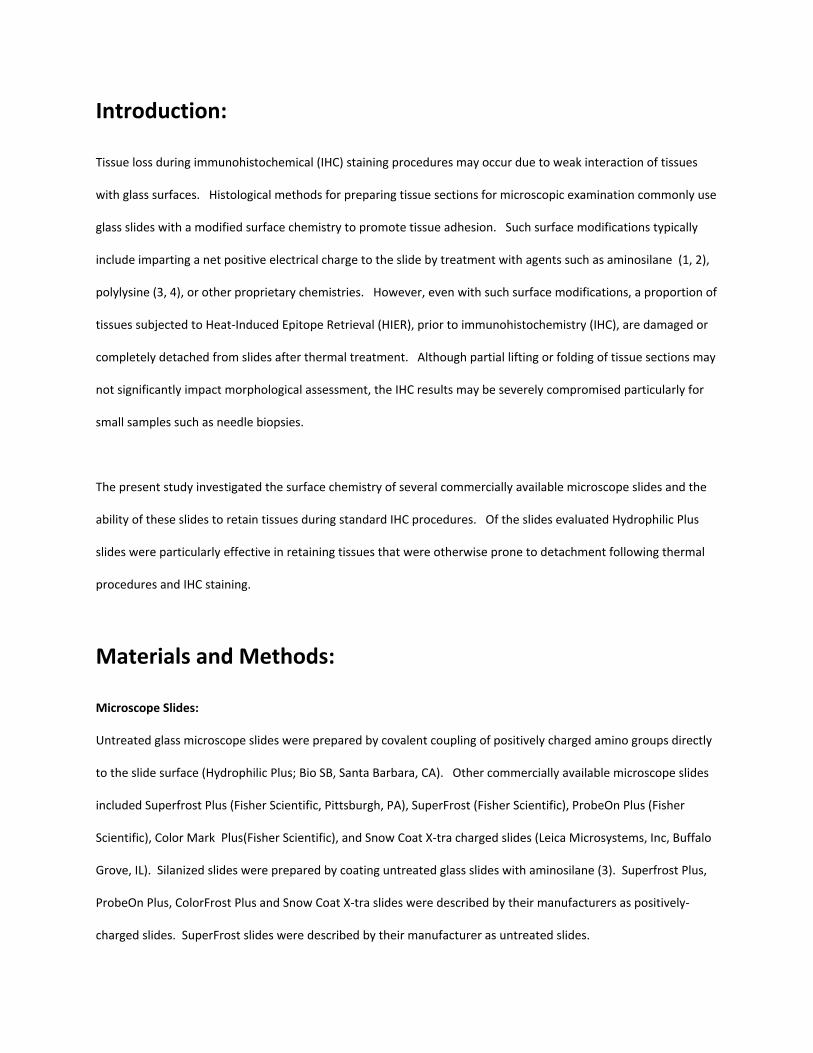
Slide wettability was determined by analysis of drop spreading on the surface of wetted slides. The buffer (without
detergent) spread over 935.6 3.6 mm2 on untreated microscope slides (SuperFrost) and over 949.5 14.3
mm2 on Hydrophilic Plus slides (85 0.3% and 86.3 1.3% respectively). In contrast, the buffer was able to cover
only 165 0.9 mm2 (15%) of ProbeOn Plus working surface (Figure 1).
Figure 1. Distribution of Buffer on Hydrophobic and Hydrophilic Surfaces of Microscope Slides.
Distribution of buffer on slide surfaces. Image was captured at five minutes after application of dyed
buffer and shows drop spread on untreated (SuperFrost), hydrophobic (ProbeOn), and hydrophilic
(Hydrophilic Plus) slides.
The contact angles of drops formed on the surface of microscope slides were used to further assess surface
characteristics, with smaller contact angles indicating greater hydrophilicity and larger contact angles indicating
greater hydrophobicity (Figure 2). Image analysis of the contact angle of drops provided the following results.
Hydrophilic Plus slides had significantly smaller contact angles (15.80 0.70) compared to both SuperFrost
(untreated) (24.90 2.170) and ProbeOn Plus (positively‐charged) (38.80 2.330) slides (Figure 2).
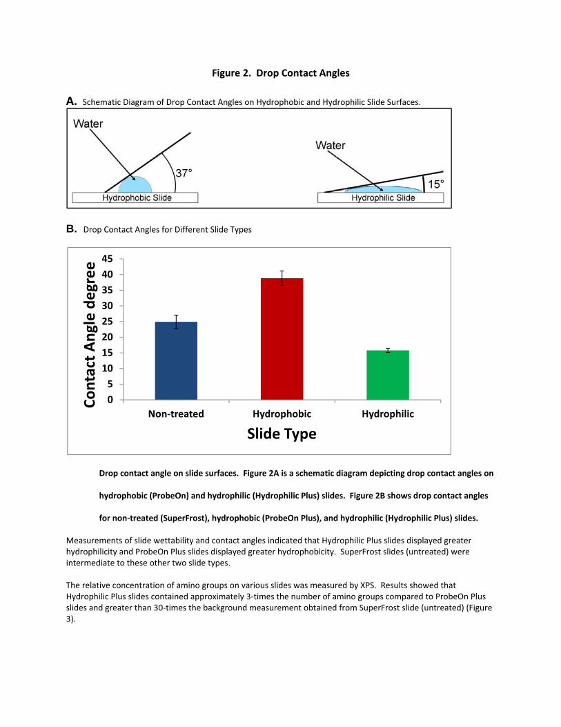
Figure 2. Drop Contact Angles A. Schematic Diagram of Drop Contact Angles on Hydrophobic and Hydrophilic Slide Surfaces.
B. Drop Contact Angles for Different Slide Types
Drop contact angle on slide surfaces. Figure 2A is a schematic diagram depicting drop contact angles on
hydrophobic (ProbeOn) and hydrophilic (Hydrophilic Plus) slides. Figure 2B shows drop contact angles
for non‐treated (SuperFrost), hydrophobic (ProbeOn Plus), and hydrophilic (Hydrophilic Plus) slides.
Measurements of slide wettability and contact angles indicated that Hydrophilic Plus slides displayed greater hydrophilicity and ProbeOn Plus slides displayed greater hydrophobicity. SuperFrost slides (untreated) were intermediate to these other two slide types. The relative concentration of amino groups on various slides was measured by XPS. Results showed that Hydrophilic Plus slides contained approximately 3‐times the number of amino groups compared to ProbeOn Plus slides and greater than 30‐times the background measurement obtained from SuperFrost slide (untreated) (Figure 3).
05
1015202530354045
Non‐treated Hydrophobic Hydrophilic
Contact A
ngle degree
Slide Type
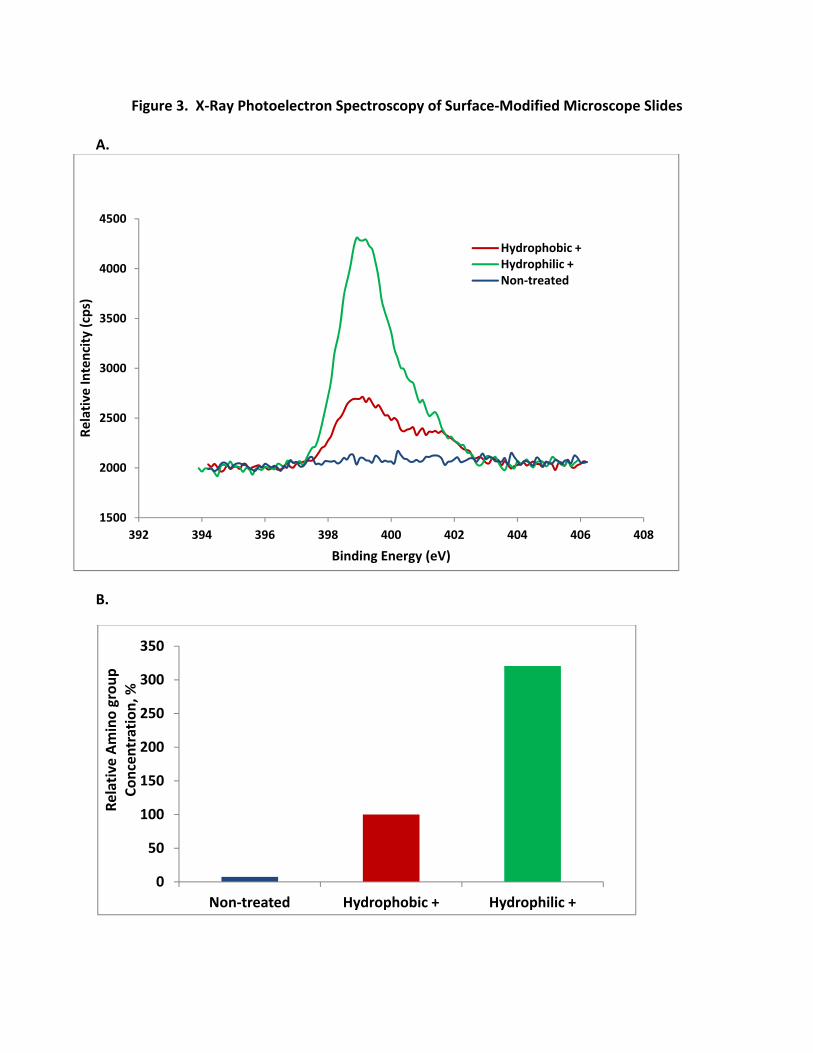
Figure 3. X‐Ray Photoelectron Spectroscopy of Surface‐Modified Microscope Slides
A.
B.
1500
2000
2500
3000
3500
4000
4500
392 394 396 398 400 402 404 406 408
Relative In
tencity (cps)
Binding Energy (eV)
Hydrophobic +Hydrophilic +Non‐treated
0
50
100
150
200
250
300
350
Non‐treated Hydrophobic + Hydrophilic +
Relative Amino grou
p Co
ncen
tration, %
A. XPS analysis of hydrophobic (ProbeOn Plus) and hydrophilic (Hydrophilic Plus) slides.
B. XPS analysis of non‐treated (SuperFrost), hydrophobic (ProbeOn Plus), and hydrophilic (Hydrophilic
Plus) slides.
Tissue adhesion was evaluated on various types of microscope slides. The results showed that Hydrophilic Plus
slides retained the greatest number of tissues and the highest percentage of tissue area (range = 90 – 100%) (Table
1).
Table 1. Percent Tissue Retention and IHC Signal
Percentage of Average Tissue Retention and IHC SignalSlide Name Range for percent Tissue Retention IHC Score
Hydrophilic Plus 90 ‐ 100% 3‐ 4+
Superfrost Plus 10‐ 50% 3‐ 3+
ProbeOn Plus 10‐ 50% 3‐ 3+
ColorMark Plus 5 ‐ 40% 3‐ 3+
Snow Coat X‐tra 5 ‐ 30% 3‐ 3+
Silanized 5 ‐ 40% 3‐ 3+
Each tissue was assigned a percentage score reflecting the amount of tissue remaining on the slide relative to the whole tissue. The range of percentage scores for each type of slide was recorded. The IHC score represents
the range of scores for seven different antibodies.
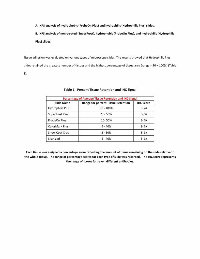
Figure 4. Macroscopic Comparison of Tissue Retention on Hydrophobic and Hydrophilic Slides
Tissue adhesion to hydrophobic (ProbeOn Plus) and hydrophilic (Hydrophilic Plus) slides. Upper tissue
on each slide (tonsil) was selected because of its strong adherence characteristics. Bottom tissue on
each slide (breast) was selected because of its tendency for detachment.
Representative photographs of the Hydrophilic Plus and ProbeOn Plus slides following IHC are shown in Figure 4
(macroscopic) and Figure 5 (microscopic). Furthermore, even when tissues remained adherent to the slides the
Hydrophilic Plus slides displayed fewer wrinkles and folding artifacts relative to the ProbeOn Plus slides (Figure 6).
An unexpected observation was the finding of more intense IHC staining (0.25 to 0.5 points) in some specimens
mounted on the Hydrophilic Plus slides when compared to other slides (Table 1). A representative
photomicrograph of staining for melanoma is shown in Figure 7.
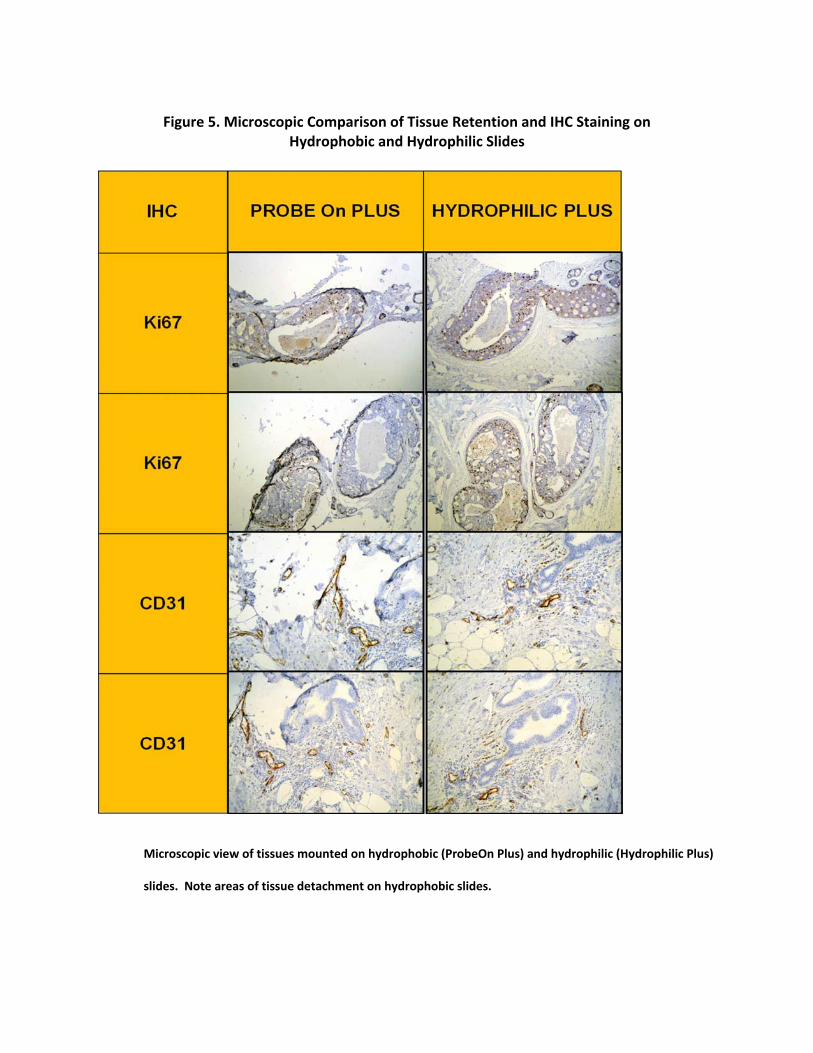
Figure 5. Microscopic Comparison of Tissue Retention and IHC Staining on
Hydrophobic and Hydrophilic Slides
Microscopic view of tissues mounted on hydrophobic (ProbeOn Plus) and hydrophilic (Hydrophilic Plus)
slides. Note areas of tissue detachment on hydrophobic slides.
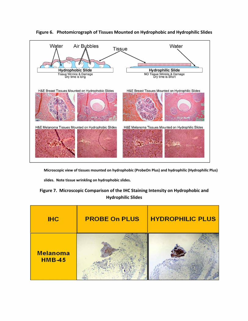
Figure 6. Photomicrograph of Tissues Mounted on Hydrophobic and Hydrophilic Slides
Microscopic view of tissues mounted on hydrophobic (ProbeOn Plus) and hydrophilic (Hydrophilic Plus)
slides. Note tissue wrinkling on hydrophobic slides.
Figure 7. Microscopic Comparison of the IHC Staining Intensity on Hydrophobic and Hydrophilic Slides
Immunohistochemical staining of melanoma with HMB45 antibody on hydrophobic (ProbeOn Plus) and
hydrophilic (Hydrophilic Plus) slides. Note increased staining intensity on hydrophilic slide.
Discussion:
Stable and strong adhesion of biological sample to the microscope slide surface is important for achieving
successful sample preparation and staining. Untreated glass surfaces of microscope slides often do not provide
strong enough retention for many biological samples, and this has led to the widespread adoption of positively‐
charged microscope slides for routine IHC analysis. Slides with positively charged surfaces are produced by
covalent couping of amino groups to slide surfaces and are sold commercially under a wide variety of names.
Other common methods for preparing positively charged surfaces include treating slides with aminosilane or
polylysine. Although these chemistries have resulted in slides with significantly improved tissue retention
characteristics, there is still a need for further improvements particularly when combined with termal methods of
antigen retrieval.
In the present study we have evaluated several of types of slides for tissue retention and performance in IHC
procedures. Given that the amino groups impart a net positive charge to the glass surface, it is likely that these
charges would increase the electrostatic attraction between the biological samples and the slides resulting in
stronger tissue retention. However, some slide surface modifications result in a hydrophobic surface with a
relatively low spatial density of amino groups such that tissue retention may not be optimal. Our results clearly
showed that Hydrophilic Plus slides retained a significantly higher percentage of tissues and had decreased
wrinkling artifacts in comparison to the other types of slides examined. Furthermore, the surface of Hydrophilic
Plus slides displayed approximately 3‐times the number of amino groups compared to ProbeOn Plus slides. Poly‐L‐
lysine is another method employed to improve adhesive properties of glass slides. Such coating usually is less
stable than aminosilane and can also increase hydrophobicity of coated slides.
It was interesting to note that more intense IHC signals were observed in some specimens mounted on the
Hydrophilic Plus slides. Although this increased staining was not observed in every case there were sufficient
examples to suggest a trend toward stronger IHC staining with the Hydrophilic Plus slides. The wetting property of
microscope slide surfaces is important in influencing outcome. Rapid and even spreading of reagents over the
entire working area of the slide is essential for uniform results. Our studies showed that Hydrophilic Plus slides
had significantly smaller drop contact angles and more rapid drop spread compared to other slides with more
hydrophobic surfaces. These hydrophilic characteristics may play a role in uniform distribution of reagents during
IHC staining, thereby reducing slide‐to‐slide variability.
The results of the present study showed that the Hydrophilic Plus slides were superior to the other slides tested in
their capacity to retain tissues during standard antigen retrieval and IHC staining procedures, without affecting
tissue morphology or IHC staining. These slides were shown to be a reliable and superior alternative to other
slides with traditional hydrophobic positively charged surfaces and could provide another effective method for
improving tissue retention during IHC analysis.
References:
1. Van Prooijen‐Knegt AC, Raap AK, Van der Burg MJ, Van der Ploeg M. Spreading and staining of human
metaphase chromosomes on aminoalkyl silane‐treated glass slides. Histochem J 14:333‐344, 1982.
2. Metwalli E, Haines D, Becker O, Conzone S, Pantarco, CG. Surface characterizations of mon‐, di‐, and tri‐
aminosilane‐treated glass substrates. J Colloid Interface Sci 298: 825‐831, 2006.
3. Husain OA, Millet JA, Grainger JM. Use of polylysine‐coated slides in preparation of cell samples for
diagnostic cytology. J Clin Pathol 33: 309‐311, 1980.
4. Mazia D, Schatten G, Salo W. Adhesion of cells to surfaces coated with polylysine. J Cell Bio 66:198‐200,
1975.
5. Collins TJ. Image J for microscopy. BioTechniques 43: 25‐30, 2007.

6. Araujo YC, Toledo PG, Leon V, Gonzalez HY. Wettability of silane‐treated glass slides as determined from
X‐ray photoelectron spectroscopy. J Colloid Interface Sci 176: 485‐490, 1995.
7. Stalder AF, Kulik G, Sage D, Barbieri L, Hoffmann P. A snake‐based approach to accurate determination of
both contact points and contact angles. Colloids and Surfaces A: Physicochem. Eng. Aspects 286: 92–103,
2006.