Embed Size (px)
Citation preview
NANO EXPRESS Open Access
Hydrophobic Drug-Loaded PEGylatedMagnetic Liposomes for Drug-ControlledReleaseAndri Hardiansyah1,2, Ming-Chien Yang2*, Ting-Yu Liu3*, Chih-Yu Kuo4, Li-Ying Huang2 and Tzu-Yi Chan3
Abstract
Less targeted and limited solubility of hydrophobic-based drug are one of the serious obstacles in drug deliverysystem. Thus, new strategies to enhance the solubility of hydrophobic drug and controlled release behaviors wouldbe developed. Herein, curcumin, a model of hydrophobic drug, has been loaded into PEGylated magneticliposomes as a drug carrier platform for drug controlled release system. Inductive magnetic heating (hyperthermia)-stimulated drug release, in vitro cellular cytotoxicity assay of curcumin-loaded PEGylated magnetic liposomes andcellular internalization-induced by magnetic guidance would be investigated. The resultant of drug carriers coulddisperse homogeneously in aqueous solution, showing a superparamagnetic characteristic and could inductivemagnetic heating with external high-frequency magnetic field (HFMF). In vitro curcumin release studies confirmedthat the drug carriers exhibited no significant release at 37 °C, whereas exhibited rapid releasing at 45 °C. However,it would display enormous (three times higher) curcumin releasing under the HFMF exposure, compared with thatwithout HFMF exposure at 45 °C. In vitro cytotoxicity test shows that curcumin-loaded PEGylated magneticliposomes could efficiently kill MCF-7 cells in parallel with increasing curcumin concentration. Fluorescencemicroscopy observed that these drug carriers could internalize efficiently into the cellular compartment of MCF-7cells. Thus, it would be anticipated that the novel hydrophobic drug-loaded PEGylated magnetic liposomes incombination with inductive magnetic heating are promising to apply in the combination of chemotherapy andthermotherapy for cancer therapy.
Keywords: PEGylated liposomes, Curcumin, Magnetic nanoparticles, High-frequency magnetic field, Drug controlledrelease
BackgroundScience and technology in medicine still dealing andcontinue to develop the optimum strategies to inhibitand kill the cancerous cells. Common cancer therapy,including surgery, chemotherapy, and radiotherapy, stillremain challenges due to the presence of various sideeffects related to the ineffectiveness treatment of thosetherapy. Thus, new strategy is in needed to overcomethe serious obstacles in cancer treatment. Nanotechnol-ogy and nanomedicine offer new opportunity for cancertreatment. In this respect development of nanoparticles
with various feature and functions along with theinnovation of the cancer treatment methodology has beenconducted experimentally in in vitro and in vivo [1, 2].Liposome is one of the nanoparticles that have been
widely used as a drug carrier for encapsulation of numer-ous drug and agents both for cancer or non-cancer treat-ment, which is a spherical bilayer membrane exhibited awell-developed of unique and important properties thatneeded for cancer therapy including a good biocompatibil-ity, appropriate size, drug loading ability, and versatilesurface functionalization [3, 4]. For instance, liposomessurfaces can be readily modified by tethering various sub-stances with specific functions. Polyethylene glycol (PEG)could be attached into the liposomes surface in order toenhance the circulation time of liposomes in the blood-stream [5, 6]. Furthermore, liposomes vesicles with size
* Correspondence: [email protected]; [email protected] of Materials Science and Engineering, National TaiwanUniversity of Science and Technology, Taipei 10607, Taiwan3Department of Materials Engineering, Ming Chi University of Technology,New Taipei City 24301, TaiwanFull list of author information is available at the end of the article
© The Author(s). 2017 Open Access This article is distributed under the terms of the Creative Commons Attribution 4.0International License (http://creativecommons.org/licenses/by/4.0/), which permits unrestricted use, distribution, andreproduction in any medium, provided you give appropriate credit to the original author(s) and the source, provide a link tothe Creative Commons license, and indicate if changes were made.
Hardiansyah et al. Nanoscale Research Letters (2017) 12:355 DOI 10.1186/s11671-017-2119-4
approximately a hundred nanometers or less exhibitedenhanced permeability and retention (EPR) effects whichfurther develop liposomes as passively-targeted nanoma-terial [7, 8]. The nanomaterial localization phenomenapresented mainly in inflammation and cancer regions.However, a passive targeting of drug carriers at the cancer-ous site is not sufficient to obtain optimum therapeutic ef-ficacy of the drug. Thus, the development of externally orinternally active stimuli would gain an interesting role forpromoting localization and action-in-pathological site [9].According to the structure and morphology, liposomes
were fabricated by the hydrophilic and hydrophobic re-gion. Various drugs and agents have been encapsulatedinside the region to develop some specific functions. Inthis respect, magnetic nanoparticles have been embed-ded into the liposomes, namely magnetic liposomes [3,10–12] or magnetoliposomes [11, 13–16], to achievespecific functions in magnetic-related characteristic suchas contrast agent [17], magnetic-targeted ability[18], andheating generation [3, 19]. Specifically, through the guid-ance of external magnetic field, magnetic liposomescould be directed into the specific area of tumor cells,then promote another specific function, including drugrelease [3, 16, 20, 21] and killing the cancerous cells [3,13, 16, 22, 23]. High-frequency magnetic field (HFMF)has been developed as a system to assist the magnetic-based nanoparticles developed the specific functionbased on the interaction between the magnetic-basednanoparticles and HFMF exposure [1–3, 24, 25].Chemotherapeutic drug has an important function in
the diseases treatment, such as cancer therapy. However,the common chemotherapeutic cancer drugs, such asdoxorubicin, exhibited toxicity and serious adverseeffects [26]. Thus, the development of therapeutic agentsor drugs with no side effects to the normal cells is inneeded as an important strategy in the treatment ofcancer or tumor cells [27]. Recently, a number ofnatural-based compounds have been investigated. Cur-cumin, a natural phenolic compound have attracted anumerous multidisciplinary researchers in natural medi-cine, food technology, and biomaterials science [1], andhas been commonly used as a traditional medicine andadditive ingredients for foods. For the chemotherapeuticproperties, curcumin exhibited beneficial properties, in-cluding antioxidant, anti-inflammatory, antimicrobial,anticancer, and wound healing characteristics [27].Curcumin has been demonstrated to inhibit proliferationof cancer cell and to induce apoptosis without promot-ing adverse effects [28]. The characteristic of curcuminreported against various cancer cells indicate its abilityto affect different targets through their interference invarious cellular mechanisms [29]. However, theutilization of curcumin for further applications has beenlimited due to its low aqueous solubility properties and
low systemic bioavailability. Previous studies revealedthat the detection of curcumin concentration in serumwas extremely low although a high concentration of cur-cumin has been orally-administered [30]. Recently, re-searchers have also been combined curcumin into thevarious features of nanomaterial to enhance the watersolubility, thereby increasing its circulation time andbioavailability thus enhance its ability to target the can-cerous cells [1, 28, 30–34].In the present study, liposome-based drug carrier
would be developed by encapsulation of oil-phase mag-netic nanoparticles and curcumin in the polyethyleneglycol-modified liposomes (PEGylated liposomes). Thestructural and morphology characterizations, high-frequency magnetic field (HFMF)-induced drug release,in vitro cellular cytotoxicity and cellular internalization-induced by magnetic guidance would be investigated.
MethodsMaterialsSynthetic lipid 1,2-dipalmitoyl-sn-glycero-3-phosphocho-line (DPPC) (purity > 99%) and 1,2-distearoyl-sn-glycero-3-phosphoethanolamine-N-[carbonyl methoxy(polyethy-lene glycol)-2000 were purchased from Avanti polarlipid, AL, USA. Cholesterol, curcumin, 1,10-dioctadecy-3,3,30,30-tetramethylindocarbocyanine perchlorate (Dil),ferric chloride tetrahydrate (FeCl2.4H2O), 4′,6-diami-dino-2-phenylindole (DAPI), oleic acid and chloroformwere purchased from Sigma-Aldrich, St. Louis, MO,USA. Ferric chloride hexahydrate (FeCl3.6H2O) was pur-chased from Shimakyu’s Pure Chemical, Osaka, Japan.Ethanol (95%) was purchased from Acros, USA. For thecell culture experiments, fibroblasts (L-929) cells wereobtained from ATCC CRL-1503TM and human breastcancer (MCF-7) cells were obtained from Food IndustryResearch and Development Institute (Taiwan). Dulbec-co’s modified Eagle’s medium-high glucose (DMEM),trypsin, dimethylsulfoxide (DMSO), trypan blue, and 3-(4,5-dimethylthiazo-2-yl)-2,5-diphenyl tetrazolium brom-ide (MTT) powder were purchased from Sigma Aldrich,St. Louis, MO, USA. Fetal bovine serum (FBS) waspurchased from BD Biosciences, San Jose, CA, USA.High-purity water purified by a Milli Q Plus water puri-fier system (Milipore, USA), with a resistivity of 18.3MΩcm was used in all experiments. All the chemicalswere used without further purification.
Preparation of Curcumin-Loaded PEGylated MagneticLiposomesLiposomes-based drug carrier were prepared through thewell-established thin-film hydration method followed by ex-trusion techniques as the method described previously withminor modification [3, 4]. Briefly, 1,2-dipalmitoyl-sn-gly-cero-3-phosphocholine (DPPC): cholesterol: 1,2-distearoyl-
Hardiansyah et al. Nanoscale Research Letters (2017) 12:355 Page 2 of 11
sn-glycero-3-phosphoethanolamine N [carbonyl-methoxy(polyethylene glycol)-2000 were mixed at a composition of80:20:5 mol%. Oleic acid coated magnetic nanoparticles(OAMNP) have been prepared via co-precipitation method(Supplementary Information). Lipid mixtures, curcumin,and OAMNP were dissolved homogeneously in chloro-form: methanol mixture (3:1 v/v) then subjected into rotaryevaporation system (N-1200 series, Eyela®, Tokyo RikakikaiCo., Ltd., Tokyo, Japan), thus resulting a thin dry lipid film.Hydration process of thin dry lipid film was accomplishedby adding PBS pH 7.4 at 60 °C for 1 h then subsequentlyplaced to a bath-type sonicator for harvesting the resultingliposomes. Un-encapsulated magnetic nanoparticles wereseparated through 1000g centrifugation for 15 min andmagnetic separation [23]. Afterward, the resultant lipo-somes were homogenized using ultrasonicator (Probe-typesonicator, VCX 750, Vibra-Cell TM, SONICS®, Sonics andMaterials, Inc., Newton, CT, USA). Eventually, the suspen-sion was extruded several times through a 0.22-μm filter toreduce the size and for sterilization. The resulting productwas termed as curcumin-loaded PEGylated magnetic lipo-somes and then stored at 4 °C prior to characterizations. Li-posomes uptake was visualized by using fluorescent Dilmarker [31]. Empty liposomes without curcumin and oleicacid-coated magnetic nanoparticles were used as a control.
CharacterizationsStructure and morphology of curcumin-loaded PEGylatedmagnetic liposomes were characterized by transmissionelectron microscopy, TEM-7650, Hitachi, Chiyoda-ku,Japan. Prior to the TEM observation, an aliquot of suspen-sion of samples was diluted with water until opticallyclear. Phosphotungstic acid (PTA) was used as the stainingagent for PEGylated liposomes. For OAMNP andcurcumin-loaded PEGylated magnetic liposomes, TEMimaging was conducted without using PTA. The sampleswere not stained as the magnetic nanoparticles can be vi-sualized directly due to their high electron density [35].Furthermore, the average particle size and zeta (ζ) poten-tial of the sample were determined at 25 °C and pH 7.4 byusing dynamic light scattering (DLS) spectrophotometer,Horiba Instrument, Horiba, Kyoto, Japan with helium-neon laser with wavelength of 633 nm, scattering angle of90°, and refractive index of 1.33 at 25 °C. Zeta (ζ) potentialwas determined with the same apparatus with DLSthrough electrophoretic mobility measurement and calcu-lated using Helmholtz-Smoluchowski’s equation.
Inductive Magnetic Heating by HFMFInductive magnetic heating (hyperthermia) experimentwas conducted by using high-frequency magnetic field(HFMF) system as the method reported previously withminor modification [1, 3, 36]. Briefly, the samples werepositioned to the center of copper coil in the HFMF
generator for 30 min. The change of the temperaturewas recorded by an alcohol thermometer. PBS was usedas a control. Each experiment was performed triplicate.
Magnetic CharacterizationsThe magnetization study was conducted to evaluate themagnetic characteristics of the synthesized PEGylatedmagnetic liposomes in response to an externally appliedmagnetic field stimuli based on the method describedwith minor modification [10]. 1,10-dioctadecy-3,3,30,30-tetramethylindocarbocyanine perchlorate (Dil) was usedas a fluorescent marker. Briefly, an aliquot of Dil-loadedPEGylated magnetic liposomes was diluted with PBS. Analiquot of the diluted sample was placed on a glass slideand positioned at a certain distance among permanentmagnetic field. Fluorescent images (fluorescence micro-scope, Olympus, Japan) of the samples at certain intervaltimes was taken to define the movement of the formula-tion along the direction of applied magnetic field.Magnetization as a function of the field were also evalu-ated using a vibrating sample magnetometer (VSM)Lakeshore model 7400 at room temperature.
Encapsulation Efficiency and In Vitro Drug Release StudiesEncapsulation efficiency defined as the ratio of encapsu-lated drug to the total drug in the system. Briefly, samplesof complete liposomes preparation are centrifuged at10,000g for 15 min and the absorbance of the clear super-natant was measured by UV-Vis spectroscopy at 425 nm.The encapsulation efficiency was calculated as [37, 38]:Encapsulation efficiency (%) = total drug-total free
drug/total drug × 100%Calibration curve was obtained by plotting absorbance
of a serial dilution of curcumin from 2 to 20 μg mL−1 at425 nm using UV spectroscopy. A linear equation wasfitted as A = 0.1534C + 0.0447, R2 = 0.991, where A isabsorbance and C is the drug concentration.The curcumin release profile from curcumin-loaded
PEGylated magnetic liposomes was determined by dialy-sis method. The curcumin release study was carried outat temperatures of 37 °C and 45 °C. 0.5% tween-80 with20% ethanol (v/v) in PBS was used as receptor medium[39]. Briefly, 1 mL of the suspensions were dialyzedagainst 20 mL receptor medium. At certain timeintervals, 1 mL of receptor medium was taken out foranalysis and fresh receptor medium solution was replen-ished and its concentration of the released drugs wasmeasured by UV spectroscopy at 425 nm. The cumula-tive release was calculated as follows [33]:
Cumulative release %ð Þ ¼ Rt=D � 100%
where D and Rt represent the initial amount of curcuminloaded and the cumulative amount of curcumin released
Hardiansyah et al. Nanoscale Research Letters (2017) 12:355 Page 3 of 11
at time t, respectively. Each experiment was performedthree times. Furthermore, Curcumin release behaviorfrom the curcumin-loaded PEGylated magnetic lipo-somes was also conducted by applying externally HFMFexposure to elaborate the mechanism of inductive mag-netic heating (hyperthermia)-triggering. Briefly, the sam-ples were placed under HFMF for 30 min. An aliquot ofcurcumin was taken from the test tube during the testfor 10 min first and continued 10 min until 30 minduring HFMF exposure. The quantity of curcumin wasquantified as described by the aforementioned method.
Cell CultureThe cells cultures of fibroblast (L-929) and MCF-7 cellswere conducted through the incubation of the cellsunder saturated humid conditions at 37 °C with 5%CO2. The cells were cultured with DMEM containing10 vol.% fetal bovine serum (FBS) and 1 vol.% antibioticantimycotic solution. Medium was changed every dayuntil reaching approximately 70 to 80% confluency.
3-(4,5-dimethylthiazo-2-yl)-2,5-Diphenyl TetrazoliumBromide (MTT) AssayCell growth and cytotoxicity were determined using MTTassay. In 96 well plates, L-929 and MCF-7 cells (10,000cells per well) were cultured in each well. These plateswere divided into several groups and incubated under sat-urated humid conditions at 37 °C and 5% CO2. After 24 hof incubation, the medium was replenished. Among theseplates, some plates were added with 200 μL of PEGylatedliposomes or PEGylated magnetic liposomes. After cultur-ing for 48 h, MTT solution were added to each well. Afterincubating for another 3 h at 37 °C, the medium was with-drawn and replaced with 200 μL DMSO and allowed tostand about 15 min for complete reaction. Furthermore,the plates were shaken, and the readings were taken at570 nm using an ELISA reader (Sunrise, Tecan, Männe-dorf, Switzerland). Proliferation or viability of the cellswas calculated as follows: Proliferation (%) = Ac/A0 ×100%. Cytotoxicity of curcumin-loaded PEGylated mag-netic liposomes was conducted through aforementionedprocedure with the variation of curcumin concentrationagainst MCF-7 cells.
Intracellular Uptake and FusionThe intracellular uptake and fusion behavior of PEGy-lated magnetic liposomes formulations toward MCF-7cell was performed using 1,10-dioctadecy-3,3,30,30-tetramethylindocarbocyanine perchlorate (Dil) as afluorescent marker. Dil is a lipophilic dye with anorange-red-fluorescence and its liposomes encapsula-tion characteristic similar to curcumin [31]. Magnetictargeting experiments were conducted as methoddescribed previously with minor modification [40].
Briefly, MCF-7 cells (20,000 cells/well) were culturedin each well of 4-well Nunc Lab-Tek chamber slides(Thermo Scientific, Rochester, New York, USA) andincubated at 37 °C in a 5% CO2 incubator for 24 h.Further, cells were incubated with Dil-loaded PEGy-lated magnetic liposomes and then exposed in thepresence of external magnets (neodymium-based mag-nets with the magnetic strength: 26-50 MGOe,Taiwan) for 3 h. As control, cells were also incubatedwith PEGylated liposomes, PEGylated magnetic lipo-somes, Dil-loaded PEGylated liposomes and Dil-loaded PEGylated magnetic liposomes, but exposed inthe absence of external magnets. After incubation,cells were washed several times with PBS and fixedwith 4 wt% paraformaldehyde for 10 min. Further, thefixing solution was aspirated and traces of fixingagents were removed by rinsing several times withPBS. Cells were then stained using 4′,6-diamidino-2-phenylindole (DAPI) for 10 min. Eventually, the stain-ing solution was aspirated and traces of staining agentwere removed by rinsing several times with PBS.Slides were mounted using a Vectashield mountingmedium (H-1000), Vector Laboratories Inc., (Burlin-game, California, USA). The cellular internalizationand fusion behavior were observed under fluorescencemicroscope (Olympus, Japan).
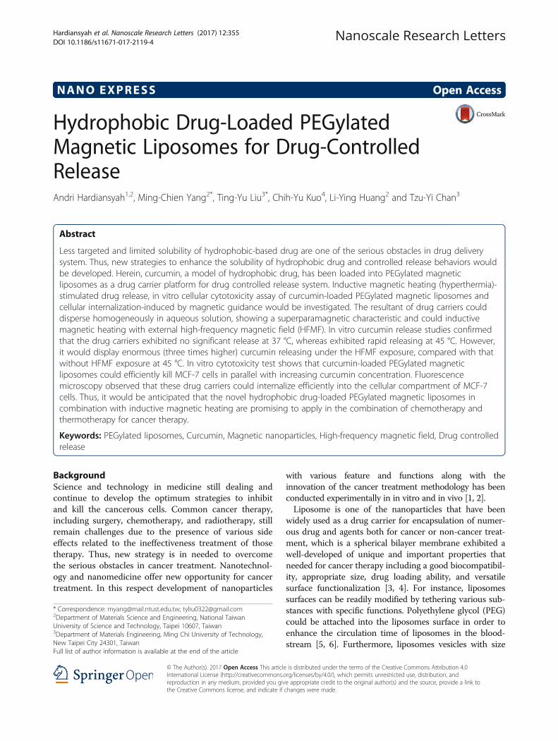
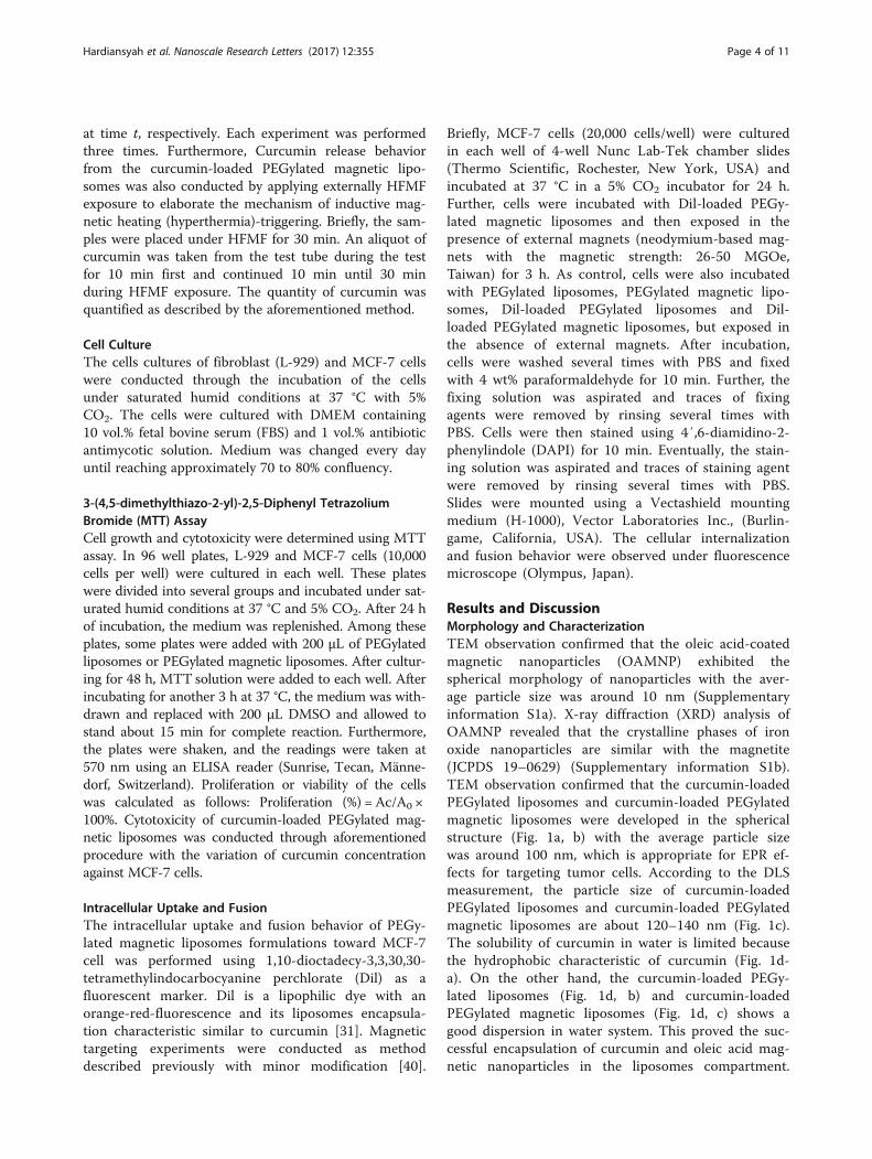
Results and DiscussionMorphology and CharacterizationTEM observation confirmed that the oleic acid-coatedmagnetic nanoparticles (OAMNP) exhibited thespherical morphology of nanoparticles with the aver-age particle size was around 10 nm (Supplementaryinformation S1a). X-ray diffraction (XRD) analysis ofOAMNP revealed that the crystalline phases of ironoxide nanoparticles are similar with the magnetite(JCPDS 19–0629) (Supplementary information S1b).TEM observation confirmed that the curcumin-loadedPEGylated liposomes and curcumin-loaded PEGylatedmagnetic liposomes were developed in the sphericalstructure (Fig. 1a, b) with the average particle sizewas around 100 nm, which is appropriate for EPR ef-fects for targeting tumor cells. According to the DLSmeasurement, the particle size of curcumin-loadedPEGylated liposomes and curcumin-loaded PEGylatedmagnetic liposomes are about 120–140 nm (Fig. 1c).The solubility of curcumin in water is limited becausethe hydrophobic characteristic of curcumin (Fig. 1d-a). On the other hand, the curcumin-loaded PEGy-lated liposomes (Fig. 1d, b) and curcumin-loadedPEGylated magnetic liposomes (Fig. 1d, c) shows agood dispersion in water system. This proved the suc-cessful encapsulation of curcumin and oleic acid mag-netic nanoparticles in the liposomes compartment.
Hardiansyah et al. Nanoscale Research Letters (2017) 12:355 Page 4 of 11
Encapsulation efficiency of curcumin in PEGylatedliposomes and PEGylated magnetic liposomes wasabout 78.06 ± 0.57% and 76.15 ± 1.6%, respectively. Inthis respect, the hydrophobic lipid bilayer compart-ment of liposomes provide the region for the encap-sulation of curcumin [28].The colloidal stability, dispersion system, and the
interaction of nanoparticles with cells are related withthe electric charge of the particle surface which is repre-sent by zeta (ζ) potential. For the reference, the zeta po-tential of non-PEGylated liposomes display the negativecharge of −17 mV. On the other hand, the zeta potentialof PEGylated liposomes and curcumin-loaded PEGylatedmagnetic liposomes increased to −2.86 and −3.17 mV,respectively. The increased zeta potential indicated thecharge shielding effect of polyethylene glycol (PEG) and
curcumin. These characteristics prevent liposomes to befusion and aggregation to enhance the colloidal stabilitysimultaneously [31, 41].
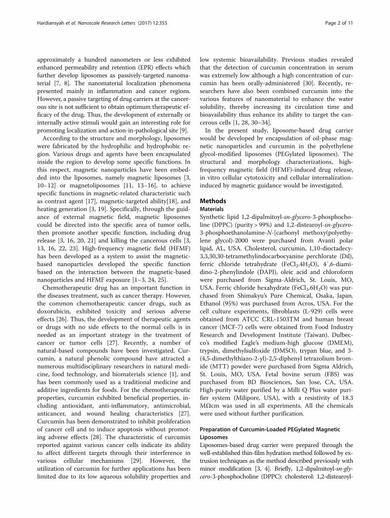
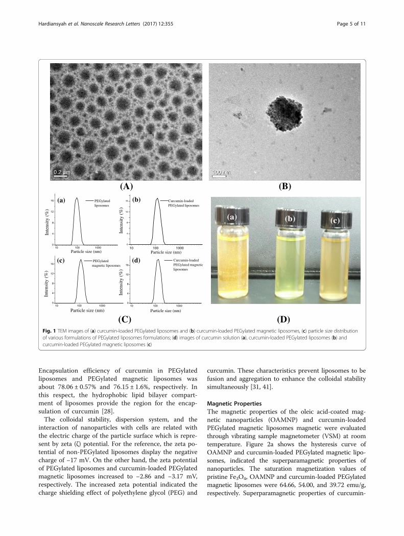
Magnetic PropertiesThe magnetic properties of the oleic acid-coated mag-netic nanoparticles (OAMNP) and curcumin-loadedPEGylated magnetic liposomes magnetic were evaluatedthrough vibrating sample magnetometer (VSM) at roomtemperature. Figure 2a shows the hysteresis curve ofOAMNP and curcumin-loaded PEGylated magnetic lipo-somes, indicated the superparamagnetic properties ofnanoparticles. The saturation magnetization values ofpristine Fe3O4, OAMNP and curcumin-loaded PEGylatedmagnetic liposomes were 64.66, 54.00, and 39.72 emu/g,respectively. Superparamagnetic properties of curcumin-
(A)
)
(a) (b) (c)
(C)
10 100 10000
4
8
12
16
Curcumin-loaded PEGylated magnetic liposomes
PEGylated liposomes
Curcumin-loaded PEGylated liposomes
Inte
nsit
y (%
))%(
ytisnetnI
Inte
nsit
y (%
))%(
ytisn etnI
PEGylated magnetic liposomes
10 100 10000
4
8
12
16
10 100 10000
4
8
12
16
(d)(c)
(b)
Particle size (nm)
Particle size (nm) Particle size (nm)
(a)
10 100 10000
4
8
12
16
Particle size (nm)
(B)
(D)Fig. 1 TEM images of (a) curcumin-loaded PEGylated liposomes and (b) curcumin-loaded PEGylated magnetic liposomes, (c) particle size distributionof various formulations of PEGylated liposomes formulations; (d) images of curcumin solution (a), curcumin-loaded PEGylated liposomes (b) andcurcumin-loaded PEGylated magnetic liposomes (c)
Hardiansyah et al. Nanoscale Research Letters (2017) 12:355 Page 5 of 11
loaded PEGylated magnetic liposomes are important forbiomedical applications to prevent aggregation and enablerapid dispersal in the absence of magnetic field. Thedecreasing of magnetization values of curcumin-loadedmagnetic liposomes might be due to the modification ofnon-magnetic phospholipids bilayers. The lipid bilayerinterferes the domain alignment and inhibited the inter-action of the OAMNP encapsulated within the lipid bilay-ers to the external magnets (neodymium-based magnets)exposure [28, 42]. Figure 2b shows that the curcumin-loaded PEGylated magnetic liposomes could interacteasily with the external magnets exposure.Figure 2c shows the magnetic movement of Dil-loaded
PEGylated magnetic liposomes triggered in the externalmagnets exposure by florescence microscopy. A homoge-neous distribution of Dil-loaded PEGylated magneticliposomes were monitored in the absence of external mag-nets. Upon application of external magnets exposure, theDil-loaded PEGylated magnetic liposomes rapidly movedtowards the magnet as a function of times. These charac-teristics clearly demonstrate that the formulations couldattracted to the external magnets exposure.
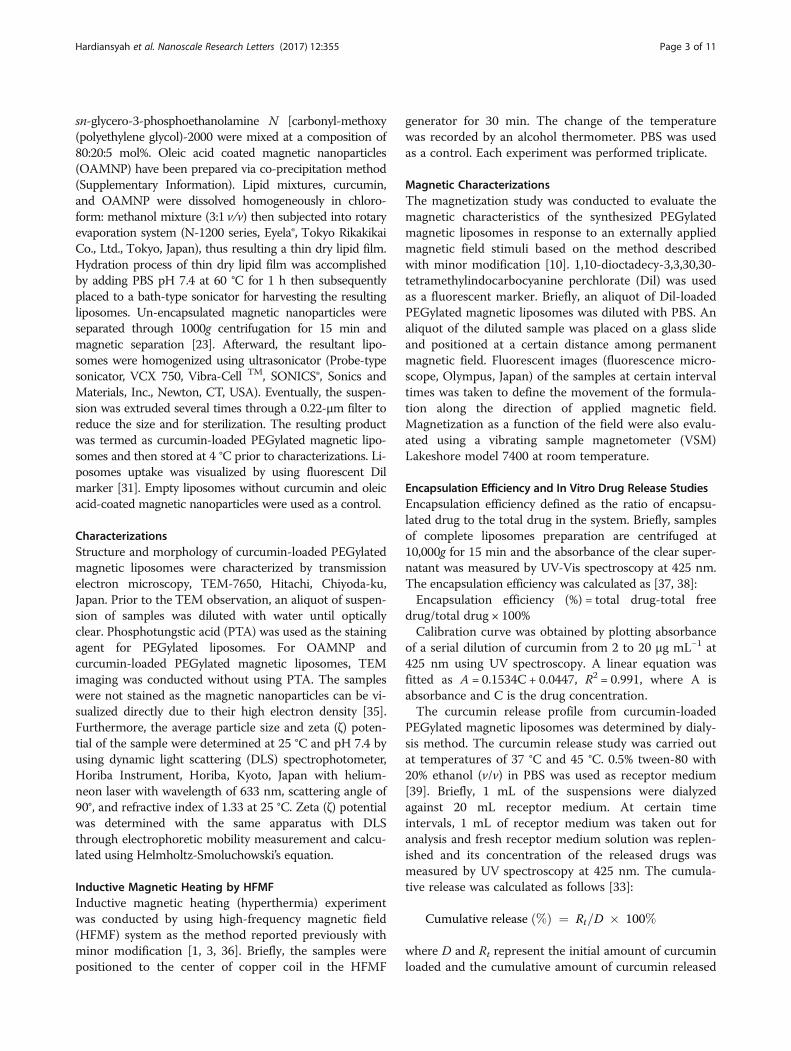
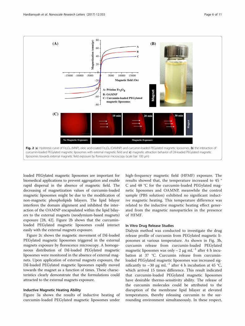
Inductive Magnetic Heating AbilityFigure 3a shows the results of inductive heating ofcurcumin-loaded PEGylated magnetic liposomes under
high-frequency magnetic field (HFMF) exposure. Theresults showed that, the temperature increased to 45 °C and 48 °C for the curcumin-loaded PEGylated mag-netic liposomes and OAMNP, meanwhile the controlsample (PBS solution) exhibited no significant induct-ive magnetic heating. This temperature difference wasrelated to the inductive magnetic heating effect gener-ated from the magnetic nanoparticles in the presenceof HFMF.
In Vitro Drug Release StudiesDialysis method was conducted to investigate the drugrelease profile of curcumin from PEGylated magnetic li-posomes at various temperature. As shown in Fig. 3b,curcumin release from curcumin-loaded PEGylatedmagnetic liposomes was only ~ 2 μg mL−1 after 4 h incu-bation at 37 °C. Curcumin release from curcumin-loaded PEGylated magnetic liposomes was increased sig-nificantly to ~30 μg mL−1 after 4 h incubation at 45 °C,which arrived 15 times difference. This result indicatedthat curcumin-loaded PEGylated magnetic liposomeshave desirable thermo-sensitivity ability. The release ofthe curcumin molecules could be attributed to thedisruption of the membrane lipid bilayer at elevatedtemperatures, thereby releasing curcumin to the sur-rounding environment simultaneously. In these respect,
(C)
(A) (B)
-15000 -10000 -5000 0 5000 10000 15000
-80
-60
-40
-20
0
20
40
60
80
C
A
)rg/ume(
noitazitengaM
Magnetic field (Oe)
B
A: Pristine Fe3O4B: OAMNPC: Curcumin-loaded PEGylated magnetic liposomes
No Magnetic Exposure
10 min 20 min 30 min
Magnetic Exposure
Fig. 2 (a) Hysteresis curve of Fe3O4 (MNP), oleic acid-coated Fe3O4 (OAMNP) and curcumin-loaded PEGylated magnetic liposomes, (b) the interaction ofcurcumin-loaded PEGylated magnetic liposomes with external magnetic field and (c) magnetic attraction behavior of Dil-loaded PEGylated magneticliposomes towards external magnetic field exposure by florescence microscopy (scale bar: 100 μm)
Hardiansyah et al. Nanoscale Research Letters (2017) 12:355 Page 6 of 11
the structure and fluidity of the lipid bilayers are greatlyinfluenced by the phase transition temperature whichfurther affects the release of curcumin from thecurcumin-loaded PEGylated magnetic liposomes. In thisrespect, physiological temperature (37 °C) is below thetransition temperature of phosphatidylcholine as themain structural component of these liposomes, thus therelease of curcumin in physiological environments isinhibited. However, a hyperthermia temperature of 45 °Cis above the transition temperature, thus increasing therelease of curcumin through the structural disruption oflipid bilayer [28].Figure 3c shows the cumulative drug release of
curcumin-loaded PEGylated magnetic liposomes underHFMF exposure and curcumin release profile moni-tored by UV-visible spectroscopy (Fig. 3d). The cumu-lative curcumin release from curcumin-loadedPEGylated magnetic liposomes with HFMF treatmentarrived to ~12 μg mL−1 after 30 min of HFMF expos-ure. Meanwhile, cumulative release of curcumin fromcurcumin-loaded PEGylated magnetic liposomes afterincubated at 37 and 45 °C for 30 min were only 0.8 and4.5 μg mL−1, respectively. These phenomena might berelated to the incorporation of oleic acid-coated
magnetic nanoparticles in the curcumin-loaded PEGy-lated magnetic liposomes which generate localizedheating under HFMF stimulus, followed by increasingthe permeability of lipid bilayer, thus enhanced the cur-cumin release from liposomes compartment [25, 43].
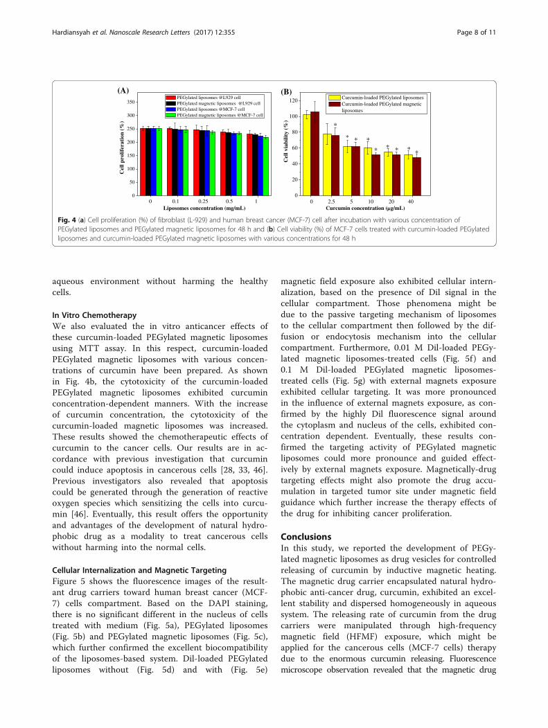
Cytotoxicity StudiesMTT assay was conducted to evaluate the cellularcytotoxicity of PEGylated liposomes and PEGylatedmagnetic liposomes toward fibroblast (L-929) and hu-man breast cancer (MCF-7) cells, thereby indicatingthe effect of those drug carriers on the growth andproliferation of cells. Based on the cell proliferationof L-929 and MCF-7 cells (Fig. 4a), the cells prolifer-ate well in the incubation with PEGylated liposomesand PEGylated magnetic liposomes. This might bedue to the better biocompatibility of the liposome-based system with the cell compartment [28, 33, 42,44, 45]. These results confirmed that the drug carriersexhibited no cytotoxicity against L-929 cell and MCF-7 cells, suggesting good biocompatibility of the drugcarriers. It is a potential to use the novel magneticcarriers to deliver the chemotherapeutic drugs in
0 5 10 15 20 25 30
36
39
42
45
48
51
*
*erutarep
meT
(o C)
Time (min)
PBS solution OAMNP Curcumin-loaded PEGylated magnetic
liposomes
0 50 100 150 200 2500
5
10
15
20
25
30
35
esaelerevitalu
muC
(µL
m/g)
Time (min)
T=45 oC
T=37 oC *
300 350 400 450 500 550 600
) u.a(yti snetnI
Wavelength (nm)
HFMF 10 minHFMF 20 minHFMF 30 min
Thermo (37 oC) 30 min
Thermo (45 oC) 30 min
(A) (B)
(C) (D)
0 10 20 30
0
2
4
6
8
10
12
14
16 Thermo-triggered release (T=37 oC)
Thermo-triggered release (T=45 oC) HFMF-triggered release
*
esaelergur d
evitalumu
C(
Lm/g
)
Time (min)
*
Fig. 3 (a) Inductive heating ability of the oleic acid-coated magnetic nanoparticles (OAMNP) and the curcumin-loaded PEGylated magnetic liposomesunder HFMF exposure. PBS solution was used as a control sample (*p< 0.05), (b) cumulative drug release profile of curcumin-loaded PEGylated magneticliposomes at various temperature (* p< 0.05 against the sample incubated at 37 °C), (c) cumulative drug release of curcumin-loaded PEGylated magneticliposomes under HFMF exposure (*p< 0.05) and (d) curcumin release profile monitored by UV-Visible spectroscopy
Hardiansyah et al. Nanoscale Research Letters (2017) 12:355 Page 7 of 11
aqueous environment without harming the healthycells.
In Vitro ChemotherapyWe also evaluated the in vitro anticancer effects ofthese curcumin-loaded PEGylated magnetic liposomesusing MTT assay. In this respect, curcumin-loadedPEGylated magnetic liposomes with various concen-trations of curcumin have been prepared. As shownin Fig. 4b, the cytotoxicity of the curcumin-loadedPEGylated magnetic liposomes exhibited curcuminconcentration-dependent manners. With the increaseof curcumin concentration, the cytotoxicity of thecurcumin-loaded magnetic liposomes was increased.These results showed the chemotherapeutic effects ofcurcumin to the cancer cells. Our results are in ac-cordance with previous investigation that curcumincould induce apoptosis in cancerous cells [28, 33, 46].Previous investigators also revealed that apoptosiscould be generated through the generation of reactiveoxygen species which sensitizing the cells into curcu-min [46]. Eventually, this result offers the opportunityand advantages of the development of natural hydro-phobic drug as a modality to treat cancerous cellswithout harming into the normal cells.
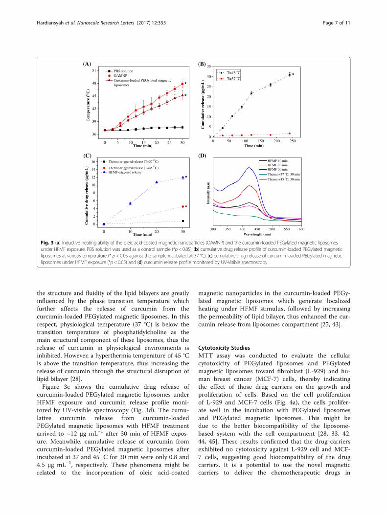
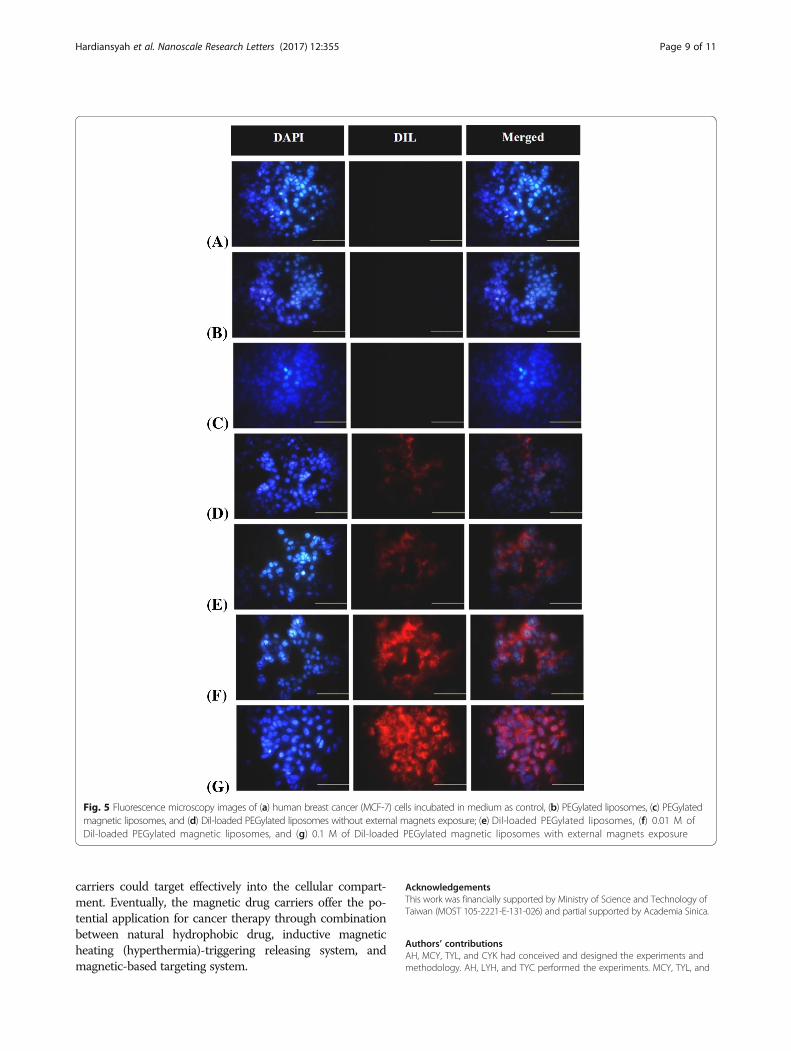
Cellular Internalization and Magnetic TargetingFigure 5 shows the fluorescence images of the result-ant drug carriers toward human breast cancer (MCF-7) cells compartment. Based on the DAPI staining,there is no significant different in the nucleus of cellstreated with medium (Fig. 5a), PEGylated liposomes(Fig. 5b) and PEGylated magnetic liposomes (Fig. 5c),which further confirmed the excellent biocompatibilityof the liposomes-based system. Dil-loaded PEGylatedliposomes without (Fig. 5d) and with (Fig. 5e)
magnetic field exposure also exhibited cellular intern-alization, based on the presence of Dil signal in thecellular compartment. Those phenomena might bedue to the passive targeting mechanism of liposomesto the cellular compartment then followed by the dif-fusion or endocytosis mechanism into the cellularcompartment. Furthermore, 0.01 M Dil-loaded PEGy-lated magnetic liposomes-treated cells (Fig. 5f ) and0.1 M Dil-loaded PEGylated magnetic liposomes-treated cells (Fig. 5g) with external magnets exposureexhibited cellular targeting. It was more pronouncedin the influence of external magnets exposure, as con-firmed by the highly Dil fluorescence signal aroundthe cytoplasm and nucleus of the cells, exhibited con-centration dependent. Eventually, these results con-firmed the targeting activity of PEGylated magneticliposomes could more pronounce and guided effect-ively by external magnets exposure. Magnetically-drugtargeting effects might also promote the drug accu-mulation in targeted tumor site under magnetic fieldguidance which further increase the therapy effects ofthe drug for inhibiting cancer proliferation.
ConclusionsIn this study, we reported the development of PEGy-lated magnetic liposomes as drug vesicles for controlledreleasing of curcumin by inductive magnetic heating.The magnetic drug carrier encapsulated natural hydro-phobic anti-cancer drug, curcumin, exhibited an excel-lent stability and dispersed homogeneously in aqueoussystem. The releasing rate of curcumin from the drugcarriers were manipulated through high-frequencymagnetic field (HFMF) exposure, which might beapplied for the cancerous cells (MCF-7 cells) therapydue to the enormous curcumin releasing. Fluorescencemicroscope observation revealed that the magnetic drug
0
50
100
150
200
250
300
350
0 0.1 0.25 0.5 1
)%(
noitarefilorplleC
Liposomes concentration (mg/mL)
PEGylated liposomes @L929 cell PEGylated magnetic liposomes @L929 cell PEGylated liposomes @MCF-7 cell PEGylated magnetic liposomes @MCF-7 cell
0
20
40
60
80
100
120
*****
**
*
)%(
ytil ibaivlleC
Curcumin-loaded PEGylated liposomes Curcumin-loaded PEGylated magnetic
liposomes
*
0 2.5 5 10 20 40Curcumin concentration (µg/mL)
(A) (B)
Fig. 4 (a) Cell proliferation (%) of fibroblast (L-929) and human breast cancer (MCF-7) cell after incubation with various concentration ofPEGylated liposomes and PEGylated magnetic liposomes for 48 h and (b) Cell viability (%) of MCF-7 cells treated with curcumin-loaded PEGylatedliposomes and curcumin-loaded PEGylated magnetic liposomes with various concentrations for 48 h
Hardiansyah et al. Nanoscale Research Letters (2017) 12:355 Page 8 of 11
carriers could target effectively into the cellular compart-ment. Eventually, the magnetic drug carriers offer the po-tential application for cancer therapy through combinationbetween natural hydrophobic drug, inductive magneticheating (hyperthermia)-triggering releasing system, andmagnetic-based targeting system.
AcknowledgementsThis work was financially supported by Ministry of Science and Technology ofTaiwan (MOST 105-2221-E-131-026) and partial supported by Academia Sinica.
Authors’ contributionsAH, MCY, TYL, and CYK had conceived and designed the experiments andmethodology. AH, LYH, and TYC performed the experiments. MCY, TYL, and
Fig. 5 Fluorescence microscopy images of (a) human breast cancer (MCF-7) cells incubated in medium as control, (b) PEGylated liposomes, (c) PEGylatedmagnetic liposomes, and (d) Dil-loaded PEGylated liposomes without external magnets exposure; (e) Dil-loaded PEGylated liposomes, (f) 0.01 M ofDil-loaded PEGylated magnetic liposomes, and (g) 0.1 M of Dil-loaded PEGylated magnetic liposomes with external magnets exposure
Hardiansyah et al. Nanoscale Research Letters (2017) 12:355 Page 9 of 11
CYK contributed ideas and material characterizations analysis. AH, MCY, andTYL wrote the manuscript. All authors read and approved the finalmanuscript.
Competing interestsThe authors declare that they have no competing interests.
Publisher’s NoteSpringer Nature remains neutral with regard to jurisdictional claims inpublished maps and institutional affiliations.
Author details1Department of Metallurgy and Materials Engineering, Bandung Institute ofTechnology and Science, Bekasi 17530, Indonesia. 2Department of MaterialsScience and Engineering, National Taiwan University of Science andTechnology, Taipei 10607, Taiwan. 3Department of Materials Engineering,Ming Chi University of Technology, New Taipei City 24301, Taiwan. 4Instituteof Polymer Science and Engineering, National Taiwan University, Taipei10617, Taiwan.
Received: 17 January 2017 Accepted: 1 May 2017
References1. Kuo C-Y, Liu T-Y, Chan T-Y, Tsai S-C, Hardiansyah A, Huang L-Y, Yang M-C,
Lu R-H, Jiang J-K, Yang C-Y et al (2016) Magnetically triggered nanovehiclesfor controlled drug release as a colorectal cancer therapy. Colloids Surf B:Biointerfaces 140:567–573
2. Kuo C-Y, Liu T-Y, Hardiansyah A, Lee C-F, Wang M-S, Chiu W-Y (2014) Self-assembly behaviors of thermal- and pH- sensitive magnetic nanocarriers forstimuli-triggered release. Nanoscale Res Lett 9:1–11
3. Hardiansyah A, Huang LY, Yang MC, Liu TY, Tsai SC, Yang CY, Kuo CY, ChanTY, Zou HM, Lian WN, Lin CH (2014) Magnetic liposomes for colorectalcancer cells therapy by high-frequency magnetic field treatment. NanoscaleRes Lett 9:497
4. Hardiansyah A, Huang L-Y, Yang M-C, Purwasasmita BS, Liu T-Y, Kuo C-Y,Liao H-L, Chan T-Y, Tzou H-M, Chiu W-Y (2015) Novel pH-sensitive drugcarriers of carboxymethyl-hexanoyl chitosan (chitosonic[registered sign]acid) modified liposomes. RSC Adv 5:23134–23143
5. Klibanov AL, Maruyama K, Beckerleg AM, Torchilin VP, Huang L (1991)Activity of amphipathic poly(ethylene glycol) 5000 to prolong thecirculation time of liposomes depends on the liposome size and isunfavorable for immunoliposome binding to target. Biochim Biophys ActaBiomembr 1062:142–148
6. Allen C, Dos Santos N, Gallagher R, Chiu GN, Shu Y, Li WM, Johnstone SA,Janoff AS, Mayer LD, Webb MS, Bally MB (2002) Controlling the physicalbehavior and biological performance of liposome formulations through useof surface grafted poly(ethylene glycol). Biosci Rep 22:225–250
7. Abraham SA, Waterhouse DN, Mayer LD, Cullis PR, MaddenTD, Bally MB(2005) The liposomal formulation of doxorubicin. Methods in enzymology,edition. San Diego: Elsevier Academic Press 391:71–97
8. El Maghraby GM, Barry BW, Williams AC (2008) Liposomes and skin: fromdrug delivery to model membranes. Eur J Pharm Sci 34:203–222
9. Sawant RR, Torchilin VP (2010) Liposomes as ‘smart’ pharmaceuticalnanocarriers. Soft Matter 6:4026–4044
10. Nahar K, Absar S, Patel B, Ahsan F (2014) Starch-coated magnetic liposomesas an inhalable carrier for accumulation of fasudil in the pulmonaryvasculature. Int J Pharm 464:185–195
11. Marie H, Lemaire L, Franconi F, Lajnef S, Frapart Y-M, Nicolas V, Frébourg G,Trichet M, Ménager C, Lesieur S: Superparamagnetic Liposomes for MRIMonitoring and External Magnetic Field-Induced Selective Targeting ofMalignant Brain Tumors. Adv Funct Mater.2015:n/a-n/a.
12. Di Corato R, Béalle G, Kolosnjaj-Tabi J, Espinosa A, Clément O, Silva AKA,Ménager C, Wilhelm C (2015) Combining magnetic hyperthermia andphotodynamic therapy for tumor ablation with photoresponsive magneticliposomes. ACS Nano 9(3):2904–2916
13. Yoshida M, Sato M, Yamamoto Y, Maehara T, Naohara T, Aono H, SugishitaH, Sato K, Watanabe Y (2012) Tumor local chemohyperthermia usingdocetaxel-embedded magnetoliposomes: interaction of chemotherapy andhyperthermia. Eur J Gastroenterol Hepatol 27:406–411
14. Bolfarini GC, Siqueira-Moura MP, Demets GJ, Morais PC, Tedesco AC (2012)In vitro evaluation of combined hyperthermia and photodynamic effectsusing magnetoliposomes loaded with cucurbituril zinc phthalocyaninecomplex on melanoma. J Photochem Photobiol B Biol 115:1–4
15. Qiu D, An X (2013) Controllable release from magnetoliposomes bymagnetic stimulation and thermal stimulation. Colloids Surf B: Biointerfaces104:326–329
16. Guo H, Chen W, Sun X, Liu Y-N, Li J, Wang J (2015) Theranosticmagnetoliposomes coated by carboxymethyl dextran with controlledrelease by low-frequency alternating magnetic field. Carbohydr Polym 118:209–217
17. Martina MS, Fortin JP, Menager C, Clement O, Barratt G, Grabielle-Madelmont C, Gazeau F, Cabuil V, Lesieur S (2005) Generation ofsuperparamagnetic liposomes revealed as highly efficient MRI contrastagents for in vivo imaging. J Am Chem Soc 127:10676–10685
18. Kubo T, Sugita T, Shimose S, Nitta Y, Ikuta Y, Murakami T (2000) Targeteddelivery of anticancer drugs with intravenously administered magneticliposomes in osteosarcoma-bearing hamsters. Int J Oncol 17:309–315
19. Yanase M, Shinkai M, Honda H, Wakabayashi T, Yoshida J, Kobayashi T(1998) Intracellular hyperthermia for cancer using magnetite cationicliposomes: an in vivo study. Jpn J Cancer Res 89:463–469
20. Babincová M, Čičmanec P, Altanerová V, Altaner Č, Babinec P (2002) AC-magnetic field controlled drug release from magnetoliposomes: design of amethod for site-specific chemotherapy. Bioelectrochemistry 55:17–19
21. Tai LA, Tsai PJ, Wang YC, Wang YJ, Lo LW, Yang CS (2009) Thermosensitiveliposomes entrapping iron oxide nanoparticles for controllable drug release.Nanotechnology 20:135101
22. Kulshrestha P, Gogoi M, Bahadur D, Banerjee R (2012) In vitro application ofpaclitaxel loaded magnetoliposomes for combined chemotherapy andhyperthermia. Colloids Surf B: Biointerfaces 96:1–7
23. Peng Z, Wang C, Fang E, Lu X, Wang G, Tong Q (2014) Co-delivery ofdoxorubicin and SATB1 shRNA by thermosensitive magnetic cationicliposomes for gastric cancer therapy. PLoS One 9:e92924
24. Hu S-H, Liu T-Y, Liu D-M, Chen S-Y (2007) Controlled pulsatile drug releasefrom a ferrogel by a high-frequency magnetic field. Macromolecules 40:6786–6788
25. Liu TY, Hu SH, Liu KH, Shaiu RS, Liu DM, Chen SY (2008) Instantaneous drugdelivery of magnetic/thermally sensitive nanospheres by a high-frequencymagnetic field. Langmuir 24:13306–13311
26. Rahman A, More N, Schein PS (1982) Doxorubicin-induced chronic cardiotoxicityand its protection by liposomal administration. Cancer Res 42:1817–1825
27. Salem M, Rohani S, Gillies ER (2014) Curcumin, a promising anti-cancertherapeutic: a review of its chemical properties, bioactivity and approachesto cancer cell delivery. RSC Adv 4:10815–10829
28. Nigam S, Kumar A, Thouas GA, Bahadur D, Chen Q (2013) Curcumin deliveryusing magnetic liposomes. J Nanopharmaceutics Drug Delivery 1:365–375
29. Anand P, Sundaram C, Jhurani S, Kunnumakkara AB, Aggarwal BB (2008)Curcumin and cancer: an “old-age” disease with an “age-old” solution.Cancer Lett 267:133–164
30. Dhule SS, Penfornis P, Frazier T, Walker R, Feldman J, Tan G, He J, Alb A,John V, Pochampally R (2012) Curcumin-loaded gamma-cyclodextrinliposomal nanoparticles as delivery vehicles for osteosarcoma. Nanomed:Nanotechnol, Biol Med 8:440–451
31. Dhule SS, Penfornis P, He J, Harris MR, Terry T, John V, Pochampally R (2014)The combined effect of encapsulating curcumin and C6 ceramide inliposomal nanoparticles against osteosarcoma. Mol Pharmacol 11:417–427
32. Saengkrit N, Saesoo S, Srinuanchai W, Phunpee S, Ruktanonchai UR (2014)Influence of curcumin-loaded cationic liposome on anticancer activity forcervical cancer therapy. Colloids Surf B: Biointerfaces 114:349–356
33. Huang Q, Zhang L, Sun X, Zeng K, Li J, Liu Y-N (2014) Coating ofcarboxymethyl dextran on liposomal curcumin to improve the anticanceractivity. RSC Adv 4:59211–59217
34. Li L, Ahmed B, Mehta K, Kurzrock R (2007) Liposomal curcumin with andwithout oxaliplatin: effects on cell growth, apoptosis, and angiogenesis incolorectal cancer. Mol Cancer Ther 6:1276–1282
35. Pradhan P, Giri J, Rieken F, Koch C, Mykhaylyk O, Doblinger M, Banerjee R,Bahadur D, Plank C (2010) Targeted temperature sensitive magneticliposomes for thermo-chemotherapy. J Control Release 142:108–121
36. Kuo C-Y, Liu T-Y, Hardiansyah A, Chiu W-Y: Magnetically polymericnanocarriers for targeting delivery of curcumin and hyperthermiatreatments toward cancer cells. J. Polym. Sci A Polym Chem. 2016:n/a-n/a.
Hardiansyah et al. Nanoscale Research Letters (2017) 12:355 Page 10 of 11
37. Hasan M, Belhaj N, Benachour H, Barberi-Heyob M, Kahn CJ, Jabbari E,Linder M, Arab-Tehrany E (2014) Liposome encapsulation of curcumin:physico-chemical characterizations and effects on MCF7 cancer cellproliferation. Int J Pharm 461:519–528
38. Rejinold NS, Muthunarayanan M, Divyarani VV, Sreerekha PR, Chennazhi KP,Nair SV, Tamura H, Jayakumar R (2011) Curcumin-loaded biocompatiblethermoresponsive polymeric nanoparticles for cancer drug delivery. JColloid Interface Sci 360:39–51
39. Chen Y, Wu Q, Zhang Z, Yuan L, Liu X, Zhou L (2012) Preparation ofcurcumin-loaded liposomes and evaluation of their skin permeation andpharmacodynamics. Molecules 17:5972–5987
40. Yallapu MM, Othman SF, Curtis ET, Bauer NA, Chauhan N, Kumar D, Jaggi M,Chauhan SC (2012) Curcumin-loaded magnetic nanoparticles for breastcancer therapeutics and imaging applications. Int J Nanomedicine7:1761–1779
41. Bloemen M, Brullot W, Luong TT, Geukens N, Gils A, Verbiest T (2012)Improved functionalization of oleic acid-coated iron oxide nanoparticles forbiomedical applications. J Nanopart Res 14:1100
42. Ding X, Cai K, Luo Z, Li J, Hu Y, Shen X (2012) Biocompatible magneticliposomes for temperature triggered drug delivery. Nanoscale 4:6289–6292
43. Lu Z, Prouty MD, Guo Z, Golub VO, Kumar CSSR, Lvov YM (2005) Magneticswitch of permeability for polyelectrolyte microcapsules embedded withCo@Au nanoparticles. Langmuir 21:2042–2050
44. Pradhan P, Giri J, Banerjee R, Bellare J, Bahadur D (2007) Preparation andcharacterization of manganese ferrite-based magnetic liposomes forhyperthermia treatment of cancer. J Magn Magn Mater 311:208–215
45. Béalle G, Di Corato R, Kolosnjaj-Tabi J, Dupuis V, Clément O, Gazeau F,Wilhelm C, Ménager C (2012) Ultra magnetic liposomes for MR imaging,targeting, and hyperthermia. Langmuir 28:11834–11842
46. Syng-Ai C, Kumari AL, Khar A (2004) Effect of curcumin on normal andtumor cells: role of glutathione and bcl-2. Mol Cancer Ther 3:1101–1108
Submit your manuscript to a journal and benefi t from:
7 Convenient online submission
7 Rigorous peer review
7 Immediate publication on acceptance
7 Open access: articles freely available online
7 High visibility within the fi eld
7 Retaining the copyright to your article
Submit your next manuscript at 7 springeropen.com
Hardiansyah et al. Nanoscale Research Letters (2017) 12:355 Page 11 of 11
![The Human In Vivo Biomolecule Corona onto PEGylated ... · [12,14] Despite the clinical track record of liposomes for more than 20 years, the role that protein corona plays in liposomal](https://img.pdfslide.net/doc/110x75/5e4301a72e39bd4bb705d596/the-human-in-vivo-biomolecule-corona-onto-pegylated-1214-despite-the-clinical.jpg)


![The Potential of Liposomes with Carbonic Anhydrase IX to Deliver … · 2017-05-06 · cardiotoxicity risk [53]. Using the liposomal formulation (including those with pegylated liposomes),](https://img.pdfslide.net/doc/110x75/5ea2c664385ce23fa374888c/the-potential-of-liposomes-with-carbonic-anhydrase-ix-to-deliver-2017-05-06-cardiotoxicity.jpg)