Embed Size (px)
Citation preview
Innate lymphoid cells responding to IL-33 mediate airway-hyperreactivity independent of adaptive immunity
Hye Young Kim, PhDa,*, Ya-Jen Chang, PhDa,*, Srividya Subramanian, PhDa, Hyun-HeeLee, PhDa, Lee A. Albacker, PhDa, Ponpan Matangkasombut, MD,PhDa, Paul B. Savage,PhDb, Andrew N. J. McKenzie, PhDc, Dirk E. Smith, PhDd, James B. Rottman, PhDe,Rosemarie H. DeKruyff, PhDa, and Dale T. Umetsu, MD,PhDa
aDivision of Immunology and Allergy, Children’s Hospital, Harvard Medical School, Boston, MA,USAbBrigham Young University, Provo, Utah, USAcMRC Laboratory of Molecular Biology, Hills Road, Cambridge CB20QH, UKdDepartment of Inflammation Research, Amgen, Seattle, WA, USAeDepartment of Pathology, Amgen, Cambridge, MA, USA
AbstractBackground—Asthma has been considered an immunological disease mediated by Th2 cellsand adaptive immunity. However, clinical and experimental observations suggest that additionalpathways may regulate asthma, particularly in non-allergic forms of asthma, such as asthmaassociated with air pollution, stress, obesity and infection.
Objectives—Our goal was to understand Th2 cell-independent conditions which might lead toairway hyperreactivity (AHR), a cardinal feature of asthma.
Methods—We examined a mouse model of experimental asthma, in which AHR was inducedwith glycolipid antigens, which activate natural killer T (NKT) cells.
Results—In this model, AHR developed rapidly when mice were treated with NKT cell-activating glycolipid antigens, even in the absence of conventional CD4+ T cells. The activatedNKT cells directly induced alveolar macrophages to produce IL-33, which in turn activated NKTcells as well as natural helper cells, a newly described non-T, non-B, innate lymphoid cell type, toincrease production of IL-13. Surprisingly, this glycolipid-induced AHR pathway required notonly IL-13, but also IL-33 and its receptor, ST2, since it was blocked by an anti-ST2 mAb, andwas greatly reduced in ST2−/− mice. When adoptively transferred into IL-13−/− mice, bothwildtype natural helper cells and NKT cells were sufficient for the development of glycolipidinduced AHR.
© 2011 American Academy of Allergy, Asthma and Immunology. Published by Mosby, Inc. All rights reserved.Address correspondence to: Dr. Dale T. Umetsu, Division of Immunology, Karp Laboratories, Rm 10127, Children’s Hospital,Harvard Medical School, One Blackfan Circle, Boston, MA 02115, USA. Phone: 617.919.2439; Fax:617.730.0384;[email protected].*These individuals contributed equally to the completion of these studies.Publisher's Disclaimer: This is a PDF file of an unedited manuscript that has been accepted for publication. As a service to ourcustomers we are providing this early version of the manuscript. The manuscript will undergo copyediting, typesetting, and review ofthe resulting proof before it is published in its final citable form. Please note that during the production process errors may bediscovered which could affect the content, and all legal disclaimers that apply to the journal pertain.Conflict of interest: Dirk E. Smith and James B. Rottman are employees and shareholders of Amgen Corporation. The other authorshave declared that no conflicts of interest exist.
NIH Public AccessAuthor ManuscriptJ Allergy Clin Immunol. Author manuscript; available in PMC 2013 January 1.
Published in final edited form as:J Allergy Clin Immunol. 2012 January ; 129(1): 216–227.e6. doi:10.1016/j.jaci.2011.10.036.
NIH
-PA Author Manuscript
NIH
-PA Author Manuscript
NIH
-PA Author Manuscript
Conclusion—Since plant pollens, house dust and some bacteria contain glycolipids that candirectly activate NKT cells, these studies suggest that AHR and asthma can fully develop, or begreatly enhanced, through innate immune mechanisms, involving IL-33, natural helper cells andNKT cells.
KeywordsInnate; NKT; Natural Helper cells; TH2; IL-33; IL-13; glycolipid; asthma
IntroductionAsthma, which has increased dramatically in prevalence over the past 2-3 decades, is amajor public health problem that affects 300 million individuals worldwide 1. Althoughallergic inflammation guided by CD4+ Th2 cells and eosinophils is thought to play adominant role in the pathogenesis of asthma, several clinical and experimental observationssuggest that additional pathological mechanisms may underlie the development of at leastsome forms of asthma 2. For example, non-allergic asthma, triggered by environmentalfactors, such as air pollutants (e.g., smoke, diesel particles, and ozone), stress, obesity andinfection, appear to develop independently of Th2 cells 3-7. In addition, non-Th2 factorssuch as IFN-γ, IL-17 and neutrophils are frequently found in the lungs of patients withasthma, particularly in the lungs of patients with severe asthma or of patients withcorticosteroid resistant asthma. Moreover, Th2-targeted therapies, including anti-IL-4 mAb,anti-IL-5 mAb and IL-13 antagonists, have not been uniformly effective as hoped in manyclinical trials of asthma 8. These findings suggest that asthma is heterogeneous, and thatother cell types and pathways that include innate immune cells and their associatedcytokines, may also regulate the development of asthma 9.
To further understand the conditions under which airway hyperreactivity (AHR), a cardinalfeature of asthma, might occur in the absence of Th2 cells and adaptive immunity, weexamined a model of experimental asthma, in which natural killer T (NKT) cells activatedwith glycolipid antigens, induced AHR 10. In systems where AHR is induced with allergen,the presence of both IL-13-producing Th2 cells and NKT cells is required 11. In contrast,when induced directly by glycolipid-activated NKT cells, AHR occurred rapidly in theabsence of Th2 cells and adaptive immunity, for example in MHC class II−/− mice treatedwith NKT cell-activating glycolipid antigens, including glycolipid antigens isolated frombacteria. We now show surprisingly, that glycolipid antigen-induced AHR required IL-33and its receptor, ST2, since it was blocked by an anti-ST2 mAb, and was greatly reduced inST2−/− mice. Importantly, in the lungs, a newly described non-T, non-B, innate lymphoidcell type called natural helper cells or nuocytes responded to IL-33, produced by alveolarmacrophages and dendritic cells that directly interacted with NKT cells. Both natural helpercells and NKT cells responded to IL-33 by producing significant quantities of IL-13, whichinduced AHR 12, 13. When adoptively transferred to IL-13−/− mice, both wildtype NKT cellsand natural helper cells were sufficient for the development of glycolipid induced AHR.Since plant pollens 14, house dust 15 and some bacteria, including bacteria found in the lungsof patients with poorly controlled asthma 16-18, contain glycolipids that can directly activateNKT cells, we suggest that AHR and airway inflammation can develop in the completeabsence of Th2 cells, or be greatly enhanced, through innate immune mechanisms, involvingIL-33, natural helper cells and NKT cells.
Kim et al. Page 2
J Allergy Clin Immunol. Author manuscript; available in PMC 2013 January 1.
NIH
-PA Author Manuscript
NIH
-PA Author Manuscript
NIH
-PA Author Manuscript
Material and MethodsMice
Wild-type BALB/c ByJ, Rag2−/− mice were generated by Dr. Shizuo Akira, and B6.129-H2dlAb1-Ea/J (MHC II−/− mice) on the C57BL/6 background were purchased from TheJackson Laboratory (Bar Harbor, Maine). CD1d−/− and Jα18−/− mice were gifts fromMichael Grusby (Harvard School of Public Health) and Masaru Taniguchi/ToshinoriNakayama (Chiba University) respectively. ST2−/− and IL4−/−/IL-13−/− mice weregenerated by Andrew McKenzie (Oxford, UK). Female mice were studied at 6~8 wks of ageand were age matched. The Animal Care and Use Committee, Children’s Hospital Bostonapproved all animal protocols.
Antibodies and Reagentsα-GalactosylCeramide (α-GalCer), Sphingomonas glycolipid (PS-30), and its correspondingvehicle control were synthesized by Paul B. Savage (Brigham Young University, ProvoUtah). Recombinant human IL-33, anti-mouse ST2 blocking Ab and the isotype control ratIgG1k Ab were generated at Amgen (Thousand Oaks, CA).
Measurement of AHRMice were anesthetized with pentobarbitol (7.5-10mg/mice) and AHR was assessed byinvasive measurement of airway resistance, in which anesthetized and tracheostomized micewere mechanically ventilated modified version of a described method (BuxcoElectronics)11.
ELISATotal lung homogenate or cell culture supernatants were collected and measured by IL-33ELISA kit (ebioscience).
Statistical testsData are given as mean ± SEM, and were analyzed by ANOVA or unpaired student’s t-tests(two-tailed) (Prism 4; GraphPad Software Inc., San Diego, CA). p<0.05(*), p <0.01 (**) andp<0.001 (***).
Resultsα-GalCer induced AHR is IL-33 dependent
Administration of α-GalCer to wild-type BALB/c mice rapidly (within 24 hrs) induced arobust AHR response associated with significant airway inflammation. The rapiddevelopment (within 24 hrs) of α-GalCer-induced AHR suggested that innate rather thanadaptive mechanisms were involved. We therefore assessed the role of an IL-33 - ST2(IL-33 receptor) axis, and showed that AHR and airway inflammation induced by α-GalCerwas blocked by treatment with an ST2 blocking mAb (Fig. 1A, 1B and 1C). These resultscontrast with that of allergen-induced AHR, which occurs independently of ST2 andIL-33 19, 20. The requirement for IL-33 and ST2 in α-GalCer-induced AHR was confirmedby challenging ST2−/− mice with α-GalCer, which resulted in a reduced AHR and airwayinflammatory response, compared to that observed in wild-type mice (Fig.1D, E).Histological evaluation of the lungs confirmed the significant reduction in airwayinflammation in the ST2−/− mice treated with α-GalCer compared to wild-type BALB/cmice (Fig. 1F), indicating that the induction of AHR by α-GalCer occurred in an ST2dependent, and presumably an IL-33 dependent, manner.
Kim et al. Page 3
J Allergy Clin Immunol. Author manuscript; available in PMC 2013 January 1.
NIH
-PA Author Manuscript
NIH
-PA Author Manuscript
NIH
-PA Author Manuscript
α-GalCer increases production of IL-33 in the lungThe development of α-GalCer induced AHR was associated with a rapid increase in IL-33 inextracts of the lung, in terms of mRNA (Fig. 2A) and protein (Fig. 2B). Assessment of IL-33by immunohistochemistry indicated that IL-33 was produced by alveolar macrophages andtype II pneumocytes (identified as surfactant protein C+) after α-GalCer stimulation (Fig.2C, D). Furthermore, IL-33 immunoreactivity was weakly detected in airway epithelial cells,and may have increased after α-GalCer stimulation (Fig. 2C). To confirm the apparent IL-33expression in these populations, we assessed IL-33 by intracellular cytokine staining ofdispersed lung cells. IL-33 was found in alveolar macrophages (F4/80+CD11c+), interstitalmacrophages (F4/80+ CD11c−) 21 and DCs (F4/80−CD11c+), and the number of these cellsgreatly increased after challenge with α-GalCer (Fig. 2E and 2F). Taken together, theseresults indicated that IL-33 production in the lungs by alveolar macrophages, DCs and typeII pneumocytes significantly increased after α-GalCer challenge. Note that IL-33 expressionby type II pneumocytes was nuclear in location, as has been previously described, possiblyfunctioning as a transciptional repressor 22, 23.
NKT cells, but not Th2 cells induce IL-33 in macrophages and DCsTo determine the mechanism by which α-GalCer induced IL-33 production in the antigenpresenting cells, we co-cultured NKT cells with macrophages, DCs, an airway epithelial cellline (MLE12) and a Type II pneumocyte line (A549). Co-culture of NKT cells with alveolarmacrophages or CD11c+ lung DCs greatly increased IL-33 mRNA levels, compared tomacrophages or DCs cultured alone, and this was further increased by the addition of α-GalCer (Fig. 3A, B). Culture of NKT cells with the airway epithelial cell line, MLE, did notincrease IL-33 mRNA (Fig. 3C), but culture of human NKT cells with a Type II pneumocyteepithelial cell line, A549, which expresses CD1d (Supl Fig. 1), greatly increased IL-33mRNA (Fig. 3D). The induction of IL-33 mRNA by NKT cells in alveolar macrophages andDCs was specific and CD1d dependent, since it was blocked by anti-CD1d mAb (HB323)(Fig 3A, B and D), but not by anti-CD40L or anti-ST2 mAb(Fig 3E, F). The response of thealveolar macrophages cultured with NKT cells in the absence of α-GalCer was most likelydue to the presence of an endogenous glycolipid present in the antigen presenting cell. Thedependence on CD1d was further demonstrated by co-culture of the NKT cells with alveolarmacrophages or lung DC from CD1d−/− mice, which failed to produce IL-33 protein (Fig.3G, H). The secretion of IL-33 protein required direct contact between the NKT cell andalveolar macrophage (Fig. 3I and Supl Fig. 2A), and was partially reduced by blockade ofmacrophage apoptosis (Fig. 3J). The induction of IL-33 required NKT cells and not Th2cells, since OVA-specific Th2 cells (generated from T cells from DO11.10 transgenic mice)co-cultured with alveolar macrophages, DCs or airway epithelial cell lines and OVA, causedonly minimal induction of IL-33 mRNA in these cells (Supl Fig. 2B-D). These resultssuggest that NKT cells activated by α-GalCer, but not OVA-activated Th2 cells, inducedalveolar macrophages, DCs and type II pneumocytes to express increased amounts of IL-33.
Sphingomonas glycolipid also induces AHR in ST2 dependent mannerThe list of glycolipid antigens recognized by NKT cells is growing rapidly, and includesseveral endogenous glycolipids 24, glycolipids found in pollen 14, house dust 15, as well as ina number of bacteria 16, 17. Fig. 4A shows that one glycolipid from a Sphingomonas species,PS30, which is known to activate NKT cells, induced significant AHR and airwayinflammation in wild-type BALB/c mice, but not in ST2−/− mice (Fig. 4A, 4B), indicating arequirement for the IL-33/ST2 axis. The importance of PS30 is highlighted by recent studiesshowing that bacteria in the Sphingomonadaceae family, which include Sphingomonasspecies, are commonly found in the lungs of patients with poorly controlled asthma, and areassociated with the presence of AHR 18. Moreover, administration of PS30 to miceincreased IL-33 expression by CD11c+ DCs and F4/80+ macrophages (Fig. 4C), and
Kim et al. Page 4
J Allergy Clin Immunol. Author manuscript; available in PMC 2013 January 1.
NIH
-PA Author Manuscript
NIH
-PA Author Manuscript
NIH
-PA Author Manuscript
increased production of IL-33 in the lung (Fig. 4D). In addition, PS30 activated NKT cells,which increased expression of CD25 and CD69 (Fig. 4E). These data suggest thatglycolipids from bacteria can activate NKT cells to induce AHR, in an IL-33/ST2 dependentfashion.
α-GalCer induced AHR is associated with an increase in natural helper cellsSince IL-13 is required for α-GalCer induced AHR 12, we asked which cell types wererequired. Following the administration of α-GalCer, we found that a large fraction of non-Tnon-B innate cells produced the bulk of IL-13 (Fig. 5A). Recently, a novel innate lymphoidcell type called natural helper cells, nuocytes, multipotent progenitor cells or innate Type 2cells, has been identified that responds to IL-33 by producing large quantities of IL-13 25-28.These innate lymphocytes are present in fat-associated lymphoid clusters 25 and inmesenteric lymph nodes of helminth-infected mice 26, 27, and in the lungs of influenza Avirus infected mice 7. Surprisingly, we found natural helper cells/nuocytes in the lung,identified as lineage− (CD3, CD19, FcεRIα, CD11b, CD11c and CD49b), ST2+ and c-Kit+.Flow cytometric analysis showed that 1-2% of the total the lung cells from naïve mice wasnatural helper cells (Fig. 5B). After α-GalCer treatment, the total numbers of these ST2+ c-Kit+ natural helper cells expressing Sca-1 and producing IL-13 greatly increased althoughthe percentage of Lin−ST2+ cells were slightly reduced (Fig. 5C). In addition, PS30, aglycolipid from Sphingomonas also increased the number of Sca1+ natural helper cells in thelung (Supl. Fig. 3). Moreover, treatment of ST2−/− mice with α-GalCer failed to increaseIL-13 production in lung natural helper cells (Fig. 5D). Taken together, these results suggestthat α-GalCer or Sphingomonas glycolipid treatment induced AHR by activating NKT cells,which induced macrophages, DCs and Type II pneumocytes to produce IL-33, which in turnactivated natural helper cells to produce IL-13, a known inducer of AHR 12, 13.
IL-33 induces AHR in the absence of adaptive immunityPrevious studies have reported that intra-nasal administration of IL-33 induces AHR andairway inflammation in RAG−/− mice 29, but the specific cells responding to the IL-33 wasnot clear, particularly since IL-33 was not required for allergen-induced AHR 7, 19, 20,although the resolution of AHR occurred more rapidly when IL-33 was neutralized 30. Weconfirmed that administration of IL-33 to RAG2−/− mice for 3 consecutive days resulted inthe induction of AHR and airway inflammation without adaptive immune cells (Supl. Fig 4).Importantly, administration of IL-33 greatly increased the number of natural helper cells inthe lungs of both wildtype and RAG2−/− mice (Fig. 6A). The natural helper cells producedlarge quantities of IL-13 (Fig. 6B) as well as IL-5 (data not shown) in response to IL-33.IL-13 production by the natural helper cells was required for IL-33 induced AHR, aschallenge of IL-13−/− mice with IL-33 failed to induce AHR (Fig. 6C), and airwayinflammation (Fig. 6D). IL-5 production by natural helper cells did not result in airwayeosinophilia, most likely because IL-33 induces epithelial cells to produce CXCL1(KC)/IL-8 31, 32, a chemokine that attracts neutrophils, and because IL-33 directly activatesneutrophils. Although IL-33 can activate mast cells to produce IL-4 and IL-13, which mightinduce AHR, administration of IL-33 to mast cell deficient mice still resulted in AHR,suggesting that mast cells are not the primary target cell of IL-33 in asthma 29. These resultstaken together indicate that activation of natural helper cells producing IL-13 by IL-33 issufficient for the induction of AHR in absence of adaptive immunity.
Adoptive transfer of IL-13 producing natural helper cells or NKT cellsWe assessed the role of natural helper cells or NKT cells in this model with reconstitutionexperiments (Fig. 7A). Adoptive transfer of a very limited number of lung natural helpercells (104/mouse) from wild-type mice into IL-13−/− mice fully reconstituted α-GalCerinduced AHR and airway inflammation, where as adoptive transfer of lung natural helper
Kim et al. Page 5
J Allergy Clin Immunol. Author manuscript; available in PMC 2013 January 1.
NIH
-PA Author Manuscript
NIH
-PA Author Manuscript
NIH
-PA Author Manuscript
cells without α-GalCer stimulation failed to reconstitute AHR (Fig. 7B,C). These datasuggested that the IL-13 producing natural helper cells were sufficient for α-GalCer inducedAHR. In addition, the role of IL-13 producing NKT cells was confirmed by a separateadoptive transfer experiment, showing that adoptive transfer of purified wild-type NKT cellsalso reconstituted α-GalCer induced AHR and airway inflammation (Fig. 7C, D).Furthermore, ST2 expression on pulmonary NKT cells or primary NKT cell lines wasincreased after intranasal challenge with α-GalCer (Supl. Fig. 5A,5B), and stimulation of theNKT cells with both α-GalCer and IL-33 synergistically enhanced cytokine production andexpression of activation markers from NKT cells (Supl. Fig. 5C, 5D). Therefore, theseresults suggest that α-GalCer stimulation increased NKT cell responsiveness to IL-33.Further, these results suggest that either IL-13 producing natural helper cells or NKT cellsare sufficient for the development of α-GalCer-induced AHR, and that IL-33 plays animportant role in the development of innate forms of asthma.
DiscussionIn these studies, we found that innate lymphoid cells including natural helper cells and NKTcells, utilizing an IL-33/ST2 receptor axis, could induce the development of AHR in theabsence of Th2 cells and adaptive immunity. In this pathway, activation of NKT cellsresulted in the induction of IL-33 production in alveolar macrophages and DCs, which thendrove the subsequent activation and expansion of natural helper cells and/or NKT cellsproducing IL-13 and the development of AHR (Fig. 8). Because NKT cells can be directlyactivated by glycolipids from pollens and house dust 15, 33, this pathway may greatlyenhance the development of allergen-induced asthma. Moreover, this innate pathway may bevery important in patients with non-allergic forms of asthma, since this IL-33-mediatedpathway could develop in the absence of Th2 cells and adaptive immunity, and becauseNKT cells can be activated by glycolipids expressed during viral infection 6, and ozoneexposure 4, by pollen, or by bacteria 16, 34-36, including Sphingomonas species, which arefound in the lungs of patients with poorly controlled asthma and associated with AHR 18.Therefore, these studies help to explain the heterogeneity in asthma, which appears todevelop through several distinct pathogenic pathways, some involving allergic/adaptivemechanisms, while other involving non-allergic/innate mechanisms 2.
For example, although Th2 cells have been shown to drive the development of AHR inexperimental models of allergic asthma, in another model of asthma, Sendai virus infectionprecipitated chronic lung disease associated with AHR, in the absence of Th2 cells 6. Thispathway involved alternatively activated alveolar macrophages interacting with NKTcells 6, 37, although the role of natural helper cells was not evaluated. Exposure to airpollution may result in another Th2 cell-independent pathway to asthma, which can bemodeled by exposing mice repeatedly to ozone, a major component of air pollution,resulting in severe AHR associated with airway neutrophils rather than eosinophils 4. ThisAHR response required NKT cells producing IL-17, but not adaptive immunity. Our currentstudies indicate that an additional innate pathway, involving NKT cell-driven IL-33production by alveolar macrophages and dendritic cells can lead to the development ofexperimental asthma in the absence of adaptive immunity.
IL-33 is a member of the IL-1 family, and has been shown to play an important role inmediating an inflammatory response required for parasite expulsion 38, and in severalautoimmune and inflammatory disorders 39, 40. While studies of intestinal parasites suggestan important role of IL-33 in diseases associated with Th2 cytokines, the precise role ofIL-33 in the lungs and in asthma has not been fully investigated. IL-33 is present in thelungs of patients with severe asthma 41, is present in the blood of patients undergoinganaphylaxis 42, and can activate DCs to prime T cells to produce Th2 cytokines 43. In
Kim et al. Page 6
J Allergy Clin Immunol. Author manuscript; available in PMC 2013 January 1.
NIH
-PA Author Manuscript
NIH
-PA Author Manuscript
NIH
-PA Author Manuscript

addition, IL33 has been identified in genome wide association studies in humans as asusceptibility gene for asthma 44, 45. However, the conditions that result in IL-33 secretion inthe lungs have not been clear, although IL-33 has been shown to be produced by airwayepithelial cells activated by TLR4 ligation by dust mite allergen 46, or in the context ofinfluenza A virus infection 7. Our studies now indicate that IL-33 can be produced byalveolar macrophages in the lungs after direct activation by NKT cells.
Although administration of recombinant IL-33 was previously shown to induce thedevelopment of AHR even in the absence of Th2 cells (e.g., in Rag−/− mice) 29, 47, the celltype in these models responding to IL-33 and required for AHR was not clear, although inretrospect may have been natural helper cells. IL-33 binds to its receptor ST2, which isexpressed by Th2 cells 48, eosinophils 49, mast cells, basophils 50, by some NKT cells 51, aswell as by natural helper cells (nuocytes or multipotent progenitor cells) 25-27. Natural helpercells have only recently been described and have not been appreciated in the past 25-27, andthe precise characteristics of natural helper cells are still being delineated. Natural helpercells do not express lineage (Lin) markers, but express c-Kit, IL-7R, and with activation,Sca-1, and may be related to another innate non-T, non-B lymphoid cell type that is Lin− c-Kit−, RORγt+, IL-23 receptor+, IL-17+ and involved in the development of colitis in mice 52
and in humans 53. Natural helper cells described in mice may be similar to a non-T, non-Bcell population found in the sputum of patients with asthma 54, and recently in human nasalpolyp tissue 55. We now show that natural helper cells are present in the lungs of mice, andin combination with NKT cells and alveolar macrophages play an overlooked role in thedevelopment of AHR. Although IL-33 can affect several cell types that contribute to asthma(eosinophils, basophils, macrophages and Th2 cells), we demonstrated that IL-33 inducedIL-13 production in natural helper cells and in NKT cells, which then mediated thedevelopment of glycolipid-induced AHR.
The identification of a role for natural helper cells in AHR extends the range of cell typesthat can mediate AHR and asthma, perhaps reflecting the heterogeneous nature of asthma,which likely develops through several distinct pathways. Th2 cells, which also produceIL-13 and IL-5, are present in the lungs of many patients with asthma, and are essential inallergic asthma. Thus, in experimental models, Th2 cells responding to allergen are requiredfor the induction of allergen-induced AHR, usually in combination with NKT cells 11, 56,although Th2 cells, when adoptively transferred in large numbers are capable by themselvesof inducing AHR 57. NKT cells are also essential for the induction of AHR in some models,independent of Th2 cells, as in ozone induced AHR by producing IL-17 4 and in Sendaivirus induced AHR by producing IL-13 6. We now show that natural helper cells producingIL-13 are also sufficient for inducing the development of AHR, independent of Th2 cells,but in partnership with NKT cells. Natural helper cells, when activated through a pathwayinvolving influenza A virus, may also induce AHR independent of Th2 cells or NKT cells 7.Although each of these pathways may mediate a distinct form of asthma and may occurindependently of each other, it is possible that in some patients, these distinct pathways maycoexist, for example in patients with multiple triggers for asthma. In such patients, theseseparate pathways may synergize, and result in more severe disease.
In summary, we identified an immunological pathway for AHR that occurs independently ofTh2 cells, but which is dependent on NKT cells and natural helper cells. This pathway isalso dependent on the IL-33 receptor ST2, and on IL-33 produced by alveolar macrophages,dendritic cells and Type II pneumocytes. We suggest that some environmental agents, suchas bacteria, pollens and house dust antigen, which can directly activate NKT cells, maytrigger the development of AHR and asthma through such an innate pathway. Since thisinnate pathway may be relatively resistant to current treatments for asthma (e.g.,
Kim et al. Page 7
J Allergy Clin Immunol. Author manuscript; available in PMC 2013 January 1.
NIH
-PA Author Manuscript
NIH
-PA Author Manuscript
NIH
-PA Author Manuscript
corticosteroids), a greater understanding of NKT cells, natural helper cells and IL-33 in thispathway could lead to improved therapies for this heterogeneous disorder.
Supplementary MaterialRefer to Web version on PubMed Central for supplementary material.
Abbreviations
AHR airway hyperreactivity
NKT Natural Killer T
DC Dendtritic cell
α-GalCer α-GalactosylCeramide
WT wild-type
References1. GINA. Global strategy for asthma management and prevention. 2010. www.ginasthma.org2. Kim HY, DeKruyff RH, Umetsu DT. The many paths to asthma: phenotype shaped by innate and
adaptive immunity. Nat Immunol. 2010; 11:577–84. [PubMed: 20562844]3. Johnston RA, Zhu M, Rivera-Sanchez YM, Lu FL, Theman TA, Flynt L, et al. Allergic airway
responses in obese mice. Am J Respir Crit Care Med. 2007; 176:650–8. [PubMed: 17641156]4. Pichavant M, Goya S, Meyer EH, Johnston RA, Kim HY, Matangkasombut P, et al. Ozone exposure
in a mouse model induces airway hyperreactivity that requires the presence of natural killer T cellsand IL-17. J Exp Med. 2008; 205:385–93. [PubMed: 18250191]
5. Li N, Hao M, Phalen RF, Hinds WC, Nel AE. Particulate air pollutants and asthma. A paradigm forthe role of oxidative stress in PM-induced adverse health effects. Clin Immunol. 2003; 109:250–65.[PubMed: 14697739]
6. Kim EY, Battaile JT, Patel AC, You Y, Agapov E, Grayson MH, et al. Persistent activation of aninnate immune response translates respiratory viral infection into chronic lung disease. Nat Med.2008; 14:633–40. [PubMed: 18488036]
7. Chang Y-J, Kim H, Albacker L, Baumgarth N, McKenzie A, Smith D, et al. Innate lymphoid cellsmediate influenza-induced airway hyperreactivity independent of adaptive immunity. NatureImmunol. 2011; 12:631–8. [PubMed: 21623379]
8. Anderson GP. Endotyping asthma: new insights into key pathogenic mechanisms in a complex,heterogeneous disease. Lancet. 2008; 372:1107–19. [PubMed: 18805339]
9. Matangkasombut P, Pichavant M, Dekruyff RH, Umetsu DT. Natural killer T cells and theregulation of asthma. Mucosal Immunol. 2009; 2:383–92. [PubMed: 19587638]
10. Meyer EH, Goya S, Akbari O, Berry GJ, Savage PB, Kronenberg M, et al. Glycolipid activation ofinvariant T cell receptor+ NK T cells is sufficient to induce airway hyperreactivity independent ofconventional CD4+ T cells. Proc Natl Acad Sci U S A. 2006; 103:2782–7. [PubMed: 16478801]
11. Akbari O, Stock P, Meyer E, Kronenberg M, Sidobre S, Nakayama T, et al. Essential role of NKTcells producing IL-4 and IL-13 in the development of allergen-induced airway hyperreactivity.Nature Medicine. 2003; 9:582–88.
12. Wills-Karp M, Luyimbazi J, Xu X, Schofield b, Neben TY, Karp CL, et al. Interleukin-13: centralmediator of allergic asthma. Science. 1998; 282:2258–61. [PubMed: 9856949]
13. Grunig G, Warnock M, Wakil AE, Venkayya R, Brombacher F, Rennick DM, et al. Requirementfor IL-13 independently of IL-4 in experimental asthma. Science. 1998; 282:2261–3. [PubMed:9856950]
14. Agea E, Russano A, Bistoni O, Mannucci R, Nicoletti I, Corazzi L, et al. Human CD1-restricted Tcell recognition of lipids from pollens. J Exp Med. 2005; 202:295–308. [PubMed: 16009719]
Kim et al. Page 8
J Allergy Clin Immunol. Author manuscript; available in PMC 2013 January 1.
NIH
-PA Author Manuscript
NIH
-PA Author Manuscript
NIH
-PA Author Manuscript
15. Wingender G, Rogers P, Batzer G, Lee MS, Bai D, Pei B, et al. Invariant NKT cells are requiredfor airway inflammation induced by environmental antigens. J Exp Med. 2011
16. Mattner J, Debord KL, Ismail N, Goff RD, Cantu C 3rd, Zhou D, et al. Exogenous and endogenousglycolipid antigens activate NKT cells during microbial infections. Nature. 2005; 434:525–9.[PubMed: 15791258]
17. Kinjo Y, Wu D, Kim G, Xing GW, Poles MA, Ho DD, et al. Recognition of bacterialglycosphingolipids by natural killer T cells. Nature. 2005; 434:520–5. [PubMed: 15791257]
18. Huang YJ, Nelson CE, Brodie EL, Desantis TZ, Baek MS, Liu J, et al. Airway microbiota andbronchial hyperresponsiveness in patients with suboptimally controlled asthma. J Allergy ClinImmunol. 2011; 127:372–81. e1-3. [PubMed: 21194740]
19. Hoshino K, Kashiwamura S, Kuribayashi K, Kodama T, Tsujimura T, Nakanishi K, et al. Theabsence of interleukin 1 receptor-related T1/ST2 does not affect T helper cell type 2 developmentand its effector function. J Exp Med. 1999; 190:1541–8. [PubMed: 10562328]
20. Mangan NE, Dasvarma A, McKenzie AN, Fallon PG. T1/ST2 expression on Th2 cells negativelyregulates allergic pulmonary inflammation. Eur J Immunol. 2007; 37:1302–12. [PubMed:17407196]
21. Bedoret D, Wallemacq H, Marichal T, Desmet C, Quesada Calvo F, Henry E, et al. Lunginterstitial macrophages alter dendritic cell functions to prevent airway allergy in mice. J ClinInvest. 2009; 119:3723–38. [PubMed: 19907079]
22. Carriere V, Roussel L, Ortega N, Lacorre DA, Americh L, Aguilar L, et al. IL-33, the IL-1-likecytokine ligand for ST2 receptor, is a chromatin-associated nuclear factor in vivo. Proc Natl AcadSci U S A. 2007; 104:282–7. [PubMed: 17185418]
23. Kouzaki H, Iijima K, Kobayashi T, O’Grady SM, Kita H. The danger signal, extracellular ATP, isa sensor for an airborne allergen and triggers IL-33 release and innate Th2-type responses. JImmunol. 2011; 186:4375–87. [PubMed: 21357533]
24. Zhou D, Mattner J, Cantu C 3rd, Schrantz N, Yin N, Gao Y, et al. Lysosomal glycosphingolipidrecognition by NKT cells. Science. 2004; 306:1786–9. [PubMed: 15539565]
25. Moro K, Yamada T, Tanabe M, Takeuchi T, Ikawa T, Kawamoto H, et al. Innate production ofT(H)2 cytokines by adipose tissue-associated c-Kit(+)Sca-1(+) lymphoid cells. Nature. 2010;463:540–4. [PubMed: 20023630]
26. Neill DR, Wong SH, Bellosi A, Flynn RJ, Daly M, Langford TK, et al. Nuocytes represent a newinnate effector leukocyte that mediates type-2 immunity. Nature. 2010; 464:1367–70. [PubMed:20200518]
27. Saenz SA, Siracusa MC, Perrigoue JG, Spencer SP, Urban JF Jr, Tocker JE, et al. IL25 elicits amultipotent progenitor cell population that promotes T(H)2 cytokine responses. Nature. 2010;464:1371–6. [PubMed: 20393462]
28. Price AE, Liang HE, Sullivan BM, Reinhardt RL, Eisley CJ, Erle DJ, et al. Systemically dispersedinnate IL-13-expressing cells in type 2 immunity. Proc Natl Acad Sci U S A. 2010; 107:11489–94.[PubMed: 20534524]
29. Kondo Y, Yoshimoto T, Yasuda K, Futatsugi-Yumikura S, Morimoto M, Hayashi N, et al.Administration of IL-33 induces airway hyperresponsiveness and goblet cell hyperplasia in thelungs in the absence of adaptive immune system. Int Immunol. 2008; 20:791–800. [PubMed:18448455]
30. Kearley J, Barker JE, Robinson DS, Lloyd CM. Resolution of airway inflammation andhyperreactivity after in vivo transfer of CD4+CD25+ regulatory T cells is interleukin 10dependent. J Exp Med. 2005; 202:1539–47. [PubMed: 16314435]
31. Verri WA Jr. Souto FO, Vieira SM, Almeida SC, Fukada SY, Xu D, et al. IL-33 induces neutrophilmigration in rheumatoid arthritis and is a target of anti-TNF therapy. Ann Rheum Dis. 2010;69:1697–703. [PubMed: 20472598]
32. Yagami A, Orihara K, Morita H, Futamura K, Hashimoto N, Matsumoto K, et al. IL-33 mediatesinflammatory responses in human lung tissue cells. J Immunol. 2010; 185:5743–50. [PubMed:20926795]
33. Agea E, Russano A, Bistoni O, Mannucci R, Nicoletti I, Corazzi L, et al. Human CD1-restricted Tcell recognition of lipids from pollens. J Exp Med. 2005; 202:295–308. [PubMed: 16009719]
Kim et al. Page 9
J Allergy Clin Immunol. Author manuscript; available in PMC 2013 January 1.
NIH
-PA Author Manuscript
NIH
-PA Author Manuscript
NIH
-PA Author Manuscript
34. Kinjo Y, Wu D, Kim G, Xing G-W, Poles M, Ho DD, et al. Recognition of bacterialglycosphingolipids by natural killer T cells. Nature. 2005; 434:520–5. [PubMed: 15791257]
35. Kinjo Y, Tupin E, Wu D, Fujio M, Garcia-Navarro R, Benhnia MR, et al. Natural killer T cellsrecognize diacylglycerol antigens from pathogenic bacteria. Nat Immunol. 2006; 7:978–86.[PubMed: 16921381]
36. Kim S, Lalani S, Parekh VV, Vincent TL, Wu L, Van Kaer L. Impact of bacteria on the phenotype,functions, and therapeutic activities of invariant NKT cells in mice. J Clin Invest. 2008; 118:2301–15. [PubMed: 18451996]
37. Subrata LS, Bizzintino J, Mamessier E, Bosco A, McKenna KL, Wikstrom ME, et al. Interactionsbetween innate antiviral and atopic immunoinflammatory pathways precipitate and sustain asthmaexacerbations in children. J Immunol. 2009; 183:2793–800. [PubMed: 19620293]
38. Townsend MJ, Fallon PG, Matthews DJ, Jolin HE, McKenzie AN. T1/ST2-deficient micedemonstrate the importance of T1/ST2 in developing primary T helper cell type 2 responses. J ExpMed. 2000; 191:1069–76. [PubMed: 10727469]
39. Mu R, Huang HQ, Li YH, Li C, Ye H, Li ZG. Elevated Serum Interleukin 33 Is Associated withAutoantibody Production in Patients with Rheumatoid Arthritis. J Rheumatol. 2010
40. Pastorelli L, Garg RR, Hoang SB, Spina L, Mattioli B, Scarpa M, et al. Epithelial-derived IL-33and its receptor ST2 are dysregulated in ulcerative colitis and in experimental Th1/Th2 drivenenteritis. Proc Natl Acad Sci U S A. 2010; 107:8017–22. [PubMed: 20385815]
41. Prefontaine D, Lajoie-Kadoch S, Foley S, Audusseau S, Olivenstein R, Halayko AJ, et al.Increased expression of IL-33 in severe asthma: evidence of expression by airway smooth musclecells. J Immunol. 2009; 183:5094–103. [PubMed: 19801525]
42. Pushparaj PN, Tay HK, H’Ng SC, Pitman N, Xu D, McKenzie A, et al. The cytokineinterleukin-33 mediates anaphylactic shock. Proc Natl Acad Sci U S A. 2009; 106:9773–8.[PubMed: 19506243]
43. Besnard AG, Togbe D, Guillou N, Erard F, Quesniaux V, Ryffel B. IL-33-activated dendritic cellsare critical for allergic airway inflammation. Eur J Immunol. 2011; 41:1675–86. [PubMed:21469105]
44. Moffatt MF, Gut IG, Demenais F, Strachan DP, Bouzigon E, Heath S, et al. A large-scale,consortium-based genomewide association study of asthma. N Engl J Med. 2010; 363:1211–21.[PubMed: 20860503]
45. Gudbjartsson DF, Bjornsdottir US, Halapi E, Helgadottir A, Sulem P, Jonsdottir GM, et al.Sequence variants affecting eosinophil numbers associate with asthma and myocardial infarction.Nat Genet. 2009; 41:342–7. [PubMed: 19198610]
46. Hammad H, Chieppa M, Perros F, Willart MA, Germain RN, Lambrecht BN. House dust miteallergen induces asthma via Toll-like receptor 4 triggering of airway structural cells. Nat Med.2009; 15:410–6. [PubMed: 19330007]
47. Schmitz J, Owyang A, Oldham E, Song Y, Murphy E, McClanahan TK, et al. IL-33, aninterleukin-1-like cytokine that signals via the IL-1 receptor-related protein ST2 and induces Thelper type 2-associated cytokines. Immunity. 2005; 23:479–90. [PubMed: 16286016]
48. Lohning M, Hutloff A, Kallinich T, Mages HW, Bonhagen K, Radbruch A, et al. Expression ofICOS in vivo defines CD4+ effector T cells with high inflammatory potential and a strong bias forsecretion of interleukin 10. J Exp Med. 2003; 197:181–93. [PubMed: 12538658]
49. Cherry WB, Yoon J, Bartemes KR, Iijima K, Kita H. A novel IL-1 family cytokine, IL-33, potentlyactivates human eosinophils. J Allergy Clin Immunol. 2008; 121:1484–90. [PubMed: 18539196]
50. Liew FY, Pitman NI, McInnes IB. Disease-associated functions of IL-33: the new kid in the IL-1family. Nat Rev Immunol. 2010; 10:103–10. [PubMed: 20081870]
51. Smithgall MD, Comeau MR, Yoon BR, Kaufman D, Armitage R, Smith DE. IL-33 amplifies bothTh1- and Th2-type responses through its activity on human basophils, allergen-reactive Th2 cells,iNKT and NK cells. Int Immunol. 2008; 20:1019–30. [PubMed: 18550585]
52. Buonocore S, Ahern PP, Uhlig HH, Ivanov II, Littman DR, Maloy KJ, et al. Innate lymphoid cellsdrive interleukin-23-dependent innate intestinal pathology. Nature. 2010; 464:1371–5. [PubMed:20393462]
Kim et al. Page 10
J Allergy Clin Immunol. Author manuscript; available in PMC 2013 January 1.
NIH
-PA Author Manuscript
NIH
-PA Author Manuscript
NIH
-PA Author Manuscript
53. Geremia A, Arancibia-Carcamo CV, Fleming MP, Rust N, Singh B, Mortensen NJ, et al. IL-23-responsive innate lymphoid cells are increased in inflammatory bowel disease. J Exp Med. 2011
54. Allakhverdi Z, Comeau MR, Smith DE, Toy D, Endam LM, Desrosiers M, et al. CD34+hemopoietic progenitor cells are potent effectors of allergic inflammation. J Allergy ClinImmunol. 2009; 123:472–8. [PubMed: 19064280]
55. Mjosberg JM, Trifari S, Crellin NK, Peters CP, van Drunen CM, Piet B, et al. Human IL-25- andIL-33-responsive type 2 innate lymphoid cells are defined by expression of CRTH2 and CD161.Nat Immunol. 2011
56. Lisbonne M, Diem S, de Castro Keller A, Lefort J, Araujo L, Hachem P, et al. Cutting edge:invariant V alpha 14 NKT cells are required for allergen-induced airway inflammation andhyperreactivity in an experimental asthma model. J Immunol. 2003; 171:1637–41. [PubMed:12902459]
57. Hansen G, Berry G, DeKruyff RH, Umetsu DT. Allergen-specific Th1 cells fail to counterbalanceTh2 cell-induced airway hyperreactivity but cause severe airway inflammation. J. Clin. Invest.1999; 103:175–83. [PubMed: 9916129]
Kim et al. Page 11
J Allergy Clin Immunol. Author manuscript; available in PMC 2013 January 1.
NIH
-PA Author Manuscript
NIH
-PA Author Manuscript
NIH
-PA Author Manuscript
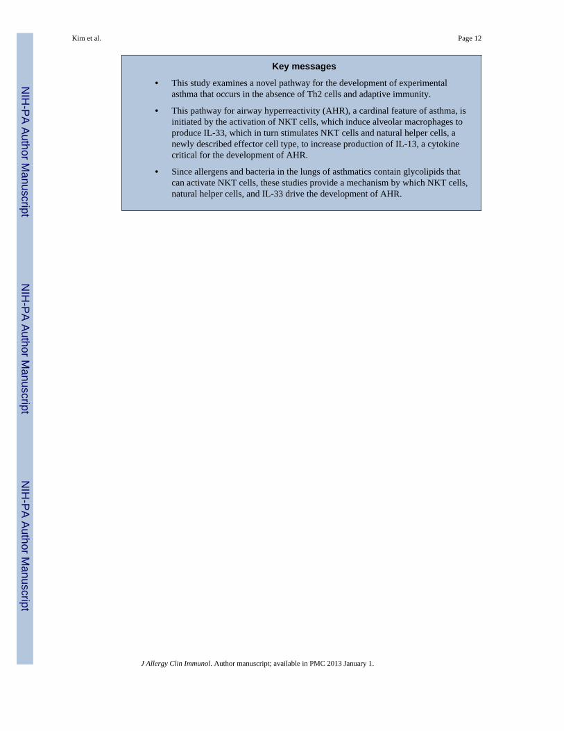
Key messages
• This study examines a novel pathway for the development of experimentalasthma that occurs in the absence of Th2 cells and adaptive immunity.
• This pathway for airway hyperreactivity (AHR), a cardinal feature of asthma, isinitiated by the activation of NKT cells, which induce alveolar macrophages toproduce IL-33, which in turn stimulates NKT cells and natural helper cells, anewly described effector cell type, to increase production of IL-13, a cytokinecritical for the development of AHR.
• Since allergens and bacteria in the lungs of asthmatics contain glycolipids thatcan activate NKT cells, these studies provide a mechanism by which NKT cells,natural helper cells, and IL-33 drive the development of AHR.
Kim et al. Page 12
J Allergy Clin Immunol. Author manuscript; available in PMC 2013 January 1.
NIH
-PA Author Manuscript
NIH
-PA Author Manuscript
NIH
-PA Author Manuscript
Capsule summary
In an experimental mouse model, asthma developed through a pathway involving IL-33-producing alveolar macrophages, natural helper cells and NKT cells. These results mayexplain non-allergic forms of asthma that develop independently of Th2 cells.
Kim et al. Page 13
J Allergy Clin Immunol. Author manuscript; available in PMC 2013 January 1.
NIH
-PA Author Manuscript
NIH
-PA Author Manuscript
NIH
-PA Author Manuscript

Figure 1. Blockade of IL-33 receptor, ST2, abrogates α-GalCer induced AHR(A) Anti-mouse ST2 blocking Ab or rat IgG1k isotype control Ab were given intravenouslyto the mice 24 hrs before intranasal administration of 0.5μg α-GalCer or vehicle. This datashows the mean ± SEM % of saline value and representative of three experiments. α-GalCer+ anti-ST2 mAb treated group was compared with α-GalCer + isotype control mAb treatedgroup. p<0.05 (*) and p<0.01 (**).(B) Data represent the number of cells per ml in BAL fluid. Mac, macrophage; Neu,neutrophils; Eos, eosinophils; Lymph, lymphocytes. p<0.01 (**) and p<0.001 (***).(C) 24h after intranasal α-GalCer challenge, lung tissues from each group were sectioned,and stained with hematoxylin/eosin (X40)(D) Littermate control and ST2−/− mice were challenged with α-GalCer or vehicle, andAHR was determined by invasive measurement of airway resistance (RL) as in (A). p<0.05(*)(E) Number of cells in BAL fluid was counted as in (B). Mac, macrophage; Neu,neutrophils; Eos, eosinophils; Lymph, lymphocytes. p<0.05 (*) and p<0.01 (**).(F) Lung tissues from each group were stained with hematoxylin/eosin as Fig. 1C. Data arerepresentative of at least three experiments.
Kim et al. Page 14
J Allergy Clin Immunol. Author manuscript; available in PMC 2013 January 1.
NIH
-PA Author Manuscript
NIH
-PA Author Manuscript
NIH
-PA Author Manuscript
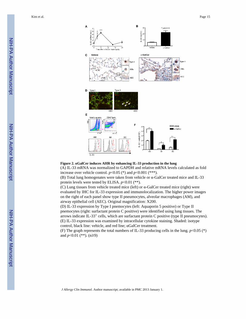
Figure 2. αGalCer induces AHR by enhancing IL-33 production in the lung(A) IL-33 mRNA was normalized to GAPDH and relative mRNA levels calculated as foldincrease over vehicle control. p<0.05 (*) and p<0.001 (***).(B) Total lung homogenates were taken from vehicle or α-GalCer treated mice and IL-33protein levels were tested by ELISA. p<0.01 (**).(C) Lung tissues from vehicle treated mice (left) or α-GalCer treated mice (right) wereevaluated by IHC for IL-33 expression and immunolocalization. The higher power imageson the right of each panel show type II pneumocytes, alveolar macrophages (AM), andairway epithelial cell (AEC). Original magnification: X200.(D) IL-33 expression by Type I pnemocytes (left: Aquaporin 5 positive) or Type IIpnemocytes (right: surfactant protein C positive) were identified using lung tissues. Thearrows indicate IL-33+ cells, which are surfactant protein C positive (type II pneumocytes).(E) IL-33 expression was examined by intracellular cytokine staining. Shaded: isotypecontrol, black line: vehicle, and red line; αGalCer treatment.(F) The graph represents the total numbers of IL-33 producing cells in the lung. p<0.05 (*)and p<0.01 (**). (n≥9)
Kim et al. Page 15
J Allergy Clin Immunol. Author manuscript; available in PMC 2013 January 1.
NIH
-PA Author Manuscript
NIH
-PA Author Manuscript
NIH
-PA Author Manuscript
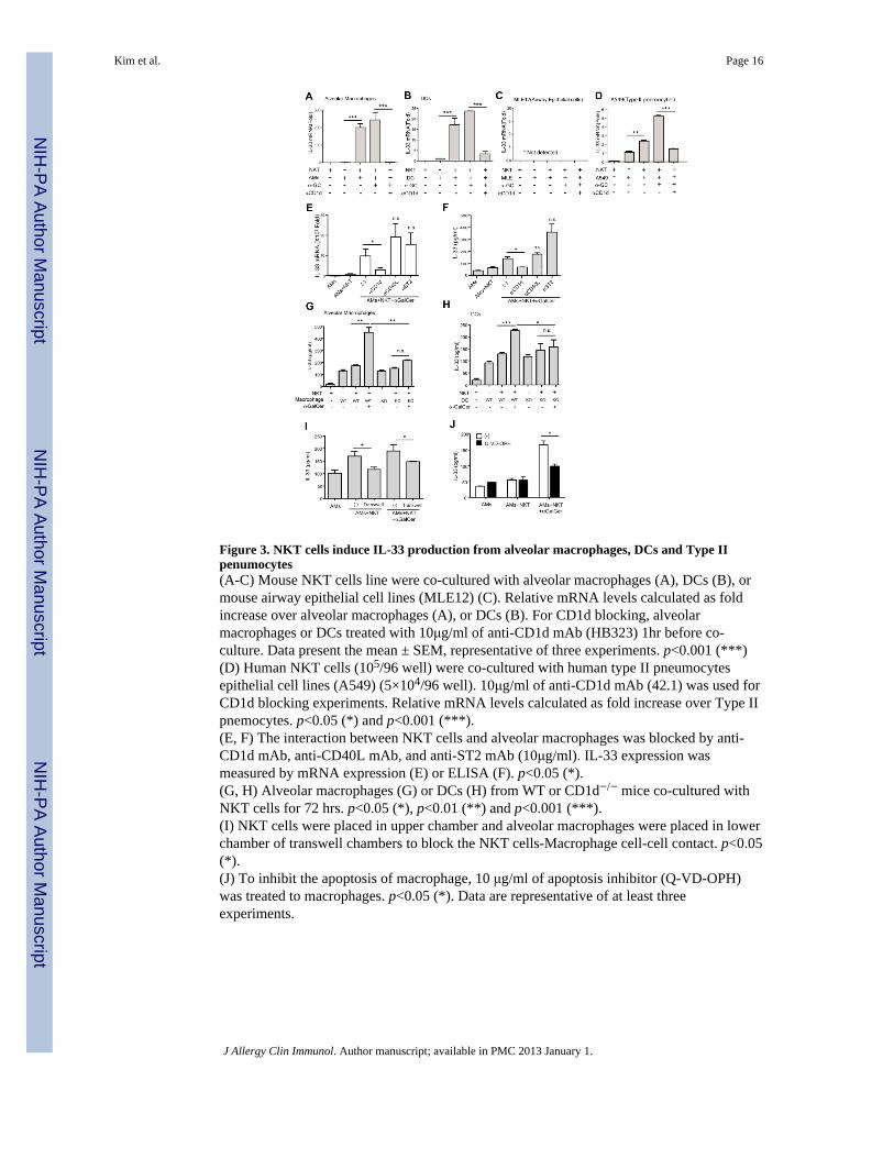
Figure 3. NKT cells induce IL-33 production from alveolar macrophages, DCs and Type IIpenumocytes(A-C) Mouse NKT cells line were co-cultured with alveolar macrophages (A), DCs (B), ormouse airway epithelial cell lines (MLE12) (C). Relative mRNA levels calculated as foldincrease over alveolar macrophages (A), or DCs (B). For CD1d blocking, alveolarmacrophages or DCs treated with 10μg/ml of anti-CD1d mAb (HB323) 1hr before co-culture. Data present the mean ± SEM, representative of three experiments. p<0.001 (***)(D) Human NKT cells (105/96 well) were co-cultured with human type II pneumocytesepithelial cell lines (A549) (5×10⁴/96 well). 10μg/ml of anti-CD1d mAb (42.1) was used forCD1d blocking experiments. Relative mRNA levels calculated as fold increase over Type IIpnemocytes. p<0.05 (*) and p<0.001 (***).(E, F) The interaction between NKT cells and alveolar macrophages was blocked by anti-CD1d mAb, anti-CD40L mAb, and anti-ST2 mAb (10μg/ml). IL-33 expression wasmeasured by mRNA expression (E) or ELISA (F). p<0.05 (*).(G, H) Alveolar macrophages (G) or DCs (H) from WT or CD1d−/− mice co-cultured withNKT cells for 72 hrs. p<0.05 (*), p<0.01 (**) and p<0.001 (***).(I) NKT cells were placed in upper chamber and alveolar macrophages were placed in lowerchamber of transwell chambers to block the NKT cells-Macrophage cell-cell contact. p<0.05(*).(J) To inhibit the apoptosis of macrophage, 10 μg/ml of apoptosis inhibitor (Q-VD-OPH)was treated to macrophages. p<0.05 (*). Data are representative of at least threeexperiments.
Kim et al. Page 16
J Allergy Clin Immunol. Author manuscript; available in PMC 2013 January 1.
NIH
-PA Author Manuscript
NIH
-PA Author Manuscript
NIH
-PA Author Manuscript
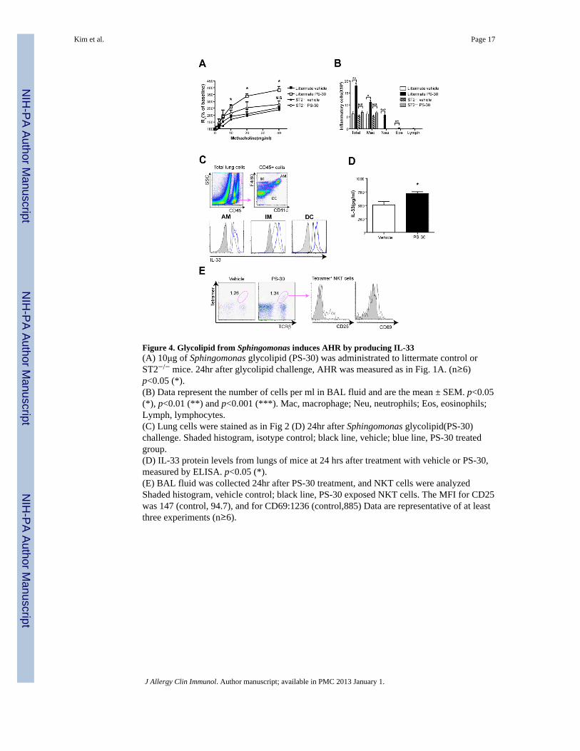
Figure 4. Glycolipid from Sphingomonas induces AHR by producing IL-33(A) 10μg of Sphingomonas glycolipid (PS-30) was administrated to littermate control orST2−/− mice. 24hr after glycolipid challenge, AHR was measured as in Fig. 1A. (n≥6)p<0.05 (*).(B) Data represent the number of cells per ml in BAL fluid and are the mean ± SEM. p<0.05(*), p<0.01 (**) and p<0.001 (***). Mac, macrophage; Neu, neutrophils; Eos, eosinophils;Lymph, lymphocytes.(C) Lung cells were stained as in Fig 2 (D) 24hr after Sphingomonas glycolipid(PS-30)challenge. Shaded histogram, isotype control; black line, vehicle; blue line, PS-30 treatedgroup.(D) IL-33 protein levels from lungs of mice at 24 hrs after treatment with vehicle or PS-30,measured by ELISA. p<0.05 (*).(E) BAL fluid was collected 24hr after PS-30 treatment, and NKT cells were analyzedShaded histogram, vehicle control; black line, PS-30 exposed NKT cells. The MFI for CD25was 147 (control, 94.7), and for CD69:1236 (control,885) Data are representative of at leastthree experiments (n≥6).
Kim et al. Page 17
J Allergy Clin Immunol. Author manuscript; available in PMC 2013 January 1.
NIH
-PA Author Manuscript
NIH
-PA Author Manuscript
NIH
-PA Author Manuscript
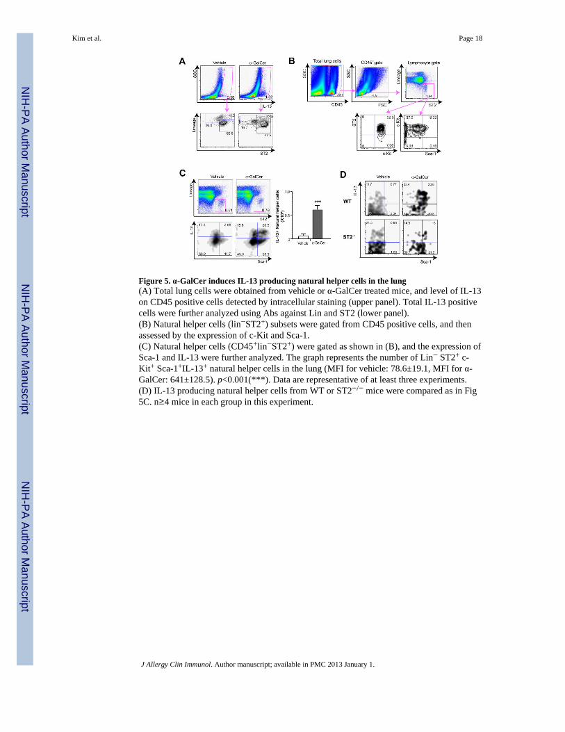
Figure 5. α-GalCer induces IL-13 producing natural helper cells in the lung(A) Total lung cells were obtained from vehicle or α-GalCer treated mice, and level of IL-13on CD45 positive cells detected by intracellular staining (upper panel). Total IL-13 positivecells were further analyzed using Abs against Lin and ST2 (lower panel).(B) Natural helper cells (lin−ST2+) subsets were gated from CD45 positive cells, and thenassessed by the expression of c-Kit and Sca-1.(C) Natural helper cells (CD45+lin−ST2+) were gated as shown in (B), and the expression ofSca-1 and IL-13 were further analyzed. The graph represents the number of Lin− ST2+ c-Kit+ Sca-1+IL-13+ natural helper cells in the lung (MFI for vehicle: 78.6±19.1, MFI for α-GalCer: 641±128.5). p<0.001(***). Data are representative of at least three experiments.(D) IL-13 producing natural helper cells from WT or ST2−/− mice were compared as in Fig5C. n≥4 mice in each group in this experiment.
Kim et al. Page 18
J Allergy Clin Immunol. Author manuscript; available in PMC 2013 January 1.
NIH
-PA Author Manuscript
NIH
-PA Author Manuscript
NIH
-PA Author Manuscript
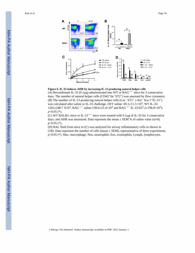
Figure 6. IL-33 induces AHR by increasing IL-13 producing natural helper cells(A) Recombinant IL-33 (0.1μg) administrated into WT or RAG−/− mice for 3 consecutivedays. The number of natural helper cells (CD45+lin−ST2+) was assessed by flow cytometry.(B) The number of IL-13 producing natural helper cells (Lin− ST2+ c-Kit+ Sca-1+IL-13+)was calculated after saline or IL-33 challenge. (WT saline: 69.1±11.5×10³, WT IL-33:1265±248.7 X10³, RAG−/− saline:138.6±25.4×10³ and RAG−/− IL-33:927.2±196.8×10³).p<0.05 (*).(C) WT BALB/c mice or IL-13−/− mice were treated with 0.1μg of IL-33 for 3 consecutivedays, and AHR was measured. Data represent the mean ± SEM % of saline value (n≥4).p<0.05 (*).(D) BAL fluid from mice in (C) was analyzed for airway inflammatory cells as shown in(1B). Data represent the number of cells (mean ± SEM), representative of three experiments.p<0.05 (*). Mac, macrophage; Neu, neutrophils; Eos, eosinophils; Lymph, lymphocytes.
Kim et al. Page 19
J Allergy Clin Immunol. Author manuscript; available in PMC 2013 January 1.
NIH
-PA Author Manuscript
NIH
-PA Author Manuscript
NIH
-PA Author Manuscript
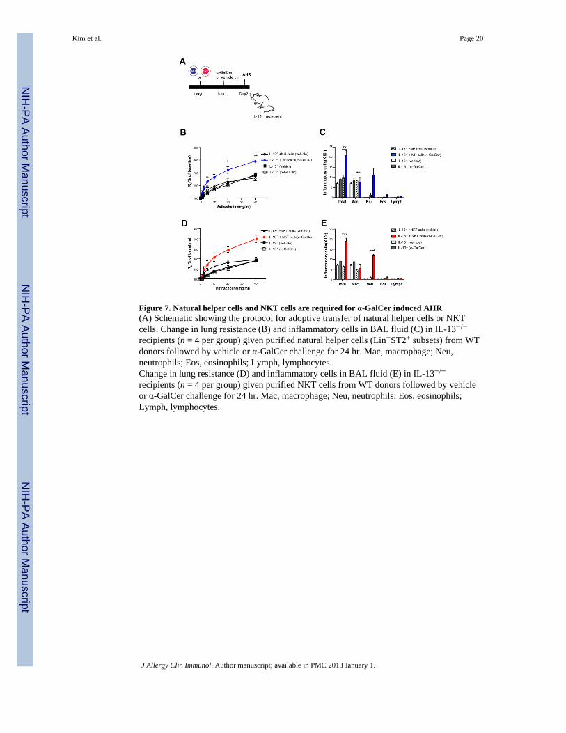
Figure 7. Natural helper cells and NKT cells are required for α-GalCer induced AHR(A) Schematic showing the protocol for adoptive transfer of natural helper cells or NKTcells. Change in lung resistance (B) and inflammatory cells in BAL fluid (C) in IL-13−/−
recipients (n = 4 per group) given purified natural helper cells (Lin−ST2+ subsets) from WTdonors followed by vehicle or α-GalCer challenge for 24 hr. Mac, macrophage; Neu,neutrophils; Eos, eosinophils; Lymph, lymphocytes.Change in lung resistance (D) and inflammatory cells in BAL fluid (E) in IL-13−/−
recipients (n = 4 per group) given purified NKT cells from WT donors followed by vehicleor α-GalCer challenge for 24 hr. Mac, macrophage; Neu, neutrophils; Eos, eosinophils;Lymph, lymphocytes.
Kim et al. Page 20
J Allergy Clin Immunol. Author manuscript; available in PMC 2013 January 1.
NIH
-PA Author Manuscript
NIH
-PA Author Manuscript
NIH
-PA Author Manuscript
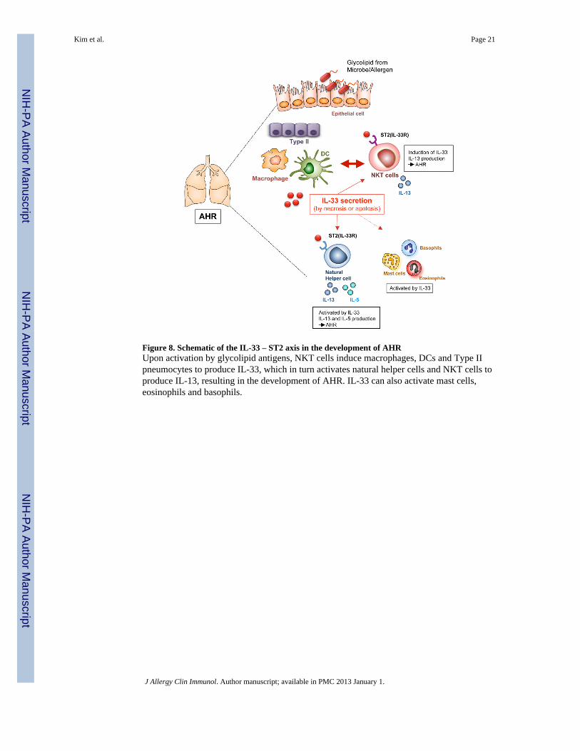
Figure 8. Schematic of the IL-33 – ST2 axis in the development of AHRUpon activation by glycolipid antigens, NKT cells induce macrophages, DCs and Type IIpneumocytes to produce IL-33, which in turn activates natural helper cells and NKT cells toproduce IL-13, resulting in the development of AHR. IL-33 can also activate mast cells,eosinophils and basophils.
Kim et al. Page 21
J Allergy Clin Immunol. Author manuscript; available in PMC 2013 January 1.
NIH
-PA Author Manuscript
NIH
-PA Author Manuscript
NIH
-PA Author Manuscript