Embed Size (px)
Citation preview
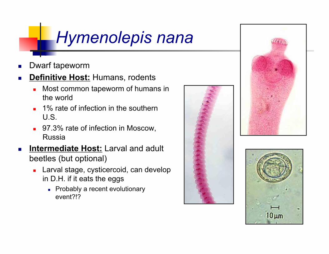
Hymenolepis nana Dwarf tapeworm Definitive Host: Humans, rodents
Most common tapeworm of humans in the world
1% rate of infection in the southern U.S.
97.3% rate of infection in Moscow, Russia
Intermediate Host: Larval and adult beetles (but optional) Larval stage, cysticercoid, can develop
in D.H. if it eats the eggs Probably a recent evolutionary
event?!?
Hymenolepis nana
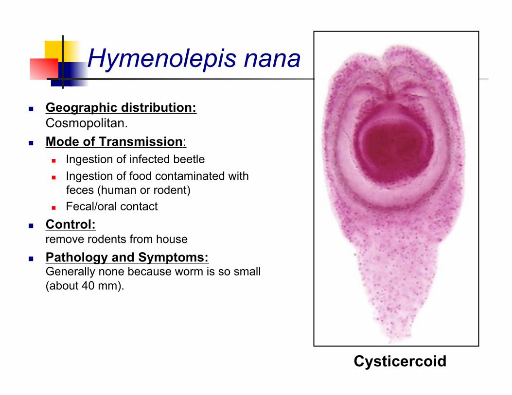
Geographic distribution: Cosmopolitan.
Mode of Transmission: Ingestion of infected beetle Ingestion of food contaminated with
feces (human or rodent) Fecal/oral contact
Control: remove rodents from house
Pathology and Symptoms: Generally none because worm is so small (about 40 mm).
Cysticercoid
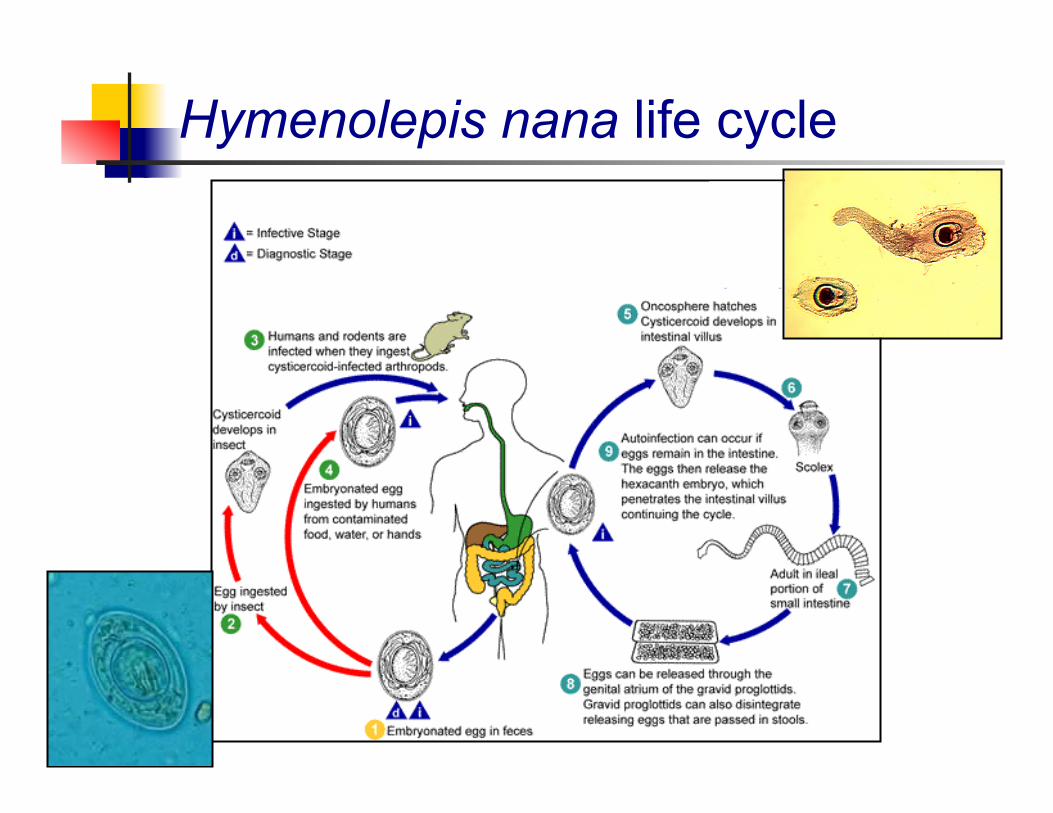
Hymenolepis nana life cycle
Hymenolepis diminuta
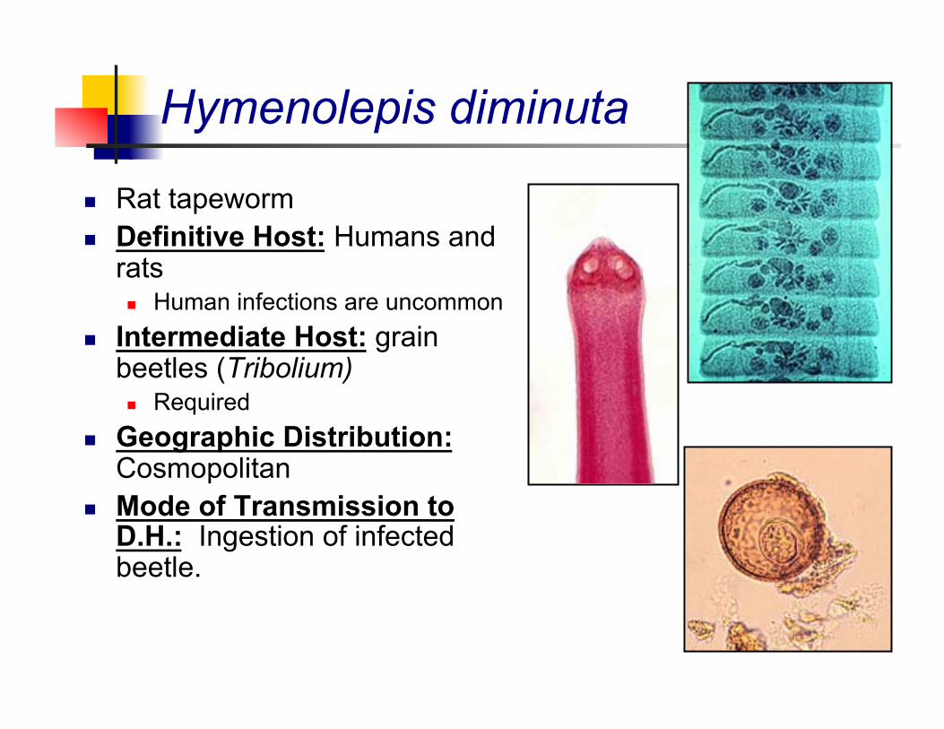
Rat tapeworm Definitive Host: Humans and
rats Human infections are uncommon
Intermediate Host: grain beetles (Tribolium) Required
Geographic Distribution: Cosmopolitan
Mode of Transmission to D.H.: Ingestion of infected beetle.
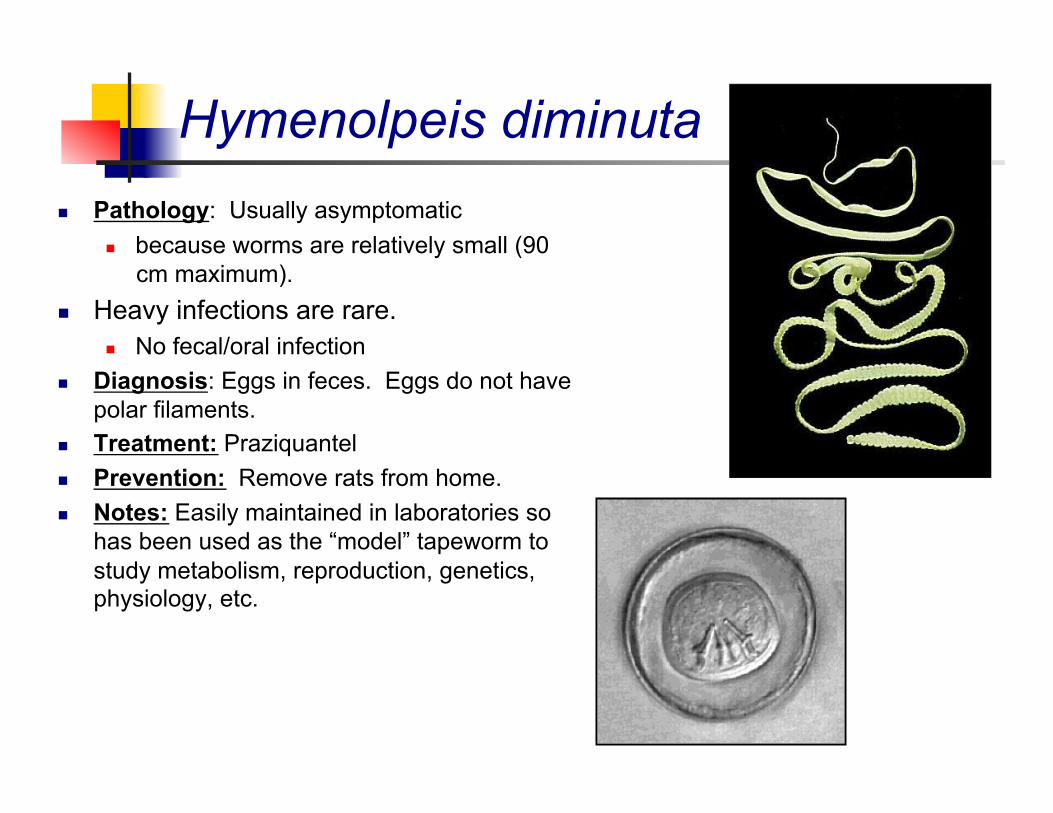
Hymenolpeis diminuta Pathology: Usually asymptomatic
because worms are relatively small (90 cm maximum).
Heavy infections are rare. No fecal/oral infection
Diagnosis: Eggs in feces. Eggs do not have polar filaments.
Treatment: Praziquantel Prevention: Remove rats from home. Notes: Easily maintained in laboratories so
has been used as the “model” tapeworm to study metabolism, reproduction, genetics, physiology, etc.
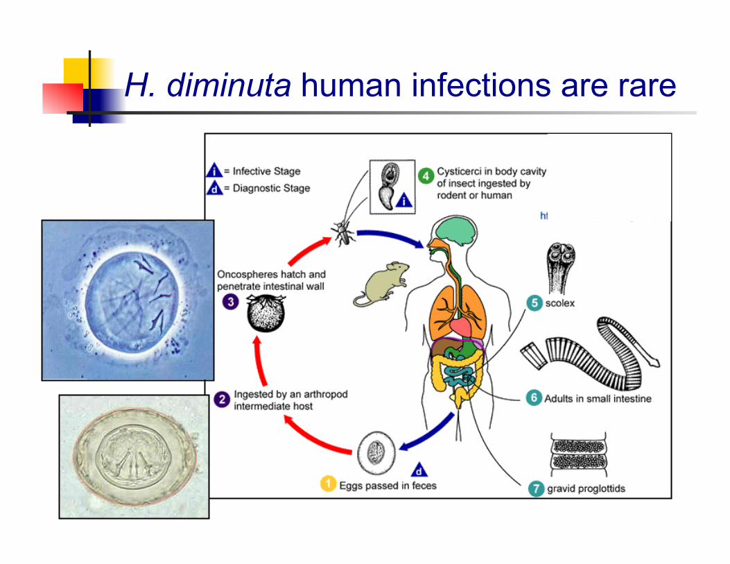
H. diminuta human infections are rare
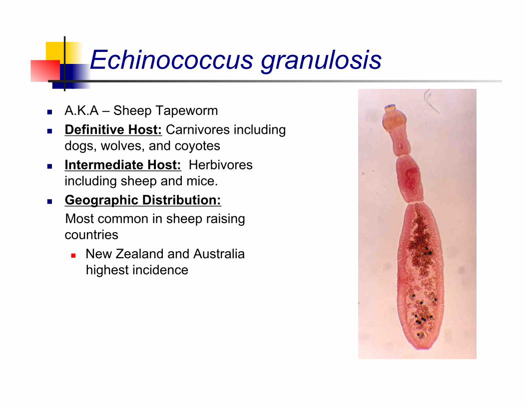
Echinococcus granulosis
A.K.A – Sheep Tapeworm Definitive Host: Carnivores including
dogs, wolves, and coyotes Intermediate Host: Herbivores
including sheep and mice. Geographic Distribution: Most common in sheep raising
countries New Zealand and Australia
highest incidence
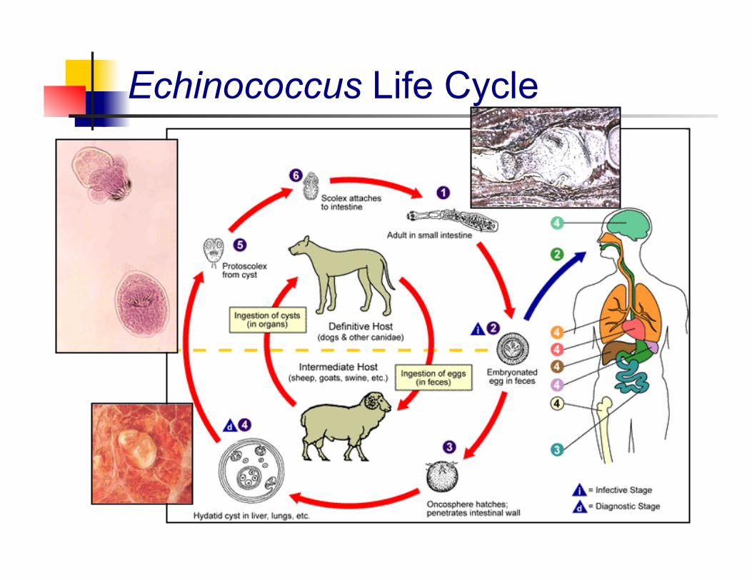
Echinococcus Life Cycle
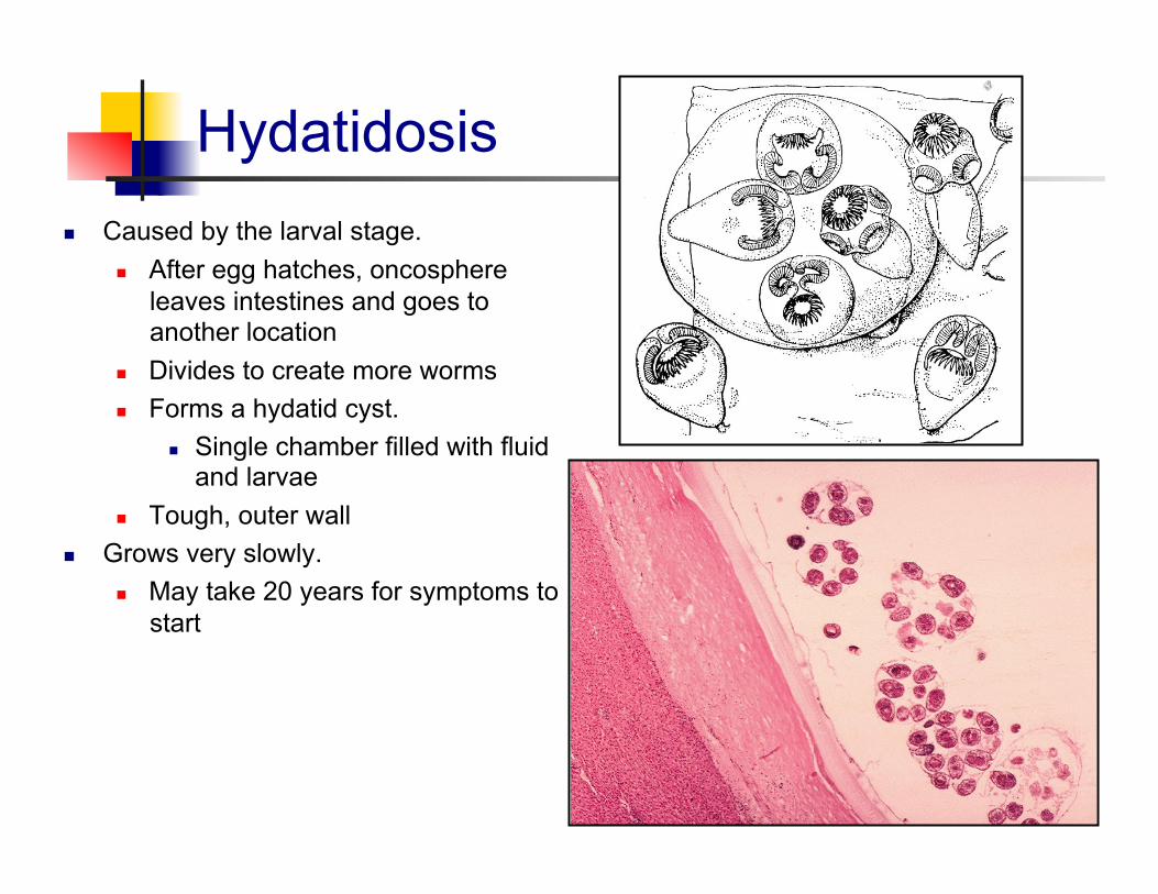
Hydatidosis Caused by the larval stage.
After egg hatches, oncosphere leaves intestines and goes to another location
Divides to create more worms Forms a hydatid cyst.
Single chamber filled with fluid and larvae
Tough, outer wall Grows very slowly.
May take 20 years for symptoms to start
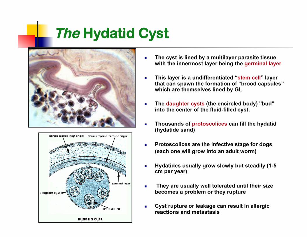
The cyst is lined by a multilayer parasite tissue with the innermost layer being the germinal layer
This layer is a undifferentiated “stem cell” layer that can spawn the formation of “brood capsules” which are themselves lined by GL
The daughter cysts (the encircled body) "bud" into the center of the fluid-filled cyst.
Thousands of protoscolices can fill the hydatid (hydatide sand)
Protoscolices are the infective stage for dogs (each one will grow into an adult worm)
Hydatides usually grow slowly but steadily (1-5 cm per year)
They are usually well tolerated until their size becomes a problem or they rupture
Cyst rupture or leakage can result in allergic reactions and metastasis
The Hydatid Cyst
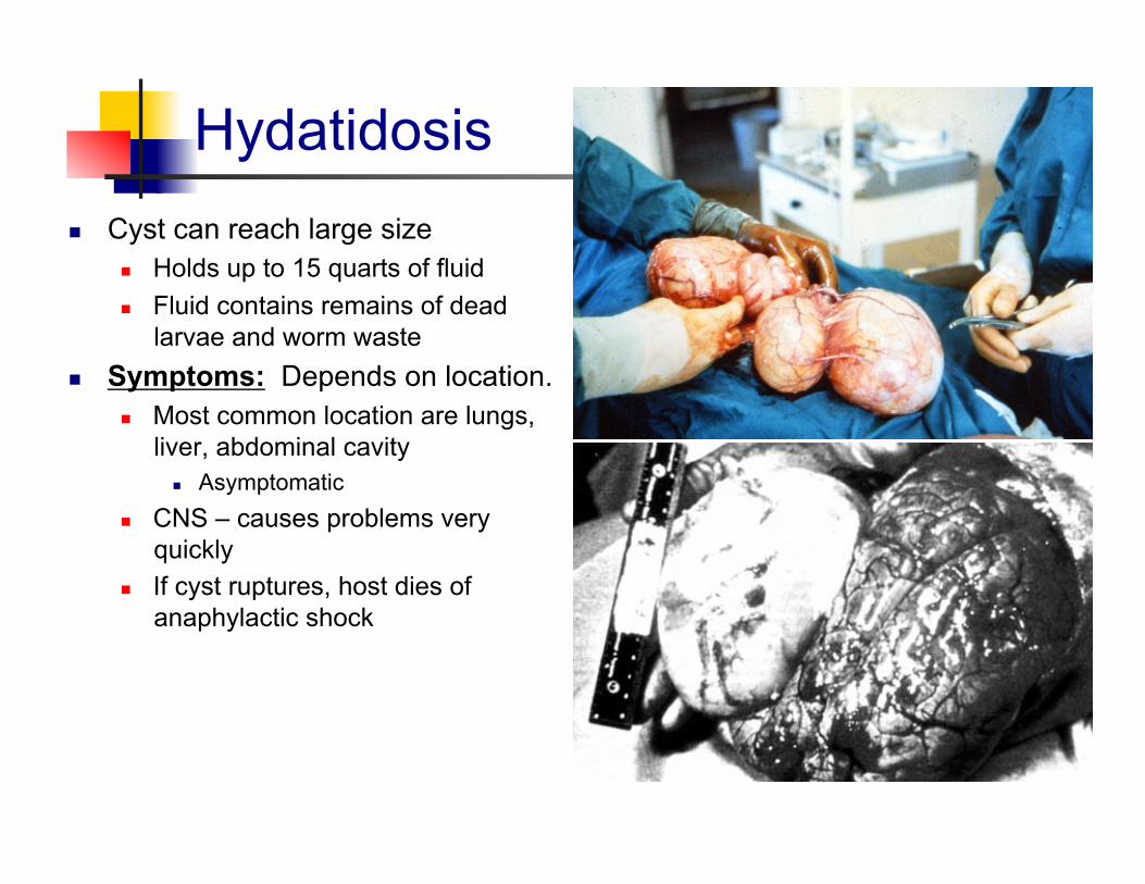
Hydatidosis Cyst can reach large size
Holds up to 15 quarts of fluid Fluid contains remains of dead
larvae and worm waste Symptoms: Depends on location.
Most common location are lungs, liver, abdominal cavity
Asymptomatic CNS – causes problems very
quickly If cyst ruptures, host dies of
anaphylactic shock
Hydatidosis
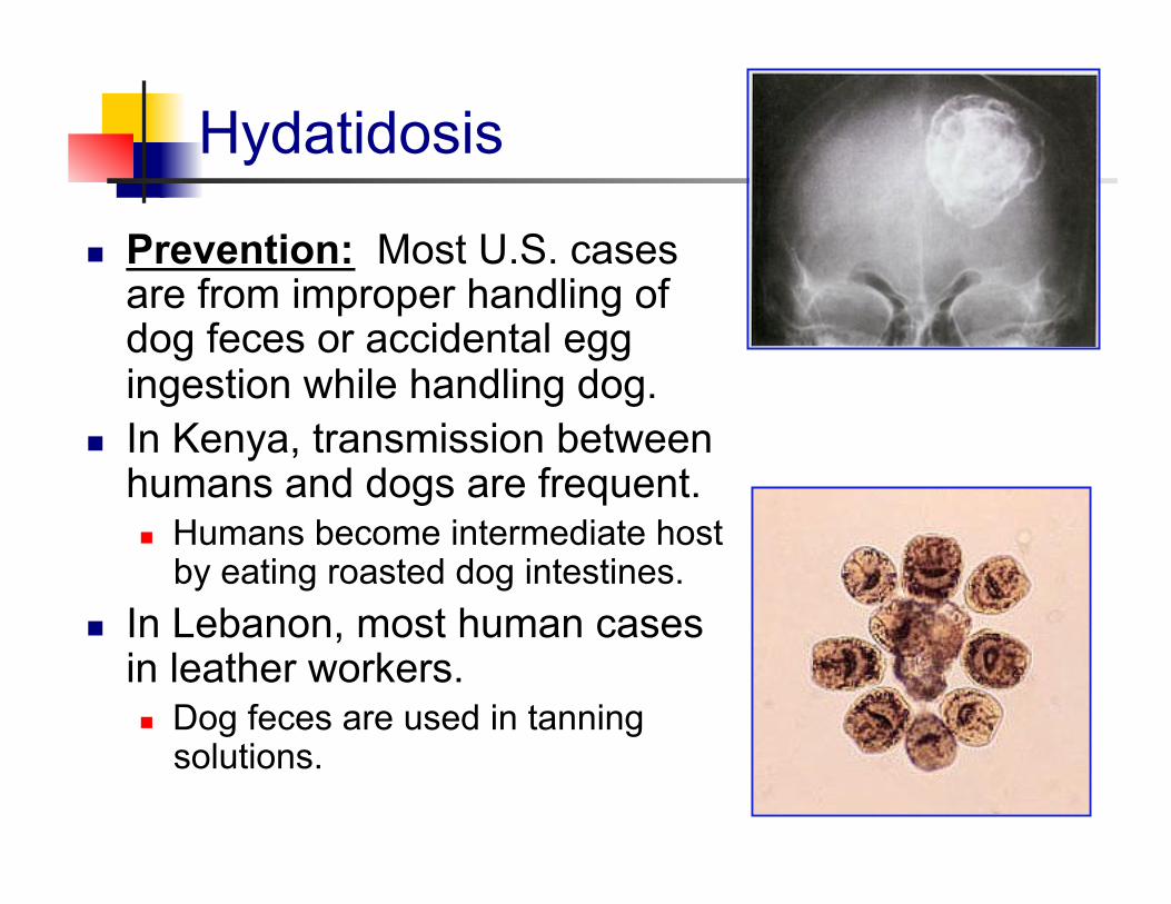
Prevention: Most U.S. cases are from improper handling of dog feces or accidental egg ingestion while handling dog.
In Kenya, transmission between humans and dogs are frequent. Humans become intermediate host
by eating roasted dog intestines. In Lebanon, most human cases
in leather workers. Dog feces are used in tanning
solutions.
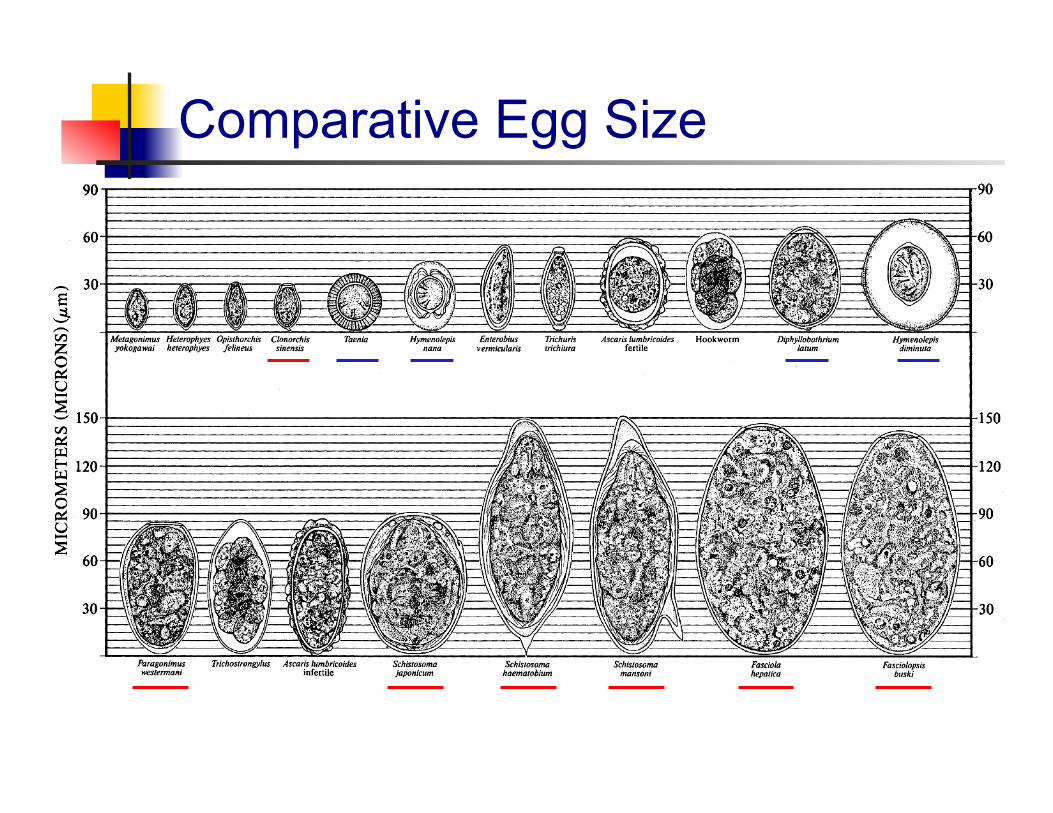
Comparative Egg Size
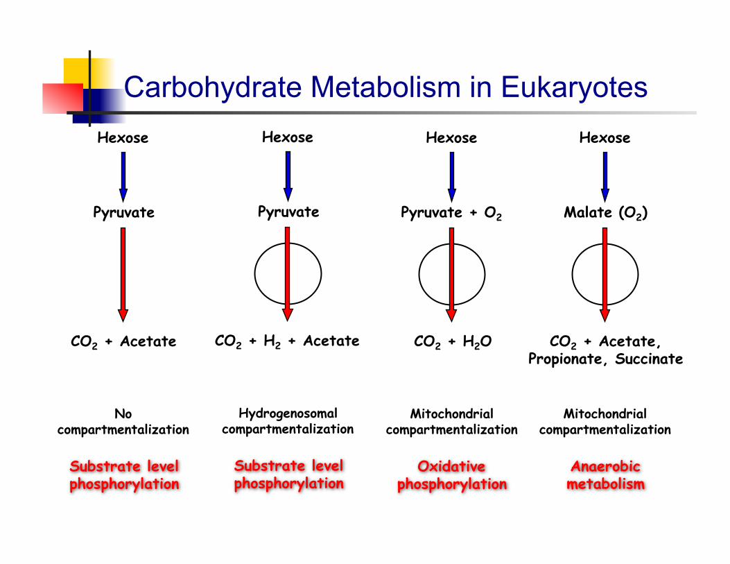
Carbohydrate Metabolism in Eukaryotes Hexose Hexose
Pyruvate
CO2 + H2 + Acetate
Hydrogenosomal compartmentalization
Pyruvate
CO2 + Acetate
No compartmentalization
Substrate level phosphorylation
Substrate level phosphorylation
Mitochondrial compartmentalization
Anaerobic metabolism
Hexose
Malate (O2)
CO2 + Acetate, Propionate, Succinate
Hexose
Pyruvate + O2
CO2 + H2O
Mitochondrial compartmentalization
Oxidative phosphorylation
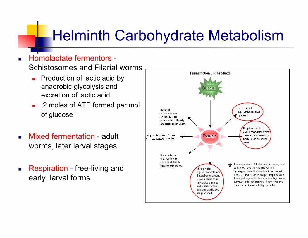
Helminth Carbohydrate Metabolism Homolactate fermentors -
Schistosomes and Filarial worms Production of lactic acid by
anaerobic glycolysis and excretion of lactic acid
2 moles of ATP formed per mol of glucose
Mixed fermentation - adult worms, later larval stages
Respiration - free-living and early larval forms
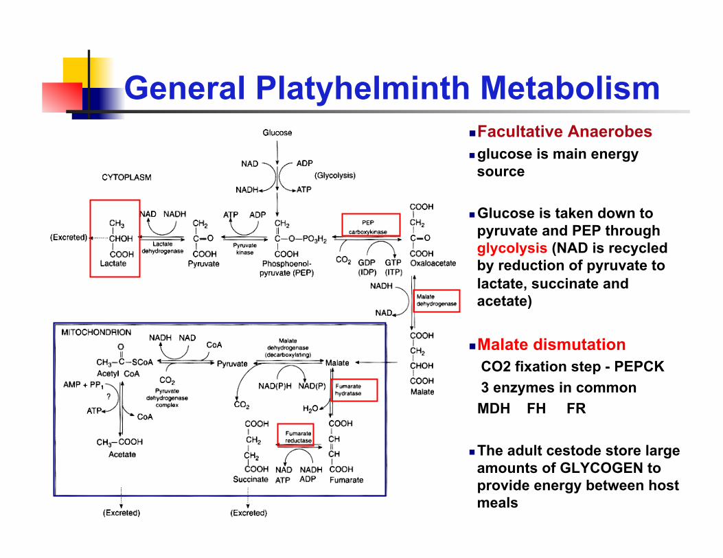
General Platyhelminth Metabolism Facultative Anaerobes glucose is main energy
source
Glucose is taken down to pyruvate and PEP through glycolysis (NAD is recycled by reduction of pyruvate to lactate, succinate and acetate)
Malate dismutation CO2 fixation step - PEPCK 3 enzymes in common MDH FH FR
The adult cestode store large amounts of GLYCOGEN to provide energy between host meals
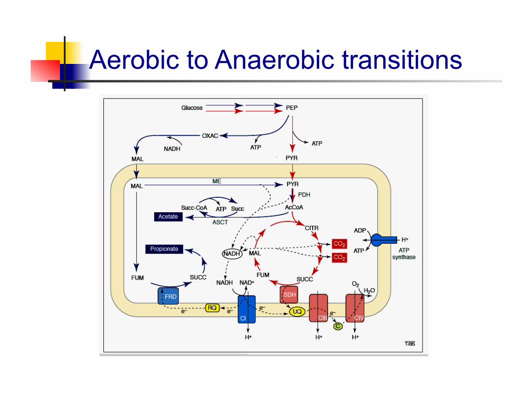
Aerobic to Anaerobic transitions
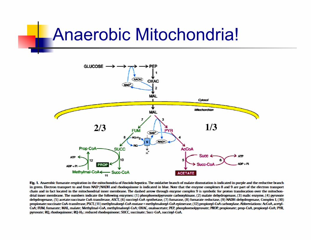
Anaerobic Mitochondria!
1/3 2/3

Platyhelminth Treatments Quinoline derivatives
Praziquantel Oxamniquine
Benzimidazole derivatives Mebendazole Albendazole Thiabendazole
Other drugs Niclosamide Metrifonate
Why Praziquantel?
Early 20th century relied heavily on antimonials
Effective against a number of species
Most widely used drug Safe, fairly cheap to make
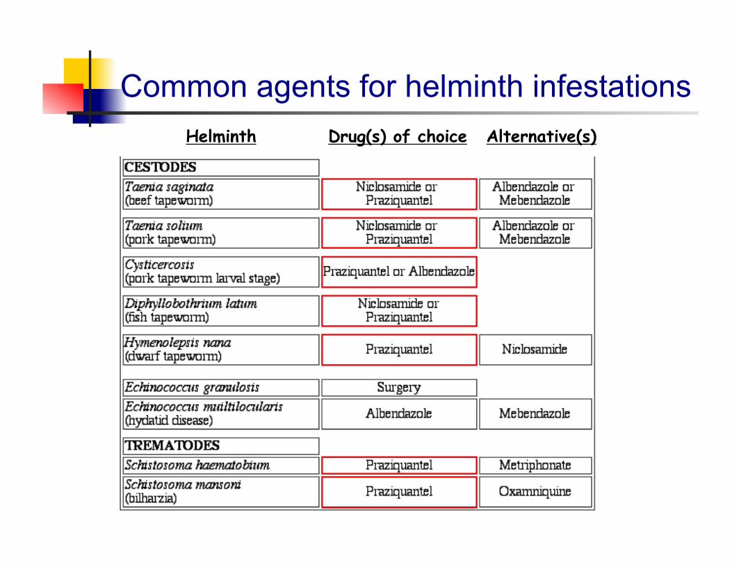
Common agents for helminth infestations Drug(s) of choice Alternative(s) Helminth
A little history on praziquantel Large number of pyrazino isoquinoline compounds were
synthesized by Merck as potential tranquilizers (~late 1960s)
Partnership between Merck and Bayer led to the first screening of the compounds as possible anthelmintics (mid-1970s)
EMBAY 8440 - now known as praziquantel - was an effective antitrematode and anticestode compound.
First human volunteers tested in 1978, by 1980 praziquantel had become the drug of choice to treat schistosomiasis as well as a number of other worm infections
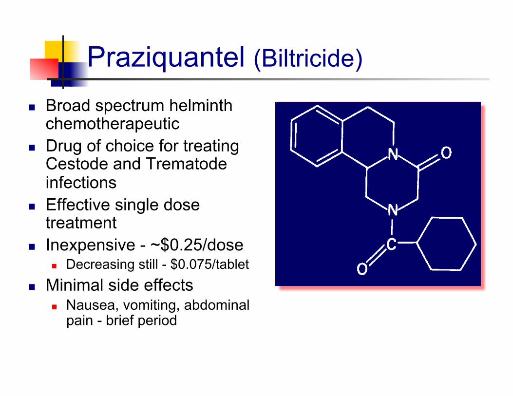
Praziquantel (Biltricide) Broad spectrum helminth
chemotherapeutic Drug of choice for treating
Cestode and Trematode infections
Effective single dose treatment
Inexpensive - ~$0.25/dose Decreasing still - $0.075/tablet
Minimal side effects Nausea, vomiting, abdominal
pain - brief period
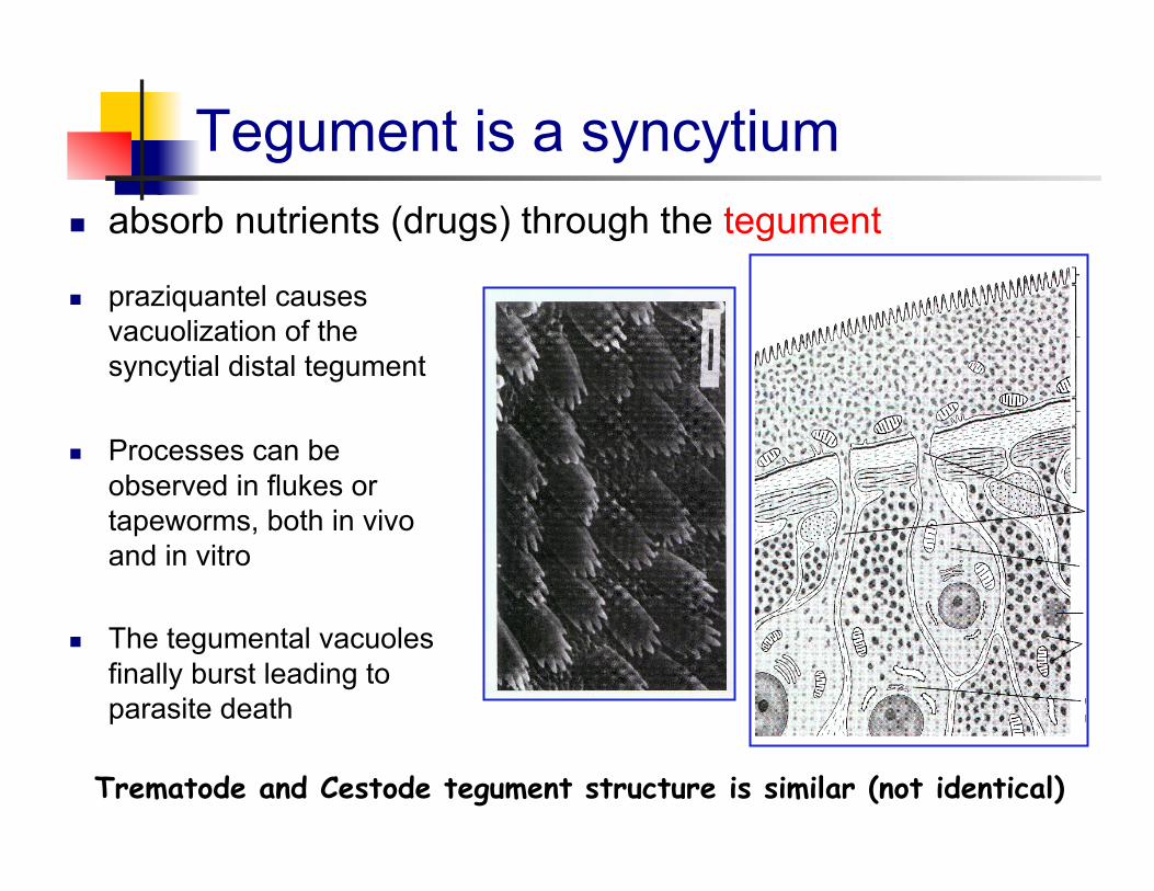
Tegument is a syncytium absorb nutrients (drugs) through the tegument
praziquantel causes vacuolization of the syncytial distal tegument
Processes can be observed in flukes or tapeworms, both in vivo and in vitro
The tegumental vacuoles finally burst leading to parasite death
Trematode and Cestode tegument structure is similar (not identical)
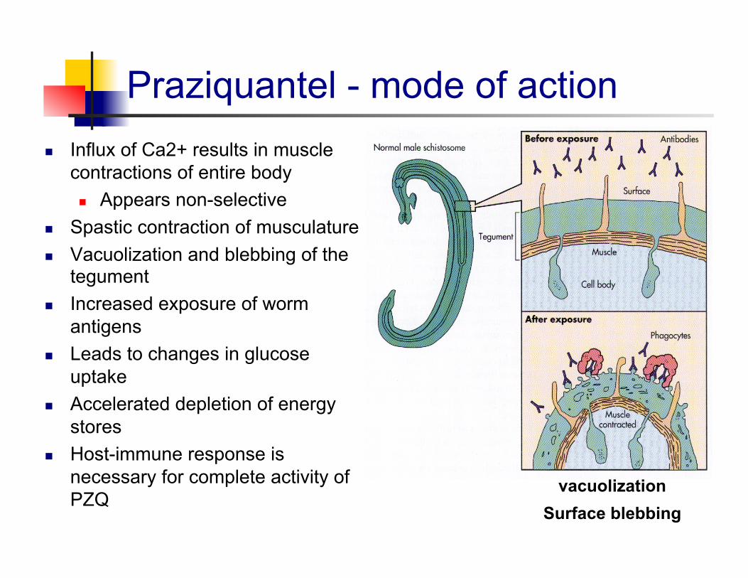
Praziquantel - mode of action Influx of Ca2+ results in muscle
contractions of entire body Appears non-selective
Spastic contraction of musculature Vacuolization and blebbing of the
tegument Increased exposure of worm
antigens Leads to changes in glucose
uptake Accelerated depletion of energy
stores Host-immune response is
necessary for complete activity of PZQ
vacuolization Surface blebbing
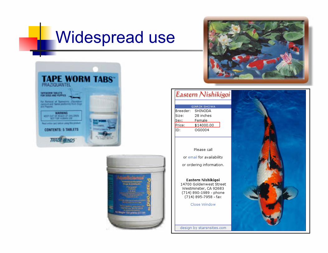
Widespread use
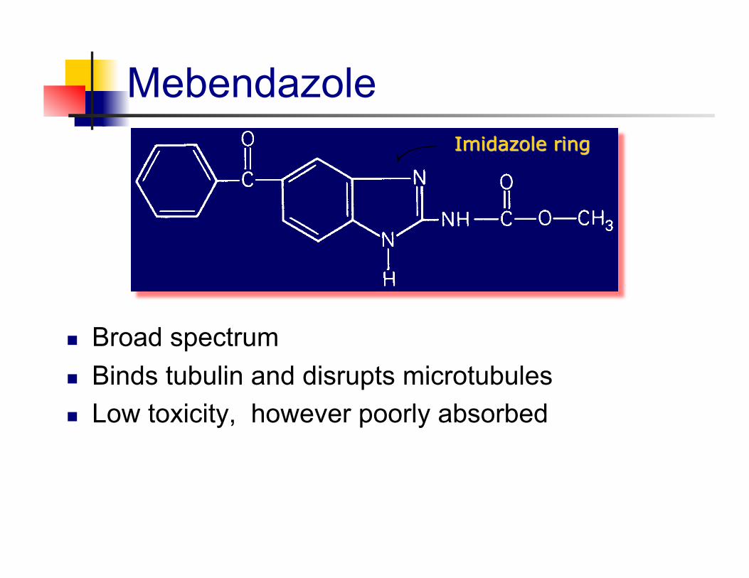
Mebendazole
Broad spectrum Binds tubulin and disrupts microtubules Low toxicity, however poorly absorbed
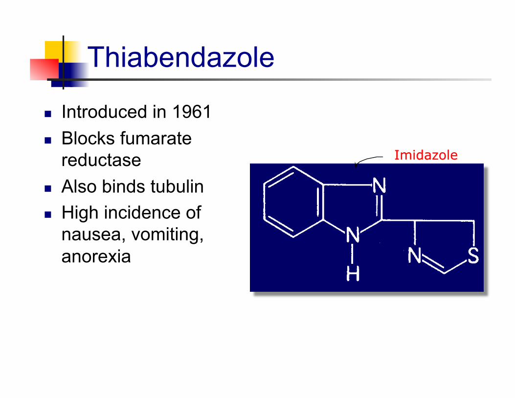
Thiabendazole
Introduced in 1961 Blocks fumarate
reductase Also binds tubulin High incidence of
nausea, vomiting, anorexia
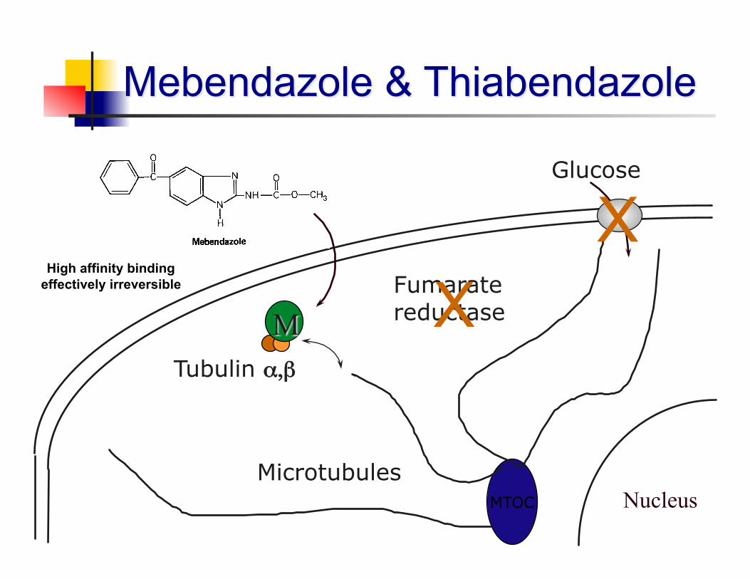
Nucleus MTOC
Microtubules
Tubulin α,β
Glucose
X Fumarate reductase X
High affinity binding effectively irreversible
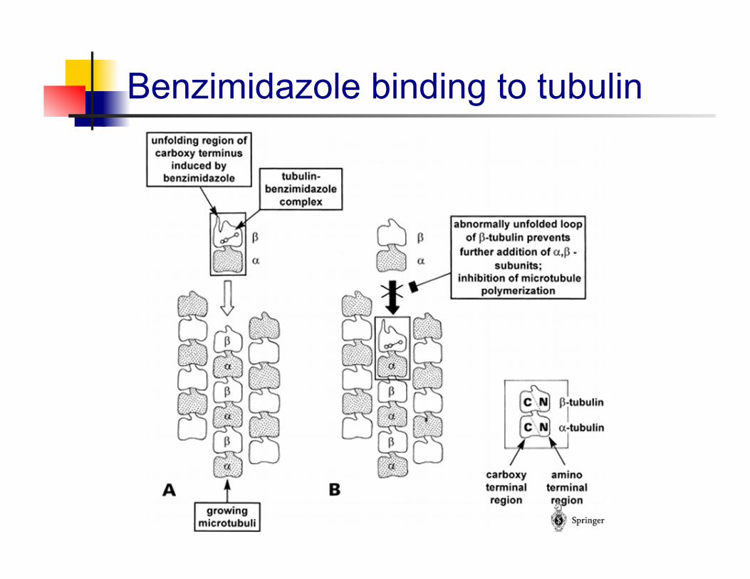
Benzimidazole binding to tubulin
Applied Parasitology
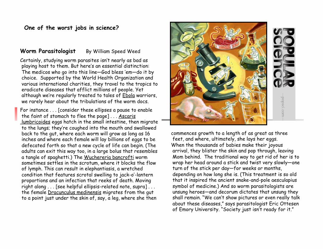
commences growth to a length of as great as three feet, and where, ultimately, she lays her eggs. When the thousands of babies make their joyous arrival, they blister the skin and pop through, leaving Mom behind. The traditional way to get rid of her is to wrap her head around a stick and twist very slowly—one turn of the stick per day—for weeks or months, depending on how long she is. (This treatment is so old that it inspired the ancient snake-and-pole aesculapius symbol of medicine.) And so worm parasitologists are unsung heroes—and decorum dictates that unsung they shall remain. “We can’t show pictures or even really talk about these diseases,” says parasitologist Eric Ottesen of Emory University. “Society just isn’t ready for it.”
Worm Parasitologist By William Speed Weed
Certainly, studying worm parasites isn’t nearly as bad as playing host to them. But here’s an essential distinction: The medicos who go into this line—God bless ’em—do it by choice. Supported by the World Health Organization and various international charities, they travel to the tropics to eradicate diseases that afflict millions of people. Yet although we’re regularly treated to tales of Ebola warriors, we rarely hear about the tribulations of the worm docs. For instance . . . [consider these ellipses a pause to enable the faint of stomach to flee the page] . . . Ascaris lumbricoides eggs hatch in the small intestine, then migrate to the lungs; they’re coughed into the mouth and swallowed back to the gut, where each worm will grow as long as 16 inches and where each female will lay billions of eggs to be defecated forth so that a new cycle of life can begin. (The adults can exit this way too, in a large bolus that resembles a tangle of spaghetti.) The Wuchereria bancrofti worm sometimes settles in the scrotum, where it blocks the flow of lymph. This can result in elephantiasis, a wretched condition that features scrotal swelling to jack-o’-lantern proportions and an infection that reeks of death. Moving right along . . . [see helpful ellipsis-related note, supra] . . . the female Dracunculus medinensis migrates from the gut to a point just under the skin of, say, a leg, where she then
One of the worst jobs in science?

![Chapter 3 · Hymenolepis diminuta, the rat tapeworm, is now one of the most widely used helminths for therapeutic purposes [, 6]. 5 However, H. diminuta is not currently approved](https://img.pdfslide.net/doc/110x75/606e8113c1aaa16ee622e0a7/chapter-3-hymenolepis-diminuta-the-rat-tapeworm-is-now-one-of-the-most-widely.jpg)


![[PPT]Slide sem título - Instituto Formação · Web viewHymenolepis nana CLASSIFICAÇA0 FILO Platyelminthes CLASSE Cestoda FAMILIA Hymenolepididae GÊNERO Hymenolepis ESPÉCIES Hymenolepis](https://img.pdfslide.net/doc/110x75/5be6982b09d3f2d8348d81a4/pptslide-sem-titulo-instituto-formacao-web-viewhymenolepis-nana-classificaca0.jpg)