Embed Size (px)
Citation preview

1
1
Hypersensitivity
• Chapter 25
• Types of Hypersensitivity
• Immunological mechanisms of Hypersensitivity
• Factors that predispose to Hypersensitivity
• Clinical manifestations of Hypersensitivity
2
Hypersensitivity
3
Cells in Hypersensitivity Reactions
• A - in skin andmucosal tissues
• B - Granulocytes:circulate in the bloodand extravasate intotissues
• C - Mononuclearcells: monocytes(macrophage) andlymphocytes
4
3 Types of Hypersensitivity
2
5
Type I Immediate
Hypersensitivity
6
Type I Immediate Hypersensitivity
• Example Mosquito bites: the swelling and redness
• 1st bite generates immune response to mosquito salivary protein
antigens =
– IgE antibodies are produced.
• IgE abs are immobilized on surface of Mast cells by high affinity
IgE FcR
• 2nd bite: salivary Ag bind IgE.
– Signal transduction to Mast cells to release histamine and serotonin into
tissues
• Capillary endothelial cells pull apart and fluids seep into tissues
bringing in Complement and cytokines
7
Type I Immediate Hypersensitivity
• Example Mosquito bites: itching sensation
• Histamine binds to type c nerve fibers that sense pain and initiate
the itching sensation
• A “wheal”or fluid-filled itchy bump is formed.
• Further induction of capillary dilation and “flare” reaction
develops - increased redness
• More scratching results in more extensive dilation of capillaries
8
Type I
Immediate
Hypersensitivity

3
9
• After the wheal and flare response subsides,
• Second wave of immune mediators are made by the mast
cells
• The arachidonic acid derivatives (fatty acid derivatives):
– Cysteinyl leukotrienes
• Recruit cells into the injured area and form a bump or
papule that lasts hours or days
– Caused by PMNs and eosinophils during first 2 hours
– Then the mononuclear cells arrive…
– All fight any infectious agent injected by the mosquito bite…
Type I Immediate Hypersensitivity
10
Inflammatory
response
• LTs are all cysteineleukotrienes
– Zileuton = Zyflo
– Montelukast = Singulair
– Zafirlukast = Accolate
• PGs are allprostaglandins– NSAIDS = ibuprofen etc
– Rofecoxib = Vioxx
– Celecoxib = Celebrex
• TX are thromboxanes
11
Type I
Immediate
Hypersensitivity
12
IgE FcR

4
13
• Origins of
Allergic
Responses
• Dendritic cells,
macrophage,
and eosinophils
likely bring
allergens to the
lymph nodes for
B cell activation
and Switching
to IgE
14
Common Allergens
• Exposure to
allergens
varies based
on season and
location
15
IgE FcR
Activation• Cross-linking of
IgE FcR results in
signal transduction
and subsequent
mast cell
degranulation
16
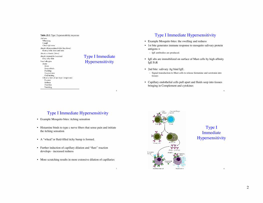
Mast Cell Activation and Degranulation
• Cross linking of IgE FcR
leads to signal transduction
events:
• cAMP production
• Activated PKC
• Elevated intracellular
calcium levels
5
17
Mast Cell Activation and Degranulation
• Active PKC
phosphorylates granule
membrane proteins
• Permeability to water and
calcium changes
• Granules swell, migrate,
fuse with the membrane
and release their granules
18
Mast Cell Activation and Degranulation
• Cross linking of IgE
FcR leads to signal
transduction events:
• Production of
Arachidonic acid
• Production of PG,
TX, and LT
• Acute inflammation
19
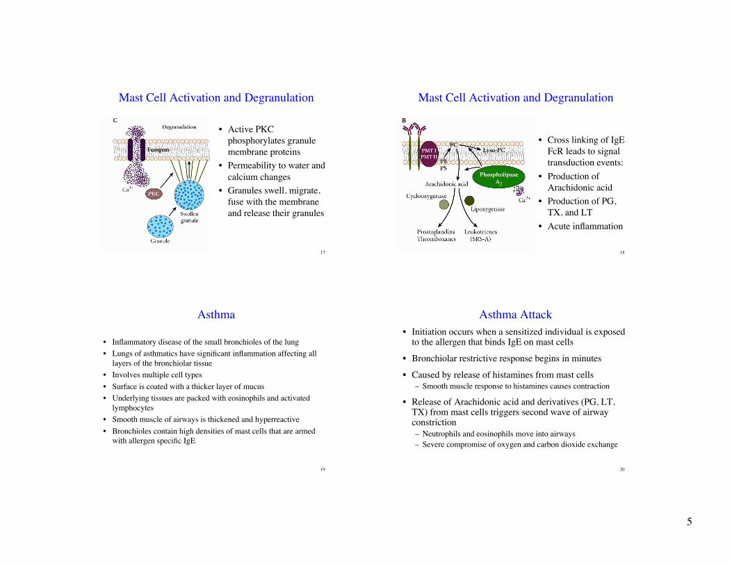
Asthma
• Inflammatory disease of the small bronchioles of the lung
• Lungs of asthmatics have significant inflammation affecting all
layers of the bronchiolar tissue
• Involves multiple cell types
• Surface is coated with a thicker layer of mucus
• Underlying tissues are packed with eosinophils and activated
lymphocytes
• Smooth muscle of airways is thickened and hyperreactive
• Bronchioles contain high densities of mast cells that are armed
with allergen specific IgE
20
Asthma Attack
• Initiation occurs when a sensitized individual is exposedto the allergen that binds IgE on mast cells
• Bronchiolar restrictive response begins in minutes
• Caused by release of histamines from mast cells
– Smooth muscle response to histamines causes contraction
• Release of Arachidonic acid and derivatives (PG, LT,TX) from mast cells triggers second wave of airwayconstriction
– Neutrophils and eosinophils move into airways
– Severe compromise of oxygen and carbon dioxide exchange

6
21
Asthma Attack
22
Industrialization and Allergies
• David Diaz-Sanchez, PhD,Marisol Penichet-Garcia, MD, and Andrew Saxon,MD. (2000) Diesel exhaust particles directly induce activated mast cells todegranulate and increase histamine levels and symptom severity. Journal ofAllergy and Clinical Immunology, 106:1140-6.
23
Type II Intermediate Hypersensitivity
• IgG Mediated Hypersensitivity
• Reactions occur when antibodies to self proteins or
carbohydrates binds to self tissues
• 4 classic types
– Maternal antibodies that cross the placenta bind to child’s
tissues and cause damage
– Antibodies against blood transfustion or organ transplant
– Antibody response to foreign substances that has adhered to
own cells
– Antibodies to own tissues
24
Type II Intermediate Hypersensitivity
• Example:
• Autoantibodies to
pancreatic beta cells
may cause insulin-
dependent diabetes
(type II diabetes)
• Cells are destroyed
through ADCC or
classical complement
cascade and MAC
activation

7
25
Type II
Hypersensitivity
• Erythroblastosis
fetalis or
• hemolytic disease of
the newborn
26
ABO Blood Group Antigens
27
• Autoantibodies
against Type IV
collagen in the
lungs and
kidneys
• Results in
complement
activation and
neutrophil
influx
• Tissue damage
Goodpasture Disease