Embed Size (px)
Citation preview

Hypertensive Disorders in Pregnancy
ByFrancis Kamwendo
@ Nkhota-kota
Adapted from Obstetrics Simplified by Diaa M. EI-Mowafi

Classification
1. PRE-EXISTING (CHRONIC) HYPERTENSION
2. PRE-ECLAMPSIA
1. Differential Diagnosis
2. Complications
3. Treatment
4. ECLAMPSIA

Pre-existing (chronic) hypertension:
– Hypertension is present before pregnancy, detected in early pregnancy (before 20 weeks in absence of vesicular mole) and postpartum.
– Examples: • essential hypertension, • secondary to chronic renal disorders e.g. pyelonephritis
and renal artery stenosis, • coarctation of the aorta, systemic lupus erythematosus
and pheochromocytoma.

PRE-EXISTING (CHRONIC) HYPERTENSION
Causes• Essential hypertension: of unknown aetiology. • Secondary to chronic renal disorder: e.g.
– Glomerulonephritis. – Hydronephrosis. – Pyelonephritis. – Renal artery stenosis.
• Secondary to cardiovascular disease: e.g. – Coarctation of the aorta. – Polyartheritis nodosa. – Systemic lupus erythematosus.

PRE-EXISTING (CHRONIC) HYPERTENSION
• Causes• Secondary to endocrine disorders: e.g. – Primary aldosteronism. – Phaeochromocytoma. – Adrenocortical tumours. – Diabetes mellitus.

Effect of Pregnancy on Chronic Hypertension
• Blood pressure falls by the second trimester in most of cases, but rises during the third trimester to a level some what above that in early pregnancy.
• Deterioration of the underlying disease.

Effect of Chronic Hypertension on Pregnancy
Maternal: • superimposed pre-
eclampsia/ eclampsia in 15-20% of cases
Foetal:
– Intrauterine growth retardation.
– Intrauterine foetal death.

Treatment
• General and medical treatment• As pre-eclampsia regarding the following:• Rest • Antihypertensives • Observation

Pregnancy-induced hypertension (PIH):
– Transient hypertension: • Late onset hypertension, without proteinuria or
pathologic oedema
– Pre-eclampsia: • Hypertension with proteinuria and / or oedema after
20 weeks of pregnancy, but may be earlier in vesicular mole.
– Eclampsia: • Pre-eclampsia + convulsions.

Superimposed pre-eclampsia or eclampsia:
– Development of pre-eclampsia or eclampsia in pre-existing hypertension detected by a further increase of 30 mmHg or more in systolic blood pressure or 15 mmHg or more in diastolic blood pressure.

PRE-ECLAMPSIA
• Incidence: 5-10%.• Aetiology:
Although eclampsia had been described since 200 years, no definite aetiology is found for PIH and it is still a disease of theories.

Predisposing factors
• Primigravidae more than multigravidae. • Pre-existing hypertension. • Previous pre-eclampsia. • Family history of pre-eclampsia. • Hyperplacentosis i.e. excessive chorionic tissue as in
hydatidiform mole, multiple pregnancy, uncontrolled diabetes mellitus and foetal haemolytic diseases.
• Obesity. •

Theories
• The uteroplacental bed• Immunological factor • Genetic factor • Renin- angiotensin system • Atrial natriuretic peptide (ANP)• Prostaglandins • Neutrophils

Pathological Changes
• Vasospasm• Coagulation status• Sodium and water retention

Diagnosis
• Signs• Symptoms

Signs
• Hypertension:• Proteinuria (albuminuria):• Oedema:
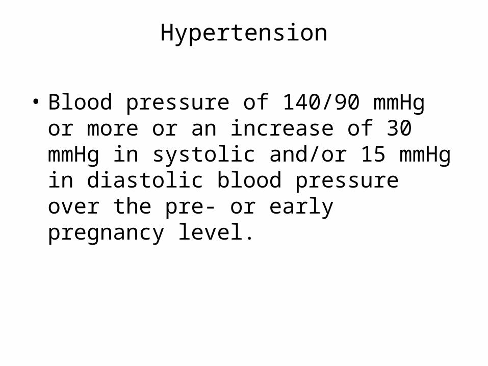
Hypertension
• Blood pressure of 140/90 mmHg or more or an increase of 30 mmHg in systolic and/or 15 mmHg in diastolic blood pressure over the pre- or early pregnancy level.

How to measure the blood pressure in pregnancy 1
• The patient should rest for at least 30 min. after arriving to the clinic.
• Remove any tight clothing from the right arm. • The patient lies comfortably on the left side that her
back makes an angle of about 30o with the bed. The right arm is supported to be with the sphygmomanometer at the same level with the patient’s sternum i.e. her heart. Each cm above or below the level of the heart induces a difference of 0.7mmHg in blood pressure reading. She should lie undisturbed in this position for 2-3 min. before blood pressure is measured.

How to measure the blood pressure in pregnancy 2
• The cuff should be applied to the right upper arm with the connecting tubes pointing downwards, the centre of the rubber bag in the cuff is directly over the brachial artery leaving ante-cubital fossa free.
• Apply cuff firmly but not tightly around the arm. • Feel the brachial artery and apply the stethoscope
directly over it without undue pressure. • Pump up cuff rapidly to 20-30 mmHg above the point
at which the pulse sound disappears, and take blood pressure reading without delay.

How to measure the blood pressure in pregnancy 3
• Let air out slowly so that mercury falls steadily by 2-3 mm/sec. • Blood pressure measurement phases (Korotkoff):
– Korotkoff I¾ Appearance of the sound¾ systolic reading. – Korotkoff II¾ Accentuation of the sound. – Korotkoff III ¾ Sound becomes harsh. – Korotkoff IV¾ Sound becomes muffled¾ diastolic reading. – Korotkoff V¾ Disappearance of the sound.
• Korotkoff I and IV is the reading for systolic and diastolic blood pressure respectively. If you wait the disappearance of the sound to take the diastolic reading (as in non-pregnant state) you may reach down to zero because of the hyperdynamic circulation during pregnancy.

How to measure the blood pressure in pregnancy 4
• Use the right arm for measuring because it is more convenient to the physician, but if the reading is 10 mmHg or more higher in the left arm use it in the future readings.
• The blood pressure should be measured in two occasions at least 6 hours apart.

Proteinuria (albuminuria)
• It is urinary protein greater than 0.3gm/L in 24 hours collection or greater than 1gm/L in two random samples obtained at least 6 hours apart.
• It indicates glomerular damage and almost always occurs after hypertension.
• Proteinuria is usually in the range of 1-3 gm daily, of which 50-60% is albumin but in severe cases it may exceed 15gm.

Oedema
• It is weight gain of more than 1 kg in any one week or 2.25 kg in any one month.
• Clinical oedema is present in about two-thirds of patients with PIH. However, two-thirds of pregnant women with clinical oedema do not develop hypertension.

Symptoms
These are usually manifestations of severe pre-eclampsia.• Headache: usually frontal but may be occipital. It is due
to cerebral oedema and hypertension. • Visual disturbances: blurring of vision, flashes of light
or blindness. • Epigastric or right upper quadrant pain: due to
enlargement and subcapsular hemorrhage of the liver. • Nausea and vomiting: due to congestion of gastric
mucosa and/ or cerebral oedema. • Oliguria or anuria: due to kidney pathology.

Investigations• Complete urine examination: for proteinuria, pus cells, RBCs, casts,
specific gravity, culture and sensitivity. • Kidney function tests: serum uric acid > 6 mg % is abnormal during
pregnancy. It is more specific for pre-eclampsia than creatinine. • Coagulation status: Platelet count, fibrinogen and FDP as DIC may
develop. • Eye fundus examination. • Tests for foetal well being: as
– ultrasound, – daily foetal movement count, – non-stress test, – oxytocin challenge test (if needed).

N.B.
• Imminent eclampsia: It is a state in which the patient is about to develop eclampsia. Usually there are: – blood pressure much higher than 160 /110 mmHg, – heavy proteinuria (+++or ++++), – hyperreflexia, – severe continuous headache, – blurring of vision, – epigastric pain.
• Fulminating pre-eclampsia: a rapidly deteriorating pre-eclampsia to be imminent eclampsia

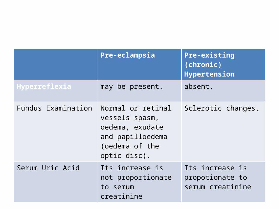
Pre-eclampsia Pre-existing (chronic) Hypertension
Parity usually primigravida
usually multigravida.
Past History of pre-eclampsia may be present
of hypertension in between pregnancies.
Hypertension after the 20th week of pregnancy (except in vesicular mole) and disappears within 6 weeks postpartum
before pregnancy, during the first 20 weeks and persists after 6 weeks postpartum.
Proteinuria If present, it develops after hypertension
If present, it develops before hypertension due to underlying renal disease.
Pre-eclampsia Pre-existing (chronic) Hypertension
Hyperreflexia may be present. absent.
Fundus Examination Normal or retinal vessels spasm, oedema, exudate and papilloedema (oedema of the optic disc).
Sclerotic changes.
Serum Uric Acid Its increase is not proportionate to serum creatinine
Its increase is propotionate to serum creatinine

Differential Diagnosis
• Other causes of hypertension• Other Causes of proteinuria• Other causes of oedema
Other Causes of proteinuria
• Contamination of urine by vaginal discharge this is excluded by examination of a midstream sample after cleansing the introitus with sterile water or saline or by using a catheter.
• Urinary infection: excluded by microscopic examination and culture of urine.
• Congestive heart failure and severe anaemia due to hypoxia of the kidney.
• Orthostatic proteinuria: Proteinuria is detected at the end of the day while it is absent in the morning. This is due to pressure of the lumbar spines on the left renal vein during standing

Other causes of oedema
• General causes: cardiac, hepatic, renal or nutritional oedema.
• Local causes: as inflammatory or deep vein thrombosis (usually unilateral).
• Pressure of the gravid uterus on the pelvic veins may produce ankle oedema.

Complications
Maternal:
– Convulsions and coma (eclampsia). – Cerebral haemorrhage. – Renal failure. – Heart failure. – Liver failure. – Disseminated intravascular
coagulation. – Abruptio placentae. – Residual chronic hypertension in
about 1/3 of cases. – Recurrent pre-eclampsia in next
pregnancies.
Foetal:
– Intrauterine growth retardation (IUGR).
– Intrauterine foetal death. – Prematurity and its
complications.

Treatment
• Prophylactic• Curative
Prophylactic
• Proper antenatal care: – To detect the high risk patients who may develop PIH through the
screening tests. – Early detection of cases who have already developed PIH and
examine them more frequently. • Low dose aspirin:
– It inhibits thromboxane production from the platelets and the AII binding sites on platelets.
– A low dose (60 mg daily) selectively inhibits thromboxane due to higher concentration of such a low dose in the portal circulation than systemic affecting the platelets when they pass through the portal circulation. The Prostacyclin production from the systemic vessels will not be affected.

Curative
• Delivery of the foetus and placenta is the only real treatment of pre-eclampsia. As the conditions are not always suitable for this, the treatment aims to prevent or minimize the maternal and foetal complications (see before) till reasonable maturation of the foetus.

General measures:Observation:
Maternal:
• blood pressure twice daily. • urine volume and
proteinuria daily, • oedema daily, • body weight twice weekly, • fundus oculi once weekly, • blood picture including
platelet count, liver and renal functions particularly serum uric acid on admission.
Foetal:
• daily foetal movement count,
• serial sonography, • non-stress and stress test if
needed.

Medical treatment• Antihypertensives:
– decrease the maternal cerebral and cardiovascular complications but do not affect the foetal outcome.
– Alpha-methyl-dopa (Aldomet): • It reduces the central sympathetic drive. • Dose: 250-500 mg every 6-8 hours up to a maximum dose of 4 gm/day. Its effect appears after 48 hours. • A loading single dose of 2 gm may act within 1-2 hours. • Side effects: headache, athenia and nightmares.
– Hydralazine (Apresoline): • It is a vasodilator, increases renal and uteroplacental blood flow. • Dose: 20 mg slowly IV initially followed by 5mg every 20 min. until diastolic blood pressure is 100-110
mmHg. This regimen is used for severe and acute hypertension. Oral hydralazine can be used in the chronic situation as a second line treatment in a dose of 25-75 mg/ 6 hours.
• Side effects: tachycardia, headache, flushing, nausea and vomiting.
– Calcium channel blockers (Nifedipine): • It is a vasodilator acting by blocking the Ca influx into smooth muscle cells. • It can be given sublingually (acts within 10 minutes) or orally (acts within 30 minutes) in a dose of 10-20 mg
2-3 times daily. • The higher the starting blood pressure the greater is the hypotensive effect. • Side effects: headache and flushing.
Obstetric measures
• Timing of delivery • Method of delivery• Intrapartum care• Postpartum care

Timing of delivery: Severe pre-eclampsia is usually treated conservatively till the end of the 36th week to ensure reasonable maturation of the foetus. Indications of termination before 36th week include:
Foetal: deteriorating placental function as judged by:
– intrauterine growth retardation,
– oligohydramnios, – reduced foetal
movements, – abnormal foetal heart
patterns, or – failing biochemical results.
Maternal: deteriorating maternal condition as judged by:
– blood pressure is sustained or exceeds 180/110 mmHg,
– urine proteinuria > 5 gm/24 hours,
– oliguria, – evidence of DIC, or – imminent or already
developed eclampsia.

Method of delivery:
– Vaginal delivery may be commenced in vertex presentation by: • amniotomy + oxytocin if the cervix is favourable. • prostaglandin vaginal tablet (PGE2) if the cervix is not
favourable.
– Caesarean section is indicated in: • Foetal distress. • Late deceleration occurs with oxytocin challenge test. • Failure of induction of labour. • Other indications as contracted pelvis, and malpresentations.

Intrapartum care:
– Close monitoring of the foetus is indicated. – Proper analgesia to the mother. – Anti-Hypertensives may be given if needed. – 2nd stage of labour may be shortened by forceps.

Postpartum care:
– Methergin (Ergometrine) is better avoided as it may increase the blood pressure.
– Continue observation of the mother for 48 hours. – Anti- hypertensive drugs are continued in a
decreasing dose for 48 hours.

Thank you