Embed Size (px)
Citation preview

Hypertensive Disorders of Pregnancy

Case report• You admitted a previously healthy
nullipara at 36 weeks’ gestation who presented with new-onset periorbital edema and is found to have blood pressure readings of 150/100 to 155/105 mmHg, 4+ protein on urinary dipstick, modest (ie, less than 2-fold) elevations in her hepatic transaminases, a platelet count of 105,000/µL and a mildly (8th percentile) growth-restricted fetus.
Question?
• Does she have severe preeclampsia? • Should you start parenteral magnesium
sulfate? • Should you give antihypertensive therapy?• Is delivery indicated?
Hypertensive Disorders of Pregnancy
• most common medical complications of pregnancy,
• 5% to 10% of all pregnancies. • 16% of maternal mortality in developed countries. • Classification include:1. chronic hypertension (30%) 2. hypertensive unique to pregnancy (70%)
( gestational hypertension and preeclampsia).
Hypertensive Disorders of Pregnancy
• The spectrum of disease ranges from non-severe to severe hypertension and multi organ dysfunction.
• The incidence is dependent on:• maternal age,• race,• underlying medical conditions.
HTN
• BP 140/90 or greater• at least two occasions at least 4 hours apart but no more
than 1 week apart.• in an upright position, after a 10-minute or longer rest
period.• For patients in the hospital, either the patient sitting up or
in the left lateral position with the patient's arm at the level of the heart.
• Avoidance tobacco or caffeine for 30 minutes preceding the measurement
• Delta HTN
Abnormal proteinuria
• 300 mg or more of protein in 24 hours. • The most accurate is obtained with a 24-hour urine
collection. • A value of 1+ or greater correlates with 30 mg/dL.• Proteinuria by dipstick is defined as 1+ or more on at least
two occasions at least 6 hours apart but no more than 1 week apart.
• 24-hour urine collection should be performed the diagnostic criteria .
• to use a clean sample, because blood, vaginal secretions, and bacteria can increase the amount of protein in urine.
Edema
• common finding in the gravid patient, (~50%)• Lower extremity edema is the most typical
form. • Pathologic edema is seen in nondependent
regions such as the face, hands, or lungs.• Excessive, rapid weight gain of 2 pounds or
more per week is another sign of fluid retention.
Classificationof Hypertension
• Gestational hypertension (most common) developing after 20 weeks gestation or during the first 24 hours postpartum without proteinuria or other signs of preeclampsia (46%___progress to preeclampcia
Transient hypertension resolves by 12 weeks postpartum – Preeclamsia or Eclampsia: – Chronic HTN – Superimpose preeclampsia
Classification of Hypertension
• Preeclamsia –Eclampsia developing after 20 weeks gestation with proteinuria; eclampsia is the occurrence of seizure activity without other identifiable causes
• Chronic hypertension prior to pregnancy, prior to 20 weeks gestation, or after 12 weeks postpartum
• Preeclampsia superimposed The development of preeclampsia or eclampsia in a woman with preexisting or chronic hypertension(New onset pr;increase pr,or HTN,or plt <100000)
Preeclampsia or eclampsia
• Multisystemic,unknown cause,only in pregnancy• 2%-- 7% incidence • higher in : nulliparous women twin gestation
(14%) and previous preeclampsia (18%). • symptoms are:• headaches• visual changes,• epigastric or right upper quadrant pain • nausea or vomiting.
Preeclampsia or eclampsia
• divided further into non-severe and severe .• non-severe preeclampsia may progress to
fulminant disease. • A particularly severe form of preeclampsia is
the HELLP syndrome
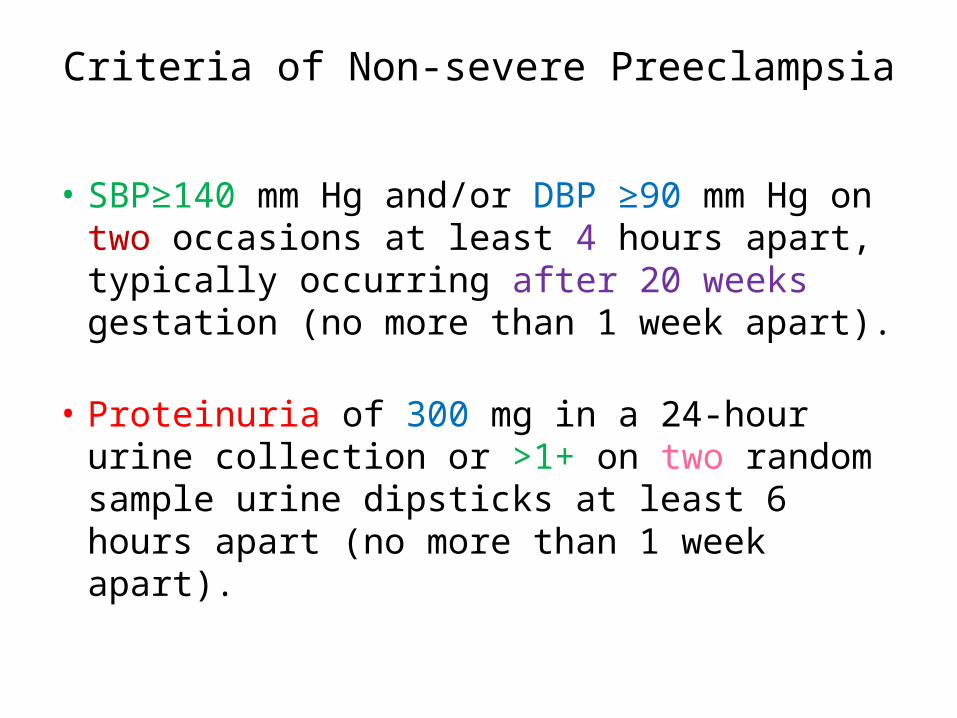
Criteria of Non-severe Preeclampsia
• SBP≥140 mm Hg and/or DBP ≥90 mm Hg on two occasions at least 4 hours apart, typically occurring after 20 weeks gestation (no more than 1 week apart).
• Proteinuria of 300 mg in a 24-hour urine collection or >1+ on two random sample urine dipsticks at least 6 hours apart (no more than 1 week apart).
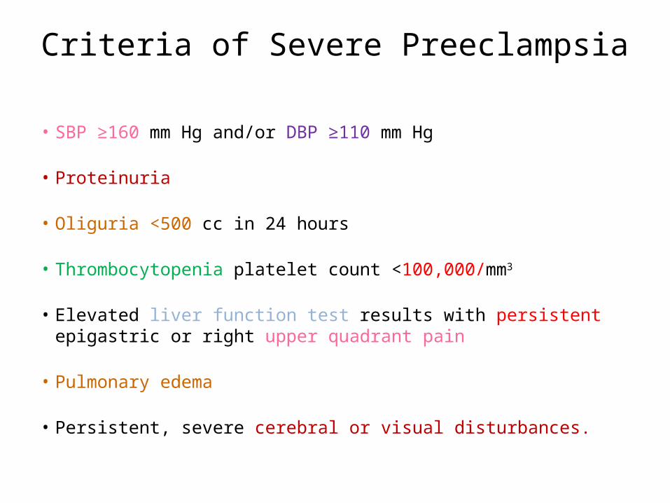
Criteria of Severe Preeclampsia
• SBP ≥160 mm Hg and/or DBP ≥110 mm Hg
• Proteinuria
• Oliguria <500 cc in 24 hours
• Thrombocytopenia platelet count <100,000/mm3
• Elevated liver function test results with persistent epigastric or right upper quadrant pain
• Pulmonary edema
• Persistent, severe cerebral or visual disturbances.
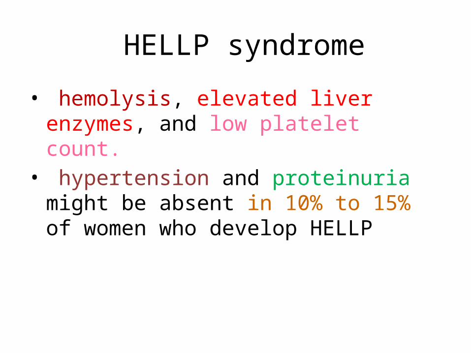
HELLP syndrome
• hemolysis, elevated liver enzymes, and low platelet count.
• hypertension and proteinuria might be absent in 10% to 15% of women who develop HELLP
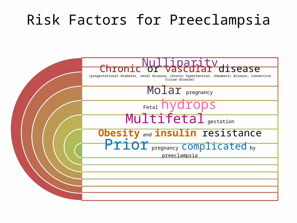
Risk Factors for Preeclampsia
NulliparityChronic or vascular disease (pregestational diabetes, renal disease, chronic
hypertension, rheumatic disease, connective tissue disease)
Molar pregnancy
Fetal hydropsMultifetal gestation
Obesity and insulin resistance
Prior pregnancy complicated by preeclampsia

Risk Factors for PreeclampsiaAntiphospholipid antibody syndrome and thrombophilia
Family history of preeclampsia or eclampsia
Fetal aneuploidy ,Maternal infections, Maternal susceptibility genes,
Extremes of maternal age
, Partner-related factors
Etiologic Theories in Preeclampsia
• The syndrome is characterized by :1. vasospasm; 2. hemoconcentration; and3. ischemic changes in the placenta, kidney, liver,
and brain.
• These abnormalities usually are seen in women with severe preeclampsia.

Etiologic Theories in PreeclampsiaAbnormal or
increased immune response
Genetic predisposition
Abnormal coagulation or thrombophilias
Abnormal angiogenesis
Endothelial cell injury

Etiologic Theories in Preeclampsia
Alterations in nitric oxide levels
Increased oxygen free radicals
Abnormal cytotrophoblast
invasion
Abnormal calcium metabolism
Dietary deficiencies
Pathophysiology
Cardiovascular• intense vasoconstriction with segmental spasm in
arterioles. • alterations in the normal interactions of vasodilatory
(prostacyclin, nitric oxide) and vasoconstrictive (thromboxane A2, endothelin) .
• higher arterial blood pressures (afterload). • hemoconcentration: • endothelial damage leak interstitial space, .• lower intravascular volumes and less tolerance for the blood
loss.
Hematologic
• thrombocytopenia (platelet count <100,000/mm3). • The suggested pathophysiology likely is vascular
endothelial damage or activation and higher levels of thromboxane A2.
• microangiopathic hemolysis, (HELLP) ( schistocytes and increased LDH levels).
• A low hematocrit may signify hemolysis, and a falsely high hematocrit may be due to hemoconcentration.
Renal
• Vasospasm renal perfusion GFR.(normal pregnancy: GFR is increased up to 50%).
• creatinine rarely rise above normal pregnancy levels (0.8 mg/dL).
• oliguria (defined as <500 cc in 24 hours) may occur due to renal insufficiency.(ATN).
• pathognomonic renal lesion in preeclampsia is called glomerular capillary endotheliosis, which is swelling of the glomerular capillary endothelial and mesangial cells.
Hepatic
• range from mildly elevated liver enzyme levels to subcapsular liver hematomas and hepatic rupture.
• 20% of maternal mortality in preeclampsia is related to hepatic complications.
• The pathologic liver lesions seen on autopsy are periportal hemorrhages, hepatocellular necrosis, ischemic lesions, intracellular fatty changes, and fibrin deposition
Central Nervous System
• Eclamptic convulsions major cause of maternal mortality.
• etiology of eclampsia : coagulopathy, fibrin deposition, and vasospasm.
• The most common finding in the brain is edema, posterior hemispheres edema&hemorrhage:.• headaches and visual disturbances ( scotomata;
blurred vision; and rarely, temporary blindness).
Fetus and Placenta
• hallmark placental lesion in preeclampsia is acute atherosis of decidual arteries.
• This is due in part to the abnormal adaptation of the spiral artery cytotrophoblast interface and results in poor perfusion.
oligohydramnios; IUGR abruption fetal distress; fetal demise.
Prediction
• No good screening test.• doppler ultrasonography: velocity of uterine
blood flow in the second trimester. Abnormal velocity waveform is characterized by a high resistance index or an early diastolic notch (unilateral or bilateral).
• No for routine screening. .
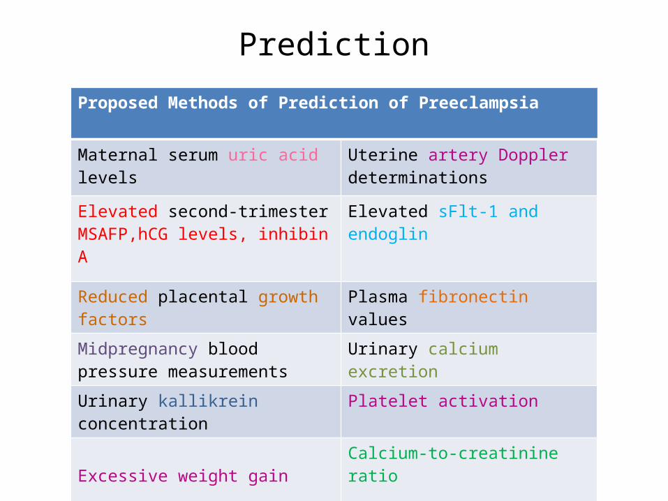
PredictionProposed Methods of Prediction of Preeclampsia
Maternal serum uric acid levels Uterine artery Doppler determinations
Elevated second-trimester MSAFP,hCG levels, inhibin A
Elevated sFlt-1 and endoglin
Reduced placental growth factors Plasma fibronectin values
Midpregnancy blood pressure measurements
Urinary calcium excretion
Urinary kallikrein concentration Platelet activation
Excessive weight gainCalcium-to-creatinine ratio
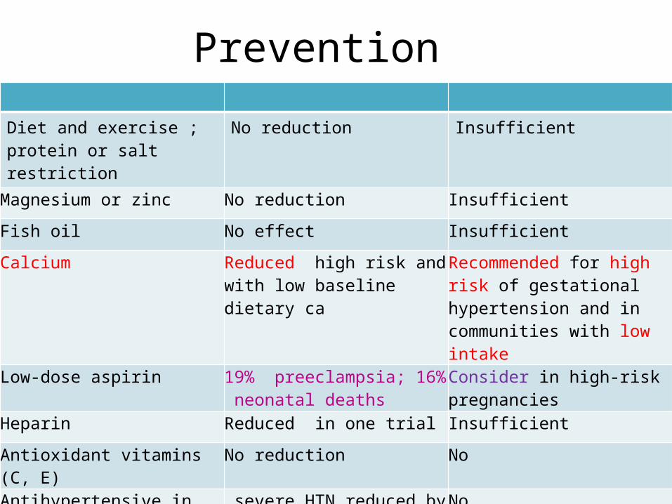
Prevention
Diet and exercise ; protein or salt restriction
No reduction Insufficient
Magnesium or zinc No reduction Insufficient
Fish oil No effect Insufficient
Calcium Reduced high risk and with low baseline dietary ca
Recommended for high risk of gestational hypertension and in communities with low intake
Low-dose aspirin 19% preeclampsia; 16% neonatal deaths
Consider in high-risk pregnancies
Heparin Reduced in one trial Insufficient
Antioxidant vitamins (C, E) No reduction No
Antihypertensive in chronic hypertension (I)
severe HTN reduced by half; no reduction in preeclampsia
No
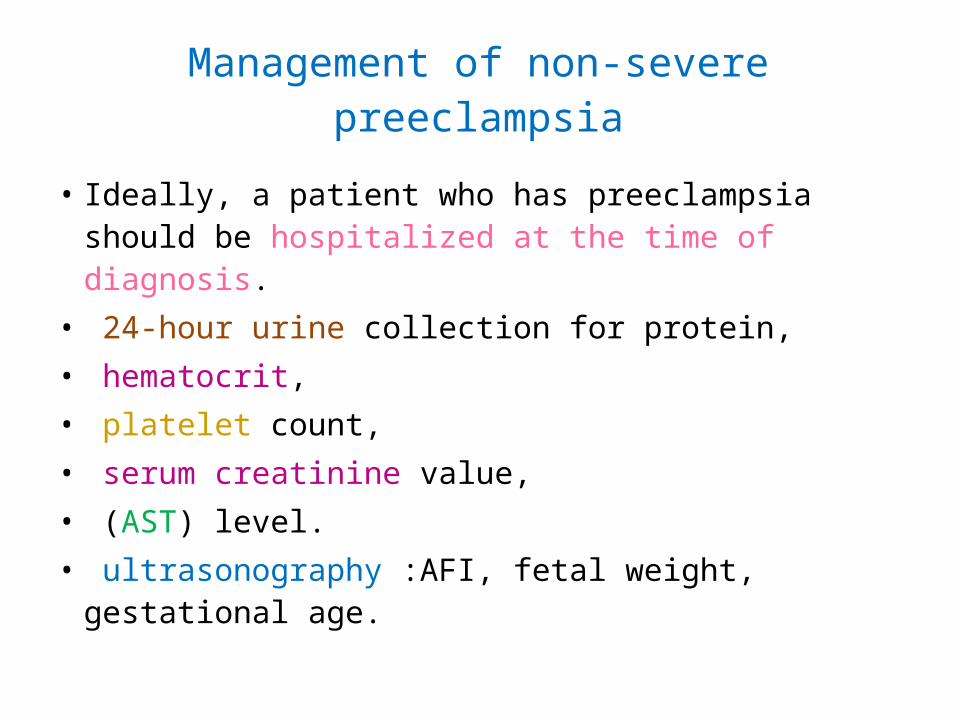
Management of non-severe preeclampsia
• Ideally, a patient who has preeclampsia should be hospitalized at the time of diagnosis.
• 24-hour urine collection for protein,• hematocrit,• platelet count,• serum creatinine value,• (AST) level.• ultrasonography :AFI, fetal weight, gestational
age.
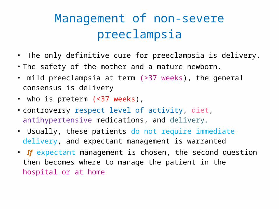
Management of non-severe preeclampsia
• The only definitive cure for preeclampsia is delivery. • The safety of the mother and a mature newborn.• mild preeclampsia at term (>37 weeks), the general consensus
is delivery• who is preterm (<37 weeks),• controversy respect level of activity, diet, antihypertensive
medications, and delivery.• Usually, these patients do not require immediate delivery, and
expectant management is warranted• If expectant management is chosen, the second question then
becomes where to manage the patient in the hospital or at home
Criteria for Home Management of non-severe Preeclampsia
• Ability to comply with recommendations
• Normal laboratory tests • No maternal symptoms• Reassuring fetal status with appropriate
growth
Maternal and Fetal Evaluation in non-severe Preeclampsia
• MaternalDaily weightUrine dipstick; 24-hour protein once weeklyMonitoring for severe preeclampsia symptomsPrenatal visits twice per weekLab tests (liver function tests, hematocrit, platelet count once or twice per week)
• FetalDaily fetal movementNonstress test twice per week or biophysical profile once per weekUltrasound for growth every 3 to 4 weeks
Prompt Evaluation
• Signs and Symptoms of Preeclampsia That Warrant :
• Nausea and vomiting• Persistent severe headache• Right upper quadrant or epigastric pain• Scotoma• Blurred vision
Management of non-severe Preeclampsia
• Induction of labor is indicated in those patients with a favorable cervix
• At 37 weeks or beyond, if the cervix is unfavorable, two options:
• either cervical ripening and delivery • continued expectant management with maternal and
fetal evaluation. • The preferred mode of delivery remains vaginal.
• A cesarean section should be performed for obstetric indications only
Management of non-severe Preeclampsia
• In the past, while in labor, patients with mild preeclampsia received intravenous magnesium sulfate (MgSO4) for seizure prophylaxis.
• There is no support in the literature for the need or the optimal timing to begin the MgSO4 infusion, and this should be left to the discretion of the physician.
• In mild the authors recommend to individualize each case for the use of MgSO4.
Management of non-severe Preeclampsia
• Pain management in labor should be individualized.• Close monitoring of blood pressure intrapartum is
necessary.• Antihypertensive medications to keep blood pressure
values below 160 / 110 mm Hg • most commonly used intravenous medications for this
purpose are labetalol and hydralazine.• not to drop the blood pressure too rapidly, lead to reduced
renal perfusion and reduced placental perfusion. • who receive MgSO4 also are at risk for postpartum
hemorrhage due to uterine atony.
Management of non-severe Preeclampsia
• Patients should be monitored closely for at least 12 to 24 hours postpartum.
• Postpartum eclampsia occurs in 25% of patients.
• It is not clear if MgSO4 should be continued in the postpartum period.
• There is no need for continued seizure prophylaxis beyond 24 hours postpartum
Maternal and Fetal Complications in Severe Preeclampsia
• MaternalAbruptio placentae (10%-40%)Disseminated coagulopathy/HELLP syndrome (10%-20%)Pulmonary edema/aspiration (2%-5%)Acute renal failure (1%-5%)Eclampsia (<1%)Liver failure or hemorrhage (<1%)Stroke (rare)Death (rare)Long-term cardiovascular morbidity
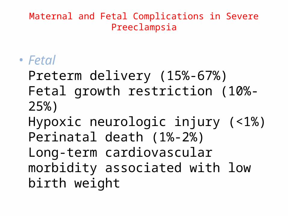
Maternal and Fetal Complications in Severe Preeclampsia
• FetalPreterm delivery (15%-67%)Fetal growth restriction (10%-25%)Hypoxic neurologic injury (<1%)Perinatal death (1%-2%)Long-term cardiovascular morbidity associated with low birth weight
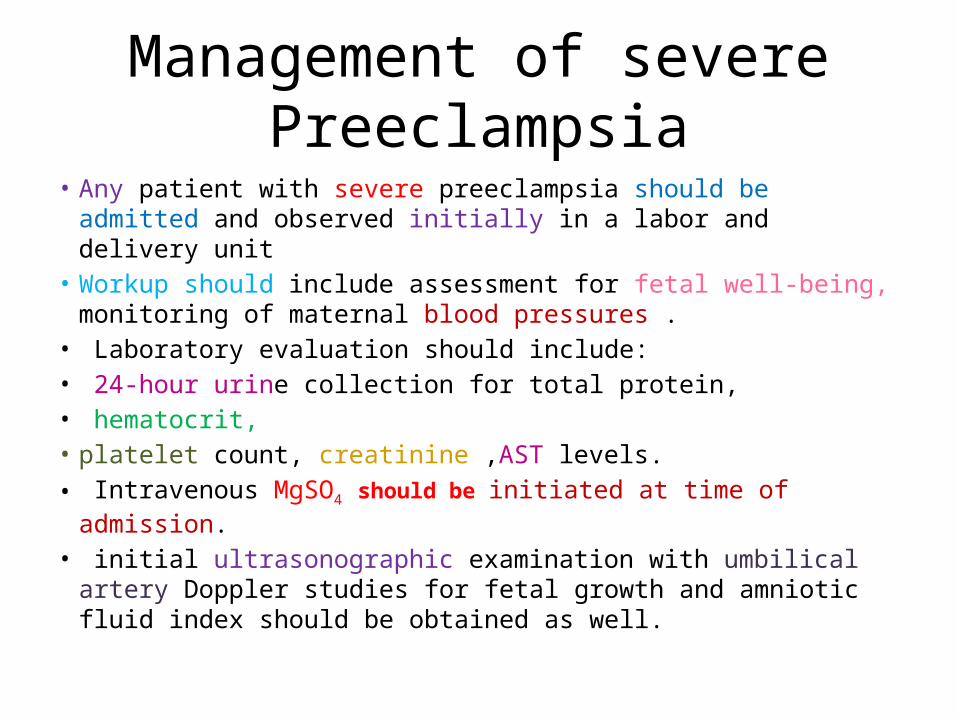
Management of severe Preeclampsia
• Any patient with severe preeclampsia should be admitted and observed initially in a labor and delivery unit
• Workup should include assessment for fetal well-being, monitoring of maternal blood pressures .
• Laboratory evaluation should include:• 24-hour urine collection for total protein,• hematocrit, • platelet count, creatinine ,AST levels.• Intravenous MgSO4 should be initiated at time of admission. • initial ultrasonographic examination with umbilical artery Doppler
studies for fetal growth and amniotic fluid index should be obtained as well.
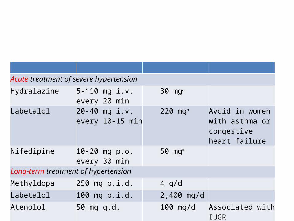
Acute treatment of severe hypertension
Hydralazine 5-“10 mg i.v. every 20 min
30 mga
Labetalol 20-40 mg i.v. every 10-15 min
220 mga Avoid in women with asthma or congestive heart failure
Nifedipine 10-20 mg p.o. every 30 min
50 mga
Long-term treatment of hypertension
Methyldopa 250 mg b.i.d. 4 g/d
Labetalol 100 mg b.i.d. 2,400 mg/d
Atenolol 50 mg q.d. 100 mg/d Associated with IUGR
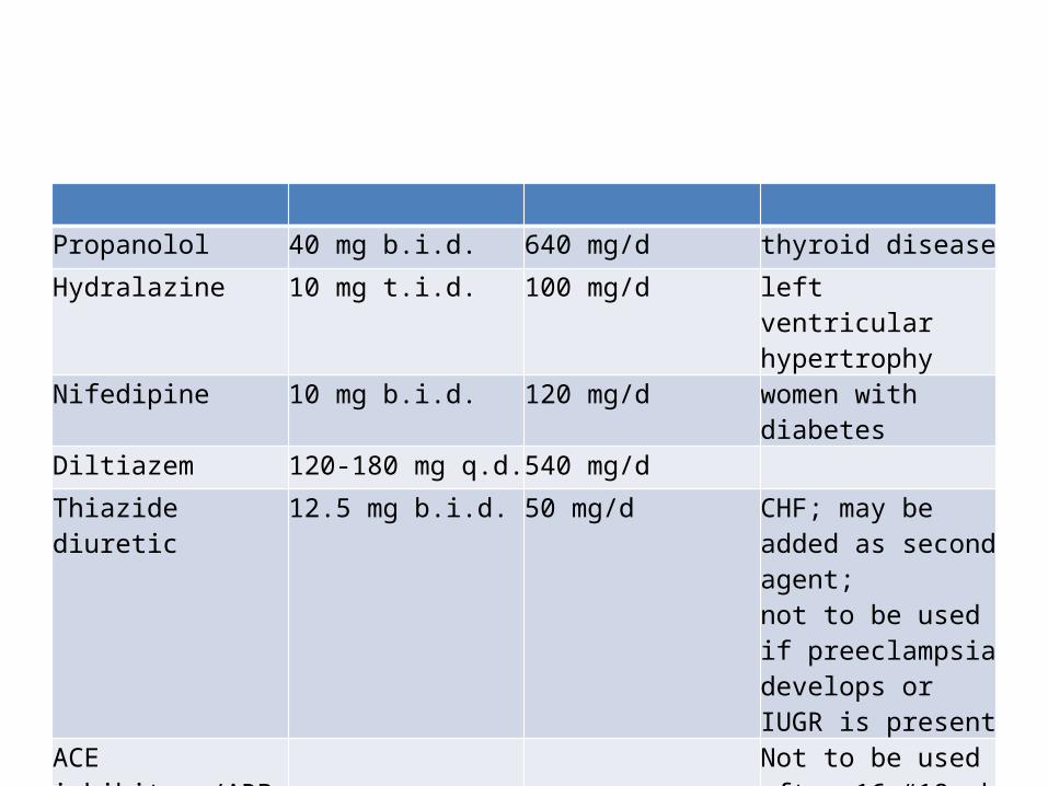
Propanolol 40 mg b.i.d. 640 mg/d thyroid disease
Hydralazine 10 mg t.i.d. 100 mg/d left ventricular hypertrophy
Nifedipine 10 mg b.i.d. 120 mg/d women with diabetes
Diltiazem 120-180 mg q.d. 540 mg/d
Thiazide diuretic 12.5 mg b.i.d. 50 mg/d CHF; may be added as second agent; not to be used if preeclampsia develops or IUGR is present
ACE inhibitors/ARB Not to be used after 16-“18 wk
Management of severe Preeclampsia
• delivery is considered in all women with severe preeclampsia
• prolonging the pregnancy may be hazardous to the mother with little benefit to the fetus.
• In patients at 33 to 34 weeks gestation, steroids should be administered and delivery planned within 48 hours unless otherwise indicated.
Management of severe Preeclampsia
• In patients with severe prematurity , expectant management significantly improves neonatal outcome.
• The goal in these patients is to gain at least 48 hours that glucocorticoids can be administered for fetal benefit.
• They should be counseled regarding the risks and benefits of expectant management
Management of severe Preeclampsia
• Ultrasonography for fetal growth every 2 to 3 weeks.• Maternal laboratory evaluation should be done daily
or every other day.• If a stable maternal and fetal course is maintained,
expectant management continued until 34 weeks. • The development of any worsening change in maternal
or fetal status warrants delivery regardless of gestational age.
• A woman with a nonviable fetus should be terminated.• above 23 weeks, expectant
Management of severe Preeclampsia
• Maternal blood pressure control• Medications orally or by the intravenous route• management of acute hypertension are
hydralazine and labetalol.• avoid a sudden drop blood pressure
precipitating cerebral ischemia, decreased renal perfusion, and decreased placental perfusion.
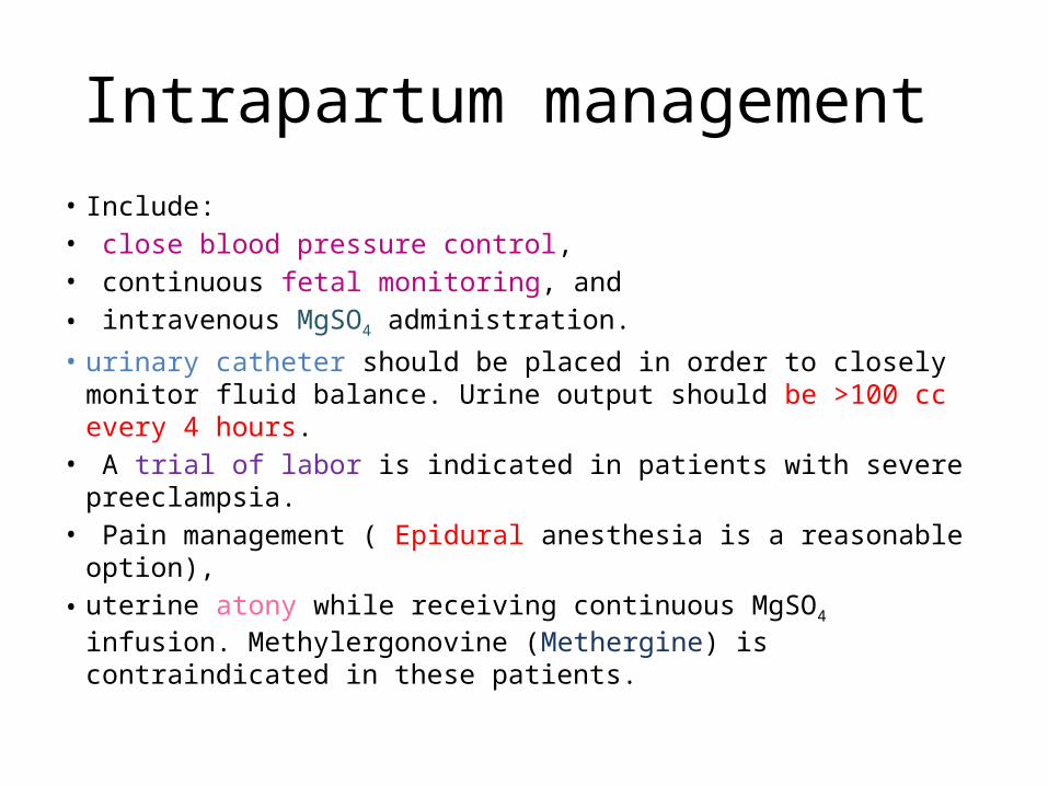
Intrapartum management • Include:• close blood pressure control,• continuous fetal monitoring, and• intravenous MgSO4 administration. • urinary catheter should be placed in order to closely monitor fluid
balance. Urine output should be >100 cc every 4 hours.• A trial of labor is indicated in patients with severe preeclampsia. • Pain management ( Epidural anesthesia is a reasonable option), • uterine atony while receiving continuous MgSO4 infusion.
Methylergonovine (Methergine) is contraindicated in these patients.
• MgSO4 continue for 24 hours postpartum. • monitor blood pressure, reflexes, and fluid
status.
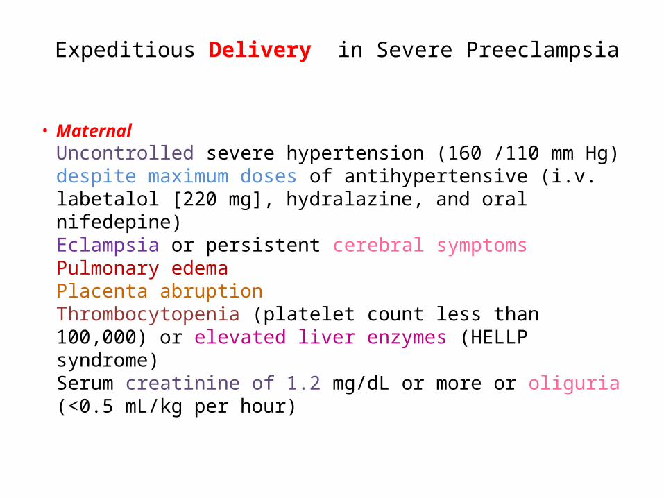
Expeditious Delivery in Severe Preeclampsia
• MaternalUncontrolled severe hypertension (160 /110 mm Hg) despite maximum doses of antihypertensive (i.v. labetalol [220 mg], hydralazine, and oral nifedepine)Eclampsia or persistent cerebral symptomsPulmonary edemaPlacenta abruptionThrombocytopenia (platelet count less than 100,000) or elevated liver enzymes (HELLP syndrome)Serum creatinine of 1.2 mg/dL or more or oliguria (<0.5 mL/kg per hour)
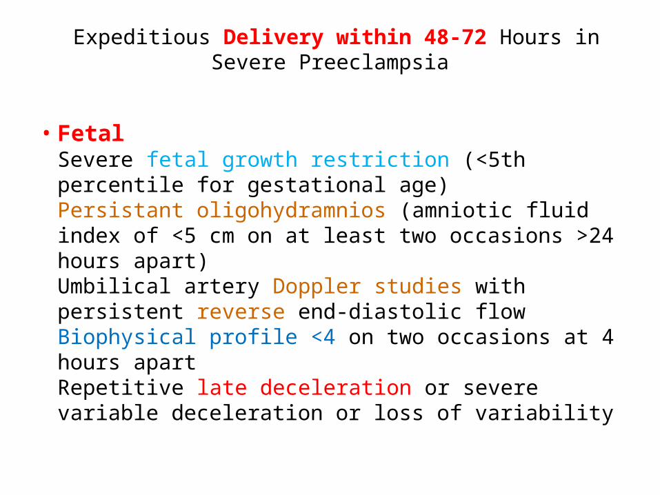
Expeditious Delivery within 48-72 Hours in Severe Preeclampsia
• FetalSevere fetal growth restriction (<5th percentile for gestational age)Persistant oligohydramnios (amniotic fluid index of <5 cm on at least two occasions >24 hours apart)Umbilical artery Doppler studies with persistent reverse end-diastolic flowBiophysical profile <4 on two occasions at 4 hours apartRepetitive late deceleration or severe variable deceleration or loss of variability
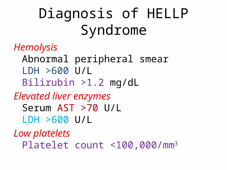
Diagnosis of HELLP Syndrome
HemolysisAbnormal peripheral smearLDH >600 U/LBilirubin >1.2 mg/dL
Elevated liver enzymesSerum AST >70 U/LLDH >600 U/L
Low plateletsPlatelet count <100,000/mm3
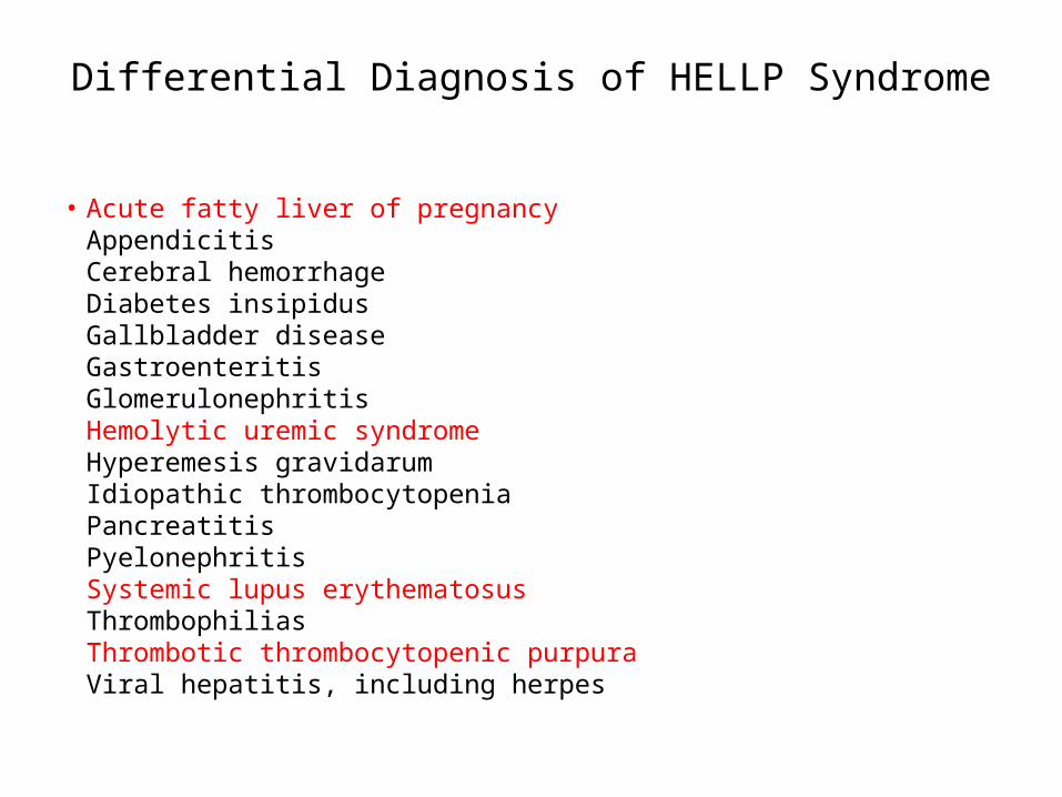
Differential Diagnosis of HELLP Syndrome
• Acute fatty liver of pregnancyAppendicitisCerebral hemorrhageDiabetes insipidusGallbladder diseaseGastroenteritisGlomerulonephritisHemolytic uremic syndromeHyperemesis gravidarumIdiopathic thrombocytopeniaPancreatitisPyelonephritisSystemic lupus erythematosusThrombophiliasThrombotic thrombocytopenic purpuraViral hepatitis, including herpes
Eclampsia
• much higher in developing countries. • major cause of maternal and perinatal morbidity
and mortality worldwide. • wide spectrum of signs and symptoms, from
mild isolated hypertension to multiorgan failure.• Prevention of eclampsia is one of the goals in
treating preeclamptic patients with MgSO4
Management of Eclampsia
• During seizure, the main therapy is supportive care.1. avoiding injury,2. maintaining oxygenation,3. minimizing the risk of aspiration.• Most seizures are self-limited, lasting 1 to 2 minutes.• hypoxia or acidosis both in mother and fetus.• MgSO4 is the drug of choice for the prevention
• 10% of eclamptic women receiving MgSO4 will have further seizures.
Management of the Eclamptic Patient
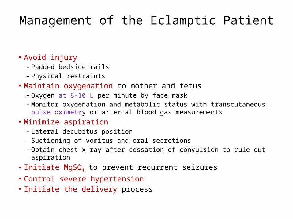
• Avoid injury – Padded bedside rails – Physical restraints
• Maintain oxygenation to mother and fetus – Oxygen at 8-10 L per minute by face mask – Monitor oxygenation and metabolic status with transcutaneous pulse oximetry or
arterial blood gas measurements• Minimize aspiration
– Lateral decubitus position – Suctioning of vomitus and oral secretions – Obtain chest x-ray after cessation of convulsion to rule out aspiration
• Initiate MgSO4 to prevent recurrent seizures • Control severe hypertension • Initiate the delivery process
Management of the Eclamptic Patient
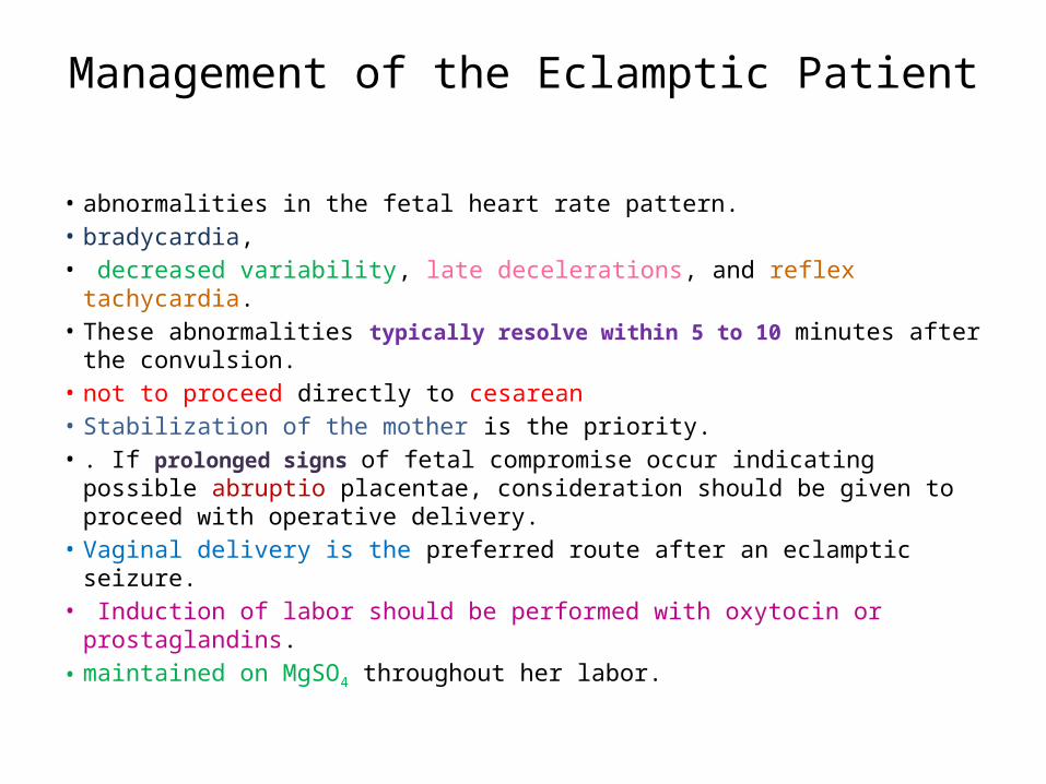
• abnormalities in the fetal heart rate pattern. • bradycardia,• decreased variability, late decelerations, and reflex tachycardia. • These abnormalities typically resolve within 5 to 10 minutes after the
convulsion.• not to proceed directly to cesarean• Stabilization of the mother is the priority.• . If prolonged signs of fetal compromise occur indicating possible abruptio
placentae, consideration should be given to proceed with operative delivery.
• Vaginal delivery is the preferred route after an eclamptic seizure.• Induction of labor should be performed with oxytocin or prostaglandins.• maintained on MgSO4 throughout her labor.
Regimens of Magnesium Sulfate in eclampsia
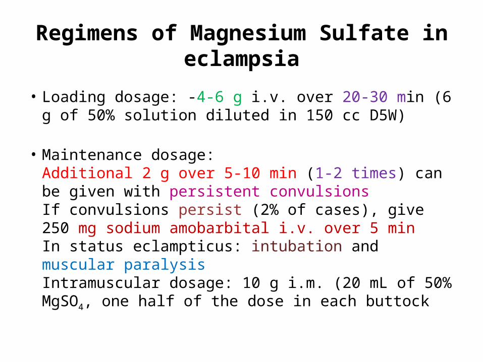
• Loading dosage: -4-6 g i.v. over 20-30 min (6 g of 50% solution diluted in 150 cc D5W)
• Maintenance dosage: Additional 2 g over 5-10 min (1-2 times) can be given with persistent convulsionsIf convulsions persist (2% of cases), give 250 mg sodium amobarbital i.v. over 5 minIn status eclampticus: intubation and muscular paralysisIntramuscular dosage: 10 g i.m. (20 mL of 50% MgSO4, one half of the dose in each buttock
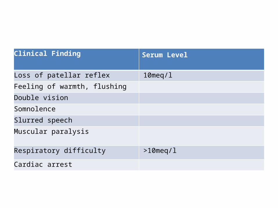
Clinical Finding Serum Level
Loss of patellar reflex 10meq/l
Feeling of warmth, flushing
Double vision
Somnolence
Slurred speech
Muscular paralysis
Respiratory difficulty >10meq/l
Cardiac arrest
Management of Magnesium Toxicity
• Discontinue MgSO4 infusion• Begin supplemental oxygen administration• Obtain serum magnesium level• Administer 1 g calcium gluconate (10 cc 10%
calcium gluconate) by slow intravenous push• Repeat calcium gluconate administration, if
necessary• If respiratory arrest occurs, begin
cardiopulmonary resuscitation.
chronic hypertension
• The management differs for the low-risk group versus the high-risk group.
• low-risk group have, by definition, mild hypertension without evidence of organ damage.
• The high-risk group has either severe hypertension (SBP 160 /110 mm Hg) or mild hypertension with evidence of organ involvement.
chronic hypertension
• Low risk:• Weekly nonstress tests should begin at 34
weeks.• increased to twice weekly with evidence of
growth restriction or oligohydramnios. • low-risk group may continue their pregnancies
up to 41 weeks,
chronic hypertension
• high-risk :• use of antihypertensive pharmacotherapy. • first-trimester 24-hour urine collection for total protein
level.• visits in the first and second trimesters should be every 2 to
3 weeks• then weekly in the third trimester if clinically indicated.• ultrasonography for estimated fetal weight and amniotic
fluid volume every 4 weeks starting at 26 weeks.• Weekly nonstress testing or biophysical profile
assessments should begin at 28 weeks.
chronic hypertension • low-risk chronic hypertension who do not develop superimposed
preeclampsia have pregnancy outcomes similar to those of the general population.
• Antihypertensive not affect perinatal outcome and is not necessary,• if the SBP reaches 160 /105 mm Hg, or target organ damage
develops. • 24-hour urine for protein determination in the first trimester • at least monthly visits in the first and second trimester• .Visits should be every 1 to 2 weeks after 32 weeks, looking
carefully for the development of superimposed preeclampsia. • ultrasonography for fetal growth and amniotic fluid assessment
every 4 weeks beginning at 32 weeks,
chronic hypertension
• visits and fetal testing may need to be increased dependent on (increasing HTN, superimposed preeclampsia, decreased AF volume, or IUGR.
• superimposed preeclampsia is suspected or uncontrolled hypertension develops, the patient should be hospitalized, preferably at a tertiary care center.
• timing of delivery is dependent on complications and gestational age.
• In general, pregnancies in patients with high-risk chronic hypertension should not be continued past 40 weeks.