Embed Size (px)
Citation preview
Liu and Guo Ann Clin Microbiol Antimicrob (2019) 18:4 https://doi.org/10.1186/s12941-018-0302-9
RESEARCH
Hypervirulent Klebsiella pneumoniae (hypermucoviscous and aerobactin positive) infection over 6 years in the elderly in China: antimicrobial resistance patterns, molecular epidemiology and risk factorChao Liu1 and Jun Guo2,3*
Abstract
Background: The definition of hypervirulent Klebsiella pneumoniae (hvKp), traditionally regarded as hypermucovis-cosity, is controversial. However, data based on both phenotype (hypermucoviscous) and genetic (aerobactin) criteria are limited.
Methods: A retrospective study was conducted in 175 geriatric patients between January 2008 and January 2014. The clinical and molecular data, including antimicrobial susceptibility testing, extended-spectrum-β-lactamase (ESBL) production, virulence gene, and multilocus sequence typing of the hvKp-group (hypermucoviscosity and aerobactin positive) were compared with those of classic K. pneumoniae (cKp) isolates.
Results: Of 175 Kp isolates, 45.7% were hvKp. In pathogenicity, K1, K2, magA, rmpA, and rmpA2 genes were strongly associated with hvKp (P < 0.01). In the hvKp group, invasive infections (P < 0.000), liver abscess (P = 0.008), abdominal infection (P = 0.002) and septic shock (P = 0.035) are significantly higher than cKp group. Patients with better nutri-tional status were frequently infected with hvKp. However, host inflammatory reaction is most severe in hvKp group. Patients with diabetes (odds ratio [OR] = 2.548) and digestive diseases (OR = 2.196) are more likely to be infected with hvKp. Importantly, the detection of hvKp isolates increased from January 2008 to January 2010, January 2010 to Janu-ary 2012, and January 2010 to January 2014 (12, 30, and 48 isolates, respectively). Overall, 16.3% of hvKp isolates pro-duced ESBLs and 20.0% were MDR-hvKp. Multivariate analysis implied that infection occurred in the ICU (OR = 5.826) and patients with indwelling stomach tubes (OR = 6.461) are independent risk factors for ESBL-hvKp infection.
Conclusions: HvKp, especially ESBL-hvKp and MDR-hvKp, is emerging in the elderly. It is essential to enhance clinical awareness and management of hvKp infections.
Keywords: Klebsiella pneumoniae, Hypervirulent, Hypermucoviscous, Aerobactin, The elderly, Risk factor
© The Author(s) 2019. This article is distributed under the terms of the Creative Commons Attribution 4.0 International License (http://creat iveco mmons .org/licen ses/by/4.0/), which permits unrestricted use, distribution, and reproduction in any medium, provided you give appropriate credit to the original author(s) and the source, provide a link to the Creative Commons license, and indicate if changes were made. The Creative Commons Public Domain Dedication waiver (http://creat iveco mmons .org/publi cdoma in/zero/1.0/) applies to the data made available in this article, unless otherwise stated.
Open Access
Annals of Clinical Microbiologyand Antimicrobials
*Correspondence: [email protected] 2 Department of Respiratory Medicine, Beijing Tsinghua Changgung Hospital, School of Clinical Medicine, Tsinghua University, No. 168 Litang Road, Changping District, Beijing 102218, ChinaFull list of author information is available at the end of the article
Page 2 of 11Liu and Guo Ann Clin Microbiol Antimicrob (2019) 18:4
IntroductionKlebsiella pneumoniae (Kp) are Gram-negative bacteria that can cause various infections. There are mainly two pathotypes that pose a threat to our health: hypervirulent (hvKp) and classical (cKp). The most common subtype of the K. pneumoniae strains is classic K. pneumoniae (cKp) notorious for their resistance to common antibiotics [1–3]. An emerging subtype, termed hypervirulent K. pneu-moniae (hvKp), was first described in 1986 [4]. The hvKp strains exhibit unique features compared to cKp. The hvKp strains exhibit hypermucoviscosity to cause vari-ous severe infections in immunocompetent and young healthy individuals in addition to diseased patients [5–9], liking pyogenic liver abscesses (PLA) [4, 10]. However, the definition of hvKp is controversial. Host, pathogen, and host–pathogen interactions should be considered comprehensively for defining hvKp. However, most pub-lished studies have focused on the bacteria alone. A pre-vious study concluded that major histocompatibility complex (MHC) variants, eating habits, nutritional sta-tus, and gut microbiota composition are essential host factors to investigate to enhance our understanding of the hypervirulence phenomenon [11]. Moreover, some controversies exist about the relationship between the virulent and morphological phenotype (hypermucovis-cosity) [12, 13]. Using in vitro and in vivo assays, various studies showed that few hypermucoviscous K. pneumo-nia (hmvKp) strains are associated with high virulence [12, 13]. In animal models, hypermucoviscous K. pneu-monia did not cause more severe infections and a higher mortality rate than non-hypermucoviscous K. pneumo-nia. In vitro and in vivo experiments showed that a few (1/5) hypermucoviscous K. pneumoniae isolates had a high virulence. Thus, identifying hvKp by the string test alone is not sufficient [11, 14].
Recently, aerobactin has been regarded as a critical vir-ulence factor for hvKp [14–16], which is often concomi-tant with the mucoid phenotype. Based on this finding, a multi-centre research in China first stated the clinical and molecular characteristics of hvKp (defined as aerobactin-positive) isolate [14]. The results showed that invasive infections (especially PLA), hypermucoviscosity and most of virulence factors (K1, K2, K20, rmpA) genes are highly associated with aerobactin-positive Kp. In addi-tion, some studies have reported that iron acquisition factors and the genes encoding the hypermucoviscous phenotype are located on the same virulence plasmid, which is not frequently present in cKp strains [5, 17–19]. Therefore, aerobactin combined with hypermucoviscos-ity may be a defining hvKp trait. Additionally, the elderly often has various underlying diseases, poor nutritional status and atypical manifestations.
To date, no data about antimicrobial susceptibility, epidemiology and risk factor of hvKp in the elderly has been described. Thus, we conducted a comparison of hvKp (hypermucoviscous- and aerobactin-positive) and cKp considering the host nutritional status, pathogen and host–pathogen interactions.
MethodsPatientsA retrospective study was conducted on K. pneumoniae culture-positive patients diagnosed at Chinese PLA Gen-eral Hospital between January 2008 and January 2014. Duplicate isolates from the same patient were excluded. The basic demographics and clinical characteristics (underlying diseases, invasive procedures, nutritional status, and survival) of patients infected by K. pneumo-niae were collected. Sequential Organ Failure Assess-ment (SOFA) scores were evaluated within the first 24 h after admission. To further assess the host response and nutritional status between the two pathotypes, we moni-tored white blood cell count (WBC), percentage of neu-trophils (NEU%), total protein (TP) and albumin (ALB) as biomarkers. The study was approved by the Chinese PLA General Hospital Ethics Committee and the Guide-lines for Human Experimentation (PR. China) were fol-lowed throughout. The main inclusion criteria were (1) the definition of the elderly has being 65 years old or older (≥ 65 year); (2) at least one K. pneumoniae positive culture; (3) Patients with all the indicators(WBC, NEU %, TP, ALB, SOFA score) were recruited in this study when their clinical specimens were identified as Kp. The exclu-sion criterions were (1) insufficient clinical data (lacking one of these above indicators) or bacterial strain sample storage and (2) co-infection cases. Infections were con-sidered to be community-acquired infections if K. pneu-moniae-positive culture was obtained from a sample isolated upon admission to the study center within 24 h. Cases without these conditions were defined as nosoco-mial infections.
Clinical K. pneumonia isolatesThese specimens were from sputum, urine, blood and drainage fluid. The standardized isolation, culture and identification were conducted in the Department of Clin-ical Microbiology. All strains were stored at − 80 °C. All the strains were identified by the API 20 NE system and the Vitek II system. Moreover, species identification was further confirmed by 16S rRNA gene sequencing. The definition of hvKp required that both hypermucoviscos-ity and aerobactin were positive. Hypermucoviscosity was confirmed by the positive string test as previously described [20].
Page 3 of 11Liu and Guo Ann Clin Microbiol Antimicrob (2019) 18:4
Antimicrobial susceptibility testing and phenotypic confirmation of extended spectrum beta lactamases (ESBL)Antimicrobial susceptibility testing was conducted using the microbroth dilution method as previously described [6]. The following antibiotic agents were included: Amikacin, Gentamicin, Ampicillin/Sulbac-tam, Aztreonam, Cefazolin, Cefepime, Ceftriaxone, Ceftazidime, Ciprofloxacin, Levofloxacin, Piperacillin/Tazobactam, Trimethoprim/Sulfamethoxazole, Imi-penem, Meropenem and Tobramycin. The results were interpreted using the 2017 Clinical and Labora-tory Standards Institute (CLSI) guidelines. ESBL was confirmed by agar dilution test using ceftazidime and cefotaxime combined with clavulanate [14]. Multidrug-resistant isolate was defined as resistant to three or more antimicrobial classes [21].
Detection of virulence‑associated gene and capsular serotype‑specific (cps) genesGenomic DNA was extracted from all K. pneumo-niae isolates. Polymerase Chain Reaction (PCR) for virulence-associated genes (such as rmpA, rmpA2, magA and aerobactin) were conducted as previously described [14, 22, 23]. Capsular serotype-specific genes (K1, K2, K5, K20, K54, and K57) were amplified by PCR [14, 24]. The primers used are listed in Additional file 1: Table S1.
Multilocus sequence typingThe primers and reaction conditions of seven house-keeping genes (gapA, mdh, phoE, tonB, infB, pgi, and rpoB) were utilized according to the K. pneumoniae MLST website (http://bigsd b.paste ur.fr.html) (Addi-tional file 1: Table S1). Allelic profiling and sequence types (STs) determination were also confirmed using the above website. In addition, for further analyses the relationship among different STs, phylogenetic analysis of housekeeping genes was performed. The concatena-tion of the seven housekeeping genes of K. pneumonia was conducted. A dendrogram was constructed from the concatenated sequences using the neighbour-join-ing method (MEGA 6.05).
Statistical analysisSPSS software (version 20.0) was used for data analysis. Measurement data were reported as the mean ± stand-ard deviation (SD), and count data were analysed as percentages. Student’s t-tests and the Wilcoxon rank-sum tests were performed for the analysis of continu-ous variables. The χ2 or Fisher’s exact test was used for categorical variables. All tests were 2-tailed. The P-value < 0.05 was considered statistically significant.
To determine the risk factors for hvKp, univariate logis-tic regression analyses were performed. All variables with a P value < 0.05 were included in the multivariate model.
ResultsPatient CharacteristicsBetween January 2008 and January 2014, 175 cases are appropriate for this study. Aerobactin-positive and hypermucoviscous strains were defined as hvKp, which was determined by PCR and string test. Eighty of 175 (45.7%) isolates were hvKp. The distribution of the main infection types in the hospital was hospital acquired pneumonia (130, 72.3%), urinary infection (28, 16.0%), abdominal infection (24, 13.7%) and bacteraemia (9, 5.14%). Overall, 170 (97.1%) patients were males and five (2.9%) were females; the mean age was 84.84 ± 8.48 years.
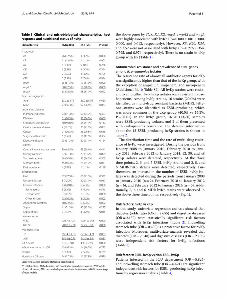
Clinical characteristics (including host response and nutritional status) of hvKp infectionThe basic clinical characteristics, host response and nutritional status of patients with hvKp infections are shown in Table 1. The mean age of patients infected with hvKp is significantly younger than the cKp group (83.2 ± 8.75 years vs 86.2 ± 8.04 years, P = 0.020). A sig-nificantly higher number of patients with hvKp had dia-betes (76.3% versus 54.7%; P = 0.003) as their underlying diseases. Compared with the cKp group, more patients with hvKp infections presented with invasive infec-tions (28.8% versus 6.3%; P = 0.000), liver abscess (10.0% vs 1.1%; P = 0.008), other abscesses (16.3% vs 3.2%; P = 0.035), sepsis shock (11.3% versus 3.2%; P = 0.035) and abdominal infection (22.5% vs 6.3%; P = 0.035). However, the rate of urinary infection in the hvKp group is lower (10.0% vs 21.1%, P = 0.047). In addition, stom-ach tube is also less common in the hvKp group (56.3% vs 74.7%, P = 0.01). With regard to the host response, both WBC (12.87 ± 4.24 vs 10.34 ± 2.95, P = 0.000) and NEU % (78.87 ± 7.60 vs 74.23 ± 7.83, P = 0.000) are higher in patients with hvKp than the cKp group. How-ever, patients infected with hvKp are more likely to have a lower TP (65.14 ± 4.93 vs 62.96 ± 4.71, P = 0.003) and ALB (35.54 ± 2.75 vs 34.45 ± 3.44, P = 0.021). It was also noted that although the SOFA score in the hvKp group is higher (6.84 ± 2.81 vs 4.93 ± 2.59, P = 0.000), the mortal-ity at 28 days (17.5% vs 17.9%, P = 0.946) was not signifi-cantly different between the two groups (Table 1).
Genetic characteristics of hvKp vs cKpPrevious reports showed that the virulence-associated genes rmpA, rmpA2, magA and (K1, K2, K5, K20, K54, and K57) genes for capsular K antigens are associated with hvKp [25–27]. All isolated strains were tested for
Page 4 of 11Liu and Guo Ann Clin Microbiol Antimicrob (2019) 18:4
the above genes by PCR. K1, K2, rmpA, rmpA2 and magA were highly associated with hvKp (P = 0.000, 0.001, 0.000, 0.000, and 0.012, respectively). However, K5, K20, K54, and K57 were not associated with hvKp (P = 0.276, 0.354, 0.795, and 0.974, respectively). There is no strain in cKp group with K5 (Table 1).
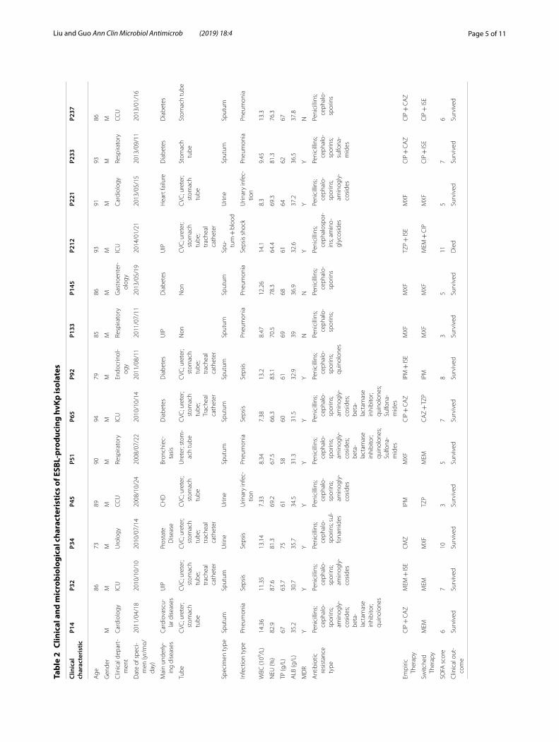
Antimicrobial resistance and prevalence of ESBL genes among K. pneumoniae isolatesThe resistance rate of almost all antibiotic agents for cKp was significantly higher than that of the hvKp group, with the exception of ampicillin, imipenem, and meropenem (Additional file 1: Table S2). All hvKp strains were resist-ant to ampicillin. Two hvKp isolates were resistant to car-bapenems. Among hvKp strains, 16 strains (20.0%) were identified as multi-drug resistant bacteria (MDR). Fifty-one strains were identified as ESBL-producing, which was more common in the cKp group (40.0% vs 16.3%, P = 0.001). In the hvKp group, 16.3% (13/80) samples were ESBL-producing isolates, and 2 of them presented with carbapenems resistance. The detailed information about the 13 ESBL-producing hvKp strains is shown in Table 2.
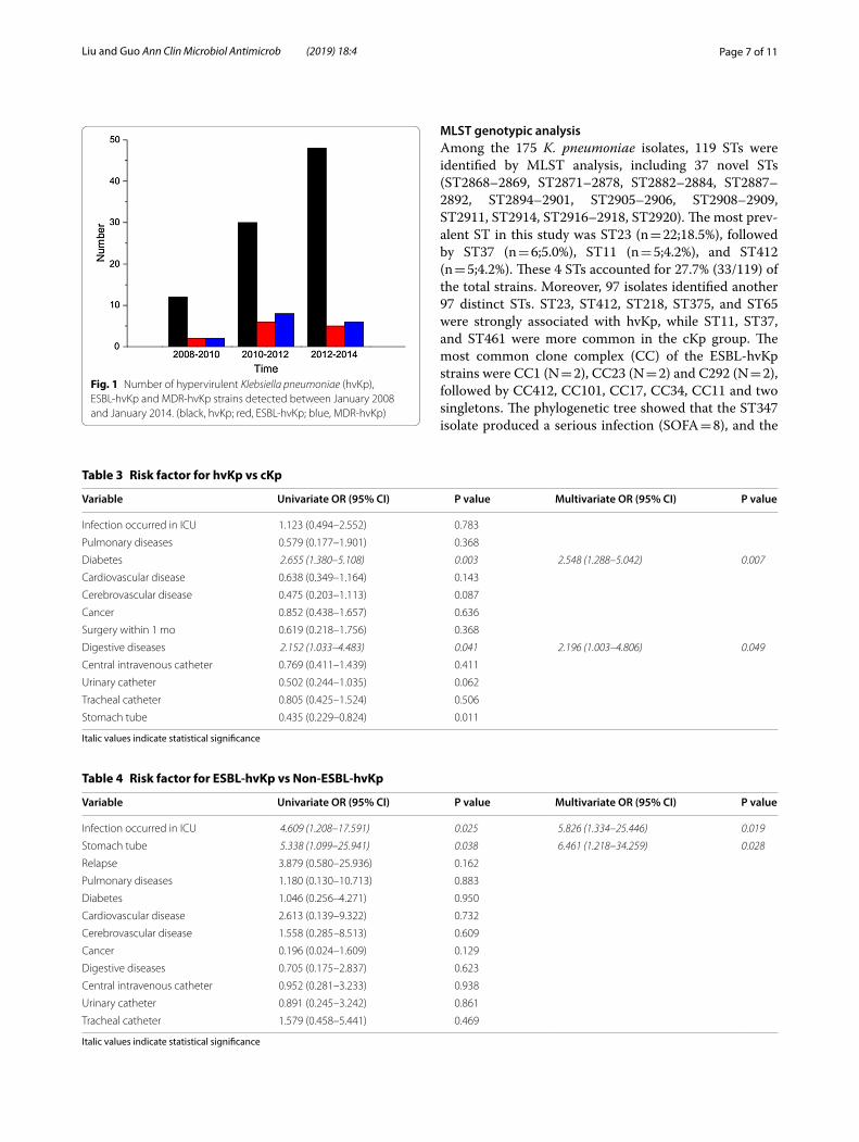
The distribution time and the rate of multi-drug resist-ance of hvKp were investigated. During the periods from January 2008 to January 2010, February 2010 to Janu-ary 2012, February 2012 to January 2014, 12, 30, and 48 hvKp isolates were detected, respectively. At the three time points, 2, 6, and 5 ESBL-hvKp strains and 2, 8, and 6 MDR-hvKp strains were detected, respectively. Fur-thermore, an increase in the number of ESBL-hvKp iso-lates was detected during the periods from January 2008 to January 2010 (n = 2), February 2010 to January 2012 (n = 6), and February 2012 to January 2014 (n = 5). Addi-tionally, 2, 8 and 6 MDR-hvKp stains were observed in the above three time points, respectively (Fig. 1).
Risk factors: hvKp vs cKpIn this study, univariate regression analysis showed that diabetes (odds ratio [OR] = 2.655) and digestive diseases (OR = 2.152) were statistically significant risk factors associated with hvKp infections (Table 2). Indwelling stomach tube (OR = 0.435) is a protective factor for hvKp infection. Moreover, multivariate analysis revealed that diabetes (OR = 2.548) and digestive diseases (OR = 2.196) were independent risk factors for hvKp infections (Table 3).
Risk factors: ESBL‑hvKp vs Non‑ESBL‑hvKpPatients infected in the ICU department (OR = 5.826) and indwelling stomach tube (OR = 6.421) are significant independent risk factors for ESBL-producing hvKp infec-tions by regression analysis (Table 4).
Table 1 Clinical and microbiological characteristics, host response and nutritional status of hvKp
Underline values indicate statistical significance
TP total protein, ALB albumin; HAP hospital acquired pneumonia, WBC white blood cell count, ESBLs extended spectrum beta lactamases, NEU% percentage of neutrophils
Characteristic HvKp (80) cKp (95) P value
K serotype
K1 26 (32.5%) 3 (3.2%) 0.000
K2 11 (13.8%) 1 (1.1%) 0.001
K5 1 (1.3%) 0 (0%) 0.276
K20 2 (2.5%) 5 (5.3%) 0.354
K54 2 (2.5%) 3 (3.2%) 0.795
K57 6 (7.5%) 7 (7.4%) 0.974
rmpA 65 (81.3%) 17 (17.9%) 0.000
rmpA2 58 (72.5%) 19 (20.0%) 0.000
magA 63 (78.8%) 58 (61.1%) 0.012
Basic demographics
Age 83.2 ± 8.75 86.2 ± 8.04 0.020
Male 77 (96.3%) 92 (96.8%) 0.837
Underlying diseases
Pulmonary disease 73 (91.3%) 90 (94.7%) 0.363
Diabetes 61 (76.3%) 52 (54.7%) 0.003
Cardiovascular disease 40 (50.0%) 58 (61.1%) 0.142
Cerebrovascular disease 9 (11.3%) 20 (21.1%) 0.082
Cancer 21 (26.3%) 28 (29.5%) 0.636
Surgery within 1 mo 6 (7.5%) 11 (11.6%) 0.364
Digestive disease 25 (31.3%) 20 (21.1%) 0.124
Catheter
Central intravenous catheter 50 (62.5%) 65 (68.4%) 0.411
Urinary catheter 57 (71.3%) 79 (83.2%) 0.059
Tracheal catheter 24 (30.0%) 33 (34.7%) 0.505
Stomach tube 45 (56.3%) 71 (74.7%) 0.01
Drainage tube 4 (5.0%) 1 (1.1%) 0.119
Infection type
HAP 62 (77.5%) 68 (71.6%) 0.372
Urinary infection 8 (10.0%) 20 (21.1%) 0.047
Invasive infection 23 (28.8%) 6 (6.3%) 0.000
Bacteraemia 5 (6.3%) 4 (4.2%) 0.543
Liver abscess 8 (10.0%) 1 (1.1%) 0.008
Other abscess 13 (16.3%) 3 (3.2%) 0.003
Abdominal infection 18 (22.5%) 6 (6.3%) 0.002
Sepsis 41 (51.3%) 40 (42.1%) 0.227
Septic shock 9 (11.3%) 3 (3.2%) 0.035
Host response
WBC 12.87 ± 4.24 10.34 ± 2.95 0.000
NEU% 78.87 ± 7.60 74.23 ± 7.83 0.000
Nutrition status
TP 65.14 ± 4.93 62.96 ± 4.71 0.003
ALB 35.54 ± 2.75 34.45 ± 3.44 0.021
SOFA score 6.84 ± 2.81 4.93 ± 2.59 0.000
Infection occurred in ICU 13 (16.3%) 14 (14.7%) 0.783
Relapse 5 (6.3%) 5 (5.3%) 0.779
Mortality at 28 days 14 (17.5%) 17 (17.9%) 0.946
Page 5 of 11Liu and Guo Ann Clin Microbiol Antimicrob (2019) 18:4
Tabl
e 2
Clin
ical
and
mic
robi
olog
ical
cha
ract
eris
tics
of E
SBL-
prod
ucin
g hv
Kp is
olat
es
Clin
ical
ch
arac
teri
stic
P14
P32
P34
P45
P51
P65
P92
P133
P145
P212
P221
P233
P237
Age
8673
8990
9479
8586
9391
9386
Gen
der
MM
MM
MM
MM
MM
MM
M
Clin
ical
dep
art-
men
tCa
rdio
logy
ICU
Uro
logy
CCU
Re
spira
tory
ICU
Endo
crin
ol-
ogy
Resp
irato
ryG
astr
oent
er-
olog
yIC
UCa
rdio
logy
Resp
irato
ryCC
U
Dat
e of
spe
ci-
men
(yr/
mo/
day)
2011
/04/
1820
10/1
0/10
2010
/07/
1420
08/1
0/24
2008
/07/
2220
10/1
0/14
2011
/08/
1120
11/0
7/11
2013
/05/
1920
14/0
1/21
2013
/05/
1520
13/0
9/11
2013
/01/
16
Mai
n un
derly
-in
g di
seas
esCa
rdio
vasc
u-la
r dis
ease
sU
IPPr
osta
te
Dis
ease
CH
DBr
onch
iec-
tasi
sD
iabe
tes
Dia
bete
sU
IPD
iabe
tes
UIP
Hea
rt fa
ilure
Dia
bete
sD
iabe
tes
Tube
CVC
; ure
ter;
stom
ach
tube
CVC
; ure
ter;
stom
ach
tube
; tr
ache
al
cath
eter
CVC
; ure
ter;
stom
ach
tube
; tr
ache
al
cath
eter
CVC
; ure
ter;
stom
ach
tube
Ure
ter;
stom
-ac
h tu
beC
VC; u
rete
r; st
omac
h tu
be;
Trac
heal
ca
thet
er
CVC
; ure
ter;
stom
ach
tube
; tr
ache
al
cath
eter
Non
Non
CVC
; ure
ter;
stom
ach
tube
; tr
ache
al
cath
eter
CVC
; ure
ter;
stom
ach
tube
Stom
ach
tube
Stom
ach
tube
Spec
imen
type
Sput
umSp
utum
Urin
eU
rine
Sput
umSp
utum
Sput
umSp
utum
Sput
umSp
u- tum
+ b
lood
Urin
eSp
utum
Sput
um
Infe
ctio
n ty
pePn
eum
onia
Seps
isSe
psis
Urin
ary
infe
c-tio
nPn
eum
onia
Seps
isSe
psis
Pneu
mon
iaPn
eum
onia
Seps
is s
hock
Urin
ary
infe
c-tio
nPn
eum
onia
Pneu
mon
ia
WBC
(109 /L
)14
.36
11.3
513
.14
7.33
8.34
7.38
13.2
8.47
12.2
614
.18.
39.
4513
.3
NEU
(%)
82.9
87.6
81.3
69.2
67.5
66.3
83.1
70.5
78.3
64.4
69.3
81.3
76.3
TP (g
/L)
6763
.775
6158
6061
6968
6164
6267
ALB
(g/L
)35
.230
.735
.734
.531
.331
.532
.939
36.9
32.6
37.2
36.5
37.8
MD
RY
YY
YY
YY
NN
YY
YN
Ant
ibio
tic
resi
stan
ce
type
Peni
cilli
ns;
ceph
alo-
spor
ins;
amin
ogly
-co
side
s; be
ta-
lact
amas
e in
hibi
tor;
quin
olon
es
Peni
cilli
ns;
ceph
alo-
spor
ins;
amin
ogly
-co
side
s
Peni
cilli
ns;
ceph
alo-
spor
ins;
sul-
fona
mid
es
Peni
cilli
ns;
ceph
alo-
spor
ins;
amin
ogly
-co
side
s
Peni
cilli
ns;
ceph
alo-
spor
ins;
amin
ogly
-co
side
s; be
ta-
lact
amas
e in
hibi
tor;
quin
olon
es;
Sulfo
na-
mid
es
Peni
cilli
ns;
ceph
alo-
spor
ins;
amin
ogly
-co
side
s; be
ta-
lact
amas
e in
hibi
tor;
quin
olon
es;
Sulfo
na-
mid
es
Peni
cilli
ns;
ceph
alo-
spor
ins;
quin
olon
es
Peni
cilli
ns;
ceph
alo-
spor
ins;
Peni
cilli
ns;
ceph
alo-
spor
ins
Peni
cilli
ns;
ceph
alos
por-
ins;
amin
o-gl
ycos
ides
Peni
cilli
ns;
ceph
alo-
spor
ins;
amin
ogly
-co
side
s
Peni
cilli
ns;
ceph
alo-
spor
ins;
sulfo
na-
mid
es
Peni
cilli
ns;
ceph
alo-
spor
ins
Empi
ric
Ther
apy
CIP
+ C
AZ
MEM
+ IS
EC
MZ
IPM
MXF
CIP
+ C
AZ
IPM
+ IS
EM
XFM
XFTZ
P +
ISE
MXF
CIP
+ C
AZ
CIP
+ C
AZ
Switc
hed
Ther
apy
MEM
MEM
MXF
TZP
MEM
CA
Z +
TZP
IPM
MXF
MXF
MEM
+ C
IPM
XFC
IP +
ISE
CIP
+ IS
E
SOFA
sco
re6
710
35
78
35
115
76
Clin
ical
out
-co
me
Surv
ived
Surv
ived
Surv
ived
Surv
ived
Surv
ived
Surv
ived
Surv
ived
Surv
ived
Surv
ived
Die
dSu
rviv
edSu
rviv
edSu
rviv
ed
Page 6 of 11Liu and Guo Ann Clin Microbiol Antimicrob (2019) 18:4
Tabl
e 2
(con
tinu
ed)
Clin
ical
ch
arac
teri
stic
P14
P32
P34
P45
P51
P65
P92
P133
P145
P212
P221
P233
P237
Strin
g te
st
leng
th (m
m)
100
3050
100
4020
200
4520
608
850
Viru
lenc
e-as
soci
ated
ge
nes
rmpA
−−
−+
++
+−
++
+−
+rm
pA2
−−
−+
++
+−
++
−−
+m
agA
++
++
++
++
++
−+
+ae
roba
ctin
++
++
++
++
++
++
+cp
s gen
es
K1−
−−
−−
−+
−−
−−
−+
K2−
−−
−−
−−
−−
−−
−−
K5−
−−
−−
−−
−−
−−
−−
K20
−−
−−
−−
−−
−−
−−
−K5
4−
−−
−−
−−
−−
−−
−−
K57
−−
−−
−−
−−
−+
−−
−M
LST
geno
typ-
ing
2899
2892
3428
8812
6441
228
9829
2023
1728
3610
123
Clo
ne c
ompl
exSi
ngle
ton
CC29
2CC
34CC
1CC
11CC
412
CC1
sing
leto
nCC
23CC
17CC
292
CC10
1CC
23
M m
ale,
ICU
inte
nsiv
e ca
re u
nit,
CCU
cor
onar
y ca
re u
nit,
UIP
usu
al in
ters
titia
l pne
umon
ia, C
HD
cor
onar
y he
art d
isea
se, C
VC c
entr
al v
enou
s ca
thet
er, C
IP c
ipro
floxa
cin,
MEM
mer
open
em, I
PM im
ipen
em, T
ZP p
iper
acill
in
tazo
bact
am, I
SE is
epam
icin
, CM
Z ce
fmet
azol
e, M
XF m
oxifl
oxac
in, C
AZ c
efta
zidi
me,
Y y
es, N
no
Page 7 of 11Liu and Guo Ann Clin Microbiol Antimicrob (2019) 18:4
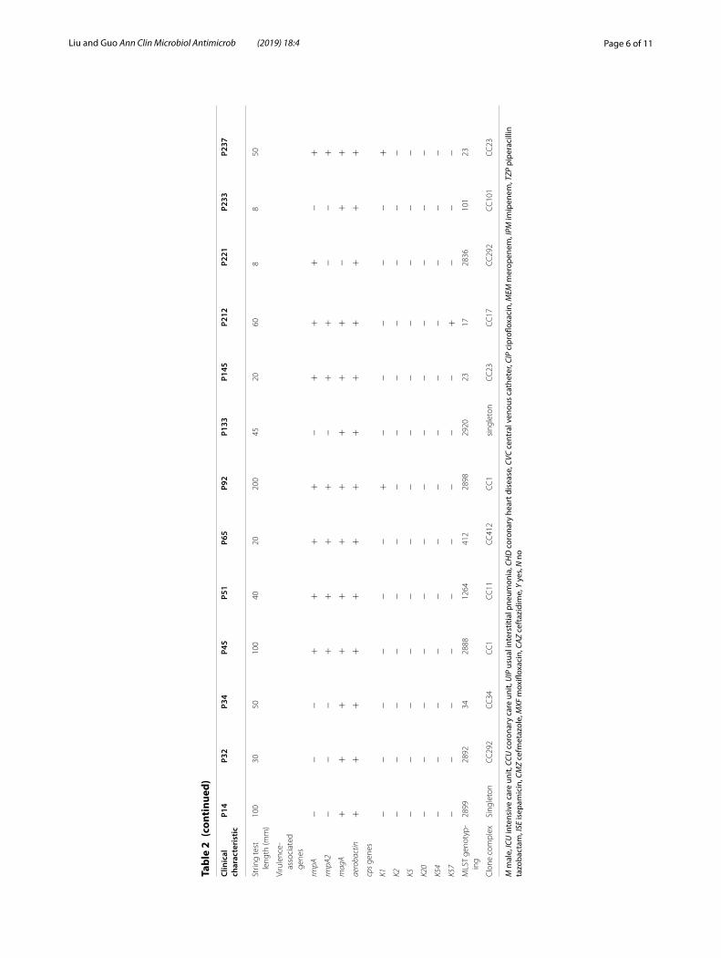
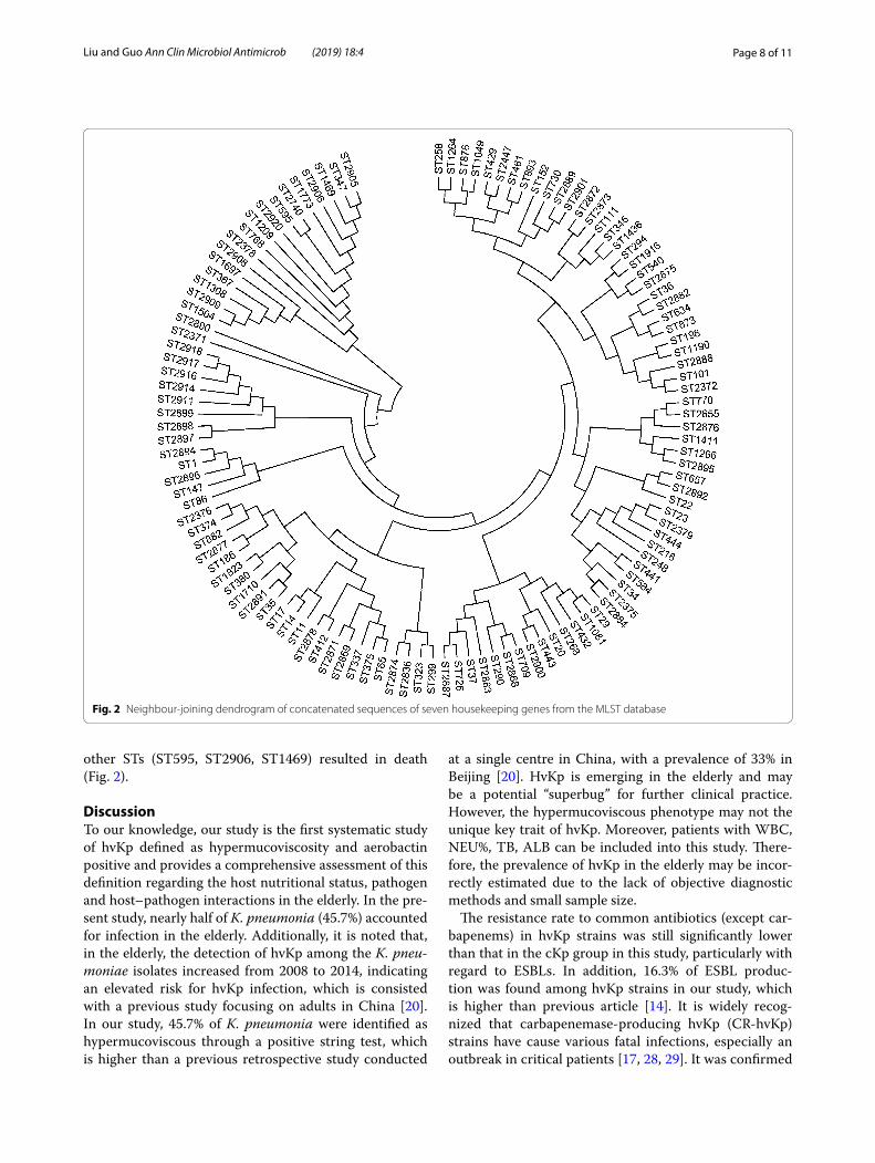
MLST genotypic analysisAmong the 175 K. pneumoniae isolates, 119 STs were identified by MLST analysis, including 37 novel STs (ST2868–2869, ST2871–2878, ST2882–2884, ST2887–2892, ST2894–2901, ST2905–2906, ST2908–2909, ST2911, ST2914, ST2916–2918, ST2920). The most prev-alent ST in this study was ST23 (n = 22;18.5%), followed by ST37 (n = 6;5.0%), ST11 (n = 5;4.2%), and ST412 (n = 5;4.2%). These 4 STs accounted for 27.7% (33/119) of the total strains. Moreover, 97 isolates identified another 97 distinct STs. ST23, ST412, ST218, ST375, and ST65 were strongly associated with hvKp, while ST11, ST37, and ST461 were more common in the cKp group. The most common clone complex (CC) of the ESBL-hvKp strains were CC1 (N = 2), CC23 (N = 2) and C292 (N = 2), followed by CC412, CC101, CC17, CC34, CC11 and two singletons. The phylogenetic tree showed that the ST347 isolate produced a serious infection (SOFA = 8), and the
Fig. 1 Number of hypervirulent Klebsiella pneumoniae (hvKp), ESBL-hvKp and MDR-hvKp strains detected between January 2008 and January 2014. (black, hvKp; red, ESBL-hvKp; blue, MDR-hvKp)
Table 3 Risk factor for hvKp vs cKp
Italic values indicate statistical significance
Variable Univariate OR (95% CI) P value Multivariate OR (95% CI) P value
Infection occurred in ICU 1.123 (0.494–2.552) 0.783
Pulmonary diseases 0.579 (0.177–1.901) 0.368
Diabetes 2.655 (1.380–5.108) 0.003 2.548 (1.288–5.042) 0.007
Cardiovascular disease 0.638 (0.349–1.164) 0.143
Cerebrovascular disease 0.475 (0.203–1.113) 0.087
Cancer 0.852 (0.438–1.657) 0.636
Surgery within 1 mo 0.619 (0.218–1.756) 0.368
Digestive diseases 2.152 (1.033–4.483) 0.041 2.196 (1.003–4.806) 0.049
Central intravenous catheter 0.769 (0.411–1.439) 0.411
Urinary catheter 0.502 (0.244–1.035) 0.062
Tracheal catheter 0.805 (0.425–1.524) 0.506
Stomach tube 0.435 (0.229–0.824) 0.011
Table 4 Risk factor for ESBL-hvKp vs Non-ESBL-hvKp
Italic values indicate statistical significance
Variable Univariate OR (95% CI) P value Multivariate OR (95% CI) P value
Infection occurred in ICU 4.609 (1.208–17.591) 0.025 5.826 (1.334–25.446) 0.019
Stomach tube 5.338 (1.099–25.941) 0.038 6.461 (1.218–34.259) 0.028
Relapse 3.879 (0.580–25.936) 0.162
Pulmonary diseases 1.180 (0.130–10.713) 0.883
Diabetes 1.046 (0.256–4.271) 0.950
Cardiovascular disease 2.613 (0.139–9.322) 0.732
Cerebrovascular disease 1.558 (0.285–8.513) 0.609
Cancer 0.196 (0.024–1.609) 0.129
Digestive diseases 0.705 (0.175–2.837) 0.623
Central intravenous catheter 0.952 (0.281–3.233) 0.938
Urinary catheter 0.891 (0.245–3.242) 0.861
Tracheal catheter 1.579 (0.458–5.441) 0.469
Page 8 of 11Liu and Guo Ann Clin Microbiol Antimicrob (2019) 18:4
other STs (ST595, ST2906, ST1469) resulted in death (Fig. 2).
DiscussionTo our knowledge, our study is the first systematic study of hvKp defined as hypermucoviscosity and aerobactin positive and provides a comprehensive assessment of this definition regarding the host nutritional status, pathogen and host–pathogen interactions in the elderly. In the pre-sent study, nearly half of K. pneumonia (45.7%) accounted for infection in the elderly. Additionally, it is noted that, in the elderly, the detection of hvKp among the K. pneu-moniae isolates increased from 2008 to 2014, indicating an elevated risk for hvKp infection, which is consisted with a previous study focusing on adults in China [20]. In our study, 45.7% of K. pneumonia were identified as hypermucoviscous through a positive string test, which is higher than a previous retrospective study conducted
at a single centre in China, with a prevalence of 33% in Beijing [20]. HvKp is emerging in the elderly and may be a potential “superbug” for further clinical practice. However, the hypermucoviscous phenotype may not the unique key trait of hvKp. Moreover, patients with WBC, NEU%, TB, ALB can be included into this study. There-fore, the prevalence of hvKp in the elderly may be incor-rectly estimated due to the lack of objective diagnostic methods and small sample size.
The resistance rate to common antibiotics (except car-bapenems) in hvKp strains was still significantly lower than that in the cKp group in this study, particularly with regard to ESBLs. In addition, 16.3% of ESBL produc-tion was found among hvKp strains in our study, which is higher than previous article [14]. It is widely recog-nized that carbapenemase-producing hvKp (CR-hvKp) strains have cause various fatal infections, especially an outbreak in critical patients [17, 28, 29]. It was confirmed
Fig. 2 Neighbour-joining dendrogram of concatenated sequences of seven housekeeping genes from the MLST database
Page 9 of 11Liu and Guo Ann Clin Microbiol Antimicrob (2019) 18:4
that the carbapenemase-producing plasmid could be suc-cessfully transferred into hvKp strains, leading to a large burden of disease for the public health [30]. In this study, MDR-hvKp is increasing and 2 hvKp isolates show high resistant to carbapenems in the elderly. It is alarming that CR-hvKp isolates are emerging, and it is a big challenge for medical workers to put forward new clinical interven-tion and prevention. Taken together, these data revealed that antimicrobial resistance is increasing among hvKp strains, which is consisted with a previous study [20]. However, the conclusion requires further investigation at multi-centres with a larger cohort of individuals to be confirmed. Moreover, the results show that the ESBL-hvKp is highly associated with magA in the study. The genetic characteristics and outer genetic environment of the two genes need to be further studied by whole genomic sequencing.
With regard to virulence factors, various types of K-antigens have been reported by now [24, 31, 32]. The most important elements are K1 and K2, which fre-quently result in serious infection [33, 34]. In our study, K1 and K2 are significantly higher in hvKp group than cKp group. RmpA/RmpA2 and MagA responsible for hypermucovicosity phenotype was proposed as another virulent factor in addition to cps K1/K2 [19, 23, 35, 36]. Our results showed that rmpA, rmpA2 and magA were closely related to hvKp group. These results revealed that most of the virulence factors are highly associated with this new definition of hvKp in the elderly.
Previous studies showed that hmvKp are frequently cause of invasive severe infection [37] in young people without underlying disease, such as PLA [2], suppura-tive endophthalmitis [38], and meningitis [39, 40]. In this study, the results show that the mean age of hvKp group is slightly younger than cKp. Invasive infection, especially liver abscess and other abscesses, occurred significantly more often with the new definition of hvKp group. In addition, the nutritional status (TP and ALB), host reac-tion (WBC and NEU %) and SOFA score of the hvKp group are significantly higher than cKp group. Moreover, the above results may also reveal that from the host, path-ogen, and host–pathogen interactions, the new definition for hvKp may be highly associated with the real hypervir-ulence. Thus, focusing only on STs, serotypes, and other pathogen genomic data may not be sufficient to define hvKp. Host, pathogen and host–pathogen interactions should be taken into consideration when defining hvKp. The inflammatory factors (such as interleukin, C-reactive protein, tumour necrosis factor) and nutritional status (prealbumin, thickness of subcutaneous fat) may be more comprehensively considered in future studies.
It is essential for clinicians to respond immediately to hvKp infections, which could cause serious infections
and a more severe inflammatory reaction than cKp, especially in the elderly, children and immunocompro-mised patients. Thus, developing a better understand-ing of the risk factors for hvKp is urgent and essential. Our results demonstrate that patients with diabetes and digestive diseases are more likely to be infected with hvKp, which is consistent with a previous study in China [14, 20]. Additionally, infections in the ICU and patients with indwelling stomach tube are risk factors for ESBL-hvKp, which may be related with potentially prolonged hospitalized course and antibiotic exposure. Clinicians should pay close attention to these risk fac-tors in clinical practice to reduce emergence of MDR isolates. Previous study [28] suggested that wards previ-ously infected with CR-hvKp should be left unoccupied for more than 2 weeks after disinfection and before the admission of new patients. However, it may be difficult to be implemented in China, a populous and develop-ing country. Thus, it is urgent to make a cluster strategy from the host nutritional status, pathogen invasiveness and host–pathogen reaction to prevent MDR-hvKp, especially CR-hvKp.
There were some limitations in our study. First, it was a retrospective study at a single centre over 6 years. More inflammatory factors and nutrition indicators were not measured. Second, in vitro and in vivo experiments, such as galleria mellonella model, mouse models and a human neutrophil assay, may be further needed for identify-ing this new definition of hvKp. Third, to further explore the pathogen genomic characteristics, whole genome sequencing may be needed for further study. A prospec-tive multi-centre study that includes more isolates, focus-ing on host, pathogen and host–pathogen interactions, is needed to better define the hvKp strains.
ConclusionsThe hvKp strains defined as hypermucoviscous and aerobactin positive are more likely to cause more severe inflammatory reaction in host and invasive infection, such as PLA and sepsis shock. To further understand hvKp, the host, pathogen and host–pathogen interac-tions may be the key element. At present, the preva-lence of hvKp in the elderly, especially ESBL-hvKp and MDR-hvKp is increasing. It is essential to enhance the clinical awareness and management of hvKp infections.
Additional file
Additional file 1: Table S1. Primers. Table S2. Comparison of antimicro-bial resistance to hvKp and cKp.
Page 10 of 11Liu and Guo Ann Clin Microbiol Antimicrob (2019) 18:4
Authors’ contributionsJG and CL were responsible for study design, performing PCR, statistical analy-ses, writing and collecting clinical data. JG performed critical data review. Both authors read and approved the final manuscript.
Author details1 Department of Respiratory Medicine, Peking Union Medical College, Chinese Academy of Medical Sciences, Beijing, China. 2 Department of Respiratory Medicine, Beijing Tsinghua Changgung Hospital, School of Clinical Medicine, Tsinghua University, No. 168 Litang Road, Changping District, Beijing 102218, China. 3 Department of Geriatric Respiratory Medicine, Chinese PLA General Hospital, Beijing, China.
AcknowledgementsWe thank the team of curators of the Institut Pasteur MLST and whole genome MLST databases for curating the data and making them publicly available at http://bigsd b.paste ur.fr.
Competing interestsThe authors declare that they have no competing interests.
Availability of data and materialsThe datasets used and/or analysed during the current study are available from the corresponding author on reasonable request.
Ethics approval and consent to participateInformed consent was not needed due to the retrospective nature of the study. The study was approved by the Chinese PLA General Hospital Ethics Committee, and the Guidelines for Human Experimentation (PR. China) were followed throughout.
FundingThis work was supported by the China postdoctoral science foundation (Grant Number 2014M562610) and the Excellent Young Program of the Organiza-tion Department of Beijing Municipal Party Committee (Grant Number 2016000057592G258).
Publisher’s NoteSpringer Nature remains neutral with regard to jurisdictional claims in pub-lished maps and institutional affiliations.
Received: 31 July 2018 Accepted: 31 December 2018
References 1. Gupta A. Hospital-acquired infections in the neonatal intensive care unit-
Klebsiella pneumoniae. Semin Perinatol. 2002;26:340–5. 2. Ko WC, Paterson DL, Sagnimeni AJ, Hansen DS, Von Gottberg A, Mohapa-
tra S, et al. Community-acquired Klebsiella pneumoniae bacteremia: global differences in clinical patterns. Emerg Infect Dis. 2002;8:160–6.
3. Podschun R, Ullmann U. Klebsiella spp. as nosocomial pathogens: epi-demiology, taxonomy, typing methods, and pathogenicity factors. Clin Microbiol Rev. 1998;11:589–603.
4. Casanova C, Lorente JA, Carrillo F, Perez-Rodriguez E, Nunez N. Klebsiella pneumoniae liver abscess associated with septic endophthalmitis. Arch Intern Med. 1989;149:1467.
5. Shon AS, Bajwa RP, Russo TA. Hypervirulent (hypermucoviscous) Klebsiella pneumoniae: a new and dangerous breed. Virulence. 2013;4:107–18.
6. Fang CT, Lai SY, Yi WC, Hsueh PR, Liu KL, Chang SC. Klebsiella pneumoniae genotype K1: an emerging pathogen that causes septic ocular or central nervous system complications from pyogenic liver abscess. Clin Infect Dis. 2007;45:284–93.
7. Decre D, Verdet C, Emirian A, Le Gourrierec T, Petit JC, Offenstadt G, et al. Emerging severe and fatal infections due to Klebsiella pneumoniae in two university hospitals in France. J Clin Microbiol. 2011;49:3012–4.
8. Pomakova DK, Hsiao CB, Beanan JM, Olson R, MacDonald U, Keynan Y, et al. Clinical and phenotypic differences between classic and
hypervirulent Klebsiella pneumonia: an emerging and under-recognized pathogenic variant. Eur J Clin Microbiol Infect Dis. 2012;31:981–9.
9. Yu WL, Fung CP, Ko WC, Cheng KC, Lee CC, Chuang YC. Polymerase chain reaction analysis for detecting capsule serotypes K1 and K2 of Klebsiella pneumoniae causing abscesses of the liver and other sites. J Infect Dis. 2007;195:1235–6 (author reply 1236).
10. Wang JH, Liu YC, Lee SS, Yen MY, Chen YS, Wang JH, et al. Primary liver abscess due to Klebsiella pneumoniae in Taiwan. Clin Infect Dis. 1998;26:1434–8.
11. Catalan-Najera JC, Garza-Ramos U, Barrios-Camacho H. Hypervirulence and hypermucoviscosity: two different but complementary Klebsiella spp. phenotypes? Virulence. 2017;8:1–13.
12. Zhang Y, Zeng J, Liu W, Zhao F, Hu Z, Zhao C, et al. Emergence of a hyper-virulent carbapenem-resistant Klebsiella pneumoniae isolate from clinical infections in China. J Infect. 2015;71:553–60.
13. Lin YC, Lu MC, Tang HL, Liu HC, Chen CH, Liu KS, et al. Assessment of hypermucoviscosity as a virulence factor for experimental Klebsiella pneumoniae infections: comparative virulence analysis with hypermu-coviscosity-negative strain. BMC Microbiol. 2011;11:50.
14. Zhang Y, Zhao C, Wang Q, Wang X, Chen H, Li H, et al. High prevalence of hypervirulent Klebsiella pneumoniae infection in china: geographic distri-bution, clinical characteristics, and antimicrobial resistance. Antimicrob Agents Chemother. 2016;60:6115–20.
15. Russo TA, Olson R, MacDonald U, Beanan J, Davidson BA. Aerobactin, but not yersiniabactin, salmochelin, or enterobactin, enables the growth/sur-vival of hypervirulent (hypermucoviscous) Klebsiella pneumoniae ex vivo and in vivo. Infect Immun. 2015;83:3325–33.
16. Russo TA, Olson R, Macdonald U, Metzger D, Maltese LM, Drake EJ, et al. Aerobactin mediates virulence and accounts for increased siderophore production under iron-limiting conditions by hypervirulent (hypermu-coviscous) Klebsiella pneumoniae. Infect Immun. 2014;82:2356–67.
17. Gu D, Dong N, Zheng Z, Lin D, Huang M, Wang L, et al. A fatal outbreak of ST11 carbapenem-resistant hypervirulent Klebsiella pneumoniae in a Chinese hospital: a molecular epidemiological study. Lancet Infect Dis. 2018;18(1):37–46.
18. Struve C, Roe CC, Stegger M, Stahlhut SG, Hansen DS, Engelthaler DM, et al. Mapping the evolution of hypervirulent Klebsiella pneumoniae. MBio. 2015;6:e00630.
19. Siu LK, Yeh KM, Lin JC, Fung CP, Chang FY. Klebsiella pneumoniae liver abscess: a new invasive syndrome. Lancet Infect Dis. 2012;12:881–7.
20. Li W, Sun G, Yu Y, Li N, Chen M, Jin R, et al. Increasing occurrence of antimicrobial-resistant hypervirulent (hypermucoviscous) Klebsiella pneu-moniae isolates in China. Clin Infect Dis. 2014;58:225–32.
21. Magiorakos AP, Srinivasan A, Carey RB, Carmeli Y, Falagas ME, Giske CG, et al. Multidrug-resistant, extensively drug-resistant and pandrug-resistant bacteria: an international expert proposal for interim standard definitions for acquired resistance. Clin Microbiol Infect. 2012;18:268–81.
22. Choi MJ, Ko KS. Loss of hypermucoviscosity and increased fitness cost in colistin-resistant Klebsiella pneumoniae sequence type 23 strains. Antimi-crob Agents Chemother. 2015;59:6763–73.
23. Yu WL, Ko WC, Cheng KC, Lee HC, Ke DS, Lee CC, et al. Association between rmpA and magA genes and clinical syndromes caused by Klebsiella pneumoniae in Taiwan. Clin Infect Dis. 2006;42:1351–8.
24. Cheng NC, Yu YC, Tai HC, Hsueh PR, Chang SC, Lai SY, et al. Recent trend of necrotizing fasciitis in Taiwan: focus on monomicrobial Klebsiella pneumo-niae necrotizing fasciitis. Clin Infect Dis. 2012;55:930–9.
25. Jung SW, Chae HJ, Park YJ, Yu JK, Kim SY, Lee HK, et al. Microbiological and clinical characteristics of bacteraemia caused by the hypermucovis-cosity phenotype of Klebsiella pneumoniae in Korea. Epidemiol Infect. 2013;141:334–40.
26. Lin JC, Yeh KM, Chang FY. The distant metastasis of pyogenic liver abscess caused by Klebsiella pneumoniae serotype K2 and the underlying disease of diabetes mellitus should be carefully interpreted. Clin Infect Dis. 2007;45:1531–2 (author reply 1532–1533).
27. Lin WH, Wang MC, Tseng CC, Ko WC, Wu AB, Zheng PX, et al. Clinical and microbiological characteristics of Klebsiella pneumoniae isolates causing community-acquired urinary tract infections. Infection. 2010;38:459–64.
28. Zhang R, Lin D, Chan EW, Gu D, Chen GX, Chen S. Emergence of carbape-nem-resistant serotype K1 hypervirulent Klebsiella pneumoniae strains in China. Antimicrob Agents Chemother. 2015;60:709–11.
Page 11 of 11Liu and Guo Ann Clin Microbiol Antimicrob (2019) 18:4
• fast, convenient online submission
•
thorough peer review by experienced researchers in your field
• rapid publication on acceptance
• support for research data, including large and complex data types
•
gold Open Access which fosters wider collaboration and increased citations
maximum visibility for your research: over 100M website views per year •
At BMC, research is always in progress.
Learn more biomedcentral.com/submissions
Ready to submit your research ? Choose BMC and benefit from:
29. Liu Y, Li XY, Wan LG, Jiang WY, Yang JH, Li FQ. Virulence and transferability of resistance determinants in a novel Klebsiella pneumoniae sequence type 1137 in China. Microb Drug Resist. 2014;20:150–5.
30. Siu LK, Huang DB, Chiang T. Plasmid transferability of KPC into a virulent K2 serotype Klebsiella pneumoniae. BMC Infect Dis. 2014;14:176.
31. Pan YJ, Fang HC, Yang HC, Lin TL, Hsieh PF, Tsai FC, et al. Capsular polysac-charide synthesis regions in Klebsiella pneumoniae serotype K57 and a new capsular serotype. J Clin Microbiol. 2008;46:2231–40.
32. Chuang YP, Fang CT, Lai SY, Chang SC, Wang JT. Genetic determinants of capsular serotype K1 of Klebsiella pneumoniae causing primary pyogenic liver abscess. J Infect Dis. 2006;193:645–54.
33. Yeh KM, Kurup A, Siu LK, Koh YL, Fung CP, Lin JC, et al. Capsular serotype K1 or K2, rather than magA and rmpA, is a major virulence determinant for Klebsiella pneumoniae liver abscess in Singapore and Taiwan. J Clin Microbiol. 2007;45:466–71.
34. Brisse S, Fevre C, Passet V, Issenhuth-Jeanjean S, Tournebize R, Diancourt L, et al. Virulent clones of Klebsiella pneumoniae: identification and evo-lutionary scenario based on genomic and phenotypic characterization. PLoS ONE. 2009;4:e4982.
35. Yeh KM, Chang FY, Fung CP, Lin JC, Siu LK. magA is not a specific virulence gene for Klebsiella pneumoniae strains causing liver abscess but is part of the capsular polysaccharide gene cluster of K. pneumoniae serotype K1. J Med Microbiol. 2006;55:803–4.
36. Fang CT, Chuang YP, Shun CT, Chang SC, Wang JT. A novel virulence gene in Klebsiella pneumoniae strains causing primary liver abscess and septic metastatic complications. J Exp Med. 2004;199:697–705.
37. Liu YM, Li BB, Zhang YY, Zhang W, Shen H, Li H, et al. Clinical and molecu-lar characteristics of emerging hypervirulent Klebsiella pneumoniae bloodstream infections in mainland China. Antimicrob Agents Chem-other. 2014;58:5379–85.
38. Abdul-Hamid A, Bailey SJ. Klebsiella pneumoniae liver abscess and endophthalmitis. BMJ Case Rep. 2013;2013:bcr2013008690.
39. Tang LM, Chen ST, Hsu WC, Chen CM. Klebsiella meningitis in Taiwan: an overview. Epidemiol Infect. 1997;119:135–42.
40. Chang WN, Huang CR, Lu CH, Chien CC. Adult Klebsiella pneumoniae meningitis in Taiwan: an overview. Acta Neurol Taiwan. 2012;21:87–96.