Embed Size (px)
Citation preview


Alaa S. Abdul Mahdi Shoman 2013
1 “BOND STRENGTH TO ROOT DENTIN AND FLUID FILTRATION TEST OF SEVERAL RESIN SEALER
AND MICRO-RAMAN SPECTROSCOPY STUDY OF DENTIN AFTER USING IRRIGATION SOLUTIONS”
“BOND STRENGTH TO ROOT DENTIN AND FLUID FILTRATION
TEST OF SEVERAL RESIN SEALER AND MICRO-RAMAN
SPECTROSCOPY STUDY OF DENTIN AFTER USING IRRIGATION
SOLUTIONS”

Editor: Editorial de la Universidad de GranadaAutor: Alaa S. Abdul Mahdi Shoman D.L.: GR 712-2014ISBN: 978-84-9028-895-5


Alaa S. Abdul Mahdi Shoman 2013
2 “BOND STRENGTH TO ROOT DENTIN AND FLUID FILTRATION TEST OF SEVERAL RESIN SEALER
AND MICRO-RAMAN SPECTROSCOPY STUDY OF DENTIN AFTER USING IRRIGATION SOLUTIONS”
Acknowledgements
My sincere acknowledgment and appreciation to all the individuals who assisted me
in this undertaking, I am truly grateful for all their contributions. First and foremost, I thank
Allah who allowed and enabled me to complete this project successfully and to the best of
my ability, I would like to extend my sincere thanks to Ministry of Higher Education, all
the field team.
I would like to extend my deepest thanks and appreciation to Professor Santiago
González López for his inspiration, encouragement, wisdom and understanding in getting
this thesis off the ground. A special thanks to Professor Mª Victoria Bolaños Carmona who
supervise through to the end and provided extremely valuable insight and direction from
the beginning with great passion and interest.
I would like also to express my thanks and appreciation to the members of the
defense committee, and to the University of Granada. I have reached this place not only by
my efforts alone, but also through your support and kindness
I wish to express my sincere gratitude to my father and mother who helped and
supported me all through the work. In addition to Messrs. Dr. Ridha Abbas, Dr. Jawad
Abbas and Amin Abbas.
Beyond the academic world, I would like to express my heartfelt gratitude for the
love and support of my husband Ammar and my three sons Hassan, Zahraa and Maryem,
who supported me all through the work on this research until it was completed.

Alaa S. Abdul Mahdi Shoman 2013
3 “BOND STRENGTH TO ROOT DENTIN AND FLUID FILTRATION TEST OF SEVERAL RESIN SEALER
AND MICRO-RAMAN SPECTROSCOPY STUDY OF DENTIN AFTER USING IRRIGATION SOLUTIONS”
INDEX
Part 1
Resumen…………………………………………………………………………….........9-20
Part 2
BOND STRENGTH TO ROOT DENTINAND FLUID
FILTRATION TEST OF SEVERALRESINSEALERS…………………………………...21
2.1Abstract……………………………………………………………………………...22-23
2.2 Introduction…………………………………………………………………………….24
A.2.2. Permeability…………………………………………...............................................25
A.2.2.1. Permeability definition…………………………………………………………....25
A.2.2.2. Importance of permeability…………………………….........................................25
A.2.2.3. Factors affecting fluid filtration……………………………………………….24-25
A.2.2.4. Mechanism of fluid filtration………………………………………………….25-26
A.2.2.5. Root canal treatment………………………………………………………………26
A.2.2.5.1. Objectives ………………………………………………………………………26
A.2.2.5.2. Three dimensional filling……………………….................................................27
A.2.2.5.3. The basic principles of root canal filling………………………………………..27
A.2.2.5.4. Properties of root canal filling material……………............................................27
A.2.2.5.5. Function of root filling…...……………………………………………………..28
A.2.2.5.6. Types and composition of endodontic filling materials….…………………......28
A.2.2.5.6.1. Gutta-percha…………………………………………………………………..28
A.2.2.5.6.2. Resin-based filling material……………..........................................................29
A.2.2.6. Sealer……………………………………..............................................................29
Alaa S. Abdul Mahdi Shoman 2013
4 “BOND STRENGTH TO ROOT DENTIN AND FLUID FILTRATION TEST OF SEVERAL RESIN SEALER
AND MICRO-RAMAN SPECTROSCOPY STUDY OF DENTIN AFTER USING IRRIGATION SOLUTIONS”
A.2.2.6.1. Function of sealer…………………………........................................................29
A.2.2.6.2. Requirements of sealer………………………………………………….............30
A.2.2.6.3. Types of endodontic sealers……………………………….……........................31
A.2.2.6.3.1. Epoxy resins……………………………..........................................................31
A.2.2.6.3.1.1. AH Plus………………………………………………………………….31-33
A.2.2.6.3.2. Methacrylate Resin Based Sealers……………………………………………34
A.2.2.6.3.2.1. EndoREZ..................................................................................................34-35
A.2.2.6.3.2.2. RealSeal endodontic obturation system………………………………...35-37
A.2.2.7. Microleakage definition…………………………………………………………..38
A.2.2.7.1. Types of microleakage……………………….....................................................38
A.2.2.7.2. Location of microleakage……………................................................................38
A.2.2.7.3. Microleakage development…………………………………………………......38
A.2.2.7.4. Adverse effect of microleakage……………………………………………........39
A.2.2.7.5. Factors affect microleakage…………………………………………………39-40
A.2.2.7.6. Microleakage modification……………………………………………………..40
A.2.2.7.7. Microleakage studies……………………………………………………………40
A.2.2.8. Fluid filtration system………………………………………………………….…41
A.2.2.8.1. Advantages of fluid filtration system…………………………………………...41
A.2.2.8.2. Properties of fluid filtration system………………………………………......…42
A.2.2.7.7.1. Factors influencing microleakage studies…………….....................................42
B.2.2. Micro Push-out bond strength……………………………………………………....42
B.2.2.1. Bond strength definition…………………………………………………………..42
B.2.2.2. Importance of adhesion……………………...........................................................43
Alaa S. Abdul Mahdi Shoman 2013
5 “BOND STRENGTH TO ROOT DENTIN AND FLUID FILTRATION TEST OF SEVERAL RESIN SEALER
AND MICRO-RAMAN SPECTROSCOPY STUDY OF DENTIN AFTER USING IRRIGATION SOLUTIONS”
B.2.2.3. Mechanism of adhesion of resin-based sealers…………………………………...43
B.2.2.4. Modification of dentin bonding…………………………………………………...44
B.2.2.5. Adhesion Studies………………………………………………………………….44
B.2.2.6. Micro Push-out test………………………..............................................................45
B.2.2.7. Factors affecting the bond strength of cement to dentin………………………….45
2.3. Objectives……………………………………………………………………………...46
2.4. Materials and Methods………………………………………………………………...48
2.4.1. Collection of teeth…………………………………………………………………...49
2.4.2. Specimen preparation……………………..................................................................49
2.4.2.1. Instrumentation procedure……………………………………………………..49-50
2.4.2.2. Obturation procedure……………………………………………………………...51
2.4.2.2.1. AH Plus…………………………………………………………………….........51
2.4.2.2.2. EndoREZ……………………….....................................................................52-53
2.4.2.2.3. RealSeal sealer…………………….................................................................54-55
2.4.3. Leakage evaluation…………………………………………………………………..55
2.4.4. Description of the fluid filtration apparatus……………………………………...56-57
2.4.5. Measurement of fluid filtration…………………………………………………..58-59
2.4.6. Preparation of samples for µPush-out test……………….....................................60-61
2.4.7. Micro Push-out test……………………………………………………………....62-64
2.5. Results…………………................................................................................................65
A. Permeability…………………………………………………………………………….65
A.2.5.1. Statistical analysis………………….......................................................................66
A.2.5.2. Results…………………………………………………………………………66-67
Alaa S. Abdul Mahdi Shoman 2013
6 “BOND STRENGTH TO ROOT DENTIN AND FLUID FILTRATION TEST OF SEVERAL RESIN SEALER
AND MICRO-RAMAN SPECTROSCOPY STUDY OF DENTIN AFTER USING IRRIGATION SOLUTIONS”
B. MICRO PUSH-OUT BOND STRENGTH……………………………………....…......68
B.2.5.1. Statistical analysis……………………………………………...............................68
B.2.5.2. Results…………………………………………………………………………….69
B.2.5.2.1.Comparison among root levels for each material…………..................................70
B.2.5.2.2. Comparisons among materials in each root level…………………………...70-72
B.2.5.2.3. Mode of failure……………………………………………………………...73-75
2.6. Conclusion………………………………………………………………………..76-787
2.7. Discussion………………………………………………………………………….78-85
Part 3
MICRO-RAMAN SPECTROSCOPY OF ROOT DENTIN
AFTER USING IRRIGATION SOLUTIONS……………………………………..……...86
3.1. Abstract………………………….............................................................................87-89
3.2. Introduction………………………………………...................................................90-92
3.2.1 Dentin………...............................................................................................................93
3.2.1.1. Physical properties……….......................................................................................93
3.2.1.2. Chemical characteristics of dentin……………………………………………..94-95
3.2.2. Dentinogenesis……………………………................................................................95
3.2.3. Organic matrix……………...................................................................................96-97
3.2.4. Mineralization……………………………………………………………………97-98
3.2.5. Root formation…………………................................................................................98
3.2.6. Dentin microstructure……………………..................................................................99
3.3. Mechanical instrumentation……………………………………………………..100-101
3.3.1. Nickel titanium elasticity…………………………….......................................101-102
Alaa S. Abdul Mahdi Shoman 2013
7 “BOND STRENGTH TO ROOT DENTIN AND FLUID FILTRATION TEST OF SEVERAL RESIN SEALER
AND MICRO-RAMAN SPECTROSCOPY STUDY OF DENTIN AFTER USING IRRIGATION SOLUTIONS”
3.3.2. Crown-down technique………………………………………………………..102-104
3.3.3. Sodium hypochlorite…………………………………………………………….....105
3.3.4. EDTA……………………………………………………………………………....106
3.3.5. Bonding agent…………………………………………………………………107-108
3.4. Confocal micro-Raman spectroscopy………………..................................................108
3.4.1. The theory of micro Raman spectroscopy……………………………………...….109
3.4.2. Raman scattering phenomenon………………………………………………..109-110
3.4.3. The components of the Raman system……………………………………….…….110
3.4.4. Micro Raman spectrometer………………………………………………….……..111
3.4.5. Micro Raman spectroscopy applications………………...................................111-112
3.4.6. Raman advantages………………………….............................................................112
3.3. Objectives……….................................................................................................113-114
3.4. Materials and methods…………….............................................................................115
3.4.1. Sample selection…………………………………………………………………...116
3.4.2. Preparation of teeth………………………………………………………...…........116
3.4.2.1. Instrumentation……………………………………………………………...116-117
3.4.2.2. Preparation of control group……………………………………………………..118
3.4.3. Grouping…………………………………………………………………………...118
3.4.4. Preparation of samples for micro-Raman……………………………………..119-120
3.4.5. Micro-Raman spectroscopy…………………………………………………...121-125
3.5. Results……………………………......................................................................126-128
3.5.1. Statistical analysis……………………….................................................................129
3.5.2. Effect of sodium hypochlorite on the organic
Alaa S. Abdul Mahdi Shoman 2013
8 “BOND STRENGTH TO ROOT DENTIN AND FLUID FILTRATION TEST OF SEVERAL RESIN SEALER
AND MICRO-RAMAN SPECTROSCOPY STUDY OF DENTIN AFTER USING IRRIGATION SOLUTIONS”
and mineral component of root canal dentin ……………………………………….129-133
3.5.3. Effect of 17% EDTA on the organic
and mineral component of root canal dentin ………………………………………..134-139
3.5.4. Effect of self-etching primer on the organic
and mineral component of root canal dentin………………………………………..139-144
3.6. Conclusion…………………..…………………………………………………..145-146
3.7. Discussion………………………….....................................................................147-154
Part 4
REFERENCE………………………….……………………………………………..155-194
Part 5
Article………………………………………………….………………………………….195
Alaa S. Abdul Mahdi Shoman 2013
9 “BOND STRENGTH TO ROOT DENTIN AND FLUID FILTRATION TEST OF SEVERAL RESIN SEALER
AND MICRO-RAMAN SPECTROSCOPY STUDY OF DENTIN AFTER USING IRRIGATION SOLUTIONS”
RESUMEN
“Fuerza adhesiva a la dentina radicular, test de filtración de fluidos de
varios selladores de resina y estudio mediante espectroscopia micro-Raman
de la dentina tras la utilización de soluciones irrigantes”

Alaa S. Abdul Mahdi Shoman 2013
10 “BOND STRENGTH TO ROOT DENTIN AND FLUID FILTRATION TEST OF SEVERAL RESIN SEALER
AND MICRO-RAMAN SPECTROSCOPY STUDY OF DENTIN AFTER USING IRRIGATION SOLUTIONS”
Fuerza adhesiva a la dentina radicular and test de filtración de fluídos de varios
selladores de resina
Introducción
Los objetivos fundamentales del tratamiento endodóncico son la limpieza y
conformación del conducto radicular y la obtención de sellado apical hermético. De forma
ideal, un sellador de conductos radiculares debe adherirse tanto a las paredes del conducto
como al material de relleno en el interior del canal radicular. Por este motivo, se han
desarrollado numerosos materiales de sellado radicular basados en los principios de la
adhesión dentinaria; estos selladores, en ocasiones, no han logrado mejorar los resultados,
en términos de fuerza adhesiva, obtenidos con la utilización del sellador no adhesivo AH
Plus231,230,212
Aunque no se ha encontrado una relación significativa entre la fuerza de adhesión y
la microfiltración apical, hemos de asumir que un sellado firme es, quizá, más importante
que la fuerza adhesiva para evitar la filtración apical72
. Lamentablemente, no existe un
método aceptado universalmente para medir la filtración apical y se han obtenido en
ocasiones resultados contradictorios que han llevado cuestionar la relevancia clínica de
estos tests99,226
.
El test mecánico de expulsión del relleno radicular, habitualmente conocido por su
nombre en inglés, Push-out bond strength test, se ha popularizado para la determinación de
la eficacia adhesiva en endodoncia, aunque la relación entre test y la filtración apical
permanece indeterminada.
Objetivos
Determinar la fuerza adhesiva a la dentina del conducto radicular y la capacidad de
sellado de dos selladores de resina de metacrilato (RealSeal and EndoREZ) y comparlas
con las del sellador no adhesivo AH Plus.
Para ello, se aplicaron los test de expulsión y de filtración de fluidos. La hipótesis nula a
verificar es que no hay diferencias en la fuerza de adhesión y las propiedades de sellado de
RealSeal, EndoREZ y AH Plus.
Alaa S. Abdul Mahdi Shoman 2013
11 “BOND STRENGTH TO ROOT DENTIN AND FLUID FILTRATION TEST OF SEVERAL RESIN SEALER
AND MICRO-RAMAN SPECTROSCOPY STUDY OF DENTIN AFTER USING IRRIGATION SOLUTIONS”
Material y métodos
Se utilizaron 60 dientes humanos unirradiculares extraídos por patología
periodontal. Tras seccionar la corona de forma perpendicular al eje longitudinal del diente,
las raíces se sometieron a preparación endodóncica manual, utilizando la técnica de paso
atrás (telescópica). La longitud de trabajo se determinó sustrayendo 1 mm de la longitud de
una lima k, de tamaño 15 (Dentsply Maillefer, Ballaigues, Switzerland) llevada hasta el
foramen apical. La preparación biomecánica se llevó a cabo manualmente, con limas k de
la misma marca, hasta el n° # 40. El irrigante endodóncico fue hipoclorito sódico al 2.5%
durante 1 minuto, tras cada lima. En las raíces del grupo sellado con RealSeal, se usó
EDTA al 17% (Colgate Oral Care Company, Waverly, Australia). En todos los grupos se
practicó una irrigación final con agua destilada durante 1 minuto y el conducto se secó con
puntas de papel.
Las raíces se asignaron al azar en grupos iguales (n=20) según el sellador a utilizar:
Grupo 1: AH PlusTM (Dentsply De Trey, Konstanz, Germany), Grupo 2: EndoREZ®
(Ultradent products, Inc. Utah, USA) y Grupo 3: RealSeal TM (SybronEndo. Glendora,
CA, USA). Los selladores se utilizaron según las instrucciones y utilizando los aditamentos
recomendados por cada fabricante.
En el grupo AH Plus se utilizó como relleno intracanal un cono maestro de
gutapercha de conicidad 40.02 y conos accesorios de los números 25 y 20. En el grupo 2,
EndoREZ se aplicó en la totalidad del conducto, obturándose con un cono de gutapercha
impregnado en resina de conicidad 40.02 (EndoREZ point) y conos adicionales de tamaños
#25, 20 de la misma marca. En el grupo 3, el imprimador RealSeal (Sybron Endo,
Glendora, CA) se dejó actuar en el conducto durante 30 s, aplicando posteriormente el
sellador en la totalidad del canal y obturando con un cono maestro RealSeal de conicidad
40.02 y puntas accesorias #25, 20 de la misma marca. En todos los casos se utilizó la
técnica de condensación lateral en frío.
Las raíces se almacenaron en cámara de humedad a 37ºC durante 24 horas, para
completar el proceso de polimerización, tras lo cual se asignaron al azar a los test de fuerza
adhesiva (10 especímenes muéstrales) o de filtración de fluidos (n=10).
Los especímenes asignados al test de filtración de fluidos se seccionaron
apicalmente, para conseguir una longitud radicular de 10 mm y se unieron a un sistema para
la medición de fluidos, diseñado por Pashley et al.199
Cada espécimen se insertó en
posición corono-apical en un tubo de goma 2mm de diámetro interior conectado a una
micropipeta de 10 ml unida, a su vez, a un reservorio de agua destilada de 250 ml situado a
Alaa S. Abdul Mahdi Shoman 2013
12 “BOND STRENGTH TO ROOT DENTIN AND FLUID FILTRATION TEST OF SEVERAL RESIN SEALER
AND MICRO-RAMAN SPECTROSCOPY STUDY OF DENTIN AFTER USING IRRIGATION SOLUTIONS”
una altura de 1 m sobre el plano de situación del espécimen. En el sistema se inyecta una
burbuja de aire cuyo desplazamiento durante 24 horas indica la cantidad de fluido que ha
permeado la raíz en ese tiempo. La conductancia hidráulica se expresó en µL/min/cm de
H2O. Para verificar la hermeticidad del sistema se usaron dos especímenes adicionales en
los que se había sellado el ápice con barniz.
Micro push-out test (µPBS)
Cada grupo compuesto por 10 especímenes muéstrales, se cortó en 4 láminas de,
aproximadamente 1 mm de grosor, denominadas Cervical1 [C1], Cervical2 [C2], Medio1
[M1] y Medio2 [M2], en dirección corono-apical.
Se sometieron a presión en una máquina Instron 3345 (Instron Ltd, High Wycombe,
UK) equipada con un vástago cilíndrico de 0.5 mm de diámetro que se situaba en contacto
con el centro del conducto radicular, en el lado apical del conducto. La velocidad de carga
fue de 0.5 mm/min. La máxima carga en el momento del fallo, cuando ocurría la extrusión
del cono de relleno radicular, se registró en Newton (N) y se dividió entre la superficie
adhesiva expresada en mm2, obteniéndose así la fuerza adhesiva expresada en Mega
Pascales (MPa).
Una vez producido el fallo, los especímenes se recuperaron y se examinaron a 40
aumentos en un estéreo-microscopio (SZ60, Olympus, Tokio, Japón), clasificando el modo
de fallo como adhesivo (entre la superficie de la dentina y el material de relleno), cohesivo
(en el material de relleno radicular) o mixto.
Análisis estadístico:
Tras explorar la normalidad y homoscedasticidad de las distribuciones, el
movimiento de fluido se comparó entre grupos mediante Análisis de la Varianza de una vía.
El análisis de los resultados del test de fuerza adhesiva se realizó mediante contrastes no
paramétricos (Kruskal-Wallis y test U de Mann Whitney, para la comparación entre grupos
y tests de Friedman y de Wilcoxon para la comparación entre los niveles radiculares). La
significación estadística se aceptó para un valor de p< 0.05.
Alaa S. Abdul Mahdi Shoman 2013
13 “BOND STRENGTH TO ROOT DENTIN AND FLUID FILTRATION TEST OF SEVERAL RESIN SEALER
AND MICRO-RAMAN SPECTROSCOPY STUDY OF DENTIN AFTER USING IRRIGATION SOLUTIONS”
RESULTADOS:
Test de permeabilidad
El sellador AH Plus/gutta percha obtuvo los mayores valores en el test de
permeabilidad (10.45 ±6.45) seguido por el EndoREZ (8.49±5.92) y RealSeal (6.20±3.82),
aunque no se obtuvieron diferencias estadísticamente significativas entre ellos.
Test de fuerza adhesiva
El sellador por sí mismo no influyó de forma significativa en los resultados del test
de adhesión, aunque sí lo hizo el nivel radicular y la interacción entre el sellador y el nivel
radicular.
En el conjunto de la muestra se obtuvieron diferencias significativas en función de
la profundidad radicular, con valores crecientes en dirección corono-apical, aunque sin
diferencias significativas entre los niveles M1 y M2.
Al considerar cada material aisladamente, AH Plus/gutapercha demostró una menor
fuerza adhesiva en el nivel C1; para RealSeal, la fuerza adhesiva aumentó en sentido
corono-apical; EndoREZ demostró un comportamiento similar en todos los niveles
radiculares.
Cuando se compararon los valores del test μPush-out entre los sistemas de sellado
radicular en cada nivel de profundidad radicular, no se observaron diferencias significativas
para los niveles más superficiales C1 y C2; sin embargo, en el nivel M1 RealSeal demostró
mayor fuerza adhesiva que EndoREZ, mientras que, en el nivel M2, EndoREZ desarrolló la
menor fuerza adhesiva entre los tres sistemas de sellado radicular.
El modo de fallo fue similar entre los tres sistemas de sellado tanto en el conjunto
de la muestra como en cada nivel de profundidad radicular. Con mayor frecuencia se
detectaron patrones de fallo adhesivos, que representaron el 60% para RealSeal y el 90%
para EndoREZ, seguidos por los fallos de tipo mixto.
Alaa S. Abdul Mahdi Shoman 2013
14 “BOND STRENGTH TO ROOT DENTIN AND FLUID FILTRATION TEST OF SEVERAL RESIN SEALER
AND MICRO-RAMAN SPECTROSCOPY STUDY OF DENTIN AFTER USING IRRIGATION SOLUTIONS”
Conclusiones
No hay diferencias significativas en la permeabilidad ni en la fuerza adhesiva a
dentina radicular medida mediante el test de expulsión μPush-out, entre los sistemas AH
Plus/gutapercha, EndoREZ y RealSeal.
ESPECTROSCOPIA MICRO-RAMAN DE LA DENTINA RADICULAR TRAS EL
USO DE SOLUCIONES IRRIGADORAS.
Introducción
El éxito del tratamiento de conductos radiculares depende del método y la cali-dad
de la instrumentación, irrigación, desinfección y de la obturación del canal radicular. La
instrumentación endodoncia produce residuos de material calcificado y orgánico, que
conocemos como barrillo y tapones dentinarios, que pueden interferir el pleno contacto y la
adherencia de los materiales de obturación endodoncia al interior de los túbulos dentinarios.
Además, esta capa de residuos que conocemos como barrillo dentinario, contiene bacterias
y sus productos. La remoción del barrillo dentinario puede permitir la penetración de los
medicamentos intra-conducto en el interior de los túbulos dentinarios para su mejor
desinfección.
Estudios previos han demostrado que las soluciones de irrigación alteran el con-
tenido mineral de la dentina radicular245,247,248
en particular, las soluciones quelantes.
Alaa S. Abdul Mahdi Shoman 2013
15 “BOND STRENGTH TO ROOT DENTIN AND FLUID FILTRATION TEST OF SEVERAL RESIN SEALER
AND MICRO-RAMAN SPECTROSCOPY STUDY OF DENTIN AFTER USING IRRIGATION SOLUTIONS”
Objetivos
Caracterizar la estructura química y la cristalinidad mineral de la dentina del
conducto radicular tras la instrumentación convencional, o su tratamiento adicional con
NaOCl durante 5 minutos, EDTA, y un imprimador autograbador, mediante espectros-
copia micro-Raman.
Específicamente, este estudio analiza las diferencias en la composición del con-tenido
orgánico e inorgánico de la dentina del conducto radicular en función de:
1. La profundidad radicular, considerando cuatro niveles en la raíz en dirección
corono-apical.
2. La aplicación de NaOCL durante 5 minute, con énfasis en las posibles dife-rencias
respecto a su aplicación durante sólo 1 minuto y a la dentina no tra-tada.
3. El uso de tratamiento intra-canal de efectos principalmente desmineralizado-res
(EDTA e imprimador de auto-grabado) en relación a la dentina del con-ducto no
tratada y a la tratada con NaOCL, de efecto preferentemente des-proteinizador.
Material y métodos
En esta fase del estudio se utilizaron 20 dientes anteriores humanos extraídos. La
corona se eliminó a nivel del límite amelo-cementario. Las raíces se asignaron
aleatoriamente a uno de los cinco grupos del estudio (n=4), en función del producto
irrigador utilizado durante la instrumentación y conformación del conducto. El Grupo I se
irrigó con NaOCl al 2.5%, durante 1 minuto durante la instrumentación. El grupo II se
instrumentó e irrigó igual que el grupo I, aplicando el NaOCl al 2.5% durante 5 min como
irrigador final. El Grupo III se instrumentó e irrigó como el grupo I, con una irrigación final
con EDTA (ácido etilen diamino tetra acético) al 17%, durante 1 minuto. En el Grupo IV,
tras la instrumentación e irrigación como en el Grupo III, se aplicó el imprimador
autograbador del sistema RealSeal, durante 30 segundos. El grupo V se instrumentó y se
lavó solamente con agua destilada durante la instrumentación sin ningún irrigador
adicional. Cada raíz se seccionó en cuatro láminas perpendicularmente al eje longitudinal
de la raíz, correspondientes a los niveles Cervical 1 (C1), Cervical 2 (C2), Medio 1 (M1) y
Medio 2 (M2).
Alaa S. Abdul Mahdi Shoman 2013
16 “BOND STRENGTH TO ROOT DENTIN AND FLUID FILTRATION TEST OF SEVERAL RESIN SEALER
AND MICRO-RAMAN SPECTROSCOPY STUDY OF DENTIN AFTER USING IRRIGATION SOLUTIONS”
De cada espécimen se obtuvieron 5 espectros Micro-Raman, en la dentina adyacente
al conducto radicular, a una distancia máxima de 4 µm respecto a la luz del canal.
Los espectros se registraron con un Espectrómetro Micro-Raman Dispersivo
JASCO NRS-5100, utilizando, como fuente de excitación un láser de Diodo Rojo de
785.11 nm y 500 mW (Torsana Starbright) y refrigeración por aire. Este espectroscopio
está dotado de Microscopio confocal con apertura seleccionable desde software y objetivos
OLYMPUS (x5, x20 y x100) y platina porta muestras automática controlable desde
software y desplazable en los tres ejes del espacio. El Software Spectra Manager II permite
el control del sistema, adquisición y análisis de los datos.
Los parámetros espectrales fueron: Raman shift de 800 a 1800 cm-1, 10
acumulaciones, 15 segundos por acumulación. En esta región espectral se pueden
identificar las vibraciones de los componentes moleculares del colágeno y del componente
mineral de la dentina. La identificación y asignación de los picos más importantes en esta
región, se ha realizado según lo descrito en la literatura258,398
: el pico más intenso a 961
cm−1
(ν1 tensión simétrica) se asigna al fosfato mineral de la dentina. El pico a 1070 cm−1
(ν1 tensión simétrica, CO32−
) corresponde al carbonato mineral. Los principales picos
asociados con los componentes orgánicos de la matriz dentinaria aparecen a 1242 cm−1
(NH-, Amida III), a 1667 cm−1
(Amida I) y 1452 (CH2 deformación-vibración)
El análisis cuantitativo de la intensidad de los picos se realizó tras ajustar la línea base
mediante la técnica multipuntos, utilizando el software SpectraManager II.
Las ratios analizadas en este estudio han sido:
Razón carbonato/fosfato. (1072/959 cm-1
). Puede proporcionar información valiosa sobre la
composición química inorgánica de la dentina.
Razón carbonato/ CH2 wagging (1072/1450 cm-1
): informa sobre las diferencias relativas
en el contenido en carbonato tipo B15.
Razón fosfato/ CH2 wagging (959/1450 cm-1
): informa sobre el contenido mineral, re-
presentado por la intensidad de la vibración en tension del fosfato respecto a la de las
vibraciones de agitación de las cadenas laterales del colágeno, a 1450 cm-1
. Esta banda se
elige como referencia por su baja sensibilidad a la orientación molecular, al contrario que la
banda de Amida I, a 1,670 cm-1 393
Alaa S. Abdul Mahdi Shoman 2013
17 “BOND STRENGTH TO ROOT DENTIN AND FLUID FILTRATION TEST OF SEVERAL RESIN SEALER
AND MICRO-RAMAN SPECTROSCOPY STUDY OF DENTIN AFTER USING IRRIGATION SOLUTIONS”
Razón Amida III/ CH2 (1243/1450 cm-1
): Indica diferencias estructurales. Se ha
obtenido un valor más alto de está razón en dentina intertubular respecto a dentina
peritubular, reflejando un mayor contenido en colágeno394
.
Razón Amida I /CH2 (1665/1450 cm-1): Un aumento de la banda Amida I respecto a la
vibración agitación CH2 indica una alteración en la calidad del colágeno, que se ha rela-
cionado con el envejecimiento395
, hidratación/deshidratación396
o daño radiológico397
.
Además, para el pico del fosfato se estimó la anchura total a la mitad de la
intensidad máxima (FWHM, full width at half máximum) se estimó para el pico de la
vibración en tensión del fosfato, ν1PO43−
a 961 cm-1
; un pico más estrecho indica una mayor
cristalinidad.
Análisis estadístico
El análisis y comparación de los datos entre los grupos del estudio se ha realizado
mediante tests no paramétricos. El test de Kruskal-Wallis y test U de Mann-Whitney, se
utilizaron para la comparación entre grupos y los tests de Friedman y de los rangos con
signo de Wilcoxon para la comparación entre los niveles radiculares. La significación
estadística se aceptó para un valor de p< 0.05.
RESULTADOS
I. Efectos del hipoclorito sódico en los components orgánico y mineral de la dentina
del conducto radicular.
La irrigación del conducto NaOCl al 2.5%, 1 minuto durante la instrumentación
(Grupo I) no demostró diferencias significativas en el componente orgánico respecto al uso
de agua destilada.
La irrigación final del conducto radicular con NaOCl al 2.5% durante 5 minutos
(Grupo II) demostró una disminución de la razón Amida I/Amida III (1667/1242 cm-1
)
debida al descenso de la intensidad de la banda de la Amida III, así como disminución de la
relación Amida I y Amida III respecto a la banda 1450 cm-1
(vibración de -CH2 de la
matriz dentinaria), en algunos niveles de profundidad radicular con respecto al grupo
control irrigado sólo con agua destilada (Grupo V). Respecto al grupo I, la aplicación
prolongada de NaOCl demostró un aumento de la razón AmidaI/AmidaIII, a todos los
Alaa S. Abdul Mahdi Shoman 2013
18 “BOND STRENGTH TO ROOT DENTIN AND FLUID FILTRATION TEST OF SEVERAL RESIN SEALER
AND MICRO-RAMAN SPECTROSCOPY STUDY OF DENTIN AFTER USING IRRIGATION SOLUTIONS”
niveles radiculares, junto con una disminución en algunos niveles de la relación Amida
III/CH2. Como hecho destacable, no se detectaron alteraciones del pico a 1670 cm-1
,
asignado a la Amida I.
En la composición mineral, la aplicación del NaOCl durante 1 minuto produjo,
respecto al grupo control, como efecto más importante una disminución de la ratio
carbonato/fosfato detectable en los tres niveles más coronales de la raíz (C1, C2 y M1).
El Grupo II, en el que se realizó una aplicación adicional de NaOCl durante 5
minutos, demostró un aumento de carbonato y fosfato a nivel superficial (C1) respecto al
grupo control, aunque la relación carbonato/fosfato disminuyó en algunos niveles
radiculares (C1 y M1). Las diferencias del grupo II respecto al grupo I (aplicación breve de
NaOCl) fueron irregulares, demostrando el grupo II niveles más elevados de fosfato y
carbonato, pero una relación entre ambos componentes mayor para el nivel C2 y menor en
el nivel M1.
II. Effectos de la irrigación con EDTA al 17%irrigation (Grupo III) sobre los
componentes orgánico y mineral de la dentina del conducto radicular.
La aplicación de EDTA como irrigante final, respecto al grupo control y al grupo I,
(irrigación con NaOCl 1 minuto), no produjo alteraciones detectables de la banda asignada
a Amida I ni de la relación Amida I/Amida III. Se detectó una disminución de la intensidad
de la banda la Amida III, con un descenso de la ratio 1245/1450 cm-1
en los niveles más
coronas y de 1268/1450 en los niveles C1 y M1.
En el componente mineral, las diferencias con el grupo control fueron irregulares
entre los distintos niveles radiculares. Respecto al grupo I, se obtuvo un aumento del
carbonato (1072/1450 cm-1
) en los niveles C1 y C2 y un disminución del fosfato mineral en
el nivel M2. En conjunto, la ratio carbonato/fosfato fue mayor en los especímenes tratados
con EDTA que la obtenida en el grupo I en tres de los cuatro niveles radiculares analizados.
Alaa S. Abdul Mahdi Shoman 2013
19 “BOND STRENGTH TO ROOT DENTIN AND FLUID FILTRATION TEST OF SEVERAL RESIN SEALER
AND MICRO-RAMAN SPECTROSCOPY STUDY OF DENTIN AFTER USING IRRIGATION SOLUTIONS”
III. Efectos del imprimador auto-grabador en los componentes orgánico y mineral de
la dentina del conducto radicular.
En el componente orgánico, las principales diferencias respecto al grupo control
consistieron en una disminución de las ratios Amida III/CH2, en los niveles C2 y M2 para
la razón 1242/1450 y en el nivel M2 para la razón 1268/1450. Se observó, en consecuencia,
una tendencia al aumento de la relación AmidaI/AmidaIII, que fue significativamente
mayor que la del grupo control en el nivel M2. Similares resultados se obtuvieron al
comparar el grupo IV con el grupo I (irrigación con NaOCl, 1 minuto), con un valor mayor
de la razón AmidaI/Amida III en el nivel C2.
Respecto al grupo III, el grupo IV (con aplicación adicional de imprimador) alteró
la intensidad de los picos de Amida III aunque de forma irregular y demostró en conjunto
un valor mayor de la razón AmidaI/Amida III, significativa en los niveles C2 y M2.
En cuanto a los efectos sobre el componente mineral, el grupo IV presentó, como
efecto más destacable, una disminución de la relación fosfato/matriz en los niveles
inferiores M1 y M2, sin afectación del carbonato mineral, al ser comparado con el grupo
control. Respecto al Grupo I, el grupo IV presentó como hallazgo más importante, una
relación ligeramente mayor carbonato/fosfato, aunque solo fue significativa para el nivel
C2.
La comparación del componente mineral en el Grupo IV respecto al Grupo III, sólo
mostró niveles más elevados de carbonato y fosfato de forma ocasional.
Finalmente, el valor de la medida de la anchura del pico de fosfato a 960 cm-1
, no
demostró diferencias significativas entre ninguno de los grupos del estudio, indicando una
escasa afectación de la cristalinidad en función del tratamiento radicular.
Alaa S. Abdul Mahdi Shoman 2013
20 “BOND STRENGTH TO ROOT DENTIN AND FLUID FILTRATION TEST OF SEVERAL RESIN SEALER
AND MICRO-RAMAN SPECTROSCOPY STUDY OF DENTIN AFTER USING IRRIGATION SOLUTIONS”
Conclusión
• La irrigación con NaOCl al 2.5% durante 1 minuto durante la instrumentación
(Grupo I) no altera los componentes orgánicos de la dentina (Amida I y Amida III).
Pero, si se realiza una irrigación final con NaOCl al 2.5% durante 5 minutos, se
produce un descenso de la Amida III en algunos niveles radiculares junto con
efectos inconstantes sobre el componente mineral de la dentina que indican una
disminución en la razón carbonato/fosfato.
• La irrigación final con EDTA durante 1 minuto tras la instrumentación con
irrigación con NaOCl al 2.5% durante 1 minuto produce escasa o nula afectación de
las bandas de carbonatos y fosfatos, aunque con un ligero incremento de la razón
carbonato/fosfato. En lo que respecta al componente orgánico, no afecta a la Amida
III ni a la razón Amida I/Amida III.
• La aplicación de imprimador auto-grabador durante 30 segundos, al final de la
instrumentación e irrigación con EDTA, produce efectos inconstantes sobre el
componente mineral, sobre todo un descenso en la concentración de fosfato. A nivel
orgánico, produce un descenso irregular de la Amida III en algunos niveles
radiculares, sin afectación de la Amida I, y un aumento ocasional de la razón Amida
I/ Amida III.
• Las variaciones en la cristalinidad no difieren significativamente entre la dentina
radicular no tratada y la tratada con irrigantes.

Alaa S. Abdul Mahdi Shoman 2013
21 “BOND STRENGTH TO ROOT DENTIN AND FLUID FILTRATION TEST OF SEVERAL RESIN SEALER
AND MICRO-RAMAN SPECTROSCOPY STUDY OF DENTIN AFTER USING IRRIGATION SOLUTIONS”
BOND STRENGTH TO ROOT DENTIN AND FLUID FILTRATION TEST OF
SEVERAL RESIN SEALERS

Alaa S. Abdul Mahdi Shoman 2013
22 “BOND STRENGTH TO ROOT DENTIN AND FLUID FILTRATION TEST OF SEVERAL RESIN SEALER
AND MICRO-RAMAN SPECTROSCOPY STUDY OF DENTIN AFTER USING IRRIGATION SOLUTIONS”

Alaa S. Abdul Mahdi Shoman 2013
23 “BOND STRENGTH TO ROOT DENTIN AND FLUID FILTRATION TEST OF SEVERAL RESIN SEALER
AND MICRO-RAMAN SPECTROSCOPY STUDY OF DENTIN AFTER USING IRRIGATION SOLUTIONS”
Objectives
To investigate the bond strength and seal ability produced by AH Plus/gutta-percha,
EndoREZ and RealSeal systems to root canal dentin.
Material and methods
Sixty extracted single-root human teeth, instrumented manually to size 40, were
divided into three groups (n=20) according to the sealer used; G1: AH Plus, G2: EndoREZ,
and G3: RealSeal sealers. After filling using the lateral condensation technique, each sealer
group was randomly divided into two subgroups according to the tests applied (n=10 for
µPush-out test and n=10 for fluid filtration test). A fluid filtration method was used for
quantitative evaluation of apical leakage. Four 1-mm-thick slices (cervical and medium
level) were obtained from each root sample and a µPush-out test was performed. Failure
modes were examined under microscopy at 40X, and a one-way ANOVA was applied to
analyze the permeability. Non-parametrical statistics for related (Friedman's and Wilcoxon's
rank tests) or unrelated samples (Kruskal-Wallis and Mann-Whitney tests) allowed for
comparisons of µPush-out strength values among materials at the different levels. Statistical
significance was accepted for p values < 0.05.
Results
There are no significant differences among fluid filtration of the three sealers. The
sealer/core material does not significantly influence the µPush-out bond strength values
(F=2.49; p=0.10), although statistically significant differences were detected with regard to
root level (Chi2=23.93; p<0.001). AH Plus and RealSeal obtained higher bond strength to
intraradicular dentin in the medium root slices.
Conclusions
There are no significant differences between the permeability and global µPush-out
bond strength to root canal dentin achieved by AH Plus/gutta-percha, EndoREZ and
RealSeal systems.
Keywords
AH Plus. Fluid filtration. Intraradicular dentin bonding. Resin sealer. Push-out bond
strength.

Alaa S. Abdul Mahdi Shoman 2013
24 “BOND STRENGTH TO ROOT DENTIN AND FLUID FILTRATION TEST OF SEVERAL RESIN SEALER
AND MICRO-RAMAN SPECTROSCOPY STUDY OF DENTIN AFTER USING IRRIGATION SOLUTIONS”

Alaa S. Abdul Mahdi Shoman 2013
25 “BOND STRENGTH TO ROOT DENTIN AND FLUID FILTRATION TEST OF SEVERAL RESIN SEALER
AND MICRO-RAMAN SPECTROSCOPY STUDY OF DENTIN AFTER USING IRRIGATION SOLUTIONS”
A.2.2. Permeability
A.2.2.1. Permeability definition
Permeability consists of the condition of being open to passage, especially fluids,
ions, bacteria, and minute particles. In physics it is the rate of diffusion through a body or
tissue under standard situations, including the movement of fluids, ions, molecules,
particulate matter and bacteria into and through a substance or tissue under different and
varying conditions.
A.2.2.2. Importance of permeability
The permeability of the dentin is crucial to support the physiology and reaction
patterns of the pulp-dentin organ. The permeability of dentin has become a fundamental
part of modern restorative dentistry, where adhesive technology plays a central role. Recent
attention to dentin permeability surrounds the penetration of resin monomers into dentin.
The penetration of resin monomer into dentin tubules and their branches, and its
impregnation of the thin layer of demineralized intertubular collagen matrix exposed as a
result of acid etching, are essential components in bonding resin based restorations to
dentin. Measurements of changes in permeability or of fluid filtration through dentin are
repeatedly used for testing the sealing ability of restorative adhesive1-5
or non-adhesive6
materials, the mobility of potentially toxic materials7-9
the effectiveness of toothpastes10
or
desensitizing materials,11-14
the uptake of substances,15
or the effect of diverse clinical
procedures16-20
.
A.2.2.3. Factors affecting fluid filtration
Many factors affect this passage, including the area exposed, the chemistry and
structure of the tissue involved, the tissue thickness, and the pressure exerted on the
process. The size of the particle is also important when testing dentin permeability as well
as any chemical interaction between the dentin and the penetrating agent, the characteristics
of the dentin (such as density), dentin calcification and topical application (dentin may have
open tubules, as in newly erupted teeth, or it may have tubules that are partly or completely
occluded by mineralized deposits), and the volume of dentinal tubules (some parts of root
dentin have relatively few tubules). These differences will affect fluid filtration and the
Alaa S. Abdul Mahdi Shoman 2013
26 “BOND STRENGTH TO ROOT DENTIN AND FLUID FILTRATION TEST OF SEVERAL RESIN SEALER
AND MICRO-RAMAN SPECTROSCOPY STUDY OF DENTIN AFTER USING IRRIGATION SOLUTIONS”
penetration of sealers into the dentin tubules. The use of fluid filtration to assess the
patency of dentinal tubules has the advantage of generating quantitative values, which are
clearly more objective than a qualitative assessment.
Clinical conditions that reportedly affect or are associated with dentin permeability
include aging, dentin hypersensitivity, different types of wear, biological reactions to
restorative materials, dental caries and bonding to dentin.
A.2.2.4. Mechanism of fluid filtration
Fluid transport through dentinal tubules is transited by either the difference in
concentration between the outer and inner dentin surfaces (diffusion) 21
or the presence of a
pressure gradient, meaning the pressure on one side is higher than on the other (convection) 22
. Most dentine permeability studies use a pressure gradient to cause fluid movement.
A.2.2.5. Root canal treatment
A.2.2.5.1. Objectives
It has been found that apical periodontitis is caused by bacteria derived from the
root canal23-25
. Therefore, one chief goal of root canal treatment is the elimination from the
root canal space of microorganisms which are the cause of pulpitis, apical
periodontitis23,26,27
and failure in endodontic treatment28
, and the prevention of reinfection.
An additional goal is to seal the root canal system from the outside environment using an
obturating material to stop leakage from the oral cavity and the periradicular tissues into the
root canal system. Chemo-mechanical preparation is considered the most important step in
the management of the infected root canal system, though it is difficult or even impossible
to remove all organisms from the canal space29
. Microorganisms present inside root canals
may remain alive in the dentinal tubules even after vigorous chemical-mechanical
preparation. Bacteria can continue in areas such as lateral canals and dentinal tubules,
where they may be protected from the disinfecting actions of irrigant and medicaments30
.
These remaining bacteria can play a role in continued periapical disease31
.
Alaa S. Abdul Mahdi Shoman 2013
27 “BOND STRENGTH TO ROOT DENTIN AND FLUID FILTRATION TEST OF SEVERAL RESIN SEALER
AND MICRO-RAMAN SPECTROSCOPY STUDY OF DENTIN AFTER USING IRRIGATION SOLUTIONS”
A.2.2.5.2. Three dimensional filling
A number of studies have shown that most teeth with apical periodontitis will be
cured despite having a positive bacterial culture at the time of root filling32
. Filling may
overcome some of the limitations of chemo-mechanical preparation, with the main
objective being to eliminate all lines of leakage from the oral cavity and the periradicular
tissues into the root canal system by producing a fluid tight seal33
; and to remove space and
seal within the root canal system any irritants that cannot be fully removed during cleaning
and shaping procedures32
. Thus, three dimensional obturation of the root canal system is
widely accepted as a key to successful endodontic therapy. Schilder et al.34
state, “The
objective of root canal procedures should be the total three dimensional filling of the root
canal and all accessory canals.” A well-fitted three dimensional root canal filling stops
percolation and microleakage of periapical exudates into the root canal space, prevents
reinfection, and produces a favorable biological environment for curing.
A.2.2.5.3. The basic principles of root canal filling
The standard root filling is a combination of sealer cement with a central core
material. The core acts like a piston upon the flowable sealer, causing it to spread, filling
voids and wetting and attaching to the instrumented dentin wall. It follows that the sealer
should possess many of the critical properties of the root filling.
A.2.2.5.4. Properties of root canal filling material
A root canal filling material must present suitable biological and physicochemical
properties. First of all, it must be inert biocompatible material, not irritating the
periradicular tissues, amenable to different obturation methods, radiopaque, antimicrobial,
easy to manipulate and easily removed for post placement or retreatment. Ideally, it would
be desirable that it stimulates reformation and biologic sealing by mineralized tissue
deposition in the apical foramen. It should be noted that some desirable technical,
practical, and even biological properties must be subordinated to the main functions of the
root filling: filling and sealing.
Alaa S. Abdul Mahdi Shoman 2013
28 “BOND STRENGTH TO ROOT DENTIN AND FLUID FILTRATION TEST OF SEVERAL RESIN SEALER
AND MICRO-RAMAN SPECTROSCOPY STUDY OF DENTIN AFTER USING IRRIGATION SOLUTIONS”
A.2.2.5.5. Function of root filling
Sundqvist and Figdor35
assigned three primary functions to the root filling: sealing
against ingrowth of bacteria from the oral cavity; entombment of remaining micro-
organisms; and complete obturation at a microscopic level to prevent stagnant fluid from
accumulating and serving as nutrients for bacteria from any source. This notion of bacterial
entombment suggests that bacteria staying within the root canal space are rendered
harmless as they are deprived of crucial nutrients and space required for growth and
multiplying.
A.2.2.5.6. Types and composition of endodontic filling materials
A.2.2.5.6.1. Gutta-percha
The most commonly used core filling material is gutta-percha. The introduction of
thermoplastic gutta-percha to dentistry in the mid-19th century was a turning point in
endodontic treatment. Plasticity combined with physical durability made it possible for the
material to move into the recesses of the root canal system and to adapt to the canal walls.
This material consists of coagulated exudates isolated from several species of the tropical
tree palaquium (sapotaceae). It is a trans-isomer of natural rubber of caoutchouc, but is
harder, more brittle and less elastic36
. Crystalline gutta-percha may occur in α- or β-phase,
and there are only minor differences in the chemical behavior and physical properties of the
two. The α phase appears in nature; the β-phase occurs during refining and is dominant in
the products used in endodontics. In their final form, gutta-percha points contain some 20%
gutta-percha and up to 80% zinc oxide. A dye and metal salts are added for color and
radiographic contrast. Some manufacturers add antimicrobials, for example calcium
hydroxide37
, chlorhexidine38
or iodoform39
, to impart some disinfectant properties to the
materials. Gutta-percha is used as filling and impression material in dentistry and
orthopedics, and as an insulator in electronics. It has also been used as a rubber substitute.
It is compressible, allowing adaptation to the walls of the canal preparation during
condensation, and it is dimensionally stable, undergoing little or no dimensional change
despite temperature changes. It is very well tolerated in tissues, being considerably less
reactive than gold or silver, and it is radiopaque, but does not spontaneously bond to the
dentin wall.
Alaa S. Abdul Mahdi Shoman 2013
29 “BOND STRENGTH TO ROOT DENTIN AND FLUID FILTRATION TEST OF SEVERAL RESIN SEALER
AND MICRO-RAMAN SPECTROSCOPY STUDY OF DENTIN AFTER USING IRRIGATION SOLUTIONS”
A.2.2.5.6.2. Resin-based filling material
Recently resin-based material was introduced. Synthetic resins have been discussed
and tested as endodontic filing materials for many decades40
. This material is an alternative
to gutta-percha in clinical and practice, first present with the introduction of Resilon. A new
root canal filling material, RealSeal, is a thermoplastic synthetic polyester, difunctional
methacrylate resin based root canal filling material that contains bioactive glass and
radiopaque fillers based on Resilon. This material handles like traditional gutta-percha and
is therefore called resin-percha41
. It is obtainable in standardized points that fit endodontic
instruments and in various tapers, as well as in accessory points for use with the lateral
condensation technique, and pellets for use with the Obtura II delivery system. Different
techniques can be used to place this material into the canal (single-cone method, cold
lateral condensation and thermoplastic techniques), with the same instruments and devices
that are used for gutta-percha condensation42-43
. The main advantage of thermoplastic resin
rather than gutta-percha as a core material resides in the extent to which it will bond to the
sealer.
A.2.2.6. Sealer
Because of the lack of a chemical union between gutta-percha and the root canal
dentin, regardless of the filling technique used44
, gutta-percha should be used with a sealer
to achieve an optimal seal. The use of sealer cement together with a core filling material is
suggested with most obturating techniques34
.
A.2.2.6.1. Function of sealer
The sealers are responsible for the principal functions of the final root filling:
sealing off the root canal system, entombment of remaining bacteria, and filling
irregularities in the prepared canal. Sealer cements produce a union between the core
material and the canal wall by filling any residual spaces45
. In addition, sealer cements
often have the ability to penetrate areas such as lateral canals and dentinal tubules.
Alaa S. Abdul Mahdi Shoman 2013
30 “BOND STRENGTH TO ROOT DENTIN AND FLUID FILTRATION TEST OF SEVERAL RESIN SEALER
AND MICRO-RAMAN SPECTROSCOPY STUDY OF DENTIN AFTER USING IRRIGATION SOLUTIONS”
A.2.2.6.2. Requirements of sealer
Requirements for an ideal root filling cement, according to Grossman40
are as
follows. It should be easily introduced into the canal, should seal the canal laterally as well
as apically, and should not shrink after being inserted. Furthermore, it should be impervious
to moisture, bacteriostatic or at least not encourage bacterial growth, radiopaque, not stain
tooth structure and not irritate periapical tissue; and it should be sterile, or quickly and
easily sterilized before insertion, and easily removed from the root canal if necessary. A
suitable endodontic sealer46
should present good adhesion to the root canal wall and gutta-
percha, and good sealing ability47
. The quality of the filling relies largely on the sealing
capacity offered by sealers48-49
, acting as a lubricant50
, while dimensionally stable to avert
fluid circulation between canal compartment and the periapex. Neither shrinkage nor
expansion is considered desirable for a root canal filling material. Shrinkage may cause slits
and passageway for bacteria and their products; expansion may create forces leading to the
fracture of dentin. An ideal root canal sealer should be nontoxic, hermetically seal the root
canal system, and have good tissue compatibility and a lasting tightness, providing
dimensional stability against shrinkage, expansion and solubility. In addition, it should be
both insoluble in tissue fluids and able to fill all the unoccupied spaces, which is expected
from a material with a suitable flow property 51-54
.
Flow is the ability of sealer cement to infiltrate into irregularities and accessory
canals of the root canal system, and it is held to be a very important property. The greater
the flow, the greater the ability to penetrate into irregularities. Vice versa, if the flow is
excessive, the danger of material extravasations to the periapex is increased, which could
harm periodontal tissues55
.
Many authorities consider that, regardless of coronal seal, a complete seal of the
root is needed to preserve long-term periapical health56
. With this goal in mind, new
endodontic sealers have arisen to improve the root canal seal beyond current possibilities
using conventional materials.
Alaa S. Abdul Mahdi Shoman 2013
31 “BOND STRENGTH TO ROOT DENTIN AND FLUID FILTRATION TEST OF SEVERAL RESIN SEALER
AND MICRO-RAMAN SPECTROSCOPY STUDY OF DENTIN AFTER USING IRRIGATION SOLUTIONS”
A.2.2.6.3. Types of endodontic sealers
A great variety of endodontic sealers are available commercially. The hierarchy of
sealers runs from zinc oxide and eugenol, calcium hydroxide, and glass ionomer, to epoxy
resin and methacrylate resin-based sealers spanning some 80 years, and they are divided
into groups according to their chemical composition.
Endodontic sealers based on zinc oxide and eugenol have been used clinically for
several decades, and they have satisfactory physico-chemical properties57
. The glass
ionomer sealers were introduced into root canal treatment because of their adhesion to
dental hard tissues58
. Sealers based on resin were then introduced, including epoxy-resin
sealers because of their adhesive ability, and methacrylate resin sealers due to the formation
of a monoblock.
A.2.2.6.3.1. Epoxy resins
Epoxy resins are organic compounds containing an epoxide group. They are
characterized by a reactive epoxy ring and are polymerized by the breaking of this ring59
.
Epoxy resins, with their strong thermosetting capacity, are often used as dental materials,
affording very good physical properties and ensuring suitable biological performance.
Excellent apical sealing has been observed with epoxy resin-based sealers60
.
A.2.2.6.3.1.1. AH Plus
AH Plus (De Trey-Dentsply, Konstanz, Germany) is an epoxy resin-based sealer
shown to have low solubility and disintegration61
and good adhesion62
. It is placed in the
canal without any dentin preparation or dentin adhesive, and can be used with any
obturating technique. It contains no eugenol, which inhibits the polymerization of resins63
and can interfere with bonding64-65
. It can be used with gutta-percha in vertical or lateral
compaction techniques. Previous studies showed that the epoxy resin-based root canal
sealer AH Plus is cytocompatible66
, and has good tissue tolerance67
.
The composition of AH Plus sealer is AH Plus paste A, containing bisphenol-A
epoxy resin, bisphenol-F epoxy resin, calcium tungstate, zirconium oxide, silica and iron
oxide pigments; and AH Plus paste B contains dibenzydiamine, aminoadamantane,
tricyclodecane-diamine, calcium tungstate, zirconium oxide, silica and silicone oil.
Alaa S. Abdul Mahdi Shoman 2013
32 “BOND STRENGTH TO ROOT DENTIN AND FLUID FILTRATION TEST OF SEVERAL RESIN SEALER
AND MICRO-RAMAN SPECTROSCOPY STUDY OF DENTIN AFTER USING IRRIGATION SOLUTIONS”
Tightness and insolubility of the polymerized material are pertinent for the function
of a root canal sealer. These properties and the viscosity during application are directly
reliant on the filler. Therefore, finely ground calcium tungstate with an average particle size
of 8 µm and finely ground zirconium oxide of 1.5 µm average particle size are used. The
mixed and polymerized AH Plus has a filler content of 76% in weight, the remainder of the
constituents being polymers, Aerosil and the pigment.
In addition to the tube delivery, the proven and unchanged AH Plus sealer chemistry
is now available as AH Plus Jet™ Mixing Syringe. The new double-barrel syringe
significantly improves working ergonomics. AH Plus Jet comes with a mixing tip, which
automatically mixes the sealer components in an ideal ratio. It features an intra-oral tip
adjustable to individual anatomic conditions through rotation and angulations. Thus, AH
Plus Jet allows direct application of the sealer into the root canal orifices. The sealer can be
clinically applied with a single hand.
AH Plus has high radiopacity, owing to new fillers with a greater absorption
capacity, thereby ensuring suitable visibility of the filling material even in thin layers. AH
Plus is characterized by very low shrinkage, or high dimensional stability, decisive for the
impermeability of the treated root canal. AH Plus has demonstrated good sealing
properties68
.
A bacterial leakage study was carried out using Enterococcus faecalis as a microbial
tracer to determine the length of time for bacteria to infiltrate through the obturated root
canal to the root apex. The conclusion drawn from this comparative experiment was that
the epoxy resin root canal sealer was more adaptable to the root canal wall and filling
material than a calcium hydroxide sealer when bacterial coronal leakage was studied69
.
The antimicrobial effects of endodontic AH Plus, investigated70
after 2, 20 and 40
days, showed slight inhibition of Streptococcus mutans at 20 days and on Actinomyces
israelii at every time interval. No effect was found on Candida albicans and
Staphylococcus aureus.
AH Plus has also been tested for possible interactions with living tissue. According
to the present level of knowledge, AHPlus can be classified as harmless and safe.
Alaa S. Abdul Mahdi Shoman 2013
33 “BOND STRENGTH TO ROOT DENTIN AND FLUID FILTRATION TEST OF SEVERAL RESIN SEALER
AND MICRO-RAMAN SPECTROSCOPY STUDY OF DENTIN AFTER USING IRRIGATION SOLUTIONS”
Summary of AH Plus features:
1. Long-term sealing properties.
2. Manifesting dimensional stability.
3. Self-adhesive properties.
4. Very high radiopacity.
5. Excellent scientific documentation in many clinical and in-vitro studies.
6. Use as reference and standard in many investigations.
7. Extensive market history.
8. Fulfillment of requirements ISO 6876:2001 (E) for dental root canal filling
materials.
Indication: Permanent obturation of root canals of teeth of the secondary dentition in
conjunction with root canal points.
Contraindication: Hypersensitivity against epoxy resins, amines, or other components of
the root canal filling material.
Removal of root canal filling: If AH plus is used in combination with gutta-percha
points, the root canal fillings can be removed using conventional techniques for the removal
of gutta-percha.
Working Time: The working time is at least 4 hours at 23°C2.
Setting Time: The setting time is at least 8 hours at 37°C2.

Alaa S. Abdul Mahdi Shoman 2013
34 “BOND STRENGTH TO ROOT DENTIN AND FLUID FILTRATION TEST OF SEVERAL RESIN SEALER
AND MICRO-RAMAN SPECTROSCOPY STUDY OF DENTIN AFTER USING IRRIGATION SOLUTIONS”
A.2.2.6.3.2. Methacrylate Resin Based Sealers
Methacrylate Resin Based Sealers (MRBS), based on polymer chemistry
technology, appeared in the late 1990´s. The organic polymer matrix in most composite
resins used is either aromatic or urethane diacrylate oligomer composite resins polymerized
by the free radical-addition mechanism. Methacrylate resin based sealers enable obturation
in a slightly moist root canal because they are hydrophilic. This hydrophilicity encourages
the creation of deep resin tags stretching into the dentinal tubules from the root canals.
Deep resin tags reinforce bonding and the clinical success of obturation. While several
formulas have been introduced, two dominate the market, and were used in this study:
EndoREZ (Ultradent Products Inc. Utah, USA) and RealSeal (Sybron Endo Glendora, CA,
USA).
This genre of bondable root canal sealers has been encouraged in view of the highly
desirable property of producing a monoblock within the root canal space71
. The term
monoblock refers to the adhesion of sealer to dentin and filling materials, the canal space
becoming perfectly filled with a gap-free solid mass that stops or reduces microleakage,
improving the fracture resistance of filled canals72-73
and simplifying the clinical technique.
A.2.2.6.3.2.1. EndoREZ
(Ultradent Products Inc., South Jordan, Utah, USA) is a hydrophilic, two
component, radiopaque, chemical or dual-curing sealer designed to bond to resin-coated
gutta-percha74
forming a monoblock. Its active ingredient is urethane dimethacrylate resin,
which affords a hermetic seal deep into dentinal tubules and accessory canals. According to
the manufacturer, EndoREZ has satisfactory sealing properties and an easy delivery
system75
. EndoREZ has the following properties:
• The first injectable, self-priming sealer —no mixing pads or primers necessary.
• Same radiopacity as gutta-percha.
• Superior flow and wetting for easy handling.
• Proven to reinforce roots and provide long-lasting obturation.
• Penetrates and adapts to intricate canals and dentinal tubules.
• Allows for easy post placement to facilitate simple post preps or retreatment.
Alaa S. Abdul Mahdi Shoman 2013
35 “BOND STRENGTH TO ROOT DENTIN AND FLUID FILTRATION TEST OF SEVERAL RESIN SEALER
AND MICRO-RAMAN SPECTROSCOPY STUDY OF DENTIN AFTER USING IRRIGATION SOLUTIONS”
EndoREZ is a second generation of bondable sealer76-78
containing zinc oxide,
barium sulphate, resins and pigments in a matrix of urethane dimethacrylate. It is non-
etching, hydrophilic in nature, and does not need the adjunctive use of dentin adhesive.
It is designed to flow into accessory canals and dentinal tubules. The increased
hydrophilicity of EndoREZ enhances its penetration, thereby aiding resin tag creation for
retention and seal after smear layer removal79
. In spite of this, however, gap formation
occurs as a result of polymerization shrinkage80
. EndoREZ is recommended for use with
either traditional gutta-percha or resin-coated gutta-percha. Low bond strength to the
dentinal wall was reported with conventional uncoated gutta-percha81,82
because of a lack of
chemical union between the polyisoprene component of gutta-percha and methacrylate-
based resins. To overcome this problem, specific EndoREZ points are used; they are
traditional gutta-percha cones with a resin coating that is a polybutadiene-diisocyanate-
methacrylate adhesive83
. This adhesive resin includes a hydrophobic portion that is
chemically compatible with the hydrophobic polyisoprene substrate, and a hydrophilic
portion that is chemically compatible with a hydrophilic methacrylate resin that creates a
chemical bond between EndoREZ sealer and the gutta-percha cone, resulting in a durable,
contiguous seal throughout the obturation infrastructure that works best with EndoREZ
points. It has been recommended for a single gutta-percha cone technique, but can be used
with other obturating methods. ENdoREZ is fully polymerized in 20-30 minutes.
In the early toxicology studies of EndoREZ by Pameijer, Zmener and Bangas,
EndoREZ was determined to be biocompatible and was introduced to the dental profession.
It was not found to have antimicrobial properties84
.
A.2.2.6.3.2.2. RealSeal endodontic obturation system
RealSeal (Sybron Endo, Glendora, CA, USA) is a synthetic polyester resin. This
radiopaque endodontic obturation material contains bioactive and radiopaque fillers.
RealSeal is based on Resilon (Pentron Clinical Technologies, Wallingford, CT) which is a
thermoplastic synthetic polymer based root canal filling material containing bioactive glass
and radiopaque fillers. RealSeal seems and handles like gutta-percha, though unlike gutta-
percha, the RealSeal points bond to an associated sealer. RealSeal demonstrates all the
advantages of gutta-percha (radiopacity, biocompatibility, retrievability, insolubility and
thermo-plasticity), and there are master and accessory cones in ISO sizes. For retreatment
purposes, it may be heat-softened or dissolved with solvents such as chloroform.
Alaa S. Abdul Mahdi Shoman 2013
36 “BOND STRENGTH TO ROOT DENTIN AND FLUID FILTRATION TEST OF SEVERAL RESIN SEALER
AND MICRO-RAMAN SPECTROSCOPY STUDY OF DENTIN AFTER USING IRRIGATION SOLUTIONS”
RealSeal sealer is a dual-cured dental composite resin sealer42
, containing a mixture
of urethane dimethacrylate, polyethylene glycol dimethacrylate, ethoxylated bisphenol A
dimethacrylate and Bis-GMA resins, silane-treated barium borosilicate glasses with a small
amount of aluminum oxide, barium sulfate, calcium hydroxide, bismuth oxychloride with
amines, peroxide, photo initiator, stabilizers and pigment. It can be used in conjunction with
Resilon points.
RealSeal is a third generation methacrylate resin-based sealer that incorporates the
use of self-etching primers, reduced from a 2-bottle system to a single-bottle system. The
primer/adhesives mostly contain 2-acrylamido -2-methyl- propansulfonic acid (Amps) as
the functional acidic monomer85
. In the single-bottle type self-etching primer, the contents
are functional acidic monomers, solvents, water that is necessary for ionization of the acidic
monomers, and self-cured catalysts incorporated into a ‘‘one-component’’ (a single bottle).
This is similar to the so-called all-in-one adhesives currently available in restorative
dentistry.
The acidic primer, which is a resinous material incorporated in a volatile liquid
carrier such as acetone or alcohol, is applied to the dentin surface, where it penetrates
through the smear layer and demineralizes the superficial dentin. The acidic primer is air
dried to remove the volatile carrier; then RealSeal sealer is applied and polymerized with
the resin already in the matrix, locking it into the dentin surface. The primer forms a hybrid
layer that bonds to the sealer, which in turns bond to the core. RealSeal Thinning Resin
(SybronEndo), an ethoxylated bisphenol-A-dimethacrylate (EBPADMA) based resinous
solvent, is also included in these systems to adjust the sealer viscosity. However, addition of
the thinning solvent to the sealer without photo activation did not increase adhesion to
dentin86
.
ADVANTAGES:
1. Designed like gutta percha for ease of use and reduced learning curve.
2. Highly radiopaque.
3. Potential reduction in microleakage.
4. May improve fracture resistance.
5. Eugenol-free.
Alaa S. Abdul Mahdi Shoman 2013
37 “BOND STRENGTH TO ROOT DENTIN AND FLUID FILTRATION TEST OF SEVERAL RESIN SEALER
AND MICRO-RAMAN SPECTROSCOPY STUDY OF DENTIN AFTER USING IRRIGATION SOLUTIONS”
6. Retrievable.
7. Immediate coronal seal when light cured.
DISADVANTAGES:
1. Small sealer syringe and potential waste with mixing tips.
2. Microbrushes may be too large.
3. Basic kits lack pellets for warm gutta-percha backfill.
4. Lacks larger cone sizes in larger taper systems.
5. Sealer may be more expensive than conventional ZOE systems.
6. More time-consuming, with additional steps.
7. Relatively sticky when heated.
8. No clinical studies.
Pending further investigation, it was found that the black material causing the
darkening of the root canals was bismuth sulfide, which resulted from the reaction between
the bismuth oxy chloride in the RealSeal root canal filler and a protein (from body fluid).
The possible causes of the bismuth sulfide formation are primarily due to technique
variations or not following the instructions of the RealSeal system perfectly, such as
overheating and incomplete flushing of NaOCl from the root canal. Overheating can
degrade the RealSeal root canal filler and may lead to improper sealing and leaking, which
can result in the leaching of proteins into the root canal and subsequent bismuth sulfide
formation. Failure to completely flush all NaOCl from the root canal can also result in the
formation of bismuth sulfide.
Alaa S. Abdul Mahdi Shoman 2013
38 “BOND STRENGTH TO ROOT DENTIN AND FLUID FILTRATION TEST OF SEVERAL RESIN SEALER
AND MICRO-RAMAN SPECTROSCOPY STUDY OF DENTIN AFTER USING IRRIGATION SOLUTIONS”
A.2.2.7. Microleakage definition
Microleakage is the leak of fluids, debris and microorganisms between the walls of
a prepared root canal and the restoration. Trowbridge87
describes microleakage as the
ingress of oral fluids into the space between the tooth structure and a restoration. These
descriptions have been widely used by researchers88,89,90
. Microleakage can occur at a
micron level or at nanometer level. Microleakage is possibly more important for endodontic
applications than bond strength, because even if materials have relatively low bond strength
to dentin, they may be considered good obturating materials if effective in stopping
microleakage 91,92,93
.
A.2.2.7.1. Types of microleakage
Leakage at the micron level (bacterial microleakage) is the passage of bacteria to the
root-filling interface. Leakage at the submicron level (nano leakage) would be the ingress
of ions and molecules through root-restoration interface, while bacteria are not able to
enter. It is agreed that fluid containing ions and molecules access dentinal tubules with ease
when the dentin surface is treated with acid-etch or other conditioning agents. Investigation
of microleakage is important in the assessment of restorative materials.
A.2.2.7.2. Location of microleakage
In practice, the use of a solid core with a sealer leaves two interfaces along which
leakage could occur: the core sealer and dentin-sealer interfaces46
. According to Timpawt94
,
endodontic sealers are used to remove the interface between the gutta-percha and the
dentinal walls. Leakage may occur at the interfaces between the sealer and dentin, or sealer
and gutta-percha, and in spaces within the sealer itself.
A.2.2.7.3. Microleakage development
There are many factors that can contribute to microleakage. Polymerization
shrinkage of materials is well documented, where the hardening phase causes contraction in
volume, creating stress and forming gaps between the canal walls and filling95
. Secondly,
some materials have the property of thermal expansion and water absorption, making them
susceptible to leakage formation96
. Thirdly, long term effects of mechanical loading and
thermal changes can cause elastic deformation and physical alteration of both the tooth
substance and filling material, resulting in microleakage87,91
.
Alaa S. Abdul Mahdi Shoman 2013
39 “BOND STRENGTH TO ROOT DENTIN AND FLUID FILTRATION TEST OF SEVERAL RESIN SEALER
AND MICRO-RAMAN SPECTROSCOPY STUDY OF DENTIN AFTER USING IRRIGATION SOLUTIONS”
A.2.2.7.4. Adverse effect of microleakage
Microleakage, whether apical or coronal, may be clinically undetectable. It is a
major factor influencing the longevity of dental restoration, as well as a clinical problem
which may cause failure of endodontic therapy97,98
.
A.2.2.7.5. Factors affect microleakage
Microleakage is influenced by many variables such as different filling techniques,
the physical and chemical properties of sealers, and the presence or absence of a smear
layer99,100
. Setting up a seal in the root canal may depend on the ability of the root canal
sealer to penetrate into the dentinal tubules101
, and the adhesion of the sealer to gutta-percha
and dentin, which, when complete, prevents apical leakage102
.
The penetration of sealer cements into dentinal tubules is believed to be a desirable
result for a number of reasons. It will increase the interface between the material and
dentin, thus improving the sealing ability, and retention of the material may be improved by
mechanical locking. The other main advantage of penetration is the potential for these
materials to exert antibacterial effects against bacteria that may reside within these areas.
Sealers that display greater penetration will have a greater propensity to entomb viable
bacteria within tubules, isolating them from potential nutrient sources. Penetration of sealer
cements into dentinal tubules is influenced by factors that include smear layer removal,
dentine permeability and filling technique103,104,105,106
. Variations in the physical and
chemical properties of sealer cements also influence the depth of penetration104
. The ability
of any one particular sealer cement to penetrate dentinal tubules consistently and effectively
will be one factor influencing the choice of material for filling. The penetration of sealer
cements into dentinal tubules may differ, given the physico-chemical properties of the
sealer such as viscosity and particle size.
As described by McComb and Smith, the smear layer that affects microleakage, the
penetration of a sealer and the bond between the sealer and the canal wall is a combination
of organic and inorganic debris present on the root canal wall after instrumentation107
. Its
presence may act as a path for the ingress and growth of bacteria108
. If filling materials leak
out of the root canal and the smear layer is not removed, it can be eliminated by bacterial
byproducts such as acids and enzymes, or it may slowly disintegrate and dissolve109
, or
joined with saliva or any liquid present are forced or smeared onto the surface and often
Alaa S. Abdul Mahdi Shoman 2013
40 “BOND STRENGTH TO ROOT DENTIN AND FLUID FILTRATION TEST OF SEVERAL RESIN SEALER
AND MICRO-RAMAN SPECTROSCOPY STUDY OF DENTIN AFTER USING IRRIGATION SOLUTIONS”
into the tubules covering the prepared surface. The smear layer and the smear plugs in the
opening of the tubules will reduce the permeability of the dentin, and interfere with the
adhesion and penetration of root canal sealers into dentinal tubules. Many studies have
reported reduction of apical leakage after removal of the layer110
.
A.2.2.7.6. Microleakage modification
Microleakage is an active process and thus varies over time. The progression of
microleakage is due to long term biochemical reactions within the material itself and
between the material and the surrounding environment, where the distance along the root
filling may increase or decrease with time87
. Moreover, dissolution of the sealer may
increase leakage111
. RealSeal is susceptible to alkaline112
and enzymatic hydrolysis,
meaning that biodegradation of RealSeal by bacterial/salivary enzymatic113
and
endodontically relevant bacteria might increase leakage. On the other hand, swelling of
gutta-percha may decrease leakage114
in the root canal. Physical and chemical properties of
the sealer, such as the thickness of the sealer layer, may also play an important role in
sealing the root canal115
because 50% of the root canal surface is covered by sealer after
lateral condensation of gutta-percha116
.
A.2.2.7.7. Microleakage studies
Microleakage studies used to comprehend the leakage pattern of filling materials
can lead to an increased perception of the mechanism and etiology of microleakage.
Establishment of the microleakage pattern is of considerable relevance for filling material
selection in dental practice89,91
. Different in vitro methodologies are used to estimate
sealing quality, and laboratory-based experimental models are used to detect and assess
leakage along root fillings. In the past, leakages of different root canal filling materials
were measured by the penetration of dyes, glucose, bacteria, radioisotopes,
microorganisms, or electro-chemical means, while dye leakage and bacterial penetration
were the most repeatedly used in laboratory models to assess microleakage after root
sealing. All of these techniques have been shown to have a variety of deficiencies. Some
investigators harbor doubts about the relevance of these tests under clinical conditions117
because the size and shape of the tracers are different from the bacteria and endotoxins that
cause periapical disease and endodontic failure118
. These tracer methods also have the
shortcoming of being semi-quantitative. In dye-penetration studies trapped air has been
shown to limit the penetration of dye119,54
. Furthermore, if the specimen is divided into
Alaa S. Abdul Mahdi Shoman 2013
41 “BOND STRENGTH TO ROOT DENTIN AND FLUID FILTRATION TEST OF SEVERAL RESIN SEALER
AND MICRO-RAMAN SPECTROSCOPY STUDY OF DENTIN AFTER USING IRRIGATION SOLUTIONS”
sections, for leakage to be measured on the cut surface, the tooth must be destroyed, so
longitudinal studies cannot be performed. It may be more pertinent to assess the amount of
fluid crossing through a canal than measuring the length of a gap in a filled canal120
.
A.2.2.8. Fluid filtration system
For the reasons mentioned above, Wu121
affirm that the fluid filtration technique
may be more suitable for assessing endodontic sealing strategies. Derkson122
developed a
fluid filtration technique to measure microleakage around coronal restorations, a technique
adapted by Wu121
to measure microleakage of root end fillings. The measuring device
consisted of a calibrated micropipette of appropriate volume, with an air bubble placed into
it, connected via tubes to the specimen on one side and the liquid reservoir on the other.
Displacement of the bubble along the micropipette, usually toward the specimen, measured
the amount of fluid movements. They described this technique as being capable of
quantitatively measuring volumetric microleakage. It has been used for 20 years to explore
the physiology of dentin123
, and the effects of various restorative treatments on dentin
permeability124
. The introduction of the fluid filtration method in endodontics125
has gained
popularity in evaluating apical or coronal microleakage. Many researchers have thereby
evaluated the sealing efficiency of root end filling materials126-130
, apical leakage of post
and core restoration in endodontically treated teeth6 and the microleakage of temporary
restorative materials131
. This method has also been used to compare the sealing efficiency
of different root canal filling methods115,132.133
.
A.2.2.8.1. Advantages of fluid filtration system
This method presents several advantages: the samples are not destroyed, permitting
the evaluation of sealing efficiency over time; no tracer is needed with the related problems
of molecular size, or affinity for dentin; and no intricate materials are required as in
bacterial penetration studies or radioactive tracer studies134
. The results are generally
expressed as µl/min/cm H2O. Fluid filtration is based on the principle that no fluid
movement will be discovered if the root canal system is completely closed. Further, the
fluid filtration technique calculates microleakage and allows for repeated measurements
because it is non-destructive135
.
Alaa S. Abdul Mahdi Shoman 2013
42 “BOND STRENGTH TO ROOT DENTIN AND FLUID FILTRATION TEST OF SEVERAL RESIN SEALER
AND MICRO-RAMAN SPECTROSCOPY STUDY OF DENTIN AFTER USING IRRIGATION SOLUTIONS”
A.2.2.8.2. Properties of fluid filtration system
This system is a sensitive, reproducible and non-destructive method, making
possible repeated observation of the same specimen over time121
. In combination with other
evaluation protocols such as SEM analysis122
, it can be used to assess and compare fluid
movement at two observation periods. It provides some level of quantitative and qualitative
analysis. It is fairly effective, cheap and straightforward.
It has some drawbacks, however. First, it does not allow for continuous recording of fluid
flow, and because some changes are related to time, discontinuous recordings impede an
accurate discernment of different parts of the recording. Secondly, if changes are to be
introduced in the environment via simulation of stimuli to the specimen, recording of the
precise timing is fundamental. Moreover, the exact location of leakage cannot be directly
determined. Bearing in mind that in the micropipette method readings are made visually,
this method is more susceptible to personal determinant errors136
.
A.2.2.7.7.1. Factors influencing microleakage studies
Factors that influence microleakage studies include, firstly, the substrate for
microleakage studies, given the limited availability of human teeth and the concern about
infection control effects. Secondly, storage factors such as time, media, and temperature for
the storage of extracted teeth can play a role in microleakage studies. The duration of tooth
storage after extraction can range from minutes to years91
. Notwithstanding, a review by
Rueggeberg137
concluded that time after extraction has no impact on bonding result.
B.2.2. Micro Push-out bond strength
B.2.2.1. Bond strength definition
In recent years obturating materials and sealers have been developed based on
dentin adhesion technologies. Adhesive materials are frequently compared using bond
strength and microleakage tests. Bond strength is defined as the ability of two materials to
adhere to each other138
, or it is the force per unit area required to break the bond between
the adhesive material and dentin. It is usually limned in megapascals (MPa) that is,
Newtons per square millimeters. According to Tagger139
“bonding” is a better term than
“adhesion”, because it suggests that the attachment between substances could have been
initiated by other factors such as mechanical interlocking.
Alaa S. Abdul Mahdi Shoman 2013
43 “BOND STRENGTH TO ROOT DENTIN AND FLUID FILTRATION TEST OF SEVERAL RESIN SEALER
AND MICRO-RAMAN SPECTROSCOPY STUDY OF DENTIN AFTER USING IRRIGATION SOLUTIONS”
B.2.2.2. Importance of adhesion
Crossman postulated that an ideal endodontic sealer should adhere firmly to both
dentin and gutta-percha140
. Adhesion of the root canal filling to the dentinal walls is
essential in both static and dynamic states, as it removes any space that permits penetration
of fluid between the filling and the wall, and withstands dislodgment of fillings during
future operation46
. The adhesion of root canal sealers to gutta-percha seems to be an
important property for preserving the integrity of the apical seal, thereby reducing apical
microleakage. Differences in the adhesive properties of endodontic sealers may be
expected, because their interaction with gutta-percha can vary along with their chemical
composition.
B.2.2.3. Mechanism of adhesion of resin-based sealers
Hybridization is the primary process used today to bond restorative resin materials
to dentin. Contrary to common belief, the dentinal tubules make only a minor contribution
to dentin adhesion. Most retention is due to micromechanical retention from the collagen
matrix in the intertubular dentin141-143
. While micromechanical retention is believed to be
the primary cause of retention overall, there is also a minor chemical interaction between
dentin and some adhesive systems144
.
Another principle of adhesion of a resin-based material to root canal walls is based
on forming a good adhesive interface to dentin as well as penetrating the adhesive agent
within the tubules and the formation of tags145,146
. Acid etching of dentin surface will
remove the smear layer and the smear plugs, leaving a mesh of collagen on the prepared
surface. The resin monomer gets into dentin tubules and their branches, and its
impregnation of the thin layer of demineralized intertubular collagen matrix exposed as a
result of acid etching147
is an essential component of bonding resin based restorations to
dentin. The thin layer of collagen exposed as a result of the demineralization, when
infiltrated by resin monomer and polymerized, forms the so-called “hybrid layer”. This
layer is an essential part of adhesive dentistry. With most products, the hybrid layer is
between 2 and 5µm in thickness148
. If failure occurs during the penetration of the monomer
into dentin, the exposed collagen fibers are not filled. If the fibers are not filled, hydrolysis
of this layer may occur due to the removal of hydroxyapatite crystals without replacement
by the resin-based material. The result is a failed union between the dentin and resin-based
material, formation of gaps and subsequent decrease in bond strength149
when
methacrylate-based cements etch the dentin through acidic primers; these cements are
called self-etching system. These adhesive cements encourage the retention to dentin walls
by chemical reaction and micromechanical retention1150
.
Alaa S. Abdul Mahdi Shoman 2013
44 “BOND STRENGTH TO ROOT DENTIN AND FLUID FILTRATION TEST OF SEVERAL RESIN SEALER
AND MICRO-RAMAN SPECTROSCOPY STUDY OF DENTIN AFTER USING IRRIGATION SOLUTIONS”
B.2.2.4. Modification of dentin bonding
Different modifications can affect the bonding of sealers to dentin over time.
Plasticization is a process in which fluids are absorbed by resins, causing them to swell,
resulting in degradation of their mechanical properties151
. Hydrolysis due to water ingress
between the hybrid layer and unaffected dentin can break the covalent bonds within
collagen fibrils and the resin polymers152
. This process is enhanced by enzymes released by
bacteria153
and from the dentin itself 154
. The breakdown products diffuse out of the
interfacial area, weakening the bond, and allowing more fluid to ingress. Collagen
degradation is thought to stem from host-derived matrix metalloproteinases (MMPs) that
are present in dentin and released slowly over time154
. MMPs are released by bacteria,
along with other enzymes153,154
, but bacteria are not necessary for collagen degradation to
occur154
. Another potential modification is the deterioration of the resin bond over time, a
process well documented in vitro155,156,157,158,159
and in vivo160,161
.
Interfacial leakage
increases as the bond degrades 162,163
. Functional forces have been shown to contribute to
the degradation of the resin bond in restorative applications164,165
. This is also true in the
root canal system, where torsional and flexural forces stress the dentin/resin interface
repeatedly during function. Repeated stress causes microfractures or cracks within the
resin151
.
B.2.2.5. Adhesion Studies
Several studies have looked into the adhesion of different kinds of root canal sealers
to root dentin and gutta-percha148,62
. Although the American Dental Association166
put out a
series of rules and tests for the study of physical properties of root canal sealers, adhesion
tests have not yet been standardized because no consensus on test parameters has been
reached among researchers. Moreover, the divergent results obtained in studies and the
difficulties in testing materials with great plasticity, such as gutta-percha and Resilon®
(RealSeal), or materials with high modules of elasticity, such as radicular posts, have led to
the development of different methodologies for determining the bond strength of
endodontic sealers to coronal or root dentin167-170
.
Alaa S. Abdul Mahdi Shoman 2013
45 “BOND STRENGTH TO ROOT DENTIN AND FLUID FILTRATION TEST OF SEVERAL RESIN SEALER
AND MICRO-RAMAN SPECTROSCOPY STUDY OF DENTIN AFTER USING IRRIGATION SOLUTIONS”
B.2.2.6. Micro Push-out test
Bond strength can be determined by several techniques. An adequate method for
evaluation of the adhesion of root canal filling materials would supply more reliable results,
allowing comparison of the materials to validate their clinical choice. Numerous studies
have looked into the bond between the sealer and the canal wall171,172
, including the effect
of the smear layer on bond strength173,174
. More recently, a "push-out" test was developed to
measure the bond between sealer, canal wall and core material81,175
. The test is intended to
evaluate the extent to which the sealer and core material are bonded into a solid mass, as
well as the strength of the bond to the canal wall. The push-out test permits an accurate
standardization of the specimens176
, and may offer a better estimation of the actual bonding
effectiveness. The push-out test would appear to be more reliable because of the absence of
premature failures and the variability of data distribution177
.
B.2.2.7. Factors affecting the bond strength of cement to dentin
The adhesion of cements to root dentin has always represented a challenge for
dental clinicians because of the unfavorable ovoid canal configuration178,179
; the ana-
tomical and histological characteristics of the root canal, including the orientation of the
dentin tubules180-184
; the influence of different root canal regions185,186
, since the number of
tubules decreases from the crown to the apical root187
; and the response to acid etching.
Consequently, dentin bonding can vary among different areas of the same root canal181
.
Bond strength values are higher for the crown (above the cementum-enamel junction) than
for the root dentin188
. Further conditioning factors are the hydration degree of the root canal
dentin; the type of resin sealer and adhesive used189,190
; the conditioning agent and
accompanying cement used; the filler content of the cement191,192
; the thickness of
cement193-195
; the use of eugenol-containing sealers; the presence or absence of smear layer;
and finally, differences in root dentin among specimens. Moreover, moisture control and
the difficulty of viewing the root canal can complicate bonding procedures. If resin-based
sealers undergo polymerization shrinkage, the quality of the bond to dentine and to core
material is affected79
. A pulling of resin sealer tags out of the tubules during polymerization
shrinkage of the sealer produces gaps along the sealer dentin interface75,196,197
.


Alaa S. Abdul Mahdi Shoman 2013
46 “BOND STRENGTH TO ROOT DENTIN AND FLUID FILTRATION TEST OF SEVERAL RESIN SEALER
AND MICRO-RAMAN SPECTROSCOPY STUDY OF DENTIN AFTER USING IRRIGATION SOLUTIONS”

Alaa S. Abdul Mahdi Shoman 2013
47 “BOND STRENGTH TO ROOT DENTIN AND FLUID FILTRATION TEST OF SEVERAL RESIN SEALER
AND MICRO-RAMAN SPECTROSCOPY STUDY OF DENTIN AFTER USING IRRIGATION SOLUTIONS”
1. Measure and compare the fluid filtration of roots treated endodontically with gutta-
percha/AH Plus, EndoREZ point/EndoREZ sealer and RealSeal point/RealSeal sealers.
2. Measure and compare the µPush-out bond strength of the roots treated endodontically
with gutta-percha/AH Plus, EndoREZ point/EndoREZ sealer, and RealSeal
points/RealSeal sealer.
3. Study regional differences when comparing the µPush out bond strength among root
thirds (1st coronal- 2nd coronal- 1st middle and 2nd middle).
4. Try to establish associations between fluid filtration and µPush out bond strength.

Alaa S. Abdul Mahdi Shoman 2013
48 “BOND STRENGTH TO ROOT DENTIN AND FLUID FILTRATION TEST OF SEVERAL RESIN SEALER
AND MICRO-RAMAN SPECTROSCOPY STUDY OF DENTIN AFTER USING IRRIGATION SOLUTIONS”

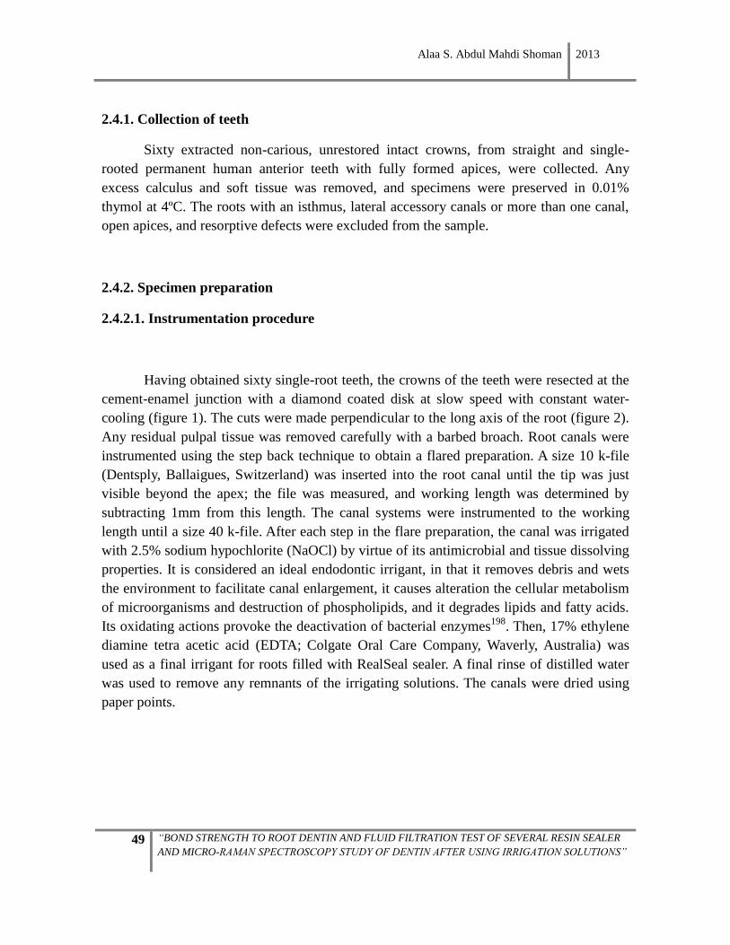
Alaa S. Abdul Mahdi Shoman 2013
49 “BOND STRENGTH TO ROOT DENTIN AND FLUID FILTRATION TEST OF SEVERAL RESIN SEALER
AND MICRO-RAMAN SPECTROSCOPY STUDY OF DENTIN AFTER USING IRRIGATION SOLUTIONS”
2.4.1. Collection of teeth
Sixty extracted non-carious, unrestored intact crowns, from straight and single-
rooted permanent human anterior teeth with fully formed apices, were collected. Any
excess calculus and soft tissue was removed, and specimens were preserved in 0.01%
thymol at 4ºC. The roots with an isthmus, lateral accessory canals or more than one canal,
open apices, and resorptive defects were excluded from the sample.
2.4.2. Specimen preparation
2.4.2.1. Instrumentation procedure
Having obtained sixty single-root teeth, the crowns of the teeth were resected at the
cement-enamel junction with a diamond coated disk at slow speed with constant water-
cooling (figure 1). The cuts were made perpendicular to the long axis of the root (figure 2).
Any residual pulpal tissue was removed carefully with a barbed broach. Root canals were
instrumented using the step back technique to obtain a flared preparation. A size 10 k-file
(Dentsply, Ballaigues, Switzerland) was inserted into the root canal until the tip was just
visible beyond the apex; the file was measured, and working length was determined by
subtracting 1mm from this length. The canal systems were instrumented to the working
length until a size 40 k-file. After each step in the flare preparation, the canal was irrigated
with 2.5% sodium hypochlorite (NaOCl) by virtue of its antimicrobial and tissue dissolving
properties. It is considered an ideal endodontic irrigant, in that it removes debris and wets
the environment to facilitate canal enlargement, it causes alteration the cellular metabolism
of microorganisms and destruction of phospholipids, and it degrades lipids and fatty acids.
Its oxidating actions provoke the deactivation of bacterial enzymes198
. Then, 17% ethylene
diamine tetra acetic acid (EDTA; Colgate Oral Care Company, Waverly, Australia) was
used as a final irrigant for roots filled with RealSeal sealer. A final rinse of distilled water
was used to remove any remnants of the irrigating solutions. The canals were dried using
paper points.
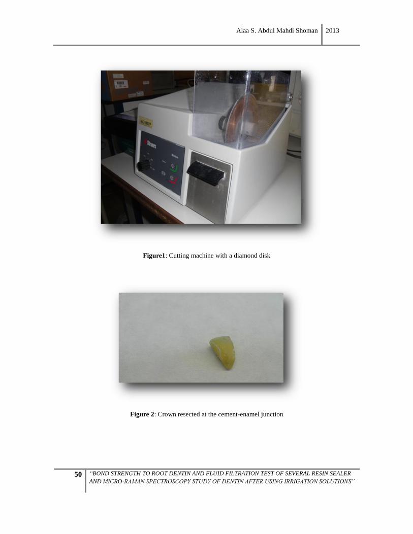
Alaa S. Abdul Mahdi Shoman 2013
50 “BOND STRENGTH TO ROOT DENTIN AND FLUID FILTRATION TEST OF SEVERAL RESIN SEALER
AND MICRO-RAMAN SPECTROSCOPY STUDY OF DENTIN AFTER USING IRRIGATION SOLUTIONS”
Figure1: Cutting machine with a diamond disk
Figure 2: Crown resected at the cement-enamel junction
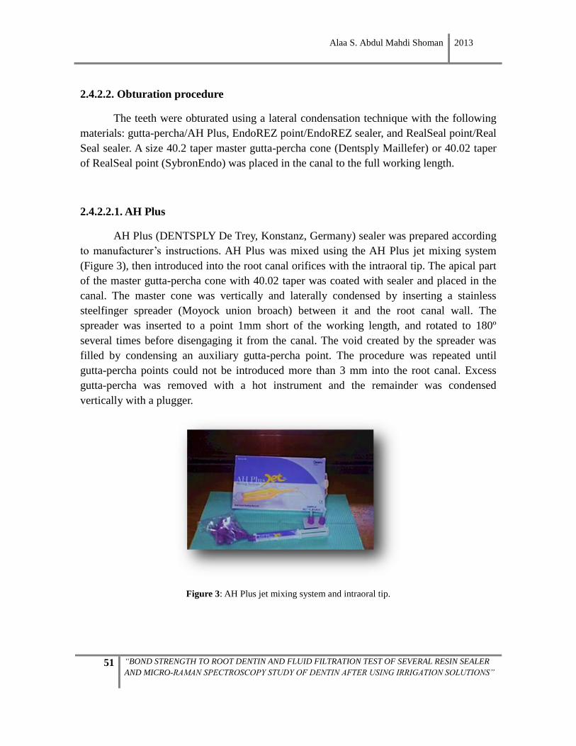
Alaa S. Abdul Mahdi Shoman 2013
51 “BOND STRENGTH TO ROOT DENTIN AND FLUID FILTRATION TEST OF SEVERAL RESIN SEALER
AND MICRO-RAMAN SPECTROSCOPY STUDY OF DENTIN AFTER USING IRRIGATION SOLUTIONS”
2.4.2.2. Obturation procedure
The teeth were obturated using a lateral condensation technique with the following
materials: gutta-percha/AH Plus, EndoREZ point/EndoREZ sealer, and RealSeal point/Real
Seal sealer. A size 40.2 taper master gutta-percha cone (Dentsply Maillefer) or 40.02 taper
of RealSeal point (SybronEndo) was placed in the canal to the full working length.
2.4.2.2.1. AH Plus
AH Plus (DENTSPLY De Trey, Konstanz, Germany) sealer was prepared according
to manufacturer’s instructions. AH Plus was mixed using the AH Plus jet mixing system
(Figure 3), then introduced into the root canal orifices with the intraoral tip. The apical part
of the master gutta-percha cone with 40.02 taper was coated with sealer and placed in the
canal. The master cone was vertically and laterally condensed by inserting a stainless
steelfinger spreader (Moyock union broach) between it and the root canal wall. The
spreader was inserted to a point 1mm short of the working length, and rotated to 180º
several times before disengaging it from the canal. The void created by the spreader was
filled by condensing an auxiliary gutta-percha point. The procedure was repeated until
gutta-percha points could not be introduced more than 3 mm into the root canal. Excess
gutta-percha was removed with a hot instrument and the remainder was condensed
vertically with a plugger.
Figure 3: AH Plus jet mixing system and intraoral tip.
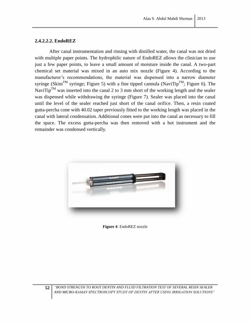
Alaa S. Abdul Mahdi Shoman 2013
52 “BOND STRENGTH TO ROOT DENTIN AND FLUID FILTRATION TEST OF SEVERAL RESIN SEALER
AND MICRO-RAMAN SPECTROSCOPY STUDY OF DENTIN AFTER USING IRRIGATION SOLUTIONS”
2.4.2.2.2. EndoREZ
After canal instrumentation and rinsing with distilled water, the canal was not dried
with multiple paper points. The hydrophilic nature of EndoREZ allows the clinician to use
just a few paper points, to leave a small amount of moisture inside the canal. A two-part
chemical set material was mixed in an auto mix nozzle (Figure 4). According to the
manufacturer’s recommendations, the material was dispensed into a narrow diameter
syringe (SkiniTM
syringe; Figure 5) with a fine tipped cannula (NaviTipTM
; Figure 6). The
NaviTipTM
was inserted into the canal 2 to 3 mm short of the working length and the sealer
was dispensed while withdrawing the syringe (Figure 7). Sealer was placed into the canal
until the level of the sealer reached just short of the canal orifice. Then, a resin coated
gutta-percha cone with 40.02 taper previously fitted to the working length was placed in the
canal with lateral condensation. Additional cones were put into the canal as necessary to fill
the space. The excess gutta-percha was then removed with a hot instrument and the
remainder was condensed vertically.
Figure 4: EndoREZ nozzle
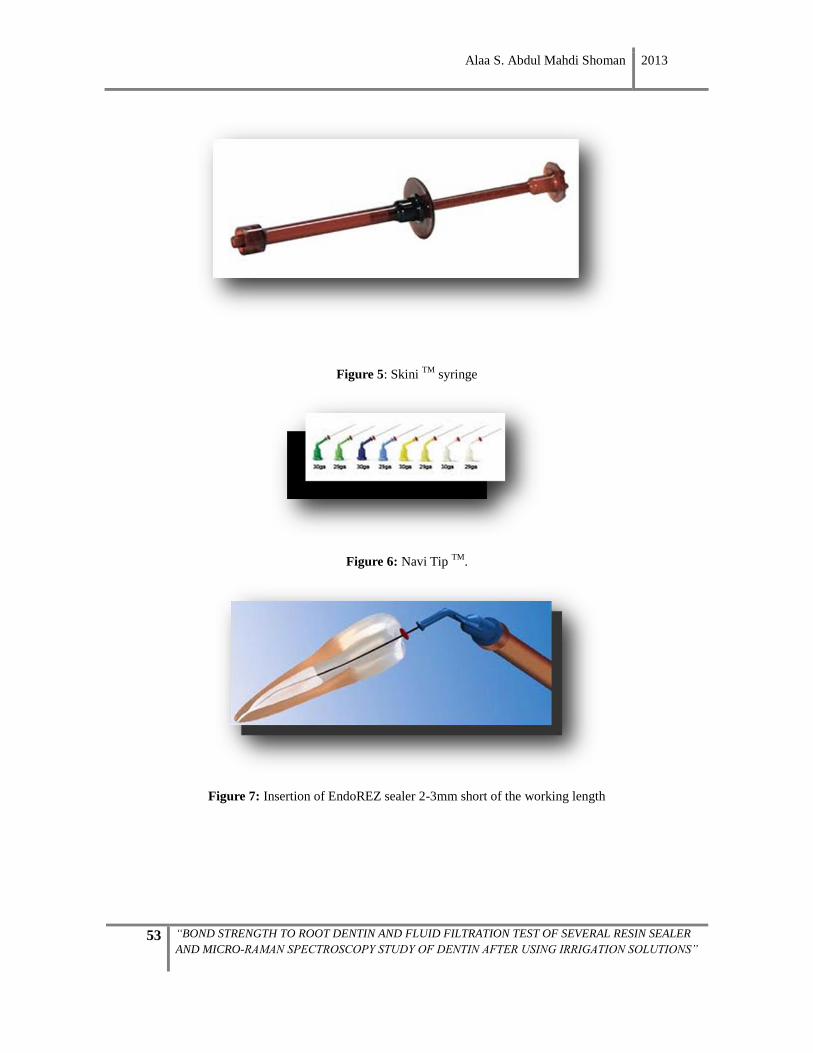
Alaa S. Abdul Mahdi Shoman 2013
53 “BOND STRENGTH TO ROOT DENTIN AND FLUID FILTRATION TEST OF SEVERAL RESIN SEALER
AND MICRO-RAMAN SPECTROSCOPY STUDY OF DENTIN AFTER USING IRRIGATION SOLUTIONS”
Figure 5: Skini TM
syringe
Figure 6: Navi Tip TM
.
Figure 7: Insertion of EndoREZ sealer 2-3mm short of the working length
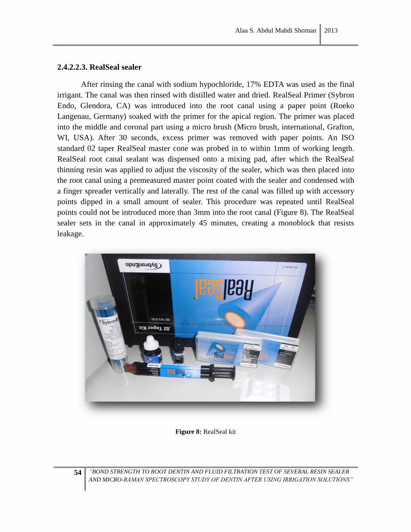
Alaa S. Abdul Mahdi Shoman 2013
54 “BOND STRENGTH TO ROOT DENTIN AND FLUID FILTRATION TEST OF SEVERAL RESIN SEALER
AND MICRO-RAMAN SPECTROSCOPY STUDY OF DENTIN AFTER USING IRRIGATION SOLUTIONS”
2.4.2.2.3. RealSeal sealer
After rinsing the canal with sodium hypochloride, 17% EDTA was used as the final
irrigant. The canal was then rinsed with distilled water and dried. RealSeal Primer (Sybron
Endo, Glendora, CA) was introduced into the root canal using a paper point (Roeko
Langenau, Germany) soaked with the primer for the apical region. The primer was placed
into the middle and coronal part using a micro brush (Micro brush, international, Grafton,
WI, USA). After 30 seconds, excess primer was removed with paper points. An ISO
standard 02 taper RealSeal master cone was probed in to within 1mm of working length.
RealSeal root canal sealant was dispensed onto a mixing pad, after which the RealSeal
thinning resin was applied to adjust the viscosity of the sealer, which was then placed into
the root canal using a premeasured master point coated with the sealer and condensed with
a finger spreader vertically and laterally. The rest of the canal was filled up with accessory
points dipped in a small amount of sealer. This procedure was repeated until RealSeal
points could not be introduced more than 3mm into the root canal (Figure 8). The RealSeal
sealer sets in the canal in approximately 45 minutes, creating a monoblock that resists
leakage.
Figure 8: RealSeal kit

Alaa S. Abdul Mahdi Shoman 2013
55 “BOND STRENGTH TO ROOT DENTIN AND FLUID FILTRATION TEST OF SEVERAL RESIN SEALER
AND MICRO-RAMAN SPECTROSCOPY STUDY OF DENTIN AFTER USING IRRIGATION SOLUTIONS”
In all specimens the length was kept identical: the roots were resected at the apex
perpendicular to the long axis of the tooth until 10 mm of root length was obtained, so as to
avoid anatomical variations and ensure standardization for the leakage measurements.
In the present study, the teeth were divided into three groups (n=20) according to the sealer
used; Group1: AH Plus, Group2: EndoREZ, and Group3: RealSeal sealers. After filling
using the lateral condensation technique, each sealer group was randomly divided into two
subgroups in view of the tests applied (n=10 for µPush-out test and n=10 for fluid filtration
test).
Group I. AH Plus sealer and gutta-percha point for fluid filtration measurement.
Group II. EndoREZ sealer and resin-coated gutta-percha point for fluid filtration
measurement.
Group III. RealSeal sealer and RealSeal points for fluid filtration measurement.
Group IV. AH Plus sealer and gutta-percha cone for µPush-out test.
Group V. EndoREZ sealer and resin-coated gutta-percha points for µPush-out test.
Group VI. RealSeal sealer and RealSeal points for µPush-out test.
The teeth of all groups were kept moist at all times by wrapping them in a distilled water-
soaked gauze at 37ºC for 24 hours to allow the sealer to set completely.
2.4.3. Leakage evaluation
Twenty-four hours after completion of the filling procedure, the roots of each group
(I, II, and III) were resected at the apex perpendicular to the long axis of the tooth until 10
mm of root length was obtained.
A layer of nail varnish was then applied on the external root surface, leaving the
coronal and apical orifices open for fluid exchange with the environment. Nail varnish was
used to limit the movement of fluid across the dentinal tubules and ensure that any fluid
flow assessed was caused by flow along the interface between the filling and the dentin. At
that point, the specimen was attached to the apparatus to measure the fluid filtration.
Alaa S. Abdul Mahdi Shoman 2013
56 “BOND STRENGTH TO ROOT DENTIN AND FLUID FILTRATION TEST OF SEVERAL RESIN SEALER
AND MICRO-RAMAN SPECTROSCOPY STUDY OF DENTIN AFTER USING IRRIGATION SOLUTIONS”
2.4.4. Description of the fluid filtration apparatus
The apparatus used to assess dentin permeability was constructed by the West of
Scotland Health Board’s Department of Clinical Physics and Bio-engineering using the
same principles as those described by Pashley199
. The system involves the evaluation of
fluid transport in specimens calculated from bubble movement. It is necessary to apply
pressure so that the fluid moves through the specimen and displaces the bubbles. For this
purpose, a reservoir of 250 ml of distilled water was used and placed 100 cm higher than
the root, to produce a pressure of 100 cm H2O at room temperature. A specific rubber tube
(2mm internal diameter) was attached to the reservoir of the system, as was a 10 ml insulin
syringe (syringe A in Figure 9), connected to introduce an air bubble into the system, and
then the rubber tube was closed with glue. The rubber tube was connected to a 10 ml
micropipette with an inner diameter of 1 mm, which served to measure movement of the air
bubble (Figure 10). The other end of the micropipette was connected to another rubber
tube, to which a 10 ml plastic syringe was attached (syringe B). Syringe B was used to pull
the air bubble into the micropipette and then adjust its position before the start of each
measurement. The apparatus was flushed to ensure that there were no air bubbles trapped in
the tubing. The diameter of the air bubble must not be smaller than the internal diameter of
the micropipette so that its movement will be a precise indicator for fluid movement in the
micropipette. At the end of the rubber tube, the root was inserted and placed in a plastic
syringe filled with distilled water. All the parts were fixed onto a wooden board, which was
kept horizontal using a spirit level.
System sensitivity can be adjusted by altering the pressure used or the diameter of
the micropipette. Various parameters could modify the test results, such as diameter of the
capillary containing the bubble, the length-measuring time, and the pressure applied123
.
This should be expressed as µl/min cm H20 instead of µl/min.
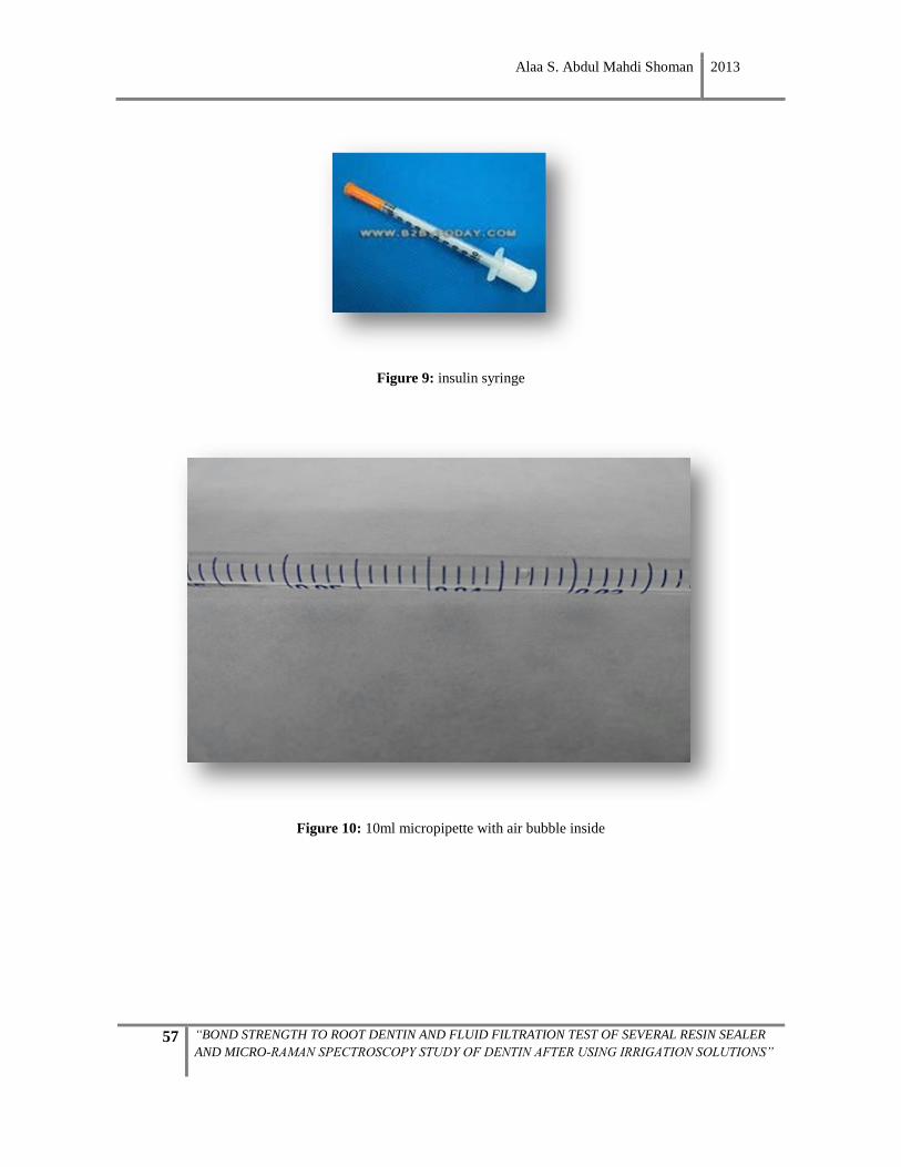
Alaa S. Abdul Mahdi Shoman 2013
57 “BOND STRENGTH TO ROOT DENTIN AND FLUID FILTRATION TEST OF SEVERAL RESIN SEALER
AND MICRO-RAMAN SPECTROSCOPY STUDY OF DENTIN AFTER USING IRRIGATION SOLUTIONS”
Figure 9: insulin syringe
Figure 10: 10ml micropipette with air bubble inside
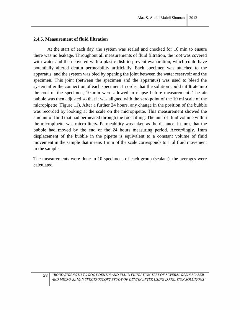
Alaa S. Abdul Mahdi Shoman 2013
58 “BOND STRENGTH TO ROOT DENTIN AND FLUID FILTRATION TEST OF SEVERAL RESIN SEALER
AND MICRO-RAMAN SPECTROSCOPY STUDY OF DENTIN AFTER USING IRRIGATION SOLUTIONS”
2.4.5. Measurement of fluid filtration
At the start of each day, the system was sealed and checked for 10 min to ensure
there was no leakage. Throughout all measurements of fluid filtration, the root was covered
with water and then covered with a plastic dish to prevent evaporation, which could have
potentially altered dentin permeability artificially. Each specimen was attached to the
apparatus, and the system was bled by opening the joint between the water reservoir and the
specimen. This joint (between the specimen and the apparatus) was used to bleed the
system after the connection of each specimen. In order that the solution could infiltrate into
the root of the specimen, 10 min were allowed to elapse before measurement. The air
bubble was then adjusted so that it was aligned with the zero point of the 10 ml scale of the
micropipette (Figure 11). After a further 24 hours, any change in the position of the bubble
was recorded by looking at the scale on the micropipette. This measurement showed the
amount of fluid that had permeated through the root filling. The unit of fluid volume within
the micropipette was micro-liters. Permeability was taken as the distance, in mm, that the
bubble had moved by the end of the 24 hours measuring period. Accordingly, 1mm
displacement of the bubble in the pipette is equivalent to a constant volume of fluid
movement in the sample that means 1 mm of the scale corresponds to 1 µl fluid movement
in the sample.
The measurements were done in 10 specimens of each group (sealant), the averages were
calculated.
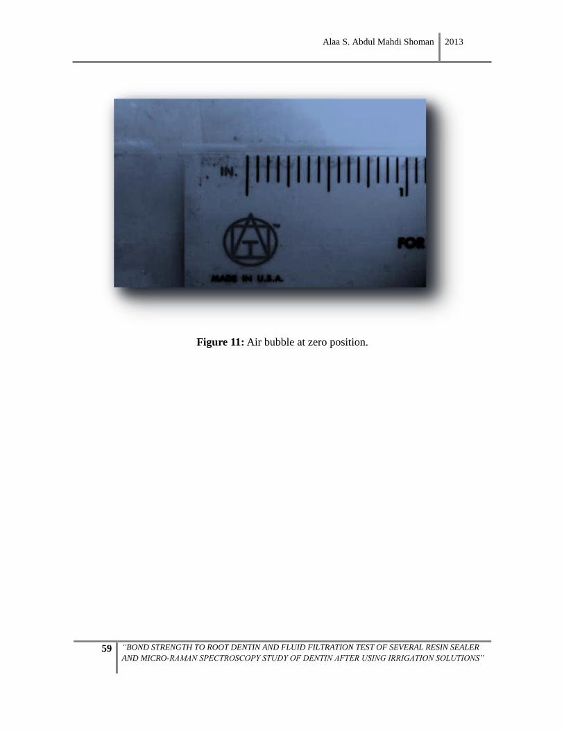
Alaa S. Abdul Mahdi Shoman 2013
59 “BOND STRENGTH TO ROOT DENTIN AND FLUID FILTRATION TEST OF SEVERAL RESIN SEALER
AND MICRO-RAMAN SPECTROSCOPY STUDY OF DENTIN AFTER USING IRRIGATION SOLUTIONS”
Figure 11: Air bubble at zero position.
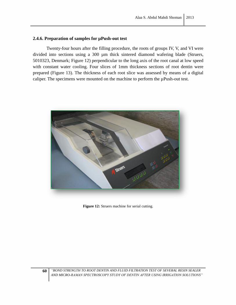
Alaa S. Abdul Mahdi Shoman 2013
60 “BOND STRENGTH TO ROOT DENTIN AND FLUID FILTRATION TEST OF SEVERAL RESIN SEALER
AND MICRO-RAMAN SPECTROSCOPY STUDY OF DENTIN AFTER USING IRRIGATION SOLUTIONS”
2.4.6. Preparation of samples for µPush-out test
Twenty-four hours after the filling procedure, the roots of groups IV, V, and VI were
divided into sections using a 300 µm thick sintered diamond wafering blade (Struers,
5010323, Denmark; Figure 12) perpendicular to the long axis of the root canal at low speed
with constant water cooling. Four slices of 1mm thickness sections of root dentin were
prepared (Figure 13). The thickness of each root slice was assessed by means of a digital
caliper. The specimens were mounted on the machine to perform the µPush-out test.
Figure 12: Struers machine for serial cutting.
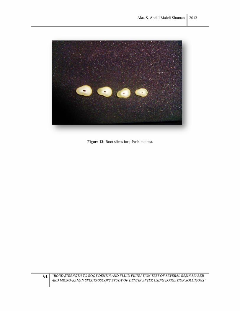
Alaa S. Abdul Mahdi Shoman 2013
61 “BOND STRENGTH TO ROOT DENTIN AND FLUID FILTRATION TEST OF SEVERAL RESIN SEALER
AND MICRO-RAMAN SPECTROSCOPY STUDY OF DENTIN AFTER USING IRRIGATION SOLUTIONS”
Figure 13: Root slices for µPush-out test.
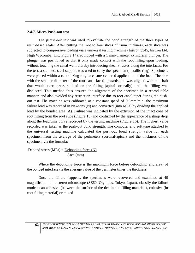
Alaa S. Abdul Mahdi Shoman 2013
62 “BOND STRENGTH TO ROOT DENTIN AND FLUID FILTRATION TEST OF SEVERAL RESIN SEALER
AND MICRO-RAMAN SPECTROSCOPY STUDY OF DENTIN AFTER USING IRRIGATION SOLUTIONS”
2.4.7. Micro Push-out test
The µPush-out test was used to evaluate the bond strength of the three types of
resin-based sealer. After cutting the root to four slices of 1mm thickness, each slice was
subjected to compressive loading via a universal testing machine (Instron 3345, Instron Ltd,
High Wycombe, UK; Figure 14), equipped with a 1 mm-diameter cylindrical plunger. The
plunger was positioned so that it only made contact with the root filling upon loading,
without touching the canal wall, thereby introducing shear stresses along the interfaces. For
the test, a stainless steel support was used to carry the specimen (metallic ring). Specimens
were placed within a centralizing ring to ensure centered application of the load. The side
with the smaller diameter of the root canal faced upwards and was aligned with the shaft
that would exert pressure load on the filling (apical-coronally) until the filling was
displaced. This method thus ensured the alignment of the specimen in a reproducible
manner, and also avoided any restriction interface due to root canal taper during the push-
out test. The machine was calibrated at a constant speed of 0.5mm/min; the maximum
failure load was recorded in Newtons (N) and converted (into MPa) by dividing the applied
load by the bonded area (A). Failure was indicated by the extrusion of the intact cone of
root filling from the root slice (Figure 15) and confirmed by the appearance of a sharp drop
along the load/time curve recorded by the testing machine (Figure 16). The highest value
recorded was taken as the push-out bond strength. The computer and software attached to
the universal testing machine calculated the push-out bond strength value for each
specimen from the average of the perimeters (coronal-apical) and the thickness of the
specimen, via the formula:
Debond stress (MPa) = Debonding force (N)
Area (mm)
Where the debonding force is the maximum force before debonding, and area (of
the bonded interface) is the average value of the perimeter times the thickness.
Once the failure happens, the specimens were recovered and examined at 40
magnification on a stereo-microscope (SZ60, Olympus, Tokyo, Japan), classify the failure
mode as an adhesive (between the surface of the dentin and filling material ), cohesive (in
root filling material) or mixed
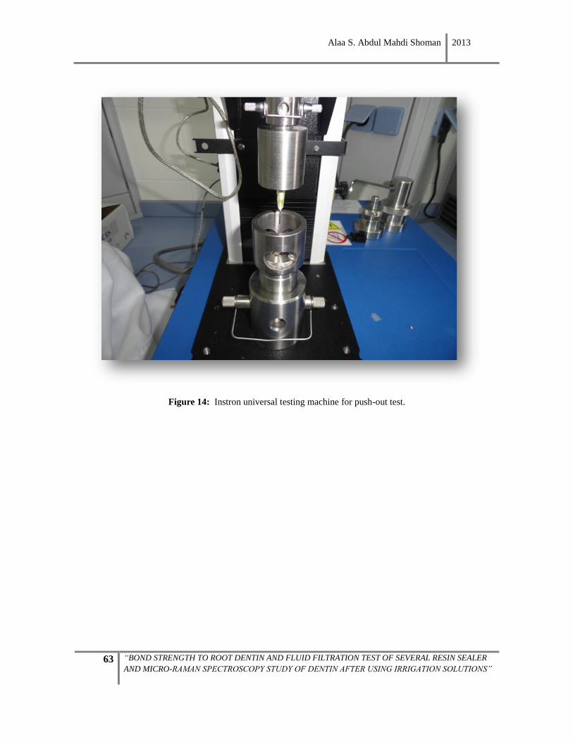
Alaa S. Abdul Mahdi Shoman 2013
63 “BOND STRENGTH TO ROOT DENTIN AND FLUID FILTRATION TEST OF SEVERAL RESIN SEALER
AND MICRO-RAMAN SPECTROSCOPY STUDY OF DENTIN AFTER USING IRRIGATION SOLUTIONS”
Figure 14: Instron universal testing machine for push-out test.
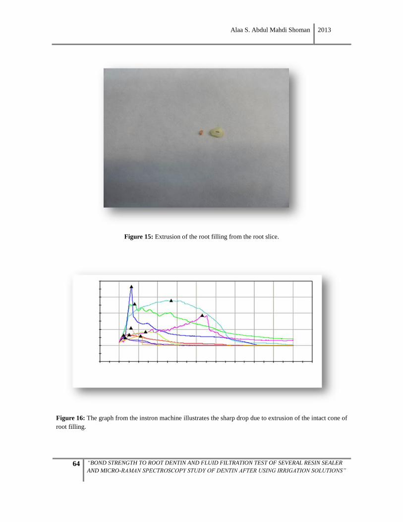
Alaa S. Abdul Mahdi Shoman 2013
64 “BOND STRENGTH TO ROOT DENTIN AND FLUID FILTRATION TEST OF SEVERAL RESIN SEALER
AND MICRO-RAMAN SPECTROSCOPY STUDY OF DENTIN AFTER USING IRRIGATION SOLUTIONS”
Figure 15: Extrusion of the root filling from the root slice.
Figure 16: The graph from the instron machine illustrates the sharp drop due to extrusion of the intact cone of
root filling.
Alaa S. Abdul Mahdi Shoman 2013
65 “BOND STRENGTH TO ROOT DENTIN AND FLUID FILTRATION TEST OF SEVERAL RESIN SEALER
AND MICRO-RAMAN SPECTROSCOPY STUDY OF DENTIN AFTER USING IRRIGATION SOLUTIONS”

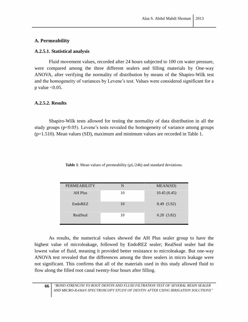
Alaa S. Abdul Mahdi Shoman 2013
66 “BOND STRENGTH TO ROOT DENTIN AND FLUID FILTRATION TEST OF SEVERAL RESIN SEALER
AND MICRO-RAMAN SPECTROSCOPY STUDY OF DENTIN AFTER USING IRRIGATION SOLUTIONS”
A. Permeability
A.2.5.1. Statistical analysis
Fluid movement values, recorded after 24 hours subjected to 100 cm water pressure,
were compared among the three different sealers and filling materials by One-way
ANOVA, after verifying the normality of distribution by means of the Shapiro-Wilk test
and the homogeneity of variances by Levene’s test. Values were considered significant for a
p value <0.05.
A.2.5.2. Results
Shapiro-Wilk tests allowed for testing the normality of data distribution in all the
study groups (p<0.05). Levene’s tests revealed the homogeneity of variance among groups
(p=1.510). Mean values (SD), maximum and minimum values are recorded in Table 1.
Table 1: Mean values of permeability (µL/24h) and standard deviations.
PERMEABILITY N MEAN(SD)
AH Plus 10 10.45 (6.45)
EndoREZ 10 8.49 (5.92)
RealSeal 10 6.20 (3.82)
As results, the numerical values showed the AH Plus sealer group to have the
highest value of microleakage, followed by EndoREZ sealer; RealSeal sealer had the
lowest value of fluid, meaning it provided better resistance to microleakage. But one-way
ANOVA test revealed that the differences among the three sealers in micro leakage were
not significant. This confirms that all of the materials used in this study allowed fluid to
flow along the filled root canal twenty-four hours after filling.
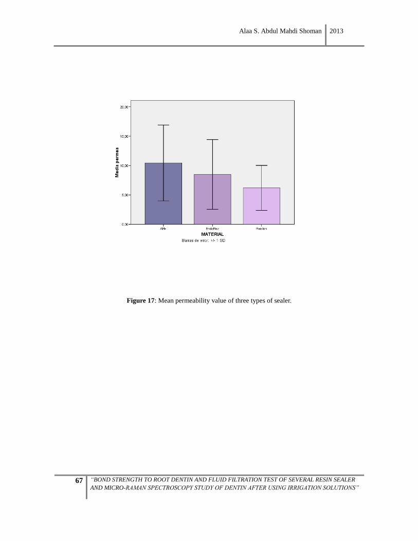
Alaa S. Abdul Mahdi Shoman 2013
67 “BOND STRENGTH TO ROOT DENTIN AND FLUID FILTRATION TEST OF SEVERAL RESIN SEALER
AND MICRO-RAMAN SPECTROSCOPY STUDY OF DENTIN AFTER USING IRRIGATION SOLUTIONS”
Figure 17: Mean permeability value of three types of sealer.
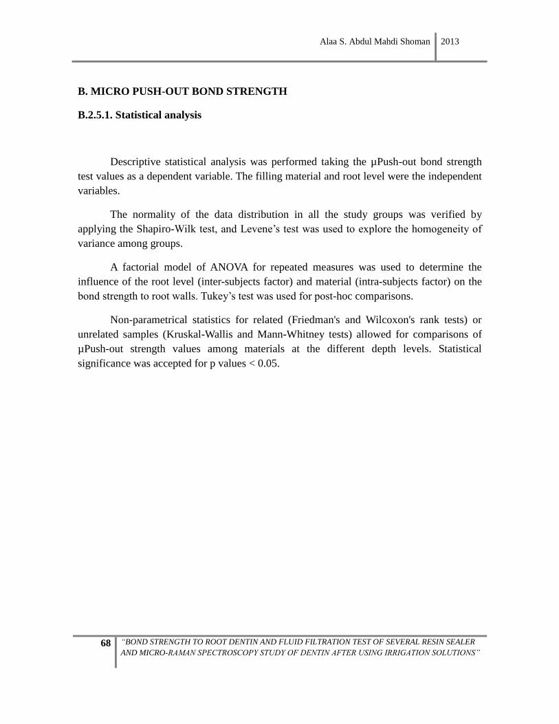
Alaa S. Abdul Mahdi Shoman 2013
68 “BOND STRENGTH TO ROOT DENTIN AND FLUID FILTRATION TEST OF SEVERAL RESIN SEALER
AND MICRO-RAMAN SPECTROSCOPY STUDY OF DENTIN AFTER USING IRRIGATION SOLUTIONS”
B. MICRO PUSH-OUT BOND STRENGTH
B.2.5.1. Statistical analysis
Descriptive statistical analysis was performed taking the µPush-out bond strength
test values as a dependent variable. The filling material and root level were the independent
variables.
The normality of the data distribution in all the study groups was verified by
applying the Shapiro-Wilk test, and Levene’s test was used to explore the homogeneity of
variance among groups.
A factorial model of ANOVA for repeated measures was used to determine the
influence of the root level (inter-subjects factor) and material (intra-subjects factor) on the
bond strength to root walls. Tukey’s test was used for post-hoc comparisons.
Non-parametrical statistics for related (Friedman's and Wilcoxon's rank tests) or
unrelated samples (Kruskal-Wallis and Mann-Whitney tests) allowed for comparisons of
µPush-out strength values among materials at the different depth levels. Statistical
significance was accepted for p values < 0.05.
Alaa S. Abdul Mahdi Shoman 2013
69 “BOND STRENGTH TO ROOT DENTIN AND FLUID FILTRATION TEST OF SEVERAL RESIN SEALER
AND MICRO-RAMAN SPECTROSCOPY STUDY OF DENTIN AFTER USING IRRIGATION SOLUTIONS”
B.2.5.2. Results
The Shapiro-Wilk test revealed that data distribution for the µPush-out test was
normal. Descriptive statistics of the µPush-out bond strength values are given in Table The
linear model of ANOVA for repeated measures demonstrated that root level (F=27.148,
p<0.001), and the interaction between the root thirds and the filling material (F=8.66,
p=0.001), significantly affected the µPush-out bond strength to the walls of the root canal.
The filling material in itself does not significantly influence the µPush-out bond strength
values (F=2.492; p=0.102).
Despite the normality of the data, Levene’s test showed that the variances were not
homogenous across all the study groups.
Table 2: Adhesion values in the push-out test. Mean (standard deviation).
Group C1 C2 M1 M2 Total N=30
AH Plus n=10
1.69(1,18)a 2.05(1,00)a,b 2.48(1,32)a,b 3.30(2,22)b 1
2.38(1.14)
EndoREZ n=10
0.95(1,08) 2.06(1,77) 1.59(0,98) 2
1.44(1,11) 2
1.51(0.94)
RealSeal n=10
0.77(0,46)a 0.94(0,68)a,b 3.41(1,88)b,c, 1
4.59(2,34)c 1
2.43(1.01)
Total 1.14(1.01)A N=30
1.69(1.31)B 2.49(1.58)C 3.11(2.31)C
Different capital letters indicate significant differences in µPush-out strength values
among root levels in total. Different lower case letters indicate significant differences
among levels for each sealer.
Numbers indicate significant differences in columns (among sealers for each depth level).
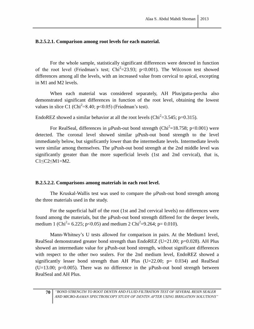
Alaa S. Abdul Mahdi Shoman 2013
70 “BOND STRENGTH TO ROOT DENTIN AND FLUID FILTRATION TEST OF SEVERAL RESIN SEALER
AND MICRO-RAMAN SPECTROSCOPY STUDY OF DENTIN AFTER USING IRRIGATION SOLUTIONS”
B.2.5.2.1. Comparison among root levels for each material.
For the whole sample, statistically significant differences were detected in function
of the root level (Friedman’s test; Chi2=23.93; p<0.001). The Wilcoxon test showed
differences among all the levels, with an increased value from cervical to apical, excepting
in M1 and M2 levels.
When each material was considered separately, AH Plus/gutta-percha also
demonstrated significant differences in function of the root level, obtaining the lowest
values in slice C1 (Chi2=8.40; p<0.05) (Friedman’s test).
EndoREZ showed a similar behavior at all the root levels (Chi2=3.545; p=0.315).
For RealSeal, differences in µPush-out bond strength (Chi2=18.758; p<0.001) were
detected. The coronal level showed similar µPush-out bond strength to the level
immediately below, but significantly lower than the intermediate levels. Intermediate levels
were similar among themselves. The µPush-out bond strength at the 2nd middle level was
significantly greater than the more superficial levels (1st and 2nd cervical), that is,
C1≤C2≤M1=M2.
B.2.5.2.2. Comparisons among materials in each root level.
The Kruskal-Wallis test was used to compare the µPush-out bond strength among
the three materials used in the study.
For the superficial half of the root (1st and 2nd cervical levels) no differences were
found among the materials, but the µPush-out bond strength differed for the deeper levels,
medium 1 (Chi2= 6.225; p<0.05) and medium 2 Chi
2=9.264; p= 0.010).
Mann-Whitney’s U tests allowed for comparison in pairs. At the Medium1 level,
RealSeal demonstrated greater bond strength than EndoREZ (U=21.00; p=0.028). AH Plus
showed an intermediate value for µPush-out bond strength, without significant differences
with respect to the other two sealers. For the 2nd medium level, EndoREZ showed a
significantly lesser bond strength than AH Plus (U=22.00; p= 0.034) and RealSeal
(U=13.00; p=0.005). There was no difference in the µPush-out bond strength between
RealSeal and AH Plus.
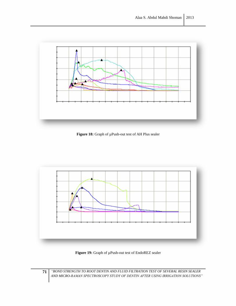
Alaa S. Abdul Mahdi Shoman 2013
71 “BOND STRENGTH TO ROOT DENTIN AND FLUID FILTRATION TEST OF SEVERAL RESIN SEALER
AND MICRO-RAMAN SPECTROSCOPY STUDY OF DENTIN AFTER USING IRRIGATION SOLUTIONS”
Figure 18: Graph of µPush-out test of AH Plus sealer
Figure 19: Graph of µPush-out test of EndoREZ sealer
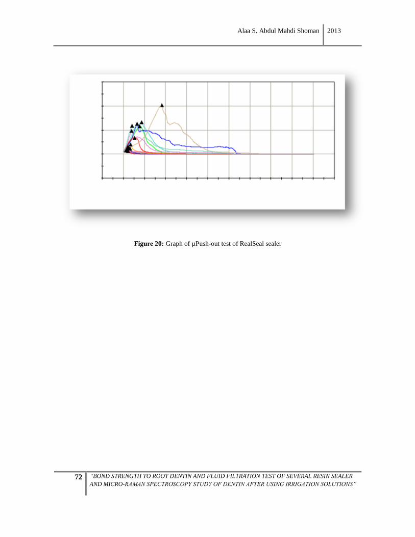
Alaa S. Abdul Mahdi Shoman 2013
72 “BOND STRENGTH TO ROOT DENTIN AND FLUID FILTRATION TEST OF SEVERAL RESIN SEALER
AND MICRO-RAMAN SPECTROSCOPY STUDY OF DENTIN AFTER USING IRRIGATION SOLUTIONS”
Figure 20: Graph of µPush-out test of RealSeal sealer
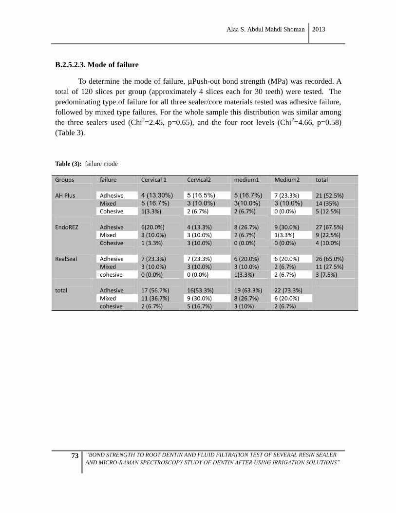
Alaa S. Abdul Mahdi Shoman 2013
73 “BOND STRENGTH TO ROOT DENTIN AND FLUID FILTRATION TEST OF SEVERAL RESIN SEALER
AND MICRO-RAMAN SPECTROSCOPY STUDY OF DENTIN AFTER USING IRRIGATION SOLUTIONS”
B.2.5.2.3. Mode of failure
To determine the mode of failure, µPush-out bond strength (MPa) was recorded. A
total of 120 slices per group (approximately 4 slices each for 30 teeth) were tested. The
predominating type of failure for all three sealer/core materials tested was adhesive failure,
followed by mixed type failures. For the whole sample this distribution was similar among
the three sealers used (Chi2=2.45, p=0.65), and the four root levels (Chi
2=4.66, p=0.58)
(Table 3).
Table (3): failure mode
Groups Failure Cervical1 Cervical2 Medium1 Medium2 Total
failure Cervical 1 Cervical2 medium1 Medium2 total
AH Plus Adhesive 4 (13.30%) 5 (16.5%) 5 (16.7%) 7 (23.3%) 21 (52.5%) Mixed 5 (16.7%) 3 (10.0%) 3(10.0%) 3 (10.0%) 14 (35%) Cohesive 1(3.3%) 2 (6.7%) 2 (6.7%) 0 (0.0%) 5 (12.5%)
EndoREZ
Adhesive
6(20.0%)
4 (13.3%)
8 (26.7%)
9 (30.0%)
27 (67.5%)
Mixed 3 (10.0%) 3 (10.0%) 2 (6.7%) 1(3.3%) 9 (22.5%) Cohesive
1 (3.3%) 3 (10.0%) 0 (0.0%) 0 (0.0%) 4 (10.0%)
RealSeal Adhesive 7 (23.3%) 7 (23.3%) 6 (20.0%) 6 (20.0%) 26 (65.0%) Mixed 3 (10.0%) 3 (10.0%) 3 (10.0%) 2 (6.7%) 11 (27.5%) cohesive 0 (0.0%) 0 (0.0%) 1(3.3%) 2 (6.7%) 3 (7.5%)
total
Adhesive
17 (56.7%)
16(53.3%)
19 (63.3%)
22 (73.3%)
Mixed 11 (36.7%) 9 (30.0%) 8 (26.7%) 6 (20.0%) cohesive 2 (6.7%) 5 (16,7%) 3 (10%) 2 (6.7%)
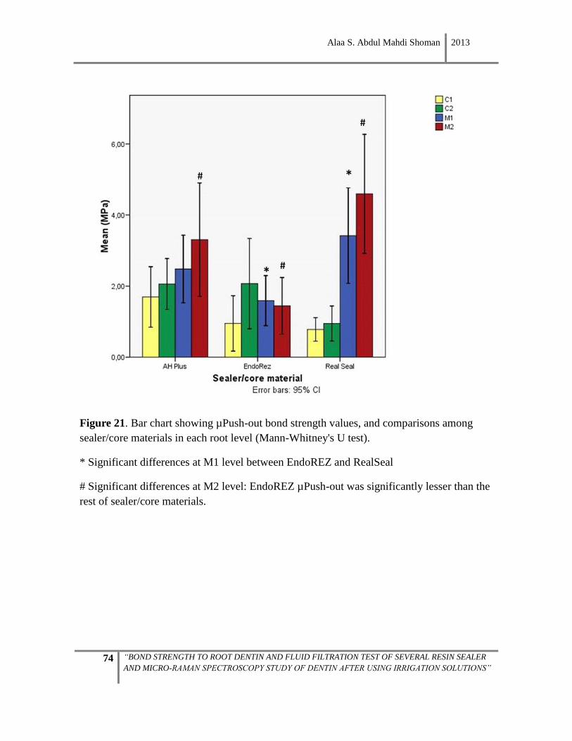
Alaa S. Abdul Mahdi Shoman 2013
74 “BOND STRENGTH TO ROOT DENTIN AND FLUID FILTRATION TEST OF SEVERAL RESIN SEALER
AND MICRO-RAMAN SPECTROSCOPY STUDY OF DENTIN AFTER USING IRRIGATION SOLUTIONS”
Figure 21. Bar chart showing µPush-out bond strength values, and comparisons among
sealer/core materials in each root level (Mann-Whitney's U test).
* Significant differences at M1 level between EndoREZ and RealSeal
# Significant differences at M2 level: EndoREZ µPush-out was significantly lesser than the
rest of sealer/core materials.
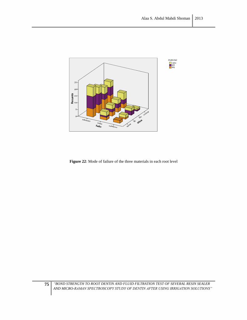
Alaa S. Abdul Mahdi Shoman 2013
75 “BOND STRENGTH TO ROOT DENTIN AND FLUID FILTRATION TEST OF SEVERAL RESIN SEALER
AND MICRO-RAMAN SPECTROSCOPY STUDY OF DENTIN AFTER USING IRRIGATION SOLUTIONS”
Figure 22: Mode of failure of the three materials in each root level
Alaa S. Abdul Mahdi Shoman 2013
76 “BOND STRENGTH TO ROOT DENTIN AND FLUID FILTRATION TEST OF SEVERAL RESIN SEALER
AND MICRO-RAMAN SPECTROSCOPY STUDY OF DENTIN AFTER USING IRRIGATION SOLUTIONS”


Alaa S. Abdul Mahdi Shoman 2013
77 “BOND STRENGTH TO ROOT DENTIN AND FLUID FILTRATION TEST OF SEVERAL RESIN SEALER
AND MICRO-RAMAN SPECTROSCOPY STUDY OF DENTIN AFTER USING IRRIGATION SOLUTIONS”
In this study, permeability and µPush-out bond strength was compared among roots
treated endodontically with three different types of sealers. Root canals were filled using
the lateral condensation technique with AH Plus, EndoREZ, and RealSeal sealers.
Microleakage was recorded for each type of sealer and none of the investigated
materials achieved a tight seal at the filling-cement-root interface at 24 hours.
Regarding the µPush-out bond strength, no significant difference was found among
the three sealers/filling materials.
The root third significantly affects the value of adhesion to the walls of the canal.
There were no significant differences between the different filling materials in any of the
four levels of the canal considered, except in the 2nd medium third. Analyzing separately
each of the materials according to root level, it appears that only the 2nd medium third
manifested significant differences in the behavior of the materials in terms of interfacial
strength achieved with RealSeal and AH Plus to intraradicular dentin —in the 2nd medium
third it was significantly higher than that achieved with EndoREZ.
This study revealed that µPush-out bond strength and permeability were not affected
by the kind of sealer. There was no relation found between the permeability and µPush-out
bond strength.

Alaa S. Abdul Mahdi Shoman 2013
78 “BOND STRENGTH TO ROOT DENTIN AND FLUID FILTRATION TEST OF SEVERAL RESIN SEALER
AND MICRO-RAMAN SPECTROSCOPY STUDY OF DENTIN AFTER USING IRRIGATION SOLUTIONS”

Alaa S. Abdul Mahdi Shoman 2013
79 “BOND STRENGTH TO ROOT DENTIN AND FLUID FILTRATION TEST OF SEVERAL RESIN SEALER
AND MICRO-RAMAN SPECTROSCOPY STUDY OF DENTIN AFTER USING IRRIGATION SOLUTIONS”
The three dimensional obturation of the root is a main objective of endodontic
treatment and proves essential for preventing apical or coronal leakage in the root canal.
Gutta-percha, used for many years as a solid material in root fillings associated with
different types of sealers200
, has suboptimal short and long term apical sealing properties
and does not provide chemical bonding to the root canal wall. Recent advance in obturation
materials have centered on the introduction of resins into the filling material in the cones,
the sealer or both. Resin-filling materials have gained popularity and are now accepted as a
root canal filling material201
. Epoxy resin AH Plus is a two component paste root canal
sealer, placed in the canal without any dentin preparation or dentin adhesive, and used with
gutta-percha and the lateral condensation technique. The dentin surface is cleaned and then
treated with a weak acid202,203
. This acid removes debris from the dentin surface, removes
the smear layer, and exposes hydroxyapatite crystals. Micromechanical retention occurs
between AH Plus sealer, based on the polymerization reaction of epoxy resin amines and
with the collagen matrix in the intertubular dentin.
Recently, new methacrylate resin-based endodontic sealers were introduced.
EndoREZ is a non-etching root canal sealer and does not require the adjunctive use of a
dentin adhesive. It is designed to flow into accessory canals and dentinal tubules. Because it
is hydrophilic in nature, it is applied in a moist environment to facilitate tag formation and
close adaptation to the canal walls.
Another type is RealSeal, a dual cured resin composite sealer. One of the claimed
advantages of using adhesive endodontic sealers with bondable polymeric root canal filling
materials is that they bond throughout the length of the root canal, producing
micromechanical retention through the formation of a thin hybrid layer that forms when the
self-etching primer demineralizes the superficial dentin and exposes the thin layer of
collagen. The resin composite sealer is then applied and polymerized.
A chemical coupling of the urethane dimethacrylate- containing RealSeal root-
filling material to the methacrylate based sealer; a continuum is anticipated by the
manufacturer that results in the creation of a monoblock between the root filling and the
intraradicular dentin204
. These new materials need to be carefully evaluated.
Leakage studies of resin-based endodontic materials are still relevant205
. Different
methods have been used to evaluate the sealing of endodontic cements, including bacterial
penetration, dye penetration, radio isotopes, light microscopy or SEM. These methods have
some disadvantages, the most important one being their qualitative, rather than quantitative
information —they can reveal the presence or absence of leakage, but not the amount206
.
This study involved the most popular method used for microleakage assessment, the fluid
Alaa S. Abdul Mahdi Shoman 2013
80 “BOND STRENGTH TO ROOT DENTIN AND FLUID FILTRATION TEST OF SEVERAL RESIN SEALER
AND MICRO-RAMAN SPECTROSCOPY STUDY OF DENTIN AFTER USING IRRIGATION SOLUTIONS”
filtration method, which was originally described by Derkson and has been extensively
used to measure dentin permeability, comparing the sealing properties of restorative
materials. More recently, Wu, applied this technique in root canals, by means of air bubble
movement inside a capillary tube. This method presents several advantages over the
traditional microleakage studies207
:
The samples are not destroyed, allowing repeated measurements over a period of
time.
No tracer is needed, thereby avoiding the related problem of molecular size and
affinity for dentin, and no intricate materials are required as in bacterial penetration
studies or radioactive tracer studies.
There is no modification of seal with this technique, because the measurements are
made directly after filling without dipping the roots in acids, alcohol or methyl
salicylate.
This technique also avoids problems caused by entrapped air or fluid, not using the
high pressures employed in vacuum studies.
The results are very accurate because every small volume can be recorded. Thus, a
better standardization of the methodology is achieved. Reliability, reproducibility and
compatibility are enhanced by using the fluid filtration system206
.
This technique is simple, less time-consuming and makes it possible to assess
microleakage in individual samples in different observation periods.
Meanwhile, obstacles associated with this method include the sealing of the space
between root and plastic tube, which is important for bias prevention; in this study super
glue was used to seal this area.
To obtain accurate results one must check:
Presence of leaky tube connection
The stability of the apparatus is important, and the pipette must be perfectly straight
to allow the air bubble to flow along its entire length
The dentin must be sealed by using nail varnish to seal the external root surface.
Alaa S. Abdul Mahdi Shoman 2013
81 “BOND STRENGTH TO ROOT DENTIN AND FLUID FILTRATION TEST OF SEVERAL RESIN SEALER
AND MICRO-RAMAN SPECTROSCOPY STUDY OF DENTIN AFTER USING IRRIGATION SOLUTIONS”
The length of specimens should ideally be kept identical, as in this study, to avoid
anatomical variations and ensure standardization for the leakage measurements.
The sensitivity of the fluid transport method system can be adjusted by altering the
pressure used and altering the diameter of the micropipette208
.
Microleakage is also affected by the type of filling technique. Here, the lateral
condensation technique was used. Its most widely accepted due to advantages such as long
term use, controlled placement of materials and relative ease of use. This technique does
not reproduce canal irregularities, but it does produce many irregularities in the final mass
of gutta-percha, leading to an inadequate dispersion of the sealer and the formation of voids
in and around the gutta-percha points. Therefore this drawback, may contribute to leakage.
In the context of bond strength, the concept of bonding resin material to enamel was
first introduced by Bounocore209
. Because of the distinction in composition and
morphology between enamel and dentin, the need for wet bonding arose. Polymer-based
sealers have been introduced as root canal sealers to obtain dentinal tubular penetration,
bond to the collagen matrix, and consequently provide adhesive strength to dentin207
.
Adhesion is defined as a process in which two surfaces of different molecular
compositions are bonded by chemical, physical or mechanical attraction. Adhesion of an
endodontic sealer is its capacity to adhere to the root canal walls and promote the union of
gutta-percha cones to each other and to the dentin62,210
by either mechanical attraction,
which occurs by the entrapment of material into another body, within natural or artificial
cavities. In contrast, chemical adhesion may result from primary valence forces, such as
covalent and metallic bonds. The micromechanical retention to a tooth structure occurs
when resin completely infiltrates dentinal surfaces and creates a hybrid or resin reinforced
layer211
. The materials to be adhered must be sufficiently close to each other. Therefore, a
primary condition is the wettability of the liquid in a solid material212
, which will provide
the required proximity between the materials, facilitating molecular attraction and
promoting adhesion213
.
Various methods have been used to evaluate the bond strength of endodontic sealer
in root dentin; including pull-out214
, micro tension and push-out215,188,168,216
techniques. The
thin slice µPush-out test is an important experimental instrument81
. It can evaluate the bond
strength in a high c-factor with high stress generation directed toward the bonding area. In
addition, the confidence of the µPush-out test may be confirmed by the low variability of
the data when the results show low standard deviations. It offers some advantages over the
microtension technique for the study of canal adhesion, in that specimens are rarely lost;
Alaa S. Abdul Mahdi Shoman 2013
82 “BOND STRENGTH TO ROOT DENTIN AND FLUID FILTRATION TEST OF SEVERAL RESIN SEALER
AND MICRO-RAMAN SPECTROSCOPY STUDY OF DENTIN AFTER USING IRRIGATION SOLUTIONS”
advantages over tensile and shear strength test are that it is less sensitive to small variations
among specimens and to variations in stress distribution during load application, and it is
easy to align samples for testing217
µPush-out force reportedly increases linearly with
greater thickness of the dentin disc218
. The optimal thickness of dentin for the performance
of this technique is controversial, however, with different authors recommending 4mm218
,
3mm219
, 2.5mm64
, and 1mm168
. A thin slice (1mm) was used in the present study because it
has proven reliable in the evaluation of bond strength in 1mm thick samples220
, and it
simplifies calculations of the bond area. Moreover, the use of thicker discs increases the
area of friction and may lead to an over-estimation of the bond strength81
.
In this study, permeability and µPush-out bond strength were compared among roots
treated endodontically with AH Plus, EndoREZ and RealSeal sealers and root canals filled
using lateral condensation technique at 24 hours. The results showed that the permeability
and global µPush-out bond strength were not affected by the kind of sealer/core material.
We used different samples for fluid filtration and µPush-out bond strength, to be
performed at exactly 24 hours´ time, thus avoiding different degrees of conversion for the
sealers. We did not light-cure the coronal portion of the RealSeal sealer as recommended by
the manufacturer, because fast-setting light-cured resin sealers produce restriction in the
flow and consequent defects or weakening of bond strength221
. Moreover, the slow
chemical reaction of methacrylate-based self-etching sealers may reduce shrinkage stress
by means of prolonged plastic flow during the setting time of the material222
. We held that
such behavior would allow for complete resin infiltration into the demineralized dentin.
Our results showed that there were no statistically significant differences in the values
of fluid filtration among the three sealers used in the study. It is noteworthy, however, that
none of the investigated sealers/core materials achieved a tight seal at the filling-cement-
root interface, a point also brought out by Vasconcelos et al.223
allowing fluid to flow along
the filled root canal twenty-four hours after filling, whereas the control showed no
filtration. The authors speculated that AH Plus did not adapt well to dentin walls because of
the hydrophobic properties of epoxy resin224
. Also, the leakage of AHplus may have
resulted from inadequate bonding between the sealer and the gutta-percha points, allowing
fluid to flow at the interface225
. In turn, for RealSeal sealer leakage may be due to
Uneven application of the self-etching primer to the root canal.
Inadequate evaporation of the solvent from the primer, promoting the formation of
hydrogel, which is leaky.
Uneven application of the root canal sealer to the root canal.
Alaa S. Abdul Mahdi Shoman 2013
83 “BOND STRENGTH TO ROOT DENTIN AND FLUID FILTRATION TEST OF SEVERAL RESIN SEALER
AND MICRO-RAMAN SPECTROSCOPY STUDY OF DENTIN AFTER USING IRRIGATION SOLUTIONS”
Inadvertent stripping of the sealer off the canal wall during the placement of cones.
Disruption of the maturing resin-root dentin bond during cold lateral condensation.
The amount of dimethacrylate in the RealSeal points might not be optimal for
effective chemical coupling to a methacrylate-based sealer.
Currently, the clinical relevance of leakage tests in vitro is questioned226
because it is
difficult to interpret results when, in our opinion, only zero filtration can be considered a
good result. Moreover, there is no correlation among various methods to evaluate
microleakage, and outcome of the tests depends on the evaluation method226,99
. In the
absence of correlation between the ex vivo sealing ability of root filled teeth and “clinical
success”227
, we believe there is a threshold for microleakage values that would prove
clinically relevant 228
.
In this study we used three totally different kinds of sealers: a conventional non-
bonding epoxy resin-based sealer, AH Plus; a first generation non-acidic diurethane
dimethacrylate and triethyleneglycol dimethacrylate, EndoREZ; and a second generation
sealer, RealSeal, based on adhesive technology with a self-etching primer and in
association with a thermoplastic synthetic polyester polymer-based root canal filling
material. It should be noted that the conventional nonbonding AH Plus/gutta-percha root
filling was equal to RealSeal and EndoREZ. These discouraging results are in line with
those of Ungor et al.217
who found that the Epiphany/Resilon combination was not superior
to the AH Plus/gutta-percha combination. Still, most studies report that AH Plus sealer
presents greater adhesion to dentin than RealSeal, regardless of root canal wall
treatment229,230,231
. Clinicians need to be aware that methacrylate resin based sealers did not
meet expectations regarding adhesion to root dentin, and at this point in their development
there are no clear benefits in their use72
.
The µPush-out bond strength results were significantly influenced by dentin location.
AH Plus and RealSeal obtained higher values in the deeper slices, owing to the fact that in
the deepest third there is more intertubular dentin and higher collagen fiber density
available for hybridization181
, and both materials (AH Plus and RealSeal) depend mainly on
intertubular dentin for micromechanical retention, whereas EndoREZ showed the lowest
values, regardless of location, because:
Alaa S. Abdul Mahdi Shoman 2013
84 “BOND STRENGTH TO ROOT DENTIN AND FLUID FILTRATION TEST OF SEVERAL RESIN SEALER
AND MICRO-RAMAN SPECTROSCOPY STUDY OF DENTIN AFTER USING IRRIGATION SOLUTIONS”
EndoREZ differs from RealSeal in that it contains only TEGMA, diurethane, di-
methacrylate and bisglycol dimethacrylate phosphate232
, which probably leads to long
but discontinuous tags, the resin tags making only a minor contribution to bond
strength141,143,233
. Adhesion is a complex process and depends on several physical and
chemical aspects of molecules and the interaction between these materials and the
dentin wall104
, so the quality of dentin hybridization is what most important146
, not
the quantity of tags is.
The interface between the gutta-percha resin coating and the resin sealer is the real
bondable interface in this system, but this interface is a weak link that failed during
polymerization shrinkage of the sealer.
The removal of the oxygen inhibition layer234
from the surface of resin coated gutta-
percha cones during packaging has been hypothesized for their weak adhesion to the
methacrylate resin-based root canal sealer, resulting in their frequent delamination
from the sealer after root canal obturation.
All sealers exhibited their lowest µPush-out bond strength values in two superficial
slices. This can be attributed to the presence of oxygen, inhibiting sealer setting and
producing a layer with low polymerization.
The values of the µPush-out test are very low, consistent with results of other studies
using the same methodology, indicating that the bonding of two methacrylate resin-based
sealers and root dentin is much weaker than with resin-dentin bonds, 25-30 MPa71
.
Furthermore, the adhesive failures between sealer/dentin interface clearly suggest an
inadequate level of adhesion between sealer and dentin in terms of bond strength235,236,237
.
This may be due to the difficulties in testing materials with great plasticity, such as gutta-
percha, RealSeal and EndoREZ points. In contrast, when the test was done with sealer and
no core material, values were higher238,235
, suggesting failure may be traced to the
sealer/material interface. In a root canal there is a highly unfavorable configuration factor
(ratio of bonded to unbounded resin surfaces)239
that contributes to maximizing the
polymerization stress of resin-based materials along the root canal walls; this may even
exceed the bond strength of dentin adhesives to dentin, resulting in gap formation along the
surfaces240,239
. Along these lines Souza et al.241
report that there is a correspondence
between the presence of gaps and microleakage.
Despite a material´s relatively low bond strength to root dentin, it may be effective in
preventing microleakage242
. The main problem is that this low µPush-out bond strength is
accompanied by fluid filtration. The adhesion of resin sealer to gutta-percha or core seems
Alaa S. Abdul Mahdi Shoman 2013
85 “BOND STRENGTH TO ROOT DENTIN AND FLUID FILTRATION TEST OF SEVERAL RESIN SEALER
AND MICRO-RAMAN SPECTROSCOPY STUDY OF DENTIN AFTER USING IRRIGATION SOLUTIONS”
to play an important role in microleakage prevention, since it does not have to be at the
sealer-dentin interface. Microleakage can affect the bonding of sealers to dentin by
plasticization (fluids are absorbed by resins)240
and hydrolysis due to water entry in the
interface. In addition, collagen degradation may occur due to host-derived matrix
metalloproteinases (MMPs) in dentin that are slowly released over time when self-etching
adhesives are used154
. RealSeal may therefore adversely affect the longevity of bonded root
canal fillings by accelerating degradation of the bond through the movement of fluid
between the hybrid layer and unaffected dentin240,154
.
This study reveals that global µPush-out bond strength and permeability were not
affected by different sealer/core materials. This leads us to partially accept the null
hypothesis, because in the two deeper slices, RealSeal and AH Plus achieved higher µPush-
out bond strength than EndoREZ. We cannot confirm any clear relationship between
permeability and µPush-out bond strength.

Alaa S. Abdul Mahdi Shoman 2013
86 “BOND STRENGTH TO ROOT DENTIN AND FLUID FILTRATION TEST OF SEVERAL RESIN SEALER
AND MICRO-RAMAN SPECTROSCOPY STUDY OF DENTIN AFTER USING IRRIGATION SOLUTIONS”
MICRO-RAMAN SPECTROSCOPY OF ROOT DENTIN AFTER
USING IRRIGATION SOLUTIONS

Alaa S. Abdul Mahdi Shoman 2013
87 “BOND STRENGTH TO ROOT DENTIN AND FLUID FILTRATION TEST OF SEVERAL RESIN SEALER
AND MICRO-RAMAN SPECTROSCOPY STUDY OF DENTIN AFTER USING IRRIGATION SOLUTIONS”

Alaa S. Abdul Mahdi Shoman 2013
88 “BOND STRENGTH TO ROOT DENTIN AND FLUID FILTRATION TEST OF SEVERAL RESIN SEALER
AND MICRO-RAMAN SPECTROSCOPY STUDY OF DENTIN AFTER USING IRRIGATION SOLUTIONS”
Objective
The purpose of this study was to evaluate and compare the effect of different
chemical agents used in endodontics on the root dentin composition.
Materials and methods
The crown was removed at the cement-enamel junction from 20 human anterior
teeth. The roots were divided into five groups, each having four roots. Group I was irrigated
with 2.5% NaOCl for 1 minute during instrumentation. Group II was irrigated with 2.5%
NaOCl for 5 minutes as a final rinse after instrumentation like group I. Group III was
treated with 17% EDTA as a final rinse for 1 minute after instrumentation like group I and
irrigation with 2.5% NaOCl for 5 minute as group II. In Group IV, RealSeal self-etching
primer was applied for 30 seconds after instrumentation, followed by irrigation as in groups
I, II and III. Group V served as the control group, treated with distilled water during
instrumentation. Each root was sectioned into 4 slices (n=80). Micro-Raman spectroscopy
was performed on these root thirds.
Results
Final irrigation of the canal with 2.5%NaOCl solution for 5 minutes (Group II)
showed a decrease in the amide I/amide III (1667/1242) ratio corresponding to the amides
III, while a lower value of the two amide III/matrix ratios, significant in some root levels,
was observed in relation with the control group (V). Regarding Group I, irrigated with
2.5% NaOCl for 1 minute during instrumentation, there was a significant increase of
1667/1242 ratio at all levels. In addition, there was a significant decrease in the 1072/960
ratio with respect to the control group at the first three levels (C1, C2 and M1). The 2.5%
NaOCl solution for 1 minute decreased the carbonate/phosphate ratio.
EDTA showed little or no affectation of the bands of carbonates and phosphates, just
slightly increasing the carbonate/phosphate ratio. Application of primer for 30 seconds
produced an inconsistent effect on the mineral component, mainly originating a decrease in
the phosphate concentration, with no affectation of amide I and non-constant affectation of
amide III. There was no significant difference in the crystallinity between the treated dentin
and the control group.
Alaa S. Abdul Mahdi Shoman 2013
89 “BOND STRENGTH TO ROOT DENTIN AND FLUID FILTRATION TEST OF SEVERAL RESIN SEALER
AND MICRO-RAMAN SPECTROSCOPY STUDY OF DENTIN AFTER USING IRRIGATION SOLUTIONS”
Conclusion
In short, 2.5% NaOCl irrigation for 1 minute during instrumentation had no effect
on organic components, while increasing time to 5 minutes led to an irregular decrease in
amide III and an inconsistent effect on the mineral component. Final irrigation with EDTA
for 1 minute after instrumentation, with 2.5% NaOCl irrigation for 1 minute during
instrumentation, produces little or no affectation of bands of carbonates and phosphates.
The application of a primer produces an inconsistent decrease in the phosphate
concentration, without affectation of amide I.
Alaa S. Abdul Mahdi Shoman 2013
90 “BOND STRENGTH TO ROOT DENTIN AND FLUID FILTRATION TEST OF SEVERAL RESIN SEALER
AND MICRO-RAMAN SPECTROSCOPY STUDY OF DENTIN AFTER USING IRRIGATION SOLUTIONS”

Alaa S. Abdul Mahdi Shoman 2013
91 “BOND STRENGTH TO ROOT DENTIN AND FLUID FILTRATION TEST OF SEVERAL RESIN SEALER
AND MICRO-RAMAN SPECTROSCOPY STUDY OF DENTIN AFTER USING IRRIGATION SOLUTIONS”
During root canal preparation, root dentin is exposed to different mechanical and
chemical factors. An understanding of the chemical composition of dentin is the first step
toward predicting the behavior of the dentin after treatment.
The success of root canal therapy depends on the method and the quality of
instrumentation, irrigation, disinfection, and three-dimensional obturation of the root canal.
Endodontic instrumentation using either manual or mechanized techniques produces a
smear layer and plugs of organic and inorganic particles of calcified tissue. The smear layer
contains additional organic elements such as pulp tissue debris, odontoblastic processes,
microorganisms, and blood cells in the dentinal tubules109
. A smear layer can create a space
between the inner wall of the root canal and the obturating materials, thus preventing the
complete locking and adherence of the root canal filling materials into the dentinal
tubules107
. It also contains bacteria and bacterial by-products and thus must be completely
removed from the root canal system243
. In addition, removal of the smear layer can allow
intra-canal medicaments to penetrate the dentinal tubules for better disinfection244
.
Among the different root canal irrigants for removal of smear layer, sodium
hypochlorite (NaOCl) and ethylene diamine tetra-acetic acid (EDTA) solution are
commonly used. It has been reported that some chemicals used for endodontic irrigation are
capable of causing alterations in the chemical composition of dentin245,246,247
. and change
the Ca/P ratio of the dentin surface246
. Any change in the Ca/P ratio may alter the original
proportion of organic and inorganic components, which in turn changes the microhardness,
permeability, and solubility characteristics of dentin248
, which may also affect the quality of
its adhesion to root canal sealers249
. This results in compromised sealer penetration and
significant apical microleakage243
. Indeed, studies have shown that different concentrations
of EDTA are capable of decreasing the microhardness of root canal dentin250,251
. Changes in
the mineral content of superficial dentin may also adversely affect the sealing ability and
adhesion of dental materials such as resin-based cements and root sealers to dentin252,253
.
Although NaOCl is not a chelating agent, it can significantly decrease the Ca/P ratio
of superficial root dentin245,247
, and its microhardness254,255
, depending on the concentration
of the solution.
Dentin is a complex, hydrated, dynamic bonding substrate149,256
. It has been
speculated that regional differences in density and orientation of dentin tubules,
mineral/collagen matrix content, presence of various form of dentin, varying smear layer
thickness, and differences in dentin hydration state as a function of intratooth location
would result in complex, non-uniform acid etching of dentin257
. The complexity and non-
uniformity of this demineralized dentin layer will directly affect subsequent
Alaa S. Abdul Mahdi Shoman 2013
92 “BOND STRENGTH TO ROOT DENTIN AND FLUID FILTRATION TEST OF SEVERAL RESIN SEALER
AND MICRO-RAMAN SPECTROSCOPY STUDY OF DENTIN AFTER USING IRRIGATION SOLUTIONS”
penetration/infiltration of bonding agents and, ultimately, adhesive bond formation with the
dentin substrate258,259
Measuring the structure, composition, or suitability for bonding of this complex,
etched dentin substrate, especially in its natural, hydrated state, is a formidable problem. To
date the most popular techniques for studying the etched dentin layer have relied on
morphologic characterization of this layer after fixation by scanning electron microscopy
(SEM)260,261
. Numerous morphological and chemical studies have been done in an effort to
obtain a clearer picture of the dentin-resin interface. Bonding systems can be characterized
by how they treat the smear layer to obtain micro-mechanical retention by infiltration of
resin into the demineralized dentin and exposed collagen fibers262
. Because of the very
small thickness of the hybrid layer, particularly with self-etching primer systems263, 264
,
methods to analyze this layer must have a very high resolution similar to that of electron
microscopy. The major drawback of electron microscopy is that information regarding the
depth of demineralization and morphology could be modified during the specimen
preparation procedure, which involves fixation, dehydration and drying.
Raman micro-spectroscopy is a useful analytical technique for studying the
composition and structure of bonding of a sample265
. It can potentially be used to obtain a
longitudinal view of the demineralized dentin layer in its wet condition. There is no need
for specimen preparation/metallization as for ESEM 266,267
. In contrast to other microscopic
techniques, Raman microscopy can be used to detect and quantify the molecular chemistry
of microscopic specimens. By combining spectroscopy with microscopy, the laser beam
can be focused on a very small spot size (0.6-0.8 µm) with the CCD camera, and molecular
information can be obtained with great spatial resolution at the microscopic level.
Specimens can be analyzed directly in water at room temperature and pressure without
destroying the sample when using a water immersion lens. The capability of performing
spatially resolved chemical analyses of microscopic regions of specimens in situ has been
applied to materials science and biological sciences. Raman micro-spectroscopy is an
exceptional tool for investigating the chemistry of interfaces or surface profiles because it
does not rely on homogenization, but rather each structure is analyzed in situ.
The aim of the present in vitro study was to assess the effect of instrumentation with
2.5%NaOCl during 1 minute, 2.5% sodium hypochlorite during 5 minute, 17% EDTA and
self-etching primer on the dentin component using micro-Raman spectroscopy.
Alaa S. Abdul Mahdi Shoman 2013
93 “BOND STRENGTH TO ROOT DENTIN AND FLUID FILTRATION TEST OF SEVERAL RESIN SEALER
AND MICRO-RAMAN SPECTROSCOPY STUDY OF DENTIN AFTER USING IRRIGATION SOLUTIONS”
3.2.1 Dentin
Dentine is the most voluminous avascular mineralized connective tissue of the
tooth, supporting enamel and enclosing the central pulp chamber, characterized by a
mineralized matrix that is mainly hydroxyapatite with an organic matrix of collagenous
proteins. Macroscopically, dentin —along with enamel, cementum, and pulp— is one of the
four major components of teeth. In the crown it is covered by enamel, in the root it is
covered by cementum. Dentin, which is less mineralized and less brittle than enamel, is
necessary for the support of enamel268
. Dentin is the calcified tissue that forms in the
internal bulk of the tooth and is a hydrated biological composite made up largely of
intertubular dentin that is collagen type I reinforced with apatite. Mineralized dentin
together with the pulp tissue forms the dentin-pulp complex, which is responsible for the
formation and maintenance of the tooth mass.269,270
3.2.1.1. Physical properties
Color: Its color is pale yellow.
Hardness: It is the second hardest surface of the body, harder than cementum yet
softer than enamel, so it decays more rapidly and is subject to severe cavities if not
properly treated; but it still acts as a support substance for enamel, which is extremely
brittle. Dentin with enamel forms a composite structure that can withstand high loads.
Elasticity: Being highly elastic, it provides flexibility and prevents fracture of the
overlying more brittle enamel. It also provides a chamber and protective barrier for
the vital pulp tissues.
Permeability: Semipermeable in both directions (to and from the pulp).
X-ray: More radiolucent than enamel and more radiopaque than cementum.
Thickness varies from 3-10mm.
Dentin is living tissue. The odontoblast residue in the pulp continues to be active
throughout the life of the tooth and can be initiated in response to stimuli.
Alaa S. Abdul Mahdi Shoman 2013
94 “BOND STRENGTH TO ROOT DENTIN AND FLUID FILTRATION TEST OF SEVERAL RESIN SEALER
AND MICRO-RAMAN SPECTROSCOPY STUDY OF DENTIN AFTER USING IRRIGATION SOLUTIONS”
3.2.1.2. Chemical characteristics of dentin
In terms of weight, mature dentin is approximately 70% inorganic material (mineral
phase), 20% organic material, and 10% water (adsorbed on the surface of the mineral or in
interstices between crystals). On a volume basis, these proportions are 45%, 33% and 22%,
respectively271
.
The mineralized substance is hydroxyapatite in the form of small plates. Hydroxyl
apatite, also called hydroxyapatite (HA), is a naturally occurring mineral form of calcium
apatite with the formula Ca5(PO4)3(OH), but it is usually written Ca10(PO4)6(OH)2 to
specify that the crystal unit cell comprises two entities. Hydroxyl apatite is the hydroxyl
end member of the complex apatite group. The OH-ion can be replaced by fluoride,
chloride or carbonate, producing fluorapatite or chlorapatite. It crystallizes in the hexagonal
crystal system. It is reported that the growth of apatite crystals occurs with the long axis
running parallel to the long axis of collagen fibrils272
, and their nucleation is associated
with non-collagenous proteins, e.g., phosphoryn273
. The organic phase is about 90% fibrous
proteins, mainly type I collagen, while the rest of the organic phase is composed of lipids,
non-collagenous matrix proteins271
and ground substance, inclusions of insoluble protein
and glycoprotein. Non-collagenous matrix proteins mainly consist of osteopontin, bone
sialoprotein, dentin sialophosphoprotein (DSPP), matrix extracellular phosphoglycoprotein
and dentin matrix protein 1 (DMP1)274,275
.
DMP1 is a bone- and tooth-specific protein initially identified from mineralized
dentin matrix576
, but also expressed in non-mineralized tissues277
, while DPP (dentin
phosphophoryn) is a dentin (circumpulpal) specific, highly phosphorylated protein
(phosphoprotein), It is the most abundant non-collagenous protein in dentin278,279,280
accounting for 50% of dentin. It plays a role in the mineralization of circumpulpal dentin,
and is present in root dentin to a lesser extent. DSP is dentin sialo protein, accounting for
5%-8% of dentin non-collagenous protein.
Alaa S. Abdul Mahdi Shoman 2013
95 “BOND STRENGTH TO ROOT DENTIN AND FLUID FILTRATION TEST OF SEVERAL RESIN SEALER
AND MICRO-RAMAN SPECTROSCOPY STUDY OF DENTIN AFTER USING IRRIGATION SOLUTIONS”
Other elements of dentin include:
Carbon makes up over 45% of human tooth and is present in both the enamel and dentin.
Calcium is very important in the formation of teeth, making up 13% of the tooth, and it is
present in both enamel and dentin.
Phosphorus makes up 8% of the enamel and dentin. Phosphorus and calcium work closely
to build strong teeth.
Oxygen forms 33% of human teeth, present in enamel and dentin.
Other elements, including sodium, magnesium and chlorine, make up the rest of the tooth.
3.2.2. Dentinogenesis
The formation of dentin, known as dentinogenesis, is a highly regulated and
controlled course of actions in which several constituents of both cellular and extracellular
nature play a role. It begins prior to the formation of enamel. Dentin is secreted by
odontoblasts, which are a special type of biological cell on the outside of dental pulp, and it
begins at the late bell stage of a developing tooth. Odontoblasts differentiate from cells of
the dental papilla. The main task of the odontoblasts is to synthesize and secrete collagens
and several non-collagenous proteins of which the dentin organic matrix is formed. In
addition, odontoblasts secrete signaling molecules, mainly of the TGF-β superfamily281
,
which are significant for cellular functionality282
. Odontoblasts control dentin matrix
mineralization at least by determining the nature of the extracellular matrix and by
controlling the influx of mineral ions283
. The odontoblasts begin to move toward the center
of the tooth, forming an extension called the odontoblast process. Thus, dentin formation
proceeds toward the inside of the tooth. Collagen is the main organic component of the
dentin, comprising about 93% of the organic materials. A complex compound of type I
collagen fibers and a carbonate-rich apatite mineral phase (Ca10 (PO4)6(OH) 2) gives a
quality of rigidity and strength to the dentin matrix284
. Citric acid is present and additional
minor organic components include glycosaminoglycan, lipids, and various protein
components.
Alaa S. Abdul Mahdi Shoman 2013
96 “BOND STRENGTH TO ROOT DENTIN AND FLUID FILTRATION TEST OF SEVERAL RESIN SEALER
AND MICRO-RAMAN SPECTROSCOPY STUDY OF DENTIN AFTER USING IRRIGATION SOLUTIONS”
3.2.2. Organic matrix
The organic matrix of dentin primarily consists of fibrous collagens and other
proteins such as proteoglycans, phosphoproteins and phospholipids, etc. The matrix
provides a framework for mineralization. Collagens comprise 90% of the dentin matrix, and
are principally type I285,286
. Type I collagen is composed of two identical α1 (I) chains and
one α2 (I) chain, and a glycine in every third amino acid position in an individual chain is
needed for the formation of a triple helix structure287,288
.
Type I collagen is predominant in bone and dentin, the most abundant collagenous
protein in the body, although there is some type III detectable in most dentin
preparations289
. Type I collagen fibers in dentin form a network with which the mineral
crystallites are aligned in a tightly packed manner290
. Electron microscopic studies of dentin
suggest that the bulk of the mineral is on the surface of the dentin collagen, with the
crystals densely packed and not reflecting the arrangement of the collagen291
.
Type I collagen is synthesized as a larger procollagen, which contains extensions at
both the N- and C-terminal ends, called the amino terminal and carboxy terminal
propeptides, which prevent premature collagen aggregation into fibrils. After procollagen
secretion from cells, extracellular modification takes place, and propeptides are removed by
specific proteinases and mature collagen molecules aggregate into a fibrous matrix288
,
which then serves as a support for mineral deposition. Thus, the collagen provides a
template, which requires the association of specific matrix proteins, upon which the mineral
can deposit in an organized fashion.
Type III collagen, a homopolymer of three α1 (III) chains, is a prominent constituent
of soft connective tissues, such as pulp tissue, where it comprises approximately half of the
collagen matrix292,293
. In addition, there is strong evidence that calcified tissues are also
able to express type III collagen. Karjalainen et al.294
has shown that mature and intact
human odontoblasts produce type III collagen after tooth development. The role of type III
collagen in normal physiological dentin mineralization is unknown.
Proteins other than collagens comprise the remaining 10% of the dentin organic
matrix. In addition, a minor part of the dentin matrix is composed of lipids, which possibly
participate in mineral formation272
. The non-collagenous proteins —dentin phos-phoprotein
(DPP; phosphophoryn) and dentin sialoprotein (DSP)— represent the most abundant
dentin-specific acid proteins in the dental matrix.295
Alaa S. Abdul Mahdi Shoman 2013
97 “BOND STRENGTH TO ROOT DENTIN AND FLUID FILTRATION TEST OF SEVERAL RESIN SEALER
AND MICRO-RAMAN SPECTROSCOPY STUDY OF DENTIN AFTER USING IRRIGATION SOLUTIONS”
DPP is a highly phosphorylated protein, hydrophilic in character. It is capable of
binding a large amount of calcium, facilitating the initial mineralization of the organic
collagen frame. DPP is secreted by odontoblasts just ahead of the mineralization front296,297
.
The function of DSP is not yet known, but it may also have a role in the matrix
mineralization reaction295
. Proteoglycans, such as decorin, bigclycan, fibromodulin and
lumican, which carry glycosaminoglycan (GAG) carbohydrate side chains within their
structures, comprise another sizeable portion of the non-collagenous proteins298
. Since
proteoglycans are also able to bind calcium299
, they may play a part in mineralization of the
organic matrix of dentin, together with acid phosphoproteins.
3.2.4. Mineralization
Dentin mineralization sharply occurs at a specific mineralization front300,272,270
. At
the onset of dentin formation, odontoblasts synthesize and secrete type I collagen and
proteoglycans, and other significant constituents to the predentin layer. In predentin,
collagen molecule fibers aggregate with their long axes in parallel into fibrils, which further
arrange to bundles, possibly with the help of proteoglycans301,302
.
Formation of hydroxyapatite and the other collagen-based tissues (cementum,
calcified cartilage, tendon, and bone) in dentin occurs through cell-mediated events. The
cells produce the extracellular matrix upon which the mineral is deposited, regulate the flux
of calcium and phosphate ions into this matrix, and produce the enzymes and growth
factors that regulate both their own function and the composition of the matrix. The
extracellular matrix of dentin consists of mineral (hydroxyapatite), collagen, non-
collagenous matrix proteins, extracellular organelles, and water303
. The hydroxyapatite
mineral crystals in dentin, as in bone, and cementum, are arranged in an oriented fashion,
with their long axis parallel to the fibril axis of the predominant matrix protein, collagen.
Mineralization occurs during dentinogenesis: the odontoblast, a highly polarized cell301
,
secretes its extracellular matrix from its basal laminar portion. The distal secretory pole of
the odontoblast also has a high concentration of calcium, which appears to be transported to
the extracellular matrix, where mineralization commences301
. Calcium ions (Ca2+) are
transported to the mineralization front by a transcellular route. This route includes a Ca2+-
activated ATPase, which in concert with Na+/Ca2+ -exchangers, calcium channels and
intracellular calcium binding proteins, maintain delicate calcium ion homeostasis in
odontoblasts304,305
. Phosphophoryn, initially released from the odontoblast in its calcium-
Alaa S. Abdul Mahdi Shoman 2013
98 “BOND STRENGTH TO ROOT DENTIN AND FLUID FILTRATION TEST OF SEVERAL RESIN SEALER
AND MICRO-RAMAN SPECTROSCOPY STUDY OF DENTIN AFTER USING IRRIGATION SOLUTIONS”
dependent conformation, is one of the factors facilitating initial crystal deposition,
presumably because it stabilizes hydroxyapatite nuclei. With dentin maturation, the
phosphophoryn that is secreted into the extracellular matrix may bind to the preformed
hydroxyapatite crystals, leading to phosphophoryn accumulation at the mineralization front
and to the regulation of crystal growth306
. Association of phosphophoryn with collagen
fibrils307
would help orient the mineral crystals relative to the collagen. Concurrently,
highly phosphorylated dentin phosphoprotein and dentin sialoprotein, phospholipids, and
possibly another pool of proteoglycans, are added to the mineralization front, where they
act as mineral nucleators and induce apatite formation. They bind to the collagen fiber
surface and enhance the ability of the fibers to bind calcium ions and, thus, mineral
deposition308
. The binding of calcium appears to be dependent on the presence of the
phosphate groups, since the dephosphorylated protein can no longer bind cations309
. Upon
binding Ca ions, the protein undergoes a conformational change, forming a structure with
abundant, ß-pleated sheets310,311
. It is this structure that has a high affinity for
hydroxyapatite. Primary dentin matrix is synthesized at a rapid rate during tooth
development272,270
. Thus, the tooth mass consists principally of primary dentin, which
outlines the pulp chamber, and therefore it may be referred to as circumpulpal dentin.
Following primary dentinogenesis, odontoblasts continue to deposit a physiological,
secondary dentin around the pulp at a slow rate, leading eventually to the reduced size of
the pulp chamber. Structurally, secondary dentin resembles primary dentin, having a similar
tubular pattern, which is, however, less regular than that of the primary dentin272,270
.
3.2.5. Root formation
The root structure promotes the exchange of nutrient from the blood into the dentin,
which is responsible for the major biomechanical changes occurring throughout the lifetime
of a tooth and its underlying components. Radicular dentin forms at a slightly later stage
than coronal dentin. It is formed during tooth eruption and is uniformly calcified in the root.
It is secreted by the odontoblast of the dental pulp312
.
The dentin in the root of a tooth forms only after the presence of Hertwig's epithelial
root sheath (HERS), near the cervical loop of the enamel organ. Root dentin is considered
different from dentin found in the crown of the tooth (known as coronal dentin) because of
the different orientation of collagen fibers (parallel to the basement membrane), the
decrease of phosphoryn protein levels, lesser mineralization, and a slower rate of deposition
of dentin.
Alaa S. Abdul Mahdi Shoman 2013
99 “BOND STRENGTH TO ROOT DENTIN AND FLUID FILTRATION TEST OF SEVERAL RESIN SEALER
AND MICRO-RAMAN SPECTROSCOPY STUDY OF DENTIN AFTER USING IRRIGATION SOLUTIONS”
3.2.6. Dentin microstructure
Although the primary function of dentin is mechanical, it is only in the last few
years that its mechanical properties have begun to be understood in terms of its hierarchical
microstructure313,314
. Prominent features in the microstructure of dentin are tubules that
radiate outward from the pulp to the dentin-enamel junction in coronal dentin, and from the
pulp canal to the cementum in the root312
. Dentinal tubules contain dentinal fluid and the
cytoplasmic processes of the cells that have formed the dentin, the odontoblasts, are
located270
. In addition, the tubule contains a complex mixture of proteins such as albumin,
transferrin, tenascin and proteoglycans272,270
. As a result, dentin has a degree of
permeability which can increase the sensation of pain and the rate of tooth decay. The
tubules´ numerical density varies with intra-tooth location, a lower number being in the
outer portions and increased density near the pulp chamber. In normal human dentin, the
tubules are lined with a highly mineralized cuff of peritubular dentin315,316
which is much
more mineralized than the surrounding intertubular dentin. The intertubular dentin matrix is
formed of mineralized collagen fibrils arranged in a felt-like structure, oriented
perpendicular to the tubules. Pashley et al.315
reported that the number of tubules and
peritubular dentin area decrease with distance from the pulp, whereas the intertubular area
increases with distance from the pulp.
The collagen fibrils are randomly oriented in a plane perpendicular to the direction
of dentin formation301
. The mineral occupies two sites within this collagen scaffold:
intrafibrillar (inside the periodically spaced gap zones in the collagen fibril) and extrafi-
brillar (in the interstices between the fibrils). The partitioning between these two sites is
uncertain, although it is believed that between 70% and 75% of the mineral may be
extrafibrillar317,318
.
Alaa S. Abdul Mahdi Shoman 2013
100 “BOND STRENGTH TO ROOT DENTIN AND FLUID FILTRATION TEST OF SEVERAL RESIN SEALER
AND MICRO-RAMAN SPECTROSCOPY STUDY OF DENTIN AFTER USING IRRIGATION SOLUTIONS”
3.3. Mechanical instrumentation
The general purposes of root canal instrumentation are cleaning, shaping and
smoothing the root canal. Schlider319
considered that root canal cleaning and formation
were the basis of endodontic treatment success. Cleaning of the root canal involves the
removal of all contents of the root canal systems before and during shaping, and removal of
vital and/ or necrotic pulp tissue, infected dentin and dentin in order to eliminate most of
the microorganisms from the root canal system320
. In turn, shaping entails the enlargement
of the canal system to facilitate the placement of a root filling321
, developing a continuous
tapering conical form of the canal and maintaining the original shape and position of the
apical foramen319
. Buchanan322
suggested avoiding an excessive enlargement on the apical
site by making minimum root canal narrowing. Walton and Torabinejad323
affirm that the
root canal should be shaped into a continuous cone shape from the apex to the coronal part.
Ram324
suggested that the root canal needed to be enlarged to a number 40 file, so the
maximal irrigation could contact apical debris and better eliminate the smear layer325
.
Mechanical instrumentation and irrigation stand as endodontic treatment principles
and an essential component supporting treatment success. Recent advances in the field of
endodontics have given rise to the use of nickel-titanium rotary instruments in dental
practice. It believed that they may improve the efficacy of root canal preparation. The
introduction of nickel-titanium rotary files to endodontics has changed the way root canal
preparations are performed, enabling more complicated root canal systems to be shaped
with fewer procedural errors. Its application would reduce the number of instruments used
for root canal cleaning and formation. Grossman et al.326
hold that mechanical cleaning is
an important measure in root canal treatment. The original canal shape is maintained better
when using rotary nickel-titanium instruments as opposed to a hand-preparation technique
with stainless steel k- flexo file327
. Another advantage is that a more conical preparation of
the root canals permits the placement of a master gutta-percha point of grater taper (non-
standardized). Musikant et al.328
concluded that the greater the taper, the more resistant the
gutta-percha cones are to apical displacement by condensing forces.
Engine-driven nickel-titanium instruments require less chair time to prepare the root
canals as compared to stainless steel hand k-files329
. In addition, it appears to produce more
centered canal preparations than stainless steel hand files330,331,332
. These devices are
moreover easy to use, which contributed to their popularity.
Alaa S. Abdul Mahdi Shoman 2013
101 “BOND STRENGTH TO ROOT DENTIN AND FLUID FILTRATION TEST OF SEVERAL RESIN SEALER
AND MICRO-RAMAN SPECTROSCOPY STUDY OF DENTIN AFTER USING IRRIGATION SOLUTIONS”
All endodontic files and reamers were manufactured from stainless steel and had
standard 0.02 tapers. According to some theories, the irrigation that a 0.02 tapered
spaceprovides may be insufficient for maximum dissolution of organic debris by sodium
hypochlorite. This can alter the treatment outcome, since the cleaning of all spaces is a very
important phase during endodontic therapy319,333
. The use of greater tapers (0.04, 0.06,
0.08, and 0.10) should allow for a more apical placement of the irrigant.
The golden roles for nickel-titanium rotary preparation:
• Assess case difficulty
• Provide adequate access
• Prepare with hand files up to size #20 prior to rotary use
• Use light touch and low rpm
• Proceed with crown-down sequence
• Replace rotary instruments frequently
3.3.1. Nickel titanium elasticity
Stainless steel files have a high stiffness that increases along with the instrument
size, and causes high lateral forces in curved canals334
. These restoring forces attempt to
return the instrument to its original shape and act on the canal wall during preparation,
influencing the amount of dentin removed, also leading to straightening in the apical,
middle and coronal thirds335,321
. The resulting transportation and canal aberrations
(including ledges, zipping and perforations) leave a significant portion of the canal wall
uninstrumented, and may create an irregular cross-sectional shape that is harder to
obturate336,337
. According to some reports, less transportation is caused with nickel-titanium
than with hand instrumentation329,330
.
Nickel-titanium files, which are two to three times more flexible than stainless steel
files, result in more uniform preparation with regard to taper, and they enhance obturation
by providing resistance. The high flexibility of nickel-titanium is critical for endodontic
files for two reasons. With highly elastic instruments, forces between the file and the canal
wall during instrumentation are reduced. This results in the file remaining centered in the
root canal space, and in a lower propensity towards canal straightening or other preparation
errors. Secondly, rotational movement in curved canals will bend rotary files once per
Alaa S. Abdul Mahdi Shoman 2013
102 “BOND STRENGTH TO ROOT DENTIN AND FLUID FILTRATION TEST OF SEVERAL RESIN SEALER
AND MICRO-RAMAN SPECTROSCOPY STUDY OF DENTIN AFTER USING IRRIGATION SOLUTIONS”
revolution. This leads to work hardening and brittle fracture, known as cyclic fatigue.
Given their super elasticity, one might surmise that Ni-Ti instruments are less
efficient, since little force is applied to the dentin as the file is deflected or bent away from
the surface. However, studies demonstrate that nickel-titanium instruments are as effective
or better than comparable stainless steel instruments in machining dentin338
.
3.3.2. Crown-down technique
In this technique, larger instruments are used first in the coronal aspect of the canal,
followed sequentially by smaller instruments in the apical aspect of the canal. Using
standardized gutta-percha points and a lateral condensation technique, a more conservative
taper is advisable. Nevertheless, the additional use of orifice openers and 0.06 tapers in the
Profile series facilitates spreader placement after canal preparation339
. This improved deep
shaping could compensate the less close fit of the 0.02 tapered master point and improve
the lateral obturation with additional cones.
The advantages of this technique are that:
The elimination of debris and microorganisms from the more coronal parts of the root
canal system prevents inoculation of apical tissues with contaminated debris.
Elimination of coronally placed interferences that might adversely influence
instrumentation.
Early movement of large volumes of irrigant and lubricant to the apical part of the
canal.
Facilitation of accurate working length determination, as coronal curvature is
eliminated early on in the preparation.
Reduction in postoperative pain that may occur with apical extrusion of debris.
Better dissolution of tissue with increased irrigant penetration.
Easier smear layer removal because of better contact with chelating agents.
Enhanced disinfection of canal irregularities due to irrigant penetration.
In a study by Blum340
in which the root canals of mandibular incisors were prepared
with Ni-Ti rotary instruments using either a step-back or crown-down technique, the
researchers found that less vertical force and torque were created with the crown down
Alaa S. Abdul Mahdi Shoman 2013
103 “BOND STRENGTH TO ROOT DENTIN AND FLUID FILTRATION TEST OF SEVERAL RESIN SEALER
AND MICRO-RAMAN SPECTROSCOPY STUDY OF DENTIN AFTER USING IRRIGATION SOLUTIONS”
technique and that instrument tips had less contact with dentin and less stress during the
early phases of instrumentation.
There are two types of preparation. The first is mechanical, done by instrument; but
instrumentation alone has a limited ability to debride and clean the canal341,342
. The second
one is chemical. Regardless of the instrumentation technique or system used, the use of
irrigant is essential for debridement of the canal system343,344
. Wu and Wesseelink345
reported an uninstrumented area in 65% of instrumented oval canal because of the
morphology of the root canals. Furthermore, because the instrumentation technique does
not sterilize the canal, debriding the remaining 40% of the canal surface involves irrigation,
which has an important role during endodontic treatment.
Chemo-mechanical action is key to optimally clean the root canal system from debris
(defined as dentin chips and residual or necrotic pulp tissue attached to the root canal wall,
which in most cases is infected346
). The smear layer is a surface film of a thickness of
approximately 1-2mm347
. A thick and non-homogenous smear layer can prevent the
efficient elimination of intra-canal microorganisms, and compromise the complete sealing
of the root canal348
. Irrigation is necessary for its removal.
During each instrumentation procedure, the canals are simultaneously washed out or
irrigated with a solution capable of disinfecting them and dissolving organic matter349
. It is
recommended to use antibacterial irrigant in combination with chelating agents in order to
remove debris as well as the inorganic/organic smear layer350
.
Irrigation with appropriate solutions contributes to the cleaning of the canal system in
several ways including:
Cleaning of debris.
Lubrication of the canal system to avoid instrument separation.
Dissolution of organic and inorganic remnants of the pulp system.
Antibacterial properties; destruction of bacteria and deactivation of antigenic
products.
Softening and eliminating the smear layer.
Penetrating into areas unreachable to instruments, thereby extending the cleaning
processes.
Alaa S. Abdul Mahdi Shoman 2013
104 “BOND STRENGTH TO ROOT DENTIN AND FLUID FILTRATION TEST OF SEVERAL RESIN SEALER
AND MICRO-RAMAN SPECTROSCOPY STUDY OF DENTIN AFTER USING IRRIGATION SOLUTIONS”
There are three main categories of irrigant:
Lubricants: they decrease the friction between the endodontic instrument and the wall of
canal and make stress-free instrumentation. Instrument lubrication can be done by using
either sterile water, sodium hypochlorite (NaOCl), or 17% EDTA (ethylene diamine tetra
acetic acid).
Disinfectants: they include the most frequently used irrigant, NaOCl, which dissolves
necrotic tissue and kills bacteria effectively.
Chelating agents: these compounds have a central metal ion surrounded by
covalently bonded atoms which possess additional bonds for chemical reaction. Chelating
agents were introduced into endodontics as an aid for the preparation of narrow and
calcified root canals in 1957. They remove the inorganic components by binding with
calcium and carrying it out of the canal. The most commonly used endodontic chelating
agent is EDTA.
The characteristics of an ideal irrigating system would be: biocompatibility,
bactericidal agent, low toxicity, low superficial tension, physical flushing of debris, smear
layer removal, capacity to dissolve tissue or organic residue, be lubricant, disinfected, and
not affect physical properties of dentin.
Other factors related to the utility of the irrigating agent include:
a) Should be readily available
b) Reasonable price
c) Suitability
d) Satisfactory shelf life
e) Simple storing
Irrigants cause changes in the mechanical properties of dentin351,352
. Studies on
modes of action and efficiency of various chemical irrigating solutions have shown their
effect on both organic and inorganic components of root canal dentin. In turn, the
mechanical, chemical, and physical properties of the dentin structure changes. Thus,
irrigating solutions cause alterations on dentin and enamel surfaces and effect interactions
with materials used for obturation and coronal restorations352
. Widely used irrigation
solutions are sodium hypochlorite, EDTA, and chlorhexidine. This study used sodium
hypochlorite.
Alaa S. Abdul Mahdi Shoman 2013
105 “BOND STRENGTH TO ROOT DENTIN AND FLUID FILTRATION TEST OF SEVERAL RESIN SEALER
AND MICRO-RAMAN SPECTROSCOPY STUDY OF DENTIN AFTER USING IRRIGATION SOLUTIONS”
3.3.3. Sodium hypochlorite
It is a chemical compound with the formula NaOCl. It is a vital and effective
endodontic irrigant, with excellent efficacy as a primary irrigant on account of its ability to
dissolve both vital and necrotic remnant pulp tissues353
. It is both an oxidizing and
hydrolyzing agent354
, bactericidal, virucidal355
, and features deodorizing action356
. Sodium
hypochlorite has been used as a wound irrigant since at least 1915357
, and as an endodontic
irrigant since 1920358
. It has a long history of successful usage in endodontics because of its
antimicrobial activity359
. Studies show that sodium hypochlorite decreases microorganism
numbers during the treatment of teeth with apical periodontitis343,360
, dissolves soft
tissue361,362
removes the smear layer, lubricates and flushes away loose debris. It moreover
has the capacity of dissolving proteins, saponification of fats and neutralization of toxic
products, it has low viscosity and a reasonable shelf life. A concentration between 0.5% and
5% is recommended for clinical use to degrade necrotic tissue363
.
Three important factors are behind its widespread use among clinicians: 1) non-
specific antimicrobial effect and ability to dissolve biofilms364,365
, as its mechanism of
action causes biosynthetic alterations in cellular metabolism and the destruction of
phospholipids, chloramine formation that restricts cellular metabolism, oxidative action
with irreversible inactivation of enzyme in bacteria, and lipid and fatty acid degradation; 2)
its unique capacity to solubilize necrotic tissue366
; and 3) its reasonable price and
availability from many commercial sources367
.
Disadvantage
Sodium hypochlorite is toxic to living tissue, except keratinized epithelia368
.
Another drawback is its high surface tension, which limits its penetration into the depth of
dentinal tubules. It causes corrosion to ultrasonic instruments, has an undesirable taste for
patients, and causes irritation of the eye369
.
Alaa S. Abdul Mahdi Shoman 2013
106 “BOND STRENGTH TO ROOT DENTIN AND FLUID FILTRATION TEST OF SEVERAL RESIN SEALER
AND MICRO-RAMAN SPECTROSCOPY STUDY OF DENTIN AFTER USING IRRIGATION SOLUTIONS”
3.3.4. EDTA
Among the different root canal irrigants for removal of smear layer, ethylene
diamine tetra acetic acid solution, or EDTA, is generally recognized as the most effective
chelating agent in endodontics. Almost all manufacturers of nickel-titanium instruments
recommend use of EDTA as a lubricant during rotary root canal instrumentation. A liquid
solution of EDTA can chemically soften the root canal dentin and dissolve the smear layer,
as well as increase dentine permeability and prepare the dentinal walls for better adhesion
of filling materials109
. Serper and Calt 370
found that 17% EDTA had a good demineralizing
effect by measuring the amount of phosphorus liberated. EDTA demineralize the inorganic
component of dentin via calcium chelation371,372
. The 17% concentration and neutral pH of
disodium EDTA is widely chosen for root canal treatment. An adverse effect of EDTA is
that it may cause dentinal erosion.
Dentin is a molecular complex with calcium ions in its composition. The chelating
agent is applied over dentin; this facilitates dentin disintegration for the EDTA373
.
EDTA provides the following benefits:
1) Helps to clean and disinfect the wall of the root dentin, since it eliminates dentinal
debris resulting from instrumentation usage in the process of forming the canal.
2) Facilitates the action of medication used inside the canal by increasing dentinal
tubule diameter as well as dentin permeability.
3) Conditions the root canal dentin wall, providing for an increased degree of adhesion
of the obturation material374
. EDTA used for one minute inside the root canal
effectively removes dentinal debris375
. Nevertheless, a 10 minute application erodes
dentin around and inside the canals.
Alaa S. Abdul Mahdi Shoman 2013
107 “BOND STRENGTH TO ROOT DENTIN AND FLUID FILTRATION TEST OF SEVERAL RESIN SEALER
AND MICRO-RAMAN SPECTROSCOPY STUDY OF DENTIN AFTER USING IRRIGATION SOLUTIONS”
3.3.5. Bonding agent
This material of low viscosity, when applied on the tooth surface, forms a thin film
after setting. This film strongly bonds to the tooth surface upon which the viscous
composite restorative resin is applied. When it sets, it forms an integrated resin restoration.
Components: 1) Etchants/conditioners, 2) Primers, 3) adhesives.
It has been reported that the quality of resin dentin adhesion is greatly influenced by
the duration of the etching process, and by the amount of dentin surface humidity following
rinsing of the acid and prior to resin infiltration376,377
. Therefore, much present research and
development in dentin adhesion aims to simplify the bonding procedures and to eliminate
all possible technical sensitivities by reducing the number of bonding steps.
A relatively new formulation comprises both a self-etching and self-priming
adhesive system with a pH low enough (1 to 2) to etch through smear layers into
underlying dentin when the concentration of acidic monomers is increased and dissolved in
30%-40% (by weight): hydroxyl ethyl methacrylate, or HEMA, a very water soluble
priming monomer. The smear layers are either incorporated into the hybrid layer if the
solution cannot be well agitated378,379
, or they are completely dissolved if the surface can be
scrubbed with the product. Importantly the self-etched, self-primed surfaces are not to be
rinsed with water. The surfaces are scrubbed for 30 seconds and then gently air dried to
evaporate the small amount of water that is in the product, in order to ionize the acidic
monomers and facilitate solubilization of calcium and phosphate ions from the etched
smear layer and underlying intact tooth structure.
The hydrophilic primer is applied to increase dentin surface energy and to facilitate
the penetration of the bonding resin monomer, generating a mixed zone of resin entangled
(intertwined) collagen fibrils, known as the hybrid layer211
or the resin-dentin inter diffusion
zone262
. Self-etching primer systems have been developed to simplify and shorten bonding
procedures380
. A great advantage is that self-etching primers are designed to be used on dry
dentin. Since no rinsing step is required, the clinician does not need to be concerned about
the level of dentin wetness. Another advantage is that a separate etching step is no longer
required. They do not etch very far into the dentin beneath the smear layer, and dentin is
simultaneously demineralized and infiltrated by the same resin component381
. This makes
removal of smear plugs in the tubules unnecessary.
Self-etching systems may potentially reduce post-operative sensitivity by providing
simultaneous infiltration of the adhesive to the depth of demineralization and dissolving the
smear layer without exposing dentinal tubules. However, in general, laboratory tests have
Alaa S. Abdul Mahdi Shoman 2013
108 “BOND STRENGTH TO ROOT DENTIN AND FLUID FILTRATION TEST OF SEVERAL RESIN SEALER
AND MICRO-RAMAN SPECTROSCOPY STUDY OF DENTIN AFTER USING IRRIGATION SOLUTIONS”
shown a reduction in bond strengths and only limited clinical data is available to date148
.
These self-etching primer adhesives can be used to etch both ground enamel and dentin
simultaneously382
. They bond equally well to superficial and deep dentin382
.
In the case of RealSeal (SybronEndo, Orange, CA), the self-etching primers are
further reduced from a 2-bottle system to a single bottle system, consisting of hydroxyethyl
meth-acrylate, sulfonic acid and water.
3.4. Confocal micro-Raman spectroscopy
Micro Raman spectrometery is used in lieu of a standard Raman spectrometer. It is a
spectroscopic technique involving a specialized Raman spectrometer integrated with an
optical microscope. A Raman microscope begins with a standard optical microscope, with
its lenses and mirrors, and adds an excitation laser, a monochromator, and a sensitivity
detector (such as a charge-coupled device, CCD, or photomultiplier tube, PMT). In many
cases CCD is the detector of choice for Raman spectroscopy.
Raman spectroscopy has become an important analytical and research tool. This
light scattering technique can be applied to study vibrational, rotational, and other low
frequency modes in material. It is based on inelastic scattering, or Raman scattering of
monochromatic light, usually from a laser in the visible, near infrared or near ultraviolet
range. The sample is illuminated through the objective by a laser of a very narrow wave
length range. Confocal Raman microscopy refers to the ability to spatially filter the analysis
volume of the sample, in the XY (lateral) and Z (depth) axes. The use of a confocal
microscope greatly improves Raman micro spectroscopy. The confocal microscope has a
pinhole (25 to 100 µm in diameter) that rejects out-of-focus signals. The smaller the
pinhole size, the better the rejection. As a result, the background signals can be reduced
significantly. Furthermore, it is possible to measure Raman spectra of the sample at
different depths by moving the focal plane in the axial direction. Confocal Raman
microscopy has very high spatial resolution, and stands as a powerful non-destructive and
non-contact method of sample analysis. Since the objective lenses of the microscope can
focus the laser beam to several and/or sub-micrometers in diameter, the resulting photon
flux is much higher than that achieved in conventional Raman setups. In the past, this
technique was used to a very limited extent, to extract information about the chemical
structure of mineralized tissue mainly due to the influence of fluorescence383
. The confocal
Raman microscope used in this study has the added benefit of enhanced fluorescence
quenching.
Alaa S. Abdul Mahdi Shoman 2013
109 “BOND STRENGTH TO ROOT DENTIN AND FLUID FILTRATION TEST OF SEVERAL RESIN SEALER
AND MICRO-RAMAN SPECTROSCOPY STUDY OF DENTIN AFTER USING IRRIGATION SOLUTIONS”
3.4.1. The theory of micro Raman spectroscopy
Spectroscopy was originally the study of the interaction between radiation and
matter as a function of wavelength (ƛ). When monochromatic radiation is incident upon a
sample, this light will interact with the sample in some fashion. It may be reflected,
absorbed or scattered. It is precisely the scattering of radiation that gives Raman
spectroscopy information about a sample´s molecular structure.
The laser light interacts with molecular vibrations, photons or other excitations in
the system, causing the energy of the laser photons to be shifted up or down. The shift in
energy reveals information about the vibrational modes in the system. Spontaneous Raman
scattering is typically very weak, and as a result the main difficulty of Raman spectroscopy
lies in separating the weak inelastically scattered light from the intense Rayleigh scattered
laser light. Scattered light is collected with a lens and sent through an interference filter or
spectrometer to separate desired Raman modes or obtain the Raman spectrum of a sample.
Raman spectrometry relies on holographic gratings and multiple dispersion stages to
achieve a high degree of laser rejection.
Raman spectra were recorded using the NRS series of benchtop, singly dispersive
micro Raman spectrometers based on JASCOS proven technology, emphasizing sensitivity,
reliability, and ease of operation through a computer-controlled optical system. With super
stability, effortless software controlled optics and no time consuming realignment, complete
operator safety is maintained by the fully enclosed automated sample chamber door, which
provides a 120-degree opening to allow full microscope access.
3.4.2. Raman scattering phenomenon
The phenomenon of inelastic light scattering is known as Raman radiation. It was
first documented in 1928 by Raman384
, who won the Nobel Prize for his work. Light is
elastically scattered when the frequency of photons in monochromatic light changes upon
interaction with a sample. The photons of the laser light are absorbed by the sample, so that
the photon excites the molecule from the ground state to a virtual energy state. When the
molecule relaxes, the photon is reemitted and the molecule returns to a different rotational
or vibrational state. The difference in energy between the original state and this new state is
the reason why the frequency of the reemitted photons is shifted lower or higher in
comparison with the original monochromatic frequency; this is known as the Raman effect.
The Raman shift provides information about vibrational, rotational and other molecular
modes.
Alaa S. Abdul Mahdi Shoman 2013
110 “BOND STRENGTH TO ROOT DENTIN AND FLUID FILTRATION TEST OF SEVERAL RESIN SEALER
AND MICRO-RAMAN SPECTROSCOPY STUDY OF DENTIN AFTER USING IRRIGATION SOLUTIONS”
Scattered light may take the form of two types: the first, called Rayleigh scattering,
is strong, having the same frequency as the incident beam. This type of signal is useless for
practical purposes of molecular characterization. The other, called Raman scattering, is very
weak. The light hits the molecule, the incident photon excites one of the electrons into a
virtual state, and, depending on the state of the electron, Stokes Raman scattering or anti-
Stokes Raman scattering can be generated. If the final vibrational state of the molecule is
more energetic than the initial state, then the emitted photon will be shifted to a lower
frequency in order for the total energy of the system to remain balanced. This shift in
frequency is designated as a Stokes shift. If the final vibrational state is less energetic than
the initial state, then the emitted photon will be shifted to a higher frequency, and this is
denoted as an anti-Stokes shift.
3.4.3. The components of the Raman system
The Raman system consists of four major components:
• Excitation source (laser)
• Sample illumination system and light collection optics.
• Wave length selector (filter or spectrophotometer).
• Detector (photo diode array, CCD, or PMT).
Raman is both qualitative and quantitative. Quantitative analysis is performed by
measuring the relative intensities of bands that are directly proportional to the relative
concentrations of the compounds.
Raman is usually not destructive, but if too much laser power is used, or if the
power is focused on a small point (in a microscope), it can be destructive, burning the
sample. Using a large sample spot or lowering the laser power significantly would reduce
the risk of sample damaging.
Alaa S. Abdul Mahdi Shoman 2013
111 “BOND STRENGTH TO ROOT DENTIN AND FLUID FILTRATION TEST OF SEVERAL RESIN SEALER
AND MICRO-RAMAN SPECTROSCOPY STUDY OF DENTIN AFTER USING IRRIGATION SOLUTIONS”
3.4.4. Micro Raman spectrometer
Optical spectrometers often said to be the most useful device used in science
overall. They serve to scatter light into its various component wavelengths and to determine
the wavelength of each resolved component. They provide much reliable information
concerning the composition, structure, and dynamics of matter. The Raman spectrometer
portion of the micro Raman is an optical instrument for measuring the intensity of light
relative to its stock shift from the wavelength of the exciting laser light. This shift is given
in wave numbers. A beam of light collected from the sample enters the device and is
separated into its stoke shifted frequencies by a diffraction grating.
The Raman spectrometer is built into the microscope, along with a digital imaging
system, so that a maximum amount of light can be collected from the smallest samples. In
this sense, micro Raman spectrometers are very flexible instruments, able to measure the
Raman spectra of microscopic areas. Raman spectra provide unique molecular fingerprints
which can be used to classify a material, and distinguish it from others.
3.4.5. Micro Raman spectroscopy applications
Raman spectroscopy is very information rich, capable of identifying different
molecules and even functional groups within larger molecules, chemical identification and
effects of bond. The bonds formed between atoms have specific vibrational frequencies that
correspond to atom masses and the strength of the bond between them. Complex molecules
therefore exhibit many peaks and can be readily identified by the pattern or fingerprint of
those peaks. There are many uses for micro Raman spectrometers as they can non-
destructively identify microscopic samples or microscopic areas of larger samples. Indeed,
they are appropriate for any application where non-destructive, microscopic, chemical
analysis or imaging is required, whether the goal is to derive qualitative or quantitative
data. Raman analysis makes it possible to determine the chemical structure of a sample
whether solid, liquid, gas, gel, slurry or powder, and identify the compounds present by
measuring molecular vibrations. It yields better spatial resolution and can be used for the
analysis of smaller samples. Raman spectroscopy is moreover suitable for the microscopic
examination of minerals and materials such as polymers and ceramics, cells and proteins.
The chemical profile of the demineralized dentin layer can be obtained using Raman
microscopy.
Alaa S. Abdul Mahdi Shoman 2013
112 “BOND STRENGTH TO ROOT DENTIN AND FLUID FILTRATION TEST OF SEVERAL RESIN SEALER
AND MICRO-RAMAN SPECTROSCOPY STUDY OF DENTIN AFTER USING IRRIGATION SOLUTIONS”
The micro Raman technique has been shown to be powerful tool for detecting and
quantifying the molecular structure of hybrid layers at a spatial resolution comparable to
optical microscopy, degree of conversion of resin-based composite385,386
, of dental resins in
different mixtures387,388
, and of adhesive systems389
.
For biological and medical specimens, Raman micro-spectroscopy generally uses
near-infrared (NIR) lasers (785 nm and 1064 nm Nd: YAG are especially common). This
reduces the risk of damaging the specimen by applying higher energy wavelengths.
General uses of Raman:
To identify organic molecules, polymers, biomolecules, and inorganic compounds
both in the bulk and in individual particles.
Raman imaging and depth profiling is used to chart the distribution of components in
mixtures.
To determine the presence of different carbon types (diamond, graphic, amorphous
carbon, etc.) and their relative proportions.
To determine inorganic oxides and their valence state.
To measure the stress and crystalline structure in semiconductors and other materials.
3.4.6. Raman advantages
Raman spectroscopy offers several advantages for microscopic analysis. Since it is a
scattering technique, specimens do not need to be fixed or sectioned (no special treatment is
required). Analysis can be performed at room temperature. Raman spectra can be collected
from a very small volume (<1 µm in diameter); these spectra allow the identification of
species present in that volume. Water does not generally interfere with Raman spectral
analysis, as it can use a water immersion lens; they are less sensitive to water content than
FTIR techniques390
. The techniques require minimal specimen preparation265
, and can be
conducted in a normal atmosphere391
. It affords good reproducibility, and the presence of a
molecule to which a spectrum is assigned can be appraised by calculating the spectrum
peak.

Alaa S. Abdul Mahdi Shoman 2013
113 “BOND STRENGTH TO ROOT DENTIN AND FLUID FILTRATION TEST OF SEVERAL RESIN SEALER
AND MICRO-RAMAN SPECTROSCOPY STUDY OF DENTIN AFTER USING IRRIGATION SOLUTIONS”

Alaa S. Abdul Mahdi Shoman 2013
114 “BOND STRENGTH TO ROOT DENTIN AND FLUID FILTRATION TEST OF SEVERAL RESIN SEALER
AND MICRO-RAMAN SPECTROSCOPY STUDY OF DENTIN AFTER USING IRRIGATION SOLUTIONS”
The aim of this study was to characterize the surface chemistry & mineral
crystallinity of root canal dentin after conventional instrumentation, or its treatment with
NaOCl for 5 min, EDTA, and a self-etching primer, by means of micro-Raman
spectroscopy.
Specifically, this study analyzes the compositional differences in the organic and inorganic
contents of the root canal dentin as a function of:
1. The depth into the root, by considering four root levels in a crown-apex direction (mean,
coronal to apical)
2. The application of NaOCl during 5 minutes, with emphasis on the differences with
respect to a shorter application of 1 minute and no-treatment.
3. The use of canal treatments with a main demineralizing effect (EDTA, self-etching
primer), as opposed to no-treatment and to mainly deproteinizing treatment with NaOCl.

Alaa S. Abdul Mahdi Shoman 2013
115 “BOND STRENGTH TO ROOT DENTIN AND FLUID FILTRATION TEST OF SEVERAL RESIN SEALER
AND MICRO-RAMAN SPECTROSCOPY STUDY OF DENTIN AFTER USING IRRIGATION SOLUTIONS”

Alaa S. Abdul Mahdi Shoman 2013
116 “BOND STRENGTH TO ROOT DENTIN AND FLUID FILTRATION TEST OF SEVERAL RESIN SEALER
AND MICRO-RAMAN SPECTROSCOPY STUDY OF DENTIN AFTER USING IRRIGATION SOLUTIONS”
3.4.1. Sample selection
Twenty extracted intact permanent maxillary and mandibular incisor teeth with
straight single roots, fully developed with closed apices, free from cracks, caries and
restoration in the root region, and not having undergone root canal treatment were selected
from anonymous subjects after their signed consent under a protocol approved by the
Ethics Committee of the School of Dentistry, University of Granada. Following extraction,
the teeth were stored in 0.1% thymol at 4ºC for a maximum of six months.
3.4.2. Preparation of teeth
3.4.2.1. Instrumentation
The crowns of teeth were sectioned at the cement-enamel junction (CEJ),
perpendicular to the long axis of the teeth using a water cooled low-speed diamond disc
(Struers, 44310019, Copenhagen, Denmark). After cutting, the crown instrumentation was
done using Profile nickel-titanium rotary instruments (Dentsply Maillefer) by means of the
crown-down technique. The canal orifices were identified and the canals were negotiated
with stainless steel K files, sizes 8 to 15, until the tip was visible at the apical foramen. This
led to three things: the establishment of the working length; canal patency; and the creation
of a pathway or glide path for rotary instruments. The working length was established as 1
mm short of the apical foramen. All roots were instrumented mechanically using an ENDO-
MATE DT apparatus (Figure 23). This ultra-slim & compact hand piece featuring Torque
Control & Auto Reverse is smart enough to memorize exact speeds and torque settings for
up to 9 nickel-titanium files from all major suppliers.
The basic steps common to all ‘crown to apical’ techniques imply early coronal and
mid-root flaring and enlargement before proceeding to the apical part of the canal. The
initial coronal flaring can be completed most efficiently with Profile orifice shapers No.3
(0.06/40) as the first instrument used. The rotating instrument was inserted, after which
slight push/pull motions were executed for about 5-10 seconds. When progression became
difficult it was stopped, and the next instrument was chosen (profile No.2 (0.06/30). As the
instrument initially moves into the coronal third of the canal, the pathway is enlarged. The
approach permits rapid irrigant penetration and facilitates further movement of small
instruments deeper into the root canal. The same movements were performed till there was
resistance, and repeated sequentially with 0.06/25, 0.06/20 and 0.04/25 to 3mm less from
the working length. This allowed an unimpeded placement of instruments to the middle and
apical parts of the canal system. Apical preparation up to the exact working length was
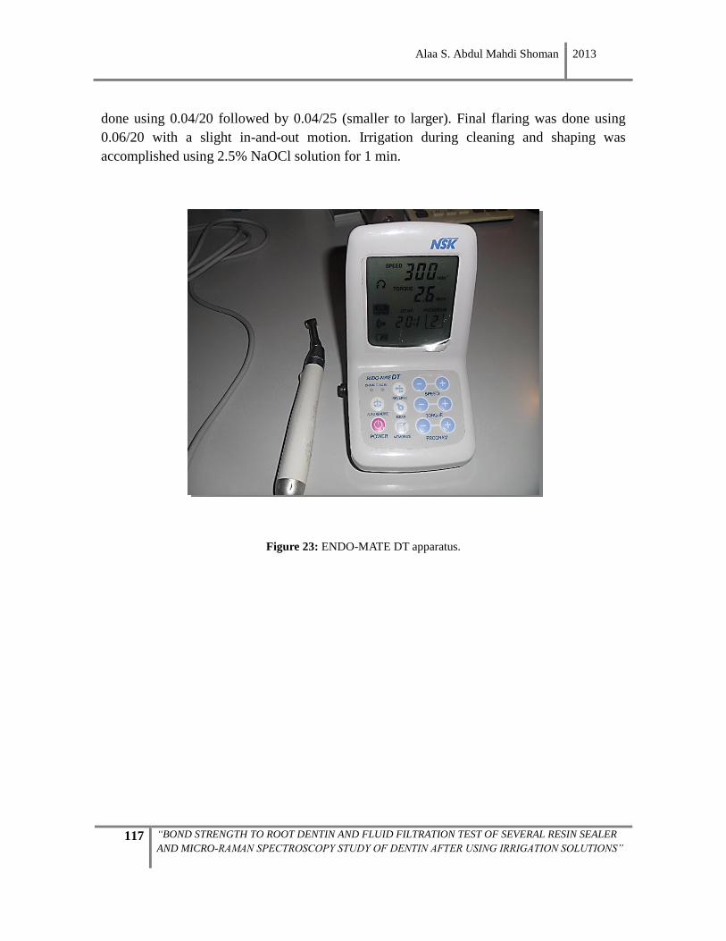
Alaa S. Abdul Mahdi Shoman 2013
117 “BOND STRENGTH TO ROOT DENTIN AND FLUID FILTRATION TEST OF SEVERAL RESIN SEALER
AND MICRO-RAMAN SPECTROSCOPY STUDY OF DENTIN AFTER USING IRRIGATION SOLUTIONS”
done using 0.04/20 followed by 0.04/25 (smaller to larger). Final flaring was done using
0.06/20 with a slight in-and-out motion. Irrigation during cleaning and shaping was
accomplished using 2.5% NaOCl solution for 1 min.
Figure 23: ENDO-MATE DT apparatus.
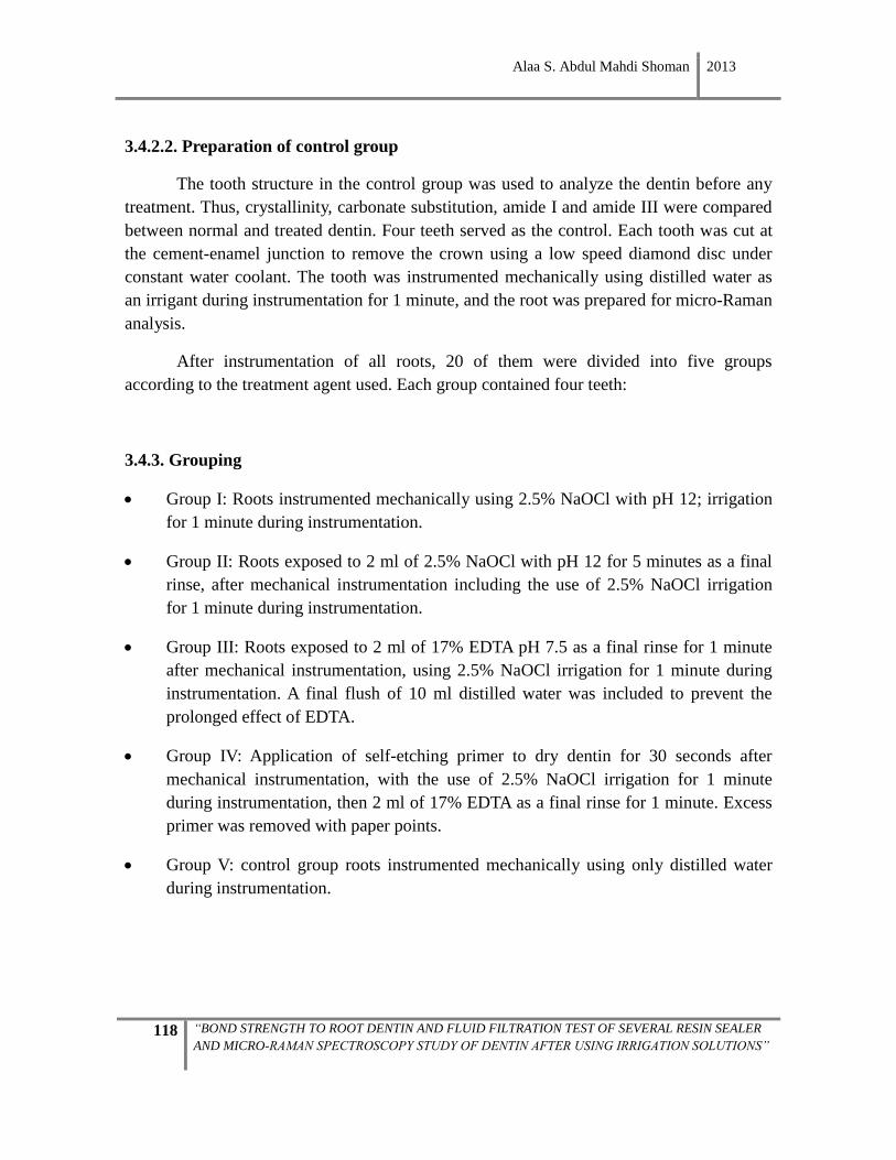
Alaa S. Abdul Mahdi Shoman 2013
118 “BOND STRENGTH TO ROOT DENTIN AND FLUID FILTRATION TEST OF SEVERAL RESIN SEALER
AND MICRO-RAMAN SPECTROSCOPY STUDY OF DENTIN AFTER USING IRRIGATION SOLUTIONS”
3.4.2.2. Preparation of control group
The tooth structure in the control group was used to analyze the dentin before any
treatment. Thus, crystallinity, carbonate substitution, amide I and amide III were compared
between normal and treated dentin. Four teeth served as the control. Each tooth was cut at
the cement-enamel junction to remove the crown using a low speed diamond disc under
constant water coolant. The tooth was instrumented mechanically using distilled water as
an irrigant during instrumentation for 1 minute, and the root was prepared for micro-Raman
analysis.
After instrumentation of all roots, 20 of them were divided into five groups
according to the treatment agent used. Each group contained four teeth:
3.4.3. Grouping
Group I: Roots instrumented mechanically using 2.5% NaOCl with pH 12; irrigation
for 1 minute during instrumentation.
Group II: Roots exposed to 2 ml of 2.5% NaOCl with pH 12 for 5 minutes as a final
rinse, after mechanical instrumentation including the use of 2.5% NaOCl irrigation
for 1 minute during instrumentation.
Group III: Roots exposed to 2 ml of 17% EDTA pH 7.5 as a final rinse for 1 minute
after mechanical instrumentation, using 2.5% NaOCl irrigation for 1 minute during
instrumentation. A final flush of 10 ml distilled water was included to prevent the
prolonged effect of EDTA.
Group IV: Application of self-etching primer to dry dentin for 30 seconds after
mechanical instrumentation, with the use of 2.5% NaOCl irrigation for 1 minute
during instrumentation, then 2 ml of 17% EDTA as a final rinse for 1 minute. Excess
primer was removed with paper points.
Group V: control group roots instrumented mechanically using only distilled water
during instrumentation.
Alaa S. Abdul Mahdi Shoman 2013
119 “BOND STRENGTH TO ROOT DENTIN AND FLUID FILTRATION TEST OF SEVERAL RESIN SEALER
AND MICRO-RAMAN SPECTROSCOPY STUDY OF DENTIN AFTER USING IRRIGATION SOLUTIONS”
3.4.4. Preparation of samples for micro-Raman
Following cleaning, shaping and treatment, each root was sectioned horizontally
perpendicular to the long axis of the root, into four slices —coronal 1, coronal 2, middle1,
middle 2— Figure 24; using a water cooled low speed diamond disc (Struers, 5010323,
Copenhagen, Denmark).
Figure 24: Four root slices with 1mm thickness
In this way, 80 root slices were obtained, having thickness 1mm. The dentin surface
of each slice was then abraded to smoothness with a series of silicon carbide paper (500,
800, 1200 and 2500) successively increasing in grit size, and polished with a fine silicon
carbide paper (4000 grit) under water coolant to obtain a mirror-like surface (Figure 25).
The final thickness of each slice was 0.3mm of flat dentin surface. The samples were
cleaned using an ultrasonic bath (with distilled water) for 30 minutes to remove excess
debris. The slices were then ready for micro-Raman spectroscopy characterization (Figure
26).
All 80 specimens were kept in a wet environment at 37°C for 24 hours until micro-
Raman analysis.
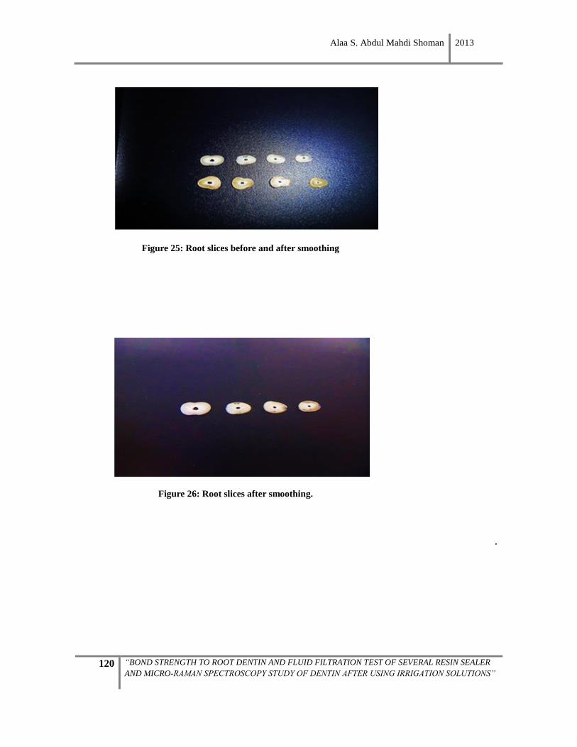
Alaa S. Abdul Mahdi Shoman 2013
120 “BOND STRENGTH TO ROOT DENTIN AND FLUID FILTRATION TEST OF SEVERAL RESIN SEALER
AND MICRO-RAMAN SPECTROSCOPY STUDY OF DENTIN AFTER USING IRRIGATION SOLUTIONS”
Figure 25: Root slices before and after smoothing
Figure 26: Root slices after smoothing.
.
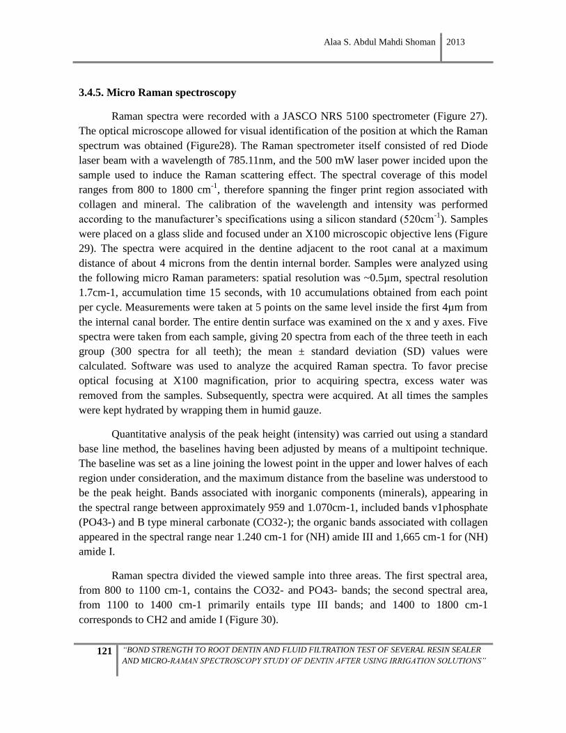
Alaa S. Abdul Mahdi Shoman 2013
121 “BOND STRENGTH TO ROOT DENTIN AND FLUID FILTRATION TEST OF SEVERAL RESIN SEALER
AND MICRO-RAMAN SPECTROSCOPY STUDY OF DENTIN AFTER USING IRRIGATION SOLUTIONS”
3.4.5. Micro Raman spectroscopy
Raman spectra were recorded with a JASCO NRS 5100 spectrometer (Figure 27).
The optical microscope allowed for visual identification of the position at which the Raman
spectrum was obtained (Figure28). The Raman spectrometer itself consisted of red Diode
laser beam with a wavelength of 785.11nm, and the 500 mW laser power incided upon the
sample used to induce the Raman scattering effect. The spectral coverage of this model
ranges from 800 to 1800 cm-1
, therefore spanning the finger print region associated with
collagen and mineral. The calibration of the wavelength and intensity was performed
according to the manufacturer’s specifications using a silicon standard (520cm-1
). Samples
were placed on a glass slide and focused under an X100 microscopic objective lens (Figure
29). The spectra were acquired in the dentine adjacent to the root canal at a maximum
distance of about 4 microns from the dentin internal border. Samples were analyzed using
the following micro Raman parameters: spatial resolution was ~0.5µm, spectral resolution
1.7cm-1, accumulation time 15 seconds, with 10 accumulations obtained from each point
per cycle. Measurements were taken at 5 points on the same level inside the first 4µm from
the internal canal border. The entire dentin surface was examined on the x and y axes. Five
spectra were taken from each sample, giving 20 spectra from each of the three teeth in each
group (300 spectra for all teeth); the mean ± standard deviation (SD) values were
calculated. Software was used to analyze the acquired Raman spectra. To favor precise
optical focusing at X100 magnification, prior to acquiring spectra, excess water was
removed from the samples. Subsequently, spectra were acquired. At all times the samples
were kept hydrated by wrapping them in humid gauze.
Quantitative analysis of the peak height (intensity) was carried out using a standard
base line method, the baselines having been adjusted by means of a multipoint technique.
The baseline was set as a line joining the lowest point in the upper and lower halves of each
region under consideration, and the maximum distance from the baseline was understood to
be the peak height. Bands associated with inorganic components (minerals), appearing in
the spectral range between approximately 959 and 1.070cm-1, included bands v1phosphate
(PO43-) and B type mineral carbonate (CO32-); the organic bands associated with collagen
appeared in the spectral range near 1.240 cm-1 for (NH) amide III and 1,665 cm-1 for (NH)
amide I.
Raman spectra divided the viewed sample into three areas. The first spectral area,
from 800 to 1100 cm-1, contains the CO32- and PO43- bands; the second spectral area,
from 1100 to 1400 cm-1 primarily entails type III bands; and 1400 to 1800 cm-1
corresponds to CH2 and amide I (Figure 30).
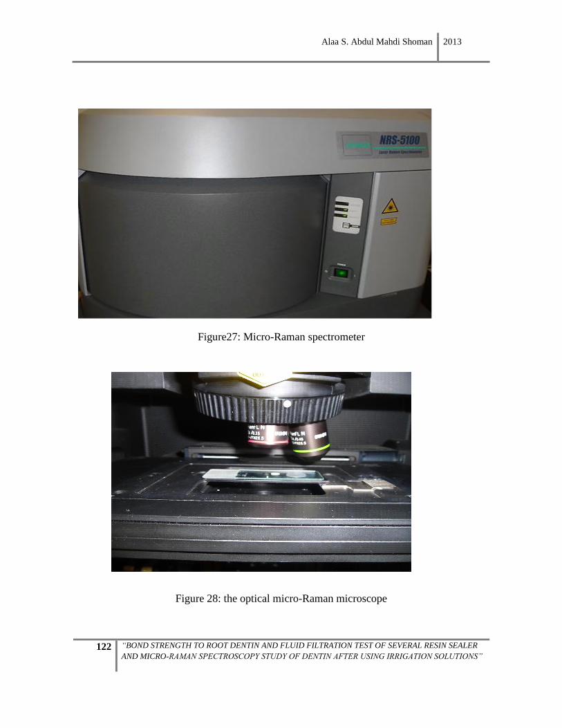
Alaa S. Abdul Mahdi Shoman 2013
122 “BOND STRENGTH TO ROOT DENTIN AND FLUID FILTRATION TEST OF SEVERAL RESIN SEALER
AND MICRO-RAMAN SPECTROSCOPY STUDY OF DENTIN AFTER USING IRRIGATION SOLUTIONS”
Figure27: Micro-Raman spectrometer
Figure 28: the optical micro-Raman microscope
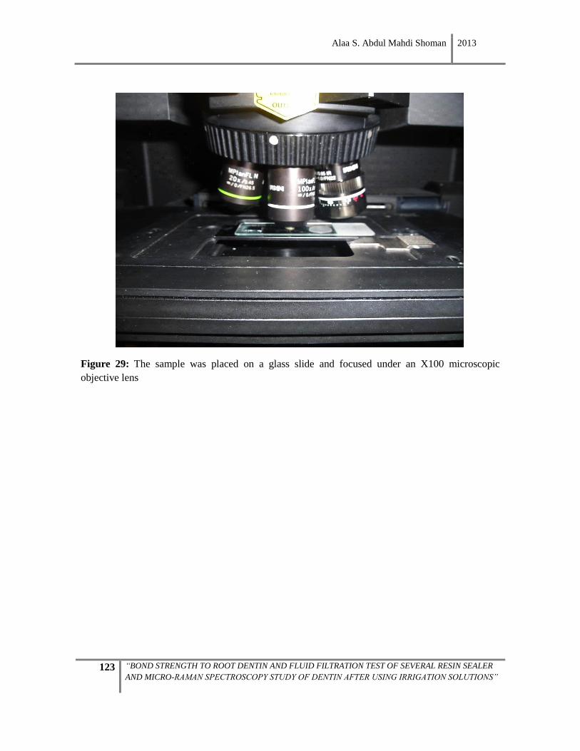
Alaa S. Abdul Mahdi Shoman 2013
123 “BOND STRENGTH TO ROOT DENTIN AND FLUID FILTRATION TEST OF SEVERAL RESIN SEALER
AND MICRO-RAMAN SPECTROSCOPY STUDY OF DENTIN AFTER USING IRRIGATION SOLUTIONS”
Figure 29: The sample was placed on a glass slide and focused under an X100 microscopic
objective lens
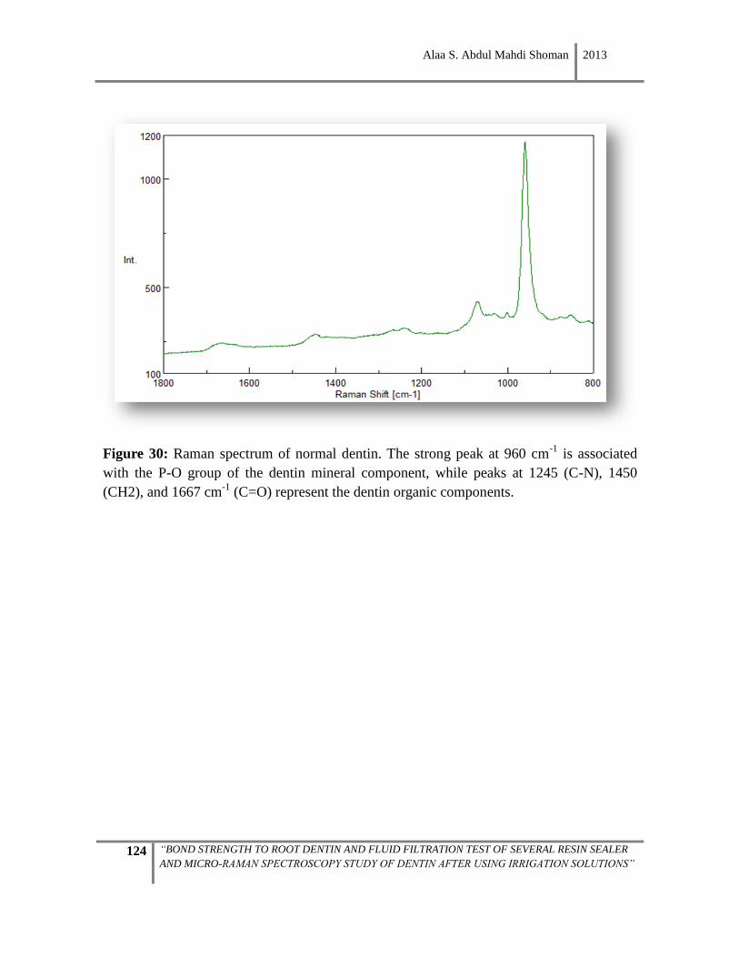
Alaa S. Abdul Mahdi Shoman 2013
124 “BOND STRENGTH TO ROOT DENTIN AND FLUID FILTRATION TEST OF SEVERAL RESIN SEALER
AND MICRO-RAMAN SPECTROSCOPY STUDY OF DENTIN AFTER USING IRRIGATION SOLUTIONS”
Figure 30: Raman spectrum of normal dentin. The strong peak at 960 cm-1
is associated
with the P-O group of the dentin mineral component, while peaks at 1245 (C-N), 1450
(CH2), and 1667 cm-1
(C=O) represent the dentin organic components.
Alaa S. Abdul Mahdi Shoman 2013
125 “BOND STRENGTH TO ROOT DENTIN AND FLUID FILTRATION TEST OF SEVERAL RESIN SEALER
AND MICRO-RAMAN SPECTROSCOPY STUDY OF DENTIN AFTER USING IRRIGATION SOLUTIONS”
Comparisons of the intensity ratios among the different dentin specimens are given below.
Carbonate to phosphate ratio: It can provide valuable insight into the inorganic chemical
composition of dentine.
Carbonate to CH2 ratio (1,072–1,450 cm-1
): The intensity ratios of CO32-
to the CH2
wagging mode were calculated to measure the relative differences in carbonate392
.
Phosphate to CH2 ratio (959–1,450 cm-1
): The mineral to organic ratio was calculated as
the ratio of the phosphate intensity to the intensity of vibrations resulting from the CH2
flapping mode of side-chains of collagen molecules. This 1,450 cm-1
band was chosen by
virtue of its low sensitivity to molecular orientation compared with amide I (at 1,670 cm-1
) 393
.
Amide III to CH2 ratio (1,243–1,450 cm-1
): It indicates structural differences. A higher ratio
was noted in intertubular dentine than in peritubular dentine, which indicates a higher
content in collagen394
.
Amide I to CH2 ratio (1,665–1,450 cm-1): An increase of the amide I band at 1,655 cm-1
relative to the CH2 band indicates altered collagen quality induced by aging395
,
hydration/dehydration396
, or radiologic damage397
.

Alaa S. Abdul Mahdi Shoman 2013
126 “BOND STRENGTH TO ROOT DENTIN AND FLUID FILTRATION TEST OF SEVERAL RESIN SEALER
AND MICRO-RAMAN SPECTROSCOPY STUDY OF DENTIN AFTER USING IRRIGATION SOLUTIONS”

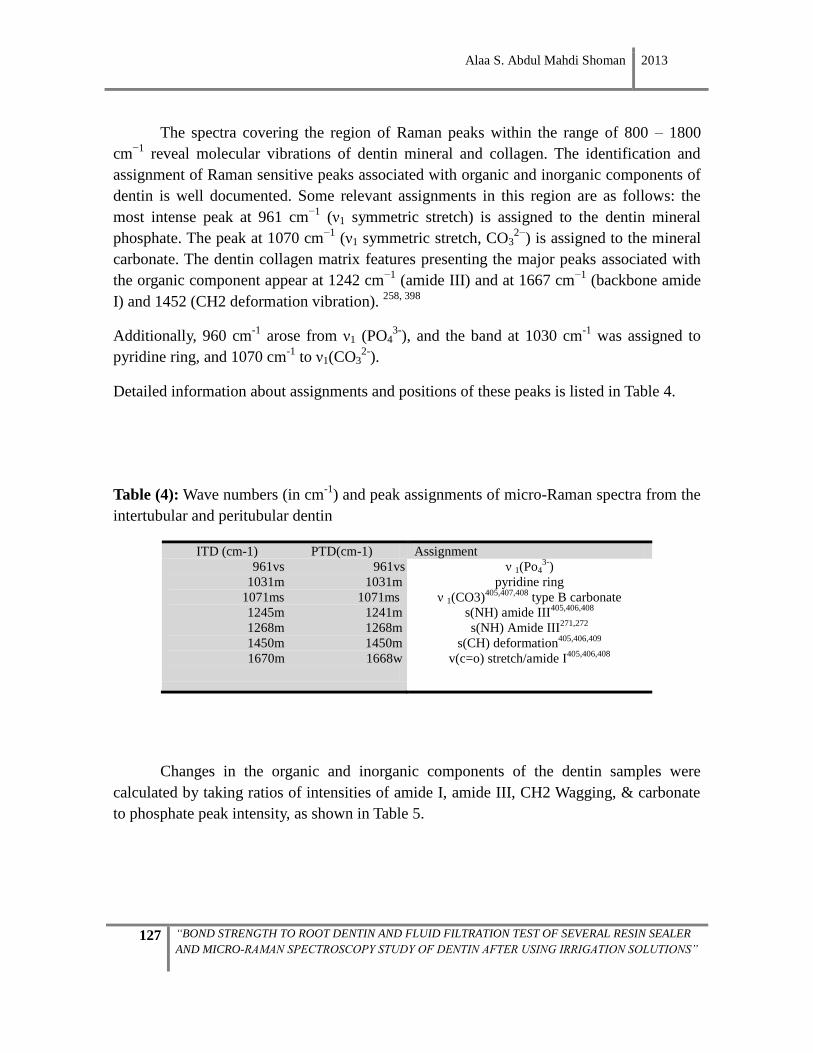
Alaa S. Abdul Mahdi Shoman 2013
127 “BOND STRENGTH TO ROOT DENTIN AND FLUID FILTRATION TEST OF SEVERAL RESIN SEALER
AND MICRO-RAMAN SPECTROSCOPY STUDY OF DENTIN AFTER USING IRRIGATION SOLUTIONS”
The spectra covering the region of Raman peaks within the range of 800 – 1800
cm−1
reveal molecular vibrations of dentin mineral and collagen. The identification and
assignment of Raman sensitive peaks associated with organic and inorganic components of
dentin is well documented. Some relevant assignments in this region are as follows: the
most intense peak at 961 cm−1
(ν1 symmetric stretch) is assigned to the dentin mineral
phosphate. The peak at 1070 cm−1
(ν1 symmetric stretch, CO32−
) is assigned to the mineral
carbonate. The dentin collagen matrix features presenting the major peaks associated with
the organic component appear at 1242 cm−1
(amide III) and at 1667 cm−1
(backbone amide
I) and 1452 (CH2 deformation vibration). 258, 398
Additionally, 960 cm-1
arose from ν1 (PO43-
), and the band at 1030 cm-1
was assigned to
pyridine ring, and 1070 cm-1
to ν1(CO32-
).
Detailed information about assignments and positions of these peaks is listed in Table 4.
Table (4): Wave numbers (in cm-1
) and peak assignments of micro-Raman spectra from the
intertubular and peritubular dentin
ITD (cm-1) PTD(cm-1) Assignment
961vs
961vs
1031m
1031m
1071ms
1071ms
1245m
1241m
1268m
1268m
1450m
1450m
1670m
1668w
ν 1(Po43-
)
pyridine ring
ν 1(CO3)405,407,408
type B carbonate
s(NH) amide III405,406,408
s(NH) Amide III271,272
s(CH) deformation405,406,409
v(c=o) stretch/amide I405,406,408
Changes in the organic and inorganic components of the dentin samples were
calculated by taking ratios of intensities of amide I, amide III, CH2 Wagging, & carbonate
to phosphate peak intensity, as shown in Table 5.
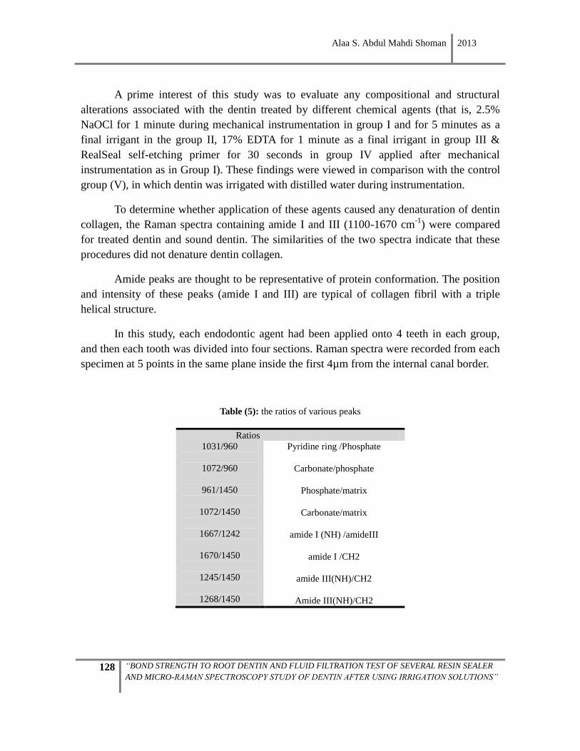
Alaa S. Abdul Mahdi Shoman 2013
128 “BOND STRENGTH TO ROOT DENTIN AND FLUID FILTRATION TEST OF SEVERAL RESIN SEALER
AND MICRO-RAMAN SPECTROSCOPY STUDY OF DENTIN AFTER USING IRRIGATION SOLUTIONS”
A prime interest of this study was to evaluate any compositional and structural
alterations associated with the dentin treated by different chemical agents (that is, 2.5%
NaOCl for 1 minute during mechanical instrumentation in group I and for 5 minutes as a
final irrigant in the group II, 17% EDTA for 1 minute as a final irrigant in group III &
RealSeal self-etching primer for 30 seconds in group IV applied after mechanical
instrumentation as in Group I). These findings were viewed in comparison with the control
group (V), in which dentin was irrigated with distilled water during instrumentation.
To determine whether application of these agents caused any denaturation of dentin
collagen, the Raman spectra containing amide I and III (1100-1670 cm-1
) were compared
for treated dentin and sound dentin. The similarities of the two spectra indicate that these
procedures did not denature dentin collagen.
Amide peaks are thought to be representative of protein conformation. The position
and intensity of these peaks (amide I and III) are typical of collagen fibril with a triple
helical structure.
In this study, each endodontic agent had been applied onto 4 teeth in each group,
and then each tooth was divided into four sections. Raman spectra were recorded from each
specimen at 5 points in the same plane inside the first 4µm from the internal canal border.
Table (5): the ratios of various peaks
Ratios
1031/960
1072/960
961/1450
1072/1450
1667/1242
1670/1450
1245/1450
1268/1450
Pyridine ring /Phosphate
Carbonate/phosphate
Phosphate/matrix
Carbonate/matrix
amide I (NH) /amideIII
amide I /CH2
amide III(NH)/CH2
Amide III(NH)/CH2
Alaa S. Abdul Mahdi Shoman 2013
129 “BOND STRENGTH TO ROOT DENTIN AND FLUID FILTRATION TEST OF SEVERAL RESIN SEALER
AND MICRO-RAMAN SPECTROSCOPY STUDY OF DENTIN AFTER USING IRRIGATION SOLUTIONS”
3.5.1. Statistical analysis
Descriptive statistics and normality plots were obtained for each treatment group
and root level. Shapiro-Wilk's test was applied to explore the data distribution. Afterwards,
non-parametrical tests were used.
To compare among treatment groups, the Kruskal-Wallis and Mann-Whitney U tests
were applied. Analysis of the effect of chemical agents among root levels was done using
Friedman’s test for multiple comparisons and Wilcoxon’s rank tests, for pairwise
comparisons. Significance was established for a p value lesser than 0.05.
3.5.2. Effect of sodium hypochlorite on the organic and mineral component of root
canal dentin (table 6 and 7)
Irrigation of the canal with 2.5%NaOCl solution during instrumentation for 1
minute —Group I— showed no significant difference in amide I, amide III and amide
I/amide III ratios with respect to the control group, irrigated with distilled water during
instrumentation.
After final irrigation, the canal with 2.5%NaOCl solution for 5 minutes —Group
II— showed a decreased amide I/amide III (1667/1242) ratio corresponding to the amides
III, in relation with control group (V) at all levels, but only significant in the lower medium
levels (M1 & M2). and there was a decrease in the amide I and amide III band about 1450
cm-1 (-CH2 vibration dentin matrix), in some rooting depth levels compared to the control
group irrigated only with distilled water (Group V. In relation with Group (I), irrigated with
2.5% NaOCl for 1 minute during instrumentation, there was a significant increase of
1667/1242 ratio at all levels, yet a lower value for the two amide III/matrix ratios that was
significant in the 1st cervical level of 1245/1450 and in the upper levels (cervical and
middle) of 1268/1450 ratio.
It should be noted that the final irrigation for 5 minutes did not cause change in the
bands 1670/1450 associated with amide I in relation to the instrumentation and control
groups.
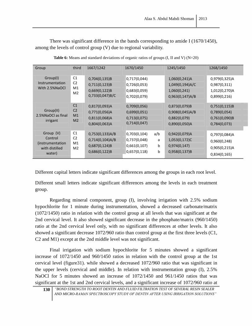
Alaa S. Abdul Mahdi Shoman 2013
130 “BOND STRENGTH TO ROOT DENTIN AND FLUID FILTRATION TEST OF SEVERAL RESIN SEALER
AND MICRO-RAMAN SPECTROSCOPY STUDY OF DENTIN AFTER USING IRRIGATION SOLUTIONS”
There was significant difference in the bands corresponding to amide I (1670/1450),
among the levels of control group (V) due to regional variability.
Table 6: Means and standard deviations of organic ratios of groups (I, II and V) (N=20)
Group third
1667/1242 1670/1450 1245/1450 1268/1450
Group(I) Instrumentation With 2.5%NaOCl
C1 C2 M1 M2
0,704(0,135)B
0,711(0,123)B
0,669(0,122)B
0,733(0,047)B/C
0,717(0,044)
0,726(0,053)
0,683(0,059)
0,702(0,079)
1,060(0,241)A
1,049(0,194)A/C
1,060(0,241)
0,963(0,147)A/B
0,979(0,325)A
0,987(0,311)
1,012(0,270)A
0,899(0,216)
Group(II)
2.5%NaOCl as final irrigant
C1 C2 M1 M2
0,817(0,093)A
0,771(0,056)A
0,811(0,068)A
0,804(0,063)A
0,709(0,056)
0,699(0,051)
0,713(0,075) 0,714(0,047)
0,873(0,079)B
0,908(0,045)A/B
0,882(0,079)
0,890(0,050)A
0,751(0,115)B
0,789(0,054)
0,761(0,090)B
0,784(0,073)
Group (V) Control
(instrumentation with distilled
water)
C1 C2 M1 M2
0,753(0,133)A/B
0,714(0,104)A/B
0,687(0,124)B
0,686(0,122)B
0,703(0,104) a/b
0,737(0,048) a
0,661(0,107) b
0,657(0,118) b
0,942(0,079)A
1,053(0,172)C
0,974(0,147)
0,958(0,137)B
0,797(0,084)A
0,960(0,248)
0,905(0,215)A
0,834(0,165)
Different capital letters indicate significant differences among the groups in each root level.
Different small letters indicate significant differences among the levels in each treatment
group.
Regarding mineral component, group (I), involving irrigation with 2.5% sodium
hypochlorite for 1 minute during instrumentation, showed a decreased carbonate/matrix
(1072/1450) ratio in relation with the control group at all levels that was significant at the
2nd cervical level. It also showed significant decrease in the phosphate/matrix (960/1450)
ratio at the 2nd cervical level only, with no significant differences at other levels. It also
showed a significant decrease 1072/960 ratio than control group at the first three levels (C1,
C2 and M1) except at the 2nd middle level was not significant.
Final irrigation with sodium hypochlorite for 5 minutes showed a significant
increase of 1072/1450 and 960/1450 ratios in relation with the control group at the 1st
cervical level (figure31). while showed a decreased 1072/960 ratio that was significant in
the upper levels (cervical and middle). In relation with instrumentation group (I), 2.5%
NaOCl for 5 minutes showed an increase of 1072/1450 and 961/1450 ratios that was
significant at the 1st and 2nd cervical levels, and a significant increase of 1072/960 ratio at
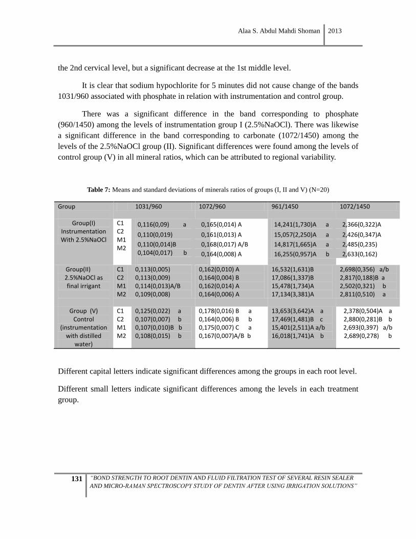
Alaa S. Abdul Mahdi Shoman 2013
131 “BOND STRENGTH TO ROOT DENTIN AND FLUID FILTRATION TEST OF SEVERAL RESIN SEALER
AND MICRO-RAMAN SPECTROSCOPY STUDY OF DENTIN AFTER USING IRRIGATION SOLUTIONS”
the 2nd cervical level, but a significant decrease at the 1st middle level.
It is clear that sodium hypochlorite for 5 minutes did not cause change of the bands
1031/960 associated with phosphate in relation with instrumentation and control group.
There was a significant difference in the band corresponding to phosphate
(960/1450) among the levels of instrumentation group I (2.5%NaOCl). There was likewise
a significant difference in the band corresponding to carbonate (1072/1450) among the
levels of the 2.5%NaOCl group (II). Significant differences were found among the levels of
control group (V) in all mineral ratios, which can be attributed to regional variability.
Table 7: Means and standard deviations of minerals ratios of groups (I, II and V) (N=20)
Group
1031/960 1072/960 961/1450 1072/1450
Group(I) Instrumentation With 2.5%NaOCl
C1 C2 M1 M2
0,116(0,09) a
0,110(0,019)
0,110(0,014)B 0,104(0,017) b
0,165(0,014) A
0,161(0,013) A
0,168(0,017) A/B
0,164(0,008) A
14,241(1,730)A a
15,057(2,250)A a
14,817(1,665)A a
16,255(0,957)A b
2,366(0,322)A
2,426(0,347)A
2,485(0,235)
2,633(0,162)
Group(II) 2.5%NaOCl as final irrigant
C1 C2 M1 M2
0,113(0,005) 0,113(0,009) 0,114(0,013)A/B 0,109(0,008)
0,162(0,010) A 0,164(0,004) B 0,162(0,014) A 0,164(0,006) A
16,532(1,631)B 17,086(1,337)B 15,478(1,734)A 17,134(3,381)A
2,698(0,356) a/b 2,817(0,188)B a 2,502(0,321) b 2,811(0,510) a
Group (V) Control
(instrumentation with distilled
water)
C1 C2 M1 M2
0,125(0,022) a 0,107(0,007) b 0,107(0,010)B b 0,108(0,015) b
0,178(0,016) B a 0,164(0,006) B b 0,175(0,007) C a 0,167(0,007)A/B b
13,653(3,642)A a 17,469(1,481)B c 15,401(2,511)A a/b 16,018(1,741)A b
2,378(0,504)A a 2,880(0,281)B b 2,693(0,397) a/b 2,689(0,278) b
Different capital letters indicate significant differences among the groups in each root level.
Different small letters indicate significant differences among the levels in each treatment
group.
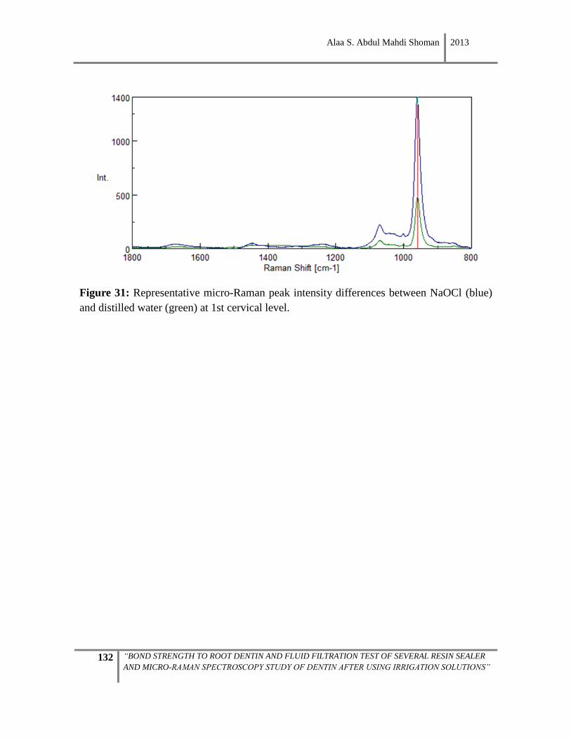
Alaa S. Abdul Mahdi Shoman 2013
132 “BOND STRENGTH TO ROOT DENTIN AND FLUID FILTRATION TEST OF SEVERAL RESIN SEALER
AND MICRO-RAMAN SPECTROSCOPY STUDY OF DENTIN AFTER USING IRRIGATION SOLUTIONS”
Figure 31: Representative micro-Raman peak intensity differences between NaOCl (blue)
and distilled water (green) at 1st cervical level.
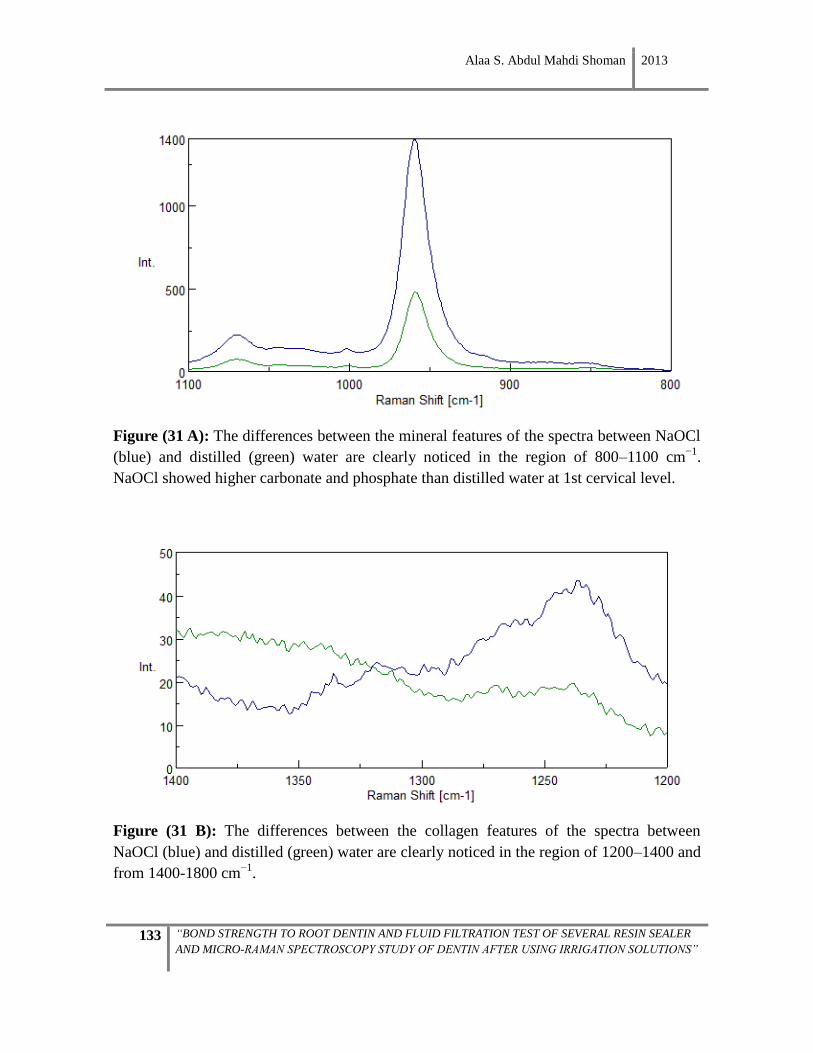
Alaa S. Abdul Mahdi Shoman 2013
133 “BOND STRENGTH TO ROOT DENTIN AND FLUID FILTRATION TEST OF SEVERAL RESIN SEALER
AND MICRO-RAMAN SPECTROSCOPY STUDY OF DENTIN AFTER USING IRRIGATION SOLUTIONS”
Figure (31 A): The differences between the mineral features of the spectra between NaOCl
(blue) and distilled (green) water are clearly noticed in the region of 800–1100 cm−1
.
NaOCl showed higher carbonate and phosphate than distilled water at 1st cervical level.
Figure (31 B): The differences between the collagen features of the spectra between
NaOCl (blue) and distilled (green) water are clearly noticed in the region of 1200–1400 and
from 1400-1800 cm−1
.
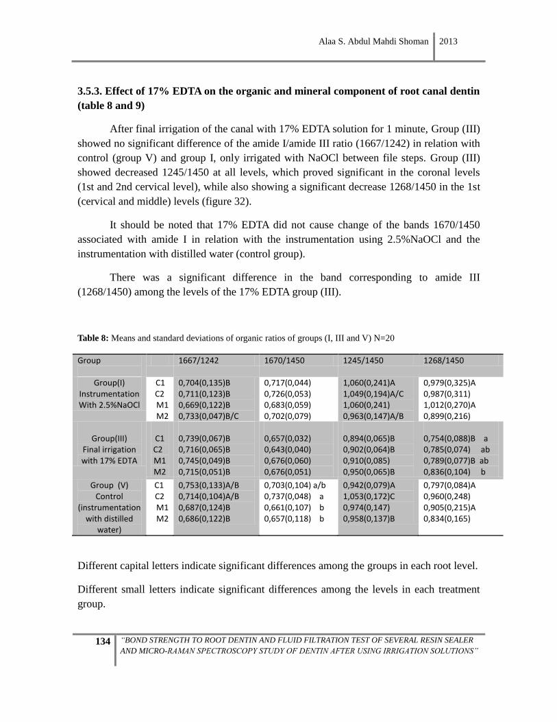
Alaa S. Abdul Mahdi Shoman 2013
134 “BOND STRENGTH TO ROOT DENTIN AND FLUID FILTRATION TEST OF SEVERAL RESIN SEALER
AND MICRO-RAMAN SPECTROSCOPY STUDY OF DENTIN AFTER USING IRRIGATION SOLUTIONS”
3.5.3. Effect of 17% EDTA on the organic and mineral component of root canal dentin
(table 8 and 9)
After final irrigation of the canal with 17% EDTA solution for 1 minute, Group (III)
showed no significant difference of the amide I/amide III ratio (1667/1242) in relation with
control (group V) and group I, only irrigated with NaOCl between file steps. Group (III)
showed decreased 1245/1450 at all levels, which proved significant in the coronal levels
(1st and 2nd cervical level), while also showing a significant decrease 1268/1450 in the 1st
(cervical and middle) levels (figure 32).
It should be noted that 17% EDTA did not cause change of the bands 1670/1450
associated with amide I in relation with the instrumentation using 2.5%NaOCl and the
instrumentation with distilled water (control group).
There was a significant difference in the band corresponding to amide III
(1268/1450) among the levels of the 17% EDTA group (III).
Table 8: Means and standard deviations of organic ratios of groups (I, III and V) N=20
Group
1667/1242 1670/1450 1245/1450 1268/1450
Group(I) Instrumentation With 2.5%NaOCl
C1 C2 M1 M2
0,704(0,135)B 0,711(0,123)B 0,669(0,122)B 0,733(0,047)B/C
0,717(0,044) 0,726(0,053) 0,683(0,059) 0,702(0,079)
1,060(0,241)A 1,049(0,194)A/C 1,060(0,241) 0,963(0,147)A/B
0,979(0,325)A 0,987(0,311) 1,012(0,270)A 0,899(0,216)
Group(III)
Final irrigation with 17% EDTA
C1
C2 M1 M2
0,739(0,067)B 0,716(0,065)B 0,745(0,049)B 0,715(0,051)B
0,657(0,032) 0,643(0,040) 0,676(0,060) 0,676(0,051)
0,894(0,065)B 0,902(0,064)B 0,910(0,085) 0,950(0,065)B
0,754(0,088)B a 0,785(0,074) ab 0,789(0,077)B ab 0,836(0,104) b
Group (V) Control
(instrumentation with distilled
water)
C1 C2 M1 M2
0,753(0,133)A/B 0,714(0,104)A/B 0,687(0,124)B 0,686(0,122)B
0,703(0,104) a/b 0,737(0,048) a 0,661(0,107) b 0,657(0,118) b
0,942(0,079)A 1,053(0,172)C 0,974(0,147) 0,958(0,137)B
0,797(0,084)A 0,960(0,248) 0,905(0,215)A 0,834(0,165)
Different capital letters indicate significant differences among the groups in each root level.
Different small letters indicate significant differences among the levels in each treatment
group.
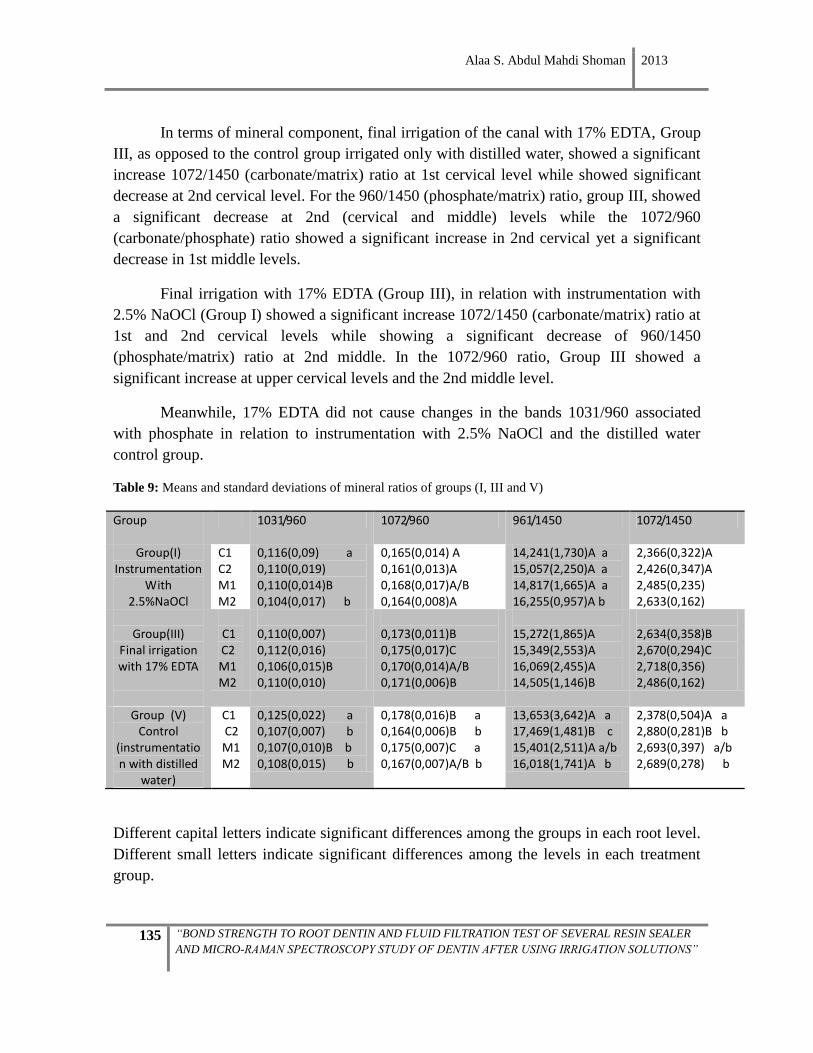
Alaa S. Abdul Mahdi Shoman 2013
135 “BOND STRENGTH TO ROOT DENTIN AND FLUID FILTRATION TEST OF SEVERAL RESIN SEALER
AND MICRO-RAMAN SPECTROSCOPY STUDY OF DENTIN AFTER USING IRRIGATION SOLUTIONS”
In terms of mineral component, final irrigation of the canal with 17% EDTA, Group
III, as opposed to the control group irrigated only with distilled water, showed a significant
increase 1072/1450 (carbonate/matrix) ratio at 1st cervical level while showed significant
decrease at 2nd cervical level. For the 960/1450 (phosphate/matrix) ratio, group III, showed
a significant decrease at 2nd (cervical and middle) levels while the 1072/960
(carbonate/phosphate) ratio showed a significant increase in 2nd cervical yet a significant
decrease in 1st middle levels.
Final irrigation with 17% EDTA (Group III), in relation with instrumentation with
2.5% NaOCl (Group I) showed a significant increase 1072/1450 (carbonate/matrix) ratio at
1st and 2nd cervical levels while showing a significant decrease of 960/1450
(phosphate/matrix) ratio at 2nd middle. In the 1072/960 ratio, Group III showed a
significant increase at upper cervical levels and the 2nd middle level.
Meanwhile, 17% EDTA did not cause changes in the bands 1031/960 associated
with phosphate in relation to instrumentation with 2.5% NaOCl and the distilled water
control group.
Table 9: Means and standard deviations of mineral ratios of groups (I, III and V)
Group
1031/960 1072/960 961/1450 1072/1450
Group(I) Instrumentation
With 2.5%NaOCl
C1 C2M1 M2
0,116(0,09) a 0,110(0,019) 0,110(0,014)B 0,104(0,017) b
0,165(0,014) A 0,161(0,013)A 0,168(0,017)A/B 0,164(0,008)A
14,241(1,730)A a 15,057(2,250)A a 14,817(1,665)A a 16,255(0,957)A b
2,366(0,322)A 2,426(0,347)A 2,485(0,235) 2,633(0,162)
Group(III)
Final irrigation with 17% EDTA
C1 C2 M1 M2
0,110(0,007) 0,112(0,016) 0,106(0,015)B 0,110(0,010)
0,173(0,011)B 0,175(0,017)C 0,170(0,014)A/B 0,171(0,006)B
15,272(1,865)A 15,349(2,553)A 16,069(2,455)A 14,505(1,146)B
2,634(0,358)B 2,670(0,294)C 2,718(0,356) 2,486(0,162)
Group (V) Control
(instrumentation with distilled
water)
C1 C2 M1 M2
0,125(0,022) a 0,107(0,007) b 0,107(0,010)B b 0,108(0,015) b
0,178(0,016)B a 0,164(0,006)B b 0,175(0,007)C a 0,167(0,007)A/B b
13,653(3,642)A a 17,469(1,481)B c 15,401(2,511)A a/b 16,018(1,741)A b
2,378(0,504)A a 2,880(0,281)B b 2,693(0,397) a/b 2,689(0,278) b
Different capital letters indicate significant differences among the groups in each root level.
Different small letters indicate significant differences among the levels in each treatment
group.
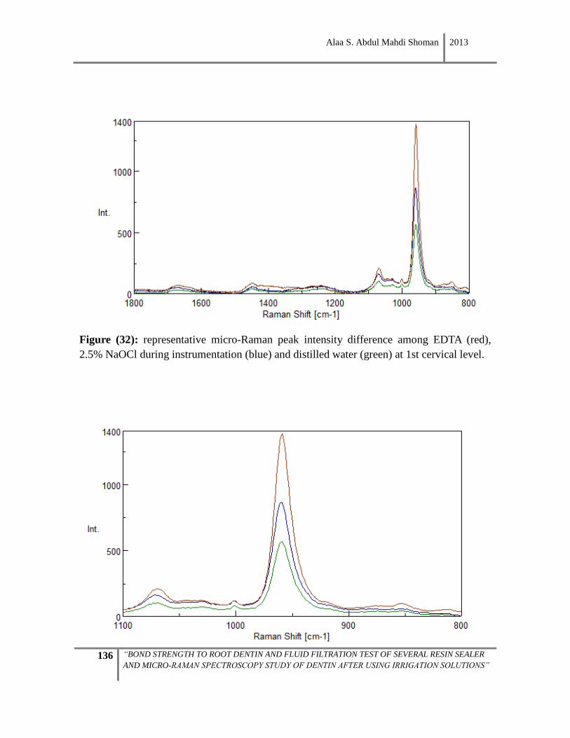
Alaa S. Abdul Mahdi Shoman 2013
136 “BOND STRENGTH TO ROOT DENTIN AND FLUID FILTRATION TEST OF SEVERAL RESIN SEALER
AND MICRO-RAMAN SPECTROSCOPY STUDY OF DENTIN AFTER USING IRRIGATION SOLUTIONS”
Figure (32): representative micro-Raman peak intensity difference among EDTA (red),
2.5% NaOCl during instrumentation (blue) and distilled water (green) at 1st cervical level.
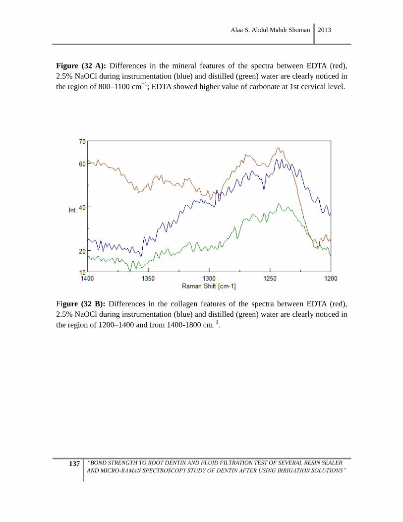
Alaa S. Abdul Mahdi Shoman 2013
137 “BOND STRENGTH TO ROOT DENTIN AND FLUID FILTRATION TEST OF SEVERAL RESIN SEALER
AND MICRO-RAMAN SPECTROSCOPY STUDY OF DENTIN AFTER USING IRRIGATION SOLUTIONS”
Figure (32 A): Differences in the mineral features of the spectra between EDTA (red),
2.5% NaOCl during instrumentation (blue) and distilled (green) water are clearly noticed in
the region of 800–1100 cm−1
; EDTA showed higher value of carbonate at 1st cervical level.
Figure (32 B): Differences in the collagen features of the spectra between EDTA (red),
2.5% NaOCl during instrumentation (blue) and distilled (green) water are clearly noticed in
the region of 1200–1400 and from 1400-1800 cm−1.
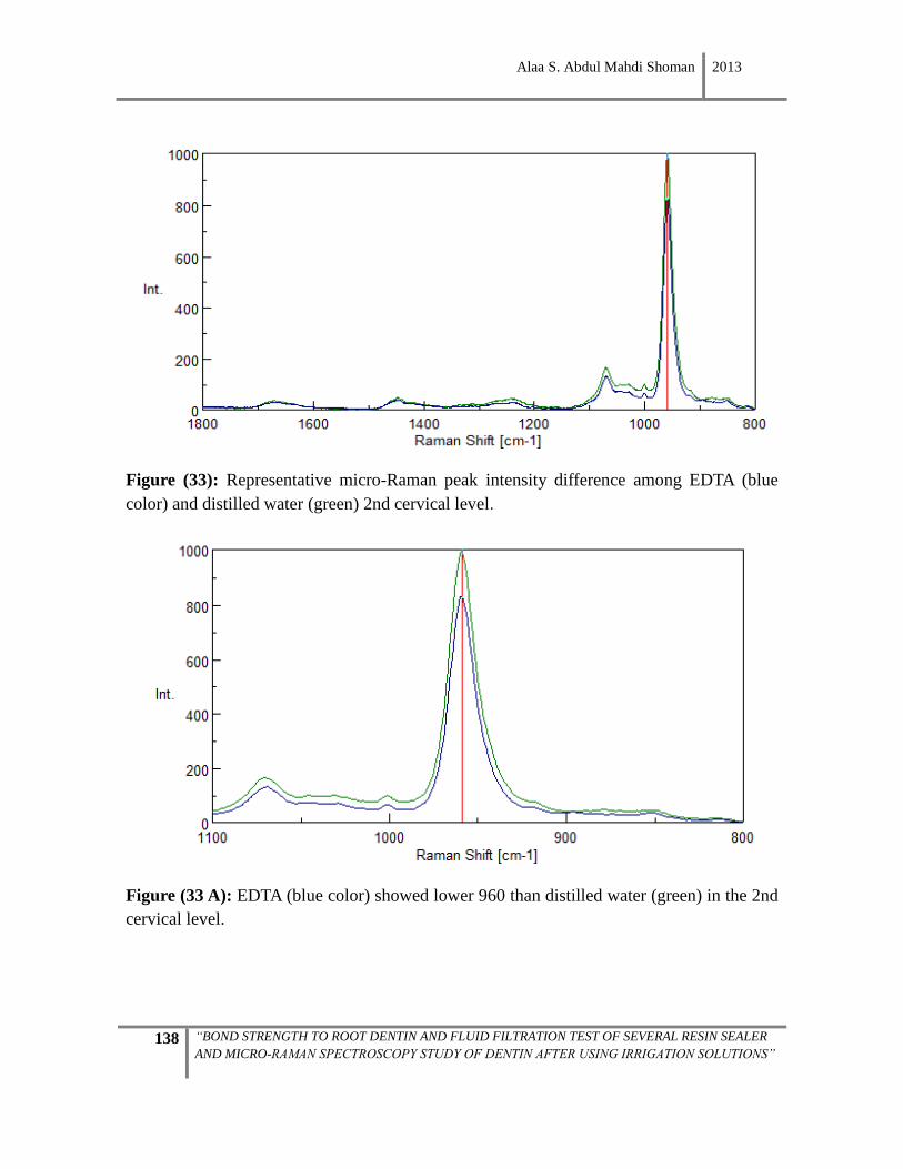
Alaa S. Abdul Mahdi Shoman 2013
138 “BOND STRENGTH TO ROOT DENTIN AND FLUID FILTRATION TEST OF SEVERAL RESIN SEALER
AND MICRO-RAMAN SPECTROSCOPY STUDY OF DENTIN AFTER USING IRRIGATION SOLUTIONS”
Figure (33): Representative micro-Raman peak intensity difference among EDTA (blue
color) and distilled water (green) 2nd cervical level.
Figure (33 A): EDTA (blue color) showed lower 960 than distilled water (green) in the 2nd
cervical level.
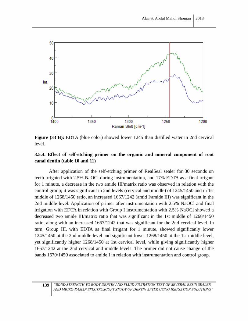
Alaa S. Abdul Mahdi Shoman 2013
139 “BOND STRENGTH TO ROOT DENTIN AND FLUID FILTRATION TEST OF SEVERAL RESIN SEALER
AND MICRO-RAMAN SPECTROSCOPY STUDY OF DENTIN AFTER USING IRRIGATION SOLUTIONS”
Figure (33 B): EDTA (blue color) showed lower 1245 than distilled water in 2nd cervical
level.
3.5.4. Effect of self-etching primer on the organic and mineral component of root
canal dentin (table 10 and 11)
After application of the self-etching primer of RealSeal sealer for 30 seconds on
teeth irrigated with 2.5% NaOCl during instrumentation, and 17% EDTA as a final irrigant
for 1 minute, a decrease in the two amide III/matrix ratio was observed in relation with the
control group; it was significant in 2nd levels (cervical and middle) of 1245/1450 and in 1st
middle of 1268/1450 ratio, an increased 1667/1242 (amid I/amide III) was significant in the
2nd middle level. Application of primer after instrumentation with 2.5% NaOCl and final
irrigation with EDTA in relation with Group I instrumentation with 2.5% NaOCl showed a
decreased two amide III/matrix ratio that was significant in the 1st middle of 1268/1450
ratio, along with an increased 1667/1242 that was significant for the 2nd cervical level. In
turn, Group III, with EDTA as final irrigant for 1 minute, showed significanly lower
1245/1450 at the 2nd middle level and significant lower 1268/1450 at the 1st middle level,
yet significantly higher 1268/1450 at 1st cervical level, while giving significantly higher
1667/1242 at the 2nd cervical and middle levels. The primer did not cause change of the
bands 1670/1450 associated to amide I in relation with instrumentation and control group.
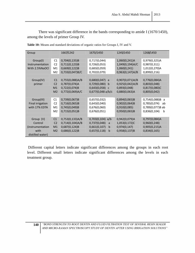
Alaa S. Abdul Mahdi Shoman 2013
140 “BOND STRENGTH TO ROOT DENTIN AND FLUID FILTRATION TEST OF SEVERAL RESIN SEALER
AND MICRO-RAMAN SPECTROSCOPY STUDY OF DENTIN AFTER USING IRRIGATION SOLUTIONS”
There was significant difference in the bands corresponding to amide I (1670/1450),
among the levels of primer Group IV.
Table 10: Means and standard deviations of organic ratios for Groups I, IV and V.
Group
1667/1242 1670/1450 1245/1450 1268/1450
Group(I) Instrumentation With 2.5%NaOCl
C1 C2 M1 M2
0,704(0,135)B 0,711(0,123)B 0,669(0,122)B 0,733(0,047)B/C
0,717(0,044) 0,726(0,053) 0,683(0,059) 0,702(0,079)
1,060(0,241)A 1,049(0,194)A/C 1,060(0,241) 0,963(0,147)A/B
0,979(0,325)A 0,987(0,311) 1,012(0,270)A 0,899(0,216)
Group(IV)
primer
C1 C2
M1 M2
0,755(0,088)A/B 0,787(0,074)A 0,722(0,074)B 0,772(0,069)A/C
0,680(0,047) a 0,729(0,080) b 0,643(0,058) c 0,677(0,048 a/b/c
0,907(0,071)A/B 0,925(0,042)A/B 0,893(0,048) 0,880(0,063)A
0,776(0,084)A 0,803(0,048) 0,817(0,080)C 0,805(0,042)
Group(III) Final irrigation with 17% EDTA
C1 C2 M1 M2
0,739(0,067)B 0,716(0,065)B 0,745(0,049)B 0,715(0,051)B
0,657(0,032) 0,643(0,040) 0,676(0,060) 0,676(0,051)
0,894(0,065)B 0,902(0,064)B 0,910(0,085) 0,950(0,065)B
0,754(0,088)B a 0,785(0,074) ab 0,789(0,077)B ab 0,836(0,104) b
Group (V) Control
(instrumentation with
distilled water)
C1 C2 M1 M2
0,753(0,133)A/B 0,714(0,104)A/B 0,687(0,124)B 0,686(0,122)B
0,703(0,104) a/b 0,737(0,048) a 0,661(0,107) b 0,657(0,118) b
0,942(0,079)A 1,053(0,172)C 0,974(0,147) 0,958(0,137)B
0,797(0,084)A 0,960(0,248) 0,905(0,215)A 0,834(0,165)
Different capital letters indicate significant differences among the groups in each root
level. Different small letters indicate significant differences among the levels in each
treatment group.
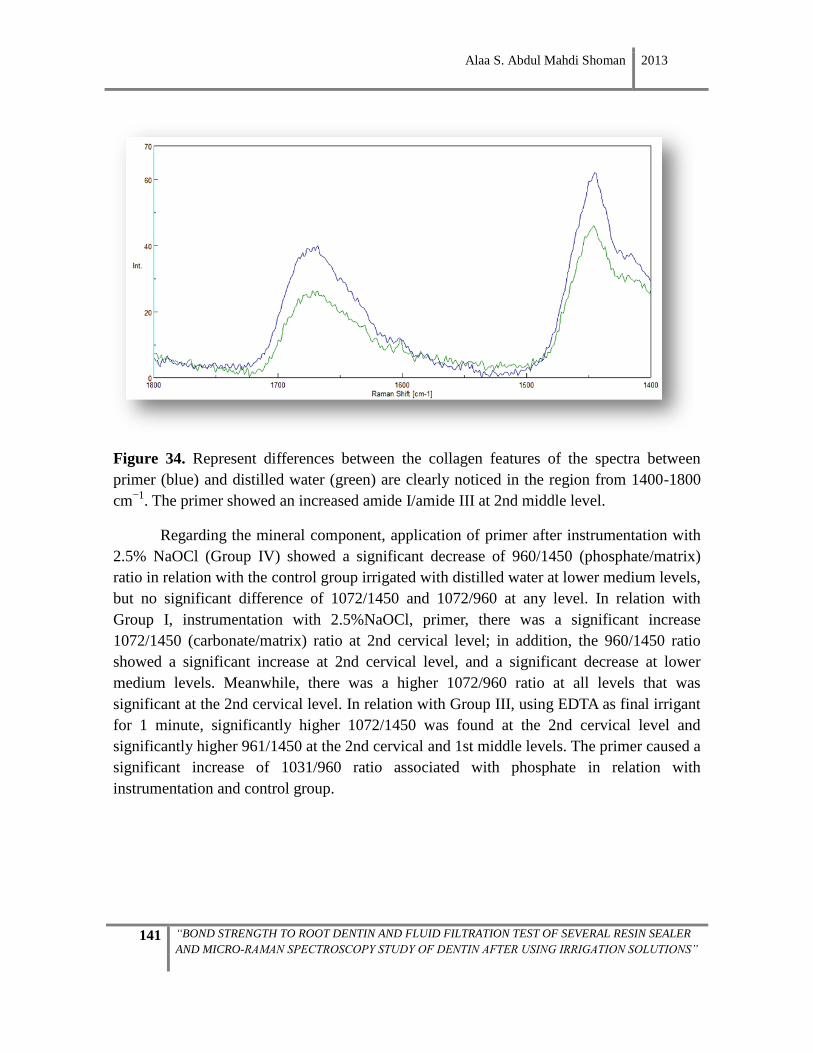
Alaa S. Abdul Mahdi Shoman 2013
141 “BOND STRENGTH TO ROOT DENTIN AND FLUID FILTRATION TEST OF SEVERAL RESIN SEALER
AND MICRO-RAMAN SPECTROSCOPY STUDY OF DENTIN AFTER USING IRRIGATION SOLUTIONS”
Figure 34. Represent differences between the collagen features of the spectra between
primer (blue) and distilled water (green) are clearly noticed in the region from 1400-1800
cm−1
. The primer showed an increased amide I/amide III at 2nd middle level.
Regarding the mineral component, application of primer after instrumentation with
2.5% NaOCl (Group IV) showed a significant decrease of 960/1450 (phosphate/matrix)
ratio in relation with the control group irrigated with distilled water at lower medium levels,
but no significant difference of 1072/1450 and 1072/960 at any level. In relation with
Group I, instrumentation with 2.5%NaOCl, primer, there was a significant increase
1072/1450 (carbonate/matrix) ratio at 2nd cervical level; in addition, the 960/1450 ratio
showed a significant increase at 2nd cervical level, and a significant decrease at lower
medium levels. Meanwhile, there was a higher 1072/960 ratio at all levels that was
significant at the 2nd cervical level. In relation with Group III, using EDTA as final irrigant
for 1 minute, significantly higher 1072/1450 was found at the 2nd cervical level and
significantly higher 961/1450 at the 2nd cervical and 1st middle levels. The primer caused a
significant increase of 1031/960 ratio associated with phosphate in relation with
instrumentation and control group.
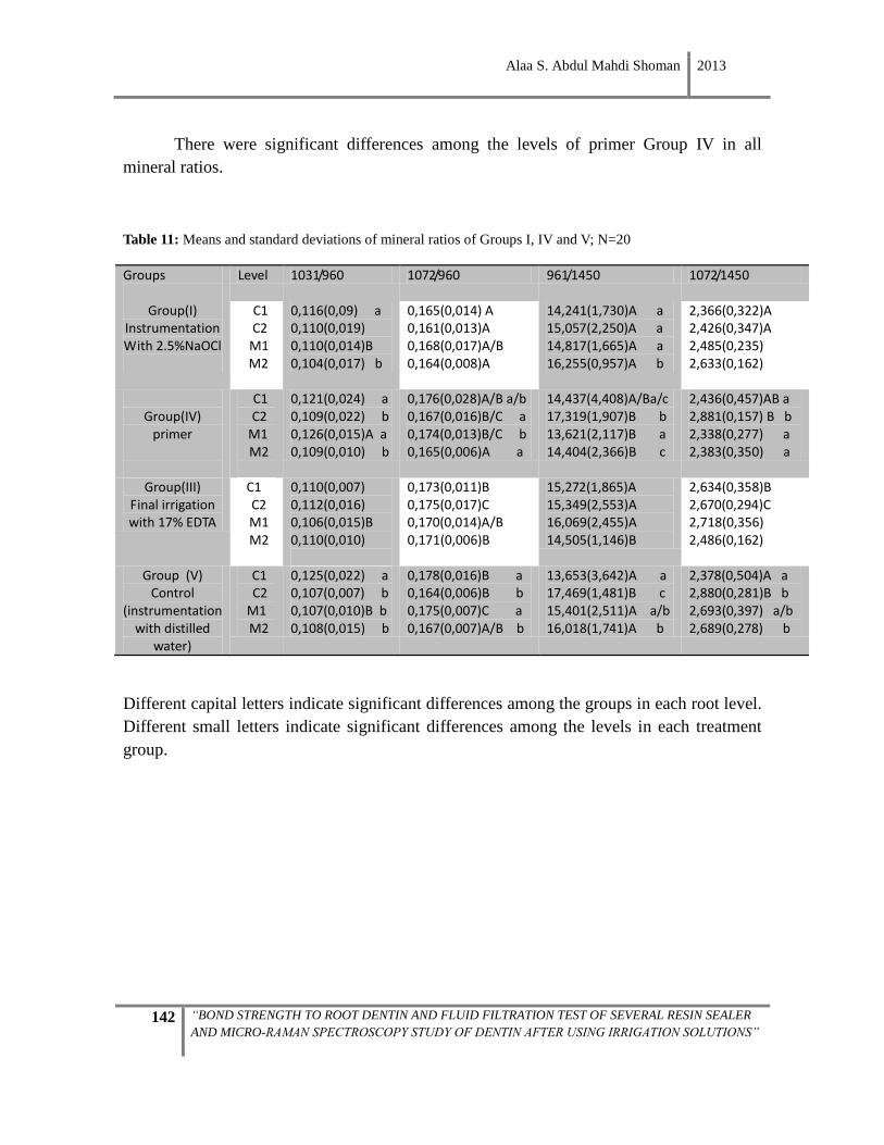
Alaa S. Abdul Mahdi Shoman 2013
142 “BOND STRENGTH TO ROOT DENTIN AND FLUID FILTRATION TEST OF SEVERAL RESIN SEALER
AND MICRO-RAMAN SPECTROSCOPY STUDY OF DENTIN AFTER USING IRRIGATION SOLUTIONS”
There were significant differences among the levels of primer Group IV in all
mineral ratios.
Table 11: Means and standard deviations of mineral ratios of Groups I, IV and V; N=20
Groups
Level 1031/960 1072/960 961/1450 1072/1450
Group(I) Instrumentation With 2.5%NaOCl
C1 C2
M1 M2
0,116(0,09) a 0,110(0,019) 0,110(0,014)B 0,104(0,017) b
0,165(0,014) A 0,161(0,013)A 0,168(0,017)A/B 0,164(0,008)A
14,241(1,730)A a 15,057(2,250)A a 14,817(1,665)A a 16,255(0,957)A b
2,366(0,322)A 2,426(0,347)A 2,485(0,235) 2,633(0,162)
Group(IV)
primer
C1 C2
M1 M2
0,121(0,024) a 0,109(0,022) b 0,126(0,015)A a 0,109(0,010) b
0,176(0,028)A/B a/b 0,167(0,016)B/C a 0,174(0,013)B/C b 0,165(0,006)A a
14,437(4,408)A/Ba/c 17,319(1,907)B b 13,621(2,117)B a 14,404(2,366)B c
2,436(0,457)AB a 2,881(0,157) B b 2,338(0,277) a 2,383(0,350) a
Group(III) Final irrigation with 17% EDTA
C1 C2 M1 M2
0,110(0,007) 0,112(0,016) 0,106(0,015)B 0,110(0,010)
0,173(0,011)B 0,175(0,017)C 0,170(0,014)A/B 0,171(0,006)B
15,272(1,865)A 15,349(2,553)A 16,069(2,455)A 14,505(1,146)B
2,634(0,358)B 2,670(0,294)C 2,718(0,356) 2,486(0,162)
Group (V) Control
(instrumentation with distilled
water)
C1 C2
M1 M2
0,125(0,022) a 0,107(0,007) b 0,107(0,010)B b 0,108(0,015) b
0,178(0,016)B a 0,164(0,006)B b 0,175(0,007)C a 0,167(0,007)A/B b
13,653(3,642)A a 17,469(1,481)B c 15,401(2,511)A a/b 16,018(1,741)A b
2,378(0,504)A a 2,880(0,281)B b 2,693(0,397) a/b 2,689(0,278) b
Different capital letters indicate significant differences among the groups in each root level.
Different small letters indicate significant differences among the levels in each treatment
group.
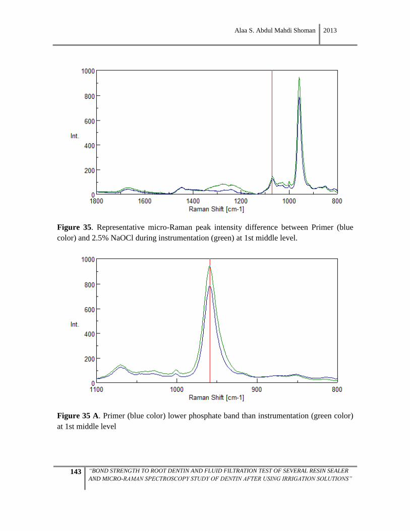
Alaa S. Abdul Mahdi Shoman 2013
143 “BOND STRENGTH TO ROOT DENTIN AND FLUID FILTRATION TEST OF SEVERAL RESIN SEALER
AND MICRO-RAMAN SPECTROSCOPY STUDY OF DENTIN AFTER USING IRRIGATION SOLUTIONS”
Figure 35. Representative micro-Raman peak intensity difference between Primer (blue
color) and 2.5% NaOCl during instrumentation (green) at 1st middle level.
Figure 35 A. Primer (blue color) lower phosphate band than instrumentation (green color)
at 1st middle level
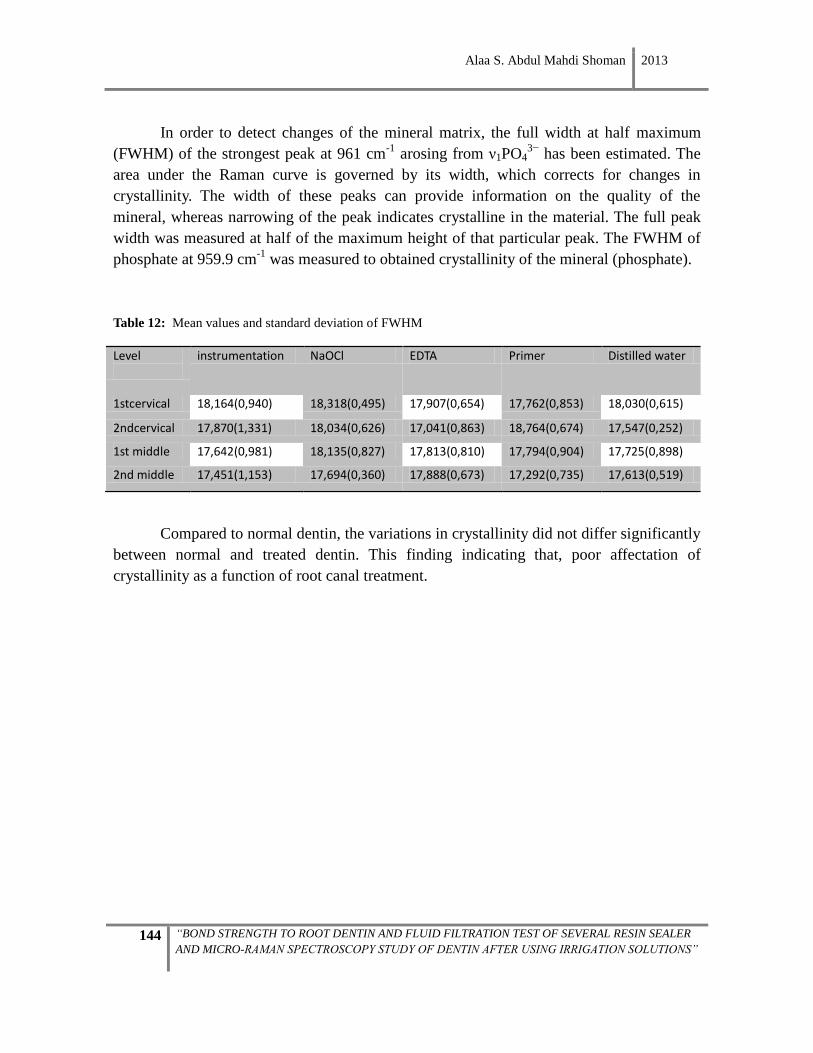
Alaa S. Abdul Mahdi Shoman 2013
144 “BOND STRENGTH TO ROOT DENTIN AND FLUID FILTRATION TEST OF SEVERAL RESIN SEALER
AND MICRO-RAMAN SPECTROSCOPY STUDY OF DENTIN AFTER USING IRRIGATION SOLUTIONS”
In order to detect changes of the mineral matrix, the full width at half maximum
(FWHM) of the strongest peak at 961 cm-1
arosing from ν1PO43−
has been estimated. The
area under the Raman curve is governed by its width, which corrects for changes in
crystallinity. The width of these peaks can provide information on the quality of the
mineral, whereas narrowing of the peak indicates crystalline in the material. The full peak
width was measured at half of the maximum height of that particular peak. The FWHM of
phosphate at 959.9 cm-1
was measured to obtained crystallinity of the mineral (phosphate).
Table 12: Mean values and standard deviation of FWHM
Level
instrumentation NaOCl EDTA Primer Distilled water
1stcervical 18,164(0,940) 18,318(0,495) 17,907(0,654) 17,762(0,853) 18,030(0,615)
2ndcervical 17,870(1,331) 18,034(0,626) 17,041(0,863) 18,764(0,674) 17,547(0,252)
1st middle 17,642(0,981) 18,135(0,827) 17,813(0,810) 17,794(0,904) 17,725(0,898)
2nd middle 17,451(1,153) 17,694(0,360) 17,888(0,673) 17,292(0,735) 17,613(0,519)
Compared to normal dentin, the variations in crystallinity did not differ significantly
between normal and treated dentin. This finding indicating that, poor affectation of
crystallinity as a function of root canal treatment.
Alaa S. Abdul Mahdi Shoman 2013
145 “BOND STRENGTH TO ROOT DENTIN AND FLUID FILTRATION TEST OF SEVERAL RESIN SEALER
AND MICRO-RAMAN SPECTROSCOPY STUDY OF DENTIN AFTER USING IRRIGATION SOLUTIONS”

Alaa S. Abdul Mahdi Shoman 2013
146 “BOND STRENGTH TO ROOT DENTIN AND FLUID FILTRATION TEST OF SEVERAL RESIN SEALER
AND MICRO-RAMAN SPECTROSCOPY STUDY OF DENTIN AFTER USING IRRIGATION SOLUTIONS”
The following important results have come out of this study:
• 2.5% NaOCl irrigation for 1 minute during instrumentation (Group I) showed no
effect on organic component (amide I, amide III); but a subsequent final irrigation
with 2.5% NaOCl solution for 5 minutes produces an irregular decrease of amide III
in some root levels, and inconsistent effect on the mineral component —most
notably a decrease in the carbonate/phosphate ratio.
• Final irrigation with EDTA for 1 minute after instrumentation, with 2.5% NaOCl
irrigation for 1 minute during instrumentation, produces little or no affectation of
the bands of carbonates and phosphates, and just a slight increase of
carbonate/phosphate ratio. In terms of the organic component, it does not affect the
amide III ratio or amide I/amide III.
• Application of primer for 30 seconds after the end of instrumentation and irrigation
with EDTA produces a consistent effect on the mineral component. It mainly leads
to a decrease in the phosphate concentration. At the organic component level, an
irregular decrease of amide III was seen in some levels, without affectation of amide
I, along with an inconsistent increase of the amide I/amide III ratio.
• The variations in crystallinity did not differ significantly between normal and
treated dentin.

Alaa S. Abdul Mahdi Shoman 2013
147 “BOND STRENGTH TO ROOT DENTIN AND FLUID FILTRATION TEST OF SEVERAL RESIN SEALER
AND MICRO-RAMAN SPECTROSCOPY STUDY OF DENTIN AFTER USING IRRIGATION SOLUTIONS”

Alaa S. Abdul Mahdi Shoman 2013
148 “BOND STRENGTH TO ROOT DENTIN AND FLUID FILTRATION TEST OF SEVERAL RESIN SEALER
AND MICRO-RAMAN SPECTROSCOPY STUDY OF DENTIN AFTER USING IRRIGATION SOLUTIONS”
The success of root canal therapy depends on the method and quality of
instrumentation, irrigation, disinfection, and three-dimensional obturation of the root canal.
In the endodontic literature, techniques for cleaning and shaping of root canals differ, and
no single-best procedure for all conditions has been established404,405,406
. Nickel-titanium
rotary systems solve most of the deficiencies of traditional stainless steel instruments.
Original canal shape can be better maintained when using rotary nickel-titanium
instruments as compared to a hand-preparation technique with stainless steel K-
Flexofiles327
. The major retention is provided by micromechanical interactions of the
bonding agent with the collagen matrix and the underlying mineralized zone in the
intertubular dentin. Chemicals should be applied on instrumented root canal surfaces in
order to remove the smear layer407,408
. Such procedures may induce considerable changes in
the surface morphology of dentin, which may also exert changes in its mechanical and
physical properties245,247,409,410
, in turn modifying its permeability and solubility
characteristics411,412
. Moreover, alteration of the inorganic phase of dentin surfaces by
acidic pretreatments modifies their surface properties, and undoubtedly, their hardness413
. It
is important to test the effect of irrigation solutions on all dentin tissues, because contact
might occur during irrigation, meaning that radicular and coronal dentin is exposed to
irrigating solution deposited in the pulp chamber; this may cause an alteration of dentin and
effect its interaction with material used for obturation414
. Earlier studies showed that
irrigating solutions significantly change the mineral content of root dentin247,245,215
. In
particular, chelating solutions could play a part in influencing the mineral content of dentin.
In the current study, we evaluated the mineral and organic contents of root canal dentin
treated with different chemical agents, using micro-Raman spectroscopy. It has been
conclusively shown that the organic element of dentin (collagenous component) is depleted
by soaking in NaOCl416,417
, while the mineral component is left relatively intact. Hence, the
recommendation of combining chelating agents with NaOCl solution, because no single
irrigation solution has been shown capable of dissolving organic remnants or
demineralizing the inorganic calcified portion of the root canal wall. If irrigation with
NaOCl is alternated with EDTA, the hydroxyapatite is also degraded and consequently
leads to greater dentin strain and a change in visco-elastic properties417
.
Root canal preparation and the use of irrigating solutions such as sodium
hypochlorite would be responsible for eliminating the majority of microorganisms in an
infected root canal system360,418
. Sodium hypochlorite is recommended and used by the
majority of dentists because this solution presents several important properties:
antimicrobial effect,360,359
tissue dissolution capacity419,420
and acceptable biologic
compatibility in less concentrated solutions421,422
. All the NaOCl solutions that are available
to the clinician are alkaline. The reason for this is two-fold: unaltered sodium hypochlorite
Alaa S. Abdul Mahdi Shoman 2013
149 “BOND STRENGTH TO ROOT DENTIN AND FLUID FILTRATION TEST OF SEVERAL RESIN SEALER
AND MICRO-RAMAN SPECTROSCOPY STUDY OF DENTIN AFTER USING IRRIGATION SOLUTIONS”
solutions are alkaline, and neutralized or acidified solutions are unstable and thus cannot be
stored or marketed that way423
. NaOCl with PH of 7.4-11.5 caused 70% protein description
from the hydroxyapatite surfaces362
. Dentin contained 22% organic material, mainly
collagen type I, which played a major mechanical role. Depletion of the organic phase after
NaOCl treatment caused mechanical changes.
Many studies have investigated the influence of different concentrations of sodium
hypochlorite on the mechanical properties of dentin410,424
. Cameron has shown that
concentrations greater than 2% remove protein remnants from root canals when combined
with ultrasound, whereas lower concentrations do not425
. A 2.5% concentration of sodium
hypochlorite, as used clinically, was chosen to establish the possible effect of such a
concentration on root dentin.
The interaction between sodium hypochlorite and organic material of the pulp tissue
results in a reduction in the pH of sodium hypochlorite420
. Understanding the mechanism of
action of sodium hypochlorite is necessary to appreciate its tissue dissolving ability. The
mechanism of its action on organic tissues was described to be mediated via dissociation of
sodium hypochlorite in water into sodium hydroxide and hypochlorous acid198
. Sodium
hydroxide (NaOH) interacts with fatty acids to form soap and glycerol (alcohol) and with
amino acids to form salt and water. Hypochlorous acid (HOCl-) interacts with amino acids
to form chloramines and water. Both of these ions were found to be essential for the
dissolution of organic tissues420
.
The tissue-dissolving ability of NaOCl has been found to be related to the duration
of exposure426
and its concentration and temperature427
. It is also dependent on the amount
of organic tissue present, the frequency and intensity of the irrigant fluid flow, and the
available surface area for interaction428
.
EDTA, which is the most widely used chelating irrigant, was first used in root canal
therapy by Nygaard–Ostby429
in 1957. EDTA is an organic acid which eliminates the
mineral part of pulp tissue430
. EDTA solution mainly contains disodium salts, which appear
to give the best chelating action. It also plays an important role in the reduction of
inflammatory reaction by inhibiting the affinity of macrophages to the vasoactive peptides
of the pulpal tissue431
. The use of EDTA is common during mechanical preparation of root
canals. Goldberg432
explains that EDTA action is not selective for dentin debris. This
demineralizing effect also acts upon the root canal walls, leaving them almost devoid of
mineralized surface, which is soft and permeable. On the contrary, it can affect the sealing
ability of the filling material433
. Chemical solutions that have softening effects on the
dentinal walls could be beneficial in the clinical setting, permitting rapid preparation and
Alaa S. Abdul Mahdi Shoman 2013
150 “BOND STRENGTH TO ROOT DENTIN AND FLUID FILTRATION TEST OF SEVERAL RESIN SEALER
AND MICRO-RAMAN SPECTROSCOPY STUDY OF DENTIN AFTER USING IRRIGATION SOLUTIONS”
negotiation of narrow/calcified root canals. However, the degree of softening and
demineralization may bear an influence on the physical and chemical properties of the root
canal dentin.
The basic mechanism of bonding is based on micro-mechanical interlocking,
derived from an exchange process that involves replacement of minerals from the hard
dental tissue by resin monomers434
. During this exchange process, the smear layer that
covers the cut tooth tissues acts as a barrier to resin infiltration. There are two approaches
for treating the smear layer: (1) by complete removal of the smear layer through rinsing
after application of a mineral acidic etchant, such as 30-40% phosphoric acid; or (2) by
demineralization of the smear layer through the use of a self-etching primer, which is a
mixture of a hydrophilic resin and acidic monomers. When the self-etching primer is
applied to the dentin surface, its acidic component either completely dissolves the smear
layer, or it creates diffusion channels through the smear layer where the resin can diffuse a
short distance into the underlying dentin435
. The extent of adhesive penetration with self-
etch systems was almost identical to the extent of dentin demineralization. Such knowledge
surrounding the depth and chemical profile of the etched dentin surface is important for
improving the reliability and durability of these systems.
The Micro-Raman technique appeared to offer a powerful new method for direct
measurement of the mineral distribution after acid etching. Raman studies are known to be
very versatile and allow for easy characterization with little or no requirement for sample
preparation436
. The combination of Raman spectroscopy with microscopy makes it easier to
characterize specimens with high resolution, giving better qualitative analysis as well as a
comprehensive representation of the degree and depth of mineralization. Micro-Raman
spectrometry allows a thorough molecular analysis of mineralized dental tissues. The
information provided is in the form of curves representing the intensity of the signal
according to the frequency, and mathematical analyses allow for any type of comparative
and quantitative analysis437
. Micro-Raman spectroscopy was used in this study to analyze
the chemical composition of dentine, given that it is a useful, cost-effective analytical
technique affording high specificity. Here, the acquired spectra are attributed to molecules
rather than to single elements, and the laser beam can be focused on a very small spot size.
A high spatial resolution at the sample surface can be achieved without dehydration of the
sample. Measurements can be taken at room temperature and pressure, wet or dry, without
destroying the sample. In relevant studies, vibrational spectroscopy provided information
on the changes in the mineral and matrix composition as demineralization occurs. The
spectroscopic parameters used here —mineral-matrix and crystallinity of the mineral—
were selected for dentin analysis because they are independent of sectioning artifacts (being
Alaa S. Abdul Mahdi Shoman 2013
151 “BOND STRENGTH TO ROOT DENTIN AND FLUID FILTRATION TEST OF SEVERAL RESIN SEALER
AND MICRO-RAMAN SPECTROSCOPY STUDY OF DENTIN AFTER USING IRRIGATION SOLUTIONS”
ratios) and they represent important properties for any mineralized tissue.
The Raman technique can be used to measure the mineral loss during
demineralization both qualitatively and quantitatively. For example, the phosphate peak in
the range of 952-962 cm−1
can be used for identifying the presence of mineral in the
demineralized layer, while ratios of 961/1454 can be analyzed for quantitative information
such as depth and degree of dentin demineralization.
There is much debate regarding the ideal time of application for each chelating
agent. Despite the huge amount of research on the topic, no clearly defined irrigation
protocol has been established. That is, there is a current lack of consensus about how long a
decalcifying agent must be in contact with the root canal walls to adequately remove the
smear layer250
. The optimal amount of time, according to diverse studies, varies from 1 to
15 minutes247,438,439
. Other researchers suggest extending the application time to 10 to 15
minutes to obtain optimal results 432,440
.
EDTA has been reported to remove the smear layer in less than one minute if the
fluid is able to reach the root canal wall surfaces441
. Serper reports that after just one minute
of exposition on dentin, EDTA begins to affect dentinal structure370
. Studies by Ari et al.255
,
De-Deus et al.250
, Eldeniz et al.442
, and Sayin et al.412
indicated that irrigation of root canals
with 17 % EDTA solution reduced the microhardness of root canal dentin as the irrigation
time was increased. The significant alteration in dentin hardness after the irrigation
treatment indicates the potent direct effect of chemical solutions on the component of
dentin structure. The decalcifying efficacy of these acids and chelating agents depends on
the root length, duration of application time, diffusion into the dentin, the pH of the
solution443
and the concentration.
In the present study, all specimens were subjected to 1-minute contact with the
2.5%NaOCl solution during instrumentation (Group I), which represents the working
solution. This duration is more accurate in terms of clinical practice. Then, 2.5%NaOCl was
used for 5 minutes in Group II to compare it with the 1 minute group (I). A final rinse 17%
EDTA for 1 minute was used in Group III; and self-etching primer RealSeal was applied in
Group IV. Because it exerts no chemical changes on dentin444
, distilled water was used as
the control. The objective of our investigation was to evaluate and compare the effect of
these chemical agents on root dentin mineral and organic compositions.
Alaa S. Abdul Mahdi Shoman 2013
152 “BOND STRENGTH TO ROOT DENTIN AND FLUID FILTRATION TEST OF SEVERAL RESIN SEALER
AND MICRO-RAMAN SPECTROSCOPY STUDY OF DENTIN AFTER USING IRRIGATION SOLUTIONS”
In terms of organic matrix, the spectral region of amides I and III were thought to be
the best possible region for studying the protein structural changes445
. The position and
intensity of these amide bands are sensitive to the molecular conformation/structure of
polypeptide chains446
and/or the orientation of collagen/proteins447
.
This indicated that the spectral differences were mainly due to molecular/structural
changes. An increase of the amide I bands indicated altered collagen quality397
. Our results
showed that amide I did not increase in groups treated with 2.5% NaOCl solution, 17%
EDTA solution and the self-etching primer of RealSeal sealer, meaning there was no
alteration of collagen quality.
Comparing the collagen spectra collected from the treated dentin and the
mineralized dentin (control), it was shown that 2.5% NaOCl solution for 1 minute during
instrumentation did not cause any effect on organic, though increasing time to 5 minutes
had an effect on the organic component (amide III) of dentin. The spectral region of amide I
was believed to be the best possible region for investigating the protein structural
changes70
. Final irrigation with 2.5% NaOCl for 5 minutes gave a significant increase of
1667/1242 ratio when compared to 2.5% NaOCl for 1 minute during instrumentation. The
increase in the 1667/1242 ratios ratio means that NaOCl causes a selective decrease of
amide III.
The band at 1071 cm-1
(СО32-
) which could be explained by substitution of the
phosphate ion with carbonate (B-type substitution). The ν1 СО32-
/ν1PO43−
represents the
relative carbonate/phosphate content in mineral. The differences in carbonate content are
important because the higher the carbonate level, the greater the susceptibility to mineral
dissolution448
. In our study, the intensity of carbonate/phosphate ratio reduced after
exposure to 2.5% NaOCl solution for 1 minute during instrumentation in the first three
levels, indicating a decrease of carbonate content of the mineral phase in these regions.
Contrary to what may be commonly accepted, the treatment of dentin with NaOCl might
not only remove the organic matrix but also some of the inorganic content, ultimately
rendering dentin much weaker than normal449
. The precise mechanism of this phenomenon
is unknown, leaving room for speculation. For instance, it may be due to the high solubility
of carbonate-apatite. Carbonate reportedly interferes with proper apatite crystallization and
has a weakening effect on the bonds in the structure, as it increases the dissolution rate and
solubility; it would therefore contribute to the susceptibility to caries450
of dental apatite
containing carbonate. Authors (Slutzky-Goldberg et al.254
, Ari et al.255
and Oliveira et al.451
conclude that sodium hypochlorite significantly reduces the microhardness of root canal
dentin254,451
. The significant alteration in dentin hardness after irrigation treatment indicates
the potent direct effect of the chemical solutions on the component of dentin structure.
Alaa S. Abdul Mahdi Shoman 2013
153 “BOND STRENGTH TO ROOT DENTIN AND FLUID FILTRATION TEST OF SEVERAL RESIN SEALER
AND MICRO-RAMAN SPECTROSCOPY STUDY OF DENTIN AFTER USING IRRIGATION SOLUTIONS”
The fact that 17% EDTA solution as a final irrigant showed an inconsistent effect on
mineral phosphate and carbonate may be due to factors inside the canal beyond our control:
• Thick and non-homogenous smear layer
• Short exposure time
• Length of canal, for which reason the irrigant did not reach the all root canal wall
surfaces.
• The surface tension did not allow for a better contact with the dentin, hence a higher
efficiency of the product. Defined as “the force between molecules that produces a
tendency for the surface area of a liquid to decrease”452
, surface tension tends to limit
the ability of a liquid to penetrate a capillary tube. Endodontic irrigants should have
very low surface tension.
• The fact that with both instrumentation techniques, partially uninstrumented areas
with remaining debris were found in all canal sections. This finding has been
described by previous authors453
.
The application of RealSeal self-etching primer for 30 seconds also showed an
inconsisten effect on mineral, which may be because of weak acidity of the self-etching
type in comparison with total etch types.
As the mineral ratio of dentin may vary considerably within the same tooth454
, five
points were taken in the 1st cervical, 2nd cervical, 1st middle, and 2nd middle root canal
wall, and the means for each sample were calculated.
Biological materials such as dentin are far less homogenous, with dentin tubule
density increasing from cervical to apical dentin187
, resulting in an inverse correlation
between dentin microhardness and tubule density455
. This may lead to deviations in the
results owing to differences in adjacent regions of the dentin tissue250
.
This study showed there was irregular significant difference among root levels of
treated groups and control group in mineral and organic ratios. It must be noted that studies
have shown that dentin is not a homogenous tissue, and that its protein components change
with age as the root matures.456
Alaa S. Abdul Mahdi Shoman 2013
154 “BOND STRENGTH TO ROOT DENTIN AND FLUID FILTRATION TEST OF SEVERAL RESIN SEALER
AND MICRO-RAMAN SPECTROSCOPY STUDY OF DENTIN AFTER USING IRRIGATION SOLUTIONS”
The study of Tesch et al.457
also showed variations in the structural and mechanical
properties of the mineral as a function of location in the mature human tooth by various
methods, including FTIR microspectroscopy. This variation is in part due to the decrease of
the dentinal tubule density and a respective decrease in the peritubular dentin density in
areas farther away from the mineralization front315
. As the mineral concentration and most
likely the nature of the organic matrix in peritubular dentin differ from that in intertubular
dentin458
, some spatial variation is anticipated.
In order to detect changes of the mineral matrix caused by treatment with chemical
agents, the full width at half maximum (FWHM) of the strongest band (at 961 cm-1
)
assigned to phosphate-ion was estimated for all groups. The peak width reflects the degree
of mineral crystallinity459
. In general, the narrower the spectral peak width is, the higher the
degree of the mineral crystallinity. Our study showed no significant differences between the
experimental groups and the control group.
It must be kept in mind that the idea of this study was to measure the effect of
chemical agents on the surface of the root canal. In this study, the root was cut
perpendicular to the long axis into four slices of 1mm thickness, polished with silicone
carbide to obtain a 0.3mm thick flat dentin surface. Then, measurement was done at a depth
of 4 µm in each slice of five points around the canal. However, Raman analysis required a
polished surface that could not be performed on the internal canal surface, because the
canal preparation was done as in clinic. This may be the reason why we did not find
significant changes in the molecular structure.
It should be underlined that in the present study, the mineral contents in the same
samples before treatment were not determined. Because the teeth were collected from
patients at random, mineral contents would have varied from one tooth to the next, so it is
unlikely that all groups of teeth were equivalent before treatment. Therefore, variation of
results among groups may depend partly on the original content of dentin. For this reason,
teeth similar to the experimental groups were used as control samples. Likewise, as no
other methods (x-ray fluorescence spectrometry analysis, scanning electron microscopy,
and energy dispersive spectrometry) were used to establish the mineral content of root
dentin before treatment, the control group served as reference245,415
.

Alaa S. Abdul Mahdi Shoman 2013
155 “BOND STRENGTH TO ROOT DENTIN AND FLUID FILTRATION TEST OF SEVERAL RESIN SEALER
AND MICRO-RAMAN SPECTROSCOPY STUDY OF DENTIN AFTER USING IRRIGATION SOLUTIONS”
REFERENCES

Alaa S. Abdul Mahdi Shoman 2013
156 “BOND STRENGTH TO ROOT DENTIN AND FLUID FILTRATION TEST OF SEVERAL RESIN SEALER
AND MICRO-RAMAN SPECTROSCOPY STUDY OF DENTIN AFTER USING IRRIGATION SOLUTIONS”
1. Bouillaguet S, Duroux B, Ciucchi B, Sano H. Ability of adhesive systems to seal
dentin surfaces: an in vitro study. J Adhes Dent. 2000;2:201-8.
2. Del Nero MO, de la Macorra, JC. Sealing and dentin bond strengths of adhesive
systems. Oper Dent 1999; 24:194-202.
3. Gale MS, Darvell, BW. Dentine permeability and tracer tests. J Dent 1999; 27:1-11.
4. Pereira PN, Sano H, Ogata M, Zheng L, Nakajima M, Tagami J, Pashley DH. Effect
of region and dentin perfusion on bond strengths of resin-modified glass ionomer
cements. J Dent. 2000;28:347-54.
5. Youngson CC, Jones JC, Fox K, Smith IS, Wood DJ, Gale M. A fluid filtration and
clearing technique to assess microleakage associated with three dentine bonding
systems. J Dent. 1999;27:223-33.
6. Bachicha WS, DiFiore PM, Miller DA, Lautenschlager EP, Pashley DH.
Microleakage of endodontically treated teeth restored with posts. J Endod.1998
;24:703-8.
7. Abou Hashieh I, Franquin JC, Cosset A, Dejou J, Camps J. Relationship between
dentine hydraulic conductance and the cytotoxicity of four dentine bonding resins in
vitro. J Dent. 1998;26:473-7.
8. Camps J, Tardieu C, Déjou J, Franquin JC, Ladaique P, Rieu R. In vitro cytotoxicity
of dental adhesive systems under simulated pulpal pressure. Dent Mater.
1997;13:34-42.
9. Schmalz G, Schuster U, Nuetzel K, Schweikl H. An in vitro pulp chamber with
three-dimensional cell cultures. J Endod. 1999;25:24-9.
10. Prati C, Chersoni S, Lucchese A, Pashley DH, Mongiorgi R. Dentin permeability
after toothbrushing with different toothpastes. Am J Dent. 1999;12:190-3.
11. Bouillaguet S, Virgillito M, Wataha J, Ciucchi B, Holz J. The influence of dentine
permeability on cytotoxicity of four dentine bonding systems, in vitro. J Oral
Rehabil. 1998;25:45-51.
12. Camps J, Pizant S, Dejou J, Franquin JC. Effects of desensitizing agents on human
dentin permeability. Am J Dent 1998;11:286-290.
Alaa S. Abdul Mahdi Shoman 2013
157 “BOND STRENGTH TO ROOT DENTIN AND FLUID FILTRATION TEST OF SEVERAL RESIN SEALER
AND MICRO-RAMAN SPECTROSCOPY STUDY OF DENTIN AFTER USING IRRIGATION SOLUTIONS”
13. Gillam DG, Khan N, Mordan NJ, Barber PM. Scanning electron microscopy (SEM)
investigation of selected desensitizing agents in the dentine disc model. Endod Dent
Traumatol. 1999;15:198-204.
14. Zhang Y, Agee K, Pashley DH, Pashley EL. The effects of Pain-Free Desensitizer on
dentine permeability and tubule occlusion over time, in vitro. J Clin Periodontol.
1998;25:884-91.
15. Attin T, Schaller HG, Hellwig E. Fluoride uptake in dentin with and without
simulating dentinal fluid flow. Clin Oral Investig. 1997;1:125-130.
16. Evans CDJ, Wilson PR. The effects of tooth preparation on pressure measured in the
pulp chamber: A laboratory study. Int J Prosthodont. 1999;12:439-443.
17. Lam CW, Wilson PR. The effect of dentine surface treatment on pulpward pressure
transmission during crown cementation: A laboratory study. Int Dent J 1998;48:196-
202.
18. Moll K, Haller B. Effect of intrinsic and extrinsic moisture on bond strength to
dentine. J Oral Rehabil. 2000;27:150-65.
19. Schaller HG, Weihing T, Strub JR. Permeability of dentine after Nd:YAG laser
treatment: an in vitro study. J Oral Rehabil. 1997;24:274-81.
20. Sekimoto T, Derkson GD, Richardson AS. Effect of cutting instruments on
permeability and morphology of the dentin surface. Oper Dent 1999;24:130-6.
21. Outhwaite WC, Livingston MJ, Pashley DH. Effects of changes in surface area,
thickness, temperature and post-extraction time on human dentine permeability. Arch
Oral Biol. 1976;21:599-603.
22. Reeder OW Jr, Walton RE, Livingston MJ, Pashley DH. Dentin permeability:
determinants of hydraulic conductance. J Dent Res. 1978;57:187-93.
23. Kakehashi S. Stanley HR, Fitzgerald R. The effect of surgical exposures of dental
pulps in germfree and conventional laboratory rats. J South Calif Dent Assoc.
1966;34:449-51.
24. Möller AJ, Fabricius L, Dahlén G, Ohman AE, Heyden G. Influence on periapical
tissues of indigenous oral bacteria and necrotic pulp tissue in monkeys. Scand J Dent
Res 1981;89:475-84.

Alaa S. Abdul Mahdi Shoman 2013
158 “BOND STRENGTH TO ROOT DENTIN AND FLUID FILTRATION TEST OF SEVERAL RESIN SEALER
AND MICRO-RAMAN SPECTROSCOPY STUDY OF DENTIN AFTER USING IRRIGATION SOLUTIONS”
25. Ingle JI, Newton CW, West JD, Gutmann JL, Glickman GN, Korzon BH, et al.
Obturation of the radicular space. In: Ingle JI, Bakland L editor. Endodontics. 5th
ed. Hamilton Ontario: BC Decker; 2002;572-668
26. Siqueira JF Jr, Rôças IN, Favieri A, Abad EC, Castro AJ, Gahyva SM. Bacterial
leakage in coronally unsealed root canals obturated with 3 different techniques. Oral
Surg Oral Med Oral Pathol Oral Radiol Endod 2000;90:647-50.
27. Siqueira JF Jr. Aetiology of root canal treatment failure: why well-treated teeth can
fail. Int Endod J. 2001;34:1-10.
28. Lin LM, Skribner JE, Gaengler P. Factors associated with endodontic treatment
failures. J Endod. 1992;18:625-7.
29. Peters LB, Wesselink PR. Periapical healing of endodontically treated teeth in one
and two visits obturated in the presence or absence of detectable microorganisms. .
Int Endod J. 2002;35:660-7.
30. Ørstavik D, Haapasalo M. Disinfection by endodontic irrigants and dressings of
experimentally infected dentinal tubules. Endod Dent Traumatol.1990;6:142-9.
31. Oguntebi BR. Dentine tubule infection and endodontic therapy implications. Int
Endod J. 1994;27:218-22.
32. Sjögren U, Figdor D, Persson S, Sundqvist G. Influence of infection at the time of
root filling on the outcome of endodontic treatment of teeth with apical
periodontitis. Int Endod J. 1997;30:297-306.
33. Gutmann JL, Witherspoon DE. Obturation of the cleaned and shape root canal
system. In: Cohens S, Burns RC eds. Pathways of the Pulp, 8th edn. St Louis, MO:
Mosby, 2002;293-364.
34. Yaccino M.J, Walker III.A.W, Carnes. L.D and Schindler G.W. Longitudinal micro
leakage evaluation of Super-EBA as a root-end sealing material. J Endod. 1999; 25:
552-554.
35. Sundqvist G, Figdor D. Endodontic treatment of apical periodontitis. In: Essential
Endodontology (Ed. Ørstavik, D, Pitt Ford, TR). Blackwell Science Ltd, 1998:242-
269.
Alaa S. Abdul Mahdi Shoman 2013
159 “BOND STRENGTH TO ROOT DENTIN AND FLUID FILTRATION TEST OF SEVERAL RESIN SEALER
AND MICRO-RAMAN SPECTROSCOPY STUDY OF DENTIN AFTER USING IRRIGATION SOLUTIONS”
36. Spångberg LSW. Endodontic filling materials. In: Smith DC, Williams DF, eds.
Biocompatibility of Dental Materials. Boca Raton: CRC Press, 1982;223-257.
37. Lohbauer U, Gambarini G, Ebert J, Dasch W, Petschelt A. Calcium release and pH-
characteristics of calcium hydroxide plus points. Int Endod J. 2005;38:683-689.
38. Lui JN, Sae-Lim V, Song KP, Chen NN. In vitro antimicrobial effect of
chlorhexidine-impregnated gutta percha points on Enterococcus faecalis. Int Endod
J. 2004;37:105-13.
39. Chogle S, Mickel AK, Huffaker SK, Neibaur B. An in vitro assessment of iodoform
gutta-percha. J Endod. 2005; 31:814-6.
40. Grossman LI. Endodontic Practice. Philadelphia: Lea & Febiger, 1978.
41. Grossman LI. Endodontic Practice. 11th.ed. Philadelphia: Lea & Febiger. 1988;194.
42. Rosenfeld EF. http://www.mielhighendo.com/clinicaltips.asp.
43. Shipper G, Ørstavik D, Teixeira FB, Trope M. An evaluation of microbial leakage in
roots filled with a thermoplastic synthetic polymer-based root canal filling material
(Resilon). J Endod. 2004;30:342-7.
44. Shipper G, Teixeira FB, Arnold RR, Trope M. Periapical inflammation after coronal
microbial inoculation of dog roots filled with gutta-percha or resilon. J Endod.
2005;31:91-6.
45. Evans JT, Simon JH. Evaluation of the apical seal produced by injected
thermoplasticized Gutta-percha in the absence of smear layer and root canal sealer. J
Endod. 1986;12:100-7.
46. Hata G, Kawazoe S, Toda T, Weine FS. Sealing ability of Thermafil with and without
sealer. J Endod. 1992;18:322-6.
47. Orstavik D, Eriksen HM, Beyer-Olsen EM. Adhesive properties and leakage of root
canal sealers in vitro. Int Endod J. 1983;16:59-63.
48. Branstetter J, von Fraunhofer JA. The physical properties and sealing action of
endodontic sealer cements: a review of the literature. J Endod. 1982;8:312-6.
Alaa S. Abdul Mahdi Shoman 2013
160 “BOND STRENGTH TO ROOT DENTIN AND FLUID FILTRATION TEST OF SEVERAL RESIN SEALER
AND MICRO-RAMAN SPECTROSCOPY STUDY OF DENTIN AFTER USING IRRIGATION SOLUTIONS”
49. Oliver CM, Abbott PV. An in vitro study of apical and coronal microleakage of
laterally condensed gutta percha with Ketac-Endo and AH-26. Aust Dent J.
1998;43:262-8.
50. Cobankara FK, Adanir N, Belli S, Pashley DH. A quantitative evaluation of apical
leakage of four root-canal sealers. Int Endod J. 2002;35:979-84.
51. Lee KW, Williams MC, Camps JJ, Pashley DH. Adhesion of endodontic sealers to
dentin and gutta-percha. J Endod. 2002;28:684-8.
52. Schilder H. Filling root canals in three dimensions. 1967. J Endod. 2006;32:281-90.
53. Spångberg LSW. Instruments, materials and devices. In: Cohen S, Burns RC editor.
Pathways of the pulp. 7th ed. St Louis (MO): Mosby; 1998;476-531
54. Grossman LI. Endodontic practice. 11th ed. Philadeplhia: Lea & Febiger. 1988.
55. Grossman LI. Physical properties of root canal cements. J Endod. 1976;2:166-75.
56. Johnson WT, Guttmann JL. Obturation of cleaned and shaped root canal system. In
Cohen S, Hargreaves K. Pathways of the pulp. 9th ed. Philadelphia, PA: Elsevier;
2007.
57. Tronstad L, Asbjørnsen K, Døving L, Pedersen I, Eriksen HM. Influence of coronal
restorations on the periapical health of endodontically treated teeth. Endod Dent
Traumatol. 2000;16:218-21.
58. Benatti O, Stolf WL, Ruhnke LA. Verification of the consistency, setting time, and
dimensional changes of root canal filling materials. Oral Surg Oral Med Oral Pathol.
1978;46:107-13.
59. Aboush YE, Jenkins CB. An evaluation of the bonding of glass-ionomer restoratives
to dentine and enamel. Br Dent J. 1986;161:179-84.
60. Gagos CH, Economides N, Stavrianos C, Kolokouris I, Kokorikos I. Adhesion of a
new methacrylate resin-based sealer to human dentin. J Endod. 2004; 30, 238-240.
61. Limkangwalmongkol S, Abbott PV, Sandler AB. Apical dye penetration with four
root canal sealers and gutta-percha using longitudinal sectioning. J Endod.
1992;18:535-9.
Alaa S. Abdul Mahdi Shoman 2013
161 “BOND STRENGTH TO ROOT DENTIN AND FLUID FILTRATION TEST OF SEVERAL RESIN SEALER
AND MICRO-RAMAN SPECTROSCOPY STUDY OF DENTIN AFTER USING IRRIGATION SOLUTIONS”
62. Versiani MA, Carvalho-Junior JR, Padilha MI, Lacey S, Pascon EA, Sousa-Neto
MD. A comparative study of physicochemical properties of AH Plus and Epiphany
root canal sealants. Int Endod J. 2006;39:464-71.
63. Sousa-Neto MD, Silva Coelho FI, Marchesan MA, Alfredo E, Silva-Sousa YT. Ex
vivo study of the adhesion of an epoxy-based sealer to human dentine submitted to
irradiation with Er:YAG and Nd: YAG lasers. Int Endod J. 2005;38:866-70.
64. Macchi RL, Capurro MA, Herrera CL, Cebada FR, Kohen S. Influence of
endodontic materials on the bonding of composite resin to dentin. Endod Dent
Traumatol. 1992;8:26-9.
65. Muniz L, Mathias P. The influence of sodium hypochlorite and root canal sealers on
post retention in different dentin regions. Oper Dent. 2005;30:533-9.
66. Ngoh EC, Pashley DH, Loushine RJ, Weller RN, Kimbrough WF. Effects of eugenol
on resin bond strengths to root canal dentin. J Endod. 2001;27:411-4.
67. Leyhausen G, Heil J, Reifferscheid G, Waldmann P, Geurtsen W. Genotoxicity and
cytotoxicity of the epoxy resin-based root canal sealer AH plus. J Endod.
1999;25:109-13.
68. Leonardo MR, da Silva LA, Almeida WA, Utrilla LS. Tissue response to an epoxy
resin-based root canal sealer. Endod Dent Traumatol. 1999;15:28-32.
69. Branstetter J, von Fraunhofer JA. The physical properties and sealing action of
endodontic sealer cements: a review of the literature. J Endod. 1982;8:312-6.
70. Haïkel Y, Freymann M, Fanti V, Claisse A, Poumier F, Watson M. Apical
microleakage of radiolabeled lysozyme over time in three techniques of root canal
obturation. J Endod. 2000;26:148-52.
71. Kaplan AE, Picca M, Gonzalez MI, Macchi RL, Molgatini SL. Antimicrobial effect
of six endodontic sealers: an in vitro evaluation. Endod Dent Traumatol. 1999;15:42-
5.
72. Tay FR, Pashley DH. Monoblocks in root canals: a hypothetical or a tangible goal. J
Endod. 2007;33:391-8.
73. Schwartz RS. Adhesive dentistry and endodontics. Part 2: bonding in the root canal
system-the promise and the problems: a review. J Endod. 2006;32:1125-34.
Alaa S. Abdul Mahdi Shoman 2013
162 “BOND STRENGTH TO ROOT DENTIN AND FLUID FILTRATION TEST OF SEVERAL RESIN SEALER
AND MICRO-RAMAN SPECTROSCOPY STUDY OF DENTIN AFTER USING IRRIGATION SOLUTIONS”
74. Teixeira FB, Teixeira EC, Thompson J, Leinfelder KF, Trope M. Dentinal bonding
reaches the root canal system. J Esthet Restor Dent 2004;16:348-54.
75. Jensen SD, Fischer DJ. Method for filling and sealing a root canal. United States
Patent & Trademark Office. Patent Number 6,811,400, November 2, 2004.
76. Sevimay S, Kalayci A. Evaluation of apical sealing ability and adaptation to dentine
of two resin-based sealers. J Oral Rehabil. 2005;32:105-10.
77. De Munck J, Vargas M, Van Landuyt K, Hikita K, Lambrechts P, Van Meerbeek B.
Bonding of an auto-adhesive luting material to enamel and dentin. Dent Mater.
2004;20:963-71.
78. Zmener O, Pameijer CH, Serrano SA, Vidueira M, Macchi RL. Significance of moist
root canal dentin with the use of methacrylate-based endodontic sealers: an in vitro
coronal dye leakage study. J Endod. 2008;34:76-9.
79. Hammad M, Qualtrough A, Silikas N. Extended setting shrinkage behavior of
endodontic sealers. J Endod. 2008;34:90-3.
80. Tay FR, Loushine RJ, Monticelli F, Weller RN, Breschi L, Ferrari M, Pashley DH.
Effectiveness of resin-coated gutta-percha cones and a dual-cured, hydrophilic
methacrylate resin-based sealer in obturating root canals. J Endod. 2005;31:659-64.
81. Bergmans L, Moisiadis P, De Munck J, Van Meerbeek B, Lambrechts P. Effect of
polymerization shrinkage on the sealing capacity of resin fillers for endodontic use.
J Adhes Dent. 2005;7:321-9.
82. Chandra N, Ghonem H. Interfacial mechanics of push-out test: theory and
experiments. Composites Part A: Applied Science and Manufacturing, 2001;32:575-
584.
83. Jainaen A, Palamara JE, Messer HH. Push-out bond strengths of the dentine-sealer
interface with and without a main cone. Int Endod J. 2007;40:882-90.
84. Haschke E. Methods of filling a root canal with adhesive endodontic cones and
polymerizable filling and sealing materials. United States Patent & Trademark
Office. Patent. 2007;28:261,563.
85. Sipert CR, Hussne RP, Nishiyama CK, Torres SA. In vitro antimicrobial activity of
Fill Canal, Sealapex, Mineral Trioxide Aggregate, Portland cement and EndoRez. Int
Endod J. 2005;38:539-43.
Alaa S. Abdul Mahdi Shoman 2013
163 “BOND STRENGTH TO ROOT DENTIN AND FLUID FILTRATION TEST OF SEVERAL RESIN SEALER
AND MICRO-RAMAN SPECTROSCOPY STUDY OF DENTIN AFTER USING IRRIGATION SOLUTIONS”
86. Jia ET. Self-etching primer adhesive and method of use thereof. United States Patent
& Trademark Office. Patent number, 2007;5:7,226,900.
87. Rached-Junior FJ, Souza-Gabriel AE, Alfredo E, Miranda CE, Silva-Sousa YT,
Sousa-Neto MD. Bond strength of Epiphany sealer prepared with resinous solvent. J
Endod. 2009;35:251-5.
88. Trowbridge HO. Model systems for determining biologic effects of microleakage.
Oper Dent. 1987;12:164-72.
89. Youngson CC, Grey NJ, Jones JG. In vitro marginal microleakage: examination of
measurements used in assessment. J Dent. 1990;18:142-6.
90. Taylor MJ, Lynch E. Microleakage. J Dent 1992;20:3-10.
91. Matharu S, Spratt DA, Pratten J, Ng YL, Mordan N, Wilson M, Gulabivala K. A new
in vitro model for the study of microbial microleakage around dental restorations: a
preliminary qualitative evaluation. Int Endod J. 2001;34:547-53.
92. Hilton TJ. Can modern restorative procedures and materials reliably seal cavities? In
vitro investigations. Part 2. Am J Dent. 2002;15:279-89.
93. Fabianelli A, Goracci C, Ferrari M. Sealing ability of packable resin composites in
class II restorations. J Adhes Dent. 2003;5:217-23.
94. Pilo R, Ben-Amar A. Comparison of microleakage for three one-bottle and three
multiple-step dentin bonding agents. J Prosthet Dent. 1999;82:209-13.
95. Timpawat S, Amornchat C, Trisuwan WR. Bacterial coronal leakage after obturation
with three root canal sealers. J Endod. 2001;27:36-9.
96. Rees JS, Jacobsen PH. The polymerization shrinkage of composite resins. Dent
Mater. 1989;5:41-4.
97. Retief DH. Do adhesives prevent microleakage? Int Dent J. 1994;44:19-26.
98. DOW PR, INGLE JI. Isotope determination of root canal failure. Oral Surg Oral
Med Oral Pathol. 1955;8:1100-4.
99. Madison S, Wilcox LR. An evaluation of coronal microleakage in endodontically
treated teeth. Part III. In vivo study. J Endod. 1988;14:455-8.
Alaa S. Abdul Mahdi Shoman 2013
164 “BOND STRENGTH TO ROOT DENTIN AND FLUID FILTRATION TEST OF SEVERAL RESIN SEALER
AND MICRO-RAMAN SPECTROSCOPY STUDY OF DENTIN AFTER USING IRRIGATION SOLUTIONS”
100. Pommel L, Jacquot B, Camps J. Lack of correlation among three methods for
evaluation of apical leakage. J Endod. 2001;27:347-50.
101. Valois CRA, Castro AJR. Comparison of the apical sealing ability of four root canal
sealers. J Bras Endod, 2002;3:317-322.
102. Cobankara FK, Adanr N, Belli S. Evaluation of the influence of smear layer on the
apical and coronal sealing ability of two sealers. J Endod. 2004;30:406-9.
103. Jeffrey IW, Saunders WP. An investigation into the bond strength between a root
canal sealer and root-filling points. Int Endod J. 1987;20:217-22.
104. White RR, Goldman M, Lin PS. The influence of the smeared layer upon dentinal
tubule penetration by plastic filling materials. J Endod. 1984;10:558-62.
105. Okşan T, Aktener BO, Sen BH, Tezel H. The penetration of root canal sealers into
dentinal tubules. A scanning electron microscopic study. Int Endod J. 1993;26:301-
5.
106. Kouvas V, Liolios E, Vassiliadis L, Parissis-Messimeris S, Boutsioukis A. Influence
of smear layer on depth of penetration of three endodontic sealers: an SEM study.
Endod Dent Traumatol. 1998;14:191-5.
107. De Deus GA, Gurgel-Filho ED, Maniglia-Ferreira C, Coutinho-Filho T. The
influence of filling technique on depth of tubule penetration by root canal sealer: a
study using light microscopy and digital image processing. Aust Endod J.
2004;30:23-8.
108. McComb D, Smith DC. A preliminary scanning electron microscopic study of root
canals after endodontic procedures. J Endod. 1975;1:238-42.
109. Mader CL, Baumgartner JC, Peters DD. Scanning electron microscopic
investigation of the smeared layer on root canal walls. J Endod. 1984;10:477-83.
110. Sen BH, Wesselink PR, Türkün M. The smear layer: a phenomenon in root canal
therapy. Int Endod J. 1995;28:141-8.
111. Economides N, Liolios E, Kolokuris I, Beltes P. Long-term evaluation of the
influence of smear layer removal on the sealing ability of different sealers.
JEndod. 1999;25:123-5.
Alaa S. Abdul Mahdi Shoman 2013
165 “BOND STRENGTH TO ROOT DENTIN AND FLUID FILTRATION TEST OF SEVERAL RESIN SEALER
AND MICRO-RAMAN SPECTROSCOPY STUDY OF DENTIN AFTER USING IRRIGATION SOLUTIONS”
112. Kontakiotis EG, Wu MK, Wesselink PR. Effect of sealer thickness on long-term
sealing ability: a 2-year follow-up study. Int Endod J. 1997;30:307-12.
113. Tay FR, Pashley DH, Williams MC, Raina R, Loushine RJ, Weller RN,
Kimbrough WF, King NM. Susceptibility of a polycaprolactone-based root canal
filling material to degradation. I. Alkaline hydrolysis. J Endod. 2005;31:593-8.
114. Finer Y, Santerre JP. Salivary esterase activity and its association with the
biodegradation of dental composites. J Dent Res. 2004;83:22-6.
115. Wu MK, Fan B, Wesselink PR. Diminished leakage along root canals filled with
gutta-percha without sealer over time: a laboratory study. Int Endod J.
2000;33:121-5.
116. Wu MK, De Gee AJ, Wesselink PR. Leakage of four root canal sealers at different
thickness. Int Endod J. 1994;27:304-8.
117. Wu MK, Ozok AR, Wesselink PR. Sealer distribution in root canals obturated by
three techniques. Int Endod J. 2000;33:340-5.
118. Shemesh H, Wu MK, Wesselink PR. Leakage along apical root fillings with and
without smear layer using two different leakage models: a two-month longitudinal
ex vivo study. Int Endod J. 2006;39:968-76.
119. Barthel CR, Moshonov J, Shuping G, Orstavik D. Bacterial leakage versus dye
leakage in obturated root canals. Int Endod J. 1999;32:370-5.
120. Goldman M, Simmonds S, Rush R. The usefulness of dye-penetration studies
reexamined. Oral Surg Oral Med Oral Pathol. 1989;67:327-32.
121. Wu MK, Wesselink PR. Endodontic leakage studies reconsidered. Part I.
Methodology, application and relevance. Int Endod J. 1993;26:37-43.
122. Wu MK, De Gee AJ, Wesselink PR. Fluid transport and dye penetration along root
canal fillings. Int Endod J. 1994;27:233-8.
123. Derkson GD, Pashley DH, Derkson ME. Microleakage measurement of selected
restorative materials: a new in vitro method. J Prosthet Dent. 1986;56:435-40.
124. Pommel L, Camps J. Effects of pressure and measurement time on the fluid
filtration method in endodontics. J Endod. 2001;27:256-8.
Alaa S. Abdul Mahdi Shoman 2013
166 “BOND STRENGTH TO ROOT DENTIN AND FLUID FILTRATION TEST OF SEVERAL RESIN SEALER
AND MICRO-RAMAN SPECTROSCOPY STUDY OF DENTIN AFTER USING IRRIGATION SOLUTIONS”
125. Camps J, Saradell JM, Dejou J, Pignoly C, Jacquot B. Influence of concentration
and application time of maleic acid on dentin permeability. Dent Mater.
1995;11:177-81.
126. Yoshimura M, Marshall FJ, Tinkle JS. In vitro quantification of the apical sealing
ability of retrograde amalgam fillings. J Endod. 1990;16:5-12.
127. Wu MK, Kontakiotis EG, Wesselink PR. Long-term seal provided by some root-
end filling materials. J Endod. 1998;24:557-60.
128. Yatsushiro JD, Baumgartner JC, Tinkle JS. Longitudinal study of the microleakage
of two root-end filling materials using a fluid conductive system. J Endod.
1998;24:716-9.
129. Forte SG, Hauser MJ, Hahn C, Hartwell GR. Microleakage of super-EBA with
and without finishing as determined by the fluid filtration method. J Endod.
1998;24:799-801.
130. Tay KC, Loushine BA, Oxford C, Kapur R, Primus CM, Gutmann JL, Loushine
RJ, Pashley DH, Tay FR. In vitro evaluation of a Ceramicrete-based root-end
filling material. J Endod. 2007;33:1438-43.
131. Pelliccioni GA, Vellani CP, Gatto MR, Gandolfi MG, Marchetti C, Prati C. Proroot
mineral trioxide aggregate cement used as a retrograde filling without addition of
water: an in vitro evaluation of its microleakage. J Endod. 2007;33:1082-5.
132. Waite RM, Carnes DL Jr, Walker WA 3rd. Microleakage of TERM used with
sodium perborate/water and sodium perborate/superoxol in the "walking bleach"
technique. J Endod. 1998;24:648-50.
133. Nagas E, Uyanik MO, Sahin C, Durmaz V, Cehreli ZC. Effects of different light-
curing units and obturation techniques on the seal of the Resilon/Epiphany
system. J Endod. 2008;34:1230-2.
134. Sagsen B, Er O, Kahraman Y, Orucoglu H. Evaluation of microleakage of roots
filled with different techniques with a computerized fluid filtration technique. J
Endod. 2006;32:1168-70.
135. Wimonchit S, Timpawat S, Vongsavan N. A comparison of techniques for
assessment of coronal dye leakage. J Endod. 2002;28:1-4.
Alaa S. Abdul Mahdi Shoman 2013
167 “BOND STRENGTH TO ROOT DENTIN AND FLUID FILTRATION TEST OF SEVERAL RESIN SEALER
AND MICRO-RAMAN SPECTROSCOPY STUDY OF DENTIN AFTER USING IRRIGATION SOLUTIONS”
136. Camps J, Pashley D. Reliability of the dye penetration studies. J Endod.
2003;29:592-4.
137. Brunette, D.M. Errors of measurement. In: Weikersheimer PB, editor. Critical
thinking. Understanding and evaluating dental research. Chicago: Quintessence;
1996.73-81.
138. Rueggeberg FA. Substrate for adhesion testing to tooth structure - review of the
literature. Dent Mater. 1991;7:2-10.
139. Craig L, Powers JM, Wataha JC. Dental materials, properties and manipulation.
8th ed. St Louis: CV Mosby,2004.23-5.
140. Tagger M, Tagger E, Tjan AH, Bakland LK. Measurement of adhesion of
endodontic sealers to dentin. J Endod. 2002;28:351-4. Walton R, Torabinejad M.
Principle and practice of endodontics. 2nd ed., Philadelphia: WB. Saunders,
1996;239-250.
141. Tagami J, Tao L, Pashley DH. Correlation among dentin depth, permeability, and
bond strength of adhesive resins. Dent Mater. 1990;6:45-50.
142. Tao L, Pashley DH. Shear bond strengths to dentin: effects of surface treatments,
depth and position. Dent Mater. 1988;4:371-8.
143. Pereira PN, Okuda M, Sano H, Yoshikawa T, Burrow MF, Tagami J. Effect of
intrinsic wetness and regional difference on dentin bond strength. Dent Mater.
1999;15:46-53.
144. Yoshida Y, Nagakane K, Fukuda R, Nakayama Y, Okazaki M, Shintani H, Inoue S,
Tagawa Y, Suzuki K, De Munck J, Van Meerbeek B. Comparative study on adhesive
performance of functional monomers. J Dent Res. 2004;83:454-8.
145. Malyk Y, Kaaden C, Hickel R, Ilie N. Analysis of resin tags formation in root canal
dentine: a cross sectional study. Int Endod J. 2010;43:47-56.
146. Perdigão J, Lopes MM, Gomes G. Interfacial adaptation of adhesive materials to
root canal dentin. J Endod. 2007;33:259-63.
147. Kugel G, Ferrari M. The science of bonding: from first to sixth generation. J Am
Dent Assoc. 2000;131:20-25.
Alaa S. Abdul Mahdi Shoman 2013
168 “BOND STRENGTH TO ROOT DENTIN AND FLUID FILTRATION TEST OF SEVERAL RESIN SEALER
AND MICRO-RAMAN SPECTROSCOPY STUDY OF DENTIN AFTER USING IRRIGATION SOLUTIONS”
148. Van Meerbeek B, De Munck J, Yoshida Y, Inoue S, Vargas M, Vijay P, Van Landuyt
K, Lambrechts P, Vanherle G. Buonocore memorial lecture. Adhesion to enamel and
dentin: current status and future challenges. Oper Dent. 2003;28:215-35.
149. Pashley DH, Carvalho RM. Dentine permeability and dentine adhesion. J Dent.
1997;25:355-72.
150. Mamootil K, Messer HH. Penetration of dentinal tubules by endodontic sealer
cements in extracted teeth and in vivo. Int Endod J. 2007;40:873-81.
151. De Munck J, Van Landuyt K, Peumans M, Poitevin A, Lambrechts P, Braem M,
Van Meerbeek B. A critical review of the durability of adhesion to tooth tissue:
methods and results. J Dent Res. 2005;84:118-32.
152. Hashimoto M, Ohno H, Endo K, Kaga M, Sano H, Oguchi H. The effect of hybrid
layer thickness on bond strength: demineralized dentin zone of the hybrid layer.
Dent Mater. 2000;16:406-11.
153. Santerre JP, Shajii L, Leung BW. Relation of dental composite formulations to their
degradation and the release of hydrolyzed polymeric-resin-derived products. Crit
Rev Oral Biol Med. 2001;12:136-51.
154. Pashley DH, Tay FR, Yiu C, Hashimoto M, Breschi L, Carvalho RM, Ito S.
Collagen degradation by host-derived enzymes during aging. J Dent Res.
2004;83:216-21.
155. De Munck J, Van Meerbeek B, Yoshida Y, Inoue S, Vargas M, Suzuki K,
Lambrechts P, Vanherle G. Four-year water degradation of total-etch adhesives
bonded to dentin. J Dent Res. 2003;82:136-40.
156. Shirai K, De Munck J, Yoshida Y, Inoue S, Lambrechts P, Suzuki K, Shintani H,
Van Meerbeek B. Effect of cavity configuration and aging on the bonding
effectiveness of six adhesives to dentin. Dent Mater. 2005;21:110-24.
157. Armstrong SR, Vargas MA, Chung I, Pashley DH, Campbell JA, Laffoon JE, Qian
F. Resin-dentin interfacial ultrastructure and microtensile dentin bond strength after
five-year water storage. Oper Dent. 2004;29:705-12.
158. De Munck J, Braem M, Wevers M, Yoshida Y, Inoue S, Suzuki K, Lambrechts P,
Van Meerbeek B. Micro-rotary fatigue of tooth-biomaterial interfaces. Biomaterials.
2005;26:1145-53.
Alaa S. Abdul Mahdi Shoman 2013
169 “BOND STRENGTH TO ROOT DENTIN AND FLUID FILTRATION TEST OF SEVERAL RESIN SEALER
AND MICRO-RAMAN SPECTROSCOPY STUDY OF DENTIN AFTER USING IRRIGATION SOLUTIONS”
159. Frankenberger R, Strobel WO, Krämer N, Lohbauer U, Winterscheidt J,
Winterscheidt B, Petschelt A. Evaluation of the fatigue behavior of the resin-dentin
bond with the use of different methods. J Biomed Mater Res B Appl Biomater.
2003;67:712-21.
160. Hashimoto M, Ohno H, Sano H, Kaga M, Oguchi H. In vivo degradation of resin-
dentin bonds in humans over 1 to 3 years. J Biomed Mater Res 2000;79:1385-1391.
161. Hashimoto M, Ohno H, Kaga M, Endo K, Sano H, Oguchi H. Resin-tooth adhesive
interfaces after long-term function. Am J Dent. 2001;14:211-5.
162. Okuda M, Pereira PN, Nakajima M, Tagami J, Pashley DH. Long-term durability of
resin dentin interface: nanoleakage vs. microtensile bond strength. Oper Dent.
2002;27:289-96.
163. Okuda M, Pereira PN, Nakajima M, Tagami J. Relationship between nanoleakage
and long-term durability of dentin bonds. Oper Dent. 2001;26:482-90.
164. Jang KT, Chung DH, Shin D, García-Godoy F. Effect of eccentric load cycling on
microleakage of Class V flowable and packable composite resin restorations. Oper
Dent. 2001;26:603-8.
165. Kubo S, Yokota H, Sata Y, Hayashi Y. The effect of flexural load cycling on the
microleakage of cervical resin composites. Oper Dent. 2001;26:451-9.
166. ANSI/ADA Specification No. 57 Endodontic Sealing Material. Chicago, USA: 127.
ANSI/ADA 2000.
167. Eldeniz AU, Erdemir A, Belli S. Shear bond strength of three resin based sealers to
dentin with and without the smear layer. J Endod. 2005;31:293-6.
168. Goracci C, Tavares AU, Fabianelli A, Monticelli F, Raffaelli O, Cardoso PC, Tay F,
Ferrari M. The adhesion between fiber posts and root canal walls: comparison
between microtensile and push-out bond strength measurements. Eur J Oral Sci.
2004;112:353-61.
169. Hiraishi N, Loushine RJ, Vano M, Chieffi N, Weller RN, Ferrari M, Pashley DH,
Tay FR. Is an oxygen inhibited layer required for bonding of resin-coated gutta-
percha to a methacrylate-based root canal sealer? J Endod. 2006;32:429-33.
170. Tagger M, Tagger E, Tjan AHL, Bakland LK. Shearing bond strength of endodontic
sealers to gutta-percha. J Endod. 2003;29:191-3.
Alaa S. Abdul Mahdi Shoman 2013
170 “BOND STRENGTH TO ROOT DENTIN AND FLUID FILTRATION TEST OF SEVERAL RESIN SEALER
AND MICRO-RAMAN SPECTROSCOPY STUDY OF DENTIN AFTER USING IRRIGATION SOLUTIONS”
171. Gogos C, Economides N, Stavrianos C, Kolokouris I, Kokorikos I. Adhesion of a
new methacrylate resin-based sealer to human dentin. J Endod. 2004;30:238-40.
172. Doyle MD, Loushine RJ, Agee KA, Gillespie WT, Weller RN, Pashley DH, Tay FR.
Improving the performance of EndoRez root canal sealer with a dual-cured two-step
self-etch adhesive. I. Adhesive strength to dentin. J Endod. 2006;32:766-70.
173. Bouillaguet S, Troesch S, Wataha JC, Krejci I, Meyer JM, Pashley DH. Microtensile
bond strength between adhesive cements and root canal dentin. Dent Mater.
2003;19:199-205.
174. Hayashi M, Takahashi Y, Hirai M, Iwami Y, Imazato S, Ebisu S. Effect of
endodontic irrigation on bonding of resin cement to radicular dentin. Eur J Oral Sci.
2005;113:70-6.
175. Thompson JI, Gregson PJ, Revell PA. Analysis of push-out test data based on
interfacial fracture energy. J Mater Sci Mater Med. 1999;10:863-8.
176. Sousa-Neto MD, Marchesan MA, Pécora JD, Brugnera-Júnior A, Silva-Sousa YTC,
Saquy PC. Effect of Er:YAG laser on adhesion of root canal sealers. J Endod.
2002;28:185-7.
177. Goracci C, Tavares AU, Fabianelli A, Cardoso PC, Tay FR, Ferrari M. The adhesion
between microtensile and push-out bond strength measurements. Eur J Oral Sci,
2004;112:353-361.
178. Dietschi D, Duc O, Krejci I, Sadan A. Biomechanical considerations for the
restoration of endodontically treated teeth: a systematic review of the literature, Part
II (Evaluation of fatigue behavior, interfaces, and in vivo studies). Quintessence Int.
2008;39:117-29.
179. Tay FR, Loushine RJ, Lambrechts P, Weller RN, Pashley DH. Geometric factors
affecting dentin bonding in root canals: a theoretical modeling approach. J Endod.
2005;31:584-9.
180. Ogata M, Okuda M, Nakajima M, Pereira PN, Sano H, Tagami J. Influence of the
direction of tubules on bond strength to dentin. Oper Dent. 2001;26:27-35.
181. Ferrari M, Mannocci F, Vichi A, Cagidiaco MC, Mjör IA. Bonding to root canal:
structural characteristics of the substrate. Am J Dent. 2000;13:255-60.
Alaa S. Abdul Mahdi Shoman 2013
171 “BOND STRENGTH TO ROOT DENTIN AND FLUID FILTRATION TEST OF SEVERAL RESIN SEALER
AND MICRO-RAMAN SPECTROSCOPY STUDY OF DENTIN AFTER USING IRRIGATION SOLUTIONS”
182. Gaston BA, West LA, Liewehr FR, Fernandes C, Pashley DH. Evaluation of
regional bond strength of resin cement to endodontic surfaces. J Endod.
2001;27:321-4.
183. Phrukkanon S, Burrow MF, Tyas MJ. The effect of dentine location and tubule
orientation on the bond strengths between resin and dentine. J Dent.1999;27:265-74.
184. Mannocci F, Pilecki P, Bertelli E, Watson . Density of dentinal tubules affects the
tensile strength of root dentin. Dent Mater. 2004;20:293-6.
185. Zhang L, Magni E, Radovic I, Wang YJ, Chen JH, Ferrari M. Effect of curing
modes of dual-curing luting systems and root regions on retention of translucent
fiber posts in root canals. J Adhes Dent. 2008;10:219-26.
186. Wang VJ, Chen YM, Yip KH, Smales RJ, Meng QF, Chen L. Effect of two fiber
post types and two luting cement systems on regional post retention using the push-
out test. Dent Mater. 2008;24:372-7.
187. Carrigan PJ, Morse DR, Furst ML, Sinai IH. A scanning electron microscopic
evaluation of human dentinal tubules according to age and location. J Endod.
1984;10:359-63.
188. Kurtz JS, Perdigão J, Geraldeli S, Hodges JS, Bowles WR. Bond strengths of tooth-
colored posts, effect of sealer, dentin adhesive, and root region. Am J Dent.
2003;16:31A-36A.
189. Radovic I, Monticelli F, Goracci C, Vulicevic ZR, Ferrari M. Self-adhesive resin
cements: a literature review. J Adhes Dent. 2008;10:251-8.
190. Zicari F, Couthino E, De Munck J, Poitevin A, Scotti R, Naert I, Van Meerbeek B.
Bonding effectiveness and sealing ability of fiber-post bonding. Dent Mater.
2008;24:967-77.
191. Carvalho CA, Monticelli F, Cantoro A, Breschi L, Ferrari M. Push-out bond
strength of fiber posts luted with unfilled resin cement. J Adhes Dent. 2009;11:65-
70.
192. Ferrari M, Carvalho CA, Goracci C, Antoniolli F, Mazzoni A, Mazzotti G, Cadenaro
M, Breschi L. Influence of luting material filler content on post cementation. J Dent
Res. 2009;88:951-6.
Alaa S. Abdul Mahdi Shoman 2013
172 “BOND STRENGTH TO ROOT DENTIN AND FLUID FILTRATION TEST OF SEVERAL RESIN SEALER
AND MICRO-RAMAN SPECTROSCOPY STUDY OF DENTIN AFTER USING IRRIGATION SOLUTIONS”
193. Perez BE, Barbosa SH, Melo RM, Zamboni SC, Ozcan M, Valandro LF, Bottino
MA. Does the thickness of the resin cement affect the bond strength of a fiber post
to the root dentin? Int J Prosthodont. 2006;19:606-9.
194. Hagge MS, Wong RD, Lindemuth JS. Effect of dowel space preparation and
composite cement thickness on retention of a prefabricated dowel. J Prosthodont.
2002;11:19-24.
195. D'Arcangelo C, Cinelli M, De Angelis F, D'Amario M. The effect of resin cement
film thickness on the pullout strength of a fiber-reinforced post system. J Prosthet
Dent. 2007;98:193-8.
196. Oruçoğlu H, Sengun A, Yilmaz N. Apical leakage of resin based root canal sealers
with a new computerized fluid filtration meter. J Endod. 2005;31:886-90.
197. Adanir N, Cobankara FK, Belli S. Sealing properties of different resin-based root
canal sealers. J Biomed Mater Res B Appl Biomater. 2006;77:1-4.
198. Estrela C, Estrela CR, Barbin EL, Spanó JC, Marchesan MA, Pécora JD.
Mechanism of action of sodium hypochlorite. Braz Dent J. 2002;13:113-7.
199. Pashley DH, Derkson GD, Tao L, Derkson M, Kalathoor S. The effects of a multi-
step dentin bonding system on dentin permeability. Dent Mater. 1988;4:60-3.
200. Teixeira FB, Teixeira EC, Thompson JY, Trope M. Fracture resistance of roots
endodontically treated with a new resin filling material. J Am Dent Assoc.
200;135:646-52. Erratum in: J Am Dent Assoc. 2004;135:868.
201. Statement on posterior resin-based composites. ADA Council on Scientific Affairs;
ADA Council on Dental Benefit Programs. J Am Dent Assoc. 1998;129:1627-8.
202. Inoue S, Van Meerbeek B, Abe Y, Yoshida Y, Lambrechts P, Vanherle G, Sano H.
Effect of remaining dentin thickness and the use of conditioner on micro-tensile
bond strength of a glass-ionomer adhesive. Dent Mater. 2001;17:445-55.
203. De Munck J, Van Meerbeek B, Yoshida Y, Inoue S, Suzuki K, Lambrechts P. Four-
year water degradation of a resin-modified glass-ionomer adhesive bonded to
dentin. Eur J Oral Sci. 2004;112:73-83. Erratum in: Eur J Oral Sci. 2004;112:205.
204. G. Shipper, D. Ørstavik, F. B. Teixeira, and M. Trope, An evaluation of microbial
leakage in roots filled with a thermoplastic synthetic polymer-based root canal
filling material (Resilon), J Endod. 2004;30:342-347.
Alaa S. Abdul Mahdi Shoman 2013
173 “BOND STRENGTH TO ROOT DENTIN AND FLUID FILTRATION TEST OF SEVERAL RESIN SEALER
AND MICRO-RAMAN SPECTROSCOPY STUDY OF DENTIN AFTER USING IRRIGATION SOLUTIONS”
205. Miletić I, Ribarić SP, Karlović Z, Jukić S, Bosnjak A, Anić I. Apical leakage of five
root canal sealers after one year of storage. J Endod. 2002;28:431-2.
206. Ozturk B, Ozer F, Belli S. An in vitro comparison of adhesive systems to seal pulp
chamber walls. Int Endod J. 2004;37:297-306.
207. Pommel Ludovic, Camps Jean. In vitro apical leakage of System B compared with
other filling techniques. J Endod. 2001; 27:449-451.
208. Fogel HM. Microleakage of posts used to restore endodontically treated teeth. J
Endod. 1995;21:376-9.
209. BUONOCORE MG. A simple method of increasing the adhesion of acrylic filling
materials to enamel surfaces. J Dent Res. 1955;34:849-53.
210. Sousa-Neto MD, Passarinho-Neto JG, Carvalho-Júnior JR, Cruz-Filho AM, Pécora
JD, Saquy PC. Evaluation of the effect of EDTA, EGTA and CDTA on dentin
adhesiveness and microleakage with different root canal sealers. Braz Dent J.
2002;13:123-8.
211. Nakabayashi N, Kojima K, Masuhara E. The promotion of adhesion by the
infiltration of monomers into tooth substrates. J Biomed Mater Res. 1982;16:265-
73.
212. Anusavice KJ. Phillips’ science of dental materials. 11th ed. Philadelphia: CV
Saunders, 2003.
213. Erickson RL. Surface interactions of dentin adhesive materials. Oper Dent.
1992;5:81-94.
214. Giachetti L, Scaminaci Russo D, Bertini F, Giuliani V. Translucent fiber post
cementation using a light-curing adhesive/composite system: SEM analysis and
pull-out test. J Dent. 2004;32:629-34.
215. Boschian Pest L, Cavalli G, Bertani P, Gagliani M. Adhesive post-endodontic
resto63+rations with fiber posts: push-out tests and SEM observations. Dent Mater.
2002;18:596-602.
216. Perdigao J, Geraldeli S, Lee LK. Push-out bond strengths of tooth-coloured posts
bonded with different adhesive systems. Am J Dent 2004;17:422-426.
Alaa S. Abdul Mahdi Shoman 2013
174 “BOND STRENGTH TO ROOT DENTIN AND FLUID FILTRATION TEST OF SEVERAL RESIN SEALER
AND MICRO-RAMAN SPECTROSCOPY STUDY OF DENTIN AFTER USING IRRIGATION SOLUTIONS”
217. Ungor M, Onay EO, Orucoglu H. Push-out bond strengths: the Epiphany-Resilon
endodontic obturation system compared with different pairings of Epiphany,
Resilon, AH Plus and gutta-percha. Int Endod J. 2006;39:643-7.
218. Le Bell AM, Tanner J, Lassila LV, Kangasniemi I, Vallittu P. Bonding of composite
resin luting cement to fiber-reinforced composite root canal posts. J Adhes Dent.
2004;6:319-25.
219. Wakefield C, Draughn R, Sneed W, Davis T. Shear bond strengths of six bonding
systems using the push-out method of in vitro testing. Oper Dent 1998;23:69-76.
220. Skidmore LJ, Berzins DW, Bahcall JK. An in vitro comparison of the intraradicular
dentin bond strength of Resilon and gutta-percha. J Endod. 2006;32,963-6.
221. Feilzer AJ, de Gee AJ, Davidson CL. Setting stresses in composites for two
different curing modes. Dent Mater. 1993;9:2-5.
222. Lawson MS, Loushine B, Mai S, Weller RN, Pashley DH, Tay FR, Loushine RJ.
Resistance of a 4-META-containing, methacrylate-based sealer to dislocation in root
canals. J Endod. 2008;34:833-7.
223. Vasconcelos BC, Bernardes RA, Duarte MA, Bramante CM, Moraes IG. Apical
sealing of root canal fillings performed with five different endodontic sealers:
analysis by fluid filtration. J Appl Oral Sci. 2011;19:324-8.
224. Zmener O, Spielberg C, Lamberghini F, Rucci M. Sealing properties of a new epoxy
resin-based root-canal sealer. Int Endod J. 1997;30:332-4.
225. Tay FR, Loushine RJ, Weller RN, Kimbrough WF, Pashley DH, Mak YF, Lai CN,
Raina R, Williams MC. Ultrastructural evaluation of the apical seal in roots filled
with a polycaprolactone-based root canal filling material. J Endod. 2005;31:514-9.
226. Karagenç B, Gençoglu N, Ersoy M, Cansever G, Külekçi G. A comparison of four
different microleakage tests for assessment of leakage of root canal fillings. Oral
Surg Oral Med Oral Pathol Oral Radiol Endod. 2006;10:110-3.
227. Oliver CM, Abbott PV. Correlation between clinical success and apical dye
penetration. Int Endod J. 2001;34:637-44.
228. Özok AR, Verhaagen B, Wesselink PR. Improving the accuracy of a fluid transport
method. Int Endod J. 2013;46:348-54.
Alaa S. Abdul Mahdi Shoman 2013
175 “BOND STRENGTH TO ROOT DENTIN AND FLUID FILTRATION TEST OF SEVERAL RESIN SEALER
AND MICRO-RAMAN SPECTROSCOPY STUDY OF DENTIN AFTER USING IRRIGATION SOLUTIONS”
229. Braga RR, Ferracane JL. Alternatives in polymerization contraction stress
management. Crit Rev Oral Biol Med. 2004;15:176-84
230. De-Deus G, Di Giorgi K, Fidel S, Fidel RA, Paciornik S. Push-out bond strength of
Resilon/Epiphany and Resilon/Epiphany self-etch to root dentin. J Endod.
2009;35:1048-50.
231. Nunes VH, Silva RG, Alfredo E, Sousa-Neto MD, Silva-Sousa YT. Adhesion of
Epiphany and AH Plus sealers to human root dentin treated with different solutions.
Braz Dent J. 2008;19:46-50.
232. Kim YK, Grandini S, Ames JM, Gu LS, Kim SK, Pashley DH, Gutmann JL, Tay
FR. Critical review on methacrylate resin-based root canal sealers. J Endod.
2010;36:383-99.
233. Gwinnett AJ. Quantitative contribution of resin infiltration/hybridization to dentin
bonding. Am J Dent, 1993;6:7-9.
234. Ruyter IE. Unpolymerized surface layers on sealants. Acta Odontol Scand,
1981;39:27-32.
235. Barbizam JV, Trope M, Tanomaru-Filho M, Teixeira EC, Teixeira FB. Bond strength
of different endodontic sealers to dentin: push-out test. J Appl Oral Sci.
2011;19:644-7.
236. Nagas E, Uyanik MO, Eymirli A, Cehreli ZC, Vallittu PK, Lassila LV, Durmaz V.
Dentin moisture conditions affect the adhesion of root canal sealers. J Endod.
2012;38:240-4.
237. Shokouhinejad N, Sharifian MR, Jafari M, Sabeti MA. Push-out bond strength of
Resilon/Epiphany self-etch and gutta-percha/AH26 after different irrigation
protocols. Oral Surg Oral Med Oral Pathol Oral Radiol Endod. 2010;110:88-92.
238. Babb BR, Loushine RJ, Bryan TE, Ames JM, Causey MS, Kim J, Kim YK, Weller
RN, Pashley DH, Tay FR. Bonding of self-adhesive (self-etching) root canal sealers
to radicular dentin. J Endod. 2009;35:578-82.
239. Carvalho RM, Pereira JC, Yoshiyama M, Pashley DH. A review of polymerization
contraction: the influence of stress development versus stress relief. Oper Dent
1996;21:17-24.
Alaa S. Abdul Mahdi Shoman 2013
176 “BOND STRENGTH TO ROOT DENTIN AND FLUID FILTRATION TEST OF SEVERAL RESIN SEALER
AND MICRO-RAMAN SPECTROSCOPY STUDY OF DENTIN AFTER USING IRRIGATION SOLUTIONS”
240. De Munck J, Van Landuyt K, Peumans M, Poitevin A, Lambrechts P, Braem M, Van
Meerbeek B. A critical review of the durability of adhesion to tooth tissue: methods
and results. J Dent Res. 2005;84:118-32.
241. Souza Sde F, Francci C, Bombana AC, Kenshima S, Barroso LP, D'Agostino LZ,
Loguercio AD. Qualitative SEM/EDS analysis of microleakage and apical gap
formation of adhesive root-filling materials. J Appl Oral Sci. 2012;20:329-34.
242. Nagas E, Altundasar E, Serper A. The effect of master point taper on bond strength
and apical sealing ability of different root canal sealers. Oral Surg Oral Med Oral
Pathol Oral Radiol Endod. 2009;107:61-4.
243. Pashley DH, Michelich V, Kehl T. Dentin permeability: Effects of smear layer
removal. J Prosthet Dent. 1981;46:531-7.
244. Haapasalo M, Orstavik D. In vitro infection and disinfection of dentinal tubules. J
Dent Res. 1987;66:1375-9.
245. Doğan H, Qalt S. Effects of chelating agents and sodium hypochlorite on mineral
content of root dentin. J Endod. 2001;27:578-80.
246. Hennequin M, Pajot J, Avignant D. Effects of different pH values of citric acid
solutions on the calcium and phosphorus contents of human root dentin. J Endod.
1994;20:551-4.
247. Ari H, Erdemir A. Effects of endodontic irrigation solutions on mineral content of
root canal dentin using ICP-AES technique. J Endod. 2005;31:187-9.
248. Rotstein I, Dankner E, Goldman A, Heling I, Stabholz A, Zalkind M. Histochemical
analysis of dental hard tissues following bleaching. J Endod. 1996;22:23-5.
249. De-Deus G, Namen F, Galan J Jr, Zehnder M. Soft chelating irrigation protocol
optimizes bonding quality of Resilon/Epiphany root fillings. J Endod. 2008;34:703-
5.
250. De-Deus G, Paciornik S, Mauricio MH. Evaluation of the effect of EDTA, EDTAC
and citric acid on the microhardness of root dentine. Int Endod J. 2006;39:401-7.
251. Cruz-Filho AM, Sousa-Neto MD, Saquy PC, Pécora JD. Evaluation of the effect of
EDTAC, CDTA, and EGTA on radicular dentin microhardness. J Endod.
2001;27:183-4.
Alaa S. Abdul Mahdi Shoman 2013
177 “BOND STRENGTH TO ROOT DENTIN AND FLUID FILTRATION TEST OF SEVERAL RESIN SEALER
AND MICRO-RAMAN SPECTROSCOPY STUDY OF DENTIN AFTER USING IRRIGATION SOLUTIONS”
252. Perinka L, Sano H, Hosoda H. Dentin thickness, hardness and Ca-concentration vs.
bond strength of dentin adhesives. Dent Mater 1992;8:229-33.
253. García-Godoy F, Loushine RJ, Itthagarun A, Weller RN, Murray PE, Feilzer AJ,
Pashley DH, Tay FR. Application of biologically-oriented dentin bonding principles
to the use of endodontic irrigants. Am J Dent. 2005;18:281-90.
254. Slutzky-Goldberg I, Maree M, Liberman R, Heling I. Effect of sodium hypochlorite
on dentin microhardness. J Endod. 2004;30:880-2.
255. Ari H, Erdemir A, Belli S. Evaluation of the effect of endodontic irrigation solutions
on the microhardness and the roughness of root canal dentin. J Endod. 2004;30:792-
5.
256. Marshall GW, Jr, Marshall SJ, Kinney JH, Balooch M. The dentin substrate:
structure and properties related to bonding. J Dent. 1997;25:441-58.
257. Tagami J, Tao L, Pashley DH, Hosoda H, Sano H. Effects of high-speed cutting on
dentin permeability and bonding. Dent Mater. 1991;7:234-9.
258. Wang Y, Spencer P, Walker MP. Chemical profile of adhesive/caries-affected dentin
interfaces using Raman microspectroscopy. J Biomed Mater Res Part A.
2007;81:279-86.
259. Wang Y, Spencer P, Yao XM. Micro-Raman imaging analysis of monomer/mineral
distribution in intertubular region of adhesive/dentin interfaces. J Biomed Opt.
2006;11:024005.
260. Perdigao J, Lambrechts P, Van Meerbeek B, Vanherle G, Lopes AL. Field emission
SEM comparison of four postfixation drying techniques for human dentin. J Biomed
Mater Res. 1995;29:1111-20.
261. Perdigão J, Lambrechts P, van Meerbeek B, Tomé AR, Vanherle G, Lopes AB.
Morphological field emission-SEM study of the effect of six phosphoric acid
etching agents on human dentin. Dent Mater. 1996;12:262-71.
262. Van Meerbeek B, Inokoshi S, Braem M, Lambrechts P, Vanherle G. Morphological
aspects of the resin-dentin interdiffusion zone with different dentin adhesive
systems. J Dent Res. 1992;71:1530-40.
263. Harada N, Inokoshi S, Tagami J. Changes in microtopography across polished
resin-dentin interfaces. Am J Dent 1998;11:137-42.
Alaa S. Abdul Mahdi Shoman 2013
178 “BOND STRENGTH TO ROOT DENTIN AND FLUID FILTRATION TEST OF SEVERAL RESIN SEALER
AND MICRO-RAMAN SPECTROSCOPY STUDY OF DENTIN AFTER USING IRRIGATION SOLUTIONS”
264. Prati C, Chersoni S, Mongiorgi R, Pashley DH. Resin-infiltrated dentin layer
formation of mew bonding systems. Oper Dent 1998;23:185-94.
265. Tsuda H, Arends J. Raman spectroscopy in dental research: a short review of recent
studies. Adv Dent Res 1997;11:539-47.
266. Dusevich VM, Eick JD. Evaluation of demineralized dentin contraction by stereo
measurements using environmental and conventional scanning electron microscopy.
Scanning. 2002;24:101-5.
267. Gilbert LC, Doherty RE. Using ESEM and SEM to compare the performance of
dentin conditioners. Microsc Res Tech. 1993;25:419-23.
268. Johnson, Clarke. "Biology of the Human Dentition." Page accessed July 18, 2007.
269. Ten Cate AR. Structure of oral tissues. In: Ten Cate AR (ed) Oral Histology.
Development, Structure and Function. 4th edition. Mosby, St. Louis,1994;45-57.
270. Torneck CD. Dentin-pulp complex. In: Ten Cate AR (ed) Oral Histology.
Development, Structure and Function. 4th edition. Mosby, St. Louis, 1994;169-217.
271. Nanci A. Ten Gates oral histology: development, structure, and function, chapter 8
dentin-pulp complx. 6th ed. St. Louis: Mosby; 2003.
272. Linde A, Goldberg M. Dentinogenesis. Crit Rev Oral Biol Med. 1993;4:679-728.
273. Boskey AL. The role of extracellular matrix components in dentin mineralization.
Crit Rev Oral Biol Med. 1991;2:369-87.
274. Robey PG. Vertebrate mineralized matrix proteins: structure and function. Connect
Tissue Res 1996;35:131-6.
275. Papagerakis P, Berdal A, Mesbah M, Peuchmaur M, Malaval L, Nydegger J,
Simmer J, Macdougall M. Investigation of osteocalcin, osteonectin, and dentin
sialophosphoprotein in developing human teeth. Bone. 2002;30:377-85.
276. George A, Sabsay B, Simonian PA, Veis A. Characterization of a novel dentin
matrix acidic phosphoprotein. Implications for induction of biomineralization. J
Biol Chem. 1993;268:12624-30.
Alaa S. Abdul Mahdi Shoman 2013
179 “BOND STRENGTH TO ROOT DENTIN AND FLUID FILTRATION TEST OF SEVERAL RESIN SEALER
AND MICRO-RAMAN SPECTROSCOPY STUDY OF DENTIN AFTER USING IRRIGATION SOLUTIONS”
277. Terasawa M, Shimokawa R, Terashima T, Ohya K, Takagi Y, Shimokawa H.
Expression of dentin matrix protein 1 (DMP1) in nonmineralized tissues. J Bone
Miner Metab 2004;22:430-8.
278. Veis, A., Bones and teeth, in Extracellular Matrix Biochemistry, Piez, K. and Reddi,
A. H., Eds., Elsevier, New York, 1984;329.
279. Veis, A., Biochemical studies of vertebrate tooth mineralization, in
Biomineralization. Chemical and Biochemical Perspectives, Mann, S., Webb, J., and
Williams, R. J. P., Eds., VCH, Weinheim, FRG, 1989;190.
280. Dimuzio MT, Veis A. The biosynthesis of phosphophoryns and dentin collagen in
the continuously erupting rat incisor. J Biol Chem. 1978;253:6845-52.
281. Cassidy N, Fahey M, Prime SS, Smith AJ. Comparative analysis of transforming
growth factor-beta isoforms 1-3 in human and rabbit dentine matrices. Arch Oral
Biol. 1997;42:219-23.
282. Bessho K, Tanaka N, Matsumoto J, Tagawa T, Murata M. Human dentin-matrix-
derived bone morphogenetic protein. J Dent Res. 1991;70:171-5.
283. Ten Cate AR. Reaction paper: odontoblasts. J Dent Res, 1985; 64:549-51.
284. Ten Cate AR. Hard tissue formation and destruction. In: Ten Cate AR (ed) Oral
Histology, Development, Structure and Function.4th edition. Mosby. St.
Louis,1994;111-119.
285. Gage JP. Electrophoretic characterization of peptides from normal mature human
dentin. Arch Oral Biol 1984;29:575-80.
286. Lukinmaa PL, Waltimo J. Immunohistochemical localization of types I, V, and VI
collagen in human permanent teeth and periodontal ligament. J Dent Res 1992;71:
391-7.
287. Beier G, Engel J. The renaturation of soluble collagen. Products formed at different
temperatures. Biochemistry, 1966;5:2744-55.
288. Kielty CM, Hopkinson I, Grant ME. Collagen: The collagen family: Structure,
assembly and organization in the extracellular matrix. In: Royce PM & Steinmann B
(eds) Connective Tissue and Its Heritable Disordes. Molecular, Genetics, and
Medical Aspects. Wiley-Liss, Inc., New York, 1993; 103-147.
Alaa S. Abdul Mahdi Shoman 2013
180 “BOND STRENGTH TO ROOT DENTIN AND FLUID FILTRATION TEST OF SEVERAL RESIN SEALER
AND MICRO-RAMAN SPECTROSCOPY STUDY OF DENTIN AFTER USING IRRIGATION SOLUTIONS”
289. Butler, WT. Dentin collagen: Chemical structure and role in mineralization, the
morphology of Dentin and Dentinogenesis, Vol. II, Linde, A., Ed., CRC Press, Boca
Raton, FL, 1984;37-54.
290. Höhling HJ, Kreilos R, Neubauer G, Boyde A. Electron microscopy and electron
microscopical measurements of collagen mineralization in hard tissues. Z Zellforsch
Mikrosk Anat. 1971;122:36-52.
291. Höhling, HJ., Special aspects of biomineralization of dental tissues, in Handbook of
Microscopic Anatomy, Vol. 6, Teeth, Oksche A, Vollrath L (eds) Springer Berlin,
1989;475-524.
292. Shuttleworth CA, Ward JL, Hirschmann PN. The presence of type III collagen in
the developing tooth. Biochim Biophys Acta. 1978;535:348-55.
293. Van Amerongen JP, Lemmens IG, Tonino GJ. The concentration, extractability and
characterization of collagen in human dental pulp. Arch Oral Biol 1983;28:339-45.
294. Karjalainen S, Söderling E, Pelliniemi L, Foidart JM. Immunohistochemical
localization of types I and III collagen and fibronectin in the dentin of carious
human teeth. Arch Oral Biol 1986;31:801-6.
295. Gu K, Chang S, Ritchie HH, Clarkson BH, Rutherford RB. Molecular cloning of a
human dentin sialophosphoprotein gene. Eur J Oral Sci 2000;108:35-42.
296. Butler WT, Munksgaard EC, Richardson WS, 3rd
. Dentin proteins: chemistry,
structure and biosynthesis. J Dent Res 1979;58:817-24.
297. Takagi Y, Fujisawa R, Sasaki S. Identification of dentin phosphophoryn localization
by histochemical stainings. Connect Tissue Res 1986:14:279-92.
298. Embery G, Hall R, Waddington R, Septier D, Goldberg M. Proteoglycans in
dentinogenesis. Crit Rev Oral Biol Med 2001;12:331-49.
299. Embery G, Rees S, Hall R, Rose K, Waddington R, Shellis P. Calcium- and
hydroxyapatite-binding properties of glucuronic acid-rich and iduronic acid-rich
glycosaminoglycans and proteoglycans. Eur J Oral Sci. 1998;106:267-73.
300. Jontell M, Linde A. Non-collagenous proteins of predentin from dentinogenically
active bovine teeth. Biochem J. 1983;214:769-76.
Alaa S. Abdul Mahdi Shoman 2013
181 “BOND STRENGTH TO ROOT DENTIN AND FLUID FILTRATION TEST OF SEVERAL RESIN SEALER
AND MICRO-RAMAN SPECTROSCOPY STUDY OF DENTIN AFTER USING IRRIGATION SOLUTIONS”
301. Jones SJ, Boyde A. Ultrastructure of dentin and dentinogenesis. In: Linde A (ed)
Dentin and Dentinogenesis. Vol 1. CRC Press, Boca Raton, 1984;81-134.
302. Boskey AL. Noncollagenous matrix proteins and their role in mineralization. Bone
Miner 1989;6:111-23.
303. Mjör IA. The morphology of dentin and dentinogenesis. In: LindeA, ed. Dentin and
dentinocenesis. Vol 1. Boca Raton : CRC Press, 1984:1-21.
304. Lundgren T, Linde A. Na+/Ca2+ antiports in membranes of rat incisor
odontoblasts. J Oral Pathol 1988;17:560-3.
305. Magloire H, Joffre A, Azerad J, Lawson DE. Localization of 28 kDa calbindin in
human odontoblasts. Cell Tissue Res 1988; 254:341-6.
306. Nakama T, Nakamura O, Daikuhara Y, Semba T. A monoclonal antibody against
dentin phosphophoryn recognizes a bone protein(s) appearing at the beginning of
ossification. Calcif Tissue Int. 1988;43:263-7.
307. Maier GD, Lechner JH, Veis A. The dynamics of formation of a collagen-
phosphophoryn conjugate in relation to the passage of the mineralization front in rat
incisor dentin. J Biol Chem. 1983;258:1450-5.
308. Goldberg M, Boskey AL. Lipids and biomineralizations. Prog Histochem
Cytochem 1996;31:1-187.
309. Fujisawa, R., Kuboki, Y., and Sasaki, S., Changes in interaction of bovine dentin
phosphophoryn with calcium and hydroxyapatite by chemical modification, Calcif.
Tissue Int.,1986;39:248
310. Renugoplakrishnan, V., Uchiyama, A., Horowitz, P. M., Rapaka, R. S., Suzuki, M.
L., Lefteriou, B., and Glimcher, M. J., Preliminary studies of the secondary structure
in solution of two phosphoproteins of chicken bone matrix by circular dichroism
and Fourier transform infrared spectroscopy, Calcif. Tissue Int.,1986;39:166
311. Evans JS, Chan SL. NMR studies on bovine phosphophoryn, a polyelectrolyte
mineral matrix protein. I. Evidence for polyelectrolyte clustering of phosphoserine
and aspartic acid residues, Biochemistry, submitted.
312. Ross, Michael H., Gordon I. Kaye, and Wojciech Pawlina. Histology: a text and
atlas. 4th edition. 2003;448.
Alaa S. Abdul Mahdi Shoman 2013
182 “BOND STRENGTH TO ROOT DENTIN AND FLUID FILTRATION TEST OF SEVERAL RESIN SEALER
AND MICRO-RAMAN SPECTROSCOPY STUDY OF DENTIN AFTER USING IRRIGATION SOLUTIONS”
313. Angker L, Swain MV, Kilpatrick N. Micro-mechanical characterization of the
properties of primary tooth dentine. J Dent 2003;31:261-7.
314. Kinney JH, Marshall SJ, Marshall GW. The mechanical properties of human dentin:
a critical review and re-evaluation of the dental literature. Crit Rev Oral Biol Med
2003;14:13-29.
315. Pashley DH. Dentin: a dynamic substrate-a review. Scanning Microsc 1989;3:161-
74; discussion 174-76.
316. Ten Cate AR. Oral histology-development, structure and function. St. Louis, MO:
Mosby (Publishers), 4th ed., 1994;173.
317. Bonar LC, Lees S, Mook HA. Neutron diffraction studies of collagen in fully
mineralized bone. J Mol Biol. 1985;181:265-70.
318. Pidaparti RM, Chandran A, Takano Y, Turner CH. Bone mineral lies mainly outside
collagen fibrils: predictions of a composite model for osteonal bone. J
Biomech.1996; 29:909-16.
319. Schilder H. Cleaning and shaping the root canal. Dent Clin North Am.
1974;18:26996
320. Consensus report of the European Society of Endodontology on quality guidelines
for endodontic treatment. Int Endod J. 1994;27:115-24.
321. Alodeh MH, Doller R, Dummer PM. Shaping of simulated root canals in resin
blocks using the step-back technique with K-files manipulated in a simple in/out
filling motion. Int Endod J. 1989;22:107-17.
322. Buchanan LS. The standardized-taper root canal preparation--Part 1. Concepts for
variably tapered shaping instruments. Int Endod J. 2000;33:516-29.
323. Walton R, Torabinejad M. Principles and practice of endodontics. 2nd ed.
Philadelphia: W.B. Saunders Co.; 1996.
324. Ram Z. Effectiveness of root canal irrigation. Oral Surg Oral Med Oral Pathol
1977;44:306-12.
325. Peters OA, Barbakow F. Effects of irrigation on debris and smear layer on canal walls
prepared by two rotary techniques: a scanning electron microscopic study. J Endod.
2000;26:6-10.
Alaa S. Abdul Mahdi Shoman 2013
183 “BOND STRENGTH TO ROOT DENTIN AND FLUID FILTRATION TEST OF SEVERAL RESIN SEALER
AND MICRO-RAMAN SPECTROSCOPY STUDY OF DENTIN AFTER USING IRRIGATION SOLUTIONS”
326. Grossman L.I, Oliet S, Del Rio C. Endodontic practice 11th ed. Lea &
Febiger,1988;11:189.
327. Bertrand MF, Lupi-Pégurier L, Médioni E, Muller M, Bolla M. Curved molar root canal
preparations using Hero 642 rotary nickel-titanium instruments. Int Endod J. 2001
Dec;34:631-6.
328. Musikant BL, Cohen BI, Deutsch AS. Simplified obturation of tapered canal
preparations. Compend Contin Educ Dent. 1998;19:1152-5.
329. Glossen CR, Haller RH, Dove SB, del Rio CE. A comparison of root canal
preparations using Ni-Ti hand, Ni-Ti engine-driven, and K-Flex endodontic
instruments. J Endod. 1995 ;21:146-51.
330. Tharuni SL, Parameswaran A, Sukumaran VG. A comparison of canal preparation
using the K-file and Lightspeed in resin blocks. J Endod. 1996;22:474-6.
331. Short JA, Morgan LA, Baumgartner JC. A comparison of canal centering ability of four
instrumentation techniques. J Endod. 1997;23:503-7.
332. Portenier I, Lutz F, Barbakow F. Preparation of the apical part of the root canal by the
Lightspeed and step-back techniques. Int Endod J. 1998;31:103-11.
333. Cohen S, Burns RC. Pathways of the pulp, 3rd ed. St. Louis: CV Mosby, 1984;
181-201.
334. Craig RG, McIlwain ED, Peyton FA. Bending and torsion properties of endodontic
instruments. Oral Surg Oral Med Oral Pathol. 1968; 25: 239-54.
335. Park H, Yoon S. A study on the stress of files and canal transportation in a curved
canal. J Endod. 1998; 24: 293.
336. Roig-Cayón M, Brau-Aguadé E, Canalda-Sahli C, Moreno-Aguado V. A comparison
of molar root canal preparations using Flexofile, Canal Master U, and Heliapical
Instruments. J Endod. 1994;20:495-9.
337. Schäfer E, Tepel J, Hoppe W. Properties of endodontic hand instruments used in
rotary motion. Part 2. Instrumentation of curved canals. J Endod. 1995; 21:493-7.
338. Tucker DM, Wenckus CS, Bentkover SK. Canal wall planning by engine-driven
nickel-titanium instruments, compared with stainless-steel hand instrumentation. J
Endod. 1997;23:170-3.
Alaa S. Abdul Mahdi Shoman 2013
184 “BOND STRENGTH TO ROOT DENTIN AND FLUID FILTRATION TEST OF SEVERAL RESIN SEALER
AND MICRO-RAMAN SPECTROSCOPY STUDY OF DENTIN AFTER USING IRRIGATION SOLUTIONS”
339. Kavanagh D, Lumley PJ. An in vitro evaluation of canal preparation using Profile
.04 and .06 taper instruments. Endod Dent Traumatol. 1998;14:16-20
340. Blum JY, Machtou P, Micallef JP. Location of contact areas on rotary Profile
instruments in relationship to the forces developed during mechanical preparation on
extracted teeth. Int Endod J, 1999;32:108-14.
341. Hülsmann M, Schade M, Schäfers F. A comparative study of root canal preparation
with HERO 642 and Quantec SC rotary NiTi instruments. Int Endod J, 2001;34:538-
46.
342. Hülsmann M, Gressmann G, Schäfers F. A comparative study of root canal
preparation using FlexMaster and HERO 642 rotary Ni-Ti instruments. Int Endod J.
2003;36:358-66.
343. Byström A, Sundqvist G. Bacteriologic evaluation of the efficacy of mechanical
root canal instrumentation in endodontic therapy. Scand J Dent Res. 1981;89:321-8.
344. Stewart GG. The importance of chemomechanical preparation of the root canal.
Oral Surg Oral Med Oral Pathol. 1955;8:993-7.
345. Wu MK, Wesselink PR. A primary observation on the preparation and obturation of
oval canals. Int Endod J, 2001;34:137-41.
346. Hülsmann M, Rümmelin C, Schäfers F. Root canal cleanliness after preparation
with different endodontic handpieces and hand instruments: a comparative SEM
investigation. J Endod. 1997;23:301-6.
347. American Association of Endodontists Glossary. Contemporary Terminology for
Endodontics, (6th ed). American Association of Endodontists, Chicago,1998.
348. Petschelt A, Stumpf B, Raab W. [Adhesion of root canal sealers with and without
smear layer]. Dtsch Zahnarztl Z. 1987;42:743-6.
349. Ingle J, Himel T, Hawrish C. Endodontic cavity preparation. In: Ingle J, Bakland L,
editors. 5th ed. Hamilton, Ontaria: BC Decker; 2002;498.
350. Grandini S, Balleri P, Ferrari M. Evaluation of Glyde File Prep in combinationwith
sodium hypochlorite as a root canal irrigant. J Endod. 2002;28:300-3.
Alaa S. Abdul Mahdi Shoman 2013
185 “BOND STRENGTH TO ROOT DENTIN AND FLUID FILTRATION TEST OF SEVERAL RESIN SEALER
AND MICRO-RAMAN SPECTROSCOPY STUDY OF DENTIN AFTER USING IRRIGATION SOLUTIONS”
351. Grigoratos D, Knowles J, Ng YL, Gulabivala K. Effect of exposing dentine to
sodium hypochlorite and calcium hydroxide on its flexural strength and elastic
modulus. Int Endod J. 2001;34:113-9.
352. Sim TP, Knowles JC, Ng YL, Shelton J, Gulabivala K. Effect of sodium
hypochlorite on mechanical properties of dentine and tooth surface strain. Int Endod
J. 2001;34:120-32.
353. Jeansonne MJ, White RR. A comparison of 2.0% chlorhexidine gluconate and
5.25% sodium hypochlorite as antimicrobial endodontic irrigants. J Endod.
1994;20:276-8.
354. Pashley EL, Birdsong NL, Bowman K, Pashley DH. Cytotoxic effects of NaoCl on
vital tissues. J Endod. 1985;11:525-8.
355. Best M, Springthorpe VS, Sattar SA. Feasibility of a combined carrier test for
disinfectants: studies with a mixture of five types of microorganisms. Am J Infect
Control. 1994;22:152-62.
356. Guerisoli DM, Marchesan MA, Walmsley AD, Lumley PJ, Pecora JD. Evaluation of
smear layer removal by EDTAC and sodium hypochlorite with ultrasonic agitation.
Int Endod J. 2002;35:418-21.
357. Dakin HD. On the use of certain antiseptic substances in the treatment of infected
wounds. Br Med J 1915;2:318-20.
358. Crane AB. A practicable root canal technic. Philadelphia: Lea & Febiger, 1920:69.
359. Estrela CRA. Eficácia antimicrobiana de soluções irrigadoras de canais radiculares.
[Master's thesis]. Goiânia: Federal University of Goiás; 2000;80 Internet site:
http://estrela.neomundi.com.br/
360. Byström A, Sundqvist G. Bacteriologic evaluation of the effect of 0.5 percent
sodium hypochlorite in endodontic therapy. Oral Surg Oral Med Oral Pathol.
1983;55:307-12.
361. Gutiérrez JH, Jofré A, Villena F. Scanning electron microscope study on the action
of endodontic irrigants on bacteria invading the dentinal tubules. Oral Surg Oral
Med Oral Pathol. 1990;69:491-501.
362. Haikel Y, Gorce F, Allemann C, Voegel JC. In vitro efficiency of endodontic
irrigation solutions on protein desorption. Int Endod J. 1994;27:16-20.
Alaa S. Abdul Mahdi Shoman 2013
186 “BOND STRENGTH TO ROOT DENTIN AND FLUID FILTRATION TEST OF SEVERAL RESIN SEALER
AND MICRO-RAMAN SPECTROSCOPY STUDY OF DENTIN AFTER USING IRRIGATION SOLUTIONS”
363. Cunningham WT, Balekjian AY. Effect of temperature on collagen-dissolving
ability of sodium hypochlorite endodontic irrigant. Oral Surg Oral Med Oral Pathol.
1980;50:596-71.
364. McDonnell G, Russell AD. Antiseptics and disinfectants: activity, action, and
resistance. Clin Microbiol Rev. 1999;12:147-79. Review. Erratum in: Clin
Microbiol Rev 2001;14:227.
365. Bryce G, O'Donnell D, Ready D, Ng YL, Pratten J, Gulabivala K. Contemporary
root canal irrigants are able to disrupt and eradicate single- and dual-species
biofilms. J Endod. 2009;35:1243-8.
366. Naenni N, Thoma K, Zehnder M. Soft tissue dissolution capacity of currently used
and potential endodontic irrigants. J Endod. 2004;30:785-7.
367. Frais S, Ng Y, Gulabivala K. Some factors affecting the concentration of available
chlorine in commercial sources of sodium hypochlorite. Int Endod J. 2001;34:206-
15.
368. Johnson BR, Remeikis NA. Effective shelf-life of prepared sodium hypochlorite
solution. J Endod. 1993;19:40-3.
369. Marais JT. Cleaning efficacy of a new root canal irrigation solution: a preliminary
evaluation. Int Endod J. 2000;33:320-325.
370. Serper A, Calt S. The demineralizing effect of EDTA at different concentration and
pH. J Endod. 2002; 28:501-02.
371. Oyarzún A, Cordero AM, Whittle M. Immunohistochemical evaluation of the
effects of sodium hypochlorite on dentin collagen and glycosaminoglycans. J
Endod. 2002;28:152-156.
372. Saito K, Webb TD, Imamura GM, Goodell GG. Effect of shortened irrigation times
with 17% ethylene diamine tetra-acetic acid on smear layer removal after rotary
canal instrumentation. J Endod. 2008;34:1011-1014.
373. Tronstad L. Endodoncia clínica. Editorial Masson-Salvat Odontología. Barcelona,
España 1993;106.
374. Goldberg S. Endodoncia, técnica y fundamentos. Editorial Médica Panamericana.
Madrid, España 2002;130-31.
Alaa S. Abdul Mahdi Shoman 2013
187 “BOND STRENGTH TO ROOT DENTIN AND FLUID FILTRATION TEST OF SEVERAL RESIN SEALER
AND MICRO-RAMAN SPECTROSCOPY STUDY OF DENTIN AFTER USING IRRIGATION SOLUTIONS”
375. González PG, Liñán FM, Ortiz VM, Ortiz VG, Del Real LA, Guerrero-Lara G.
Estudio comparativo in vitro de tres acondicionadores de dentina para evaluar
apertura de los túbulos dentinarios en conductos radiculares. Rev Odont Mex
2009;13:217-233.
376. Tay FR, Gwinnett AJ, Wei SH. The overwet phenomenon: a transmission electron
microscopic study of surface moisture in the acid-conditioned, resin-dentin
interface. Am J Dent 1996;9:161-6.
377. Kanca J 3rd. Wet bonding: effect of drying time and distance. Am J Dent.
1996;9:273-6.
378. Tay FR, Gwinnett JA, Wei SH. Micromorphological spectrum from overdrying to
overwetting acid-conditioned dentin in water-free acetone-based, single-bottle
primer/adhesives. Dent Mater. 1996;12:236-44.
379. Unernori M, Matsuya Y, Akashi A, Goto Y, Akarnine A. Composite resin
restoration and postoperative sensitivity: clinical follow-up in an undergraduate
program. J Dent. 2001;29:7-12.
380. Van Meerbeek B, Van Landuyt K, De Munck J, Hashimoto M, Peumans M,
Lambrechts P, Yoshida Y, Inoue S, Suzuki K. Technique-sensitivity of contemporary
adhesives. Dent Mater J 2005;24:1-13.
381. Tay FR, King NM, Chan KM, Pashley DH. How can nanoleakage occur in self-
etching adhesive systems that demineralize and infiltrate simultaneously? J Adhes
Dent 2002;4:255-69.
382. Swift J. Bonding systems for restorative materials-a comprehensive review.
Pediatric Dent. 1998;20:80-84.
383. Shea DA, Morris MD. Bone tissue fluorescence reduction for visible laser Raman
spectroscopy. Applied Spectroscopy. 2002;56:182-86.
384. C. V. Raman, A New Radiation, Indian Journal of Physics, 1982;2:387-98.
385. Soares LES, Rocha R, Martin AA, Pinheiro ALB, Zampieri M. Monomer
conversion of composite dental resins photoactivated by a halogen lamp and a LED:
a FT-Raman spectroscopy study. Quim Nova. 2005;28:229-32.
Alaa S. Abdul Mahdi Shoman 2013
188 “BOND STRENGTH TO ROOT DENTIN AND FLUID FILTRATION TEST OF SEVERAL RESIN SEALER
AND MICRO-RAMAN SPECTROSCOPY STUDY OF DENTIN AFTER USING IRRIGATION SOLUTIONS”
386. Tarle Z, Knezevic A, Demoli N, Meniga A, Sutalo J, Unterbrink G, Ristic M,
Pichler G. Comparison of composite curing parameters: effects of light source and
curing mode on conversion, temperature rise and polymerization shrinkage. Oper
Dent 2006;31:219-26.
387. Feilzer AJ, Dauvillier BS. Effect of TEGDMA/BisGMA ratio on stress
development and viscoelastic properties of experimental two-paste composites. J
Dent Res 2003;8:824-828.
388. Sideridou I, Tserki V, Papanastasiou G. Effect of chemical structure on degree of
conversion in light-cured dimethacrylate-based dental resins. Biomaterials 2002;23:
1819-1829.
389. Ye Q, Wang Y, Williams K, Spencer P. Characterization of photopolymerization of
dentin adhesives as a function of light source and irradiance. J Biomed Mater Res B
Appl Biomater. 2007;80:440-6.
390. Xu J, Stangel I, Butler IS, Gilson DF. An FT-Raman spectroscopic investigation of
dentin and collagen surfaces modified by 2-hydroxyethylmethacrylate. J Dent Res
1997;76:596-601.
391. Van Meerbeek B, Mohrbacher H, Celis JP, Roos JR, Braem M, Lambrechts P,
Vanherle G. Chemical characterization of the resin-dentin interface by micro-Raman
spectroscopy. J Dent Res 1993; 72:1423-8.
392. Karan K, Yao X, Xu C, Wang Y. Chemical profile of the dentin substrate in non-
carious cervical lesions. Dent Mater 2009;25:205-12.
393. Falgayrac G, Facq S, Leroy G, Cortet B, Penel G. New method for Raman
investigation of the orientation of collagen fibrils and crystallites in the Haversian
system of bone. Appl Spectrosc 2010;64:775-80.
394. Xu C, Wang Y. Chemical composition and structure of peritubular and intertubular
human dentine revisited. Arch Oral Biol 2012;57:383-391.
395. Ager JW, Nalla RK, Breeden KL, Ritchie RO. Deep-ultraviolet Raman
spectroscopy study of the effect of aging on human cortical bone. J Biomed Opt.
2005;10:034012.
Alaa S. Abdul Mahdi Shoman 2013
189 “BOND STRENGTH TO ROOT DENTIN AND FLUID FILTRATION TEST OF SEVERAL RESIN SEALER
AND MICRO-RAMAN SPECTROSCOPY STUDY OF DENTIN AFTER USING IRRIGATION SOLUTIONS”
396. Ager JW 3rd, Nalla RK, Balooch G, Kim G, Pugach M, Habelitz S, Marshall GW,
Kinney JH, Ritchie RO. On the increasing fragility of human teeth with age: a deep-
UV resonance Raman study. J Bone Miner Res. 2006;21:1879-87.
397. Barth HD, Launey ME, MacDowell AA, Ager JW, Ritchie RO. On the effect of X-
ray irradiation on the deformation and fracture behavior of human cortical bone.
Bone 2010;46:1475-1485.
398. Tsuda H, Ruben J, Arends J. Raman spectra of human dentin mineral. Eur J Oral
Sci. 1996;104:123-31.
399. Kirchner MT. Edwards HGM. Lucy D. Pollard AM. Ancient and modern specimens
of human teeth: a Fourier transform Raman spectroscopic study. J Raman Spectrosc
1997;28:171-8.
400. Penel G, Leroy G, Rey C, Bres E. MicroRaman spectral study of the PO4 and CO3
vibrational modes in synthetic and biological apatites. Calcif Tissue Int
1998;63:475-81.
401. Nishino M, Yamashita S, Aoba T, Okazaki M, Moriwaki Y. The laser-Raman
spectroscopic studies on human enamel and precipitated carbonate-containing
apatites. J Dent Res 1981;60:751-5.
402. Liu YY, Hsu CYS. Laser-induced compositional changes on enamel: a FT-Raman
study. J Dent 2007;35:226-30.
403. Naumann D, Keller S, Helm D, Schultz C, Schrader B, Spectroscopy Ft-IR. Ft-
Raman spectroscopy are powerful analytical tools for the noninvasive
characterization of intact microbial-cells. J Mol Struct 1995;347:399-405.
404. Roane JB, Sabala CL, Ducanson MG. The “balanced force” concept for
instrumentation of curved canals. J Endod. 1985;11:203-211.
405. Morgan LF, Montgomery S. An evaluation of the crown-down pressure-less
technique. J Endod 1984;10:491-8.
406. Marshall FJ, Pappin JA. A crown-down pressureless preparation root canal
enlargement technique. Portland: Oregan Health Sciences University; 1980.
407. Yamada RS, Armas A, Goldman M, Lin PS. A scanning electron microscopic
comparison of a high volume final flush with several irrigating solutions: part 3. J
Endod 1983;9:137-42.
Alaa S. Abdul Mahdi Shoman 2013
190 “BOND STRENGTH TO ROOT DENTIN AND FLUID FILTRATION TEST OF SEVERAL RESIN SEALER
AND MICRO-RAMAN SPECTROSCOPY STUDY OF DENTIN AFTER USING IRRIGATION SOLUTIONS”
408. Torabinejad M, Handysides R, Khademi AA, Bakland LK. Clinical implications of
the smear layer in endodontics: a review. Oral Surg Oral Med Oral Pathol Oral
Radiol Endod 2002;94:658-66.
409. Grigoratos D, Knowles J, Ng YL, Gulabivala K. Effect of exposing dentine to
sodium hypochlorite and calcium hydroxide on its flexural strength and elastic
modulus. Int Endod J. 2001;34:113-9.
410. Sim TP, Knowles JC, Ng YL, Shelton J, Gulabivala K. Effect of sodium
hypochlorite on mechanical properties of dentine and tooth surface strain. Int Endod
J. 2001;34:120-32.
411. Calt S, Serper A. Smear layer removal by EGTA. J Endod. 2000; 26:459-61.
412. Sayin TC, Serper A, Cehreli ZC, Otlu HG. The effect of EDTA, EGTA, EDTAC,
and tetracycline-HCl with or without subsequent NaOCl treatment on the
microhardness of root canal dentin. Oral Surg Oral Med Oral Pathol Oral Radiol
Endod 2007;104:418-24.
413. Van Meerbeek B, Willems G, Celis JP, Roos JR, Braem M, Lambrechts P, Vanherle
G. Assessment by nano-indentation of the hardness and elasticity of the resin-dentin
bonding area. J Dent Res. 1993;72:1434-42.
414. Jovanka Gasic, Mirjana Abramovic, Goran Radicevic, Jelena Dakovic, Mariola
Stojanovic. The effect of irrigating solution on Ca/P ratio in root dentin, Stom Glas
S, 52, 2005;52:20-26.
415. Gurbuz T, Ozdemir Y, Kara N, Zehir C, Kurudirek M. Evaluation of root canal
dentin after Nd:YAG laser irradiation and treatment with five different irrigation
solutions: a preliminary study. J Endod. 2008;34:318-21.
416. Driscoll CO, Dowker SE, Anderson P, Wilson RM, Gulabivala K. Effects of sodium
hypochlorite solution on root dentine composition. J Mater Sci Mater Med.
2002;13:219-23.
417. Reddington LP, Knowles JC, Nazhat SN, Young A, Gulabivala K. An in vitro
evaluation of the visco-elastic behaviour and composition of dentine matrix soaked
in EDTA and NaOCl independently and in combination. Int Endod J. 2003; 36: 939.
Alaa S. Abdul Mahdi Shoman 2013
191 “BOND STRENGTH TO ROOT DENTIN AND FLUID FILTRATION TEST OF SEVERAL RESIN SEALER
AND MICRO-RAMAN SPECTROSCOPY STUDY OF DENTIN AFTER USING IRRIGATION SOLUTIONS”
418. Estrela C, Bammann LL. Medicação Intracanal. In: Endodontia - Princípios
biológicos e mecânicos. Estrela C, Figueiredo JAP. Eds. São Paulo: Artes Médicas;
1999;572-653.
419. Nair PNR. Apical periodontites: a dynamic encounter between root canal infection
and host response. Periodontol 2000,1997;13:121-48.
420. Spanó JC, Barbin EL, Santos TC, Guimarães LF, Pécora JD. Solvent action of
sodium hypochlorite on bovine pulp and physico-chemical properties of resulting
liquid. Braz Dent J. 2001;12:154-7.
421. Holland R, Soares IJ, Soares IM. Influence of irrigation and intracanal dressing on
the healing process of dog’s teeth with apical periodontitis. Endod Dent Traumatol
1992;8:223-229.
422. Costa CAS. Teste de biocompatibilidade dos materiais odontológicos. In:
Metodologia Científica: ensino e pesquisa em odontologia. Estrela C. Ed. São
Paulo: Artes Médicas; 2001;161-194.
423. Camps J, Pommel L, Aubut V, Verhille B, Satoshi F, Lascola B, About I. Shelf life,
dissolving action, and antibacterial activity of a neutralized 2.5% sodium
hypochlorite solution. Oral Surg Oral Med Oral Pathol Oral Radiol Endod.
2009;108:66-73.
424. Fuentes V, Ceballos L, Osorio R, Toledano M, Carvalho RM, Pashley DH. Tensile
strength and microhardness of treated human dentin. Dent Mater. 2004;20:522-9.
425. Cameron JA. The synergistic relationship between ultrasound and sodium
hypochlorite: a scanning electron microscope evaluation. J Endod. 1987;13:541-5.
426. Senia ES, Marshall FJ, Rosen S. The solvent action of sodium hypochlorite on pulp
tissue of extracted teeth. Oral Surg Oral Med Oral Pathol 1971;31:96-103.
427. Cunningham WT, Balekjian AY. Effect of temperature on collagen-dissolving ability
of sodium hypochlorite endodontic irrigant. Oral Surg Oral Med Oral Pathol 1980;
49:175-7.
428. Moorer WR, Wesselink PR. Factors promoting the tissue dissolving capability of
sodium hypochlorite. Int Endod J. 1982;15:187-96.
429. Zehnder M. Root canal irrigants. J Endod. 2006; 32:389-98.
Alaa S. Abdul Mahdi Shoman 2013
192 “BOND STRENGTH TO ROOT DENTIN AND FLUID FILTRATION TEST OF SEVERAL RESIN SEALER
AND MICRO-RAMAN SPECTROSCOPY STUDY OF DENTIN AFTER USING IRRIGATION SOLUTIONS”
430. Hottel TL, el-Refai NY, Jones JJ. A comparison of the effects of three chelating
agents on the root canals of extracted human teeth. J Endod. 1999;25:716-7.
431. Segura JJ, Calvo JR, Guerrero JM, Sampedro C, Jimenez A, Llamas R. The
disodium salt of EDTA inhibits the binding of vasoactive intestinal peptide to
macrophage membranes: endodontic implications. J Endod. 1996;22:337-40.
432. Goldberg F, Spielberg C. The effect of EDTAC and the variation of its working
time analyzed with scanning electron microscopy. Oral Surg Oral Med Oral Pathol.
1982;53:74-7.
433. Goldberg F, Bernat MI, Spielberg C, Massone EJ, Piovano SA. Analysis of the
effect of ethylenediaminetetraacetic acid on the apical seal of root canal fillings. J
Endod. 1985;11:544-7.
434. Van Meerbeek B, Vargas M, Inoue S, Yoshida Y, Peumans M, Lambrechts P,
Vanherle G. Adhesives and cements to promote preservation dentistry. Oper Dent
2001;119-44.
435. Watanabe I, Nakabayashi N, Pashley DH. Bonding to ground dentin by a phenyl-P
self-etching primer. J Dent Res 1994;73:1212-20.
436. Carden A, Morris MD. Application of vibrational spectroscopy to the study of
mineralized tissues (review). J Biomed Opt. 2000;5:259-68.
437. Tramini P, Pélissier B, Valcarcel J, Bonnet B, Maury L. A Raman spectroscopic
investigation of dentin and enamel structures modified by lactic acid. Caries Res.
2000;34:233-40.
438. Sen BH, Ertürk O, Pişkin B. The effect of different concentrations of EDTA on
instrumented root canal walls. Oral Surg Oral Med Oral Pathol Oral Radiol Endod.
2009;108:622-7.
439. Pérez-Heredia M, Ferrer-Luque CM, González-Rodríguez MP, Martín-Peinado FJ,
González-López S. Decalcifying effect of 15% EDTA, 15% citric acid, 5%
phosphoric acid and 2.5% sodium hypochlorite on root canal dentine. Int Endod J.
2008;41:418-23.
440. Calt S, Serper A. Time-dependent effects of EDTA on dentin structures. J Endod.
2002;28:17-9.
Alaa S. Abdul Mahdi Shoman 2013
193 “BOND STRENGTH TO ROOT DENTIN AND FLUID FILTRATION TEST OF SEVERAL RESIN SEALER
AND MICRO-RAMAN SPECTROSCOPY STUDY OF DENTIN AFTER USING IRRIGATION SOLUTIONS”
441. Cohen S, Burns R.C. Pathways of the Pulp. 8th Edition, Mosby, India, 2002.
442. Eldeniz AU, Erdemir A, Belli S. Effect of EDTA and citric acid solutions on the
microhardness and the roughness of human root canal dentin. J Endod.
2005;31:107-10.
443. Violich DR, Chandler NP. The smear layer in endodontics - a review. Int Endod J.
2010 ;43:2-15.
444. Cruz-Filho AM, Paula EA, Pécora JD, Sousa-Neto MD. Effect of different EGTA
concentrations on dentin microhardness. Braz Dent J 2002;23:188-90.
445. Wang Y, Spencer P. Analysis of acid-treated dentin smear debris and smear layers
using confocal Raman microspectroscopy. J Biomed Mater Res 2002;60:300-8.
446. Renugopalakrishnan V, Carreira LA, Collette TW, Dobbs JC, Chandraksasan G,
Lord RC. Non-uniform triple helical structure in chick skin type I collagen on
thermal denaturation: Raman spectroscopic study. Z Naturforsch C. 1998;53:383-8.
447. Kazanci M, Roschger P, Paschalis EP, Klaushofer K, Fratzl P. Bone osteonal tissues
by Raman spectral mapping: orientation-composition. J Struct Biol 2006;156:489-
96.
448. WentrupByrne E, Armstrong CA, Armstrong RS, Collins BM. Fourier transform
Raman microscopic mapping of the molecular components in a human tooth. J
Raman Spectrosc 1997;28:151-158.
449. Inaba D, Ruben J, Takagi O, Arends J. Effect of sodium hypochlorite treatment on
remineralization of human root dentine in vitro. Caries Res 1996;30:218-24.
450. Legeros RZ, Trautz OR, Legeros JP, Klein E, Shirra WP. Apatite crystallites: effects
of carbonate on morphology. Science. 1967;155:1409-11.
451. Oliveira LD, Carvalho CA, Nunes W, Valera MC, Camargo CH, Jorge AO. Effects
of chlorhexidine and sodium hypochlorite on the microhardness of root canal
dentin. Oral Surg Oral Med Oral Pathol Oral Radiol Endod. 2007;104:125-8.
452. Taşman F, Cehreli ZC, Oğan C, Etikan I. Surface tension of root canal irrigants. J
Endod. 2000;26:586-7.
Alaa S. Abdul Mahdi Shoman 2013
194 “BOND STRENGTH TO ROOT DENTIN AND FLUID FILTRATION TEST OF SEVERAL RESIN SEALER
AND MICRO-RAMAN SPECTROSCOPY STUDY OF DENTIN AFTER USING IRRIGATION SOLUTIONS”
453. Schwarze T, Geurtsen W. Comparative qualitative SEM study of automated vs.
hand instrumentation of root canals. Deutsche Zahnarztliche Zeitschrift
1996;51:227-30.
454. Craig RG, Gehring PE, Peyton FA. Relation of structure to the microhardness of
human dentine. J Dent Res. 1959;38:624-30.
455. Pashley D, Okabe A, Parham P. The relationship between dentin microhardness and
tubule density. Endod Dent Traumatol 1985;1:176-9.
456. Clarkson BH, Chang SR, Holland GR. Phosphoprotein analysis of sequential
extracts of human dentin and the determination of the subsequent remineralization
potential of these dentin matrices. Caries Res. 1998;32:357-64.
457. Tesch W, Eidelman N, Roschger P, Goldenberg F, Klaushofer K, Fratzl P. Graded
microstructure and mechanical properties of human crown dentin. Calcif Tissu Int.
2001;69:147-57.
458. Weiner S, Veis A, Beniash E, Arad T, Dillon JW, Sabsay B, Siddiqui F. Peritubular
dentin formation: crystal organization and the macromolecular constituents in
human teeth. J Struct Biol. 1999;126:27-41.
459. Freeman JJ, Wopenka B, Silva MJ, Pasteris JD. Raman spectroscopic detection of
changes in bioapatite in mouse femora as a function of age and in vitro fluoride
treatment. Calcif Tissue Int. 2001;68:156–62.


Alaa S. Abdul Mahdi Shoman 2013
195 “BOND STRENGTH TO ROOT DENTIN AND FLUID FILTRATION TEST OF SEVERAL RESIN SEALER
AND MICRO-RAMAN SPECTROSCOPY STUDY OF DENTIN AFTER USING IRRIGATION SOLUTIONS”
Article

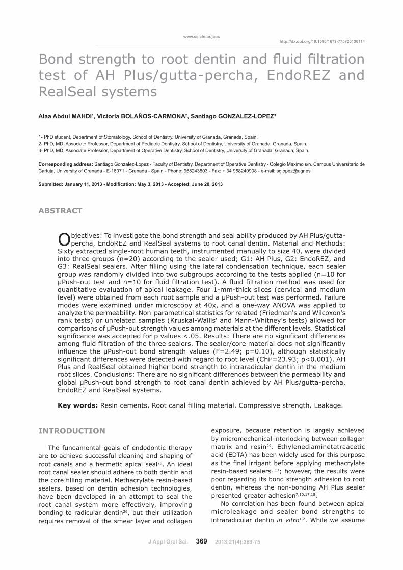
J Appl Oral Sci. 369
www.scielo.br/jaoshttp://dx.doi.org/10.1590/1679-775720130114
ABSTRACT
���������������������� �������� ������� ���test of AH Plus/gutta-percha, EndoREZ and RealSeal systems
Alaa Abdul MAHDI1, Victoria BOLAÑOS-CARMONA2, Santiago GONZALEZ-LOPEZ3
1- PhD student, Department of Stomatology, School of Dentistry, University of Granada, Granada, Spain.2- PhD, MD, Associate Professor, Department of Pediatric Dentistry, School of Dentistry, University of Granada, Granada, Spain.3- PhD, MD, Associate Professor, Department of Operative Dentistry, School of Dentistry, University of Granada, Granada, Spain.
Corresponding address: Santiago Gonzalez-Lopez - Faculty of Dentistry, Department of Operative Dentistry - Colegio Máximo s/n. Campus Universitario de Cartuja, University of Granada - E-18071 - Granada - Spain - Phone: 958243803 - Fax: + 34 958240908 - e-mail: [email protected]
���������� �� ������������������� ������ ���������������������������������
Objectives: To investigate the bond strength and seal ability produced by AH Plus/gutta-percha, EndoREZ and RealSeal systems to root canal dentin. Material and Methods:
Sixty extracted single-root human teeth, instrumented manually to size 40, were divided into three groups (n=20) according to the sealer used; G1: AH Plus, G2: EndoREZ, and ������������������������ ����� �������������������� ������� ������������group was randomly divided into two subgroups according to the tests applied (n=10 for �����!�������������"#$������ ������� ������%������ ������� ���&�����'����������quantitative evaluation of apical leakage. Four 1-mm-thick slices (cervical and medium level) were obtained from each root sample and a μPush-out test was performed. Failure modes were examined under microscopy at 40x, and a one-way ANOVA was applied to analyze the permeability. Non-parametrical statistics for related (Friedman's and Wilcoxon's rank tests) or unrelated samples (Kruskal-Wallis' and Mann-Whitney's tests) allowed for comparisons of μPush-out strength values among materials at the different levels. Statistical � �� ������'������+������+�3�����5�$6����������7�������� �� ������� �������&������ ������� ������������������7�����8���&�� ������������ �� ������9� ������ ��������!����<�����������3����� >?"@�BCD�+"$�#$%��������������� �� ����9�� �� ������� ������'�������' ���������������3��>I� 2=23.93; p<0.001). AH Plus and RealSeal obtained higher bond strength to intraradicular dentin in the medium ������ ����I������ �����7�������� �� ������� ������<�'�����+&�< � �9�����global μPush-out bond strength to root canal dentin achieved by AH Plus/gutta-percha, EndoREZ and RealSeal systems.
Key words:��� ���&������������������� ���&�� ����I�&+�� 3���������U�X���
INTRODUCTION
The fundamental goals of endodontic therapy are to achieve successful cleaning and shaping of root canals and a hermetic apical seal25. An ideal root canal sealer should adhere to both dentin and ��������� ���&�� ����[����9����� �!<����sealers, based on dentin adhesion technologies, have been developed in an attempt to seal the root canal system more effectively, improving bonding to radicular dentin26, but their utilization requires removal of the smear layer and collagen
exposure, because retention is largely achieved by micromechanical interlocking between collagen matrix and resin29. Ethylenediaminetetraacetic acid (EDTA) has been widely used for this purpose ����������� �����<����++�9 ���&����9����resin-based sealers5,13; however, the results were poor regarding its bond strength adhesion to root dentin, whereas the non-bonding AH Plus sealer presented greater adhesion7,10,17,18.
No correlation has been found between apical microleakage and sealer bond strengths to intraradicular dentin in vitro1,2. While we assume
2013;21(4):369-75
J Appl Oral Sci. 370
that preventing effective microleakage is perhaps more important for endodontic application than bond strength26, there is no universally accepted method for the evaluation of leakage33. Each has its � & ��� �����< �����3���<9���� ���������� ���and/or diffusion laws24. Comparisons between different methods point to contradictory results, and, therefore, questionable clinical relevance12,24.
Push-out bond strength testing has become a common method for determining the effectiveness of adhesion between endodontic materials and intraradicular dentin. Zicari, et al.34 (2008) have correlated the push-out bond strength and sealing �< � �9�' �������� ������� ���&������������ 3��&������� ��9���������<!+����<��� ����<���to date there is no documented correlation using methacrylate resin-based sealers. We believe that this aspect is very important, because any ����� ��������9���9� ����9������ ������� 3�interface. Hence, the purpose of this in vitro study was to quantitatively assess the bond strength to dentin root canal and the sealing properties of two methacrylate resin-based sealers and their corresponding core points (RealSeal and EndoREZ), then compare them with the gold standard conventional nonbonding AH Plus/gutta-percha. 7�� �����!���� ���� ���� �� �� ����� ��� &�����were used to test the null hypothesis that there is no difference in the bond strength and sealing properties of RealSeal, EndoREZ systems and AH Plus/gutta-percha.
MATERIAL AND METHODS
We used 60 human anterior teeth, recently extracted for periodontal reasons from anonymous subjects under a protocol (nº 11/2011) approved by the Ethics Committee of the School of Dentistry, University of Granada. All teeth had single straight root canals and closed apices and none showed caries lesions or had received restorative or root canal treatment. After extraction, any calculus and soft tissue was removed and they were stored in 0.1% thymol diluted in distilled water at 4°C for use in this study within 3 months following extraction.
The crowns of the teeth were cut perpendicular to the long axis of the root at the cement-enamel junction with a diamond coated disk at slow speed with constant water-cooling. Root canals were instrumented using the step back technique to �<�� ��������++��� ����}��������������'�X ���length was determined visually by subtracting 1mm ��&���������������~#$��!���>����+�9�[� �����Ballaigues, Switzerland) at the apical foramen. Biomechanical preparation was performed manually ' ����!������� ��� ��~B$������������+� ��������++��� �������������'��� �����' ���@�6��sodium hypochlorite during 1 min. Then, 17% EDTA
'����������������� ��������#�& ��������� ���of distilled water (1 min) to remove any remnants of the irrigating solutions. The canals were dried using paper points.
The roots were randomly divided into three equal groups (n=20) according to the root canal sealer used; G1: AH Plus (Dentsply Maillefer, Ballaigues, Switzerland), G2: EndoREZ (Ultradent Products, Inc., South Jordan, Utah, USA) and G3: RealSeal (Sybron Endo, Glendora, CA, USA).
?�� ��� ���+� ����� ' ��� ��� ������ ��� ����was mixed using AH Plus jet mixing system, and ���� �������� ������������������ ����' ������intraoral tip. A 40.02 taper master gutta-percha cone was placed into the canal and then the canal '���������+��� ��� ��� ����� ������ �������� ���technique and accessory gutta-percha points size #25 and #20 dipped in a small amount of sealer.
EndoREZ was mixed in an ultra-mixer and dispensed using a narrow diameter syringe (Skini Syringe) (Ultradent Products Inc., South Jordan, ������ ���%� ' ��� �� ��!� ++�� �������� ��3 � 7 +�(Ultradent Products, Inc., South Jordan, Utah, ���%��������'������������9�' ����������������Then a 40.02 resin coated gutta-percha cone (EndoREZ point) (Ultradent Products, Inc., South Jordan, Utah, USA) was placed inside the canal, and additional EndoREZ points size #25 and #20 were placed into the canal using the cold lateral condensation technique.
?�� ��� ���+� ����� ' ��� �������� �������Primer (SybronEndo, Glendora, CA, USA) was introduced into the root canal with a paper point (Roeko, Langenau, Germany) soaked with the primer for the apical region. The primer was placed into the middle and coronal part using a micro brush (Microbrush International, Grafton, WI, USA). After 30 seconds, excess primer was removed with paper points. The sealer was dispensed directly from the tip of its auto mix dual-chamber syringe, according to the manufacturer’s instructions. A 40.02 taper RealSeal master point was placed into the canal and then additional RealSeal accessory points size #25 and #20 were placed into the canal using the cold lateral condensation technique.
In all specimens, the excess of points was removed with a hot instrument and the remainder was condensed vertically. They were resected at the apex perpendicular to the long axis of the tooth until 10 mm of root length was obtained. Each root was transferred to a test tube (Eppendorf }<� ����[�� ����+� �%���������� ������& � ��at 37°C for 24 hours to complete sealer setting. All preparation and obturation procedures were done by one operator.
Finally, each sealer group (n=20) was randomly divided into two subgroups according to tests �++� ��>�"#$���������!�������������"#$������ ��
�����������!��������������� ��"����#�� �����������$��%�&#��'���� �����! ��*��+*8� ��+� #�� #��������
2013;21(4):369-75
J Appl Oral Sci. 371
����� ������%�
Leakage evaluationThe external root surfaces of subgroups for the
�� �� ����� ��� ���� '� ��3�� ' ��� �� ��9� ����� ��3�� �����3 ������������������+ ����� ����open. The specimens were attached to a device ��� &���� ��� +&�< � �9� <9� �� �� ����+���described by Pashley, et al.22 (1988). The roots were inserted into a silicon tube (2 mm internal diameter), and attached with cyanoacrylate glue on the outer surface of the tube. A pressure tank of 250 ml of distilled water was placed 100 cm higher than the root to create a pressure of 100 cm H2O. A 10 ml micropipette was inserted and attached with cyanoacrylate glue to measure movement of the air bubble introduced into the system with a micro syringe. Fluid pressure was applied from the coronal area in apical direction. 7�� 3���&� ��� �� �� ����+��� '��� &����� <9�observing the movement of air bubbles within a 10 ml micropipette at 24 hours.
Previously, the system was sealed and checked for 10 min to ensure there was no leakage. The root was covered with water and then covered with a plastic dish to prevent evaporation. Each specimen was attached to the device, and then the system was bled by opening the joint between the water reservoir and the specimen. The air bubble was then aligned with the zero point of the 10 ml scale. Permeability was measured as the distance that the bubble had moved by the end of 24 hours. This &���&��� ���'�� ����&���������� ��'� �������+&��������������������� ��� ������������was done with two additional roots, covering the apex with two layers of nail varnish, to ensure that ���'��������X������� ��&�3&�����9'��within the device.
Micro Push-out testTen roots for each group were cut into four
slices 1 mm thick, from cervical to apical direction (Cervical1 [C1], Cervical2 [C2], Middle1 [M1], Middle2 [M2]), using a 300 μm thick sintered diamond wafering blade perpendicular to the long axis of the root canal at low speed with constant water cooling. The thickness of each root slice was assessed by means of a digital caliper. Each slice was subjected to compressive loading via a universal testing machine (Instron 3345, Instron Ltd, High Wycombe, UK) equipped with a 0.5 mm-diameter cylindrical plunger. The plunger was positioned �������� �����9������������������ ���������� ����without touching the canal wall or introducing shear stress along the interfaces. Thus, the side with the smaller diameter of the root canal faced upwards and was aligned with the shaft that would ���+������������������ ���>�+ ���!�������9%�
��� �� ��� ��� ��� '��� � �+������ 7�� &��� �� '���calibrated at a constant speed of 0.5 mm/min, and the maximum failure load was recorded in Newton (N). Failure was shown by the extrusion of the intact �������������� �����&���������� ���������&��by the appearance of a sharp drop along the load/time curve recorded by the testing machine. The computer and software attached to the universal testing machine calculated the μPush-out bond strength value for each specimen, and converted them into MPa by dividing the applied load by the bonded area.
The s l i c e s we re exam ined unde r a ���& �����+����B$��&��� ���� ��������& ��&��������� �������� 3������� ���&�� ���' ������ �� ����������� 3�' �� ����� ���&�� ������mixed failure.
Statistical analysisThe normality of the data distribution in all the
����9����+��'���3 ���<9��++�9 ���������+ �!Wilk test, and Levene’s test was used to explore the homogeneity of variance among groups.
Fluid movement values at 24 hours were compared among the three different sealers and ��� ���&�� ����<9�&���������!'�9�����������Tukey’s tests.
To analyze μPush-out bond strength values, an ANOVA for repeated measures was first �++� ������+������ ��������� ������� �3��>' �� �!��<���������%���������8��� ���&�� ���(between-subjects factor) on the μPush-out bond strength to root walls. As the sample groups did ���� ������� ��&������� � �9� �� &����� ���!parametrical tests were used to compare μPush-out bond strength values among materials (Kruskal-Wallis’ and Mann-Whitney’s U test) and root levels (Friedman’s tests for multiple comparisons and Wilcoxon’s rank tests, for pairwise comparison).
The Chi2 test was used to analyze the mode of failure distribution among groups. Differences were ���� ���� �� ���������+5$�$6�
RESULTS
Permeability test[����� ������� ���3�����>��%�������� ��
Table 1. The AH Plus/gutta-percha group showed the highest value and the RealSeal group the lowest 3���������������'����� �� ������� ������among the three sealer/core materials. This study makes manifest that all the materials used allowed �� �������'���������������������'��9����������������� ���
Micro Push-out bond strengthDescriptive statistics of the μPush-out bond
strength are given in Table 2.
MAHDI AA, BOLAÑOS-CARMONA V, GONZALEZ-LOPEZ S
2013;21(4):369-75
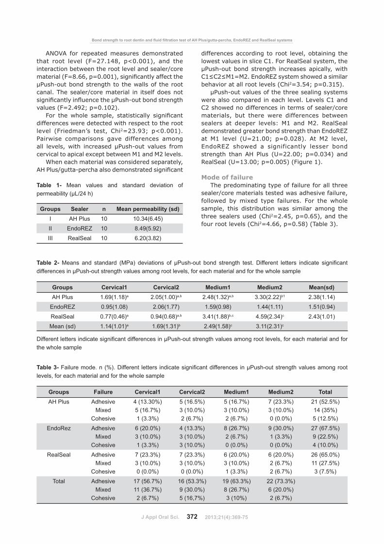
J Appl Oral Sci. 372
ANOVA for repeated measures demonstrated that root level (F=27.148, p<0.001), and the interaction between the root level and sealer/core &�� ���>?"������+"$�$$#%��� �� ������9����������μPush-out bond strength to the walls of the root canal. The sealer/core material in itself does not � �� ������9� ��������������!����<�����������values (F=2.492; p=0.102).
?�� ��� '���� ��&+��� ���� �� ����9� � �� ������differences were detected with respect to the root level (Friedman’s test, Chi2=23.93; p<0.001). Pairwise comparisons gave differences among all levels, with increased μPush-out values from cervical to apical except between M1 and M2 levels.
When each material was considered separately, �������8�����!+����������&��������� �� ������
differences according to root level, obtaining the lowest values in slice C1. For RealSeal system, the μPush-out bond strength increases apically, with I#�I@�[#"[@�����������9��&����'����� & ���behavior at all root levels (Chi2=3.54; p=0.315).
μPush-out values of the three sealing systems were also compared in each level. Levels C1 and C2 showed no differences in terms of sealer/core materials, but there were differences between sealers at deeper levels: M1 and M2. RealSeal demonstrated greater bond strength than EndoREZ at M1 level (U=21.00; p=0.028). At M2 level, EndoREZ showed a significantly lesser bond strength than AH Plus (U=22.00; p=0.034) and RealSeal (U=13.00; p=0.005) (Figure 1).
Mode of failureThe predominating type of failure for all three
sealer/core materials tested was adhesive failure, followed by mixed type failures. For the whole sample, this distribution was similar among the three sealers used (Chi2=2.45, p=0.65), and the four root levels (Chi2=4.66, p=0.58) (Table 3).
Groups Sealer n Mean permeability (sd)I AH Plus 10 10.34(6.45)
II EndoREZ 10 8.49(5.92)
III RealSeal 10 6.20(3.82)
Table 1- Mean values and standard deviation of ����������� ��������
Groups Cervical1 Cervical2 Medium1 Medium2 Mean(sd)AH Plus 1.69(1.18)a 2.05(1.00)a,b 2.48(1.32)a,b 3.30(2.22)b1 2.38(1.14)
EndoREZ 0.95(1.08) 2.06(1.77) 1.59(0.98) 1.44(1.11) 1.51(0.94)
RealSeal 0.77(0.46)a 0.94(0.68)a,b 3.41(1.88)b,c 4.59(2.34)c 2.43(1.01)
Mean (sd) 1.14(1.01)a 1.69(1.31)b 2.49(1.58)c 3.11(2.31)c
����������������������������������������������������!"���������#�������"����""��#��$��"�������������������"��the whole sample
Table 2-� %����� ���� �������� %���� ��#���"��� "�� �����!"�� �"��� ������� ��&� ��������� ����� �������� ����������differences in μPush-out strength values among root levels, for each material and for the whole sample
Groups Failure Cervical1 Cervical2 Medium1 Medium2 TotalAH Plus Adhesive
MixedCohesive
4 (13.30%)5 (16.7%)1 (3.3%)
5 (16.5%)3 (10.0%)2 (6.7%)
5 (16.7%)3 (10.0%)2 (6.7%)
7 (23.3%)3 (10.0%)0 (0.0%)
21 (52.5%)14 (35%)5 (12.5%)
EndoRez AdhesiveMixed
Cohesive
6 (20.0%)3 (10.0%)1 (3.3%)
4 (13.3%)3 (10.0%)3 (10.0%)
8 (26.7%)2 (6.7%)0 (0.0%)
9 (30.0%)1 (3.3%)0 (0.0%)
27 (67.5%)9 (22.5%)4 (10.0%)
RealSeal AdhesiveMixed
Cohesive
7 (23.3%)3 (10.0%)0 (0.0%)
7 (23.3%)3 (10.0%)0 (0.0%)
6 (20.0%)3 (10.0%)1 (3.3%)
6 (20.0%)2 (6.7%)2 (6.7%)
26 (65.0%)11 (27.5%)3 (7.5%)
Total AdhesiveMixed
Cohesive
17 (56.7%)11 (36.7%)2 (6.7%)
16 (53.3%)9 (30.0%)5 (16,7%)
19 (63.3%)8 (26.7%)3 (10%)
22 (73.3%)6 (20.0%)2 (6.7%)
Table 3-�*�������"��&��� ;�&���������� ����� ������������������������������ ��������!"���������#�������"����""�levels, for each material and for the whole sample
�����������!��������������� ��"����#�� �����������$��%�&#��'���� �����! ��*��+*8� ��+� #�� #��������
2013;21(4):369-75
J Appl Oral Sci. 373
DISCUSSION
In this study, permeability and μPush-out bond strength were compared among roots treated endodontically with AH Plus, EndoREZ and ���������������������������������� ���������condensation technique at 24 hours. The results showed that the permeability and global μPush-out bond strength is not affected by the kind of sealer/core material.
Microleakage studies are most commonly used to measure the ex vivo sealing ability of a ���� ������ ��� ���� ������� 3� ���� &������ ��3�been proposed or recommended for evaluation of leakage33�� }�� �� �� ����9�� ��� ������� �� �� ��transport model was chosen to evaluate the sealing ability of three different root canal sealers, in view of its advantages: the samples are not destroyed, it provides quantitative measurement, and the results are precise, as small volumes can be recorded15,19,24. We used different samples for �� ������� ������������!����<���������������<�performed at exactly 24 hours´ time, thus avoiding different degrees of conversion for the sealers. We did not light-cure the coronal portion of the RealSeal
sealer as recommend by manufacturer’s instruction, because fast-setting light-cured resin sealers +����� �� �� ��� �� ��� ��'� ���� ���������defects or weakening of bond strength9. Moreover, the slow chemical reaction of methacrylate-based self-etching sealers may reduce shrinkage stress by &�������+�������+���� ����'��� ��������� ���time of the material14. We held that such behavior '���������'������&+���� �� ������ ��� �������demineralized dentin.
Our results showed that there were no statistically � �� ������� ������ �����3���������� ������� ���among the three sealers used in the study. It is noteworthy, however, that none of the investigated sealers/core materials achieved a tight seal at the ��� ��!�&��!���� ���������+� ��������<������out by Vasconcelos, et al.32�>@$##%������' ����� ����� ��'� ������ ��� ����� ���� ������ �'��9!���������������� ����'�����������������'���������� ����I����9������� � �����3���������X���tests in vitro has been questioned12, because it is � ���������� ��+��������'���� ������+ � ����only zero filtration can be considered a good result. Moreover, there is no correlation among various methods to evaluate microleakage, and
Figure 1- Bar chart showing μPush-out bond strength values and comparisons among sealer/core materials in eachroot level (Mann-Whitney's U test)
=>�����������������������"���?��"J?K�����J���>�����%N��#�Q>�����������������������%���#�V�?��"J?K������!"��X�������������������������������"���������"����������
MAHDI AA, BOLAÑOS-CARMONA V, GONZALEZ-LOPEZ S
2013;21(4):369-75
J Appl Oral Sci. 374
the outcome of the tests depends on the evaluation method12,24. In the absence of correlation between the ex vivo���� ����< � �9���������������������“clinical success”20, we believe there is a threshold for microleakage values that would prove clinically relevant21.
The adhesion tests of methacrylate bond sealer to dentin have not yet been standardized. The μPush-out test seems to be more reliable because it allows for adequate standardization of the specimens, the absence of premature failures and the variability of data distribution, while supplying a better estimation of the actual bonding effectiveness11. In all of the studies reviewed, biomechanical preparation is done with mechanical instrumentation because it is easier to perform and provides a more appropriate standardization of root canals. We used the step back technique and lateral condensation because it is still widely used throughout the world; moreover, there is no study of endodontic sealer adhesion and μPush-out, <���������&���������� �&��������������&�X����������!��������&��� ����������+��&�
In this study, we used three totally different kinds of sealers: a conventional nonbonding epoxy resin-<����������������D����������� ������!�� � ��diurethane dimethacrylate and triethyleneglycol dimethacrylate, EndoREZ; and a second generation sealer, RealSeal, based on adhesive technology with a self-etching primer and in association with a thermoplastic synthetic polyester polymer-based ������������� ���&�� ����}���������<�����������the conventional nonbonding AH Plus/gutta-percha ���� ��� ��� '��� ����� ��� ������� ���� ���������These discouraging results are in line with those of Ungor, et al.31 (2006), who found that the Epiphany/Resilon combination was not superior to the AH Plus/gutta-percha combination. Still, most studies report that AH Plus sealer presents greater adhesion to dentin than RealSeal, regardless of root canal wall treatment3,7,18. Clinicians need to be aware that methacrylate resin-based sealers did not meet expectations regarding adhesion to root dentin, and, at this point in their development, there are �������<����� ���� ���26.
The μPush-out bond strength results were � �� ������9� �������<9���� ������� ������������and RealSeal obtained higher values in the deeper slices, while EndoREZ showed the lowest values, regardless of location. All sealers exhibited their low �����!����<�����������3����� ���'����+�� ���slices. This can be explained by the presence of oxygen, inhibiting sealer setting and producing a layer with low polymerization.
Because we adopted a clinical approach, we were not able to ascertain whether an increase of the adhesive strength to the apex will remain in the most apical sections. Manual instrumentation
until 40 ISO width produce very limited widening of the canal in the apical section, making it impossible to perform μPush-out tests without values having a frictional component with the canal walls. Previous authors have tested apices, but no realistic enlargements were made regarding canal preparation with drills for a post2 or using a tapered diamond bur18.
The values of the μPush-out test are very low, consistent with results of other studies using the same methodology, indicating that the bonding of two methacrylate resin-based sealers and root dentin is much weaker than with resin-dentin bonds, 25-30 MPa30. Furthermore, the adhesive failures between sealer/dentin interface clearly suggest an inadequate level of adhesion between sealer and dentin in terms of bond strength2,17,27. This may <����������� ������ �� ����� ���&�� ����' ���great plasticity, such as gutta-percha, RealSeal and EndoREZ points; in contrast, when the test was done with sealer and no core material, values were higher1,2, suggesting failure may be traced to the sealer/material interface. In a root canal ��� ����� ���9�����3��<���������� ���������(ratio of bonded to unbounded resin surfaces)6 that contributes to maximizing the polymerization stress of resin-based materials along the root canal walls; this may even exceed the bond strength of dentin adhesives to dentin, resulting in gap formation along the surfaces4,6,8. Along these lines, Souza, et al.28 (2012) report that there is a correspondence between the presence of gaps and microleakage.
Despite the material´s relatively low bond strength to root dentin, it may be effective in preventing microleakage16. The main problem is that this low μPush-out bond strength is accompanied by �� ������� ����7������ ������� ��������������!percha or core seems to play an important role in microleakage prevention, since it does not have to be at the sealer-dentin interface. Microleakage can affect the bonding of sealers to dentin by +���� � ��� ���>�� ������<��<��<9�� ��%8 and hydrolysis due to water entry in the interface. In addition, collagen degradation may occur due to host-derived matrix metalloproteinases (MMPs) in dentin that are slowly released over time when self-etching adhesives are used23. RealSeal may therefore adversely affect the longevity of bonded ������������� ����<9������� ��������� ���������<��������������&�3&�������� ��<�'�����hybrid layer and unaffected dentin8,23.
This study reveals that global μPush-out bond strength and permeability were not affected by different sealer/core materials: this leads us to partially accept the null hypothesis, because in the two deeper slices, RealSeal and AH Plus achieved higher μPush-out bond strength than EndoREZ. �������������&���9�������� ���� +�<�'��
�����������!��������������� ��"����#�� �����������$��%�&#��'���� �����! ��*��+*8� ��+� #�� #��������
2013;21(4):369-75
J Appl Oral Sci. 375
permeability and μPush-out bond strength.
CONCLUSIONS
This study makes manifest that AH Plus/gutta-percha, EndoREZ and RealSeal systems allowed �� �������'������������������������� ������'��9!���������������� �������� �� ������� ������were found among global μPush-out bond strengths �������������8��� ���&�� ���D�<����������� ������ ����� �������<9�'� �����������!����strength achieved with RealSeal and AH Plus to intraradicular dentin was found to be greater in the medium root slices.
ACKNOWLEDGEMENT
This investigation was supported by Research Grant no. CGL2011-25906.
REFERENCES
1- Babb BR, Loushine RJ, Bryan TE, Ames JM, Causey MS, Kim J, et al. Bonding of self-adhesive (self-etching) root canal sealers to radicular dentin. J Endod. 2009;35:578-82.2- Barbizam JV, Trope M, Tanomaru-Filho M, Teixeira EC, Teixeira FB. Bond strength of different endodontic sealers to dentin: push-out test. J Appl Oral Sci. 2011;19:644-7.3- Braga RR, Ferracane JL. Alternatives in polymerization contraction stress management. Crit Rev Oral Biol Med. 2004;15:176-84.4- Braga RR, Ferracane JL, Condon JR. Polymerization contraction stress in dual-cure cements and its effect on interfacial integrity of bonded inlays. J Dent. 2002;30:333-40.5- Carvalho AS, Camargo CH, Valera MC, Camargo SE, Mancini MN. Smear layer removal by auxiliary chemical substances in biomechanical preparation: a scanning electron microscope study. J Endod.2008; 34:1396-400.6- Carvalho RM, Pereira JC, Yoshiyama M, Pashley DH. A review of +��9& ��� ���������� ������� ���������������3��+&���versus stress relief. Oper Dent. 1996;21:17-24.7- De-Deus G, Di Giorgi K, Fidel S, Fidel RA, Paciornik S. Push-out bond strength of Resilon/Epiphany and Resilon/Epiphany self-etch to root dentin. J Endod. 2009;35:1048-50.8- De Munck J, Van Landuyt K, Peumans M, Poitevin A, Lambrechts P, Braem M, et al. A critical review of the durability of adhesion to tooth tissue: methods and results. J Dent Res. 2005;84:118-32.9- Feilzer AJ, de Gee AJ, Davidson CL. Setting stresses in composites for two different curing modes. Dent Mater. 1993;9:2-5.10- Fisher MA, Berzins DW, Bahcall JK. An in vitro comparison of bond strength of various obturation materials to root canal dentin using a push-out test design. J Endod. 2007;33:856-8.11-Goracci C, Tavares AU, Fabianelli A, Cardoso PC, Tay FR, ?� �[��7������ ���<�'���<�+�������������������'������comparison between microtensile and push-out bond strength measurements. Eur J Oral Sci. 2004;112:353-61.#@!�������������������������9�[��I���3�������X� ������comparison of four different microleakage tests for assessment �����X������������������� �����������������[�������������Oral Radiol Endod. 2006;102:110-3.#�!� ���&��� ��� ���X��� �������� I�&+� ���� ��� ��� �����9�of three chelating agents in smear layer removal. J Endod. 2008;34:599-602.
14- Lawson MS, Loushine B, Mai S, Weller RN, Pashley DH, Tay FR, et al. Resistance of a 4-META-containing, methacrylate-based sealer to dislocation in root canals. J Endod. 2008;34:833-7.#6!�[ �� ��}���� ��}������!� <� �������X �����U�X�������3�����canal sealers. Int Endod J. 1999;32:415-8.16- Nagas E, Altundasar E, Serper A. The effect of master point taper on bond strength and apical sealing ability of different root canal sealers. Oral Surg Oral Med Oral Pathol Oral Radiol Endod. 2009;107:e61-4.17- Nagas E, Uyanik MO, Eymirli A, Cehreli ZC, Vallittu PK, Lassila LV, et al. Dentin moisture conditions affect the adhesion of root canal sealers. J Endod. 2012;38:240-4.18- Nunes VH, Silva RG, Alfredo E, Sousa-Neto MD, Silva-Sousa YT. Adhesion of Epiphany and AH Plus sealers to human root dentin treated with different solutions. Braz Dent J. 2008;19:46-50.19- Oliver CM, Abbott PV. An in vitro study of apical and coronal microleakage of laterally condensed gutta-percha with Ketac-Endo and AH-26. Aust Dent J. 1998;43:262-8.20- Oliver CM, Abbott PV. Correlation between clinical success and apical dye penetration. Int Endod J. 2001;34:637-44.21- Özok AR, Verhaagen B, Wesselink PR. Improving the accuracy ������� ������+���&������}������������@$#�DB���B�!6B�22- Pashley DH, Derkson GD, Tao L, Derkson M, Kalathoor S. The effects of a multi-step dentin bonding system on dentin permeability. Dent Mater. 1988;4:60-3.23- Pashley DH, Tay FR, Yiu C, Hashimoto M, Breschi L, Carvalho RM, et al. Collagen degradation by host-derived enzymes during aging. J Dent Res. 2004;83:216-21.24- Pommel L, Jacquot B, Camps J. Lack of correlation among three methods for evaluation of apical leakage. J Endod. 2001;27:347-50.25- Santos JN, Carrilho MR, De Goes MF, Zaia AA, Gomes BP, Souza-Filho FJ, et al. Effect of chemical irrigants on the bond strength of a self-etching adhesive to pulp chamber dentin. J Endod. 2006;32:1088-90.26- Schwartz RS. Adhesive dentistry and endodontics. Part 2: bonding in the root canal system-the promise and the problems: a review. J Endod. 2006;32:1125-34.@ !����X��� ����������� ����[������� �[����<� �[�������!����bond strength of Resilon/Epiphany self-etch and gutta-percha/AH26 after different irrigation protocols. Oral Surg Oral Med Oral Pathol Oral Radiol Endod. 2010;110:e88-92.28- Souza SF, Francci C, Bombana AC, Kenshima S, Barroso LP, D’Agostino LZ, et al. Qualitative SEM/EDS analysis of microleakage �����+ ������+���&�� ���������� 3����!��� ���&�� ��������++��Oral Sci. 2012;20:329-34.29- Tagami J, Tao L, Pashley DH. Correlation among dentin depth, permeability, and bond strength of adhesive resins. Dent Mater. 1990;6:45-50.30- Tay FR, Pashley DH. Monoblocks in root canals: a hypothetical or a tangible goal. J Endod. 2007;33:391-8.31- Ungor M, Onay EO, Orucoglu H. Push-out bond strengths: the Epiphany-Resilon endodontic obturation system compared with different pairings of Epiphany, Resilon, AH Plus and gutta-percha. Int Endod J. 2006;39:643-7.32- Vasconcelos BC, Bernardes RA, Duarte MA, Bramante CM, [����}����+ ������� ������������������� ����+��&��' ����3�� ������������ ������������9� ��<9��� ������� �������++������Sci. 2011;19:324-8.33- Wu MK, Wesselink PR. Endodontic leakage studies reconsidered. Part I. Methodology, application and relevance. Int Endod J. 1993;26:37-43.34- Zicari F, Couthino E, De Munck J, Poitevin A, Scotti R, NaertI,et �������� ������� 3����������� ����< � �9�����<!+����<��� ����Dent Mater. 2008;24:967-77.
MAHDI AA, BOLAÑOS-CARMONA V, GONZALEZ-LOPEZ S
2013;21(4):369-75