Embed Size (px)
Citation preview
ICD-10-PCS General
Code Set Training2013
Part 3
ii ICD-10-PCSGeneralCodeSetTraining ©2013AAPC.Allrightsreserved.071113
DisclaimerThis course was current at the time it was published. This course was prepared as a tool to assist the participant in understanding how to prepare for ICD-10-CM. Although every reasonable effort has been made to assure the accuracy of the information within these pages, the ultimate responsibility of the use of this information lies with the student. AAPC does not accept responsibility or liability with regard to errors, omissions, misuse, and misinterpretation. AAPC employees, agents, and staff make no representation, warranty, or guarantee that this compilation of information is error-free and will bear no responsibility, or liability for the results or consequences of the use of this course.
AAPC does not accept responsibility or liability for any adverse outcome from using this study program for any reason including undetected inaccuracy, opinion, and analysis that might prove erroneous or amended, or the coder’s misunderstanding or misapplication of topics. Application of the information in this text does not imply or guarantee claims payment. Inquiries of your local carrier(s)’ bulletins, policy announcements, etc., should be made to resolve local billing requirements. Payers’ interpretations may vary from those in this program. Finally, the law, applicable regulations, payers’ instructions, interpretations, enforcement, etc., may change at any time in any particular area.
This manual may not be copied, reproduced, dismantled, quoted, or presented without the expressed written approval of the AAPC and the sources contained within. No part of this publication covered by the copyright herein may be reproduced, stored in a retrieval system or transmitted in any form or by any means (graphically, electronically, or mechanically, including photocopying, recording, or taping) without the expressed written permission from AAPC and the sources contained within.
Clinical Examples Used in this BookAAPC believes it is important in training and testing to reflect as accurate a coding setting as possible to students and examinees. All examples and case studies used in our study guides and exams are actual, redacted office visit and procedure notes donated by AAPC members.
To preserve the real world quality of these notes for educational purposes, we have not re-written or edited the notes to the stringent grammatical or stylistic standards found in the text of our products. Some minor changes have been made for clarity or to correct spelling errors originally in the notes, but essentially they are as one would find them in a coding setting.
©2013 AAPC2480 South 3850 West, Suite B, Salt Lake City, Utah 84120800-626-CODE (2633), Fax 801-236-2258, www.aapc.com
Revised 071113. All rights reserved.
CPC®, CPC-H®, CPC-P®, CPMA®, CPCO™, and CPPM® are trademarks of AAPC.
©2013AAPC.Allrightsreserved. www.aapc.com iii071113
ICD-10 ExpertsRhonda Buckholtz, CPC, CPMA, CPC-I, CGSC, CPEDC, CENTC, COBGC VP, ICD-10 Training and Education
Shelly Cronin, CPC, CPMA, CPC-I, CANPC, CGSC, CGIC, CPPM Director, ICD-10 Training
Betty Hovey, CPC, CPMA, CPC-I, CPC-H, CPB, CPCD Director, ICD-10 Development and Training
Jackie Stack, CPC, CPC-I, CEMC, CFPC, CIMC, CPEDC ICD-10 Education and Training Specialist
Cyndi Stewart, CPC, CPC-H CPMA, CPC-I Director, ICD-10 Training and Education
Peggy Stilley, CPC, CPB, CPMA, CPC-I, COBGC Director, ICD-10 Development and Training
Contents
Chapter 3 . . . . . . . . . . . . . . . . . . . . . . . . . . . . . . . . . . . . . . . . . . . . . . . . . . . . . . . . . . . . . 45Medical and Surgical—00 Central Nervous System and 01 Peripheral Nervous System . . . . . . . . . . . . . . . . . . . . . . . . . . . . . . 45
02 Heart & Great Vessels, 03 Upper Arteries, 04 Lower Arteries, 05 Upper Veins, 06 Lower Veins . . . . . . . . . . . . . . . . . . . . . . 48
07 Lymphatic and Hemic Systems . . . . . . . . . . . . . . . . . . . . . . . . . . . . . . . . . . . . . 53
08 Eye 54
09 Ear, Nose, and Sinus . . . . . . . . . . . . . . . . . . . . . . . . . . . . . . . . . . . . . . . . . . . . . . 57
0B Respiratory System . . . . . . . . . . . . . . . . . . . . . . . . . . . . . . . . . . . . . . . . . . . . . . . 60
0C Mouth and Throat 0D Gastrointestinal System 0F Hepatobiliary System and Pancreas . . . . . . . . . . . . . . . . . . . . . . . . . . . . . . . . . 63
0G Endocrine System . . . . . . . . . . . . . . . . . . . . . . . . . . . . . . . . . . . . . . . . . . . . . . . . 67
0H Skin and Breast . . . . . . . . . . . . . . . . . . . . . . . . . . . . . . . . . . . . . . . . . . . . . . . . . . 68
0J Subcutaneous Tissue and Fascia 0K Muscles 0L Tendons 0M Bursae and Ligaments . . . . . . . . . . . . . . . . . . . . . . 71
0N Head and Facial Bones, 0P Upper Bones, 0Q Lower Bones, 0R Upper Joints, 0S Lower Joints . . . . . . . . . . . . . . . . . . . . . . . 74
0T Urinary System . . . . . . . . . . . . . . . . . . . . . . . . . . . . . . . . . . . . . . . . . . . . . . . . . . 76
0U Female Reproductive System . . . . . . . . . . . . . . . . . . . . . . . . . . . . . . . . . . . . . . 79
0V Male Reproductive System . . . . . . . . . . . . . . . . . . . . . . . . . . . . . . . . . . . . . . . . 82
©2013AAPC.Allrightsreserved. www.aapc.com 45071113
ICD-10-PCS
Chapter 3 Medical and Surgical—00 Central Nervous System and 01 Peripheral Nervous SystemThe central nervous system (CNS) is the part of the nervous system that integrates the information that it receives from, and coordinates the activity of, all parts of the body. It contains the majority of the nervous system and consists of the brain and the spinal cord. Together with the peripheral nervous system, it has a fundamental role in the control of behavior. The CNS is contained within the dorsal cavity, with the brain in the cranial cavity and the spinal cord in the spinal cavity. The skull protects the brain, while the spinal cord is protected by the vertebrae, and both are enclosed in the meninges.
The BrainThe brain monitors and regulates the body’s actions and reactions. It continuously receives sensory information, and rapidly analyzes this data and then responds accordingly by controlling bodily actions and functions. The brainstem controls breathing, heart rate, and other autonomic processes that is independent of conscious brain functions. The cerebellum is responsible for the body’s balance, posture, and the coordination of movement.
There are four lobes to the brain:
� Frontal lobe � Parietal lobe � Occipital lobe � Temporal lobe
The lobes are named after the bones of the skull that overlie them.
Spinal CordThe spinal cord is a long, thin, tubular bundle of nervous tissue and support cells that extends from the brain (the medulla oblongata specifically). The brain and spinal cord together make up the central nervous system. The spinal cord begins at the Occipital bone and extends down to the space between the first and second lumbar vertebrae; it does not extend the entire length of the vertebral column. The enclosing bony vertebral column protects the relatively shorter spinal cord. The spinal cord functions primarily in the transmission of neural signals between the brain and the rest of the body but also contains neural circuits that can independently control numerous reflexes and central pattern generators.
The spinal cord has three major functions: A. Serve as a conduit for motor information, which travels down the spinal cordB. Serve as a conduit for sensory information, which travels up the spinal cordC. Serve as a center for coordinating certain reflexes
46 ICD-10-PCSGeneralCodeSetTraining ©2013AAPC.Allrightsreserved.071113
ICD-10-PCS
Cranial NervesThere are 12 pairs of cranial nerves originating from the brain stem, and located in the cranium. The function of each of these nerves is:
� Olfactory (cranial nerve I)—smell � Optic (cranial nerve II)—vision � Oculomotor (cranial nerve III)—eyelid and eyeball movement, pupil dilation � Trochlear (cranial nerve IV)—turns eye downward and laterally � Trigeminal (cranial nerve V)—chewing, face, mouth, touch, pain � Abducens (cranial nerve VI)—turns eye laterally � Facial (cranial nerve VII)—facial expressions, secretion of tears and saliva, taste � Auditory (vestibulocochlear) (cranial nerve VIII)— hearing, equilibrium � Glossopharyngeal (cranial nerve IX)—taste, senses carotid blood pressure � Vagus (cranial nerve X)—senses aortic blood pressure, slows heart rate, stimulates diges-
tive organs, taste � Spinal accessory (cranial nerve XI)—controls trapezius and sternocleidomastoid,
controls swallowing movements � Hypoglossal (cranial nerve XII)—controls tongue movements
The peripheral nervous system, or PNS, consists of the nerves and ganglia outside of the brain and spinal cord. The main function of the PNS is to connect the central nervous system (CNS) to the limbs and organs. Unlike the CNS, the PNS is not protected by the bone of spine and skull, or by the blood–brain barrier, leaving it exposed to toxins and mechanical injuries. The peripheral nervous system is divided into the somatic nervous system and the autonomic nervous system; some textbooks also include sensory systems.
There are two types of neurons, carrying nerve impulses in different directions. These two groups of neurons are:
� The sensory neurons are afferent neurons, which relay nerve impulses toward the central nervous system.
� The motor neurons are efferent neurons, which relay nerve impulses away from the central nervous system.
The spinal cord can be divided into segments according to the nerve roots that branch off of it. The nerve roots run through the bony canal, and at each level a pair of nerve roots exits from the spine.
There are 31 pairs of spinal nerves:
� 8 pairs of cervical nerves � 12 pairs of thoracic nerves � 5 pairs of lumbar nerves � 5 pairs of sacral nerves � 1 pair of coccygeal nerves
©2013AAPC.Allrightsreserved. www.aapc.com 47071113
ICD-10-PCS
ProceduresSclerotherapy Procedures—Sclerotherapy is a procedure used to treat blood vessels or blood vessel malformations (vascular malformations). A medicine is injected into the vessels, which makes them shrink.
Sclerotherapy is one method, along with surgery, radiofrequency and laser ablation, for treatment of varicose veins and venous malformations. In ultrasound-guided sclerotherapy, ultrasound is used to visualize the underlying vein so the physician can deliver and monitor the injection. Sclero-therapy is often done under ultrasound guidance after venous abnormalities have been diagnosed with duplex ultrasound.
EXAMPLE:A physician performs sclerotherapy of a brachial plexus lesion, utilizing an alcohol injection.
The correct code for this scenario is 01533ZZ: Percutaneous destruction of brachial plexus.
0—Medical and Surgical1—Peripheral Nervous System5—Destruction3—Brachial Plexus3—PercutaneousZ—No DeviceZ—No Qualifier
Carpal Tunnel Syndrome Procedures—Carpal Tunnel Syndrome (CTS) is idiopathic median neuropathy at the carpal tunnel. Long-standing CTS leads to permanent nerve damage with constant numbness, atrophy of some of the muscles of the thenar eminence, and weakness of palmar abduction.
Palliative treatments for CTS include use of night splints and corticosteroid injections. The only scientifically established disease-modifying treatment is surgery to cut the transverse carpal liga-ment. Release of the transverse carpal ligament is known as “carpal tunnel release” surgery. The goal is to divide the transverse carpal ligament in two. When it is cut across, it no longer presses down on the nerve inside, relieving the pressure.
There are several carpal tunnel release surgery variations, but two major types are:
� Open Carpal Tunnel Release—Involves an incision on the palm about an inch or two in length. Through it the skin, subcutaneous tissue, palmar fascia, and transverse carpal tunnel ligament are all incised.
� Endoscopic Release—Involves two smaller incisions through which the instrumentation is placed, including a synovial elevator, probes, knives, and an endoscope for visualization.
48 ICD-10-PCSGeneralCodeSetTraining ©2013AAPC.Allrightsreserved.071113
ICD-10-PCS
EXAMPLE:A physician performs an open carpal tunnel release.The correct code for this scenario is 01N50ZZ: Open release of median nerve.0—Medical and Surgical1—Peripheral Nervous SystemN—Release5—Median Nerve0—OpenZ—No DeviceZ—No Qualifier
Coding Note: Carpal tunnel release deals with the median nerve, thus the Body Part character was 5.
Nerve Transfer Procedures—A nerve transfer is a surgical technique that may be used when a patient has a nerve injury resulting in complete loss of muscle function or sensation. Nerve trans-fers involve taking nerves or branches of nerves and transferring them to restore function to a more crucial nerve that has been damaged for some reason (eg, trauma).
EXAMPLE:A physician performs an open hypoglossal-facial nerve transfer on a patient with long-term facial paralysis.The correct code for this scenario is 00XS0ZM: Open transfer of hypoglossal nerve to facial nerve.0—Medical and Surgical0—Central Nervous SystemX—TransferS—Hypoglossal Nerve0—OpenZ—No DeviceM—Facial Nerve
Coding Note: In transfer procedures, the Body Part character is the nerve that is being moved, and the Qualifier character is the receiving nerve.
02 Heart & Great Vessels, 03 Upper Arteries, 04 Lower Arteries, 05 Upper Veins, 06 Lower VeinsThe main components of the cardiovascular system are the heart, the veins, and the blood vessels. It includes: the pulmonary circulation, a “loop” through the lungs where blood is oxygenated; and the systemic circulation, a “loop” through the rest of the body to provide oxygenated blood.
©2013AAPC.Allrightsreserved. www.aapc.com 49071113
ICD-10-PCS
HeartThe heart is a muscular, four-chambered organ responsible for pumping blood throughout the blood vessels by repeated, rhythmic contractions. The term cardiac means “related to the heart.” The upper chambers are called the atria and the lower chambers are called the ventricles. The atria receive and the ventricles discharge. The right and left sides are separated by a septum.
The heart is enclosed in a double-walled sac that contains the heart and the roots of the great vessels called the pericardium. There are two layers to the pericardial sac: fibrous pericardium and serous pericardium. The serous pericardium is the deeper layer and is divided into another two layers: a parietal layer and a visceral layer. When the visceral layer comes in contact with the heart it is known as the epicardium.
The heart has four valves: the tricuspid, the mitral, the aortic, and the pulmonary. The valves are divided into the atrioventricular (AV) and the semilunar (SL) valves. The atrioventricular valves are the mitral valve and the tricuspid valve. The two semilunar valves are the aortic and the pulmonary (or pulmonic) valve. They are in the arteries leaving the heart. They do not have chordae tendineae like the AV valves and are more like the valves in the veins.
Conduction SystemThe heart is able to move blood throughout the body as a result of its conduction system. The system contains pacemaker cells, nodes, the Bundle of His, and the Purkinje fibers. The pacemaker cells have the ability to generate an electrical impulse, to pass that impulse to other cells, and to shorten the fibers in the heart when receiving the impulse. The sinoatrial (SA) node is located in the right atrium by the superior vena cava. It is the normal pacemaker of the heart and generates an electrical impulse between 60–100 times per minute. The SA node fires and sends an impulse through the right and left atria causing an atrial contraction. The atrioventriclar (AV) node is located lower in the septal wall of the right atrium. It slows the impulse conduction down between the atria and the ventricles to allow time for the atria to fill with blood before the ventricles contract. The impulse then travels to the Bundle of His, which is muscle fibers that branch off to the right and left. Then the impulse arrives at the Purkinje fibers at the end of the bundle branches. The impulses generated during the heart cycle produce electrical currents, which are conducted through body fluids to the skin, where they can be detected by electrodes and recorded on an electrocardiogram.
Blood VesselsThere are three varieties of blood vessels: arteries, veins, and capillaries. During blood circulation, the arteries carry blood away from the heart. The capillaries connect the arteries to veins. Then the veins carry the blood back to the heart.
ProceduresHeart catheterization procedures are the insertion of a catheter into a chamber or vessel of the heart. It may be done for investigational and/or interventional purposes. Cardiac catheterization is a general term for a group of procedures that are performed using this method, including coronary and ventricular angiography. Once the catheter has been placed, many other procedures can be performed, like percutaneous coronary interventions (PCI), angioplasty, balloon stents, and elec-trophysiology (EP) studies. The ICD-10-PCS code choice is driven by the root operation.
50 ICD-10-PCSGeneralCodeSetTraining ©2013AAPC.Allrightsreserved.071113
ICD-10-PCS
EXAMPLE:Physician performs a left heart catheterization with laser destruction of arrhythmogenic focus in the A-V node. The correct code for this scenario is 02583ZZ: Percutaneous destruction of conduction mecha-nism of the heart and great vessels.0—Medical and Surgical 2—Heart and Great Vessels 5—Destruction8—Conduction Mechanism 3—Percutaneous Z—No Device Z—No Qualifier
Coding Note: The procedure is performed through the catheterization, but a destruction is performed. The A-V node is part of the conduction system in the heart (as discussed earlier in the chapter), so to find the correct PCS code, one must look under “Destruction, conduction mecha-nism” in order to find the right table.
Bypass Graft Procedures—A diseased portion of artery or vein is bypassed by attachment of a healthy vessel (conduit) above and below the diseased vessel to increase the flow of blood. A healthy vessel is taken from elsewhere in the patient’s body. This is known as harvesting or procurement. The surgeon may harvest an artery, a vein, or both. The saphenous vein from the leg is one of the more common vessels harvested for use in bypass grafts.
EXAMPLE:Physician performs an open CABG of LAD using left internal mammary artery (LIMA) with the patient off-bypass. One conduit is used (LIMA) and is placed on one artery (LAD). The correct code for this scenario is 02100Z9: Open CABG of one artery with the left internal mammary artery as the conduit.0—Medical and Surgical 2—Heart and Great Vessels 1—Bypass 0—Coronary Artery, One Site 0—Open Z—No Device 9—Internal Mammary, Left
Valve Procedures—Valves can be repaired or replaced. If replaced, they can be biological or mechanical.
©2013AAPC.Allrightsreserved. www.aapc.com 51071113
ICD-10-PCS
EXAMPLE:A physician replaces a mitral valve on a patient with MVP with a porcine (swine) valve by open technique. The correct code for this scenario is 02RG08Z: Open mitral valve replacement with porcine valve.0—Medical and Surgical 2—Heart and Great Vessels R—ReplacementG—Mitral Valve 0—Open 8—Zooplastic Tissue Z—No Qualifier
Coding Note: Zooplastic tissue transfer is the surgical transfer of tissue from an animal to a human. In this case the tissue comes from a pig and is used to replace the patient’s mitral valve.
Swan-Ganz Procedures—Pulmonary artery catheterization (PAC) is the insertion of a catheter into a pulmonary artery. It is frequently referred to as a Swan-Ganz catheter (after the inventors). Its purpose is diagnostic; it is used to detect heart failure or sepsis, monitor therapy, and evaluate the effects of drugs. The Swan-Ganz catheter allows direct, simultaneous measurement of pressures in the right atrium, right ventricle, pulmonary artery, and the filling pressure (“wedge” pressure) of the left atrium. The PCS code will be driven by the root operation (placement, removal, reposi-tioning, etc.).
EXAMPLE:A physician percutaneously places a Swan-Ganz catheter in the superior vena cava. The correct code for this scenario is 02HV32Z: Percutaneous placement of a Swan-Ganz in the superior vena cava. 0—Medical and Surgical 2—Heart and Great Vessels H—Insertion V—Superior Vena Cava 3—Percutaneous 2—Monitoring Device Z—No Qualifier
Coding Note: The look up process is uniquely different between ICD-9-CM Volume III and ICD-10-PCS. The Swan-Ganz is coded to the device value of Monitoring Device because it monitors pulmonary artery output.
Pacemaker and Defibrillator Procedures—Pacemakers and pacing cardioverter defibrillators (ICD) are implanted as a means of preventing or treating rhythm disturbances that occur in the heart’s electrical system. Bradycardia (slow heartbeat) is one the most commonly reported clinical
52 ICD-10-PCSGeneralCodeSetTraining ©2013AAPC.Allrightsreserved.071113
ICD-10-PCS
indications for the insertion of a pacemaker. Biventricular pacing is the emerging treatment for patients with congestive heart failure (the heart is unable to pump enough blood to meet the needs of the body). Pacing cardioverter defibrillators are known to be effective in the treatment of ventricular arrhythmias.
The components of both pacemaker and ICD system include a pulse generator (battery) and elec-trodes (leads). To insert this device, the subclavian vein or jugular vein is cannulated. Electrodes are advanced to the desired site (e.g right ventricle/right atrium) via fluoroscopic guidance and tested for sensing, pacing, & impedance. A subcutaneous pocket is created for the pulse generator. This device is tested and secured to the leads. The pulse generator pocket is closed.
EXAMPLE:A physician percutaneously adjusts a pacemaker lead in the left ventricle of a patient. The correct code for this scenario is 02WA3MZ: Percutaneous adjustment of pacemaker lead. 0—Medical and Surgical 2—Heart and Great Vessels W—Revision A—Heart 3—Percutaneous M—Cardiac Lead Z—No Qualifier
Embolization Procedures—is the selective occlusion of blood vessels by purposely introducing emboli in order to deprive tumors (or other pathologic processes) of their perfusion (blood supply).
EXAMPLE:A physician performs a percutaneous embolization of the vascular supply to an intracranial artery due to a meningioma. The code for this scenario is 03LG3DZ: Percutaneous emboliza-tion of an intracranial artery 0—Medical and Surgical 3—Upper Arteries L—Occlusion G—Intracranial Artery 3—Percutaneous D—Intraluminal Device Z—No Qualifier
Coding Note: The fact that the patient has a meningioma has no bearing on the coding of the case. PCS contains no diagnostic information in the code set.
©2013AAPC.Allrightsreserved. www.aapc.com 53071113
ICD-10-PCS
07 Lymphatic and Hemic SystemsThe lymphatic system is the part of the immune system comprising a network of conduits called lymphatic vessels that carry a clear fluid called lymph toward the heart. Lymphoid tissue is found in many organs, particularly the lymph nodes, and in the lymphoid follicles associated with the digestive system such as the tonsils. The system also includes all the structures dedicated to the circulation and production of lymphocytes, which includes the spleen, thymus, bone marrow and the lymphoid tissue associated with the digestive system.
Lymph is the fluid that is formed when interstitial fluid enters the initial lymphatic vessels of the lymphatic system. The lymph is then moved along the lymphatic vessel network by either intrinsic or extrinsic contractions. Lymphatic tissue is a specialized connective tissue that contains large quantities of lymphocytes.
The lymphatic system has multiple interrelated functions:
� It is responsible for the removal of interstitial fluid from tissues � It absorbs and transports fatty acids and fats as chyle from the circulatory system � It transports immune cells to and from the lymph nodes into the bones � The lymph transports antigen-presenting cells (APCs) such as dendritic cells, to the
lymph nodes where an immune response is stimulated
ProceduresLymphadenectomy consists of the surgical removal of one or more groups of lymph nodes. It is almost always performed as part of the surgical management of cancer.
This is usually done because many types of cancer have a marked tendency to produce lymph node metastasis early on in their natural history. This is particularly true of melanoma, head and neck cancer, differentiated thyroid cancer, breast cancer, lung cancer, gastric cancer, and colorectal cancer. For example, a radical neck dissection for head and neck cancer and thyroid cancer.
EXAMPLE:A physician performs an open bilateral total lymphadenectomy of the axillae due to breast cancer.The correct codes for this scenario are 07T50ZZ AND 07T60ZZ: Open resection left and right axillary lymph nodes.0—Medical and Surgical7—Lymphatic and Hemic SystemsT—Resection5—Lymphatic, Right Axillary0—OpenZ—No DeviceZ—No Qualifier
AND
54 ICD-10-PCSGeneralCodeSetTraining ©2013AAPC.Allrightsreserved.071113
ICD-10-PCS
0—Medical and Surgical7—Lymphatic and Hemic SystemsT—Resection6—Lymphatic, Left Axillary0—OpenZ—No DeviceZ—No Qualifier
Coding Note: There is no bilateral code for this procedure, so two codes are necessary to indicate the complete procedure.
08 EyeEyes are organs that detect light, and convert it to electro-chemical impulses in neurons. The simplest photoreceptors in conscious vision connect light to movement. In higher organisms the eye is a complex optical system which collects light from the surrounding environment; regulates its intensity through a diaphragm; focuses it through an adjustable assembly of lenses to form an image; converts this image into a set of electrical signals; and transmits these signals to the brain, through complex neural pathways that connect the eye, via the optic nerve, to the visual cortex and other areas of the brain.
Rod and cone cells in the retina allow conscious light perception and vision including color differ-entiation and the perception of depth. The human eye can distinguish about 10 million colors.
The outermost layer of the eye is composed of the cornea and sclera. The middle layer consists of the choroid, ciliary body, and iris. The innermost is the retina, which gets its circulation from the vessels of the choroid as well as the retinal vessels, which can be seen in an ophthalmoscope.
Extraocular MusclesEach eye has six muscles that control its movements: the lateral and medial rectus, the inferior and superior rectus, the inferior and superior oblique. When the muscles exert different tensions, a torque is exerted on the globe that causes it to turn, in almost pure rotation, with only about one millimeter of translation.
The functions of the muscles are:
� Superior rectus—elevation, intorsion (rotation towards the center of the body) and adduction
� Inferior rectus—depression, adduction, and lateral rotation of the eye � Lateral rectus—abduction or moving the pupil away from the midline of the body � Medical rectus—adduction or moving the pupil towards the midline of the body � Superior oblique—abduction, depression, and internal rotation of the eye � Inferior oblique—lateral rotation, elevation, and abduction of the eye
©2013AAPC.Allrightsreserved. www.aapc.com 55071113
ICD-10-PCS
ProceduresPhacoemulsification Procedures—Are cataract surgery in which the eye’s internal lens is emulsi-fied with an ultrasonic handpiece and aspirated from the eye. Aspirated fluids are replaced with irrigation of balanced salt solution, thus maintaining the anterior chamber, as well as cooling the handpiece. Under ICD-10-PCS these are considered Replacements.
EXAMPLE:A physician performs a percutaneous phacoemulsification of a left eye cataract with a pros-thetic lens insertion. The correct code for this scenario is 08RK3JZ: Percutaneous replacement of the left lens with a synthetic substitute.0—Medical and Surgical 8—Eye R—Replacement K—Lens, Left 3—Percutaneous J—Synthetic Substitute Z—No Qualifier
Keratoplasty Procedures—A penetrating keratoplasty is a corneal transplant. A patient’s damaged cornea is replaced by the cornea from the eye of a human cadaver from an eye bank. This is the single most common type of human transplant surgery performed and has the highest success rate.
A lamellar keratoplasty is a partial transplant. This procedure consists in leaving just the patient’s own Descemet membrane and endothelium, while transplanting approximately 95 percent of the cornea.
With inlay and onlay keratoplasty, small devices made of biocompatible materials are surgically inserted into the cornea to improve vision. Under the ICD-10-PCS system, penetrating keratoplasty procedures are coded to Replacements and inlay and onlay keratoplasty procedures are coded to Supplements as they are not removing the cornea.
EXAMPLE:A physician performs a percutaneous penetrating keratoplasty of the left cornea with donor matched cornea. The correct code in this scenario is 08R93KZ: Percutaneous replacement of the left cornea with an nonautologous tissue substitute.0—Medical and Surgical 8—Eye R—Replacement 9—Cornea, Left 3—Percutaneous K—Nonautologous Tissue Substitute Z—No Qualifier
56 ICD-10-PCSGeneralCodeSetTraining ©2013AAPC.Allrightsreserved.071113
ICD-10-PCS
Coding Note: Since this is a penetrating keratoplasty, the cornea is removed and replaced, so it is coded to the root operation of Replacement.
EXAMPLE:A physician performs a left onlay lamellar keratoplasty using an autograft. The correct code in this scenario is 08U9X7Z: External approach lamellar onlay keratoplasty using an autologous tissue substitute.0—Medical and Surgical8—EyeU—Supplement9—Cornea, LeftX—External7—Autologous Tissue SubstituteZ—No Qualifier
Coding Note: The onlay lamellar keratoplasty is coded to the root operation Supplement as the cornea is not removed.
Cataract Procedures—A cataract is a clouding that develops either in the crystalline lens of the eye or in its envelope. Cataracts vary in degree from slight to complete opacity and obstruct the passage of light. Cataracts typically progress slowly to cause vision loss and are potentially blinding if untreated. The condition usually affects both eyes, but almost always one eye is affected earlier than the other.
A senile cataract is one that occurs in the elderly. It is characterized by an initial opacity in the lens, with subsequent swelling and final shrinkage with complete loss of transparency. Early in develop-ment of age-related (senile) cataracts the power of the lens may be increased, causing myopia and the gradual yellowing and opacification of the lens may reduce the perception of blue colors. In Morgagnian cataracts (also a senile cataract) the cortex liquefies over time to form a milky white fluid which can cause severe inflammation if the lens capsule ruptures and leaks. Untreated, the cataract can cause phacomorphic glaucoma.
EXAMPLE:A physician performs a percutaneous phacoemulsification of the right eye of a patient with a cataract and inserts a prosthetic lens.The correct code for this scenario is 08RJ3JZ: Percutaneous replacement of a right lens with a synthetic substitute.0—Medical and Surgical8—EyeR—ReplacementJ—Lens, Right3—PercutaneousJ—Synthetic SubstituteZ—No Qualifier
©2013AAPC.Allrightsreserved. www.aapc.com 57071113
ICD-10-PCS
09 Ear, Nose, and SinusThe ear is the anatomical organ that detects sound. It not only acts as a receiver for sound, but also plays a major role in the sense of balance and body position. The ear is part of the auditory system. The ear consists of three divisions; the external ear, the middle ear, and the inner ear.
Outer Ear (Pinna, Ear Canal, Surface of Ear Drum)The external ear includes the pinna (or auricle), the ear canal, and the very most superficial layer of the tympanic membrane. Cerumen is produced by glands in the skin of the outer portion of the ear canal. The external ear ends at the most superficial layer of the tympanic membrane. The pinna helps direct sound through the ear canal to the tympanic membrane.
Middle EarThe middle ear, an air-filled cavity behind the tympanic membrane, includes the three ear bones or ossicles: the malleus (or hammer), incus (or anvil), and stapes (or stirrup). The opening of the Eustachian tube is also within the middle ear. The malleus has a long process (the manubrium, or handle) that is attached to the mobile portion of the eardrum. The incus is the bridge between the malleus and stapes. The stapes is the smallest named bone in the human body. The three bones are arranged so that movement of the tympanic membrane causes movement of the malleus, which causes movement of the incus, which causes movement of the stapes. When the stapes footplate pushes on the oval window, it causes movement of fluid within the cochlea.
Inner EarThe inner ear includes both the organ of hearing (the cochlea) and a sense organ that is attuned to the effects of both gravity and motion (labyrinth or vestibular apparatus). The balance portion of the inner ear consists of three semicircular canals and the vestibule. The inner ear is encased in the hardest bone of the body. Within the cochlea are three fluid filled spaces: the scala tympani, the scala vestibuli and the scala media. The eighth cranial nerve comes from the brain stem to enter the inner ear.
NoseThe visible part of the nose is the protruding part of the face that bears the nostrils. The shape of the nose is determined by the ethmoid bone and the nasal septum, which consists mostly of carti-lage and which separates the nostrils.
The nose has an area of specialized cells, which are part of the olfactory system. The nasal root is the top of the nose, forming an indentation at the suture where the nasal bones meet the frontal bone. The anterior nasal spine is the thin projection of bone at the midline on the lower nasal margin, holding the cartilaginous center of the nose.
The Paranasal SinusesThe paranasal sinuses are air-filled spaces within the bones of the skull and face. Sinus is a Latin word that means fold or pocket. They are divided into four groups: maxillary, frontal, ethmoid, and sphenoid. They are named according to the bones within which they lie.
58 ICD-10-PCSGeneralCodeSetTraining ©2013AAPC.Allrightsreserved.071113
ICD-10-PCS
The paranasal sinuses are lined with ciliated respiratory epithelium.
� Maxillary sinuses are the largest of the paranasal sinuses are located under the eyes in the maxillary bone and are pyramidal in shape. This sinus has an alveolar recess, bound by the alveolar process, a zygomatic recess, bound by the zygoma (cheekbone), and an infraorbital recess, bound by the inferior orbital surface. The sinus is lined with muco-periosteum.
� Frontal sinuses are located in the frontal bone, superior to the eyes. They are absent at birth and become fully developed by puberty. They are rarely symmetrical and the septum between them is frequently deviated. The mucous membrane in this sinus is innervated by the supraorbital nerve, which carries nerve fibers for mucous secretion from the facial nerve and is supplied by the supraorbital artery and anterior ethmoidal artery.
� Ethmoidal sinuses (also called the ethmoidal air cells): These sinuses are formed from several discrete air cells within the ethmoid bone between the nose and the eyes. The ethmoidal air cells consist of numerous thin-walled cavities in the ethmoidal labyrinth. They lie between the upper parts of the nasal cavities and the orbits, separated by thin bony laminae. The ethmoidal sinuses are classified as anterior, middle, and posterior. The anterior drains into the middle meatus of the nose by way of the infundibulum. The middle drains into the middle meatus of the nose on or above the bulla ethmoidalis, and the posterior drains into the superior meatus above the middle nasal concha.
Sometimes one or more of the posterior opens into the sphenoidal sinus. The ethmoidal air cells receive sensory fibers from the anterior and posterior ethmoidal nerves, and the orbital branches of the pterygopalatine ganglion, which carry nerve fibers for mucous secretion from the facial nerve.
� Sphenoidal sinuses: These sinuses are located in the sphenoid bone at the center of the skull base under the pituitary gland. They are rarely symmetrical and vary in size and shape. Each sinus opens into the roof of the nasal cavity. They drain into the back portion of the nose through a small opening called an ostium. The mucous membrane receives sensory innervation by the posterior ethmoidal nerves and fibers of the facial nerve that synapse at the pterygopalatine ganglion.
ProceduresCochlear Implant Procedures—A surgically implanted electronic device that provides a sense of sound to the profoundly deaf or severely hard of hearing patient. The cochlear implant is often referred to as a bionic ear.
©2013AAPC.Allrightsreserved. www.aapc.com 59071113
ICD-10-PCS
EXAMPLE:A physician performs a right open insertion of a single channel cochlear implant. The correct code for this scenario is 09HD05Z: Open insertion of single channel cochlear prosthesis in the right ear.0—Medical and Surgical 9—Ear, Nose, Sinus H—Insertion D—Inner Ear, Right 0—Open 5—Hearing Device, Single Channel Cochlear ProsthesisZ—No qualifier
Rhinoplasty Procedures—Consists of plastic surgery for reconstructing the form, restoring the function, or aesthetically enhancing the nose. It may be performed for many reasons, including: nasal trauma, respiratory impediments, cosmetic purposes, and congenital defects.
EXAMPLE:A physician performs an open cosmetic rhinoplasty with septal reduction and tip elevation using a local tissue graft. The correct code in this scenario is 090K07Z: Open alteration of the nose with autologous tissue substitute. 0—Medical and Surgical 9—Ear, Nose, Sinus 0—Alteration K—Nose 0—Open 7—Autologous Tissue Substitute Z—No Qualifier
Sinus Procedures—Procedures performed on the paranasal sinuses may be either open or endoscopic. The number of endoscopic procedures is increasing, including balloon sinuplasty. The procedures may be used to drain, open, or remove sinus tissue, blocked sinuses, or polyps.
EXAMPLE:A physician performs an endoscopic drainage of bilateral ethmoid sinuses. The correct codes for this scenario are 099V4ZZ AND 099U4ZZ: Endoscopic drainage of left and right ethmoid sinus.0—Medical and Surgical 9—Ear, Nose, Sinus 9—Drainage
60 ICD-10-PCSGeneralCodeSetTraining ©2013AAPC.Allrightsreserved.071113
ICD-10-PCS
V—Ethmoid Sinus, Left 4—Percutaneous Endoscopic Z—No Device Z—No Qualifier
AND
0—Medical and Surgical 9—Ear, Nose, Sinus 9—Drainage U—Ethmoid Sinus, Right 4—Percutaneous Endoscopic Z—No Device Z—No Qualifier
0B Respiratory SystemThe respiratory system allows for the exchanges of gases between the human body and the outside environment. From an ICD-10-PCS perspective, the respiratory system includes the trachea, bronchi, and the lungs.
Molecules of oxygen and carbon dioxide are passively exchanged, by diffusion, between the gaseous external environment and the blood. This exchange process occurs in the alveolar region of the lungs.
TracheaThere are about fifteen to twenty incomplete C-shaped cartilaginous rings, which reinforce the anterior and lateral sides of the trachea to protect and maintain the airway. The rings are also incomplete so that the trachea may collapse slightly so food can pass down the esophagus when swallowed. The trachealis muscle connects the ends of the incomplete rings. The esophagus lies posteriorly to the trachea. A flap-like epiglottis closes the opening to the larynx during swallowing to prevent swallowed matter from entering the trachea.
Lungs and BronchiIn the mediastinum at the level of T4 the trachea divides into the left and right mainstem bronchi, which are passages of airway in the respiratory tract that conduct air into the lungs. The right main bronchus is wider, shorter, and more vertical than the left main bronchus. The right main bron-chus subdivides into three lobar bronchi, while the left main bronchus divides into two. The lobar bronchi divide into tertiary bronchi, also known as segmental bronchi, each of which supplies a bronchopulmonary segment. A bronchopulmonary segment is a division of a lung separated from the rest of the lung by a connective tissue septum. The segmental bronchi divide into many primary bronchioles, which further divide into terminal bronchioles. Each terminal bronchiole gives rise to several respiratory bronchioles, which go on to divide into two to 11 alveolar ducts. There are five or six alveolar sacs associated with each alveolar duct.
©2013AAPC.Allrightsreserved. www.aapc.com 61071113
ICD-10-PCS
The lungs sit in the two cavities on either side of the heart. Though similar in appearance, the two are not identical. Both are separated into lobes by fissures, with three lobes on the right and two on the left. The lobes are further divided into segments and then into lobules. While the medial border of the right lung is nearly vertical, the left lung is not. It contains a cardiac notch, which is a concave impression molded to accommodate the shape of the heart.
ProceduresTracheal Procedures—Common procedures on the trachea include endotracheal intubation, which is the placement of a flexible plastic tube into the trachea to maintain an open airway or to serve as a conduit through which to administer certain drugs. It may be placed orotracheal or naso-tracheal. It is indicated in many situations when illness or a medical procedure prevents a person from maintaining a clear airway, breathing, and oxygenating the blood. In these circumstances, oxygen supplementation using a simple face mask is inadequate.
EXAMPLE:A physician performs an endotracheal tube extubation. The correct code in this scenario is 0BPIXFZ: Removal of a device from the trachea. 0—Medical and Surgical B—Respiratory System P—Removal 1—Trachea X—External F—TracheostomyZ—No Qualifier
Lung Procedures—There are many procedures that are performed on the lungs. For lung cancer, the most common types of surgery performed include:
Wedge Excision—A wedge excision removes the portion of your lung that includes the tumor, and some surrounding tissue. It is most commonly performed when a tumor is diagnosed very early, or if surgery that is more extensive would interfere too much with a patient’s breathing.
Lobectomy—A lobectomy is the removal of a lobe of the lungs. The right lung has 3 lobes and the left lung has 2 lobes. A “bilobectomy” refers the removal of 2 lobes. This is the most common surgical procedure done to treat lung cancer. If an entire lobe of the lung is removed, under ICD-10-PCS, it would be considered a root operation of Resection. If a portion of a lobe is removed (like a segmentectomy), under ICD-10-PCS, it would be considered a root operation of Excision.
Pneumonectomy—A pneumonectomy involves the removal of an entire lung, and can result in a significant loss of lung function. A pneumonectomy may be considered if a tumor is too large to be removed by the other methods available, or if the tumor is located in a more central location in the lung. This is considered a Resection under ICD-10-PCS.
62 ICD-10-PCSGeneralCodeSetTraining ©2013AAPC.Allrightsreserved.071113
ICD-10-PCS
EXAMPLE:A physician performs a left upper lobe lung segmentectomy by endoscopic technique on a patient with lung cancer. The correct for this scenario is 0BBG4ZZ: Endoscopic excision of the upper left lobe0—Medical and SurgicalB—Respiratory SystemB—ExcisionG—Upper Lung Lobe, Left4—Percutaneous EndoscopicZ—No DeviceZ—No Qualifier
Coding Note: Since only a segment was removed, this procedure is considered a root operation of Excision.
Other common procedures would include biopsies, which may be performed by open or endoscopic technique. These are performed for diagnostic purposes in the lungs, bronchi, and trachea.
EXAMPLE:A physician performs a bilateral diagnostic bronchoscopy with biopsy in the main bronchi.The correct codes for this scenario are 0BB38ZX AND 0BB78ZX: Endoscopic right and left main bronchus biopsy.0—Medical and SurgicalB—Respiratory SystemB—Excision3—Main Bronchus, Right8—Via Natural or Artificial Opening EndoscopicZ—No DeviceX—DiagnosticAND0—Medical and SurgicalB—Respiratory SystemB—Excision7—Main Bronchus, Left8—Via Natural or Artificial Opening EndoscopicZ—No DeviceX—Diagnostic
Coding Note: In the scenario above, the approach is endoscopic, but the scope is placed through the mouth, so the correct approach is Via Natural or Artificial Opening Endoscopic.

©2013AAPC.Allrightsreserved. www.aapc.com 63071113
ICD-10-PCS
Pleurodesis is a procedure that is performed in which the pleural space is artificially obliterated. It involves the adhesion of the two pleura. It is performed to prevent recurrence of pneumothorax or recurrent pleural effusion. It can be done chemically or surgically.
A chemical pelurodesis involves the introduction of chemicals such as bleomycin, tetracycline, povidone iodine, or a slurry of talc into the pleural space through a chest drain. The instilled chemicals cause irritation between the parietal and the visceral layers of the pleura, which closes off the space between them and prevents further fluid from accumulating. A surgical pleurodesis involves mechanically irritating the parietal pleura via thoracotomy or thoracoscopy, often using a rough pad.
EXAMPLE:A physician performs a right-sided talc pleurodesis on a patient with a pleural effusion.The correct code for this scenario is 0B5N3ZZ: Endoscopic right pleural destruction.0—Medical and Surgical B—Respiratory System5—DestructionN—Pleura, Right3—PercutaneousZ—No DeviceZ—No Qualifier
Coding Note: There would also be a code from Section 3, Administration for the applicable injec-tion code (3E0L3GC).
0C Mouth and Throat 0D Gastrointestinal System 0F Hepatobiliary System and PancreasThe digestive system is made up of the gastrointestinal tract (GI tract), also known as the alimentary canal. The mouth, pharynx, esophagus, stomach, small intestine, large intestine, rectum, and anus all make up the digestive tract. Associated digestive structures include three pairs of salivary glands, the pancreas, the liver, and the gallbladder, each with a very important role. The appendix—a short, blind-ended tube attached to the large intestine—has no known function. Food is moved through the digestive tract by muscular contractions called peristalsis until it is eliminated from the body.
The pharynx is divided into three sections: the nasopharynx (epipharynx), the oropharynx (meso-pharynx), and the laryngopharynx (hypopharynx). The pharynx is part of both the digestive and respiratory system; it is also important in vocalization.
The nasopharynx or epipharynx is the most important part in digestive and the respiratory system. The nasopharynx is the uppermost portion of the pharynx. It extends from the base of the skull to the upper surface of the soft palate. It includes the space between the internal nares and the soft palate and lies superior to the oral cavity. The pharyngeal tonsils, more commonly referred to as the adenoids, are lymphoid tissue structures located in the posterior wall of the nasopharynx.
64 ICD-10-PCSGeneralCodeSetTraining ©2013AAPC.Allrightsreserved.071113
ICD-10-PCS
The oropharynx lies behind the oral cavity, extending from the uvula to the level of the hyoid bone. It opens anteriorly into the mouth, while in its lateral wall, between the two palatine arches, is the palatine tonsil. The anterior wall consists of the base of the tongue and the epiglottic vallecula; the lateral wall is made up of the tonsil, tonsillar fossa, and tonsillar (faucial) pillars; the superior wall consists of the inferior surface of the soft palate and the uvula. Because both food and air pass through the pharynx, a flap of connective tissue called the epiglottis closes over the glottis when food is swallowed to prevent aspiration.
The laryngopharynx or hypopharynx is the lowest part of the pharynx; it is the part of the throat that connects to the esophagus. It lies inferior to the epiglottis and extends to the location where this common pathway diverges into the larynx and esophagus. At that point, the laryngopharynx is continuous with the esophagus posteriorly. The esophagus conducts food and fluids to the stomach; air enters the larynx anteriorly. During swallowing, food has the “right of way”, and air passage temporarily stops. Corresponding roughly to the area located between the 4th and 6th cervical vertebrae, the superior boundary of the laryngopharynx is at the level of the hyoid bone.
The upper gastrointestinal tract consists of the esophagus and the stomach. The stomach is a hollow organ, or “container,” that holds food while it is being mixed with enzymes that continue the process of breaking down food into a usable form, called chyme. It has three major parts: the fundus, which is the upper rounded portion of the stomach, the body, which is the central part of the stomach, and the pylorus, which is the lower tubular part of the stomach.
The lower gastrointestinal tract includes most of the small intestine, large intestine, the rectum, and the anus. The small intestine has three parts: the duodenum, the jejunum, and the ileum. It is in this portion of the GI system that digestive enzymes from the pancreas and gallbladder (bile) mix together. The digestive enzymes break down proteins and bile emulsifies fats into micelles.
The colon (or large intestine) is a 6-foot long muscular tube that connects the small intestine to the rectum. The large intestine is made up of the cecum, the ascending (right) colon, the transverse (across) colon, the descending (left) colon, and the sigmoid colon, which connects to the rectum. The appendix is a small tube attached to the cecum. The large intestine is a highly specialized organ that is responsible for processing waste so that emptying the bowels is easy and convenient.
The rectum is an 8-inch chamber that connects the colon to the anus.
The anus is the final part of the digestive tract. It is a two-inch long canal consisting of the pelvic floor muscles and the two anal sphincters. The internal sphincter is always tight, except when stool enters the rectum.
Accessory Organs of the Gastrointestinal SystemThe liver is an organ in the abdomen. The liver does many important things in the body, including:
� Makes bile � Stores glucose � Allows glucose into the blood when our blood glucose levels decrease � Takes protein and fat and turns it into glucose � Makes some fats and cholesterol
©2013AAPC.Allrightsreserved. www.aapc.com 65071113
ICD-10-PCS
� Metabolizes many things in the blood: � hemoglobin � proteins like enzymes, insulin, and serum amyloid a � ammonia � toxins (substances that are poisons) and waste from the body
� Stores vitamins and minerals � Makes many proteins
The pancreas is an organ that releases hormones and enzymes to help digestion. It releases substances through special cells called the Islets of Langerhans. The pancreas plays roles in both the GI and endocrine system. It helps break down carbohydrates, fats, and proteins.
ProceduresEndoscopy Procedures—These procedures allow a physician to view inside the lining of the digestive tract with an endoscope. Endoscopy procedures comprise a large portion of diagnostic and therapeutic surgeries performed in the gastrointestinal system. They can diagnose and/or treat ulcers, GI bleeding, muscle spasms, perforations, polyps, etc.
EXAMPLE 1:A physician performs an EGD with gastric biopsy. The correct code for this scenario is 0DB68ZX: Diagnostic endoscopy of the stomach.0—Medical and SurgicalD—Gastrointestinal System B—Excision 6—Stomach 8—Via Natural or Artificial Opening Endoscopic Z—No Device X—Diagnostic
EXAMPLE 2: A physician performs a colonoscopy that is discontinued at the sigmoid colon. The correct code for this scenario is 0DJD8ZZ: Endoscopic inspection of the colon. 0—Medical and Surgical D—Gastrointestinal System J—Inspection D—Lower Intestinal Tract 8—Via Natural or Artificial Opening Endoscopic Z—No Device Z—No Qualifier
Coding Note: Since the colonoscopy was discontinued before any biopsies are noted, the procedure is considered a root operation of Inspection.
66 ICD-10-PCSGeneralCodeSetTraining ©2013AAPC.Allrightsreserved.071113
ICD-10-PCS
EXAMPLE 3: A physician performs a sigmoidoscopy with sigmoid polypectomy. The correct code for this scenario is 0DBN8ZZ: Endoscopic excision of polyp in the sigmoid colon.0—Medical and Surgical D—Gastrointestinal System B—Excision N—Sigmoid Colon 8—Via Natural or Artificial Opening EndoscopicZ—No Device Z—No Qualifier
Excision and Resection Procedures—Removal of the gall bladder, appendix, and parts of the GI tract are commonly performed. If part of the organ is removed, it is an excision. If all of the organ (or breakdown of the organ by PCS table) is performed, it is a resection.
EXAMPLE 1: A physician performs an open resection of the cecum on a patient. The correct code for this scenario is 0DTH0ZZ: Open removal of the cecum.0—Medical and SurgicalD—Gastrointestinal System T—Resection H—Cecum 0—OpenZ—No DeviceZ—No Qualifier
Coding Note: Although the cecum is a portion of the colon, it is broken out to a specific body part in the fourth character. Therefore, it is considered a Resection, not an Excision.
EXAMPLE 2: A physician performs a removal of the tail of the pancreas on a patient. The correct code in this scenario is 0FBG0ZZ: Open removal of part of the pancreas. 0—Medical and Surgical F—Hepatobiliary System and PancreasB—Excision G—Pancreas 0—Open Z—No Device Z—No Qualifier
©2013AAPC.Allrightsreserved. www.aapc.com 67071113
ICD-10-PCS
0G Endocrine SystemThe endocrine system is an organ system of ductless glands, each of which secretes a type of hormone directly into the bloodstream to regulate the body. It consists off the following glands: pituitary, thyroid, pineal, parathyroid, thymus, adrenal, pancreas, ovaries, and testes. The system secretes hormones into the blood via the endocrine glands.
The ICD-10-PCS table references the following glands in the Endocrine system:
Pituitary GlandThe pituitary gland, also called the hypophysis, is a gland about the size of a pea. It is a protrusion off the bottom of the hypothalamus at the base of the brain, and rests in the sella turcica, a small, bony cavity, covered by the diaphragma sellae, a dural fold. The pituitary is functionally connected to the hypothalamus by the median eminence via the infundibular stem (pituitary stalk). The pitu-itary fossa, in which the pituitary gland sits, is situated in the sphenoid bone in the middle cranial fossa at the base of the brain. The pituitary gland secretes six hormones that regulate homeostasis.
Thyroid GlandThe thyroid gland is one of the largest endocrine glands and is responsible for regulating metabo-lism, such as body temperature and weight. It is composed of two cone-like lobes, the lobus dexter (right lobe), and the lobus sinister (left lobe), which are connected by the isthmus.
The thyroid hormones contain iodine, which the thyroid needs to manufacture these hormones. If a person lacks iodine in his or her diet, the thyroid cannot make the hormones, causing a deficiency. In response to the bodies feedback loops calling for more thyroid hormones, the thyroid gland then enlarges to attempt to compensate. This disorder is called goiter.
Parathyroid GlandsThe parathyroid glands are small endocrine glands in the neck that produce parathyroid hormone (PTH or parathormone). There are four parathyroid glands, which are usually located on the posterior surface of the thyroid gland, and are about the size of a grain of rice. Parathyroid glands control the amount of calcium in the blood and within the bones.
Pineal GlandThe pineal gland is a small endocrine gland located near the center of the brain, between the two hemispheres. It resembles a pine-cone and is reddish-grey in color. The pineal gland is stimulated by nerves from the eyes. It produces the serotonin derivative melatonin, a hormone that affects the modulation of wake/ sleep patterns and seasonal functions. Melatonin also affects thyroid and adrenal cortex functions.
Thymus GlandThe thymus gland sits in the middle of the pleural cavity and aids in developing the immune system. The only known function of the thymus is the production and “education” of T-lympho-cytes (T cells), which are critical cells of the adaptive immune system. The thymus is composed of two identical lobes and is located anatomically in the anterior superior mediastinum, in front of the heart and behind the sternum.
68 ICD-10-PCSGeneralCodeSetTraining ©2013AAPC.Allrightsreserved.071113
ICD-10-PCS
Adrenal GlandsThe adrenal glands, or suprarenal glands, sit on top of each kidney and are divided into two parts. The right adrenal is triangular shaped and the left is semilunar shaped. They are mainly respon-sible for releasing hormones in conjunction with stress through the synthesis of corticosteroids, like cortisol and catecholamines, like epinephrine. Adrenal glands also affect kidney function by secreting aldosterone. Each adrenal gland is separated into two distinct structures, the adrenal cortex and medulla, both of which produce hormones. The cortex mainly produces cortisol, aldo-sterone, and androgens, while the medulla chiefly produces epinephrine and norepinephrine.
ProceduresA thyroidectomy is an operation that involves the surgical removal of all or part of the thyroid gland. Surgeons often perform a thyroidectomy when a patient has thyroid cancer, hyperthy-roidism, or some other condition of the thyroid gland.
Types of Thyroidectomy include:
� Hemithyroidectomy—Entire isthmus is removed along with 1 lobe � Partial thyroidectomy—Removal of gland in front of trachea after mobilization � Near total thyroidectomy—Both lobes except the lower pole which is very close to
recurrent laryngeal nerve and parathyroid is removed � Total thyroidectomy—Entire gland is removed � Hartley Dunhill operation—Removal of 1 entire lateral lobe with isthmus and partial/
subtotal removal of opposite lateral lobe
EXAMPLE:A physician performs a left hemithyroidectomy.The correct code in this scenario is 0GTG0ZZ: Left thyroid removal.0—Medical and SurgicalG—Endocrine SystemT—ResectionG—Thyroid Gland Lobe, Left0—OpenZ—No DeviceZ—No Qualifier
0H Skin and BreastThe integumentary system is made up of the structures that cover the body: skin, hair, nails, seba-ceous glands, and sweat glands. It is the largest organ system in the body. It functions as a protec-tive barrier against outside invasion to harmful substances. It also regulates body temperature, synthesizes vitamin D, and contains touch and pressure receptors.
The skin itself has two parts—the epidermis (the outermost layer) and the dermis underneath. The epidermis consists of five layers containing keratin and pre-keratin substances. The dermis is deeper and thicker and consists of two layers containing fibrous connective tissue, collagen, and
©2013AAPC.Allrightsreserved. www.aapc.com 69071113
ICD-10-PCS
other types of cells. The “living” part of the skin is in the dermis—hair bulbs, glands, nerve recep-tors, etc.
BreastsThe breast of a female contains the mammary gland that secretes milk to feed infants. Both men and women develop breasts from the same embryological tissues, however, at puberty, female sex hormones (estrogen), promote breast development that does not occur in men. Breasts are actually modified sweat glands, which produce milk in women.
ProceduresBreast Procedures—There are many procedures that can be performed on the breast. Cosmetic procedures for aesthetic purposes, removal procedures for therapeutic or prophylactic purposes, and reconstructions performed post-surgically to restore form.
EXAMPLE 1: A physician performs an open bilateral breast augmentation on a patient with insertion of silicone implants. The correct code for this scenario is 0H0V0JZ: Open augmentation of both breasts using a synthetic substitute. 0—Medical and Surgical H—Skin and Breast 0—AlterationV—Breast, Bilateral0—OpenJ—Synthetic Substitute Z—No Qualifier
Coding Note: Since this is a cosmetic procedure, in PCS it is considered an Alteration.
Example 2: A physician performs a left total mastectomy on a patient with breast cancer.
The correct codes for this scenario are 0HTU0ZZ: Open removal of left breast.
0—Medical and Surgical H—Skin and Breast T—Resection U—Breast, Left0—Open Z—No Device Z—No Qualifier
Coding Note: Since the entire left breast was removed, this is considered a root operation of Resection.
70 ICD-10-PCSGeneralCodeSetTraining ©2013AAPC.Allrightsreserved.071113
ICD-10-PCS
Skin Procedures—Since the skin is one organ system, there are no procedures in PCS under Resection for the skin. There are many procedures found under the other root operations. For example, skin lesion removals would be found under the root operation of Excision, skin flaps would be found under the root operation Transfer, and skin substitutes would be found under the root operation of Replacement.
EXAMPLE 1: A physician performs an excision of a malignant melanoma from the skin of a patient’s left upper armThe correct code in this scenario is 0HBCXZZ: Excision of the skin on the left upper arm. 0—Medical and Surgical H—Skin and Breast B—ExcisionC—Skin, Left Upper ArmX—External Z—No DeviceZ—No Qualifier
EXAMPLE 2: A physician performs cryotherapy on 2 warts: one on the patient’s right hand and one on the patient’s left handThe correct codes for this scenario are 0H5GXZZ AND 0H5FXZZ: Destruction of wart on the skin of the left and right hand.0—Medical and SurgicalH—Skin and Breast 5—Destruction G—Skin, Left Hand X—External Z—No Device Z—No Qualifier
AND
0—Medical and Surgical H—Skin and Breast5—DestructionF—Skin, Right Hand X—External Z—No Device Z—No Qualifier
©2013AAPC.Allrightsreserved. www.aapc.com 71071113
ICD-10-PCS
0J Subcutaneous Tissue and Fascia 0K Muscles 0L Tendons 0M Bursae and LigamentsSubcutaneous Tissue and FasciaThe subcutaneous tissue lies immediately below the dermis and is mainly used for fat storage. It consists primarily of loose connective tissue and lobules of fat. It contains larger blood vessels and nerves than those found in the dermis.
Subcutaneous fat is found just beneath the skin as opposed to visceral fat which is found in the peritoneal cavity. Subcutaneous fat can be measured using body fat calipers giving a rough estimate of total body adiposity.
The fascia is a layer of fibrous tissue that permeates the human body. It is a connective tissue that surrounds muscles, groups of muscles, blood vessels, and nerves, binding those structures together. It consists of several layers: a superficial fascia, a deep fascia, and a subserous (or visceral) fascia and extends uninterrupted from the head to the tip of the toes.
Fasciae are dense regular connective tissues, containing closely packed bundles of collagen fibers oriented in a wavy pattern parallel to the direction of pull.
Muscular SystemThe human body contains more than 650 individual muscles attached to the skeleton, which helps to keep bones in place and provides the pulling power for us to move around.
The main job of the muscular system is to provide movement for the body. There are three different types of muscle tissues: skeletal, cardiac, and smooth. Each has the ability to contract, which then allows body movements and functions.
There are two types of muscles in the system and they are the involuntary muscles, and the volun-tary muscles. Skeletal muscle is a system of pairs that relax and contract to move a joint. Most skeletal muscles are attached to bones by tendons. Tendons are strong sheets of connective tissue, and are identical to ligaments. Tendons and ligaments are different in their function only. Tendons attach muscle to bone and ligaments attach bone to bone.
Tendons, Ligaments, and BursaeA tendon is a tough band of fibrous connective tissue that usually connects muscle to bone and is capable of withstanding tension. Tendons are similar to ligaments and fasciae as they are all made of collagen except that ligaments join one bone to another bone, and fasciae connect muscles to other muscles. Tendons and muscles work together.
A tendon connects muscle to bone. Tendons essentially enable one to move since they act as inter-mediaries between the muscles creating the motion of the bones.
Ligaments are similar to tendons, but they connect bone to bone and help to stabilize joints. They are composed mostly of bands of tough, fibrous dense regular connective tissue comprising attenu-ated collagenous fibers.
A bursa is a small fluid-filled sac lined by synovial membrane with an inner capillary layer of fluid that provides a cushion between bones and tendons and/or muscles around a joint. This helps to
72 ICD-10-PCSGeneralCodeSetTraining ©2013AAPC.Allrightsreserved.071113
ICD-10-PCS
reduce friction between the bones and allows free movement. Bursae are filled with synovial fluid and are found around most major joints of the body.
ProceduresArthroscopy Procedures—Arthroscopic procedures can be performed either to evaluate or to treat many orthopedic conditions including torn floating cartilage, torn surface cartilage, ACL reconstruction, and trimming damaged cartilage.
The advantage of arthroscopy over traditional open surgery is that the joint does not have to be opened up fully. Instead, for example, for knee arthroscopy only two small incisions are made—one for the arthroscope and one for the surgical instruments to be used in the knee cavity to fully remove the kneecap. This reduces recovery time and may increase the rate of surgical success due to fewer traumas to the connective tissue. There is also less scarring, because of the smaller incisions.
The joints that are most commonly examined and treated by arthroscopy are the knee, shoulder, elbow, wrist, ankle, foot, and hip.
EXAMPLE:A physician performs a right shoulder arthroscopy with coracoacromial ligament release.The correct code in this scenario is 0MN14ZZ: Endoscopic right shoulder release of the right shoulder bursa and ligament.0—Medical and SurgicalM—Bursae and LigamentsN—Release1—Shoulder Bursa and Ligament, Right4—Percutaneous EndoscopicZ—No DeviceZ—No Qualifier
Fasciocutaneous Flap Procedures are tissue flaps that include skin, subcutaneous tissue and the underlying fascia. If they are raised without skin, they are referred to as fascial flaps. Fasciocuta-neous flaps are used to provide coverage when a skin graft or random skin flap is insufficient for coverage, like in the coverage over tendon or bones.
Muscle Flap Procedures use only muscle for defect coverage. It is used primarily to provide a well-vascularized soft tissue that is relatively resistant to infection, helps wounds heal, and offers a vascularized surface for skin grafts. The muscle flap is commonly used to eradicate infection and to revascularize bone. It is commonly used for lower extremity reconstructions and sternal wounds. When taken with both its nerve and vascular pedicle, muscle flaps can be transferred as functional units for use in extremity and facial reanimation.
©2013AAPC.Allrightsreserved. www.aapc.com 73071113
ICD-10-PCS
EXAMPLE:A physician performs an open fasciocutaneous flap closure of the left thigh. The correct code for this scenario is 0JXM0ZC: Open transfer of a flap to left upper leg using a fasciocutaneous flap.0—Medical and SurgicalJ—Subcutaneous Tissue and FasciaX—TransferM—Subcutaneous Tissue and Fascia, Left Upper Leg0—OpenZ—No DeviceC—Skin, Subcutaneous Tissue, and Fascia
Coding Note: The qualifier identifies the body layers, or depth, of the flap. In this case skin, subcu-taneous tissue, and fascia was used in the flap.
Tendon Release Procedures—A surgical procedure in which a tenotomy is performed to allow it to retract towards the junction of the muscle and tendon. Any tendon could be treated with this procedure, although tendons in the eyes and the extremities are the ones most commonly treated.
The purpose of tendon release is to identify and surgically remove the area producing symptoms while protecting the normal surrounding tissues and their attachments. Tendon release may also involve debridement of unhealthy surrounding soft tissues or bone to promote improved healing. It is typically done to relieve contracture or decrease friction irritation. Sometimes the tendon is transposed to maintain muscle function.
EXAMPLE:A physician performs a percutaneous left Achilles tendon release.The correct code in this scenario is 0LNP3ZZ: Percutaneous left lower leg tendon release.0—Medical and SurgicalL—TendonsN—ReleaseP—Lower Leg Tendon, Left3—PercutaneousZ—No DeviceZ—No Qualifier
74 ICD-10-PCSGeneralCodeSetTraining ©2013AAPC.Allrightsreserved.071113
ICD-10-PCS
0N Head and Facial Bones, 0P Upper Bones, 0Q Lower Bones, 0R Upper Joints, 0S Lower JointsSkeletal SystemThe bones of the body fall into four general categories: long bones, short bones, flat bones, and irregular bones.
� Long bones are longer than they are wide and work as levers. The bones of the upper and lower extremities (humerus, tibia, femur, ulna, metacarpals, etc.) are of this type.
� Short bones are short, cube-shaped, and found in the wrists and ankles. � Flat bones have broad surfaces for protection of organs and attachment of muscles (ex.
ribs, cranial bones, bones of shoulder girdle). � Irregular bones are all others that do not fall into the previous categories. They have
varied shapes, sizes, and surfaces features and include the bones of the vertebrae and a few in the skull.
Of the 206 bones in the body, 80 of them are axial bones, which include the head, facial, hyloid, auditory, trunk, ribs, and sternum. The word axial refers to the bones being located along the central axis of the body. The other 126 are appendicular bones, which include arms, shoulders, wrists, hands, legs, hips, ankles, and feet. The word appendicular refers to the bones related to movement that are “appended” to the axial structure.
JointsThere are many types of joints throughout the body. The way the body moves depends on each joint individually. There are three types of joints: immovable, partly movable, and synovial. Immovable joints, like those connecting the cranial bones, have edges that interlock tightly. Partly movable joints allow some degree of flexibility and usually have cartilage between the bones such as the vertebrae. Synovial joints permit the greatest degree of flexibility and have the ends of the bones covered with connective tissue that is filled with synovial fluid, such as the hip.
Ball-and-socket joints provide the most range of movement. Hinge joints allow movement in only one direction, and are considered the simplest type of joint. Gliding joints permit a wide range of mostly sideways movements—as well as movements in one direction—a pivot joint near the top of the spine allows the head to swivel and bend. A saddle joint is more versatile than either a hinge joint or a gliding joint. It allows movement in two directions.
Procedures Fracture Procedures—Treatment of bone fractures and are broadly classified as surgical or conservative, the latter basically referring to any non-surgical procedure, such as pain management, immobilization or other non-surgical stabilization. A similar classification is open versus closed treatment, in which open treatment refers to any treatment in which the fracture site is surgically opened, regardless of whether the fracture itself is an open or closed fracture.
ImmobilizationBone fractures are typically treated by restoring fractured pieces of bone to their natural positions and maintaining those positions while the bone heals. The fractured limb is usually immobilized
©2013AAPC.Allrightsreserved. www.aapc.com 75071113
ICD-10-PCS
with a plaster or fiberglass cast or splint, which holds the bones in position and immobilizes the joints above and below the fracture. When the initial post-fracture edema or swelling goes down, the fracture may be placed in a removable brace or orthosis.
Occasionally smaller bones, such as phalanges of the toes and fingers, may be treated without the cast, by buddy wrapping them, which serves a similar function to making a cast. By allowing only limited movement, fixation helps preserve anatomical alignment while enabling callus formation, towards the target of achieving union.
SurgeryWhen a joint surface is damaged by a fracture, surgery is also commonly recommended to make an accurate anatomical reduction and restore the smoothness of the joint. Occasionally bone grafting is used to treat a fracture. Sometimes bones are reinforced with metal with the use of plates, screws, rods, and fixation systems.
EXAMPLE:A physician performs an open fracture reduction of a patient’s right tibia.The correct code in this scenario is 0QSG0ZZ: Repositioning right tibia.0—Medical and SurgicalQ—Lower BonesS—RepositionG—Tibia, Right0—OpenZ—No DeviceZ—No Qualifier
Arthrodesis Procedures—The artificial induction of joint ossification between two bones via surgery. It is most commonly performed on joints in the spine, hand, ankle, and foot. It can be done in several ways:
� A bone graft can be created between the two bones using an autograft or an allograft from a bone bank.
� Bone autograft is generally preferred by surgeons because, as well as eliminating the risks associated with allografts, bone autograft contains osteoblasts so the graft actually forms new bone itself as well as acting as a matrix or scaffold to new bone growing from the bones being bridged. The main drawback of bone autograft is the limited supply available for harvest.
� Bone allograft has the advantage of being available in larger quantities than auto-graft. The treatment process the bone goes through following harvest kills living bone or bone marrow cells. This significantly reduces the risk of graft rejection such that no anti-rejection drugs are needed.
� A variety of synthetic bone substitutes are commercially available and they mimic the structure of cancellous bone.
� Metal implants can be attached to the two bones to hold them together in a position, which favors bone growth.
76 ICD-10-PCSGeneralCodeSetTraining ©2013AAPC.Allrightsreserved.071113
ICD-10-PCS
At the completion of surgery and healing, which takes place over a period of several weeks to over a year, the two adjoining bones are fused and no motion takes place between them. This can have the effect of actually strengthening the bones.
EXAMPLE:A physician performs an open arthrodesis of the sacrococcygeal joint using an autograft.The correct code in this scenario is 0SG507Z: Open fusion of the sacrococcygeal joint using autologous tissue substitute.0—Medical and SurgicalS—Lower JointsG—Fusion5—Sacrococcygeal Joint0—Open7—Autologous Tissue SubstituteZ—No Qualifier
0T Urinary SystemUrinary SystemThe urinary system is the organ system that produces, stores, and eliminates urine. The structure of the urinary tract includes two kidneys, two ureters, the bladder, the urethra, and two sphincter muscles.
The kidneys are bean-shaped organs that lie in the abdomen around or just below the ribcage and close to the lumbar spine. Attached to the kidneys at their functional endpoints are the ureters, which lie more medial and run down to the trigone of urinary bladder.
The tube through which the urine flows out of the body is called the urethra. When urine is passed, the urethral sphincter at the base of the bladder relaxes, the detrusor contracts, and urine is voided via the urethra.
ProceduresCalculus Procedures—Treatment varies by stone type, but include:
� Lithotomy is a surgical method for removal of calculi that cannot exit naturally through the urethra, ureter or biliary duct. The procedure is performed by means of a surgical incision to remove the calculus.
� Lithotripsy refers to the type of procedure in which the calculi are crushed. There are different forms of lithotripsy, including:
� ESWL or extracorporeal shock wave lithotripsy is how stones are crushed by acoustic pulse (extracorporeal lithotripsy).
� Laser lithotripsy is a scope inserted into the urinary tract to locate the calculus. Then a laser fiber is inserted through the scope and the laser is directly emitted to the stone, disintegrating it. The remaining pieces are washed out of the urinary tract.
©2013AAPC.Allrightsreserved. www.aapc.com 77071113
ICD-10-PCS
� PCNL or Percutaneous nephrolithotomy, pertains to a retrograde pyelogram that is performed to locate the calculus. A percutaneous nephrolithotomy needle (PCN) is passed into the pelvis of the kidney and confirmed under fluoroscopy. A guide wire is passed through the needle into the pelvis. The needle is then withdrawn, leaving the guide wire in place. Dilators are placed over the guide wire and a sheath is intro-duced. Then a nephroscope is passed inside and small stones are removed. Larger stones may be crushed first, then removed.
EXAMPLE:A physician performs a transurethral cystoscopy with fragmentation of bladder calculus.The correct code in this scenario is 0TFB8ZZ: Endoscopic fragmentation of stone in the bladder.0—Medical and SurgicalT—Urinary SystemF—FragmentationB—Bladder8—Via Natural or Artificial Opening EndoscopicZ—No DeviceZ—No Qualifier
Coding Note: Since the scope is placed through the urethra, the approach is Via Natural or Artifi-cial Opening, Endoscopic.
Urinary Incontinence Procedures—Several procedures have been developed to treat stress incon-tinence. Most surgical procedures fall into two main categories: sling procedures and bladder neck suspension procedures.
� Sling Procedures—The most common surgery to treat stress incontinence uses strips of the body’s tissue or synthetic material such as mesh to create a pelvic sling or hammock around the bladder neck and the urethra. The sling provides support to keep the urethra closed.
� Bladder Neck Suspension Procedures—This procedure is designed to provide support to the urethra and bladder neck. Through an open technique, the surgeon stitches the tissue near the bladder neck and secures the stitches to a ligament near the pubic bone or in the cartilage of the pubic bone itself. This reinforces the urethra and bladder neck.
78 ICD-10-PCSGeneralCodeSetTraining ©2013AAPC.Allrightsreserved.071113
ICD-10-PCS
EXAMPLE:A physician performs an open sling operation of the bladder neck for urinary incontinence utilizing mesh.The correct code for this scenario is 0TUC0JZ: Open supplement of the bladder neck using a synthetic substitute.0—Medical and SurgicalT—Urinary SystemU—SupplementC—Bladder Neck0—OpenJ—Synthetic SubstituteZ—No Qualifier
Urinary Diversion Procedures—May be any one of several surgical procedures performed to reroute urine flow from its normal pathway. It may be necessary for diseased or defective ureters, bladder or urethra, either temporarily or permanently. Some diversions result in a stoma.
EXAMPLE:A physician performs an open urinary diversion of the left ureter using an ileal conduit to the skin.The correct code in this scenario is 0T170ZC: Ileocutaneous open bypass of the left ureter. 0—Medical and SurgicalT—Urinary System1—Bypass7—Ureter, Left0—OpenZ—No DeviceC—Ileocutaneous
Cystoscopy Procedures—Endoscopies of the urinary bladder via the urethra. It is carried out with a cystoscope. Some cystoscopes use optical fibers that carry an image from the tip of the instrument to a viewing piece at the other end. There are two main types of cystoscopy—flexible and rigid—differing in the flexibility of the cystoscope.
©2013AAPC.Allrightsreserved. www.aapc.com 79071113
ICD-10-PCS
EXAMPLE:A physician performs a transurethral diagnostic cystoscopyThe correct code in this scenario is 0TJB8ZZ:Endoscopic inspection of the bladder.0—Medical and SurgicalT—Urinary SystemJ—InspectionB—Bladder8—Via Natural or Artificial Opening EndoscopicZ—No DeviceZ—No Qualifier
0U Female Reproductive SystemThe female reproductive system is designed to carry out several functions. It produces the female egg cells necessary for reproduction, called the ova or oocytes. The system is designed to transport the ova to the site of fertilization. Conception, the fertilization of an egg by a sperm, normally occurs in the fallopian tubes.
There are female reproductive organs located inside and outside the female body. There are four main external structures of the female reproductive system: the labia majora, the labia minora, the Bartholin’s gland, and the clitoris.
The internal reproductive organs in the female include the vagina, uterus, ovaries, and fallopian tubes. The vagina is approximately four inches in length, and joins the cervix to the outside of the body. In the middle of the cervix is a small round opening, called the external orifice that leads to the uterus.
The uterus is divided into two parts: the cervix, which is the lower part that opens into the vagina, and the main body of the uterus, called the corpus.
The ovaries are small, oval-shaped glands that are located on either side of the uterus.
The fallopian tubes are narrow tubes that are attached to the upper part of the uterus and serve as pathways for the ova to travel from the ovaries to the uterus.
Procedures Hysteroscopic Procedures—A hysteroscope is a thin, telescope-like instrument that is inserted into the uterus through the vagina and cervix. Hysteroscopy may be either diagnostic or operative.
Diagnostic hysteroscopy is used to diagnose some uterine abnormalities, and may also be used to confirm the results of other tests such as hysterosalpingography (HSG). Other instruments or tech-niques, such as dilation and curettage (D&C) and laparoscopy, are sometimes used in conjunction with the hysteroscopy. Diagnostic hysteroscopy can be used to diagnose certain conditions such as abnormal uterine bleeding, infertility, repeated miscarriages, adhesions, fibroid tumors, polyps, or to locate displaced intrauterine devices (IUDs).
An operative hysteroscopy may be used, instead of open abdominal surgery, to both diagnose and treat certain conditions such as uterine adhesions, septums, or fibroids which can often be removed
80 ICD-10-PCSGeneralCodeSetTraining ©2013AAPC.Allrightsreserved.071113
ICD-10-PCS
through the hysteroscope. Sometimes a hysteroscopy employs instrumentation, such as a resecto-scope, to treat some cases of abnormal bleeding. The resectoscope is a telescope-like instrument with a wire loop, a rollerball, or a roller cylinder tip at the end. Electric current at the end of the tip is used to destroy the uterine lining. This is considered an endometrial ablation.
EXAMPLE:A physician performs a hysteroscopy with balloon dilation of bilateral fallopian tubes. The correct code in this scenario is 0U778ZZ: Endoscopic dilation of bilateral fallopian tubes.
0—Medical and SurgicalU—Female Reproductive System7—Dilation7—Fallopian Tubes, Bilateral8—Via Natural or Artificial Opening EndoscopicZ—No DeviceZ—No Qualifier
Laparoscopy Procedures—Include the typical pelvic laparoscopy which involves a small incision in the belly button or lower abdomen. Laparoscopies may be diagnostic or operative.
Diagnostic laparoscopies may be performed to identify the cause of pain in the abdomen and pelvis.
Laparoscopy may detect or diagnose the following conditions:
� Appendicitis � Cancer, such as ovarian cancer � Ectopic pregnancy � Endometriosis � Cholecystitis � Pelvic inflammatory disease
Operative laparoscopy may be utilized to perform a number of procedures by inserting various instruments into the laparoscope while using the video monitor as a guide.
Laparoscopy may be used to repair/replace/remove organs, such as:
� Cholecystectomy � Oophorectomy � Salpingectomy � Herniorrhaphy
©2013AAPC.Allrightsreserved. www.aapc.com 81071113
ICD-10-PCS
EXAMPLE:A physician performs a laparoscopic destruction of endometriosis on a patient’s bilateral ovaries.The correct code in this scenario is 0U524ZZ: Endoscopic destruction of ovaries.0—Medical and SurgicalU—Female Reproductive System5—Destruction2—Ovaries, Bilateral4—Percutaneous EndoscopicZ—No DeviceZ—No Qualifier
Dilation and Curettage (D&C)—one of the most common surgical procedures performed on women. Dilation and Curettage also provides important information about whether uterine cancer is present. A D&C may be utilized to diagnosed and/or treat a problem such as heavy or prolonged menstruation, unexplained bleeding between periods, or polyps. Dilation and curettage is also commonly performed following miscarriage or abortion in cases where the uterus fails to fully empty its content.
EXAMPLE:A physician performs a D & C of a patient’s uterus after a miscarriage. The correct code for this scenario is 0UB97ZZ: Excision of lining of uterus.0—Medical and SurgicalU—Female Reproductive SystemB—Excision9—Uterus7—Via Natural or Artificial OpeningZ—No DeviceZ—No Qualifier
Cervical Cerclage Procedures—Cervical cerclage or sometimes referred to as tracheloplasty is used for the treatment of cervical incompetence or insufficiency.
This procedure consists of a strong suture being inserted into and around the cervix early in the pregnancy, usually between weeks 12 to 14, and then removed towards the end of the pregnancy when the greatest risk of miscarriage has passed.
There are three types of cerclage:
� A McDonald cerclage, described in 1957, is the most common, and is essentially a pursestring stitch used to cinch the cervix shut; the cervix stitching involves a band of suture at the upper part of the cervix while the lower part has already started to efface.
82 ICD-10-PCSGeneralCodeSetTraining ©2013AAPC.Allrightsreserved.071113
ICD-10-PCS
� A Shirodkar cerclage is very similar, but the sutures pass through the walls of the cervix so they’re not exposed. This type of cerclage is less common and technically more difficult than a McDonald. The Shirodkar procedure sometimes involves a permanent stitch around the cervix that will not be removed and a Cesarean section will be necessary to deliver the baby.
� An abdominal cerclage is the least common type, and is permanent and involves stitching at the very top of the cervix, inside the abdomen. This is usually only done if the cervix is too short to attempt a standard cerclage, or if a vaginal cerclage has failed or is not possible.
EXAMPLE:A physician performs a Shirodkar cerclage on a pregnant patient with an incompetent cervix.The correct code for this scenario is 0UVC7ZZ: Partially closing the cervix.0—Medical and SurgicalU—Female Reproductive SystemV—RestrictionC—Cervix7—Via Natural or Artificial OpeningZ—No DeviceZ—No Qualifier
0V Male Reproductive SystemMale Genital SystemThe male genital system consists of internal and external genital organs involved in sexual repro-duction. The penis is the external genital organ, and the internal genital organs include the testes, reproductive ducts known as the epididymis, ductus deferens, and the ejaculatory duct. Associated glands include the prostate and seminal bulbo-urethral glands.
The penis is made of several parts: the glans, the corpus cavernosum, and the corpus spongiosum.
The testes are located in the scrotum, which keeps them outside the body.
Inside each testis there are connective tissues and interstitial cells surrounded by hundreds of semi-niferous tubules, which are where sperm cells are created.
There are two ducts connecting the left and right epididymis to the ejaculatory ducts that enable the movement of sperm.
The spermatic cord is a collection of layered tissues and bundled fibers, which pass through the abdomen and into the testes. Normally, men have two spermatic cords, one for each testicle.
©2013AAPC.Allrightsreserved. www.aapc.com 83071113
ICD-10-PCS
ProceduresProstatectomy Procedures—Performed on abnormalities of the prostate, such as a tumor, or if the gland itself becomes enlarged for any reason restricting the normal flow of urine along the urethra. There are several forms of prostatectomy procedures, including:
� Transurethral resection of the prostate (TURP)—A cystoscope is passed up the urethra to the prostate, where the surrounding prostate tissue is excised. This is a common operation for benign prostatic hyperplasia (BPH).
� Conventional (monopolar) TURP—The conventional TURP method in tissue removal utilizes a wire loop with electrical current flowing in one direction through the resecto-scope to cut the tissue. A grounding ESU pad and irrigation by a nonconducting fluid is required to prevent this current from disturbing surrounding tissues.
� Bipolar TURP—Bipolar TURP is a newer technique that uses bipolar current to remove the tissue. Bipolar TURP allows saline irrigation and eliminates the need for an ESU grounding pad thus preventing TUR syndrome and reducing other complications.
� Laser prostate surgery—With laser prostate surgery a fiber optic cable pushed through the urethra is used to transmit lasers such as holmium-Nd:YAG high powered “red” or potassium titanyl phosphate(KTP) to vaporize the adenoma.
� Open prostatectomy—In an open prostatectomy the prostate is accessed through an incision that allows manual manipulation and open visualization through the incision. The most common types of open prostatectomy are radical retropubic prostatectomy (RRP) and radical perineal prostatectomy (RPP).
� Radical retropubic prostatectomy—With RRP, an incision is made in the lower abdomen, and the prostate is removed, by going behind the pubic bone (retropubic).
� Laparoscopic radical prostatectomy—This procedure involves four small incisions made in the abdomen used to remove the entire prostate for treatment of prostate cancer through a laparoscope.
EXAMPLE:A physician performs a radical retropubic prostatectomy by open technique on a male patient with prostate cancer.The correct code for this scenario is 0VT00ZZ: Open resection of prostate.0—Medical and SurgicalV—Male Reproductive SystemT—Resection0—Prostate0—OpenZ—No DeviceZ—No Qualifier
Vasectomy Procedures—Surgical procedures for male sterilization and/or birth control. During the procedure, the vasa deferentia of a man are severed, and then tied/sealed in a manner such to prevent sperm from entering into the ejaculate. Vasectomies are usually performed in a doctor’s office or medical clinic.
84 ICD-10-PCSGeneralCodeSetTraining ©2013AAPC.Allrightsreserved.071113
ICD-10-PCS
There are several methods by which a surgeon might complete a vasectomy procedure, all of which occlude at least one side of each vas deferens.
The traditional incision approach of vasectomy involves numbing of the scrotum with local anes-thetic after which a scalpel is used to make two small incisions on each side of the scrotum at a loca-tion that allows the surgeon to bring each vas deferens to the surface for excision. The vasa deferentia are cut, separated and ligating, electrocauterization, or clamping seals are then used on at least one side. Currently, there are several variations to this method that improve healing, effectiveness and help mitigate long-term pain such as Post-vasectomy pain syndrome (PVPS), including:
� No-Scalpel vasectomy, also known as a “key-hole” vasectomy, in which a sharp hemostat, is used to puncture the scrotum. This method has come in to widespread use as the resulting smaller “incision” or puncture wound typically limits bleeding and hematomas.
� “Open-Ended” vasectomy is where the testicular end of the vas deferens is not sealed, which allows continued streaming of sperm into the scrotum. This method may avoid testicular pain and reduce long-term complications, like PVPS.
There are also methods to occlude the vasa deferentia, which may have a better chance of reversal, including:
� “Pro-Vas” vasectomy does not require cutting the vas deferens, but rather uses a clip to squeeze shut the flow of sperm. Vasectomy reversal may be performed by virtue of simply removing the Vas-Clip.
� “Intra-Vas Device” is where the vasa deferentia can also be occluded by an Intra-Vas Device or “IVD.” A small cut is made after which a soft silicone or urethane plug is inserted in to each vas tube thereby occluding sperm. This method allows for the vas to remain intact. IVD reversal can be performed under the same conditions.
� “Injected Plugs” is similar to the Intra-Vas Device technique, there are 2 types of injected plugs. Medical-grade polyurethane (MPU) or medical-grade silicone rubber (MSR), starts as a liquid polymer that is injected in to the vas deferens after which the liquid is clamped in place until is solidifies.
EXAMPLE:A physician performs a bilateral VasClip procedure in a male patient who may wish to have children in the future.The correct code in this scenario is 0VLQ0CZ: Open occlusion of bilateral vas deferens with an extraluminal device.0—Medical and SurgicalV—Male Reproductive SystemL—OcclusionQ—Vas Deferens, Bilateral0—OpenC—Extraluminal DeviceZ—No Qualifier
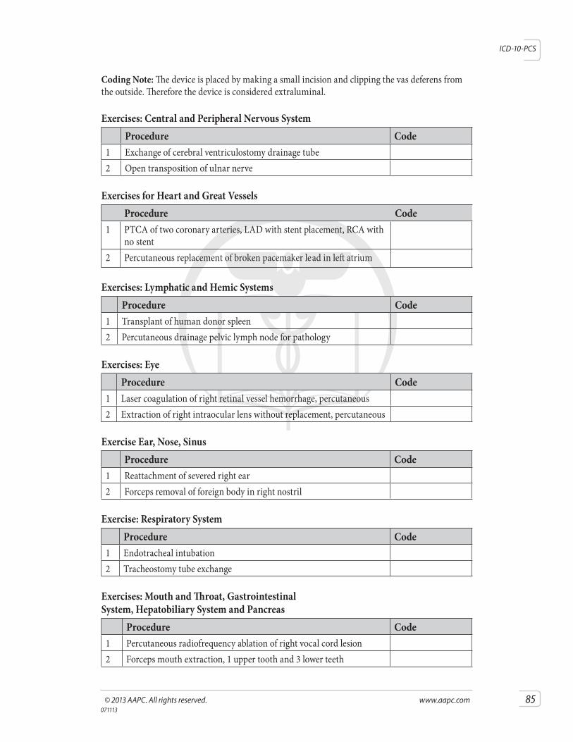
©2013AAPC.Allrightsreserved. www.aapc.com 85071113
ICD-10-PCS
Coding Note: The device is placed by making a small incision and clipping the vas deferens from the outside. Therefore the device is considered extraluminal.
Exercises: Central and Peripheral Nervous SystemProcedure Code
1 Exchange of cerebral ventriculostomy drainage tube2 Open transposition of ulnar nerve
Exercises for Heart and Great VesselsProcedure Code
1 PTCA of two coronary arteries, LAD with stent placement, RCA with no stent
2 Percutaneous replacement of broken pacemaker lead in left atrium
Exercises: Lymphatic and Hemic SystemsProcedure Code
1 Transplant of human donor spleen2 Percutaneous drainage pelvic lymph node for pathology
Exercises: EyeProcedure Code
1 Laser coagulation of right retinal vessel hemorrhage, percutaneous 2 Extraction of right intraocular lens without replacement, percutaneous
Exercise Ear, Nose, SinusProcedure Code
1 Reattachment of severed right ear 2 Forceps removal of foreign body in right nostril
Exercise: Respiratory SystemProcedure Code
1 Endotracheal intubation 2 Tracheostomy tube exchange
Exercises: Mouth and Throat, Gastrointestinal System, Hepatobiliary System and Pancreas
Procedure Code 1 Percutaneous radiofrequency ablation of right vocal cord lesion 2 Forceps mouth extraction, 1 upper tooth and 3 lower teeth
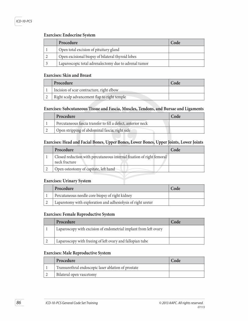
86 ICD-10-PCSGeneralCodeSetTraining ©2013AAPC.Allrightsreserved.071113
ICD-10-PCS
Exercises: Endocrine SystemProcedure Code
1 Open total excision of pituitary gland 2 Open excisional biopsy of bilateral thyroid lobes 3 Laparoscopic total adrenalectomy due to adrenal tumor
Exercises: Skin and BreastProcedure Code
1 Incision of scar contracture, right elbow 2 Right scalp advancement flap to right temple
Exercises: Subcutaneous Tissue and Fascia, Muscles, Tendons, and Bursae and Ligaments Procedure Code
1 Percutaneous fascia transfer to fill a defect, anterior neck 2 Open stripping of abdominal fascia, right side
Exercises: Head and Facial Bones, Upper Bones, Lower Bones, Upper Joints, Lower Joints Procedure Code
1 Closed reduction with percutaneous internal fixation of right femoral neck fracture
2 Open osteotomy of capitate, left hand
Exercises: Urinary SystemProcedure Code
1 Percutaneous needle core biopsy of right kidney 2 Laparotomy with exploration and adhesiolysis of right ureter
Exercises: Female Reproductive SystemProcedure Code
1 Laparoscopy with excision of endometrial implant from left ovary
2 Laparoscopy with freeing of left ovary and fallopian tube
Exercises: Male Reproductive SystemProcedure Code
1 Transurethral endoscopic laser ablation of prostate 2 Bilateral open vascetomy


![Conditionals - cs.virginia.eduup3f/cs1111/slides/1111-09-decision.pdf · Conditionals CS 1111 Introduction to Programming Fall 2018 [The Coder’s Apprentice, §6-6.2] ... This is](https://img.pdfslide.net/doc/110x75/5c5e8aa309d3f28e068c2fb7/conditionals-cs-up3fcs1111slides1111-09-decisionpdf-conditionals-cs-1111.jpg)