Embed Size (px)
Citation preview
Int.J.Curr.Microbiol.App.Sci (2017) 6(3): 1655-1675
1655
Original Research Article https://doi.org/10.20546/ijcmas.2017.603.192
Identification and Characterization of Bile Salt
Hydrolyzing Lactobacillus Isolates
Pradip Kumar Sharma1*
, Pradeep Kumar Sharma1 and Naresh Kumar
2,
Suman2 and Niti Dhingra
3
1Department of Microbiology, Ch. Charan Singh University Meerut, India
2Dairy Microbiology Division, National Dairy Research Institute, Karnal, India
3Department of Biotechnology, DAV College, Sec, 10, Chandigarh, India
*Corresponding author
A B S T R A C T
Introduction
In recent years, different investigations
support the importance of probiotics as a part
of healthy diet for humans and animals and as
a way to provide a natural, safe and effective
barrier against microbial infections (Angmo et
al., 2016). World Health Organization
(WHO) has laid down the definition of
probiotics as ‘‘live microbial food
supplements which, when administered in
adequate amounts confer health benefit on the
host” (FAO/WHO, 2001). Among the usually
used microorganisms, lactic acid bacteria
International Journal of Current Microbiology and Applied Sciences ISSN: 2319-7706 Volume 6 Number 3 (2017) pp. 1655-1675 Journal homepage: http://www.ijcmas.com
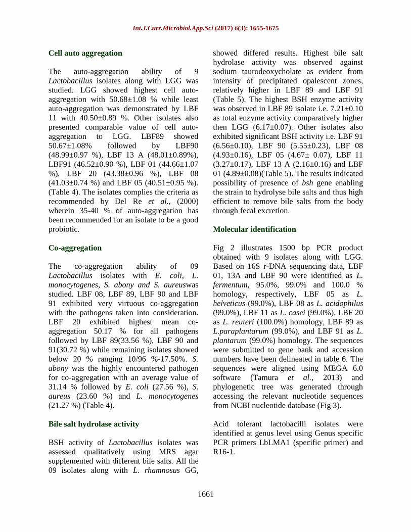
Thirty nine, out of 46 acid tolerant Lactobacillus spp., isolated from traditional dairy
products, fermented foods and human fecal samples, were preliminary identified at genus
level as Lactobacillus and evaluated for probiotic properties which included bile tolerance,
simulated gastrointestinal juice, cell surface hydrophobicity, cell auto-aggregation, co-
aggregation and bile salts hydrolase activity. Thirty three isolates could resist well at 1.5 %
bile while 26 isolates showed tolerance towards simulated gastric juice (pH 2.0, 3 h of
incubation) and simulated pancreatic juice (pH 8.0). Only 9 isolates could exhibited >50%
cell surface hydrophobicity wherein LBF89, LBF91 and LBF90 exhibited highest cell
surface hydrophobicitywith n-hexadecane i.e. 73.47±2.15 %, 72.25±1.69 % and
71.65±1.90 % respectively. LBF89 showed highest cell auto-aggregation (50.67±1.08%)
while least auto-aggregation was demonstrated by LBF 11. LBF 20 exhibited highest mean
co-aggregation 50.17 % with E. coli, L. monocytogenes, S. abony and S. aureus. S. abony
was the highly encountered pathogen with an average value of 31.14 % ofco-aggregation.
Highest bile salt hydrolase activity was observed in LBF 89 and LBF 91with sodium
taurodeoxycholate. Bile salt hydrolase activity was quantitatively determined wherein the
highest activity i.e.7.21±0.10, was observed in LBF 89 isolate followed by LBF 91 with
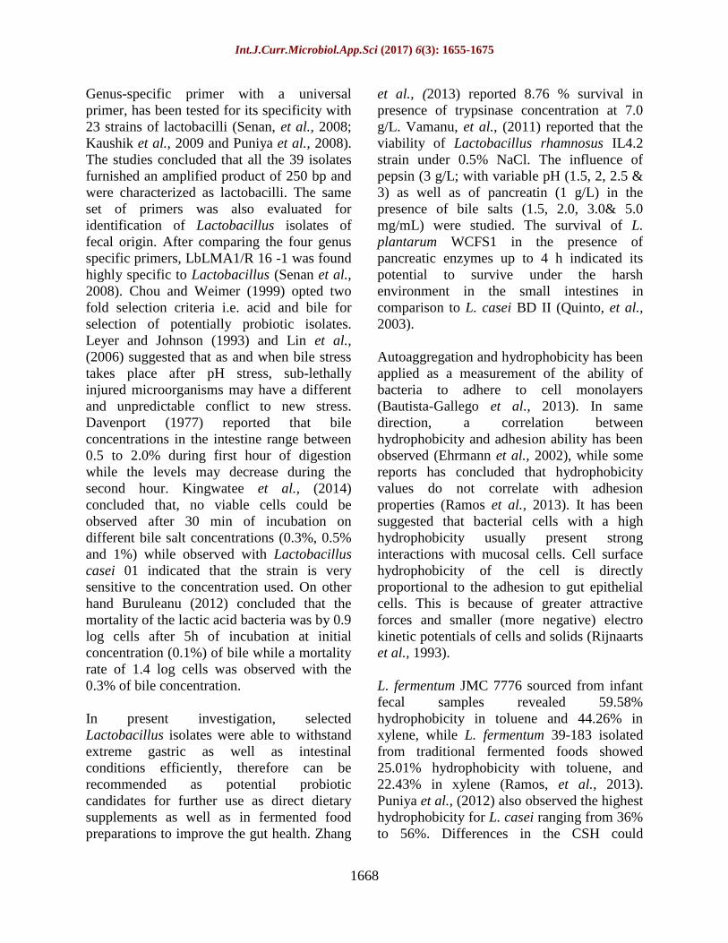
6.56±0.10 as total enzyme activity. Both the isolates were identified using PCR as
Lactobacillus plantarum but distantly placed in the phylogenetic tree. Among the selected
isolates, LBF 89 and LBF 91 showed the best probiotic potential with high tolerance to
bile, simulated gastrointestinal juice and exhibited high bile salt hydrolase activity. Both
the isolates possess application potential for functional foods and health-associated
products.
K e y w o r d s
Probiotic,
Lactobacillus, BSH
activity, Molecular
identification.
Accepted:
24 February 2017
Available Online:
10 March 2017
Article Info
Int.J.Curr.Microbiol.App.Sci (2017) 6(3): 1655-1675
1656
(LAB) are regarded as a major group of
probiotic bacteria (Collins and Gibson, 1999).
LAB is non-pathogenic, technologically
suitable for industrial processes, acid &
tolerant and produce antimicrobial substances
(Mojgani et al., 2015).
LAB are classified as generally recognized as
safe (GRAS) microorganisms because of their
long and safe use as starter cultures in
fermented food products. Most of the
probiotic organisms belong to the genera
Lactobacillus and Bifido bacterium (Prasad et
al., 1998), however, species belonging to the
genera Lactococcus, Enterococcus and
Saccharomyces (Salminen and von Wright,
1998; Sanders and in’t Veld, 1999) are also
considered as probiotic microorganisms. As
per the recommendations of Joint FAO/WHO
working group, two currently most widely
used in vitro tests for selection of probiotics
are resistance to gastric acidity and bile salts,
as evident by survival and growth studies,
(Vijaya et al., 2015). Other functional
properties for characterization of probiotics
are production of antimicrobial compounds
and cholesterol assimilation (Park et al.,
2007; Xie et al., 2015).
The mechanism through which probiotics
may antagonize pathogens involves
production of antimicrobial compounds such
as lactic acid, acetic acid, hydrogen peroxide
and bacteriocins. Other properties of probiotic
organisms include, reduced deportment of
pathogenic microorganism, lowered risk
factors for coronary artery disease and a dose
dependent reduction in the symptoms of
irritable bowel syndrome (Vries et al., 2006).
Several probiotics bacteria are found to
produce bile salt hydrolase (BSH) that helps
to reduce serum cholesterol (Miremadi et al.,
2014) and hence BSH activity is also
considered as an additional criterion for the
selection of probiotics.
Materials and Methods
Forty six acid tolerant, gram positive, catalase
negative strains isolated from traditional dairy
products, fermented foods and human fecal
samples were taken under this study. These
isolates were selected as acid tolerant after
extensive study for their survival towards
different pH i.e. 2.0, 3.0 and 6.5 (data not
shown). The selected isolates were grown in
de Man, Rogosa, and Sharpe (MRS) broth at
37 oC for 16-18 h and sub-cultured twice prior
to conducting the experiments. E. coli, L.
monocytogenes, S. abony and S. aureus used
in co-aggregation study, were obtained from
ATCC.
Molecular characterization at genus level
Genomic DNA was isolated using GeNei pure
Bacterial DNA purification kit as per
manufacturer’s instructions. Genus specific
PCR primers LbLMA1-rev 5´-CTC AAA
ACT AAA CAA AGT TTC-3´ (specific
primer) and R16-1 -5´-CTT GTA CAC ACC
GCC CGT CA-3, corresponding to the
flanking terminal sequence of the 16S rRNA
gene giving rise to 250 bp PCR product
(Dubernet et al., 2002) were used for
preliminary identification of the isolates as
lactobacilli using PCR (Bile tolerance).
Bile tolerance was tested according to the
method described by Gilliland et al.,
(1984).Overnight activated culture of
lactobacilli isolates were harvested by
centrifugation (7000 x g at 4oC for 10 min)
and re-suspended in equal volume of MRS
broth supplemented with 0.5 %, 1.0 % and 1.5
% of ox bile (Hi-media) and incubated at 37 oC. Broth without ox bile served as control.
The survival (%) of the isolates at different
time interval was calculated as follows:
Survival %=(Number of viable cells
survived/Number of initial viable cells
Int.J.Curr.Microbiol.App.Sci (2017) 6(3): 1655-1675
1657
inoculated) x100 (Tambekar and Bhutada,
2010).
Survival under simulated juice
To test the viability in presence of pepsin,
simulated gastric juice was prepared by
suspending 3.0 mg / mL pepsin in sterile
saline solution (0.85% NaCl, w/v) and pH
was adjusted to 2.5. Simultaneously, for
assessing survival ability in presence of
pancreatin, simulated intestinal fluid was
prepared by dissolving bile salt Na-
Taurodeoxycholate (0.3%) and pancreatin
(1.0 mg/mL) in sterile saline solution (0.85%
NaCl, w/v) adjusted to pH 8.0. The whole
preparation was sterilized by passing through
0.22 µm syringe filter (Kos, et al., 2000).
Isolates were grown in MRS broth for 16-18
hours and cells were harvested using
refrigerated centrifuge (7000 x g at 4oC for 10
min). Pellet was washed using potassium
phosphate buffer and re-suspended in (10
mM, pH 6.8) in same buffer to 1.0 OD using
plate reader (Perkin Elmer).
One mL of each fluids were mixed with 200
μL of bacterial cell suspension and incubated
at 37°C. Afterwards, aliquots were taken at
different time interval viz., 0 min, 90 min and
180 min and plated for total viable count
using MRS Agar media. Survival (%) of the
isolates was calculated as follows: Survival
%= (Number of viable cells survived /
Number of initial viable cells inoculated)
x100.
Cell surface hydrophobicity
Cell surface hydrophobicity (CSH)was
determined as per the method described by
Rosenberg et al., (1980) using n-hexadecane.
For CSH, overnight grown cells in MRS broth
were harvested by centrifugation at (7000 x g
at 4oC for 10 min) and washed with potassium
phosphate buffer (PB) 10mM, pH 7.0. The
pellet was re-suspended in the same buffer
and adjusted to final OD of 0.7 at 595 nm
absorbance using Perkin Elmer Victor X3
plate reader. Lactobacilli suspension (3.0 mL)
and n-hexadecane (1.0 mL) were taken in a
tube and mixed by vortexing. The preparation
was incubated at 37°C for 10 min for
temperature equilibration.
The mixture was again vortex for a while and
incubated at 37°C for 1.0 hour for phase
separation. The hydrocarbon layer was
allowed to rise completely. Aqueous phase
was removed carefully and checked for
absorbance using spectrophotometer (595 nm)
after 10 min (initial OD) and 1.0 h (final OD).
Cell surface hydrophobicity (%) was
calculated as follows: Cell surface
hydrophobicity (%)=[(Abs initial-Abs
final)/Abs final)] ×100.
Cell auto-aggregation
Auto-aggregation assay was performed
according to Del Re et al (2000).
Lactobacillus isolates were grown for 18 h at
37°C using MRS broth. Overnight grown
cells were harvested from the broth by
centrifugation at (7000 x g at 4oC for 10 min)
phosphate buffer (PB) 10mM, pH 7.0 and re-
suspended in the same buffer to an
absorbance of 0.5 at 595 nm (Abs initial) to
give viable counts of approximately 108 CFU
/ mL. The suspension was centrifuge and the
pellet were re-suspended in equal volume of
sterilized MRS broth.
The suspension were allowed to stand at 37°C
for 5 h. Afterwards, 1.0 mL of the upper
suspension were taken to measure the
absorbance (Abs final) by using sterile broth
as reference. The percent difference between
the initial and final absorbance was taken as
an index of auto-aggregation using as
following formula: Auto-aggregation % =
[(Absorbance initial-Absorbance final)
/Absorbance initial] x 100.

Int.J.Curr.Microbiol.App.Sci (2017) 6(3): 1655-1675
1658
Co-aggregation
Co-aggregation ability of each isolate was
determined by the method described by Del
Re et al., (2000). Each isolate was inoculated
into MRS broth and incubated at 37 oC for 18
hours. At the same time, the bacterial
indicators which included E. coli, L.
monocytogenes, S. abony and S. aureus were
inoculated in BHI broth and incubated at 35oC
for 16-18 h. Overnight grown cells of
Lactobacillus isolates were harvested by
centrifugation at (7000 x g at 4oC for 10 min)
and washed with potassium phosphate buffer
(PB) 10mM, pH 6.8. The pellet was re-
suspended in the same buffer so as to obtain
1.0 OD (595 nm). Indicator organisms were
also spin and washed using P.B (pH 6.8). The
pellet was suspended in the same buffer so as
to obtain 0.6 OD (595 nm). Suspension of
Lactobacillus bacteria and pathogens were
taken in 1:1 ratio and incubated at 35oC for 4
h. Suspension of individual isolate and
pathogens were taken as respective controls.
Absorbance of the suspension was monitored
at 595 nm over a period of 4 hours. Co-
aggregation % was calculated according to
Handley’s equation (Handley et al., 1987).
Co-aggregation
(%) =
[APath+ALAB/2]-
AMix X100
[APath-ALAB/2]
APath: Absorbanceof pathogens; ALAB:
Absorbance of Lactobacillus isolates; AMix:
Absorbance of mixture containing pathogens
and Lactobacillus isolates.
Bile salt hydrolase activity
Direct plate assay
The qualitative Bile salt hydrolase (BSH)
activity of the isolates was assessed by direct
plate assay (Schillinger et al., 2005).
Lactobacillus isolates were streaked on MRS
agar plates supplemented with 0.5 % (w/v)
filter sterilized bile salts (sodium
taurodeoxycholate hydrate; sodium
taurocholate; sodium deoxycholate;
deoxycholic acid and cholic acid; Sigma,
USA) along with 0.37g/L of CaCl2. The plates
were incubated anaerobically in an anaerobic
gas pack jar at 37oC for 72 h. MRS agar plates
without bile salt supplementation were used
as control. The presence of precipitated bile
acid around colonies (opaque halo) or the
formation of opaque granular white colonies
with a silvery shine was considered as a
positive reaction
Quantitative assay for bile salt hydrolase
activity
The bile salt hydrolase (BSH) assay was also
performed by measuring amino acids released
from hydrolysis of conjugated bile salts by
Lactobacillus isolates as described by Liong
and Shah, (2005). Exponentially growin
Lactobacilli cells were harvested at (7000 x g
at 4oC for 10 min), washed twice with 0.1 M
sodium phosphate buffer (pH 6.8) and re-
suspended in same buffer to 1·0 OD at 600
nm. Five mL of bacterial suspension were
sonicated for 3.0 min with constant cooling
using ice followed by centrifugation at 10,000
rpm at 4oC for 10 min. The reaction mixture
contained 100 µL cell suspension, 100 µL of
10 mM conjugated bile salt (sodium
glycocholate) and 1.8 mL of 0.1 M sodium
phosphate buffer (pH 6). Reaction mixtures
were incubated at 37°C for 30 min. Equal
volume of reaction mixture and 15 % tri-
chloroacetic acid (TCA,w/v, 200 µL each)
were taken and centrifuge at 12,000 rpm for
15 min at 4oC. The amount of amino acids
present in the supernatant was measured. An
aliquot of 0·2 mL of supernatant obtained
after centrifugation was added to 1.0 mL of
distilled water and 1.0 mL of ninhydrin
reagent (0·5 mL of 1.0% ninhydrin in 0·5 M
citrate buffer, pH 5·5, 1·2 mL of 30%
Int.J.Curr.Microbiol.App.Sci (2017) 6(3): 1655-1675
1659
glycerol and 0·2 mL of 0·5 M citrate buffer,
pH 5·5). The preparation was vortex
vigorously and kept in dry block heater at 95-
100 oC for 14 min. After cooling, absorbance
was determined at 570nm using glycine as
standards. One unit of BSH activity (U/mL)
was defined as the amount of enzyme that
liberated 1 nmol of amino acid substrate per
min per absorbance at 570nm.
Molecular identification of selected
Lactobacillus isolates at species level
Polymerase chain reaction
Amplification of selected Lactobacillus
isolates was executed using universal
prokaryotic primers for the 16S rRNA gene
pA (5’–AGAGTTTGATCCTGGCTCAG;
nucleotide 8 to 27 of the 16S rRNA gene of E.
coli) and pH (5’–
AAGGAGGTGATCCAGCCGCA;
nucleotide 1541 to 1522 of the 16S rRNA
gene of E. coli; (Rodas et al., 2003) yielding a
PCR product of ~1500 bp.
Gel electrophoresis of PCR products and
sequencing
A 100-1500 bp ladder (Fermentas,) was used
as a molecular mass marker. Bands appearing
at 1500 bp were considered for sequencing
followed by BLAST analsyis.PCR products
were sent to M/s. Invitrogen Bioservices India
Pvt. Ltd. Udhog Vihar, Gurgaon, India and
were directly sequenced using the forward
and reverse primers. The sequences were
further used to identify the isolate at species
level by using basic local alignment sequence
tool (BLAST) for similarity search at NCBI
website using GenBank data (Altschul et al.,
1997).
Statistical analysis
Data were statistically analyzed with
GraphPad Prism 5.1 software. One-way
analysis of variance was used to study
significant difference between means, with
significance level at P = 0.05.
Results and Discussion
Preliminary identification of isolates by
genus specific PCR
Molecular methods are more reliable and
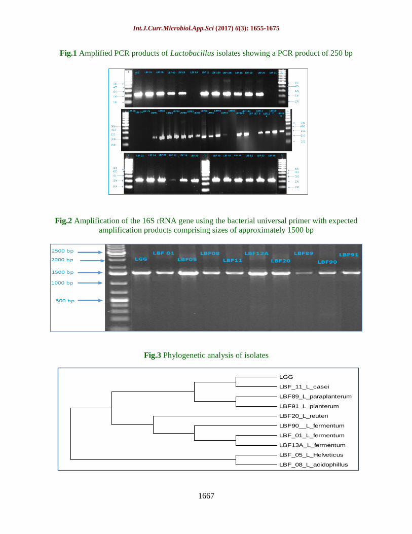
accurate than that of biochemical ones. Fig 1
shows 250 bp PCR product obtained with 39
isolates out of 46 acid tolerant Lactobacillus
isolates. Reference Lactobacillus culture was
also used as a positive control. However, 8
isolates were failed to amplify using the
above procedure showing that they may not
be of genus Lactobacillus.
Tolerance against bile
Bile resistance is one of the important criteria
for selection of probiotic (Lee and Salminen,
1995; Dunne et al., 2001). Resistance to bile
helps probiotic bacteria to reach the small
intestine and colon and subsidize in balancing
the intestinal microflora (Tambekar and
Bhutada, 2010). After 3 hour of incubation,
At 0.5 % bile, 5 isolates LBF 08, LBF 20,
LBF 11, LBF 01 and LBF197 showed 70 %
or above survival which is very close to LGG
(81.22±0.75 %). Moreover, 20 isolates
showed more than 60 % survival ranging
61.2%-69.4%; 8 isolates with more than 50 %
survival (52.5%-59.53%) while remaining 06
isolates exhibited survival between 22.0 -36.0
%.
Whereas, 9 isolates showed 60 % or above
survival (61.0 %-76.1%) at 1.0 % bile
concentration which is comparable to LGG
(76.10±0.62 %) after 3 hours of incubation. In
addition to this, 22 isolates showed more than
50 % survival (51.46%-59.95%) while 2
isolates with more than 40 % survival. Only,
06 isolates exhibited 18.0-29.0 % survival at
1.0 % bile concentration.

Int.J.Curr.Microbiol.App.Sci (2017) 6(3): 1655-1675
1660
At highest concentration of bile i.e. 1.5 %,
only 5 isolates showed 41.19-42.36 %
survival along with LGG (46.19±0.68 %)
after 3 hours of incubation. Moreover, 25
isolates showed 31.37%-39.79% and 3
isolates with 26.21%-28.47% survival. Apart
from this, 6 isolates exhibited less than 10 %
survival (Table 1). The isolates which
tolerated 1.5 % of bile concentration after 3
hours exposure with 10 % or above survival
were considered as bile tolerant and selected
for further investigation (Pennacchia, 2004).
A total of 33 isolates were selected out of 39
isolates for further characterization.
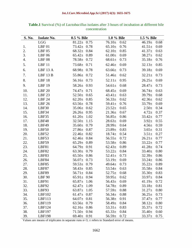
Survival under simulated gastric and
pancreatic juice
During transit from oral to colon, probiotic
bacteria are supposed to survive through
stomach followed by intestine and exert their
health promoting effects as metabolically
viable active cells after reaching in the colon
(Malek et al., 2010).All 33 isolates were able
to survive under simulated gastric juice
containing pepsin at pH 2.0 up to 3 h of
incubation with variable survival rates. Out of
33 isolates, only 2 isolates (LBF 92 and LBF
198) registered survival of 72.53±0.50 % and
73.16± 0.54 %, respectively. In addition to
this, 13 Lactobacillus isolates showed more
than 60.0 % survival but less than 70 %
survival ranging from 60.46± 0.41 % to
69.71±0.45 %with variable degree of log
reduction. LGG showed 68.12±0.84 %
survival under these conditions which is
comparable to 13 different isolates. Apart
from this, 12 Lactobacillus isolates exhibited
survival between 50-60 % ranging from
52.17±0.43 to 59.62±0.49 %. However, 6
isolates showed less than 20 % survival
ranging from 6.71±0.06 % to 18.97±0.09 %
(Table 2).
Under simulated pancreatic juice (pH 8.0), all
33 isolates were able to survived by 3 h of
incubation with variable degree of log
reduction. Out of 33 isolates, 2 isolates (LBF
01 and LBF 20) exhibited 23.30±0.26 % and
21.26±0.18 % survival which is comparable
with reference culture LGG (22.19±0.27 %).
Moreover, 19 Lactobacillus isolates were able
to withstand the pancreatic juice with survival
ranging from 10.26 % to 19.83 % survival. In
addition to this, 7Lactobacillus isolates
(21.21%) revealed less than 10 % ranging
from 1.59 to 9.35 % while 5 Lactobacillus
isolates showed below 1.0 % ranging from
0.30 % to 0.91 % (Table 2). These 5 isolates
also revealed less survival for simulated
gastric juice. Overall, only 27 isolates were
selected after scrutiny of tolerance towards
bile and good survival under simulated gastric
and pancreatic juice.
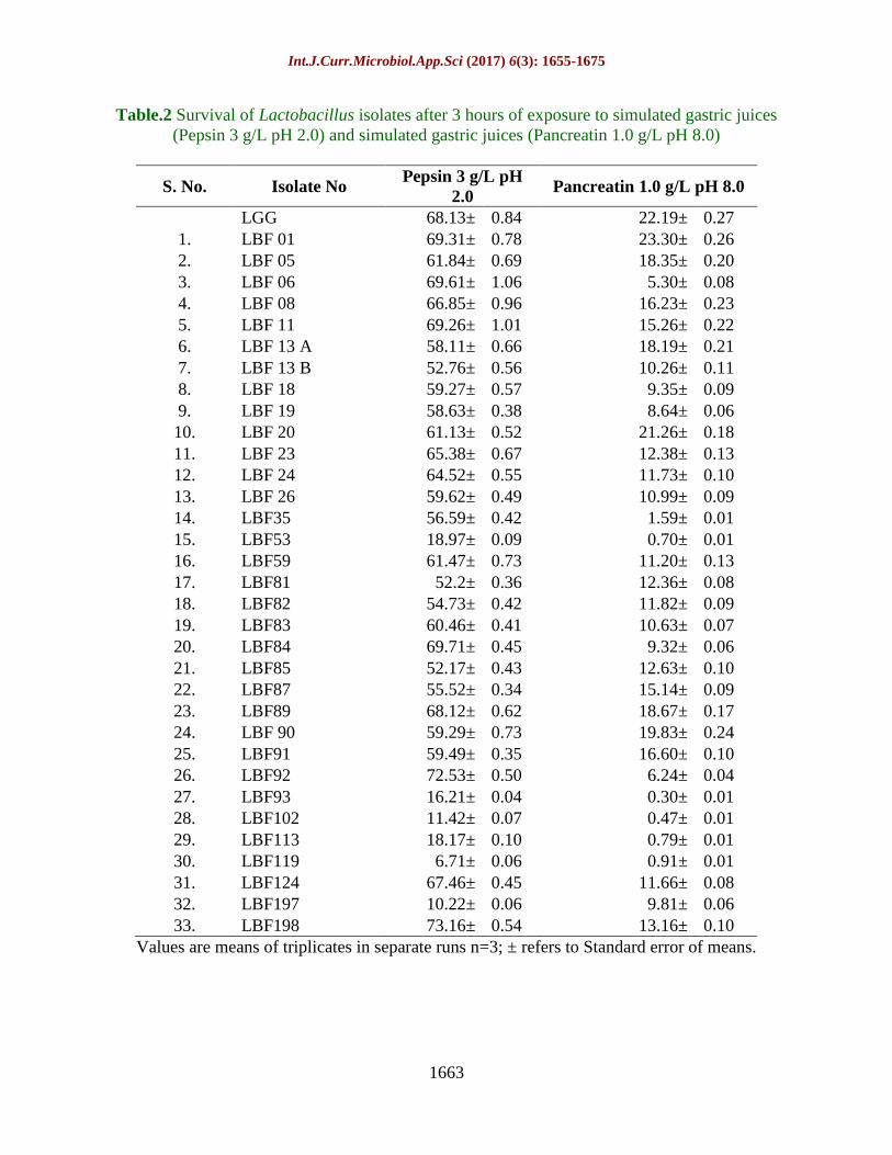
Cell surface hydrophobicity
The colonization in intestinal wall is
considered as one of the prominent criteria for
selection of probiotics. CSH was assessed by
measuring the adhesion ability of the isolates
to the intestinal epithelium (Niguez-
Palomares et al., 2008; Tuomola et al., 2001).
Significant differences in hydrophobicity
were observed within Lactobacillus isolates.
Out of 27 isolates, 3 isolates LBF89, LBF91
and LBF90 exhibited highest value of CSH
i.e. 73.47±2.15 %, 72.25±1.69 % and
71.65±1.90 %, respectively, which is very
close to the reference strain LGG
(74.10±2.24%) with n-hexadecane. In
addition to this, 4 Lactobacillus isolates,
designated as LBF 05, LBF 01, LBF 08 and
LBF 11 showed 65.45±2.15 %, 64.72±2.93
%, 63.41±2.05 % and 62.87±2.09% CSH,
respectively. LBF 20 and LBF 13A also
confirmed respectable level (58.87±2.06%
and 56.38±2.16%, respectively) of CSH.
However, rest 17 isolates exhibited CSH in
the range of 1.73 % to 21.69 % (Table 3).
Int.J.Curr.Microbiol.App.Sci (2017) 6(3): 1655-1675
1661
Cell auto aggregation
The auto-aggregation ability of 9
Lactobacillus isolates along with LGG was
studied. LGG showed highest cell auto-
aggregation with 50.68±1.08 % while least
auto-aggregation was demonstrated by LBF
11 with 40.50±0.89 %. Other isolates also
presented comparable value of cell auto-
aggregation to LGG. LBF89 showed
50.67±1.08% followed by LBF90
(48.99±0.97 %), LBF 13 A (48.01±0.89%),
LBF91 (46.52±0.90 %), LBF 01 (44.66±1.07
%), LBF 20 (43.38±0.96 %), LBF 08
(41.03±0.74 %) and LBF 05 (40.51±0.95 %).
(Table 4). The isolates complies the criteria as
recommended by Del Re et al., (2000)
wherein 35-40 % of auto-aggregation has
been recommended for an isolate to be a good
probiotic.
Co-aggregation
The co-aggregation ability of 09
Lactobacillus isolates with E. coli, L.
monocytogenes, S. abony and S. aureuswas
studied. LBF 08, LBF 89, LBF 90 and LBF
91 exhibited very virtuous co-aggregation
with the pathogens taken into consideration.
LBF 20 exhibited highest mean co-
aggregation 50.17 % for all pathogens
followed by LBF 89(33.56 %), LBF 90 and
91(30.72 %) while remaining isolates showed
below 20 % ranging 10/96 %-17.50%. S.
abony was the highly encountered pathogen
for co-aggregation with an average value of
31.14 % followed by E. coli (27.56 %), S.
aureus (23.60 %) and L. monocytogenes
(21.27 %) (Table 4).
Bile salt hydrolase activity
BSH activity of Lactobacillus isolates was
assessed qualitatively using MRS agar
supplemented with different bile salts. All the
09 isolates along with L. rhamnosus GG,
showed differed results. Highest bile salt
hydrolase activity was observed against
sodium taurodeoxycholate as evident from
intensity of precipitated opalescent zones,
relatively higher in LBF 89 and LBF 91
(Table 5). The highest BSH enzyme activity
was observed in LBF 89 isolate i.e. 7.21±0.10
as total enzyme activity comparatively higher
then LGG (6.17±0.07). Other isolates also
exhibited significant BSH activity i.e. LBF 91
(6.56±0.10), LBF 90 (5.55±0.23), LBF 08
(4.93±0.16), LBF 05 (4.67± 0.07), LBF 11
(3.27±0.17), LBF 13 A (2.16±0.16) and LBF
01 (4.89±0.08)(Table 5). The results indicated
possibility of presence of bsh gene enabling
the strain to hydrolyse bile salts and thus high
efficient to remove bile salts from the body
through fecal excretion.
Molecular identification
Fig 2 illustrates 1500 bp PCR product
obtained with 9 isolates along with LGG.
Based on 16S r-DNA sequencing data, LBF
01, 13A and LBF 90 were identified as L.
fermentum, 95.0%, 99.0% and 100.0 %
homology, respectively, LBF 05 as L.
helveticus (99.0%), LBF 08 as L. acidophilus
(99.0%), LBF 11 as L. casei (99.0%), LBF 20
as L. reuteri (100.0%) homology, LBF 89 as
L.paraplantarum (99.0%), and LBF 91 as L.
plantarum (99.0%) homology. The sequences
were submitted to gene bank and accession
numbers have been delineated in table 6. The
sequences were aligned using MEGA 6.0
software (Tamura et al., 2013) and
phylogenetic tree was generated through
accessing the relevant nucleotide sequences
from NCBI nucleotide database (Fig 3).
Acid tolerant lactobacilli isolates were
identified at genus level using Genus specific
PCR primers LbLMA1 (specific primer) and
R16-1.
Int.J.Curr.Microbiol.App.Sci (2017) 6(3): 1655-1675
1662
Table.1 Survival (%) of Lactobacillus isolates after 3 hours of incubation at different bile
concentration
S. No. Isolate No. 0.5 % Bile 1.0 % Bile 1.5 % Bile
LGG 81.22± 0.75 76.10± 0.62 46.19± 0.68
1. LBF 01 73.42± 0.78 65.10± 0.76 41.51± 0.69
2. LBF 05 68.32± 0.84 62.10± 0.85 41.37± 0.63
3. LBF 06 65.42± 0.89 61.00± 0.69 38.27± 0.62
4. LBF 08 78.58± 0.72 68.61± 0.73 35.18± 0.76
5. LBF 11 73.68± 0.71 62.46± 0.69 32.13± 0.85
6. LBF 13 A 68.98± 0.78 63.66± 0.73 39.18± 0.69
7. LBF 13 B 55.86± 0.72 51.46± 0.62 32.21± 0.73
8. LBF 18 56.16± 0.73 52.11± 0.95 26.25± 0.69
9. LBF 19 58.26± 0.93 54.61± 0.68 28.47± 0.73
10. LBF 20 74.47± 0.71 68.45± 0.69 36.74± 0.63
11. LBF 23 52.50± 0.65 43.41± 0.63 39.79± 0.68
12. LBF 24 62.20± 0.85 56.33± 0.62 42.36± 0.62
13. LBF 26 63.56± 0.78 59.41± 0.76 33.79± 0.69
14. LBF30 35.06± 0.62 23.52± 0.65 2.50± 0.34
15. LBF34 26.26± 0.95 21.36± 0.67 4.72± 0.37
16. LBF35 61.20± 1.02 56.85± 0.86 33.42± 0.77
17. LBF48 32.50± 1.15 28.63± 0.69 3.92± 0.55
18. LBF49 35.60± 0.79 28.99± 0.64 4.56± 0.59
19. LBF50 27.86± 0.87 23.89± 0.83 3.65± 0.31
20. LBF52 22.46± 0.82 18.74± 0.54 3.51± 0.27
21. LBF53 62.46± 0.84 56.55± 0.72 26.21± 0.77
22. LBF59 65.29± 0.89 53.58± 0.86 33.22± 0.77
23. LBF81 64.79± 0.91 62.43± 0.89 41.28± 0.74
24. LBF82 63.30± 0.79 53.22± 0.84 33.40± 0.80
25. LBF83 65.50± 0.86 52.41± 0.73 32.39± 0.86
26. LBF84 56.07± 0.73 53.19± 0.69 33.24± 0.86
27. LBF85 59.53± 0.79 49.64± 0.73 35.22± 0.89
28. LBF87 58.43± 0.85 53.54± 0.63 35.58± 0.84
29. LBF89 56.71± 0.84 52.75± 0.68 35.30± 0.83
30. LBF 90 65.91± 0.94 59.95± 0.62 33.97± 0.84
31. LBF91 65.87± 1.06 56.43± 0.69 41.19± 0.72
32. LBF92 62.47± 1.09 54.78± 0.89 33.18± 0.81
33. LBF93 63.87± 1.05 57.59± 0.88 31.27± 0.80
34. LBF102 61.47± 0.87 56.34± 0.88 36.25± 0.73
35. LBF113 64.07± 0.81 56.38± 0.91 37.47± 0.77
36. LBF119 63.56± 0.79 58.49± 0.84 38.12± 0.80
37. LBF124 62.16± 0.86 53.31± 0.83 35.37± 0.72
38. LBF197 71.50± 0.94 65.33± 0.84 35.40± 0.60
39. LBF198 69.40± 0.91 56.59± 0.72 33.37± 0.75 Values are means of triplicates in separate runs n=3; ± refers to Standard error of means.
Int.J.Curr.Microbiol.App.Sci (2017) 6(3): 1655-1675
1663
Table.2 Survival of Lactobacillus isolates after 3 hours of exposure to simulated gastric juices
(Pepsin 3 g/L pH 2.0) and simulated gastric juices (Pancreatin 1.0 g/L pH 8.0)
S. No. Isolate No Pepsin 3 g/L pH
2.0 Pancreatin 1.0 g/L pH 8.0
LGG 68.13± 0.84 22.19± 0.27
1. LBF 01 69.31± 0.78 23.30± 0.26
2. LBF 05 61.84± 0.69 18.35± 0.20
3. LBF 06 69.61± 1.06 5.30± 0.08
4. LBF 08 66.85± 0.96 16.23± 0.23
5. LBF 11 69.26± 1.01 15.26± 0.22
6. LBF 13 A 58.11± 0.66 18.19± 0.21
7. LBF 13 B 52.76± 0.56 10.26± 0.11
8. LBF 18 59.27± 0.57 9.35± 0.09
9. LBF 19 58.63± 0.38 8.64± 0.06
10. LBF 20 61.13± 0.52 21.26± 0.18
11. LBF 23 65.38± 0.67 12.38± 0.13
12. LBF 24 64.52± 0.55 11.73± 0.10
13. LBF 26 59.62± 0.49 10.99± 0.09
14. LBF35 56.59± 0.42 1.59± 0.01
15. LBF53 18.97± 0.09 0.70± 0.01
16. LBF59 61.47± 0.73 11.20± 0.13
17. LBF81 52.2± 0.36 12.36± 0.08
18. LBF82 54.73± 0.42 11.82± 0.09
19. LBF83 60.46± 0.41 10.63± 0.07
20. LBF84 69.71± 0.45 9.32± 0.06
21. LBF85 52.17± 0.43 12.63± 0.10
22. LBF87 55.52± 0.34 15.14± 0.09
23. LBF89 68.12± 0.62 18.67± 0.17
24. LBF 90 59.29± 0.73 19.83± 0.24
25. LBF91 59.49± 0.35 16.60± 0.10
26. LBF92 72.53± 0.50 6.24± 0.04
27. LBF93 16.21± 0.04 0.30± 0.01
28. LBF102 11.42± 0.07 0.47± 0.01
29. LBF113 18.17± 0.10 0.79± 0.01
30. LBF119 6.71± 0.06 0.91± 0.01
31. LBF124 67.46± 0.45 11.66± 0.08
32. LBF197 10.22± 0.06 9.81± 0.06
33. LBF198 73.16± 0.54 13.16± 0.10
Values are means of triplicates in separate runs n=3; ± refers to Standard error of means.
Int.J.Curr.Microbiol.App.Sci (2017) 6(3): 1655-1675
1664
Table.3 Cell surface hydrophobicity (%) of Lactobacillus isolates
S. No. Isolates CSH (%)
LGG 74.04± 2.24
1. LBF 01 64.84± 2.39
2. LBF 05 65.40± 2.15
3. LBF 06 7.36± 1.99
4. LBF 08 63.35± 2.05
5. LBF 11 62.72± 2.09
6. LBF 13 A 56.71± 2.16
7. LBF 13 B 11.00± 0.87
8. LBF 18 13.82± 0.65
9. LBF 19 21.65± 1.08
10. LBF 20 58.90± 2.06
11. LBF 23 2.39± 0.20
12. LBF 24 2.99± 0.54
13. LBF 26 6.03± 0.56
14. LBF35 7.61± 0.27
15. LBF53 4.70± 0.80
16. LBF81 4.56± 0.14
17. LBF82 7.19± 0.51
18. LBF83 12.63± 0.78
19. LBF84 3.39± 0.23
20. LBF85 2.12± 0.17
21. LBF87 5.71± 0.15
22. LBF89 73.48± 2.15
23. LBF90 71.61± 1.90
24. LBF91 72.28± 1.69
25. LBF92 1.71± 0.12
26. LBF124 2.94± 0.15
27. LBF198 5.36± 0.10 Values are means of triplicates in separate runs n=3; ± refers to Standard error of means

Int.J.Curr.Microbiol.App.Sci (2017) 6(3): 1655-1675
1665
Table.4 Cell auto-aggregation and Co-aggregation ability of selected Lactobacillus isolates
S.
NO. Isolate
Auto-aggregation
(%)
Co-aggregation (%)
E. coli L. monocytogenes S. abony S. aureus Mean Co-
aggregation
LGG 50.68± 1.085 48.49± 0.31 72.01± 0.34 37.77± 0.30 48.03± 0.30 51.57±0.51
1. LBF 01 44.66± 1.077 12.09± 0.29 17.85± 0.29 4.30± 0.28 9.59± 0.32 10.96±0.49
2. LBF 05 42.86± 0.910 9.91± 0.29 17.68± 0.27 10.63± 0.28 7.73± 0.26 11.49±0.43
3. LBF 08 41.03± 0.746 26.49± 0.28 30.63± 0.26 24.74± 0.25 21.68± 0.30 11.28±0.43
4. LBF 11 40.50± 0.893 8.34± 0.26 17.25± 0.27 6.43± 0.26 13.11± 0.34 15.77±0.44
5. LBF 13A 48.01± 0.893 11.39± 0.24 25.86± 0.22 12.92± 0.27 12.90± 0.28 17.50±0.43
6. LBF 20 43.38± 0.969 23.59± 0.27 31.63± 0.26 14.56± 0.32 0.22± 0.32 50.17±0.50
7. LBF 89 50.67± 1.081 55.74± 0.30 53.96± 0.34 45.10± 0.28 45.90± 0.32 33.56±0.48
8. LBF 90 48.99± 0.972 43.53± 0.33 25.09± 0.33 31.74± 0.27 33.88± 0.32 30.72±0.46
9. LBF 91 46.52± 0.902 35.99± 0.35 19.44± 0.39 24.47± 0.32 42.98± 0.34 30.72±0.46
Mean 45.73±0.95 27.56±0.95 21.27±0.28 31.14±0.27 23.60±0.31
Values are means of triplicates in separate runs n=3; ± refers to Standard error of means
Int.J.Curr.Microbiol.App.Sci (2017) 6(3): 1655-1675
1666
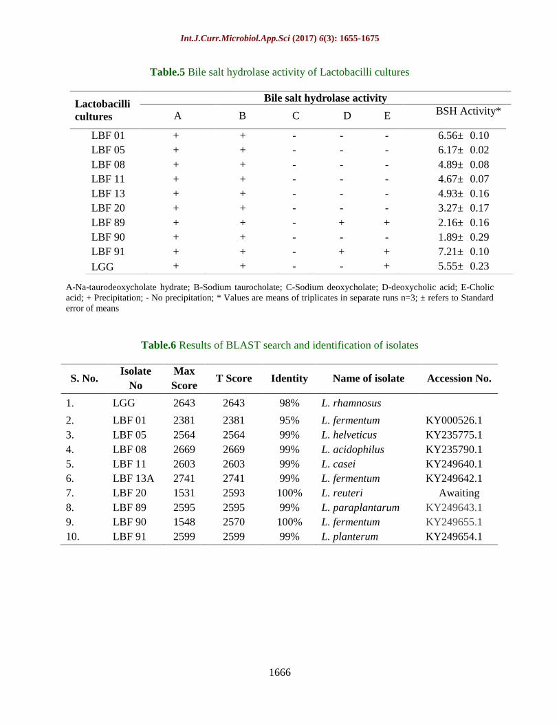
Table.5 Bile salt hydrolase activity of Lactobacilli cultures
A-Na-taurodeoxycholate hydrate; B-Sodium taurocholate; C-Sodium deoxycholate; D-deoxycholic acid; E-Cholic
acid; + Precipitation; - No precipitation; * Values are means of triplicates in separate runs n=3; ± refers to Standard
error of means
Table.6 Results of BLAST search and identification of isolates
S. No. Isolate
No
Max
Score T Score Identity Name of isolate Accession No.
1. LGG 2643 2643 98% L. rhamnosus
2. LBF 01 2381 2381 95% L. fermentum KY000526.1
3. LBF 05 2564 2564 99% L. helveticus KY235775.1
4. LBF 08 2669 2669 99% L. acidophilus KY235790.1
5. LBF 11 2603 2603 99% L. casei KY249640.1
6. LBF 13A 2741 2741 99% L. fermentum KY249642.1
7. LBF 20 1531 2593 100% L. reuteri Awaiting
8. LBF 89 2595 2595 99% L. paraplantarum KY249643.1
9. LBF 90 1548 2570 100% L. fermentum KY249655.1
10. LBF 91 2599 2599 99% L. planterum KY249654.1
Lactobacilli
cultures
Bile salt hydrolase activity
A B C D E BSH Activity*
LBF 01 + + - - - 6.56± 0.10
LBF 05 + + - - - 6.17± 0.02
LBF 08 + + - - - 4.89± 0.08
LBF 11 + + - - - 4.67± 0.07
LBF 13 + + - - - 4.93± 0.16
LBF 20 + + - - - 3.27± 0.17
LBF 89 + + - + + 2.16± 0.16
LBF 90 + + - - - 1.89± 0.29
LBF 91 + + - + + 7.21± 0.10
LGG + + - - + 5.55± 0.23
Int.J.Curr.Microbiol.App.Sci (2017) 6(3): 1655-1675
1667
Fig.1 Amplified PCR products of Lactobacillus isolates showing a PCR product of 250 bp
Fig.2 Amplification of the 16S rRNA gene using the bacterial universal primer with expected
amplification products comprising sizes of approximately 1500 bp
Fig.3 Phylogenetic analysis of isolates
LGG
LBF_11_L_casei
LBF89_L_paraplanterum
LBF91_L_planterum
LBF20_L_reuteri
LBF90__L_fermentum
LBF_01_L_fermentum
LBF13A_L_fermentum
LBF_05_L_Helveticus
LBF_08_L_acidophillus
Int.J.Curr.Microbiol.App.Sci (2017) 6(3): 1655-1675
1668
Genus-specific primer with a universal
primer, has been tested for its specificity with
23 strains of lactobacilli (Senan, et al., 2008;
Kaushik et al., 2009 and Puniya et al., 2008).
The studies concluded that all the 39 isolates
furnished an amplified product of 250 bp and
were characterized as lactobacilli. The same
set of primers was also evaluated for
identification of Lactobacillus isolates of
fecal origin. After comparing the four genus
specific primers, LbLMA1/R 16 -1 was found
highly specific to Lactobacillus (Senan et al.,
2008). Chou and Weimer (1999) opted two
fold selection criteria i.e. acid and bile for
selection of potentially probiotic isolates.
Leyer and Johnson (1993) and Lin et al.,
(2006) suggested that as and when bile stress
takes place after pH stress, sub-lethally
injured microorganisms may have a different
and unpredictable conflict to new stress.
Davenport (1977) reported that bile
concentrations in the intestine range between
0.5 to 2.0% during first hour of digestion
while the levels may decrease during the
second hour. Kingwatee et al., (2014)
concluded that, no viable cells could be
observed after 30 min of incubation on
different bile salt concentrations (0.3%, 0.5%
and 1%) while observed with Lactobacillus
casei 01 indicated that the strain is very
sensitive to the concentration used. On other
hand Buruleanu (2012) concluded that the
mortality of the lactic acid bacteria was by 0.9
log cells after 5h of incubation at initial
concentration (0.1%) of bile while a mortality
rate of 1.4 log cells was observed with the
0.3% of bile concentration.
In present investigation, selected
Lactobacillus isolates were able to withstand
extreme gastric as well as intestinal
conditions efficiently, therefore can be
recommended as potential probiotic
candidates for further use as direct dietary
supplements as well as in fermented food
preparations to improve the gut health. Zhang
et al., (2013) reported 8.76 % survival in
presence of trypsinase concentration at 7.0
g/L. Vamanu, et al., (2011) reported that the
viability of Lactobacillus rhamnosus IL4.2
strain under 0.5% NaCl. The influence of
pepsin (3 g/L; with variable pH (1.5, 2, 2.5 &
3) as well as of pancreatin (1 g/L) in the
presence of bile salts (1.5, 2.0, 3.0& 5.0
mg/mL) were studied. The survival of L.
plantarum WCFS1 in the presence of
pancreatic enzymes up to 4 h indicated its
potential to survive under the harsh
environment in the small intestines in
comparison to L. casei BD II (Quinto, et al.,
2003).
Autoaggregation and hydrophobicity has been
applied as a measurement of the ability of
bacteria to adhere to cell monolayers
(Bautista-Gallego et al., 2013). In same
direction, a correlation between
hydrophobicity and adhesion ability has been
observed (Ehrmann et al., 2002), while some
reports has concluded that hydrophobicity
values do not correlate with adhesion
properties (Ramos et al., 2013). It has been
suggested that bacterial cells with a high
hydrophobicity usually present strong
interactions with mucosal cells. Cell surface
hydrophobicity of the cell is directly
proportional to the adhesion to gut epithelial
cells. This is because of greater attractive
forces and smaller (more negative) electro
kinetic potentials of cells and solids (Rijnaarts
et al., 1993).
L. fermentum JMC 7776 sourced from infant
fecal samples revealed 59.58%
hydrophobicity in toluene and 44.26% in
xylene, while L. fermentum 39-183 isolated
from traditional fermented foods showed
25.01% hydrophobicity with toluene, and
22.43% in xylene (Ramos, et al., 2013).
Puniya et al., (2012) also observed the highest
hydrophobicity for L. casei ranging from 36%
to 56%. Differences in the CSH could
Int.J.Curr.Microbiol.App.Sci (2017) 6(3): 1655-1675
1669
attributed to variable expression of cell
surface proteins among different strains of a
species as well as to environmental conditions
that could affect the expression of cell surface
proteins (Kaushik et al., 2009). Hence, based
on cell surface hydrophobicity, only 9 isolates
(LBF 01, LBF 05, LBF 08, LBF 11, LBF 13
A, LBF 20, LBF89, LBF90, LBF91) were
selected for further characterization.Cell
adhesion, a multistep process, involves
contact of the bacterial cell membrane and
intermingling surfaces while aggregation may
take place between cells of the same strain
(auto-aggregation) or between different
species and strains (co-aggregation).
Aggregation has been considered as an
important mechanism for genetic exchange,
adhesion, and colonization in the host
environments, as well as Immuno modulation
of colonic mucosa (Cesena et al., 2001,
Voltan et al., 2007).
Savedboworn et al., (2014) concluded that
most of the LAB exhibited a strong auto-
aggregation after 5 h of incubation like
KMUTNB 5-8 strain showed highest auto-
aggregation ability of 96.09%. On another
hand some isolates (KMUTNB 5-27) could
not show any auto-aggregation. Probiotic and
pathogenic bacteria have been reported to
form joint aggregate and the process is known
as co-aggregation (Surono, 2004) resulting in
effectively inhibiting and killing them by
secreting antimicrobial compounds that act
directly on pathogens (Bao et al., 2010). Li et
al., (2015) studied the co-aggregation ability
of 18 lactic acid bacteria isolated from
traditional fermented foods and found all the
isolates to have co-aggregation ability with
Salmonella sp. ranging from 5.15% to
29.54%. Co-aggregation of L. acidophilus
M92 with two other potential probiotic strains
(L. plantarum L4, E. faecium L3) and two
enteropathogens (S. abonymurium and E. coli)
was examined wherein L. acidophilus M92
exhibited 4.36% co-aggregation with L.
plantarum L4, 19.46% with E. faecium L3,
15.11% with E. coli 3014 and 15.70% with S.
abonymurium (Kos et al., 2003).
The efficient co-aggregation ability of
probiotic bacteria against gram positive
bacteria could be attributed to the similar cell
wall morphology of LAB and gram positive
pathogens and their hydrophobic nature
making it easier to bond altogether (Arief et
al., 2015). Moreover, lactic acid bacteria
strains could control the microenviroment
around the pathogens and increase the
concentration of excreted antimicrobial
substances in the process of co-aggregation
(Li et al., 2015) which constitute an important
host defense mechanism against infection in
the gastrointestinal tract (Reid et al., 1988).
Free bile acids formed by the deconjugation
of conjugated bile salts are less soluble and
are less likely to be reabsorbed by the
intestinal lumen compared to their conjugated
equivalent, and are lost from the human body
through feces (Center, 1993). Ahn et al.,
(2003) and Begley et al., (2006), described
precipitation of bile salt by BSH activity of
probiotic strains. Bile salt hydrolase activity
against sodium tauroglycocholate but low
intensity of precipitation was noticed as
compared to sodium taurodeoxycholate.
Probiotics such as L. acidophilus have been
reported to possess bile salt hydrolase (BSH)
or cholylglycine hydrolase (the enzyme that
catalyzes the hydrolysis of glycine- and
taurine-conjugated bile salts into amino acid
residues and free bile salts). BSH has been
reported to be present in several bacterial
species of the gastrointestinal tract, such as
Lactobacillus sp.,B. longum, C. perfringens
and B. fragilis ssp. fragilis(Corzo & Gilliland,
1999). Human intestinal pH of 6.5 and a
glycocholate to taurocholate ratio of 2:3 were
found glycine conjugated bile salt to be more
efficiently deconjugated by strains of L.
acidophilus from both human and porcine
Int.J.Curr.Microbiol.App.Sci (2017) 6(3): 1655-1675
1670
origins than taurine conjugated bile salt(Corzo
& Gilliland,1999). L. buchneri JCM 1069 and
Lactobacillus kefir BCCM 9480 expressed
substrate specific BSH based on the structure
of the steroid moiety of the bile salt conjugate
(De Smet et al., 1995; Moser & Savage,
2001). Brashears et al., (1998) postulated if
the deconjugation mechanism is important in
decreasing serum cholesterol then bacterial
strains that prefer to deconjugate sodium
glycocholate, may have more potential to
lower serum cholesterol concentrations and
hence reducing the risk of heart problems.
Accordingly, Liong and Shah (2005)
concluded that L. acidophilus ATCC 33200,
4357, 4962 and L. casei ASCC 1521 possess
high deconjugation activity towards sodium
glycocholate and sodium taurocholate and
hence may exert better in vivo deconjugation
properties. Similarly, it can be concluded that
the isolates, especially LBF 08, LBF 89,
LBF90 and LBF 91 may be explored for
controlling the serum cholesterol after in-vivo
experiments.
The nucleotide sequence of 16S ribosomal
DNA (rDNA) not only provides accurate and
specific identification of unknown isolates but
also helps to study the diversity of the
microbiological population (Drancourt et al.,
2000; Greetham et al., 2002; Heilig et al.,
2002).
Biodiversity of Lactobacillus genus by S-G-
Lab-0677- a-A-17 in combination with primer
Bact-0011f on bacterial DNA isolated from
fecal and other intestinal samples resulting in
a 700 bp PCR products has been studied by
Heilig et al., (2002). Sakamoto et al., (2011)
reported that Lactobacillus species, including
L. namurensis and L. acetotolerans,
predominate the long aged nukadoko, a
traditional Japanese fermented rice bran bed
used for pickling vegetables while
investigated with molecular tools. Moreover,
Zubaidah et al., (2012) isolated L. plantarum
from fermented rice bran for its synbiotic
effect and based on phylogenetic analysis
concluded that most strains isolated from
fermented rice bran products are highly
similar to L. johnsonii. Adeyemo and
Onilude, (2014) isolated 20 L. plantarum
from spontaneously fermented cereals and
identified using classical methods as well as
molecular methods by amplification of 16S
rDNA genes. The author concluded that 15 %
of isolates were misidentified while used
conventional approach. 16 S r RNA gene
sequencing is one of the most reliable
molecular tools for identification of bacterial
isolates because it’s one of the highly
conserved region of an organism’s genome
and hence has been targeted for molecular
identification of isolates.
In conclusion LAB play an important role in
the majority of food fermentations, and a
wide variety of strains are routinely employed
as starter cultures in the manufacture of dairy,
meat, vegetables and bakery products. The
preparation of indigenous fermented food
generally depends on a spontaneous or chance
inoculation by naturally occurring LAB and
application of starter cultures is still at very
early stages. One of the major influences of
these microorganisms is the extended shelf
life of the fermented product by comparison
to that of the raw substrate. Among the
bacteria producing antimicrobials, LAB has
fascinated investigators very much as they
enjoy GRAS status. In the present study, 46
acid tolerant Lactobacillus isolates,
sequestered from different sources were taken
into consideration wherein only 9 isolates
were selected on the basis of their high
probiotic attributes. Among these 9 isolates,
two isolates LBF 89 and LBF 91 had the
highest bile tolerance and further in-vitro
assessment reveled that it also showed high
tolerance in gastrointestinal tract. Moreover,
these two isolates have significant level of
cell surface hydrophobicity, cell auto-
Int.J.Curr.Microbiol.App.Sci (2017) 6(3): 1655-1675
1671
aggregation and preferable co-aggregation
properties.
These isolates also demonstrated high level of
bile salt hydrolase activity which is very
important factor for hypocholesterolemic
effect. Both the isolates were identified as
Lactobacillus planterum but distantly placed
in the phylogenetic tree. Accordingly, the
isolates could be potentially used in
functional food and health products especially
where cholesterol reduction infood is the
main target. Further in vivo study is required
to establish the hypocholesterolemic effect
and its mechanism(s) involved in the
reduction of cholesterol by such promising
isolates.
References
Adeyemo, S.M. and Onilude, A.A. 2014.
Molecular identification of
Lactobacillus plantarum isolated from
fermenting cereals. Int. J. Biotechnol.
Mol. Biol. Res., 56: 59-67.
Ahn, Y.T., Kim, G.B. and Lim, Y.S. 2003.
Deconjugation of bile salts by
Lactobacillus acidophilus isolates. Int.
Dairy. J. 13: 303–311.
Altschul, S.F., Madden, T.L., Schaffer, A.A.,
Zhang, J., Zhang, Z., Miller, W. and
Lipman, D.J., 1997. Gapped BLAST
and PSI-BLAST: a new generation of
protein database search programs. Nucl.
Acids Res., 25: 3389–3402.
Angmo, K., Kumari, A., Bhalla, T.C., 2016.
Probiotic characterization of lactic acid
bacteria isolated from fermented foods
and beverage of Ladakh. LWT-Food.
Sci. Technol., 66: 428–435.
Arief I I, Jenie B S L, Astawan M, Fujiyama
K and Witarto A B. 2015. Identification
and probiotic characteristics of lactic
acid bacteria isolated from Indonasian
local beef. Asian
J. Anim. Sci., 9(1): 25-36.
Bao, Y., Zhang, Y., Zhang, Y., Liu, Y.,
Wang, S., Dong, X., Wang, Y., Zhang,
H. 2010. Screening of potential
probiotic properties of Lactobacillus
fermentum isolated from traditional
dairy products. Food Control, 21: 695-
701.
Bautista-Gallego, J., Arroyo-L´opez, F.N.,
Rantsiou, K., Jimenez-Dıaz, R.,
Garrido-Fernandez, A., and Cocolin, L.
2013. Screening of lactic acid bacteria
isolated from fermented table olives
with probiotic potential. Food. Res. Int.,
50) 1: 135-142.
Begley, M., Hill, C. and Gahan, C.G.M. 2006.
Bile salt hydrolase activity in probiotics.
Appl. Environ. Microbiol., 72(3): 1729–
1738.
Brashears, M.M., Gilliland, S.E. and Buck,
L.M. 1998: Bile salt deconjugation and
cholesterol removal from media by
Lactobacillus casei. J. Dairy Sci., 81:
2103-2110.
Buruleanu, J.L. 2012. Acid and bile tolerance
of probiotic bacteria used for lactic acid
fermentation of vegetable. J. Sci. Arts.,
118: 57-62.
Center, S. A. Ed. 1999. Serum bile acid in
companion animal medicine. In
Micheal, S. L. Gastroenterology: The
1990s pp. 625–657. Philadelphia:
Saunders.
Cesena, C., L. Morelli, M. Alander, T.
Siljander, E. Tuomola, S. Salminen, T.
Mattila-Sandholm, T. Vilpponen-
Salmela, and A. von Wright. 2001.
Lactobacillus crispatus and its
nonaggregating mutant in human
colonization trials. J. Dairy Sci., 84:
1001–1010.
Chou, L.S. and Weimer, B. 1999. Isolation
and characterization of acid- and bile-
tolerant isolates from strains of
Lactobacillus acidophilus. J. Dairy Sci.,
82: 23-31.
Collins, M.D. and Gibson, G.R., 1999.
Int.J.Curr.Microbiol.App.Sci (2017) 6(3): 1655-1675
1672
Probiotics, prebiotics and synbiotics:
approaches for modulating the
microbial ecology of the gut. Am. J.
Clin. Nutr., 69: 1052S–1057S.
Corzo, G., and Gilliland, S. E. 1999. Bile salt
hydrolase activity of three strains of
Lactobacillus acidophilus. J. Dairy Sci.,
82: 472–480.
Davenpot, H.W., Ed. 1997). Physiology of the
digestive tract. 4th
Ed. Year Book
Medical Publishers Incorporated,
Chicago, IL. pp. 232
De, Smet. I., Van. Hoorde. L., Vande.
Woestyne. M., Cristianes. H. and
Verstraete. W. 1995. Significance of
bile salt hydrolytic activities of
Lactobacilli. J. Appl. Bacteriol.,
79:292–30.
Del Re, B., Sgorbati, B., Miglioli, M. and
Palenzona, D. 2000. Adhesion
autoaggregation and hydrophobicity of
13 strains of Bifidobacterium longum.
Lett. Microbiol., 31: 438-442.
Drancourt, M., Bollet, C., Carlioz, A.,
Martelin, R., Grayral, J.P. and Raoult,
D. 2000. 16S Ribosomal DNA sequence
analysis of a large collection of
environmental and clinical
unidentifiable bacterial isolates. J. Clin.
Microbiol., 38: 3623–3630.
Dubernet, S., Desmasures, N. and Guéguen,
M. 2002. A PCR-based method for
identification of lactobacilli at the genus
level, FEMS Microbiol. Lett., 214: 271–
275.
Dunne, C., O’Mahony, L., Murphy, L.,
Thornton, G., Morrissey, D.,
O’Halloran, S., Feeney, M., Flynn, S.,
Fitzgerald, G., Daly, C., Kiely, B.,
O’Sullivan, G.C., Shanahan, F. and
Collins, J.K. 2001. In vitro selection
criteria for probiotic bacteria of human
origin: correlation with in vivo findings.
M. J. Clin. Nutr., 73(2): 386-392
Ehrmann, M.A., Kurzak, P., Bauer, J., and
Vogel, R.F. 2002. Characterization of
lactobacilli towards their use as
probiotic adjuncts in poultry. J. Appl.
Microbiol., 92(5): 966–975.
Fernandez, M.F., Boris, S. and Barbes, C.
2003. Probiotic properties of human
lactobacilli strains to be used in the
gastrointestinal tract. J. Appl.
Microbiol., 94: 449- 455.
Gilliland, S.E., Staley, T.E., Bush, L.J. 1984.
Importance of bile tolerance of
Lactobacillus acidophilus used as
dietary adjuct. J. Dairy. Sci., 67:3045-
3055
Greetham, H.L., Giffard, C., Hutson, R.A.,
Collins, M.D. and Gibson, G. R., 2002.
Bacteriology of the Labrador dog gut: a
cultural and genotypic approach. J.
Appl. Microbiol., 93: 640-646.
Gupta, A. 2015. Characterization of
potentially active new probiotic strains
isolated from different sources and to
study their prospects as nutraceutical
agents. Doctoral Thesis, Dr. Yashwant
Singh Parmar University of Horticulture
and Forestry, Solan, India.
Handley, P.S., Harty, D.W.S., Wyatt, J.E.,
Brown, C.R., Doran, J.P., Gibbs, A.C.C.
1987. A comparison of the adhesion,
coaggregation and cell-surface
hydrophobicity properties of fibrillar
and fimbriate strains of Streptococcus
salivarius. J. Gen. Microbiol., 133:
3207-3217.
Heilig, H.G., Zoetendal, E.G., Vaughan, E.E.,
Marteau, P., Akkermans, A.D., de Vos,
W.M. 2002. Molecular diversity of
Lactobacillus spp. and other lactic acid
bacteria in the human intestine as
determined by specific amplification of
16S ribosomal DNA. Appl. Environ.
Microbiol., 68: 114-123.
Kaushik, J.K., Kumar, A., Duary, R.K.,
Mohanty, A.K. and Grover, S. 2009.
Functional and Probiotic Attributes of
an Indigenous Isolate of Lactobacillus
plantarum. PLoS ONE, 412: 8099.
Int.J.Curr.Microbiol.App.Sci (2017) 6(3): 1655-1675
1673
Kingwatee, N., Apichartsrangkoon, A.,
Chaikham, P., Pankasemsuk, T. and
Changrue, V. 2014. Survivability and
metabolic activity of Lactobacillus
casei 01 incorporating lychee juice plus
inulin under simulated gastrointestinal
environment. Int. Food. Res. J., 211:
83-89.
Kos, B., Suskovic, J., Goreta, J. and Matosic,
S. 2000. Effect of Protectors on the
Viability of Lactobacillus acidophilus
M92 in Simulated Gastrointestinal
Conditions. Food. Technol.
Biotechnolo., 382: 121-127.
Kos, B., Suskovic, J., Vukovic, S., Simpraga,
M., Frece, J. and Matosic, S. 2003.
Adhesion and aggregation ability of
probiotic strain Lactobacillus
acidophilus M92. J. Appl. Microbiol.,
94: 981-987.
Kumar M., Ghosh M. and Ganguli A.
2012. Mitogenic response and probiotic
characteristics of lactic acid bacteria
isolated from indigenously pickled
vegetables and fermented
beverages. World J. Microbiol.
Biotechnol., 28: 703–711.
Lee, Y.K. and Salminen, S. 1995. The coming
of age of probiotics. Trends. Food. Sci.
Technol., 6: 241–245.
Leyer, G.L. and Johnson, E.A. 1993. Acid
adaptation induces cross-protection
against environmental stresses in
Salmonella abonymurium. Appl.
Environ. Microbiol., 59: 1842.
Li, Q., Liu, X., Dong, M., Zhou, J. and Wang,
Y. 2015. Aggregation and adhesion
abilities of 18 lactic acid bacteria strains
isolated from traditional fermented
food. Int. J. Agri. Policy Res. 3(2): 84-
92.
Lin, W.H., Hwang, C.F., Chen, L.W. and
Tsen, H.Y. 2006. Viable counts,
characteristic evaluation for commercial
lactic acid bacteria products. Food
Microbiol., 23: 74-81.
Liong, M.T. and Shah, N.P. 2005 Bile salt
deconjugation ability, bile salt
hydrolase activity and cholesterol co-
precipitation ability of lactobacilli
strains. Int. Dairy J., 15: 391–398.
Miremadi, F., Ayyash, M., Sherkat, F.,
Stojanovska, L., 2014. Cholesterol
reduction mechanisms and fatty acid
composition of cellular membranes of
probiotic Lactobacilli and
Bifidobacteria. J. Funct. Foods., 9:
295–305.
Mojgani, N., Fatimah, H.F. and Vaseji, N.
2015. Characterization of indigenous
Lactobacillus strains for probiotic
properties. Jundishapur J. Microbiol.,
8(2):1-2.
Moser, S.A. and Savage, D. 2001. Bile salt
hydrolase activity and resistance to
toxicity of conjugated bile salts are
unrelated properties in lactobacilli. Appl
Environ Microbiol 678: 3476-3480.
Niguez-Palomares, C.I., Perez-Morales, R.
and Acedo-Felix, E. 2008. Evaluation of
probiotic properties in
Lactobacillusisolated from small
intestine of piglets. Revista
Latinoamericana de Microbiologia.,
493-4: 46–54.
Park, Y.H., Jong, G.K., Young, W.S., Sae,
H.K. and Kwang, Y.W. 2007. Effect of
dietary inclusion ofLactobacillus
acidophilus ATCC 43121 on cholesterol
metabolism in rats. J. Microbiol.
Biotechnol., 17: 655–662.
Pennacchia, C., D. Ercolini, G. Blaiotta, O.
Pepe, G. Mauriello and F. Villani. 2004.
Selection of Lactobacillus strains from
fermented sausages for their potential
use as probiotics. Meat Sci., 67: 309-
317.
Prasad, J., Gill, H., Smart, J. and Gopal, P.K.
1998. Selection and characterization of
Lactobacillus and Bifidobacterium
strains for use as probiotics. Int. Dairy
J., 8: 993–1002.
Int.J.Curr.Microbiol.App.Sci (2017) 6(3): 1655-1675
1674
Puniya, A.K., Chaitanya, S., Tyagi, A. K., De,
S and Singh, K. 2008. Conjugated
linoleic acid producing potential of
lactobacilli isolated from the rumen of
cattle. J. Ind. Microbiol. Biotechnol.,
35: 1223–122
Puniya, M., Sangu, K.P.S, Bharadwaj, A,
Gupta, D., Kumar, S., Dhewa, T. and
Pant, S. 2012. Probiotic and functional
attributes of Lactobacillus spp. isolated
from human faeces. J. Res.
Antimicrobiol., 1: 032-042.
Quinto, E.A., Sahagun, J., Idurot, H., Medina,
S. and Sy, G. 2003. Lactobacillus
isolate USTCMS 1071: A potential
swine probiotic for a safer and cleaner
environment. Acta Manilana., 51: 49-
56.
Ramos, C.L., Thorsen, L., Schwan, R.F. and
Jespersen, L. 2013. Strain specific
probiotics properties of Lactobacillus
fermentum, Lactobacillus plantarum
and Lactobacillus brevis isolates from
Brazilian food products. Food
Microbiol., 36(1): 22–29.
Reid. G., McGroarty, J. A., Angotti, R. and
Cook, R. L. 1988. Lactobacillus
inhibitor production against Escherichia
coli and coaggregation ability with
uropathogens. Can. J. Microbiol., 34:
344–351.
Rijnaarts, H.H.M, Norde, W., Bouwer, E.J.,
Lyklema, J. and Zehnder, A.J.B. 1993.
Bacterial adhesion under static and
dynamic conditions. Appl. Environ.
Microbiol., 59: 3255-3265
Rodas, A. M., Ferrer, S. and Pardo, I. 2003.
16S-ARDRA, a tool for identification of
lactic acid bacteria isolated from grape
must and wine. Syst. Appl. Microbiol.,
26: 412–422.
Rosenberg, M., Gutnick, D. and Rosenberg,
E. 1980. Adherence of bacteria to
hydrocarbons: A simple method for
measuring cell hydrophobicity. FEMS
Microbiol. Lett., 9: 29-33.
Salminen, S., von Wright, A., Morelli, L.,
Marteau, P., Brassart, D., de Vos,
W.M., Fonde´n, R., Saxelin, M.,
Collins, K., Mogensen, G., Birkeland,
S.-E. and Mattila-Sandholm, T. 1998b.
Demonstration of safety of probiotics
— A review. Int J Food Microbiol., 44:
93–106.
Sanders, M.E., in’t Veld, J.H., 1999. Bringing
a probiotic-containing functional food
to the market: microbiological, product,
regulatory and labeling issues. Antonie
Van Leeuwenhoek 76:293–315.
Savedboworn, W., Riansa-ngawong, W.,
Sinlapacharoen, W., Pajakang, S. and
Patcharajarukit, B. 2014. Assessment of
probiotic properties in lactic acid
bacteria isolated from fermented
vegetables. Int. J. App. Sci. Tech., 74:
53-65.
Schillinger, U., Guigas, C. and Holzapfel,
W.H. 2005. In vitro adherence and other
properties of lactobacilli used in
probiotic yoghurt like products. Int.
Dairy. J., 15:1289-1297.
Senan, S., Grover, S. and Batish, V.K. 2008.
Comparison of specificity of different
primer pairs for the development of
multiplex PCR assays for rapid
identification of dairy Lactobacilli. Int.
J. Sci. Technol., 32: 123- 137.
Surono, I. S. 2004. Probiotic, fermented milk
and healthy. YAPPMI, Jakarta,
Inonasia, pp. 20-40.
Tambekar, D.H. and Bhutada, S.A. 2010.
Studies on antimicrobial activity and
characteristics of bacteriocins produced
by Lactobacillus strains isolated from
milk of domestic animals. The Internet.
J. Microbiol., 8:1-6.
Tamura, K., Stecher, G., Peterson, D.,
Filipski, A. and Kumar, S. 2013.
MEGA6: Molecular Evolutionary
Genetics Analysis version 6.0. Mol.
Biol. Evol., 30: 2725-9.
Tuomola, E., Crittenden R., Playne M.,
Int.J.Curr.Microbiol.App.Sci (2017) 6(3): 1655-1675
1675
Isolauri E., and Salminen S. 2001.
Quality assurance criteria for probiotic
bacteria. American J. Clin. Nutrition.,
732: 393S–398S.
Vamanu, E., Vamanu, A., Pelinescu, D., Nita,
S. and Rusu, N. 2011. The Viability of
the Lactobacillus Rhamnosus IL4.2
Strain in Simulated Gastrointestinal
Conditions Animal Science and
Biotechnologies., 441: 459-464.
Vijaya, K.B., Vijayendra, S.V.N. and Reddy,
O.V.S., 2015. Trends in dairy and non-
dairy probiotic products-A review. J.
Food Sci. Technol., 52: 6112–6124.
Voltan, S., Castagliuolo, I., Elli, M., Longo,
S., Brun, P., D’Inca, R., Porzionato, A.,
Macchi, V., Palu, G., Sturniolo, G. C.,
Morelli, L. and Martines. D. 2007.
Aggregating phenotype in Lactobacillus
crispatus determines intestinal
colonization and TLR2 and TLR4
modulation in murine colonic mucosa.
Clin. Vaccine Immunol., 14:1138–1148.
Vries, M.C., Vaughan, E.E., Kleerebezem, M.
and de Vos, W.M., 2006. Lactobacillus
plantarum survival, functional and
potential probiotic properties in the
human intestinal tract. Int. Dairy J.
16:1018-1028.
Xie, Y., Zhang, H., Liu, H., Xiong, L., Gao,
X., Jia, H., Lian, Z., Tong, N. and Han,
T. 2015. Hypocholesterolemic effects of
Kluyveromyces marxianus M3 isolated
from Tibetan mushrooms on diet
induced hypercholesterolemia in rat.
Brazil. J. Microbiol., 46:389-395.
Yujiang Xiang, Hyun-Joon Chung, Joo H.
Kim, Rajankumar Bhatt, Salam
Rahmatalla, Jingzhou Yang, Timothy
Marler, Jasbir S. Arora and Karim
Abdel-Malek. 2010. Predictive
dynamics: an optimization-based novel
approach for human motion simulation.
Struct. Multidisc. Optim. 41(3): 465-
479.
Zhang, W., Liu, M. and Dai, X. 2013.
Biological characteristics and probiotic
effect of Leuconostoc lactis strain
isolated from the intestine of black
porgy fish. Braz. J. Microbiol., 443:
685-691
Zubaidah, E., Nurcholis, M., Wulan, S.N. and
Kusuma, A. 2012. Comparative Study
on Synbiotic Effect of Fermented Rice
Bran by Probiotic Lactic Acid Bacteria
Lactobacillus casei and Newly Isolated
Lactobacillus plantarum B2 in Wistar
Rats,” APCBEE Procedia, 2: 170-177.
How to cite this article:
Pradip Kumar Sharma, Pradeep Kumar Sharma and Naresh Kumar, Suman and Niti Dhingra.
2017. Identification and Characterization of Bile Salt Hydrolyzing Lactobacillus Isolates.
Int.J.Curr.Microbiol.App.Sci. 6(3): 1655-1675. doi: https://doi.org/10.20546/ijcmas.2017.603.192