Embed Size (px)
Citation preview

University of ConnecticutOpenCommons@UConn
SoDM Masters Theses School of Dental Medicine
June 1984
Identification and Characterization of Wound-Associated Soluble SubstancesElizeu Alvaro Pascon
Follow this and additional works at: https://opencommons.uconn.edu/sodm_masters
Recommended CitationPascon, Elizeu Alvaro, "Identification and Characterization of Wound-Associated Soluble Substances" (1984). SoDM Masters Theses.100.https://opencommons.uconn.edu/sodm_masters/100

IDENTIFICATION AND CHARACTERIZATION OF WOUND-ASSOCIATED
SOLUBLE SUBSTANCES
ELIZEU ALVARO PASCON, D.D.S.
A THESIS
SUBMITTED IN PARTIAL FULFILLMENT OF THE
REQUIREMENTS FOR THE DEGREE OF
MASTER OF DENTAL SCIENCE
AT
THE UNIVERSITY OF CONNECTICUT
1984

APPROVAL PAGE
MASTER OF DENTAL SCIENCE
IDENTIFICATION AND CHARACTERIZATION OF WOUND-ASSOCIATED
SOLUBLE SUBSTANCES
Presented by:
ELIZEU ALVARO
Major Advisor:
Associ.ate Advisor:
Associate Advisor:
Associate Advisor:
D.D.S.
William E.<DowdenAssistant ProfessorDept 0 Endodonti s
Sevgi lB. RodanAssistant ProfessorDept. of Oral Diagnosis
THE UNIVERSITY OF CONNECTICUT
1984
ii

TO LEILAH, LUMENA, ELIZEU, JR., AND FABIANA
iii

ACKNOWLEDGEMENTS
I wish to convey a special debt of gratitude to
Charles Nicholas Bertolami, Assistant Professor, who not
only provided the guidance for the realization and
materialization of this project, but offered me an
atmosphere of enthusiasm and learning possible only through
a true and real friendship.
I thank the members of my research committee, Dr. Sevgi
B. Rodan and Dr. William E. Dowden for their advice in the
completion of the research project, and Professor Kaare
Langeland for his continuing support.
My sincere appreciation and thankfulness to Professor
Jose Gustavo de Paiva - University of Sao Paulo - for his
consideration and confidence.
I wish to thank all the members of the Department of
Endodontics who gave me encouragement. Special thanks to
Mrs. Maryann McCarthy for her untiring efforts in typing
this thesis.
iv

TABLE OF CONTENTS
INTRODUCTION 1-2
LITERATURE REVIEW 3-9
Background 3-4
Reasons for studying buffer-soluble glycoproteinsfrom open-wound granulation tissues 5-6
Soluble extracellular granulation tissueconstituents as regulatory molecules 7-9
MATERIALS AND METHODS 10-17
Wounding 10
Sampl ing 10-11
Grafting 11-12
Tissue preparation, storage, and extraction 12-13
Analytical Techniques 14
Gel filtration 14
Electrophoresis 14-15
Isoelectric focusing (IEF) 15
Amino acid analysis 16
Collagen detenninations 16-17
Effects of extraction period 17
Inhibition of non-specific protease activity 17
Measurement of NANA, and protein 18
Statistical methods 18
v
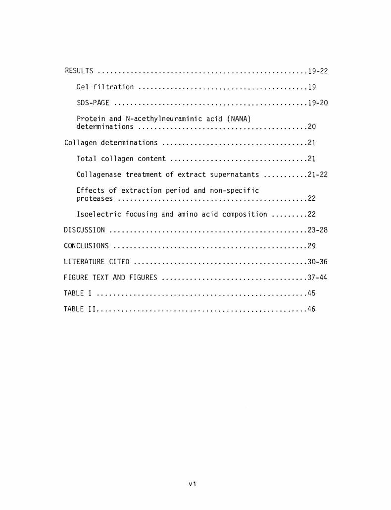
RESULTS 1·9-22
Gel filtration 19
SOS-PAGE 19-20
Protein and N-acethylneuraminic acid (NANA)determinations 20
Collagen determinations 21
Total collagen content 21
Collagenase treatment of extract supernatants 21-22
Effects of extraction period and non-specificproteases 22
Isoelectric focusing and amino acid composition 22
DISCUSSION •.•••...•.•...••...••••......•••....•••...••.•••• 23-28
CONCLUSIONS ••••••..••........•••....••..•..•.••....•...•••• 29
LITERATURE CITED ...••....•...•......•.......•..•.....•..••. 30-36
FIGURE TEXT AND FIGURES 37-44
TABLE I 45
TABLE II 46
vi

The purpose of
INTRODUCTION
this study was to identify,
characterize, and partially purify certain highly soluble
glycoproteins from experimental wounds in rabbits. An
attempt was made to determine whether the changing
concentrations of such substances could be used to
objectively monitor the repair process.
Soluble glycoproteins derived from open wound
granulation tissues have not been studied previously; but,
comparable substances have been identified in extracts from
polyvinyl sponge-induced inflammatory granulomas. 1- 5 In
granulomas, these substances experience predictable changes
in concentration as the tissue ages.
In the present work, soluble glycoproteins were
extracted from open wound granulation tissues at increasing
postwound intervals and were monitored and characterized by
methods used previously for the study of granuloma-derived
granulation tissues. The major purification technique
involved gel filtration using Sephacryl SF 200. Crude and
purified preparations were characterized by sodium dodecyl
sulfate (SDS)-polyacrylamide gel electrophoresis (PAGE),
isoelectric focusing (IEF), amino acid analysis,
glycoprotein susceptibility to a monospecific clostridial
collagenase, and measurement of soluble collagen, N-
acetylneuraminic acid (NANA), and protein. The use of these

techniques allowed the investigator to: 1) establish if
wound-derived soluble substances could be used to monitor
repair over a given interval, and 2) determine if open-wound
granulation tissue profiles resemble those from other tissue
sources (i.e., inflammatory granulomas). If soluble
glycoprotein profiles could be confirmed as having
characteristic configurations for wounds of specified age,
they would have value as objective reflections of healing
status.
2

LITERATURE REVIEW
Background
Normal healing of open full-thickness wounds of
mammalian integument occurs by the process of repair rather
than by true regeneration. 6,7 As a result, the tissues of
healed open wounds are neither structurally nor functionally
identical to those of the pre-injury state. The inevitable
scarring associated with normal healing can produce severe
impairments. 6,7 Efforts at directing repair so as to limit
scarring and contraction or to facilitate re-
epithelialization have been hampered by lack of objective
methods for assessing the effects of new agents or
techniques on healing processes. This deficiency is made
more apparent when healing occurs abnormally; aberrations in
wound repair are relatively common. 6,8 Keloids and
hypertrophic scars, for example, are debilitating
derangements to which open wounds
susceptible. 6,8 In addition, failure
are particularly
of wounds to re-
establish surface integrity and functional actiVity due to
infection, nutritional deficit, systemic disease, or drug
therapy has been well-documented. 6 By the time such
abnormalities become clinically eVident, effective
therapeutic intervention may be precluded. If wounds that
are at risk of healing abnormally could be identified during
incipient aberrancy, and if early treatment could be
3

initiated, a return to normal patterns of repair might be
accomplished. Objective criteria for monitoring wound
repair would also allow the effects of various wound
treatments to be more appropriately evaluated.
4

Reasons fQL Studying Buffer-Solubl~ Glycoprote1n~ fLQmOpen-W2Ynd Granulation Tissues
Numerous methods have been suggested for enhancing the
rate or quality of wound repair 9- 12 , but few methods have
found application in clinical practice. This disparity may
result from using sUbjective clinical criteria for
monitoring the healing process and from inevitable
investigator bias. Attempts have been made at more
objectively quantitating healing in experimental animals by
measuring rates of re-ePithelization 13 , wound
contraction,14-16 connective tissue reconstitution,16-17
collagen synthesis, 18-20 glycosaminoglycan (GAG)
production,21-23 and determination of fibroblast growth
kinetics;24 but, because species variation is significant,25
the relevance of such research to human open-wound repair
remains unclear. Certainly, methods of analysis which
require surgical removal of wound tissues are rarely
suitable for studying healing in humans. This investigation
is an experimental animal study which evaluates the
contention that highly soluble substances are present in
open wound tissues and can be used to accurately monitor the
healing process.
The existence of wound-derived soluble glycoproteins
whose concentrations change as a function of postwound
interval has never been established; but, similar substances
5

have been reported for granulation tissues of non-wound
origin (i.e., inflammatory granulomas).1-5 The close
resemblance between granulation tissue derived from
inflammatory granulomas and that from open, full thickness
skin wounds,26 justifies the speculation that a similar
assortment of glycoproteins would be identified.
Buffer-soluble granuloma glycoproteins were originally
studied to assess changes in cellular function mediated by
exposure of cells to extracellular matrix constituents. 1
When rat granuloma tissue extracts were added to cultures of
matrix-free embryonic chick tendon cells, a dramatic
suppression of collagen and other protein synthesis
occurred. 1,2 In addition, the secretion of collagen into the
medium was diminished and the normal tendency of tendon
cells to cluster during incubation was inhibited. 1,2
Another biological effect was a 10-fold drop in phagocytic
index when peritoneal macrophages were exposed in vitro to
extracts prepared from 14- and 42-day polyvinyl sponge
granulomas. 3 Subsequently, efforts at isolating and
identifying the active components of such extracts revealed
that granuloma age was
specific gel filtration
accurately
2patterns.
reflected by highly
The characteristic
changes in elution profiles observed as granuloma tissues
matured led to speculation that soluble glycoproteins play
an active role in directing tissue development. 2
6

Soluble Extracellular Granulation Tissu~ ~nstituents
~ Regulatory Molecules
Feedback regulation of reparative tissue development
has been identified as an important function for the soluble
glycoproteins of inflammatory granulomas. 2 ,27 After crude
extracts of granuloma granulation tissues were shown to
inhibit collagen synthesis and secretion,1,2 subfractions
were isolated which exhibited inhibition at extremely low
concentrations Among the many soluble
extracellular substances that might function in this
capacity, two categories of molecules, NANA-containing
glycoproteins and glycosaminoglycans (GAG), have received
particular attention.
NANA was originally implicated as a regulator of cell
function when normal and transformed cells were shown to
have different total sialoprotein contents. 28 Changes in
the synthesis/secretion of collagen and other proteins as
well as alterations in cell surface attachment mechanisms,
cellular aggregation phenomena, lymphocytic circulation,
expression of cellular antigenicity, and sensitivity of
smooth muscle cells to serotonin-induced contraction have
all been envisaged as being NANA-dependent. 29 - 33 A
neuraminidase-sensitive glycoprotein involved in collagen
synthesis and in phagocytosis has also been described. 34
Such research has been used to support the conclusion that
NANA-containing glycoproteins exert potentially important
7

influences on the changing developmental state of
granulation tissues. 2
GAG have also been envisaged as being important for
modulating cellular behavior in deve1opmenta1 35- 42 and in
21-23wound healing systems. In developmental models such as
newt limb regeneration 35, chick embryo cornea formation,36
limb bud and axial chrondogenesis,37 and chick heart and
brain deve1opment,38,40-42 the unsu1fated GAG, hyaluronic
acid, has been found to facilitate cellular division and
SUbsequent hyaluronate removal by
migration while
differentiation. 35 ,36,39
directly suppressing
tissue hyaluronidase causes a cessation of migration and
permits aggregation and differentiation to proceed in the
proper sequence for tissue organization. 35 ,42 In
healing open wounds, identical changes in GAG composition
occur. 21 - 23 In addition, a wound associated hyaluronidase
differentiation
whose activity correlates with the onset of
has been identified. 21 ,22 These
cellular
studies
support an analogy between reparative and developmental
systems and lend credence to the hypothesis that the
synthesis and degradation of hyaluronic acid help to control
and
explain
differentiation,proliferation,cellular migration,
synthesis. 35 - 42 Possible mechanisms to
hyaluronate's effects have been advanced by Ivaska. 5
Predicted changes in the concentrations of readily
extractable glycoproteins of developing granulation tissues
8

were used in this study to monitor the progression of normal
repair in experimental wounds. Since soluble glycoproteins
from open-wound tissues have not been studied previously, an
analogy between the open wound -and the granuloma systems
needed to be verified. This study sought to confirm
that soluble glycoproteins could be isolated from open wound
granulation tissues and that time-dependent changes in
soluble glycoprotein composition would occur.
9

MATERIALS AND METHODS
Woynding
Forty 2.0-3.0 kg New Zealand white rabbits (Oryctolagus
cuniculus) were caged separately and maintained on a Purina
laboratory Rabbit Chow HF 5326 diet (Ralston Purina) with
food and water taken ~ liQ. For wounding and harvesting
procedures, rabbits were anesthetized by intramuscular
injection of ketamine HCI (Bristol), 35 mg/kg and xylazine
(Haver-Lockhart), 5 mg/kg and flanks were shaved and
depilated with Nair (Carter-Wallace Inc.) Bilateral flank
wounds, 6 em x 6 em were made in all animals (Fig. 1). Skin
and superficial fascia were excised to the level of the
panniculus carnosus muscle with resected skin being frozen
and retained. Flank dressings consisted of petrolatum
gauze, gauze roll, and a light plaster cast.
Sampling
A differential wound topography permits recognition of
two distinct zones. 20- 22 Most of the open defect contains a
prOVisional tissue referred to as "central granulation
tissue". Along the wound's periphery, between central
granulation tissue and the original unwounded skin, a bi
partite zone exists which is composed of a deep layer of
granulation tissue and a superficial sheet of migrating
10

epithelium. This bi-layered region constitutes "whole edge
tissue". All samples were harvested from the central
granulation tissue region and were free of peripheral
epithelium. Sampling was performed on postwound days 3, 7,
9, and 14. For each animal, the tissues from the left and
the right wounds were pooled and extractions subsequently
performed. Excision of underlying panniculus carnosus muscle
was avoided at the time of harvesting.
Grafting
Skin grafting 1s a common therapeutic maneuver believed
to produce a significant alteration in patterns of wound
repair. Two animals were, therefore, studied for which any
changes in soluble glycoprotein profiles induced by the
application of full-thickness autogenous skin grafts could
be defined. These animals received a unilateral 6 em x 6 em
flank wound that was dressed as described above. On the
seventh day after wounding, the opposite side was wounded
and the freshly harvested skin applied as a full-thickness
graft to the week-old wound. The deep surface of the graft
was defatted to expose clean dermis, this procedure greatly
enhances the success of grafting. 43 Abdominal skin was
avoided as a source of graft material because resultant
closure of such donor sites creates undesirable tensions on
the flanks and might retard the natural course of
contraction. Petrolatum gauze, gauze roll, and plaster cast
1 1

were again used as dressings. On the fourteenth day after
original wounding (7 days after grafting), grafts were
removed and sUbjacent granulation tissue harvested; results
of previous work show that translocation of granulation
tissue to grafts is minimal. 43 The donor site was allowed
to heal for a total of 14 days and then its granulation
tissue harvested as described for open wounds.
Tissue Preparation, Storage, anQ Extraction
Harvested tissues were frozen in liquid nitrogen,
lyophilized, milled and then stored at 40 C until required.
Extl~acts were prepared by dissolVing 100 mg of milled tissue
powder in 4.0 ml of 0.05 M Tris (hydroxylmethyl)
aminomethane-HCl (Tris-HC1), 0.005 M CaC1 2 , pH 7.6, buffer
and gently stirred at 4° C for 16 hrs. as previously
described. 1,2 Samples were then centrifuged at 14,700 X g
for 5 min., insoluble pellets discarded, and the
supernatants saved for analysis. An identical extraction
was used for normal skin. Normal rabbit serum was analyzed
after dialysis against the extraction buffer. Data for
norma] skin and serum were compared with those obtained for
granulation tissues.
All samples were studied by means of gel filtration,
sodium dodecyl sulfate (SDS)-polyacrylamide
12
gel

electrophoresis (PAGE), isoelectric focusing, amino
analysis (for fractions of sufficient purity),
measurement of soluble collagen, hydroxyproline,
acetylneuraminic acid (NANA), and protein.
13
acid
and
N-

Analytical Technigues
~ Filtration
Gel filtration was performed by introducing a 500 ul
aliquot of each crude extract to a 1.6 cm x 90 cm column of
Sephacryl SF 200 (Sigma) which had been equilibrated with
the extraction buffer. Elution was performed with the same
buffer at a flow rate of 0.66 ml/min. Effluent was
monitored at 280 nm with a Buchler Fracto-Scan UV monitor;
3.2 ml fractions were collected. Peak fractions were
dialyzed against cold distilled water, lyophilized, and
stored for later analysis.
Elution profiles were described on the basis of peak
elution volumes. Changes in the profiles were expected to
reflect the changing developmental state of the granulation
tissue.
Electrophoresis
Crude extract supernatants as well as peak fractions
obtained by gel filtration were electrophoresed on 1~ SDS
-5.6% polyacrylamide gels as described by Fairbanks, Steck
et. al. 44 Staining for protein was performed with 0.05%
coomassie blue and for carbohydrate with a periodic acid
Shiff (PAS) procedure. 44 All gels were photographed and
then scanned with a Corning model 760 computing
densitometer. Protein standards consisting of lysozyme,
14

a-lactoglobulin, ferritin, ovalbumin, bovine serum albumin,
and a-galactosidase were used. Electrophoretic patterns
allowed molecular weights, approximate carbohydrate
contents, and relative purity to be determined.
Densitometric scans of gels were compared for protein
migration distances and for differences in densitometer
computed band areas.
~lectrlc fQQ.y.UnK (lEF)
Isoelectric focusing was performed as described by
Righetti and Drysdale45 using a 40% ampholin~ solution (Bio
Lyte 3/10) having a gradient of pH 4-9. Staining was
performed with coomassie blue and all gels were then
scanned. Using pI calibration standards (Pharmacia),
isoelectric points over the range pH 4-9 were determined
for purified extract subfractions. Since previous
worl{ has established the isolectric points and molecular
weights of soluble glycoproteins of inflammatory
granulomas,3 such data provided another ~asis for comparing
extracts from open wound granulation tissues and from
granulomas. Changes in isoelectric patterns occurring as a
function of postwound interval would, concej.vably, be useful
for monitoring the emergence and/or disappeal~ance of various
proteins as wound gr~nulation tissue matures and remodels.
15

Amino ~d Analysis
Those fractions that could be purified sufficiently so
as to yield a single band by SDS-PAGE were sUbjected to
amino acid analysis. Analyses were performed after
hydrolysis at 1100 C in 6 N Hel for 24 hrs. in evacuated
nitrogen-flushed tubes by the University of Connecticut
Health Center Amino Acid Analysis Facility.
Amino acid compositions of wound-derived soluble
glycoproteins were used in conjunction with SDS-PAGE and IEF
in an attempt at protein identification. Comparisons were
made between experimental substances and glycoproteins of
known composition.
Collagen Determinations
SolUble, collagen-derived peptides present in crude
extract supernatants or in isolated subfractions were
identified by evaluating extract protein susceptibility to
collagenase. Hydroxyproline content was also measured.
Samples were treated with a purified monospecific
clostridial collagenase (EC 3.4.24.3) (Millipore) and
resulting alterations in 5DS-PAGE patterns ide~tified.
Samples were divided into fractions of 250 ul; each received
162.6 ~g of N-ethylmaleimide (NEM) to inhibit nonspecific
protease activity. Half of the samples were then treated
• i- ...W1. v.1 23.2 ug of active collagenase and the remainder with
the same amount of heat-inactivated collagenase. All
16

r ti i t incubated at 37° Ceac on m x ures were for 2 hrs.
Collagenase-mediated changes in electrophoretic patterns
were monitored using the SDS-PAGE technique for collagenous
polypeptides of Noelken, Wisdom et. al. 46
Hydroxyproline levels were determined for all samples
by the alkaline hydrolysis of Huszar, Maiocca et.al. 47
~fects Qf Extraction Period
To assess the effects of varying the duration of wound
tissue extraction periods on gel filtration elution profiles
and on gel electrophoretic patterns, extract supernatants
derived from wound granulation tissue aged 3, 7, 9, and 14
days ~ere incubated in the Tris-HCl extraction buffer at 40
C for 4, 8, 12, and 16 hours respectively. Any differences
arising in the profile or electrophoretic pattern for a
given tissue reflected the influence of extraction duration.
Inhibition Qf Non-Specific Protease Activity
For each of the extract supernatants derived from the
wound tissues described above, N-ethylmaleimide (NEM) was
added to a final concentration of 2.5 mM for the purpose of
inhibiting non-specific protease activity. Resultant
elution profiles and electrophoretic patterns were then
studied and compared with NEM-untreated extracts.
17

Measurement Qf NANA, and Protein
NANA levels were estimated by the method of Aminoff. 48
Samples and standards contained in a volume of 0.5 ml were
incubated with 250 ul periodate reagent at 370 C for 30 min.
Sod i.urn arsen i te (200 ul) and th10barb i tur 1c ac id reagen t (2
ml) were added and samples and standards heated at 1000
C
for 7.5 minutes. Acid-butanol (4 ml) was added, mixtures
centrifuged, and NANA-chromophore read in the butanol layer
at 549 nm.
Lowry protein was measured using the modified folin
phenol reagent method reported by Peterson. 49
Statistical ~~~
Data for assays of tissue samples pooled according to
postwound interval or graft status were expressed as means +
one standard error of the mean (S.E.M.). Statistical
significance was determined by applying t-tests for
independent variables to the difference between means.
S i g n i f i canc e was ass i g ned at alevel 0 f p rob abiIi t y ~ 0 • 0 5 •
For gel filtration, PAGE, arld IEF experiments,
re9resentative elution profiles and gels are presented.
18
RESULTS
~ Filtration
A time-dependent change in the elution profile of
supernatants derived from rabbit skin granulation tissue was
observed (Fig. 2). Two major peaks (A and C) were seen for
each day studied, but the relative proportion of the profile
attributable to a given peak varied. Peak A was the most
prominent constituent of days 3, 9, and 14, and possessed an
approximate molecular weight of 93,000 (Fig. 3). This peak
was markedly diminished on postwound day 7, but increased
progressively throughout the second postwound ~eek. A minor
peak (E) was observed in less mature granulation tissue
(postwound days 7 and 9) and was particularly evident in
grafted granulation tissue (Figs. 2 and 4). Peak C had an
elution volume consistent with a population of substances of
differ'ing molecular weights. This possibility is suggested
by the eventual emergence of 3 discrete peaks 1n the C
position at 14 days.
SDS--PAGE
SDS-PAGE patterns of tissue extracts exhibited a
surprising degree of similarity ,·egardless of postwcund day
(Fig. 5). At least five comparable bands were identified
fo~ each extract, however, densitometric scanning
demonstrated important differences (Fig. 6). Although
19

band 3 was invariably the major component, its relative
predominence was markedly diminished on day 7 when it
comprised only 27~ of the scan (Fig. 7). Subsequently, the
the percentage of the scan attributable to band 3
progressively inoreased, reaching a maximum on day 14 of
59.1% (Fig. 7). When Sephacryl SF 200 Peak A was isolated
and electrophoresed, a single homogenous band was identified
which corresponded to band 3 of the crude extracts (Fig. 5).
The other SDS-PAGE bands (1, 2, 4, and 5) constituted
relatively low percentages of the scan on each postwound
day. Band 5 peaked on postwound day 7 when it comprised
15.4% of the scan. Bands 1, 2, and 4 were essentially
unaffected by increasing postwound intervals (Fig. 7). When
gels were stained for carbohydrate, all the coomassie
staining bands were PAS positive.
~tein an~ ~etylneuraminic~ (NANA) DeterminatiQn~
The described changes in elution profiles and 5DS-PAGE
patterns were accompanied by increasing concentrations of
extractable protein throughout the healing process (Table
I). NANA levels were uniformly low on all postwound days
except day 7 when the concentration of extractable NANA was
4- to 6.5-fold greater than at any ether postwound interval
(Table I). Isolated Peak A material was found not to
possess significant amounts of NANA.
20

Collagen Determinations
Iotal Collagen Content: An estimate of total collagen
content was made for uncentrifuged crude extracts by
measuring the release of collagen cleavage products after
treatment with purified, monospecific clostridial
collagenase. Release of cleavage products increased in
direct proportion to collagenase concentration until a
plateau was reached at 8.9 ug/mg dry tissue (Figure 8).
Collagen measured in this manner was found to be negligible
at postwound day 3, but increased thereafter, peaking on
day 9 and remaining elevated until day 14 (Fig. 8).
Although grafting appeared to diminish the collagen content
of subjacent granulation tissue, the difference between
grafted and ungrafted tissue samples was found to be
statistically insignificant (p ~ .05).
~lagenase Treatment Qf Extract ~~a~s; No
differences in electrophoretic patterns were observed
between samples treated with active or heat-inactivated
clostridial collagenase. Similarly, SDS-PAGE of samples
incubated with N-ethylmaleimide (NEM) to inhibit non
specific protease activity were indistinguishable from those
that were NEM-untreated.
Hydroxyproline determinations of extra~t supernatants
were also performed. For the periods studied, variably low
levels of hydroxyproline were e~tracted (Table I) that did
21

not reflect total wound collagen content (Fig. 8).
Effects Qf Extraction Period ~ Non-Specific Proteases
Under the conditions of the experiments (4 0 C), neither
varying the extraction time over a range of 4-16 hours nor
inclusion of NEM in extraction mixtures produced any
detectable change in gel filtration profiles or
electrophoretic patterns.
IsoelectrtQ Focusing an~ AminQ ~id ComgQsition: Only
Peak A (Band 3) could be isolated with sufficient purity to
justify IEF and amino acid composition determination. The
isoelectric point was found to be 6.9; the amino acid
composition is shown in Table II.
22
DISCUSSION
The elution profiles of open wound granulation tissues
changed as a function of postwound interval. The pattern of
change was dramatic for Sephacryl SF 200 Peak A. Peak A was
present in high concentrations on postwound day 3, was
markedly suppressed on postwound day 7, and then
progressively increased during the second postwound week
(days 9 and 14).
When isolated Peak A glycoprotein was studied by gel
electrophoresis, it was identified as a single, homogeneous
protein corresponding to the third band in elect~ophoretic
patterns of crude extract (Fig. 5, compare gels 1-13 with
gel 14). By performing densitometry on each gel, the
proportion of the pattern attributable to Band 3 could be
determined at increasing postwound intervals (Figs. 6 and
7). By this method, Band 3 was shown to vary over time in a
manner identical to that seen for Peak A by gel filtration,
I.e., initially high levels of Peak A on day 3 were
depressed on day 7 and progressively increased on days 9 and
14 (compare changes in Peak A, Fig. 2 with changes in band
3, Fig. 7).
The cause of Peak A/Band 3 (PA/B3) depression at the
end of the first postwound week is unknown, but the
observation that NANA was increased on day 7 may be
significant. NANA-containing glycoproteins are believed to
23

29exert numerous important effects upon cellular function.
33 They undergo marked changes in concentration as tissues
2 50age,' and they may function in the feedback regulation of
granulation tissue development. 2 Purified NANA-containing
glycoproteins have been shown to suppress protein and
collagen synthesis/secretion at very low concentrations
(10-6 M).2 Since Bands 1,2,4, and 5 were not diminished on
day 7, the nature of PA/B3 depression may be rather
specific, whatever its ultimate cause. Importantly, these
studies assessed protein content, not rate of synthesis, so
subtle changes in protein synthesis and/or secretion might
exist on other postwound days that would not be recognized
by tile methods employed.
Impressive changes in Peak C were also observed; Peak C
gradually increased in quantity and eventually separated
into three well-defined peaks (Fig. 2). This phenomenon may
indicate a time-dependent structural modification such as
a·gg,re"ga·tionj the concorni tant formation of small er, Peak C-
de~ived remnants may also occur. The inability to
distinguish differences in Peak C patterns by 5DS-PAGE over
time might result from reduction of disulfide bonds by
dithiothreitol and disruption of Peak C into its essential,
non-agg~egated components. In any event, the emergence of
sev~ral discrete peaks in the C position of 14-day elution
profiles implies possible value for Peak C as a monitor
substance.
24

The identity of PA/B3, Peak C and t~e other soluble
glycoproteins remains unknown. Since electrophoresis
patterns were not altered when extracts were treated with
collagenase, none of the major bands appear to be collagen
derived peptides. The absence of hydroxyproline from PA/B3
and the entirely different pattern of time-dependent change
observed for PA/B3 when compared to total wound collagen
argue against collagen as a major soluble constituent.
Soluble glycoproteins from open-wound granulation
tissues have not been studied previously, but similar
substances of non-wound granulation tissue origin have been
reported. 1-3 These earlier works may provide insight
into the nature of the materials described by this study.
Bole, Jourdain et. al. 3 isolated a saline soluble
glycoprotein from 14- and 42-day polyvinyl sponge granulomas
in rats by chromatographic fractionation of extracts on
DEAE-cellulose and Bio Gel P-150. A protein peak identified
as DEAE-celluJose C/Bio Gel P-150 II exhibited
chromatographic and electrophoretic characteristics similar
to PA/B3, but was much smaller (MW=48,500). The material
isolated by Bole, Jourdain et. al. 3 significantly depressed
phago~yto3is by peritoneal macrophages in culture.
Similarly, Aalto and Kulonen 1 described a 0.3 M Tris-HCl,
0.0015 M CaCl, pH 7.8, extract from viscose sponge-induced
inflammatory granuloma tissue that suppressed the synthesis
25

of protein and collagen by embryonal chick tendon cells.
Subsequently, a 0.5 M Tris-Hel, 0.02 M EDTA, 0.075 M boric
acid extract of rat granuloma tissue was identified that had
the same effect and that possessed a gel filtration pattern
which accurately reflected granuloma age. 2 When Aa1to,
Poti1a et. a1. 2 subjected these 7- and 21-day granuloma
extracts to Sephadex G-200 gel chromatography, they
identified three peaks (designated Sephadex G 200/1, II, and
III) that appear identical to Sephacryl SF 200 Peaks A, B,
and C obtained by the present analysis (Fig. 2). Sephadex G
200/1 and PA/B3 occupy similar chromatographic elution
positions and both increased with advancing age. 2
Isolation of
cellulose anion
Sephadex
exchange
G 200/1 followed by
chromatography2 produced
DEAE-
t~o
protein peaks which eluted at 0.15 M NaCl, a major ~rotein
subfraction (DEAE 1) and a lesser protein subfraction (DEAE
2)~ Although high levels of NANA were associated with
Sephadex G 200/1, NANA was confined exclusively to the
lesser subfraction (DEAE 2). The major subfraction (DEAE 1)
was totally free of NANA and appears to be comparable to
Sin~e the specific identification of the soluble
glycoproteins de~ived from open wound granulation tissue or
from rat granulation tissue has not been accomplished, the
possibility of a serum origin for such substances must be
26

considered. The probability of simple serum contamination
of tissue samples seems small since such contamination would
not explain the changing proportions of soluble constituents
observed as the wounds matured. In addition, the
predominant serum protein, albumin, is free of carbohydrate
and possesses a sUbstantially lower molecular weight
(63,900) than the predominant PAS-positive glycoprotein
identified in wound extracts (PA/B3 MW=93,000). Naturally,
PA/B3 could represent a higher molecular weight serum
component such as a or a macroglobulins or
immunoglobulins, or even albumin functioning as a car-ri.er
and bound to some other glycoprotein. Certainly, the amino
acid composition and isoelectric point of PA/B3 is rather
a1 bumino id • In any even t, a serum 0 r ig i 11 for this
substance would in no way invalidate its use in monitoring
the progression of normal repair given a consistent
relationship between the healing process and PA/B3
concentration. The selective uptake of serum components by
maturing wound tissues could provide a basis for the
observed effect.
The major change provoked in gel filtration patterns by
the application of skin grafts appears to be an accentuation
of Peak B. This phenomenon might be t~e result of a
retention of less mature tissue characteristics since Peak B
was more prominent in tissues harvested from younger wounds
(Days 7 and 9). In any event, the limited graft study of
27

this project supports the contention that a method known to
dramatically alter the normal healing process (i.e.,
grafting) also produces distinct changes in glycoprotein
profiles.
28
CONCLUSIONS
1) The profiles of soluble substances identified in
this study varied in a characteristic manner as a function
of postwound interval. Such profiles have apparent value as
objective reflections of healing status.
2) One soluble substance, Peak A/Band 3 (PA/B3) was
identified a~ a single, non-collagenous PAS-positive
protein, having a molecular weight of approximately 93,000
and an isoelectric point of 6.9.
3) Cullagen was not a significant component of
soluble extracts.
4) Increased levels of N-acetylneuraminic acid on day
7 may be related to an observed suppression of PA/B3.
5) Peak C substances also varied over time and may
have value as monitor substances.
6)
procedure
provoked
profiles~
Full thickness, autogenous skin grafting (a
known to dramatically alter wound healing)
distinct changes in soluble substance elution
29

LITERATURE CITED
E.: Inhibition of protein
cells by extracts from
tisue. F.E.B.S. Lett.,
1 • Aalto, M., and Kulonen,
synthesis in tendon
experimental ganulation
49:70, 1974.
2. AaIta, M., Potila, M., and Kulonen, E.: Glycoproteins
from experimental granulation tissue and their
effects on collagen synthesis in embryonic chick
tendon cells. Biochem. Biophys. Acta, 587:606,
1979.
3. Bole, G.G., Jourdain, G.W., and Wright, J.E.:
Isolation and chemical characterization of a
granuloma glycoprotein that inhibits macrophage
phagocytosis. J. Lab. Clin. Med., 86:1018, 1975.
4. Rajamaki, A., and Kulonen, E.: Purification of a
structural glycoprotein from collagenase-digested
experimental granuloma. Biochem. Biophys. Acta,
243:398, 1971.
5. Ivaska, K.: Effect of extracellular glycosaminoglycans
on the synthesis of collagen and proteoglycans by
granulation tissue cells. Acta Physiol. Scand.
Suppl., 494, 1981.
6. Peacock~ E.E., and Van Winkle, w.: HQyn~ ~~~ 2nd
Ed., W.B. Saunders Co., Philadelphia, 1976.
30

7. Bertolami, C.N.: Oral Wound Repair. In: Clinical
Dentistry, J.W. Clark (ed.), Harper and Rowe
PUblishers, Philadelphia, 1982.
8. Ketchum, L. D. , Cohen, I.K., and Master, F.W.~
Hypertrophic scars and kelo1ds.
Surg., 53: 140, 1974.
Plast. Reconstr.
9. FaUlk, W.P., Stevens, P.J., Burgess, H., Matthews, R.,
Bennett, J.P., and Hsi, B.L.: Human amnion as an
adjunct in wound healing.
1980.
Lancet, 8179:1156,
10. Calver, R.F., and Stanley, J.K.: Enhancement of
secondary wound healing by local tissue nutrition.
Clin. Trials J. (Lond), 17: 144, 1980.
11. Shafer, W.G., Beatty, R.E., and Davis, w.B.: Effect of
dilantin sodium on tensile strength of healing
wounds. Proe. Soc. Exp. BioI. Med., 98:348, 1958.
12. Ringsdorf, W.M., and Cheraskin, E •.. Vitamin C and
wound healing. Oral Surg •., 53:231, 1982.
13. Krawczyk, W.S.: A pattern of epidermal cell migration
during wound healing.
1971.
J. Cell BioI., 49:247,
14. Abercrombie, M.. , Flint, M.H., and James, D.W.:
Collagen formation and wound contraction during
repair of small excised wounds in the skin of
rats. J. Embryol. Exp. Morph., 2:264, 1954.
31

15. Abercrombie, M., James, D.W., and Newcombe, j.F.:
Wound contraction in rabbit skin studied by
splinting the wound margins. J. Anat. (Lond.),
94: 170, 1970.
16. Grillo, H.e., Watts, G.T., and Gross, J.: Studies in
wound healing. I. Contraction and the wound
contents. Ann. Surg., 148: 145, 1958.
17. Grillo, H.C.: Aspects of the origin, synthesis, and
evolution of fibrous tissue in repair. In:
Adyances in Biology Qf ~ Skin. Pergamon Press,
London, 5: 128, 1964.
18. Diegelman, R.F., Rothkopf, L.C., and Cohen, I.K.:
Measurement of collagen biosynthesis during wound
healing. J. Surge Res., 19:239, 1975.
19. Stein, H.D., and Keiser, H.R~: Collagen metabolism in
granulating wounds. J. Surg. Res., 1 '1 : 22'7, 1971.
20. Bertolami, C.N., and Donoff, R.B.: The effect of skin
grafting upon prolyl hydroxylase and hyaluronidase
activities in mammalian wound repair. J. Surge
Res., 27:359, 1979.
21. Bertolami, C.N., and Donoff, R.B.: Iiyaluronidase
activity during open wound healing in rabbits: A
preliminary report. J. Surge Res., 25:256, 1978.
22. Bertolami, C.N., and Donoff, R.B.: Identification,
characterization, and partial purification of
mammalian skin wound hyaluronidase. J. Invest.
32
Derm., 79:417, 1982.
23. Alexander, S.A., and Donoff, R.B.: The
Proc.
glycosaminoglycans of open wounds. J. Surge Res.,
29:422, 1980.
24. Diegelmann, R.F., Cohen, I.K., and McCoy, B.J.: Growth
kinetics and collagen synthesis in normal skin,
normal scar, and keloid fibroblasts in yitrQ. J~
Cell Physiol., 98:341, 1979.
25. Grillo, H.C., and Gross, J.: Studies in wound healing.
III~ Contraction in vitamin C deficiency.
Soc. Exp. BioI. Med., 101:268, 1959.
26. Gabbiani, G., Hirschel, B.J., Ryan: G.B., Statkov,
P.R., and Majno, G.: Granulation tissue as a
contractile organ: A study of structure and
function. J. Exp. Med., 135:719, 1972.
27. Aho, S., and Kulonen, E.: Effect of silica-liberated
macrophage factors on protein synthesis in cell
free systems. Exp. Cell Res., 104:31,197'7.
28. Ohta, N., Pardee, A. B. , McAuslan, B.R., and Burger,
M. M. : Si81ic acid contents and controls of normal
and malignant cells. Biochem. Bioph,y.s. Acta,
158:98, 1968.
29. Ginsburg, V• , and Neufeld, E: • F • : Complex
heterosaccharides of animals. Ann. Rev. Biochem.,
38:371, 1969.
33
30. Winzler, R.J.: Carbohydrates in cell surfaces. Int.
Rev. Cytol., 29:77, 1970.
31. Spiro, R. G. : Glycoproteins. Ann~ Rev. Biochem.,
39:599, 1970.
32. Marshall, R.D.:
41 :673, 1972.
Glycoproteins. Ann. Rev. Biochem.,
33. Rosenberg, A., and Shengrund, e.L. (eds.): Biological
Roles of Sialic Acid, p. 375, Plenum Press, New
York, 1976.
34. MII. , Ronnemaa, rr
.L • , and Kulonen, t:' •1:1 •• Effect of
neuraminidase of the synthesis of collagen and
other proteins.
1974.
Biochem. Biophys. Acta, 342:247,
35 • Toole, B • P • , and Gr 0 s s, J.: The ext rae e11u1arma t r~ i x
of the regenerating newt limb: Synthesis and
removal of hyaluronate prior to differentiation.
Dev. BioI., 25: 57, 1971.
36. Toole, B.P", and Trelstad, Hyaluronate
production and removal during cornea development
in the chick. Dev. BioI., 26:28, 1971~
37. Toole, B.P.: Hyaluronate turnover during
chondrogenesis in the developing chick limb and
axial skeleton. Dev. BioI., 29:321, 1972.
38. Orkin, R.W~, and Toole, B.P.: Hyaluronidase activity
and hyaluronate content of the developing chick
embryo heart. Dev. BioI., 66:308, 1978.
34

39. Toole, B.P., Jackson, G. t and Gross, J.: Hyaluronate
morphogenesis. Inhibition of chondrogenesis in
Vitro. Prac. Nat. Acad. Sci., 69:1384, 1972.
40. Orkin, R.W., Jackson, G., and Toole, B.P.:
Hyaluronidase activity in cultured chick embryo
skin fibroblasts. Biochem. Blophys. Res. Comm.,
77:132, 1977.
41. Polansky, J.R., Toole, B.P., and Gross, J.: Brain
hyaluronidase: Changes in activity during chick
development. Science, 183, 862, 1974.
42. Polansky, J.R., and Toole, B.P.: Hyaluronidase
activity during thyroxine-induced tadpole
metamorphosis. Dev. Bl01., 53:30, 1976.
43. Donoff, R.B., and Grillo, H.C.: The effects of skin
grafting on healing open wounds in rabbits. J.
Surge Res., 19:163-167, 1915.
44. Fairbanks, G., Steck, T.L., and Wallach, D.F.H.:
Electrophoretic analysis of the major polypeptides
of the human erythrocyte membrane. Biochem.,.
10:2606, 1971.
45. Righetti, P.G., and Drysdale, J.W.: Isoelectric
focusing - Laboratory techniques in biochemistry
and molecular biology. Work, r.s., and Work, E.
(eds.), 5:337-590, North Holland, 1976.
46. Noelken, M.E., Wisdom, B.J., and Hudson, B.G.:
Estimation of the size of collagenous polypeptides
35

by sodium dodecyl sulfate - polyacrylamide gel
electrophoresis. Anal. Biochem., 11:131, 1981.
47. Huszar, G., Maiocco, J., and Naftolin, F.: Monitoring
of collagen and collagen fragments in
chromatography of protein mixtures. Anal
Biochem., 105:424, 1980.
48. Aminoff, D.: Methods for the quantitative estimation
of N-acetylneuraminic acid and their application
to hydrolysates of sialomucoids. Biochem. J.,
81: 384, 1961.
49. Peterson, G.L.: A simplification of the protein assay
method of Lowry et. ale which is more generally
applicable. Anal. Biochem., 83:346, 1977.
50. Lehtonen, A.: The mucopolysaccharides of aging
experimental granulation tissue. Acta Physiol.
Scand. Suppl., 310, 1968.
36

FIG. 1
A • F 1 a (l k W0 U tl d b 0 undar i e s •
B. Excision of skin and superficial fascia.
c. Open wound at the level cf the panniculuscarnOSLlS.
D. Incised granulation tissue.
37


FIG. 2
Elution profiles of soluble wound extract
constituents on Sephacryl SF 200 as a function
of time. Normal skin; 1.8mg, 3-day granulation
tissue; 2.0 mg, 7-day granulation tissue;
2.5 mg, 9-day granulation tissue; 2.9 mg,
14-day granulation tissue.
38

Ec
3.0
\\\\\
\. -"*"''''''' ,\\\\
\'""-
"'"'--
~~---4"'O---80-r-i---1.."..20---....160--'-2~60....-----2M)-r-iElution Volume (ml)
Normal Skin3d GT 1.8mg7d GT 2.0mg9d GT 2.5mg
14d GT 2.9mg

FIG. 3
Molecular weight determination of
Peak A/Band 3 (PA/B3) material.
39

0.8
0.6>eu~
0.4
0.3
0.2
0.1
"u..LYSOZymc
~ f3 -Lactoglobulin
~
Pepsin
Ovalbumin
Bovine Serum Albumin
PA/B3
f3 -Galactosidase
10 20 30 40 50 60 70 80 100
Molecular Weight >< 103

FIG. 4
Elution profiles of 2.9 mg, 14-day open-wound
granulation tissue and 3.8mg, 14-day grafted
granulation tissue.
40

Ec~ 2.N
tDoceu€o.,.0c(G)
~..,asCDa:
Vt
+
O~----y--------,..----y-----,.-----y------
Elution Volume (ml)
----14d GT 2.9mg-1 ~d grafted GT 3.8mg

FIG. 5
SDS-PAGE of wound tissue extracts.
A. Gels 1-4, 3-day granulation tissue;
Gels 5-6, 7-day granulation tissue;
Gels 7-8, 9-day granulation tissue;
Gels 9-10, 14-day granulation tissue (open);
Each gel contains 25-30 ul of crude tissue
extract.
B. Gels 11-13, 14-day gr·anulation tissue
(grafted) ;
Each gel contains 25-30 ul of crude tissue
extract.
C. Gel 14, isolated Sephacryl SF 200 Peak Aj
Gels 15-16, isolated Sephacryl SF 200 Peak Cj
Gels 17-19, overloaded crude extracts derived
from 7-, 9-, and 14-day open-wound tissues.
41

.,

granulation tissue extract
on SDS-polyacrylamide.
Representative
g·.day central
electrophoresed
FIG. 6
densitometric
42
scan of

(1)(.)cccs.0L-
oen.0«Q)
>
90 80 70 60 50 40 30 20 10 0
Migration Distance (mm)

FIG. 7
Time-dependent changes in electrophoretic
banding pattern of crude wound tissue
extracts. Abscissa: Postwound interval;
Ordinate: Percentage of scan attributable
to a given band.
43

70
c: 5«S()
en"t-
o(1) ........ Band 1(»
as ----- Band 2+oJ 30c Band 3(1)0 ~.-.- Band 4'-Q) Band 5a. 20
10- --: ::::-~-;:- -;:...,....,....... ..... .,,, ••..••• .....-..-......_.~:r"""e&,;;...- •...... .. -.
O-t----.,----r---.,.-----r'----3 14
Postwound Interval

FIG. 8
Total wound collagen content in grafted
wounds as a function of time. Due
to the large standard errors for open
and grafted wounds at 14 days, the
apparent difference between these wounds
on postwound day 14 was not statistically
significant.
44

J6
1.5
14
13
!2
"'0 11G)
• JO•G)
G) 9ex:C •G) 7...,0'- 6a.0 j
E4
J
2
0
0
f
WOUND COLLAGEN CONTENT
............ __ T
-............ I...............~
I•.L
2 J 4 .s 6 1 I 9 10 11 12 13 ,~
Post Wound Day
open
grafted

TABLE I
SOLUBLE PROTEIN, NANA, AND
HYDROXYPROLINE OF WOUND TISSUE EXTRACTS.
POSTWOUND LOWRY PROTEIN NANA HYDROXYPROLINEDAY (mg/ml extract) (ug ± SEM) (ug/mg dry tissue
± SEM)
~ 3.52 ± .02 0.20 ±.O4 0.34 ±.O8..)
7 3.89 ± .29 0.78 ±.O2 0.35 ±.O8
9 5.00 ± .38 0.12 ±.O4 0.55 ±.12
i4 5.76 ± .04 0.22 ±.O3 o~ 4!~ ±.10
14 (Graft) 7.55 ±.22 0.62 ±.12
45

TABLE II
Amino AQ14 Analysis a
~acryl Sf 2QQ ~ A/SDS-Page~ 3
Glu 115 Val 61 Tyr 45
Lys 111 Gly 57 His 38
Leu 107 Al·g 56 Iso 32
Asp 90 Thr 51 Meth 8
Pro 66 Ser 51 Hyl
Ala 62 Phen 50 Hyp
aResidues/1~OOO Residues
46