Embed Size (px)
Citation preview
Identification and Functional Analysis of Trypanosomacruzi Genes That Encode Proteins of theGlycosylphosphatidylinositol Biosynthetic PathwayMariana S. Cardoso1, Caroline Junqueira2, Ricardo C. Trigueiro1, Hosam Shams-Eldin3,
Cristiana S. Macedo4, Patrıcia R. Araujo1, Dawidson A. Gomes1, Patrıcia M. Martinelli5, Jurgen Kimmel3,
Philipp Stahl3, Sebastian Niehus3, Ralph T. Schwarz3, Jose O. Previato4, Lucia Mendonca-Previato4,
Ricardo T. Gazzinelli1,2, Santuza M. R. Teixeira1*
1 Departamento de Bioquımica e Imunologia, Universidade Federal de Minas Gerais, Belo Horizonte, Minas Gerais, Brazil, 2 Centro de Pesquisas Rene Rachou, Fundacao
Oswaldo Cruz, Belo Horizonte, Minas Gerais, Brazil, 3 Institut fur Virologie – AG Parasitologie, Philipps-Universitat Marburg, Marburg, Germany, 4 Instituto de Biofısica
Carlos Chagas, Universidade Federal do Rio de Janeiro, Rio de Janeiro, Rio de Janeiro, Brazil, 5 Departamento de Morfologia, Universidade Federal de Minas Gerais, Belo
Horizonte, Minas Gerais, Brazil
Abstract
Background: Trypanosoma cruzi is a protist parasite that causes Chagas disease. Several proteins that are essential forparasite virulence and involved in host immune responses are anchored to the membrane through glycosylpho-sphatidylinositol (GPI) molecules. In addition, T. cruzi GPI anchors have immunostimulatory activities, including the ability tostimulate the synthesis of cytokines by innate immune cells. Therefore, T. cruzi genes related to GPI anchor biosynthesisconstitute potential new targets for the development of better therapies against Chagas disease.
Methodology/Principal Findings: In silico analysis of the T. cruzi genome resulted in the identification of 18 genes encodingproteins of the GPI biosynthetic pathway as well as the inositolphosphorylceramide (IPC) synthase gene. Expression of GFPfusions of some of these proteins in T. cruzi epimastigotes showed that they localize in the endoplasmic reticulum (ER).Expression analyses of two genes indicated that they are constitutively expressed in all stages of the parasite life cycle. T.cruzi genes TcDPM1, TcGPI10 and TcGPI12 complement conditional yeast mutants in GPI biosynthesis. Attempts to generateT. cruzi knockouts for three genes were unsuccessful, suggesting that GPI may be an essential component of the parasite.Regarding TcGPI8, which encodes the catalytic subunit of the transamidase complex, although we were able to generatesingle allele knockout mutants, attempts to disrupt both alleles failed, resulting instead in parasites that have undergonegenomic recombination and maintained at least one active copy of the gene.
Conclusions/Significance: Analyses of T. cruzi sequences encoding components of the GPI biosynthetic pathway indicated thatthey are essential genes involved in key aspects of host-parasite interactions. Complementation assays of yeast mutants with theseT. cruzi genes resulted in yeast cell lines that can now be employed in high throughput screenings of drugs against this parasite.
Citation: Cardoso MS, Junqueira C, Trigueiro RC, Shams-Eldin H, Macedo CS, et al. (2013) Identification and Functional Analysis of Trypanosoma cruzi Genes ThatEncode Proteins of the Glycosylphosphatidylinositol Biosynthetic Pathway. PLoS Negl Trop Dis 7(8): e2369. doi:10.1371/journal.pntd.0002369
Editor: Paul W. Denny, Durham University, United Kingdom
Received April 18, 2013; Accepted July 1, 2013; Published August 8, 2013
Copyright: � 2013 Cardoso et al. This is an open-access article distributed under the terms of the Creative Commons Attribution License, which permitsunrestricted use, distribution, and reproduction in any medium, provided the original author and source are credited.
Funding: This study was supported by grants from Conselho Nacional de Desenvolvimento Cientıfico e Tecnologico (CNPq, Brazil), Coordenacao deAperfeicoamento de Pessoal de Nıvel Superior (CAPES, Brazil), Fundacao de Amparo a Pesquisa do Estado de Minas Gerais (FAPEMIG, Brazil), The Instituto Nacionalde Ciencia e Tecnologia de Vacinas (INCTV, Brazil), Deutsche Forschungsgemeinschaft (DFG), the Deutscher Akademischer Austauschdienst (DAAD, Germany) andBundesministerium fur Bildung und Forschung (BMBF, Germany). MSC, JOP, LMP, RTG and SMRT are recipients of CNPq fellowships; PS is a recipient of fellowshipsfrom Alumni Medizin Marburg, Roland und Elfriede Schauer-Stiftung and Studien-stiftung des Deutschen Volkes (Germany). The funders had no role in studydesign, data collection and analysis, decision to publish, or preparation of the manuscript.
Competing Interests: The authors have declared that no competing interests exist.
* E-mail: [email protected]
Introduction
Glycosylphosphatidylinositol (GPI) is an abundant component
of the plasma membrane of protist parasites. In most eukaryotic
cells, GPIs are found as free molecules or as lipid anchor for
proteins that are bound to the cell surface [1]. They are complex
molecules that are synthesized in the ER by sequential addition of
sugar residues and other substituents, e.g. ethanolamine-phosphate,
to the phosphatidylinositol (PI) precursor and transported to the
cell surface, as a free GPI also known as GIPL (glycoinositol-
phospholipid) or linked to the C-terminus of a protein that
contains a GPI signal sequence [2]. Numerous studies with
different parasites clearly show that GIPLs and GPI-anchored
proteins play important roles in different processes related to
host-parasite interaction. Also, it has been suggested that,
because of the existence of differences in the structure of GPI
from several parasite species as well as between GPIs of the
parasite and their host cells [2], [3], [4], these molecules
PLOS Neglected Tropical Diseases | www.plosntds.org 1 August 2013 | Volume 7 | Issue 8 | e2369
constitute promising targets for studies towards the development
of new anti-microbial drugs [5].
Trypanosoma cruzi is a parasitic protist that causes Chagas disease,
an illness not only prevalent in Latin America, where an estimated
8 million people are infected, but a worldwide health issue for
which there is an urgent need for the development of new
chemotherapeutic agents and more effective prophylactic methods
(www.who.int/topics/chagas_disease/en/). The surface of T. cruzi
is covered by a large amount of GPI-anchored proteins whose
structure and chemical composition have been extensively studied
[6] and are expressed in all developmental stages of the parasite
life cycle [3], [7]. Analysis of the T. cruzi genome indicated that
12% of the parasite genes encode proteins anchored by GPI, a
percentage that is much higher when compared with other
organisms [8]. Many of these proteins play important roles in the
invasion process and, since they show varying sequences, they
could also participate in the processes responsible for evasion of
the host immune response [9], [10]. Two main components of the
T. cruzi surface, the trans-sialidases and mucins, which act,
respectively, as enzymes responsible for the transfer and acceptors
for sialic acid molecules, are GPI-anchored glycoproteins [11]. It
has also been demonstrated that T. cruzi GPI-anchored mucins as
well as free GPI anchors act as potent pro-inflammatory agents
that are recognized by Toll like receptors [12] and, because of
their role in activating the innate immune response, they have
been used as adjuvants in immunization protocols [13].
In Saccharomyces cerevisiae, biosynthesis of GPI is essential for cell
growth and occurs in eleven steps beginning with the transfer of a
molecule of N-acetyl-glucosamine (GlcNAc) from UDP-GlcNAc to
PI [14], [15]. After the addition of mannose molecules using
dolichol-P-mannose as a donor, followed by the transfer of
ethanolamine-phosphate (EtNP) to the third mannose residue,
GPI is transferred to proteins that have a predicted GPI addition
signal at their C-terminal end, in a reaction catalyzed by the GPI-
transamidase complex [16]. Genes encoding enzymes involved in
GPI pathway from various organisms, including protist parasites
such as Trypanosoma brucei, Leishmania mexicana and Plasmodium
falciparum have been cloned and their products characterized by
functional complementation in mammalian cells and in yeast
mutants [17], [18], [19], [20]. Although the main structure of GPI
is conserved in all organisms, several studies have shown
differences in the biosynthetic pathway and additional modifica-
tions to GPI structures present in mammalian and parasite cells
[2], [3], [4]. Substrate analogues of enzymes of the GPI
biosynthetic pathway showing trypanocidal activity have been
described [21]. Since enzymes involved in the basic steps common
to the biosynthesis of GPI in the different organisms have different
sensitivities to various inhibitors [22], [23], [24], [25], [26], [27],
we sought to characterize the genes involved in biosynthesis of GPI
anchors in T. cruzi. Orthologous sequences of all genes involved in
biosynthesis of T. cruzi GPI anchors were identified and, for three
of them, we were able to show that they complement yeast
conditional mutants of genes of this pathway. Unsuccessful
attempts to generate T. cruzi knockouts for three of these genes
suggest that GPI is an essential component of the parasite. Since
specific inhibition of GPI biosynthesis may affect the expression of
a large number of T. cruzi proteins that are essential for host-
parasite interactions, targeting this pathway can be considered a
promising strategy for the development of new chemotherapy
against Chagas disease. The availability of yeast mutants
expressing T. cruzi enzymes constitutes the first step in that
direction.
Methods
Parasite culturesEpimastigotes of the CL Brener clone of T. cruzi were
maintained in logarithmic growth phase at 28uC in liver infusion
tryptose (LIT) medium supplemented with 10% fetal bovine serum
as described by Camargo [28]. Metacyclic trypomastigotes were
obtained after metacyclogenesis in LIT medium, observed after
15–20 days of culture [28] and were used to infect Vero cells.
Intracellular amastigotes and tissue culture derived trypomasti-
gotes were obtained from Vero cells grown in Dulbecco’s Modified
Eagle Medium (DMEM) supplemented with 5% fetal bovine
serum, at 37uC and 5% CO2 as previously described [29].
In silico analysis of T. cruzi genesSequence analyses were conducted using the T. cruzi genome
database (www.tritrypdb.org) to identify all orthologous genes
involved in the parasite GPI biosynthesis. Sequences from different
organisms, such as T. brucei, P. falciparum and S. cerevisiae [16], [20],
were used as queries in Blastp analyses (www.ncbi.nlm.nih.gov/
blast/Blast.cgi) and ClustalW (www.clustal.org/) for multiple
alignments between the predicted T. cruzi protein sequences and
homologous sequences present in other organisms.
DNA and RNA extraction, northern blot and RT-PCRassays
Total DNA was purified from 109 T. cruzi epimastigotes that
were harvested from exponentially growing cultures, according to
previously described protocols [29]. Total RNA was isolated from
epimastigotes, tissue culture derived trypomastigotes and amasti-
gotes using the RNeasy kit (Qiagen). For northern blot analyses,
10 mg of total RNA/lane was separated in 1.2% agarose/MOPS/
formaldehyde gel. The RNA was transferred to Hybond-N
membrane (GE-Healthcare) and hybridized with GPI8, GPI10
and 24Sa rRNA probes previously labeled with [a-32P]-dCTP
using the Amersham Ready-to-Go DNA Labeling Beads (GE-
Healthcare), according to the suppliers protocol. The hybridiza-
tion was carried out as previously described [30] in 50%
formamide buffer at 42uC. After washing twice with 2X SSC/
0.2% SDS at 60uC for 20 min, the membranes were exposed to a
Author Summary
Chagas disease, considered one of the most neglectedtropical diseases, is caused by the blood-borne parasiteTrypanosoma cruzi and currently affects about 8 millionpeople in Latin America. T. cruzi can be transmitted byinsect vectors, blood transfusion, organ transplantationand mother-to-baby as well as through ingestion ofcontaminated food. Although T. cruzi causes life-longinfections that can result in serious damage to the heart,the two drugs currently available to treat Chagas disease,benznidazole and nifurtimox, which have been used formore than 40 years, have proven efficacy only during theacute phase of the disease. Thus, there is an urgent needto develop new drugs that are more targeted, less toxic,and more effective against this parasite. Here we describedthe characterization of T. cruzi genes involved in thebiosynthesis of GPI anchors, a molecule responsible forholding different types of glycoproteins on the parasitemembrane. Since GPI anchored proteins are essentialmolecules T. cruzi uses during infection, besides helpingunderstand how this parasite interacts with its host, thiswork may contribute to the development of bettertherapies against Chagas disease.
Trypanosoma cruzi Genes of GPI Biosynthesis
PLOS Neglected Tropical Diseases | www.plosntds.org 2 August 2013 | Volume 7 | Issue 8 | e2369
phosphor screen of the STORM 820 phosphor image (GE-
Healthcare). Reverse-transcription amplifications (RT-PCR) were
carried out with total RNA isolated from transfected yeast mutants
and T. cruzi epimastigotes according to published protocols [30].
After first strand cDNA synthesis using oligo (dT)18 or gene-
specific primers (see primer sequences in supplementary material,
Table S1) and the SuperScript II Reverse Transcriptase (Life
Technologies), the cDNAs were amplified using Taq Polymerase
(Promega) and primers specific for each gene and analyzed in 1%
agarose gels stained with ethidium bromide.
Yeast strains and culture mediaThe S. cerevisiae strain used in this work were: YPH499 (Mat a,
ura3-52, lys2-801amber, ade2-101ochre, trp1-63, his3-200, leu2-1)
(Stratagene), used as a control, and conditional lethal yeast
mutants for GPI biosynthesis (YPH499-HIS-GAL-DPM1,
YPH499-HIS-GAL-GPI3, YPH499-HIS-GAL-GPI8, YPH499-
HIS-GAL-GPI10, YPH499-HIS-GAL-GPI12, YPH499-HIS-
GAL-GPI14, YPH499-HIS-GAL-GAA1, and YPH499-HIS-
GAL-AUR1), which were generated by replacement of the
endogenous yeast promoter by a galactose regulated promoter,
as described [31]. S. cerevisiae strains were grown in YPGR medium
(1% w/v yeast extract, 2% w/v bacto-peptone, 2% w/v galactose,
1% w/v raffinose), or in SD medium (0.17% yeast nitrogen base,
0.5% ammonium sulfate, 2% glucose, containing the nutritional
supplements necessary to complement the auxotrophic samples or
to allow selection of transformants). Before complementation,
yeast clones were cultivated in SGR medium (4% galactose, 2%
raffinose, 0.17% yeast nitrogen base, 0.5% ammonium sulfate) in
which glucose is replaced by galactose/raffinose as a carbon
source.
Transformation of conditional lethal S. cerevisiae mutantsSequences encompassing the full-length coding regions of
TcDPM1, TcGPI3, TcGPI8, TcGPI10, TcGPI12, TcGPI14, TcGAA-
1, and TcIPCS were PCR amplified from total DNA of T. cruzi
epimastigotes prepared as described above, using primers specific
for each gene (Table S1). The amplicons were inserted into the S.
cerevisiae expression vector pRS426Met [32]. Full-length coding
sequences corresponding to orthologous S. cerevisiae genes were also
PCR amplified with specific primers (Table S1) and cloned into
the same vector. Transformation of yeast mutants were carried out
using the standard lithium acetate procedure [33]. Conditional
lethal mutants were transformed with pRS426Met plasmids
carrying either the S. cerevisiae (Sc) or the T. cruzi (Tc) genes and
transformed cells were plated on minimal medium lacking
histidine and uracil containing either galactose (SGR) or glucose
(SD) and incubated at 30uC.
SDS-PAGE of [2-3H]myo-inositol labeled yeast proteinsControl YPH499 cells, mutant yeasts (YPH499-HIS-GAL) and
mutant yeasts carrying pRS426Met containing yeast or T. cruzi
genes were grown in SGR to saturation and used to inoculate SD
(2% glucose), in which they were grown for about 16 h. Cells
(16108) were washed twice in SD without inositol medium (2%
glucose), resuspended in 1 ml of SD without inositol (2% glucose)
and depleted of inositol for 20 min before the addition of 30 mCi
of [2-3H]myo-inositol (American Radiolabeled Chemicals, St.
Louis, USA). Cells were labeled for 1 hour. Protein extraction
was done according to Damasceno et al. [34] with the following
modifications: radiolabeled cells were harvested, washed twice in
phosphate-buffered saline (PBS 1X) at pH 7.4, and resuspended in
100 ml of Yeast Breaking Buffer [50 mM sodium phosphate,
pH 7.4; 1 mM phenylmethylsulfonyl fluoride (PMSF); 1X prote-
ase inhibitor cocktail (Amresco, Solon, USA); 1 mM EDTA, and
5% (v/v) glycerol]. Yeast cells were lysed by the addition of acid-
washed glass beads (425–600 mm) vortexing for 1 min with 1 min
intervals on ice, repeated twenty times. The lysate was centrifuged
at 2,0006g for 5 min at 4uC and the supernatant was collected.
The remaining pellet containing cell debris and glass beads was
resuspended in 75 ml of Yeast Breaking Buffer containing 2% (w/
v) sodium dodecyl sulfate (SDS) by vortexing for 1 min with 1 min
intervals on ice, repeated five times. After removing cellular debris
by centrifugation, the lysates were combined and the proteins were
then separated by 10% SDS-polyacrylamide gel electrophoresis.
Protein bands containing labeled inositol were detected by
fluorography.
Dol-P-Man synthase assaysWild type and yeast mutant cell lysates were prepared as
previously described [35]. Briefly, exponential-phase yeast cultures
corresponding to 1.56107 cells/ml of cells grown in glucose-
containing medium (nonpermissive) or in galactose-containing
medium (permissive medium) were lysed after incubation in 1.0 ml
of 1 M sorbitol/1 mM EDTA containing Zymolyase at 37uC and
glass beads for 30 min, harvested by centrifugation (18006g,
10 min, 4uC) and resuspended in 200 ml of TM buffer (50 mM
Tris/HCl, pH 7.5, containing 5 mM MgCl2 and 0.2% 2-
mercaptoethanol). Ninety ml for lysates (corresponding to 36108
cells for each assay) were assayed directly for Dol-P-Man synthase
activity as described [36]. Briefly, incubation mixtures contained
5 ml of GDP-[3H]Man (1 mCi/ml), 1 ml of Dol-P (5 mg/ml
dispersed in 1.0% Triton X-100 by sonication) and water to give
a final volume of 10 ml. Amphomycin and tunicamycin (final
concentrations 1 mg/ml) were added to some samples. After the
addition of 90 ml of cell lysates and incubation at 30uC for 30 min,
the reactions were terminated by the addition of 1.5 ml of ice-cold
chloroform/methanol (2:1, v/v). The reactions were centrifuged
(15006g, 5 min, 4uC) and the pellet extracted twice with 500 ml of
chloroform/methanol. Equivalent amounts of radiolabeled, chlo-
roform/methanol extractable reaction products were analyzed by
TLC on Silica 60 plates (Merck) with chloroform/methanol/acetic
acid/water (25:15:4:2, by vol.) as solvent and Dol-P-Man as a
reference. Plates were screened for radioactivity with a Berthold
LB 2842 Automatic TLC-Linear Analyzer.
Parasite transfections and cellular localization of GFPfusion proteins
Full-length TcDPM1, TcGPI3, and TcGPI12 coding sequences
were PCR amplified from genomic DNA purified from cultures of
the T. cruzi epimastigotes, using forward and reverse primers
carrying XbaI and EcoRI restriction sites, respectively (Table S1).
The amplicons were inserted into the XbaI-EcoRI sites of the T.
cruzi expression vector pTREXnGFP [37], generating pTREX-
TcDPM1-GFP, pTREX-TcGPI3-GFP, and pTREX-TcGPI12-
GFP that contain TcDPM1, TcGPI3 and TcGPI12 genes fused to
the N-terminus of the green fluorescent protein (GFP). A total of
100 mg of each plasmid construction was used to transfect T. cruzi
epimastigotes as previously described [37]. Twenty four hours
post-transfection, parasites were fixed with 4% paraformaldehyde
for 30 min at 4uC, permeabilized with 0.1% Triton X-100 for
5 min at room temperature and blocked with 5% fetal bovine
serum in PBS (blocking solution) for 20 min at 4uC. Staining of the
parasite ER was done with rabbit anti-T. brucei BiP antibody ([38];
kindly provided by Renato Mortara, Universidade Federal de Sao
Paulo), at a 1:1000 dilution, and secondary goat anti-rabbit IgG
antibody conjugated to Alexa Fluor 555 (1:1000 dilution)
(Molecular Probes/Life Technologies). After nuclei staining with
Trypanosoma cruzi Genes of GPI Biosynthesis
PLOS Neglected Tropical Diseases | www.plosntds.org 3 August 2013 | Volume 7 | Issue 8 | e2369
1 mg/ml of 49,6-diamidino-2-phenylindole (DAPI, Molecular
Probes/Life Technologies), cover slides were mounted with 90%
glycerol, 10% 1 M Tris HCl pH 9.0, and 2.3% DABCO (Sigma).
Images were obtained with a fluorescence microscope (Nikon
Eclipse Ti) or with the 5 LIVE confocal microscope (Zeiss), both at
the Center of Electron Microscopy (CEMEL), at the Instituto de
Ciencias Biologicas, UFMG. Transfections of HT1080 human
fibrosarcoma cells were done with 1 mg of pcDNA3.1/NT-GFP-
TOPO (Life Technologies) containing the different T. cruzi genes
inserted in fusion with GFP (for primer sequences, see Table S1)
and the FuGENE transfection reagent (Roche), following the
manufacturer’s instructions. All plasmids were co-transfected with
pGAG-DsRed-ER, a mammalian expression vector that encodes
the Discosoma sp. red fluorescent protein (DsRed) in fusion with ER
targeting sequences and the ER retention sequence, KDEL
(Clontech).
Disruption of T. cruzi genesDNA constructs designed to delete both TcGPI8 alleles in the T.
cruzi CL Brener genome by homologous recombination were
prepared after PCR amplification of the 59 and 39 regions of the
TcGPI8 gene (for primer sequences, see Table S1). The generated
PCR products (with 487 bp and 647 bp, respectively) were cloned
sequentially into the SacI/SpeI and XhoI/XbaI sites of pCR2.1
TOPO vector (Invitrogen), flanking the neomycin phosphotrans-
ferase (NeoR) or hygromycin phosphotransferase (HygR) resistance
markers that were cloned into this vector. To improve mRNA
expression in the parasite, the 39 UTR plus downstream intergenic
sequences of the T. cruzi gliceraldehyde-3-phosphate dehydroge-
nase (gapdh) gene was inserted downstream from the HygR marker.
Similar constructs using 59 and 39 flanking sequences derived from
TcGPI3 and TcGPI10 genes were generated. Epimastigote
transfections were performed by electroporation with 50 mg
DNA as described previously [37]. Twenty-four hours after
transfection, 200 mg/ml of hygromycin B or G418 was added to
the cultures and selected populations were obtained approximately
30 days after transfection. Cloned cell lines were obtained by
plating on semisolid blood agar plates, after another 30 days of
incubation at 28uC.
Electron microscopy analyses of T. cruziEpimastigotes were fixed in 5% glutaraldehyde in 0.1 M
cacodylate buffer pH 7.2 and processed following standard
protocols, including post-fixation in osmium tetroxide followed
by block counterstained with uranyl acetate and embedding in
Epon resin. Ultrathin sections were counterstaining with lead
citrate and analyzed in the Transmission Electron Microscope
Tecnai G2-12 - SpiritBiotwin FEI - 120 kV located at the Center
of Microscopy at the Universidade Federal de Minas Gerais, Belo
Horizonte, Brazil.
Cell membrane preparation, immunoblot and flowcytometry analyses
Approximately 109 epimastigotes were lysed in 20 mM Hepes,
10 mM KCl, 1.5 mM MgCl2, 250 mM sucrose, 1 mM DTT,
0.1 mM PMSF, with five cycles of freezing in liquid nitrogen and
thawing at 37uC. Total cell lysate was centrifuged at a low speed
(2,0006g) for 10 min and the supernatant was subjected to
ultracentrifugation (100,0006g) for one hour. The resulting
supernatant was analyzed as soluble, cytoplasmic fraction (C)
whereas the pellet, corresponding to the membrane fraction (M)
was resuspended in lysis buffer. Volumes corresponding to 20 mg
of proteins from total parasite cell lysate (T), cytoplasmic (C) and
membrane (M) fractions were loaded onto a 12.5% SDS-PAGE
gel, transferred to nitrocellulose membranes, blocked with 5.0%
non-fat dry milk and incubated with the anti-mucin antibody 2B10
(gently provided by Nobuko Yoshida, Universidade Federal de
Sao Paulo), at 1:200 dilution followed by incubation with
peroxidase conjugated anti-mouse IgG and the ECL Plus reagent
(GE-Healthcare). For flow cytometric analysis, epimastigotes were
stained with anti-mucin 2B10 (dilution 1:450) and Alexa Fluor 488
conjugated secondary antibodies. Data were acquired on a
FACScan flow cytometer (Becton Dickinson).
Results
In silico identification of T. cruzi genes involved in the GPIbiosynthetic pathway
Eighteen T. cruzi genes involved in 8 steps of the GPI
biosynthetic pathway were identified based on their similarities
to the yeast, mammals, Trypanosoma brucei and Plasmodium falciparum
sequences [15], [16], [17], [20], (Table 1). For the majority of
these genes, annotated as putative T. cruzi orthologs in the
TriTrypDB (www.tritrypdb.org), both alleles, belonging to the two
CL Brener haplotypes, were identified. Since CL Brener is a
hybrid strain, as described by El-Sayed et al. [39], the two
haplotypes corresponding to the two ancestral genomes that
originated the CL Brener genome, named Esmeraldo-like and
non-Esmeraldo-like, were separated during the T. cruzi genome
assembly. In Table 1, the genes corresponding to the non-
Esmeraldo haplotype were indicated by their identification
numbers in the TriTrypDB database. For all listed genes, the
amino acid identities between the two alleles were greater than
94%. Based on these sequences and the known structure of the
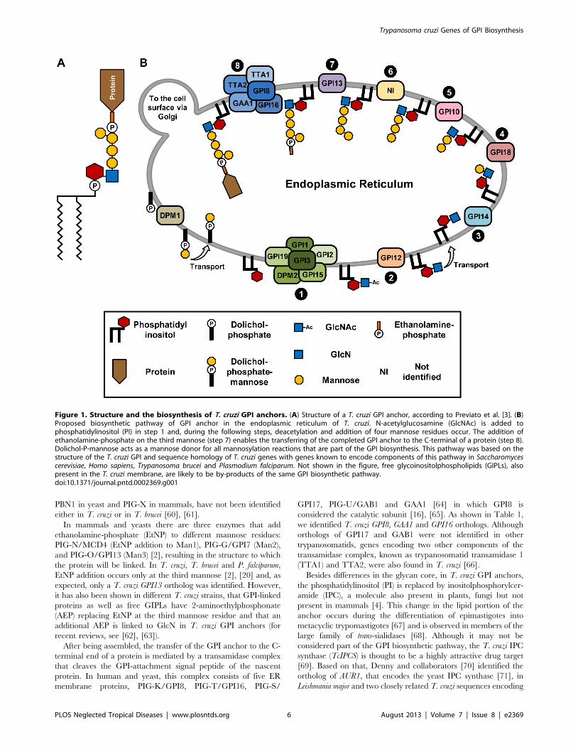
GPI anchor in this parasite (Figure 1A) [3], we proposed that the
T. cruzi GPI biosynthetic pathway occurs in the ER according to
the diagram shown in Figure 1B.
Dolichol-phosphate mannose synthase (DPM1), also named
dolichol-phosphate-b-D-mannosyltransferase, catalyses the trans-
fer of a mannose residue from GDP-mannose to dolichol-
phosphate (Dol-P) generating Dol-P-mannose, used as a donor
for all mannosylation reactions that are part of the GPI
biosynthetic pathway [40], [41]. Comparisons among DPM1 of
various organisms [42], [43], [44] showed that, together with S.
cerevisiae, T. brucei, and Leishmania mexicana [45] and in contrast to P.
falciparum DPM1, T. cruzi DPM1 belongs to a group that includes
monomeric enzymes that have a C-terminal hydrophobic tail. The
glycosyltransferase complex that is responsible for transferring N-
acetylglucosamine (GlcNAc) from UDP-GlcNAc to phosphatidy-
linositol (PI) to generate N-acetylglucosaminyl-PI (GlcNAc-PI) has
six and seven proteins, respectively, in yeast and mammalian cells
[16]. TcGPI3 was identified as the gene encoding the catalytic
subunit of the T. cruzi glycosyltransferase complex since it shares
41% and 49% of sequence identity with the yeast GPI3 and
mammalian PIG-A, respectively. Among other components of the
glycosyltransferase complex present in yeast, we identified the T.
cruzi orthologs of GPI1, GPI2, GPI15, and GPI19. In mammalian
cells, DPM2, a non-catalytic subunit of dolichol-P-mannose
synthase, is physically associated with PIG-A, PIG-C and PIG-Q
and enhances GlcNAc-PI transferase activity [46]. A T. cruzi gene
encoding a protein with 17% identity to human DPM2 and
containing a DPM2 domain, which probably acts as a regulatory
component of the N-acetyl-glucosamine transferase complex, was
also identified. Only one component of this complex, named ERI1
in yeast [47], and PIG-Y in mammals [48], was not identified
either in T. cruzi, P. falciparum or T. brucei. The T. cruzi ortholog of
yeast GPI12 (named PIG-L in mammals) [49], encoding the
Trypanosoma cruzi Genes of GPI Biosynthesis
PLOS Neglected Tropical Diseases | www.plosntds.org 4 August 2013 | Volume 7 | Issue 8 | e2369
enzyme responsible for the de-N-acetylation of GlcNAc-PI, which
has been well characterized in T. brucei [50], [51], was also
identified. Since differences in substrate recognition among the
mammal and T. brucei enzyme have been described [52], this
enzyme has been considered as a suitable target for drug
development.
As depicted in Figure 1B, the first two reactions of the GPI
biosynthetic pathway occur on the cytoplasmic face of the ER,
whereas mannosylation reactions occur in the ER lumen. After
deacetylation, the GPI precursor is transported across the ER
membrane to the ER lumen, a step that requires distinct flippases
[53]. In yeast and mammalian cells, the addition of mannose
residues to GlcN-PI after flipping this precursor into the ER lumen
requires acylation of the inositol ring and, after mannosylation and
the attachment of GPIs to proteins, this group is removed [54]. In
contrast, in T. brucei, inositol acylation occurs after the addition of
the first mannose residue [55] since both acylated and non-
acylated GPI intermediates exist during transfer of the Man2 and
Man3 to GPI intermediates [56]. Although analyses of GPI
precursors synthesized in T. cruzi cell-free systems indicated that
this organism also has the ability to acylate the inositol ring [57],
sequences encoding an enzyme responsible for acylation of the
inositol ring, named PIG-W in mammals and GWT1 in yeast [54],
[58] were not identified either in T. cruzi or in T. brucei [2]. In spite
of that, the two alleles encoding the ortholog of the enzyme
responsible for inositol deacylation, named GPIdeAc2 in T. brucei
[56], were found in the T. cruzi genome (Tc00.1047053508
153.1040 and Tc00.1047053506691.22).
All three genes encoding mannosyltransferases, responsible for
the addition of the first, second and third mannose residues to
GlcN-PI, named TcGPI14 (a-1,4-mannosyltransferase), TcGPI18
(a-1,6-mannosyltransferase) and TcGPI10 (a-1,2-mannosyltrans-
ferase), were identified in the T. cruzi genome. Since the predicted
T. cruzi proteins exhibit sequence identities with yeast and human
proteins ranging from 17% to 30%, for some of these genes,
functional assays are necessary to confirm these predictions. It is
noteworthy that no T. cruzi ortholog encoding the enzyme
responsible for the addition of the fourth residue of mannose
(step 6), named SMP3 in yeast and PIG-Z in human, was
identified. Similarly, no ortholog of the SMP3 gene was found in P.
falciparum, even though the presence of a fourth mannose residue
has been shown by structural studies of the GPI anchor from both
organisms [3], [20], [59]. Furthermore, genes encoding an
essential component of the mannosyltransferase I complex named
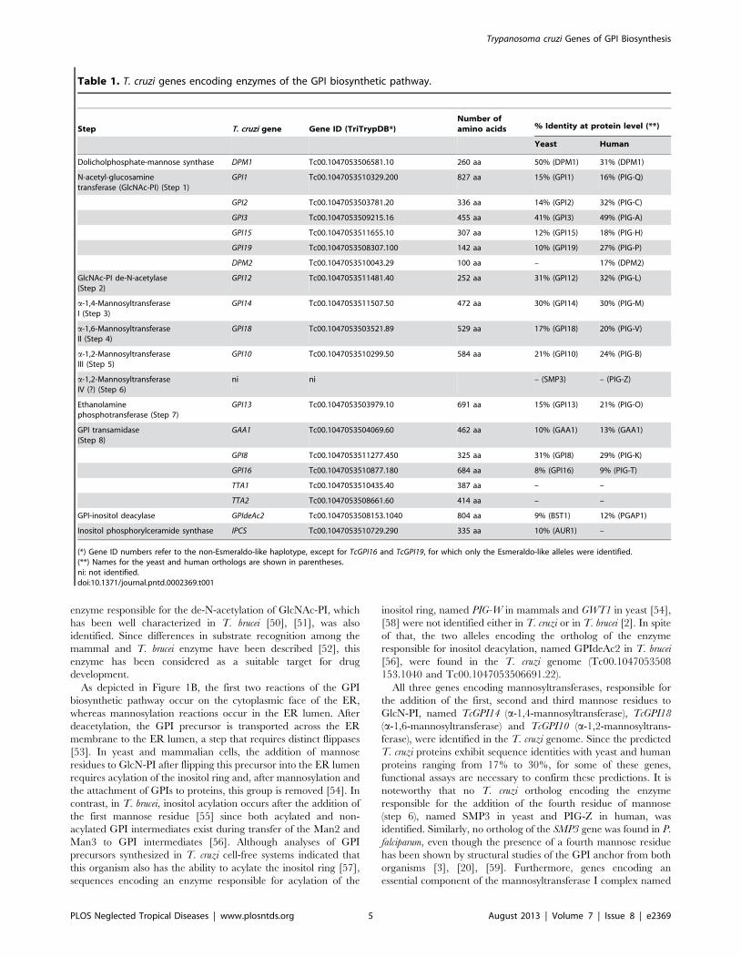
Table 1. T. cruzi genes encoding enzymes of the GPI biosynthetic pathway.
Step T. cruzi gene Gene ID (TriTrypDB*)Number ofamino acids % Identity at protein level (**)
Yeast Human
Dolicholphosphate-mannose synthase DPM1 Tc00.1047053506581.10 260 aa 50% (DPM1) 31% (DPM1)
N-acetyl-glucosaminetransferase (GlcNAc-PI) (Step 1)
GPI1 Tc00.1047053510329.200 827 aa 15% (GPI1) 16% (PIG-Q)
GPI2 Tc00.1047053503781.20 336 aa 14% (GPI2) 32% (PIG-C)
GPI3 Tc00.1047053509215.16 455 aa 41% (GPI3) 49% (PIG-A)
GPI15 Tc00.1047053511655.10 307 aa 12% (GPI15) 18% (PIG-H)
GPI19 Tc00.1047053508307.100 142 aa 10% (GPI19) 27% (PIG-P)
DPM2 Tc00.1047053510043.29 100 aa – 17% (DPM2)
GlcNAc-PI de-N-acetylase(Step 2)
GPI12 Tc00.1047053511481.40 252 aa 31% (GPI12) 32% (PIG-L)
a-1,4-MannosyltransferaseI (Step 3)
GPI14 Tc00.1047053511507.50 472 aa 30% (GPI14) 30% (PIG-M)
a-1,6-MannosyltransferaseII (Step 4)
GPI18 Tc00.1047053503521.89 529 aa 17% (GPI18) 20% (PIG-V)
a-1,2-MannosyltransferaseIII (Step 5)
GPI10 Tc00.1047053510299.50 584 aa 21% (GPI10) 24% (PIG-B)
a-1,2-MannosyltransferaseIV (?) (Step 6)
ni ni – (SMP3) – (PIG-Z)
Ethanolaminephosphotransferase (Step 7)
GPI13 Tc00.1047053503979.10 691 aa 15% (GPI13) 21% (PIG-O)
GPI transamidase(Step 8)
GAA1 Tc00.1047053504069.60 462 aa 10% (GAA1) 13% (GAA1)
GPI8 Tc00.1047053511277.450 325 aa 31% (GPI8) 29% (PIG-K)
GPI16 Tc00.1047053510877.180 684 aa 8% (GPI16) 9% (PIG-T)
TTA1 Tc00.1047053510435.40 387 aa – –
TTA2 Tc00.1047053508661.60 414 aa – –
GPI-inositol deacylase GPIdeAc2 Tc00.1047053508153.1040 804 aa 9% (BST1) 12% (PGAP1)
Inositol phosphorylceramide synthase IPCS Tc00.1047053510729.290 335 aa 10% (AUR1) –
(*) Gene ID numbers refer to the non-Esmeraldo-like haplotype, except for TcGPI16 and TcGPI19, for which only the Esmeraldo-like alleles were identified.(**) Names for the yeast and human orthologs are shown in parentheses.ni: not identified.doi:10.1371/journal.pntd.0002369.t001
Trypanosoma cruzi Genes of GPI Biosynthesis
PLOS Neglected Tropical Diseases | www.plosntds.org 5 August 2013 | Volume 7 | Issue 8 | e2369
PBN1 in yeast and PIG-X in mammals, have not been identified
either in T. cruzi or in T. brucei [60], [61].
In mammals and yeasts there are three enzymes that add
ethanolamine-phosphate (EtNP) to different mannose residues:
PIG-N/MCD4 (EtNP addition to Man1), PIG-G/GPI7 (Man2),
and PIG-O/GPI13 (Man3) [2], resulting in the structure to which
the protein will be linked. In T. cruzi, T. brucei and P. falciparum,
EtNP addition occurs only at the third mannose [2], [20] and, as
expected, only a T. cruzi GPI13 ortholog was identified. However,
it has also been shown in different T. cruzi strains, that GPI-linked
proteins as well as free GIPLs have 2-aminoethylphosphonate
(AEP) replacing EtNP at the third mannose residue and that an
additional AEP is linked to GlcN in T. cruzi GPI anchors (for
recent reviews, see [62], [63]).
After being assembled, the transfer of the GPI anchor to the C-
terminal end of a protein is mediated by a transamidase complex
that cleaves the GPI-attachment signal peptide of the nascent
protein. In human and yeast, this complex consists of five ER
membrane proteins, PIG-K/GPI8, PIG-T/GPI16, PIG-S/
GPI17, PIG-U/GAB1 and GAA1 [64] in which GPI8 is
considered the catalytic subunit [16], [65]. As shown in Table 1,
we identified T. cruzi GPI8, GAA1 and GPI16 orthologs. Although
orthologs of GPI17 and GAB1 were not identified in other
trypanosomatids, genes encoding two other components of the
transamidase complex, known as trypanosomatid transamidase 1
(TTA1) and TTA2, were also found in T. cruzi [66].
Besides differences in the glycan core, in T. cruzi GPI anchors,
the phosphatidylinositol (PI) is replaced by inositolphosphorylcer-
amide (IPC), a molecule also present in plants, fungi but not
present in mammals [4]. This change in the lipid portion of the
anchor occurs during the differentiation of epimastigotes into
metacyclic trypomastigotes [67] and is observed in members of the
large family of trans-sialidases [68]. Although it may not be
considered part of the GPI biosynthetic pathway, the T. cruzi IPC
synthase (TcIPCS) is thought to be a highly attractive drug target
[69]. Based on that, Denny and collaborators [70] identified the
ortholog of AUR1, that encodes the yeast IPC synthase [71], in
Leishmania major and two closely related T. cruzi sequences encoding
Figure 1. Structure and the biosynthesis of T. cruzi GPI anchors. (A) Structure of a T. cruzi GPI anchor, according to Previato et al. [3]. (B)Proposed biosynthetic pathway of GPI anchor in the endoplasmic reticulum of T. cruzi. N-acetylglucosamine (GlcNAc) is added tophosphatidylinositol (PI) in step 1 and, during the following steps, deacetylation and addition of four mannose residues occur. The addition ofethanolamine-phosphate on the third mannose (step 7) enables the transferring of the completed GPI anchor to the C-terminal of a protein (step 8).Dolichol-P-mannose acts as a mannose donor for all mannosylation reactions that are part of the GPI biosynthesis. This pathway was based on thestructure of the T. cruzi GPI and sequence homology of T. cruzi genes with genes known to encode components of this pathway in Saccharomycescerevisiae, Homo sapiens, Trypanosoma brucei and Plasmodium falciparum. Not shown in the figure, free glycoinositolphospholipids (GIPLs), alsopresent in the T. cruzi membrane, are likely to be by-products of the same GPI biosynthetic pathway.doi:10.1371/journal.pntd.0002369.g001
Trypanosoma cruzi Genes of GPI Biosynthesis
PLOS Neglected Tropical Diseases | www.plosntds.org 6 August 2013 | Volume 7 | Issue 8 | e2369
proteins sharing 52–53% identity with the Leishmania IPC synthase
[70]. Our analysis confirmed that the two sequences described by
Denny and collaborators [70] correspond to the two alleles of the
T. cruzi IPC synthase (TcIPCS) gene present in the CL Brener
genome, which are synthenic with the L. major and T. brucei
orthologs.
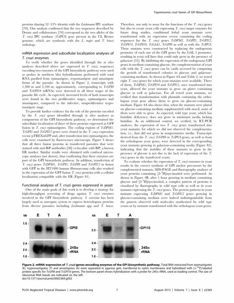
mRNA expression and subcellular localization analyses ofT. cruzi enzymes
To verify whether the genes identified through the in silico
analyses described above are expressed in T. cruzi, sequences
encoding two enzymes of the GPI biosynthetic pathway were used
as probes in northern blot hybridizations performed with total
RNA purified from epimastigote, trypomastigote and amastigote
forms of the parasite. As shown in Figure 2, transcripts with
1,300 nt and 2,100 nt, approximately, corresponding to TcGPI8
and TcGPI10 mRNAs were detected in all three stages of the
parasite life cycle. As expected, increased levels of both transcripts
were found in the two proliferative stages, epimastigotes and
amastigotes, compared to the infective, nonproliferative trypo-
mastigote stage.
To provide further evidence for the role of the proteins encoded
by the T. cruzi genes identified through in silico analyses as
components of the GPI biosynthetic pathway, we determined the
subcellular localization of three of these proteins expressed as GFP
fusion in T. cruzi epimastigotes. The coding regions of TcDPM1,
TcGPI3 and TcGPI12 genes were cloned in the T. cruzi expression
vector pTREXnGFP and, after transfection into epimastigotes, the
cells were examined by fluorescence microscopy. Figure 3 shows
that all three fusion proteins in transfected parasites that were
stained with anti-BiP antibodies [38] co-localize with BiP, a known
ER marker. Similar results were obtained with confocal micros-
copy analyses (not shown), thus confirming that these enzymes are
part of the GPI biosynthetic pathway. In addition, transfection of
T. cruzi genes TcDPM1, TcGPI3, TcGPI8 and TcGPI12 in fusion
with GFP in the HT1080 human fibrosarcoma cells also resulted
in the expression of the GFP fusion T. cruzi proteins with a cellular
localization compatible with the ER (Figure S1).
Functional analyses of T. cruzi genes expressed in yeastOne of the main goals of this work is to develop a strategy for
high-throughput screening of drugs against T. cruzi enzymes
involved in the GPI biosynthetic pathway. S. cerevisiae has been
largely used as surrogate system to express heterologous proteins
from diverse parasites including Leishmania spp and T. brucei.
Therefore, not only to assay for the functions of the T. cruzi genes
but also to create yeast cells expressing T. cruzi target enzymes for
future drug studies, conditional lethal yeast mutants were
transformed with an expression vector containing the coding
sequences for the T. cruzi genes TcDPM1, TcGPI3, TcGPI12,
TcGPI14, TcGPI10, TcGAA1, TcGPI8 as well as with the TcIPCS.
These mutants were constructed by replacing the endogenous
promoter of each one of the GPI genes by the GAL 1 promoter,
resulting in yeast cell lines that could only grow in the presence of
galactose [31]. By inhibiting the expression of the endogenous GPI
genes in medium containing glucose, the complementation of yeast
cells with the T. cruzi genes can be easily accessed by comparing
the growth of transformed colonies in glucose and galactose-
containing medium. As shown in Figure 4A and Table 2, we tested
eight T. cruzi genes for which yeast mutants were available. Three
of them, TcDPM1, TcGPI10 and TcGPI12, once transformed into
yeast, allowed the yeast mutants to grow on plates containing
glucose as well as galactose. For all tested yeast mutants, we
verified that transformation with plasmids containing the ortho-
logous yeast gene allows them to grow on glucose-containing
medium. Figure 4A also shows that, when the mutants were plated
on glucose-containing medium supplemented with uracil, none of
them were able to grow. As expected, wild type yeast, which has
histidine deficiency, does not grow in minimum media lacking
histidine. As an additional control, we verified, by RT-PCR
analyses, the expression of two T. cruzi genes transformed into
yeast mutants, for which we did not observed the complementa-
tion, i.e., that did not grow in nonpermissive media. Transcripts
derived from the T. cruzi TcGPI8 or TcIPCS genes, as well as from
the orthologous yeast genes, were detected in the corresponding
yeast mutants growing in galactose-containing media (Figure S2),
indicating that the inability of these mutants to grow in the
presence of glucose is not due to the lack of expression of the T.
cruzi genes in the transfected yeasts.
To evaluate whether the expression of T. cruzi enzymes in yeast
results in the correct synthesis of GPI anchor precursors by the
complemented mutants, SDS-PAGE and fluorography analyses of
yeast proteins containing [2-3H]myo-inositol were performed. As
shown in Figure 4B, after 1 hour growing in medium containing
glucose and [2-3H]myo-inositol, a complex pattern of proteins is
visualized by fluorography in wild type cells as well as in yeast
mutants expressing the T. cruzi genes. The protein patterns in yeast
mutants expressing TcDPM1 and TcGPI12 genes growing in
glucose-containing medium were indeed indistinguishable from
the pattern observed with molecules synthesized by wild type
yeasts or by mutants transformed with the orthologous yeast genes.
Figure 2. mRNA expression of T. cruzi genes encoding enzymes of the GPI biosynthetic pathway. Total RNA extracted from epimastigotes(E), trypomastigotes (T) and amastigotes (A) were separated in agarose gels, transferred to nylon membranes and hybridized with [a-32P]-labeledprobes specific for TcGPI8 and TcGPI10 genes. The bottom panel shows hybridization with a probe for 24Sa rRNA, used as loading control. The size ofribosomal RNA bands are indicated on the left.doi:10.1371/journal.pntd.0002369.g002
Trypanosoma cruzi Genes of GPI Biosynthesis
PLOS Neglected Tropical Diseases | www.plosntds.org 7 August 2013 | Volume 7 | Issue 8 | e2369
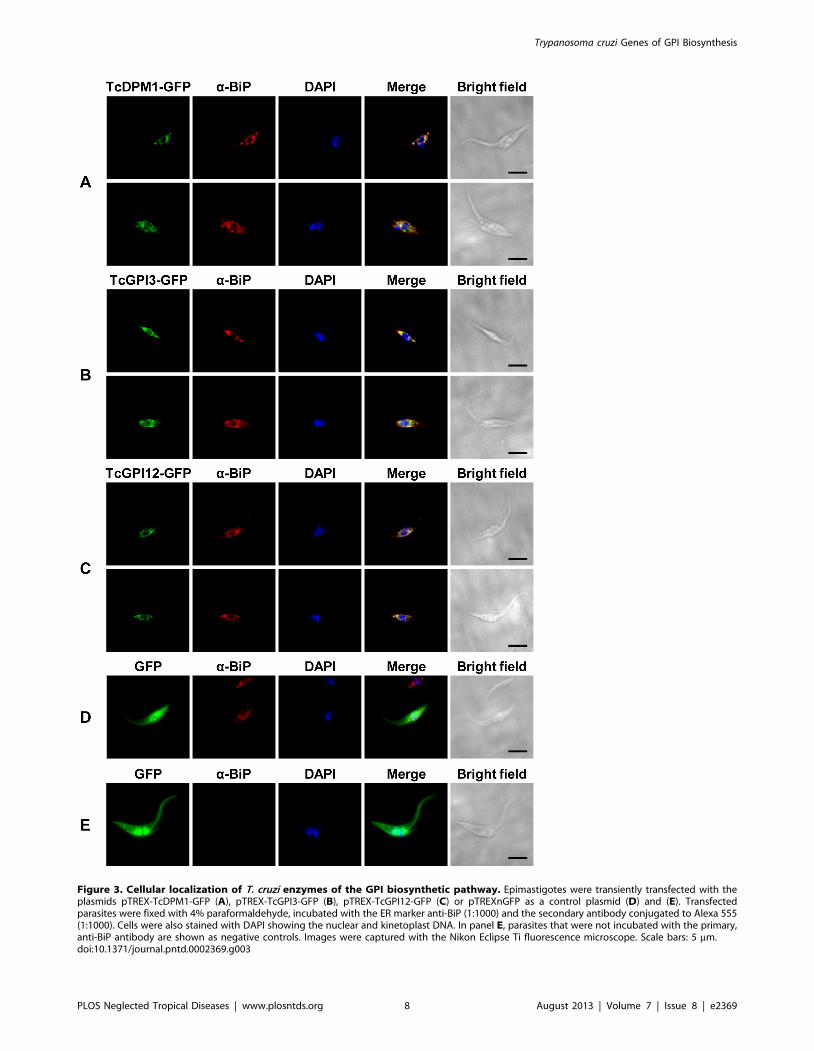
Figure 3. Cellular localization of T. cruzi enzymes of the GPI biosynthetic pathway. Epimastigotes were transiently transfected with theplasmids pTREX-TcDPM1-GFP (A), pTREX-TcGPI3-GFP (B), pTREX-TcGPI12-GFP (C) or pTREXnGFP as a control plasmid (D) and (E). Transfectedparasites were fixed with 4% paraformaldehyde, incubated with the ER marker anti-BiP (1:1000) and the secondary antibody conjugated to Alexa 555(1:1000). Cells were also stained with DAPI showing the nuclear and kinetoplast DNA. In panel E, parasites that were not incubated with the primary,anti-BiP antibody are shown as negative controls. Images were captured with the Nikon Eclipse Ti fluorescence microscope. Scale bars: 5 mm.doi:10.1371/journal.pntd.0002369.g003
Trypanosoma cruzi Genes of GPI Biosynthesis
PLOS Neglected Tropical Diseases | www.plosntds.org 8 August 2013 | Volume 7 | Issue 8 | e2369
Trypanosoma cruzi Genes of GPI Biosynthesis
PLOS Neglected Tropical Diseases | www.plosntds.org 9 August 2013 | Volume 7 | Issue 8 | e2369
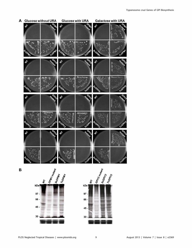
On the other hand, a much weaker signal was detected in non-
transformed yeast mutants, indicating that the expression of T.
cruzi orthologs encoding enzymes of the GPI biosynthetic pathway
restores the mutants’ ability to synthesize GPI molecules.
Corroborating the functional complementation of yeast mu-
tants with the TcDPM1 gene, thin layer chromatography (TLC)
of yeast mutants expressing the T. cruzi gene or the yeast
ScDPM1gene, as a positive control, showed the presence of
dolichol-P-mannose. Yeast cell extracts were preincubated with
dolichol-phosphate and labeled in vitro with GDP-[2-3H]mannose.
Labeled dolichol-P-mannose was detected in wild type yeast cells
as well as in DPM1 mutants that were transfected with the
TcDPM1 or with the yeast ScDPM1 gene, confirming that the
expression of the T. cruzi enzyme rescues the mutant ability to
synthesize dolichol-P-mannose (Figure S3).
T. cruzi GPI8 mutants have altered cell surfaceKnockout parasites of GPI8, GPI16 and GPI10 were generated
in T. brucei whereas a GPI8 knockout was described in L. mexicana
[18], [19], [72], [73]. To further investigate the role of GPI
anchors in T. cruzi, we tried to generate parasite cell lines in
which both alleles of TcGPI3, TcGPI8 and TcGPI10 genes were
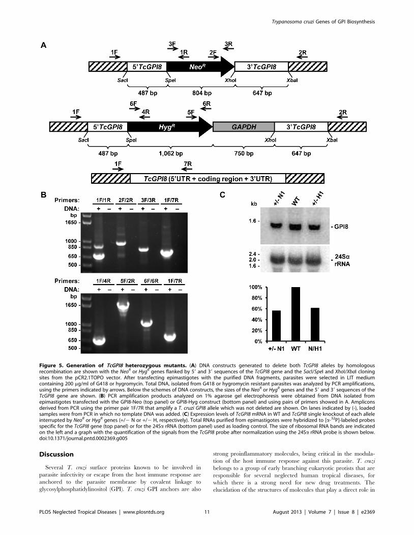
deleted by homologous recombination. Although we were able to
generate heterozygote epimastigotes carrying a drug resistance
marker inserted in each one of the TcGPI8 alleles (Figure 5A–B),
several attempts to generate double-resistant, null mutant
epimastigotes with both TcGPI8 alleles deleted were unsuccessful.
Also unexpectedly, transfection with plasmid constructs contain-
ing TcGPI3 and TcGPI10 sequences flanking the neomycin
resistance gene did not result in G418 resistant parasites,
indicating that disruption of even one allele of a gene involved
in the initial steps of the GPI biosynthesis pathway results in non-
viable parasites (not shown). Thus, our results suggest that, in
contrast to T. brucei and L. mexicana, the GPI biosynthesis may be
an essential pathway in epimastigotes of T. cruzi. In agreement
with PCR analyses that showed the disruption of single alleles of
TcGPI8 (Figure 5B), northern blot assays (Figure 5C) showed that
both heterozygous TcGPI8 mutants have the expression of
TcGPI8 mRNA reduced by about 40%. Although a few double-
resistant epimastigote clones were generated and PCR analyses
indicated that the neomycin and hygromycin resistance genes
were inserted into both TcGPI8 alleles, PCR amplifications also
indicated that additional sequences corresponding to the TcGPI8
gene were present in a different genomic location in the double
resistant parasites (Figure 6A–B). It should be noted that it was
possible to generate the double resistant parasites only after we
prepared different plasmid constructs in which the resistance
genes were linked to trans-splicing and polyadenylation signals
from the glyceraldehyde-3-phosphate dehydrogenase (gapdh) and
the ribosomal protein TcP2b (HX1) genes and performing drug
selection by gradually increasing drug concentrations. Northern
blot analyses (Figure 6C) indicate that the recombination events
that resulted in viable, double resistant parasites allowed the
expression of an aberrant TcGPI8 mRNA population. Among this
TcGPI8 mRNA population transcribed in the double resistant
mutants, mature, trans-spliced mRNAs were detected by RT-
PCR using primers specific for TcGPI8 sequences and the T. cruzi
spliced leader (Figure 6D), thus indicating that this gene is still
active in these mutants. Although no significant changes in either
growing or overall morphology of the TcGPI8 mutants were
observed, transmission electron microscopy showed striking
alterations in the dense glycocalyx that covers the parasite
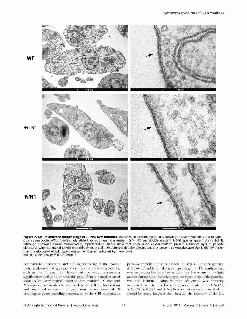
surface. As shown in Figure 7, cell membranes of epimastigotes
from TcGPI8 heterozygous mutants (+/2N) present a thinner
layer of the surface glycocalyx compared to wild type (WT)
epimastigotes. In contrast, cell membranes from both clones of
double resistant parasites (N/H), which may have suffered
recombination events involving TcGPI8 sequences, present an
increased thickness of their glycocalyx compared to the hetero-
zygous mutants (Figure 7). Although no significant differences in
the levels of mucins were detected in the heterozygous mutants,
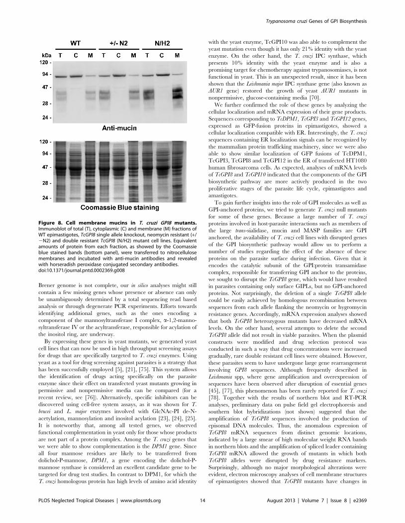
western blot analyses of membrane proteins of WT and double
resistant TcGPI8 mutants using the anti-mucin monoclonal
antibody 2B10 [74] showed increased amounts of the 35–
50 kDa glycoproteins (also known as Gp35/50 mucins) expressed
on the surface of epimastigotes of the double resistant clones
(Figure 8). Flow cytometry of epimastigotes stained with 2B10
antibodies also showed increased amounts of surface mucins in
the double resistant parasites (Figure S4).
Figure 4. Yeast complementation with T. cruzi genes encoding enzymes of the GPI biosynthetic pathway. (A) DPM1, GPI10 and GPI12yeast conditional lethal mutants (YPH499-HIS-GAL-DPM1, YPH499-HIS-GAL-GPI10 and YPH499-HIS-GAL-GPI12, respectively) were transformed withpRS426Met plasmids carrying either T. cruzi or S. cerevisiae genes encoding DPM1, GPI10 and GPI12 (TcDPM1 or ScDPM1, TcGPI10 or ScGPI10, andTcGPI12 or ScGPI12, respectively). Wild-type (WT), non-transformed mutants and transformed yeast mutants were streaked onto plates withnonpermissive, glucose-containing SD medium lacking histidine, with or without uracil or in galactose-containing medium (with uracil) andincubated at 30uC for 3 days. In the bottom panel, yeast mutants (YPH499-HIS-GAL-GPI14) transformed with pRS426Met plasmid carrying T. cruzigene (TcGPI14), which could not restore cell growth of GPI14 deficient yeast are shown. (B) GPI-anchored proteins synthesized by the conditionallethal yeast mutants expressing T. cruzi genes were separated by SDS-PAGE and analyzed after fluorography. Wild-type (WT), non-transformed yeastmutants and yeast mutants that were transformed with plasmids containing the corresponding yeast genes (ScDPM1 or ScGPI12) or with the T. cruzigenes (TcDPM1 or TcGPI12), were cultivated in medium glucose-containing in the presence of [2-3H]myo-inositol for 1 hour. Total protein extractcorresponding to 16108 cells were loaded on each lane of a 10% SDS-PAGE and the labeled proteins were visualized by fluorography (top panels). Asa loading control, Coomassie Blue stained gels prepared with equivalents amounts of total proteins are shown in the bottom panels. UntransfectedDPM1 and GPI12 mutants were grown in the presence of galactose for 2 days and then switched to glucose-containing medium for 16 hours beforeaddition of [2-3H]myo-inositol. Molecular weight markers (M) are shown on the left.doi:10.1371/journal.pntd.0002369.g004
Table 2. Functional complementation of yeast mutants by T.cruzi genes.
Yeast mutants pRS Tc
YPH499 DPM1 +
YPH499 GPI3 2
YPH499 GPI12 +
YPH499 GPI14 2
YPH499 GPI10 +
YPH499 GAA1 2
YPH499 GPI8 2
YPH499 AUR1 2
The (+) signs indicate the ability of transformed mutants to grow innonpermissive glucose-containing media.doi:10.1371/journal.pntd.0002369.t002
Trypanosoma cruzi Genes of GPI Biosynthesis
PLOS Neglected Tropical Diseases | www.plosntds.org 10 August 2013 | Volume 7 | Issue 8 | e2369
Discussion
Several T. cruzi surface proteins known to be involved in
parasite infectivity or escape from the host immune response are
anchored to the parasite membrane by covalent linkage to
glycosylphosphatidylinositol (GPI). T. cruzi GPI anchors are also
strong proinflammatory molecules, being critical in the modula-
tion of the host immune response against this parasite. T. cruzi
belongs to a group of early branching eukaryotic protists that are
responsible for several neglected human tropical diseases, for
which there is a strong need for new drug treatments. The
elucidation of the structures of molecules that play a direct role in
Figure 5. Generation of TcGPI8 heterozygous mutants. (A) DNA constructs generated to delete both TcGPI8 alleles by homologousrecombination are shown with the NeoR or HygR genes flanked by 59 and 39 sequences of the TcGPI8 gene and the SacI/SpeI and XhoI/XbaI cloningsites from the pCR2.1TOPO vector. After transfecting epimastigotes with the purified DNA fragments, parasites were selected in LIT mediumcontaining 200 mg/ml of G418 or hygromycin. Total DNA, isolated from G418 or hygromycin resistant parasites was analyzed by PCR amplifications,using the primers indicated by arrows. Below the schemes of DNA constructs, the sizes of the NeoR or HygR genes and the 59 and 39 sequences of theTcGPI8 gene are shown. (B) PCR amplification products analyzed on 1% agarose gel electrophoresis were obtained from DNA isolated fromepimastigotes transfected with the GPI8-Neo (top panel) or GPI8-Hyg construct (bottom panel) and using pairs of primers showed in A. Ampliconsderived from PCR using the primer pair 1F/7R that amplify a T. cruzi GPI8 allele which was not deleted are shown. On lanes indicated by (-), loadedsamples were from PCR in which no template DNA was added. (C) Expression levels of TcGPI8 mRNA in WT and TcGPI8 single knockout of each alleleinterrupted by NeoR or HygR genes (+/2 N or +/2 H, respectively). Total RNAs purified from epimastigotes were hybridized to [a-32P]-labeled probesspecific for the TcGPI8 gene (top panel) or for the 24Sa rRNA (bottom panel) used as loading control. The size of ribosomal RNA bands are indicatedon the left and a graph with the quantification of the signals from the TcGPI8 probe after normalization using the 24Sa rRNA probe is shown below.doi:10.1371/journal.pntd.0002369.g005
Trypanosoma cruzi Genes of GPI Biosynthesis
PLOS Neglected Tropical Diseases | www.plosntds.org 11 August 2013 | Volume 7 | Issue 8 | e2369
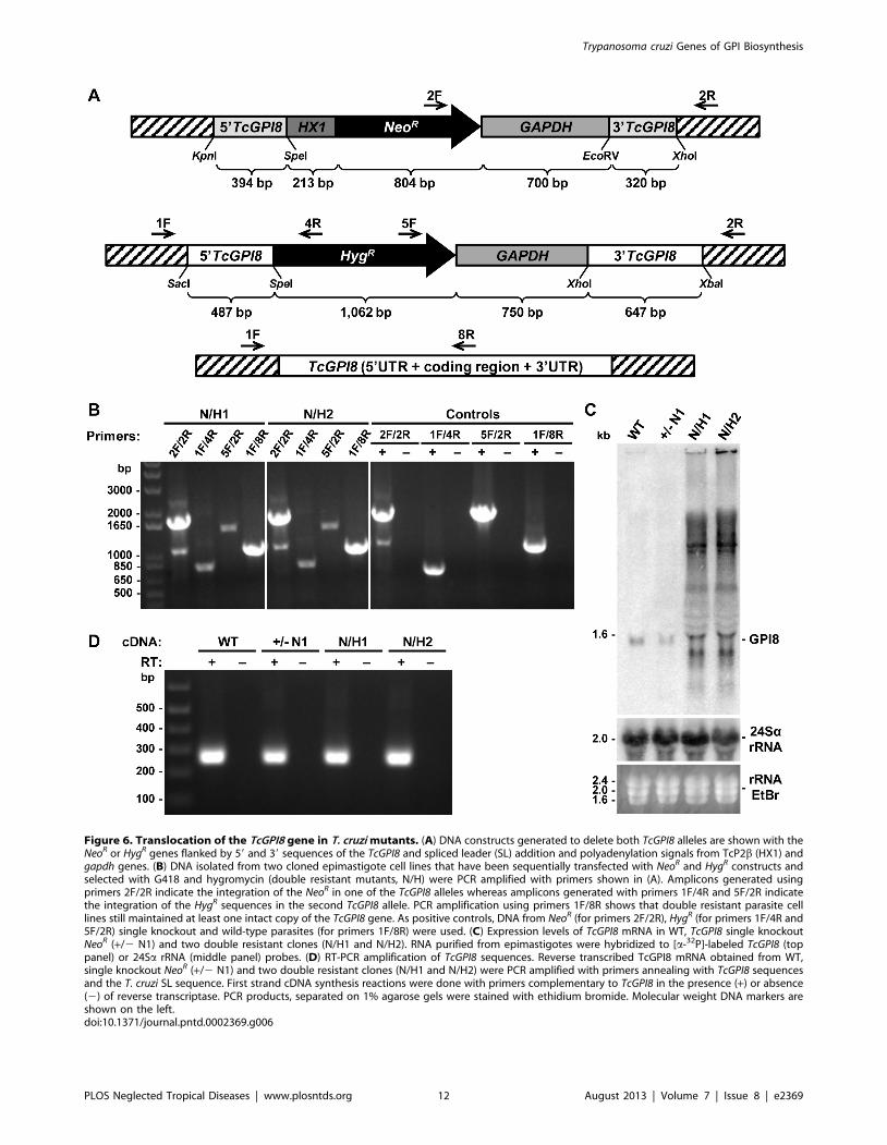
Figure 6. Translocation of the TcGPI8 gene in T. cruzi mutants. (A) DNA constructs generated to delete both TcGPI8 alleles are shown with theNeoR or HygR genes flanked by 59 and 39 sequences of the TcGPI8 and spliced leader (SL) addition and polyadenylation signals from TcP2b (HX1) andgapdh genes. (B) DNA isolated from two cloned epimastigote cell lines that have been sequentially transfected with NeoR and HygR constructs andselected with G418 and hygromycin (double resistant mutants, N/H) were PCR amplified with primers shown in (A). Amplicons generated usingprimers 2F/2R indicate the integration of the NeoR in one of the TcGPI8 alleles whereas amplicons generated with primers 1F/4R and 5F/2R indicatethe integration of the HygR sequences in the second TcGPI8 allele. PCR amplification using primers 1F/8R shows that double resistant parasite celllines still maintained at least one intact copy of the TcGPI8 gene. As positive controls, DNA from NeoR (for primers 2F/2R), HygR (for primers 1F/4R and5F/2R) single knockout and wild-type parasites (for primers 1F/8R) were used. (C) Expression levels of TcGPI8 mRNA in WT, TcGPI8 single knockoutNeoR (+/2 N1) and two double resistant clones (N/H1 and N/H2). RNA purified from epimastigotes were hybridized to [a-32P]-labeled TcGPI8 (toppanel) or 24Sa rRNA (middle panel) probes. (D) RT-PCR amplification of TcGPI8 sequences. Reverse transcribed TcGPI8 mRNA obtained from WT,single knockout NeoR (+/2 N1) and two double resistant clones (N/H1 and N/H2) were PCR amplified with primers annealing with TcGPI8 sequencesand the T. cruzi SL sequence. First strand cDNA synthesis reactions were done with primers complementary to TcGPI8 in the presence (+) or absence(2) of reverse transcriptase. PCR products, separated on 1% agarose gels were stained with ethidium bromide. Molecular weight DNA markers areshown on the left.doi:10.1371/journal.pntd.0002369.g006
Trypanosoma cruzi Genes of GPI Biosynthesis
PLOS Neglected Tropical Diseases | www.plosntds.org 12 August 2013 | Volume 7 | Issue 8 | e2369
host-parasite interactions and the understanding of the biosyn-
thetic pathways that generate these specific parasite molecules,
such as the T. cruzi GPI biosynthetic pathway, represent a
significant contribution towards this goal. Using a combination of
sequence similarity analyses based on yeast, mammal, T. brucei and
P. falciparum previously characterized genes, cellular localization
and functional expression in yeast mutants we identified 18
orthologous genes encoding components of the GPI biosynthetic
pathway present in the published T. cruzi CL Brener genome
database. In addition, the gene encoding the IPC synthase, an
enzyme responsible for a key modification that occurs in the lipid
anchor during in the infective, trypomastigote stage of the parasite,
was also identified. Although most sequences were correctly
annotated in the TriTrypDB genome database, TcGPI15,
TcGPI19, TcDPM2 and TcGPI16 were not correctly identified. It
should be noted however that, because the assembly of the CL
Figure 7. Cell membrane morphology of T. cruzi GPI8 mutants. Transmission electron microscopy showing cellular membranes of wild type T.cruzi epimastigotes (WT), TcGPI8 single allele knockout, neomycin resistant (+/2 N1) and double resistant TcGPI8 epimastigote mutants (N/H1).Although displaying similar morphologies, representative images show that single allele TcGPI8 mutants present a thinner layer of parasiteglycocalyx, when compared to wild type cells, whereas cell membranes of double resistant parasites present a glycocalyx layer that is slightly thickerthan the glycocalyx of wild type parasite membranes (indicated by the arrows).doi:10.1371/journal.pntd.0002369.g007
Trypanosoma cruzi Genes of GPI Biosynthesis
PLOS Neglected Tropical Diseases | www.plosntds.org 13 August 2013 | Volume 7 | Issue 8 | e2369
Brener genome is not complete, our in silico analyses might still
contain a few missing genes whose presence or absence can only
be unambiguously determined by a total sequencing read based
analysis or through degenerate PCR experiments. Efforts towards
identifying additional genes, such as the ones encoding a
component of the mannosyltransferase I complex, a-1,2-manno-
syltransferase IV or the acyltransferase, responsible for acylation of
the inositol ring, are underway.
By expressing these genes in yeast mutants, we generated yeast
cell lines that can now be used in high throughput screening assays
for drugs that are specifically targeted to T. cruzi enzymes. Using
yeast as a tool for drug screening against parasites is a strategy that
has been successfully employed [5], [21], [75]. This system allows
the identification of drugs acting specifically on the parasite
enzyme since their effect on transfected yeast mutants growing in
permissive and nonpermissive media can be compared (for a
recent review, see [76]). Alternatively, specific inhibitors can be
discovered using cell-free system assays, as it was shown for T.
brucei and L. major enzymes involved with GlcNAc-PI de-N-
acetylation, mannosylation and inositol acylation [23], [24], [25].
It is noteworthy that, among all tested genes, we observed
functional complementation in yeast only for those whose products
are not part of a protein complex. Among the T. cruzi genes that
we were able to show complementation is the DPM1 gene. Since
all four mannose residues are likely to be transferred from
dolichol-P-mannose, DPM1, a gene encoding the dolichol-P-
mannose synthase is considered an excellent candidate gene to be
targeted for drug test studies. In contrast to DPM1, for which the
T. cruzi homologous protein has high levels of amino acid identity
with the yeast enzyme, TcGPI10 was also able to complement the
yeast mutation even though it has only 21% identity with the yeast
enzyme. On the other hand, the T. cruzi IPC synthase, which
presents 10% identity with the yeast enzyme and is also a
promising target for chemotherapy against trypanosomiases, is not
functional in yeast. This is an unexpected result, since it has been
shown that the Leishmania major IPC synthase gene (also known as
AUR1 gene) restored the growth of yeast AUR1 mutants in
nonpermissive, glucose-containing media [70].
We further confirmed the role of these genes by analyzing the
cellular localization and mRNA expression of their gene products.
Sequences corresponding to TcDPM1, TcGPI3 and TcGPI12 genes,
expressed as GFP-fusion proteins in epimastigotes, showed a
cellular localization compatible with ER. Interestingly, the T. cruzi
sequences containing ER localization signals can be recognized by
the mammalian protein trafficking machinery, since we were also
able to show similar localization of GFP fusions of TcDPM1,
TcGPI3, TcGPI8 and TcGPI12 in the ER of transfected HT1080
human fibrosarcoma cells. As expected, analyses of mRNA levels
of TcGPI8 and TcGPI10 indicated that the components of the GPI
biosynthetic pathway are more actively produced in the two
proliferative stages of the parasite life cycle, epimastigotes and
amastigotes.
To gain further insights into the role of GPI molecules as well as
GPI-anchored proteins, we tried to generate T. cruzi null mutants
for some of these genes. Because a large number of T. cruzi
proteins involved in host-parasite interactions such as members of
the large trans-sialidase, mucin and MASP families are GPI
anchored, the availability of T. cruzi cell lines with disrupted genes
of the GPI biosynthetic pathway would allow us to perform a
number of studies regarding the effect of the absence of these
proteins on the parasite surface during infection. Given that it
encodes the catalytic subunit of the GPI:protein transamidase
complex, responsible for transferring GPI anchor to the proteins,
we sought to disrupt the TcGPI8 gene, which would have resulted
in parasites containing only surface GIPLs, but no GPI-anchored
proteins. Not surprisingly, the deletion of a single TcGPI8 allele
could be easily achieved by homologous recombination between
sequences from each allele flanking the neomycin or hygromycin
resistance genes. Accordingly, mRNA expression analyses showed
that both TcGPI8 heterozygous mutants have decreased mRNA
levels. On the other hand, several attempts to delete the second
TcGPI8 allele did not result in viable parasites. When the plasmid
constructs were modified and drug selection protocol was
conducted in such a way that drug concentrations were increased
gradually, rare double resistant cell lines were obtained. However,
these parasites seem to have undergone large gene rearrangement
involving GPI8 sequences. Although frequently described in
Leishmania spp, where gene amplification and overexpression of
sequences have been observed after disruption of essential genes
[45], [77], this phenomenon has been rarely reported for T. cruzi
[78]. Together with the results of northern blot and RT-PCR
analyses, preliminary data on pulse field gel electrophoresis and
southern blot hybridizations (not shown) suggested that the
amplification of TcGPI8 sequences involved the production of
episomal DNA molecules. Thus, the anomalous expression of
TcGPI8 mRNA sequences from distinct genomic locations,
indicated by a large smear of high molecular weight RNA bands
in northern blots and the amplification of spliced leader containing
TcGPI8 mRNA allowed the growth of mutants in which both
TcGPI8 alleles were disrupted by drug resistance markers.
Surprisingly, although no major morphological alterations were
evident, electron microscopy analyses of cell membrane structures
of epimastigotes showed that TcGPI8 mutants have changes in
Figure 8. Cell membrane mucins in T. cruzi GPI8 mutants.Immunoblot of total (T), cytoplasmic (C) and membrane (M) fractions ofWT epimastigotes, TcGPI8 single allele knockout, neomycin resistant (+/2N2) and double resistant TcGPI8 (N/H2) mutant cell lines. Equivalentamounts of protein from each fraction, as showed by the Coomassieblue stained bands (bottom panel), were transferred to nitrocellulosemembranes and incubated with anti-mucin antibodies and revealedwith horseradish peroxidase conjugated secondary antibodies.doi:10.1371/journal.pntd.0002369.g008
Trypanosoma cruzi Genes of GPI Biosynthesis
PLOS Neglected Tropical Diseases | www.plosntds.org 14 August 2013 | Volume 7 | Issue 8 | e2369
their glycocalyx layer. Even though the small reduction in the
glycocalyx layer observed in the heterozygous mutants could not
be correlated with changes in the levels of mucins, western blot
with membrane fractions, confirmed by flow cytometry using anti-
mucin antibodies indicated that double-resistant parasites present
a small increase in the amount of surface glycoproteins, most likely
due to an increased expression of the translocated copies of
TcGPI8 gene. Mucins play a critical role during infection, since
they are the acceptors of sialic acid that allows trypomastigotes to
build a negatively charged coat that protects them from killing by
host anti-a-galactopyranosyl antibodies [79]. Whether the geno-
mic rearrangements that resulted in the expression of TcGPI8 from
different genomic locations have affected the expression of other
T. cruzi genes, it remains to be determined. It will be also
important to determine which are the mechanisms employed by
the parasite that resulted in the genomic rearrangement observed
with the double resistant clones.
Interestingly, despite being viable in culture, T. brucei mutants
lacking TbGPI8 resulted in the absence of GPI-anchored surface
proteins, accumulation of non-protein-linked GPI and incapacity
of procyclic forms to establish infections in the tsetse midgut [80].
In contrast, GPI8 RNAi knock-down in bloodstream forms
resulted in accumulation of unanchored variant surface glycopro-
tein (VSG) and cell death with a phenotype indicative of blocking
cytokinesis [72]. On the other hand, L. mexicana GPI8 knockouts,
although deficient of GPI-anchored proteins, display normal
growth in culture, are capable of differentiating into amastigotes,
and are able to infect mice [19]. In addition to GPI8, procyclic T.
brucei lacking the TbGPI12 and TbGPI10 were also obtained.
Although unable to synthesize GPI structures beyond GlcNAc-PI,
TbGPI122/2 parasites are viable in culture, but are not able to
colonize the tsetse midgut [51]. Deletion of TbGPI10 also interferes
with the ability of procyclic mutants to infect tsetse flies [18].
These reports are in contrast with our results indicating that
disruption of only one allele of a gene involved in the initial steps of
the GPI pathway such as TcGPI3 or TcGPI10 resulted in non-
viable T. cruzi epimastigotes. On the other hand, similarly to the
genomic alterations we observed in the T. cruzi double resistant
TcGPI8 mutants, an attempt to create a L. mexicana knockout by
targeted deletion of the gene encoding the dolichol-phosphate-
mannose synthase resulted in amplification of this chromosomal
locus [45]. Thus, our contrasting results attempting to generate T.
cruzi null mutants of genes involved with GPI biosynthesis,
compared to similar studies described in T. brucei and L. mexicana,
suggest that, although considered closely related organisms, the
different members of the trypanosomatid family have significant
peculiarities that deserve detailed analyses of major biochemical
pathways in each parasite species.
Supporting Information
Figure S1 Cellular localization of T. cruzi proteinsexpressed in mammalian cells. The T. cruzi genes TcDPM1,
TcGPI3, TcGPI12, and TcGPI8 were cloned in fusion with GFP in
the vector pcDNA3.1/NT-GFP-TOPO and transfected into
HT1080 human fibrosarcoma cells. Forty eight hours after
transfections with pcDNA-GFP-TcDPM1 (A), pcDNA-GFP-
TcGPI3 (B), pcDNA-GFP-TcGPI12 (C), pcDNA-GFP-TcGPI8
(D) or after mock transfections (E), cells were stained with DAPI
and visualized under fluorescence microscopy. All plasmids were
cotransfected with the pGAG-DsRed-ER plasmid to visualize
cellular ER compartments. Scale bars: 20 mm.
(TIF)
Figure S2 RT-PCR mRNA analysis of yeast mutantstransformed with T. cruzi genes. Reverse-transcription and
PCR amplifications (RT-PCR) of total RNA isolated from non-
transformed yeast mutants or mutants transformed with T. cruzi
genes were analyzed by agarose gel electrophoresis. Total RNA
was isolated from GPI8 yeast mutants (top panel) or AUR1 mutants
(bottom panel). mRNA expression was analyzed in non-trans-
formed mutants (GPI8 mutants or AUR1 mutants) or mutants
transformed with pRS426Met plasmids carrying either the T. cruzi
(TcGPI8 or TcIPCS) that were grown in galactose-containing
media. For each RNA sample, pair of primers used for cDNA
amplifications, which are specific for the TcGPI8, TcIPCS, the
endogenous ScGPI8 or ScAUR1, as well as for the yeast 26S rRNA
genes, are indicated above each lane of the gel and are listed in
Table S1. It is also indicated above each lane, whether the
amplicons were generated in presence (+) or in the absence (2) of
reverse transcriptase (RT). Molecular weight DNA markers are
shown on the left.
(TIF)
Figure S3 Synthesis of dolichol-P-mannose in yeastmutants expressing the TcDMP1 gene. Thin Layer
Chromatography (TLC) of dolichol-phosphate-mannose in vitro
labeled with GDP-[2-3H]mannose was performed using mem-
brane fractions from: wild type yeast expressing the DPM1
endogenous gene (A), grown in the complete medium and
preincubated with dolichol-phosphate; (B) DPM1 mutant grown
in SD medium supplemented with uracil (nonpermissive condi-
tions); (C) wild type yeast, expressing the DPM1 endogenous gene,
grown in the YPGR medium and preincubated with amphomycin
and dolichol-phosphate; (D) DPM1 mutant transformed with the
recombinant plasmid pRS426Met containing the ScDPM1 grown
in nonpermissive medium; (E) WT yeast, containing the DPM1
endogenous gene, grown in complete but not preincubated with
amphomycin and dolichol-phosphate; (F) DPM1 mutant trans-
formed with the recombinant plasmid pRS426Met containing the
TcDPM1 grown in nonpermissive medium. The position of the
dolichol-P-mannose (Dol-P-Man) in the TLC is indicated by an
arrow.
(TIF)
Figure S4 Flow cytometry analyses of T. cruzi mutants.Wild type epimastigotes (WT), two TcGPI8 single knockouts NeoR
(+/2 N1 and +/2 N2) and double resistant clones (N/H1 and N/
H2) were stained with the anti-mucin monoclonal antibody 2B10
(dilution 1:450) and analyzed by flow cytometry. The values of
mean fluorescence intensity (MFI) for each parasite cell line are
shown below.
(TIF)
Table S1 Sequences of oligonucleotides used for PCRamplications and to generate plasmid constructs.(PDF)
Acknowledgments
We are indebted to Marco Antonio S. Campos for helpful discussions. R.T.
Schwartz is a visiting Professor at University of Lille.
Author Contributions
Conceived and designed the experiments: HS RTS RTG SMRT.
Performed the experiments: MSC CJ RCT HS CSM PRA DAG PMM
JK PS SN JOP LM. Analyzed the data: MSC CJ RCT HS RTS JOP LM
RTG SMRT. Contributed reagents/materials/analysis tools: HS PMM
RTS JOP LM RTG SMRT. Wrote the paper: MSC SMRT.
Trypanosoma cruzi Genes of GPI Biosynthesis
PLOS Neglected Tropical Diseases | www.plosntds.org 15 August 2013 | Volume 7 | Issue 8 | e2369
References
1. McConville MJ, Ferguson MA (1993) The structure, biosynthesis and function of
glycosylated phosphatidylinositols in the parasitic protozoa and highereukaryotes. Biochem J 294: 305–324.
2. Fujita M, Kinoshita T (2010) Structural remodeling of GPI anchors during
biosynthesis and after attachment to proteins. FEBS Lett 584: 1670–1677.
3. Previato JO, Wait R, Jones C, DosReis GA, Todeschini AR, et al. (2004)Glycoinositolphospholipid from Trypanosoma cruzi: structure, biosynthesis and
immunobiology. Adv Parasitol 56: 1–41.
4. De Lederkremer RM, Agusti R, Docampo R (2011) Inositolphosphoceramidemetabolism in Trypanosoma cruzi as compared with other trypanosomatids.
J Eukaryot Microbiol 58: 79–87.
5. Ferguson MA (2000) Glycosylphosphatidylinositol biosynthesis validated as adrug target for African sleeping sickness. Proc Natl Acad Sci U S A 97: 10673–
10675.
6. Previato JO, Jones C, Xavier MT, Wait R, Travassos LR, et al. (1995) Structural
characterization of the major glycosylphosphatidylinositol membrane-anchoredglycoprotein from epimastigote forms of Trypanosoma cruzi Y-strain. J Biol
Chem 270: 7241–7250.
7. Giorgi ME, de Lederkremer RM (2011) Trans-sialidase and mucins ofTrypanosoma cruzi: an important interplay for the parasite. Carbohydr Res
346: 1389–1393.
8. Nakayasu ES, Yashunsky DV, Nohara LL, Torrecilhas AC, Nikolaev AV, et al.(2009) GPIomics: global analysis of glycosylphosphatidylinositol-anchored
molecules of Trypanosoma cruzi. Mol Syst Biol 5: 261.
9. Yoshida N (2006) Molecular basis of mammalian cell invasion by Trypanosomacruzi. An Acad Bras Cienc 78: 87–111.
10. Alves MJ, Colli W (2008) Role of the gp85/trans-sialidase superfamily of
glycoproteins in the interaction of Trypanosoma cruzi with host structures.Subcell Biochem 47: 58–69.
11. Frasch AC (2000) Functional diversity in the trans-sialidase and mucin families
in Trypanosoma cruzi. Parasitol Today 16: 282–286.
12. Almeida IC, Gazzinelli RT (2001) Proinflammatory activity of glycosylpho-sphatidylinositol anchors derived from Trypanosoma cruzi: structural and
functional analyses. J Leukoc Biol 70: 467–477.
13. Junqueira C, Guerrero AT, Galvao-Filho B, Andrade WA, Salgado AP, et al.(2012) Trypanosoma cruzi adjuvants potentiate T cell-mediated immunity
induced by a NY-ESO-1 based antitumor vaccine. PLoS One 7: e36245.
14. Eisenhaber B, Maurer-Stroh S, Novatchkova M, Schneider G, Eisenhaber F(2003) Enzymes and auxiliary factors for GPI lipid anchor biosynthesis and post-
translational transfer to proteins. Bioessays 25: 367–385.
15. Pittet M, Conzelmann A (2007) Biosynthesis and function of GPI proteins in the
yeast Saccharomyces cerevisiae. Biochim Biophys Acta 1771: 405–420.16. Kinoshita T, Inoue N (2000) Dissecting and manipulating the pathway for
glycosylphosphatidylinositol-anchor biosynthesis. Curr Opin Chem Biol 4: 632–
638.
17. Ferguson MA (1999) The structure, biosynthesis and functions of glycosylpho-sphatidylinositol anchors, and the contributions of trypanosome research. J Cell
Sci 112 (Pt 17): 2799–2809.
18. Nagamune K, Nozaki T, Maeda Y, Ohishi K, Fukuma T, et al. (2000) Criticalroles of glycosylphosphatidylinositol for Trypanosoma brucei. Proc Natl Acad
Sci U S A 97: 10336–10341.
19. Hilley JD, Zawadzki JL, McConville MJ, Coombs GH, Mottram JC (2000)Leishmania mexicana mutants lacking glycosylphosphatidylinositol (GPI):pro-
tein transamidase provide insights into the biosynthesis and functions of GPI-anchored proteins. Mol Biol Cell 11: 1183–1195.
20. Delorenzi M, Sexton A, Shams-Eldin H, Schwarz RT, Speed T, et al. (2002)
Genes for glycosylphosphatidylinositol toxin biosynthesis in Plasmodiumfalciparum. Infect Immun 70: 4510–4522.
21. de Macedo CS, Shams-Eldin H, Smith TK, Schwarz RT, Azzouz N (2003)
Inhibitors of glycosyl-phosphatidylinositol anchor biosynthesis. Biochimie 85:
465–472.22. Sutterlin C, Horvath A, Gerold P, Schwarz RT, Wang Y, et al. (1997)
Identification of a species-specific inhibitor of glycosylphosphatidylinositol
synthesis. EMBO J 16: 6374–6383.23. Smith TK, Sharma DK, Crossman A, Dix A, Brimacombe JS, et al. (1997)
Parasite and mammalian GPI biosynthetic pathways can be distinguished using
synthetic substrate analogues. EMBO J 16: 6667–6675.
24. Smith TK, Sharma DK, Crossman A, Brimacombe JS, Ferguson MA (1999)Selective inhibitors of the glycosylphosphatidylinositol biosynthetic pathway of
Trypanosoma brucei. EMBO J 18: 5922–5930.
25. Smith TK, Crossman A, Borissow CN, Paterson MJ, Dix A, et al. (2001)Specificity of GlcNAc-PI de-N-acetylase of GPI biosynthesis and synthesis of
parasite-specific suicide substrate inhibitors. EMBO J 20: 3322–3332.
26. Smith TK, Crossman A, Paterson MJ, Borissow CN, Brimacombe JS, et al.(2002) Specificities of enzymes of glycosylphosphatidylinositol biosynthesis in
Trypanosoma brucei and HeLa cells. J Biol Chem 277: 37147–37153.
27. Morita YS, Paul KS, Englund PT (2000) Specialized fatty acid synthesis inAfrican trypanosomes: myristate for GPI anchors. Science 288: 140–143.
28. Camargo EP (1964) Growth and differentiation in Trypanosoma cruzi. I. Origin
of metacyclic trypanosoma in liquid media. Rev Inst Med Trop Sao Paulo 6: 93–100.
29. Teixeira SM, Kirchhoff LV, Donelson JE (1995) Post-transcriptional elements
regulating expression of mRNAs from the amastin/tuzin gene cluster ofTrypanosoma cruzi. J Biol Chem 270: 22586–22594.
30. Ausubel FM, Brent R, Kingston RE, Moore DD, Seidman JG, et al. (1995)
Current Protocols in Molecular Biology. New York: John Wiley & Sons.
31. Mazhari-Tabrizi R, Blank M, Mumberg D, Funk M, Schwarz RT, et al. (1999)Chromosomal promoter replacement in Saccharomyces cerevisiae: construction
of conditional lethal strains for the cloning of glycosyltransferases from various
organisms. Glycoconj J 16: 673–679.
32. Christianson TW, Sikorski RS, Dante M, Shero JH, Hieter P (1992)Multifunctional yeast high-copy-number shuttle vectors. Gene 110: 119–122.
33. Gietz RD, Schiestl RH, Willems AR, Woods RA (1995) Studies on the
transformation of intact yeast cells by the LiAc/SS-DNA/PEG procedure. Yeast11: 355–360.
34. Damasceno LM, Anderson KA, Ritter G, Cregg JM, Old LJ, et al. (2007)
Cooverexpression of chaperones for enhanced secretion of a single-chainantibody fragment in Pichia pastoris. Appl Microbiol Biotechnol 74: 381–389.
35. Mazhari-Tabrizi R, Eckert V, Blank M, Muller R, Mumberg D, et al. (1996)
Cloning and functional expression of glycosyltransferases from parasiticprotozoans by heterologous complementation in yeast: the dolichol phosphate
mannose synthase from Trypanosoma brucei brucei. Biochem J 316 (Pt 3): 853–
858.
36. Sharma CB, Babczinski P, Lehle L, Tanner W (1974) The role ofdolicholmonophosphate in glycoprotein biosynthesis in Saccharomyces cerevi-
siae. Eur J Biochem 46: 35–41.
37. DaRocha WD, Silva RA, Bartholomeu DC, Pires SF, Freitas JM, et al. (2004)Expression of exogenous genes in Trypanosoma cruzi: improving vectors and
electroporation protocols. Parasitol Res 92: 113–120.
38. Bangs JD, Uyetake L, Brickman MJ, Balber AE, Boothroyd JC (1993) Molecularcloning and cellular localization of a BiP homologue in Trypanosoma brucei.
Divergent ER retention signals in a lower eukaryote. J Cell Sci 105: 1101–1113.
39. El-Sayed NM, Myler PJ, Bartholomeu DC, Nilsson D, Aggarwal G, et al. (2005)The genome sequence of Trypanosoma cruzi, etiologic agent of Chagas disease.
Science 309: 409–415.
40. Orlean P (1990) Dolichol phosphate mannose synthase is required in vivo for
glycosyl phosphatidylinositol membrane anchoring, O mannosylation, and Nglycosylation of protein in Saccharomyces cerevisiae. Mol Cell Biol 10: 5796–
5805.
41. Maeda Y, Kinoshita T (2008) Dolichol-phosphate mannose synthase: structure,function and regulation. Biochim Biophys Acta 1780: 861–868.
42. Tomita S, Inoue N, Maeda Y, Ohishi K, Takeda J, et al. (1998) A homologue of
Saccharomyces cerevisiae Dpm1p is not sufficient for synthesis of dolichol-phosphate-mannose in mammalian cells. J Biol Chem 273: 9249–9254.
43. Colussi PA, Taron CH, Mack JC, Orlean P (1997) Human and Saccharomyces
cerevisiae dolichol phosphate mannose synthases represent two classes of theenzyme, but both function in Schizosaccharomyces pombe. Proc Natl Acad
Sci U S A 94: 7873–7878.
44. Shams-Eldin H, de Macedo CS, Niehus S, Dorn C, Kimmel J, et al. (2008)
Plasmodium falciparum dolichol phosphate mannose synthase represents a novelclade. Biochem Biophys Res Commun 370: 388–393.
45. Ilgoutz SC, Zawadzki JL, Ralton JE, McConville MJ (1999) Evidence that free
GPI glycolipids are essential for growth of Leishmania mexicana. EMBO J 18:2746–2755.
46. Watanabe R, Murakami Y, Marmor MD, Inoue N, Maeda Y, et al. (2000)
Initial enzyme for glycosylphosphatidylinositol biosynthesis requires PIG-P andis regulated by DPM2. EMBO J 19: 4402–4411.
47. Sobering AK, Watanabe R, Romeo MJ, Yan BC, Specht CA, et al. (2004) Yeast
Ras regulates the complex that catalyzes the first step in GPI-anchor biosynthesisat the ER. Cell 117: 637–648.
48. Murakami Y, Siripanyaphinyo U, Hong Y, Tashima Y, Maeda Y, et al. (2005)
The initial enzyme for glycosylphosphatidylinositol biosynthesis requires PIG-Y,
a seventh component. Mol Biol Cell 16: 5236–5246.
49. Watanabe R, Ohishi K, Maeda Y, Nakamura N, Kinoshita T (1999)Mammalian PIG-L and its yeast homologue Gpi12p are N-acetylglucosaminyl-
phosphatidylinositol de-N-acetylases essential in glycosylphosphatidylinositolbiosynthesis. Biochem J 339 (Pt 1): 185–192.
50. Chang T, Milne KG, Guther ML, Smith TK, Ferguson MA (2002) Cloning of
Trypanosoma brucei and Leishmania major genes encoding the GlcNAc-phosphatidylinositol de-N-acetylase of glycosylphosphatidylinositol biosynthesis
that is essential to the African sleeping sickness parasite. J Biol Chem 277:
50176–50182.
51. Guther ML, Lee S, Tetley L, Acosta-Serrano A, Ferguson MA (2006) GPI-
anchored proteins and free GPI glycolipids of procyclic form Trypanosoma
brucei are nonessential for growth, are required for colonization of the tsetse fly,and are not the only components of the surface coat. Mol Biol Cell 17: 5265–
5274.
52. Sharma DK, Smith TK, Weller CT, Crossman A, Brimacombe JS, et al. (1999)Differences between the trypanosomal and human GlcNAc-PI de-N-acetylases
of glycosylphosphatidylinositol membrane anchor biosynthesis. Glycobiology 9:
415–422.
Trypanosoma cruzi Genes of GPI Biosynthesis
PLOS Neglected Tropical Diseases | www.plosntds.org 16 August 2013 | Volume 7 | Issue 8 | e2369
53. Sanyal S, Frank CG, Menon AK (2008) Distinct flippases translocate
glycerophospholipids and oligosaccharide diphosphate dolichols across theendoplasmic reticulum. Biochemistry 47: 7937–7946.
54. Murakami Y, Siripanyapinyo U, Hong Y, Kang JY, Ishihara S, et al. (2003)
PIG-W is critical for inositol acylation but not for flipping of glycosylpho-sphatidylinositol-anchor. Mol Biol Cell 14: 4285–4295.
55. Guther ML, Ferguson MA (1995) The role of inositol acylation and inositoldeacylation in GPI biosynthesis in Trypanosoma brucei. EMBO J 14: 3080–
3093.
56. Hong Y, Nagamune K, Morita YS, Nakatani F, Ashida H, et al. (2006) Removalor maintenance of inositol-linked acyl chain in glycosylphosphatidylinositol is
critical in trypanosome life cycle. J Biol Chem 281: 11595–11602.57. Heise N, Raper J, Buxbaum LU, Peranovich TM, de Almeida ML (1996)
Identification of complete precursors for the glycosylphosphatidylinositol proteinanchors of Trypanosoma cruzi. J Biol Chem 271: 16877–16887.
58. Umemura M, Okamoto M, Nakayama K, Sagane K, Tsukahara K, et al. (2003)
GWT1 gene is required for inositol acylation of glycosylphosphatidylinositolanchors in yeast. J Biol Chem 278: 23639–23647.
59. Guther ML, de Almeida ML, Yoshida N, Ferguson MA (1992) Structural studieson the glycosylphosphatidylinositol membrane anchor of Trypanosoma cruzi
1G7-antigen. The structure of the glycan core. J Biol Chem 267: 6820–6828.
60. Ashida H, Hong Y, Murakami Y, Shishioh N, Sugimoto N, et al. (2005)Mammalian PIG-X and yeast Pbn1p are the essential components of
glycosylphosphatidylinositol-mannosyltransferase I. Mol Biol Cell 16: 1439–1448.
61. Kim YU, Ashida H, Mori K, Maeda Y, Hong Y, et al. (2007) Both mammalianPIG-M and PIG-X are required for growth of GPI14-disrupted yeast. J Biochem
142: 123–129.
62. Buscaglia CA, Campo VA, Di Noia JM, Torrecilhas AC, De Marchi CR, et al.(2004) The surface coat of the mammal-dwelling infective trypomastigote stage
of Trypanosoma cruzi is formed by highly diverse immunogenic mucins. J BiolChem 279: 15860–15869.
63. de Lederkremer RM, Agusti R (2009) Glycobiology of Trypanosoma cruzi. Adv
Carbohydr Chem Biochem 62: 311–366.64. Orlean P, Menon AK (2007) Thematic review series: lipid posttranslational
modifications. GPI anchoring of protein in yeast and mammalian cells, or: howwe learned to stop worrying and love glycophospholipids. J Lipid Res 48: 993–
1011.65. Benghezal M, Benachour A, Rusconi S, Aebi M, Conzelmann A (1996) Yeast
Gpi8p is essential for GPI anchor attachment onto proteins. EMBO J 15: 6575–
6583.66. Nagamune K, Ohishi K, Ashida H, Hong Y, Hino J, et al. (2003) GPI
transamidase of Trypanosoma brucei has two previously uncharacterized(trypanosomatid transamidase 1 and 2) and three common subunits. Proc Natl
Acad Sci U S A 100: 10682–10687.
67. Serrano AA, Schenkman S, Yoshida N, Mehlert A, Richardson JM, et al. (1995)The lipid structure of the glycosylphosphatidylinositol-anchored mucin-like sialic
acid acceptors of Trypanosoma cruzi changes during parasite differentiation
from epimastigotes to infective metacyclic trypomastigote forms. J Biol Chem
270: 27244–27253.
68. Agusti R, Couto AS, Campetella OE, Frasch AC, de Lederkremer RM (1997)
The trans-sialidase of Trypanosoma cruzi is anchored by two different lipids.
Glycobiology 7: 731–735.
69. Figueiredo JM, Dias WB, Mendonca-Previato L, Previato JO, Heise N (2005)
Characterization of the inositol phosphorylceramide synthase activity from
Trypanosoma cruzi. Biochem J 387: 519–529.
70. Denny PW, Shams-Eldin H, Price HP, Smith DF, Schwarz RT (2006) The
protozoan inositol phosphorylceramide synthase: a novel drug target that defines
a new class of sphingolipid synthase. J Biol Chem 281: 28200–28209.
71. Heidler SA, Radding JA (1995) The AUR1 gene in Saccharomyces cerevisiae
encodes dominant resistance to the antifungal agent aureobasidin A (LY295337).
Antimicrob Agents Chemother 39: 2765–2769.
72. Lillico S, Field MC, Blundell P, Coombs GH, Mottram JC (2003) Essential roles
for GPI-anchored proteins in African trypanosomes revealed using mutants
deficient in GPI8. Mol Biol Cell 14: 1182–1194.
73. Hong Y, Nagamune K, Ohishi K, Morita YS, Ashida H, et al. (2006) TbGPI16
is an essential component of GPI transamidase in Trypanosoma brucei. FEBS
Lett 580: 603–606.
74. Mortara RA, da Silva S, Araguth MF, Blanco SA, Yoshida N (1992)
Polymorphism of the 35- and 50-kilodalton surface glycoconjugates of
Trypanosoma cruzi metacyclic trypomastigotes. Infect Immun 60: 4673–4678.
75. Bilsland E, Pir P, Gutteridge A, Johns A, King RD, et al. (2011) Functional
expression of parasite drug targets and their human orthologs in yeast. PLoS
Negl Trop Dis 5: e1320.
76. Norcliffe JL, Alvarez-Ruiz E, Martin-Plaza JJ, Steel PG, Denny PW (2013) The
utility of yeast as a tool for cell-based, target-directed high-throughput screening.
Parasitology: 1–9.
77. Boitz JM, Ullman B (2010) Amplification of adenine phosphoribosyltransferase
suppresses the conditionally lethal growth and virulence phenotype of
Leishmania donovani mutants lacking both hypoxanthine-guanine and xanthine
phosphoribosyltransferases. J Biol Chem 285: 18555–18564.
78. Li ZH, Alvarez VE, De Gaudenzi JG, Sant’Anna C, Frasch AC, et al. (2011)
Hyperosmotic stress induces aquaporin-dependent cell shrinkage, polyphosphate
synthesis, amino acid accumulation, and global gene expression changes in
Trypanosoma cruzi. J Biol Chem 286: 43959–43971.
79. Pereira-Chioccola VL, Acosta-Serrano A, Correia de Almeida I, Ferguson MA,
Souto-Padron T, et al. (2000) Mucin-like molecules form a negatively charged
coat that protects Trypanosoma cruzi trypomastigotes from killing by human
anti-alpha-galactosyl antibodies. J Cell Sci 113 (Pt 7): 1299–1307.
80. Nagamune K, Acosta-Serrano A, Uemura H, Brun R, Kunz-Renggli C, et al.
(2004) Surface sialic acids taken from the host allow trypanosome survival in
tsetse fly vectors. J Exp Med 199: 1445–1450.
Trypanosoma cruzi Genes of GPI Biosynthesis
PLOS Neglected Tropical Diseases | www.plosntds.org 17 August 2013 | Volume 7 | Issue 8 | e2369