Embed Size (px)
Citation preview

Identification of Alternative Transcripts Encodingthe Essential Murine Gammaherpesvirus LyticTransactivator RTABrian S. Wakeman, Emory UniversityL. Steven Johnson, Washington University in St. LouisClinton R. Paden, Emory UniversityKathleen S. Gray, Emory UniversityHerbert W. Virgin, Washington University in St. LouisSamuel Speck, Emory University
Journal Title: Journal of VirologyVolume: Volume 88, Number 10Publisher: American Society for Microbiology | 2014-05-01, Pages 5474-5490Type of Work: Article | Final Publisher PDFPublisher DOI: 10.1128/JVI.03110-13Permanent URL: https://pid.emory.edu/ark:/25593/rmc73
Final published version: http://dx.doi.org/10.1128/JVI.03110-13
Copyright information:© 2014, American Society for Microbiology.
Accessed December 19, 2021 2:19 AM EST
Identification of Alternative Transcripts Encoding the EssentialMurine Gammaherpesvirus Lytic Transactivator RTA
Brian S. Wakeman,a L. Steven Johnson,b Clinton R. Paden,a Kathleen S. Gray,a Herbert W. Virgin,b Samuel H. Specka
Emory Vaccine Center and Department of Microbiology & Immunology, Emory University School of Medicine, Atlanta, Georgia, USAa; Department of Pathology andImmunology, Washington University School of Medicine, St. Louis, Missouri, USAb
ABSTRACT
The essential immediate early transcriptional activator RTA, encoded by gene 50, is conserved among all characterized gamma-herpesviruses. Analyses of a recombinant murine gammaherpesvirus 68 (MHV68) lacking both of the known gene 50 promoters(G50DblKo) revealed that this mutant retained the ability to replicate in the simian kidney epithelial cell line Vero but not inpermissive murine fibroblasts following low-multiplicity infection. However, G50DblKo replication in permissive fibroblastswas partially rescued by high-multiplicity infection. In addition, replication of the G50DblKo virus was rescued by growth onmouse embryonic fibroblasts (MEFs) isolated from IFN-�/�R�/� mice, while growth on Vero cells was suppressed by the addi-tion of alpha interferon (IFN-�). 5= rapid amplification of cDNA ends (RACE) analyses of RNAs prepared from G50DblKo andwild-type MHV68-infected murine macrophages identified three novel gene 50 transcripts initiating from 2 transcription initia-tion sites located upstream of the currently defined proximal and distal gene 50 promoters. In transient promoter assays, neitherof the newly identified gene 50 promoters exhibited sensitivity to IFN-� treatment. Furthermore, in a single-step growth analysisRTA levels were higher at early times postinfection with the G50DblKo mutant than with wild-type virus but ultimately fell be-low the levels of RTA expressed by wild-type virus at later times in infection. Infection of mice with the MHV68 G50DblKo virusdemonstrated that this mutant virus was able to establish latency in the spleen and peritoneal exudate cells (PECs) of C57BL/6mice with about 1/10 the efficiency of wild-type virus or marker rescue virus. However, despite the ability to establish latency,the G50DblKo virus mutant was severely impaired in its ability to reactivate from either latently infected splenocytes or PECs.Consistent with the ability to rescue replication of the G50DblKo mutant by growth on type I interferon receptor null MEFs,infection of IFN-�/�R�/� mice with the G50DblKo mutant virus demonstrated partial rescue of (i) acute virus replication in thelungs, (ii) establishment of latency, and (iii) reactivation from latency. The identification of additional gene 50/RTA transcriptshighlights the complex mechanisms involved in controlling expression of RTA, likely reflecting time-dependent and/or cell-spe-cific roles of different gene 50 promoters in controlling virus replication. Furthermore, the newly identified gene 50 transcriptsmay also act as negative regulators that modulate RTA expression.
IMPORTANCE
The viral transcription factor RTA, encoded by open reading frame 50 (Orf50), is well conserved among all known gammaher-pesviruses and is essential for both virus replication and reactivation from latently infected cells. Previous studies have shownthat regulation of gene 50 transcription is complex. The studies reported here describe the presence of additional alternativelyinitiated, spliced transcripts that encode RTA. Understanding how expression of this essential viral gene product is regulatedmay identify new strategies for interfering with infection in the setting of gammaherpesvirus-induced diseases.
Herpesviruses are large double-stranded DNA viruses. Thehallmark of all herpesvirus infections is their ability to persist
latently for the lifetime of the host, which is marked by sporadicvirus reactivation, replication, and shedding. The herpesvirusfamily is divided into three classes, the alpha-, beta-, and gamma-herpesviruses. While both alpha- and betaherpesviruses generallyexhibit broad tropism and the ability to infect a range of cell types,gammaherpesviruses are unique in that they are mainly lympho-tropic, infecting and establishing latency in T or B lymphocytes.Gammaherpesviruses are also unique in that they are associatedwith a variety of lymphomas. Kaposi’s sarcoma-associated virus(KSHV) is associated with tumor development in its namesakeKaposi’s sarcoma, as well as multicentric Castleman’s disease andprimary effusion lymphoma. Epstein-Barr virus (EBV) is tightlyassociated with the development of several human malignancies,including nasopharyngeal carcinoma, Hodgkin’s lymphoma,Burkitt’s lymphoma, and posttransplant lymphoproliferative dis-ease (1). Though both KSHV- and EBV-associated malignancies
are rare, their prevalence is elevated in people who are immuno-compromised via either immunosuppressive therapies or HIV in-fection (2–4).
While in vitro cell culture systems exist to study the two humangammaherpesviruses EBV and KSHV, these viruses have a narrowhost tropism which severely limits in vivo studies. This has gener-ated a need for robust small-animal models to further investigateprimary viral infection, establishment of latency, and reactivationin the context of natural infection. Murine gammaherpesvirus 68
Received 23 October 2013 Accepted 22 February 2014
Published ahead of print 26 February 2014
Editor: R. M. Longnecker
Address correspondence to Samuel H. Speck, [email protected].
Copyright © 2014, American Society for Microbiology. All Rights Reserved.
doi:10.1128/JVI.03110-13
5474 jvi.asm.org Journal of Virology p. 5474 –5490 May 2014 Volume 88 Number 10

(MHV68) has emerged as the leading animal model for character-izing gammaherpesvirus pathogenesis in vivo. MHV68 is a naturalpathogen of wild murid rodents and readily infects laboratorystrains of mice (5). Complete sequencing of the MHV68 genomerevealed extensive homology to those of both EBV and KSHV (6).MHV68 infects multiple organs, establishes latency in the spleenand lymph nodes, is primarily lymphotropic, and is associatedwith development of lymphoproliferative disease and lymphomas(7–9).
An area of the MHV68 genome that is well conserved in bothsequence homology and function is the gene 50 region, whichencodes the essential immediate early viral transcriptional activa-tor RTA (also known as R or BRLF1). Notably, the MHV68,KSHV, and EBV gene 50 transcripts exhibit similar splicing pat-terns, promoters, and genome locations (Fig. 1). Furthermore,ectopic expression of RTA in cells latently infected with MHV68,herpesvirus saimiri (HVS), KSHV, and EBV is capable of drivingvirus reactivation (10, 11), mediated through RTA activation ofviral lytic cycle-associated gene promoters (12, 13–15). RTA feedsinto many different lytic and latent gene pathways, acting as botha positive and negative regulator of gene transcription. The manyimportant functions exhibited by RTA make it one of the mosttightly regulated genes in gammaherpesviruses where control isestablished through the use of alternative splicing, epigeneticmodifications, feedback loops, and multiple promoters.
Recently we identified a new RTA promoter, which we namedthe distal gene 50 promoter (16). Notably, we previously showedthat this promoter is also present in KSHV and EBV (Fig. 1). Thetranscript initiated from the distal promoter encodes a 181-bpexon (E0) at coordinates bp 65909 to 66089 in the MHV68 ge-nome, which splices to the E1 exon located at coordinates bp66509 to 66796 and then splices to exon 2 at coordinates bp 67662to 69462. Notably, we have shown that a proximal gene 50 mutantvirus is able to replicate, establish latency in both splenocytes and
peritoneal exudate cells (PECs), and reactivate from PECs, sug-gesting that the distal promoter is functional (16). However, theproximal-promoter mutant is defective in reactivation fromsplenocytes (16).
Here we demonstrate that the expression of RTA can be drivenfrom multiple previously unknown promoters upstream of thedistal promoter. These promoters encode three new exons, two ofwhich represent extensions of exon 0 and the other a completelyunique exon. To further characterize these promoters, we gener-ated an MHV68 mutant lacking both the proximal and distal gene50 promoters (G50DblKo). We show the G50DblKo virus retainsthe ability to replicate to levels similar to those for the wild-type(WT) virus, but only in the absence of a type I interferon (IFN)response. Furthermore, we show that the G50DblKo virus is ex-tremely sensitive to type I interferons both in vitro and in vivo. Wealso show that despite the replication defect observed in the pres-ence of a type I interferon response, the G50DblKo virus estab-lished a latent infection in both splenocytes and PECs, although itwas unable to reactivate from either splenocytes or PECs. Impor-tantly, the replication and latency defects exhibited by theG50DblKo mutant were largely rescued by infection of mice lack-ing a type I interferon response. Finally, despite the sensitivity ofthe G50DblKo mutant virus to type I IFNs, we show that the newlyidentified promoters themselves are not directly sensitive toIFN-�, indicating that type I IFNs likely act downstream of RTAtranscription.
MATERIALS AND METHODSGeneration of G50DblKo and G50DblKo.MR viruses. The MHV68.G50DblKo virus was generated using the galK-mediated Red recom-bineering method as described previously (17, 18). Briefly, we introducedthe MHV68 bacterial artificial chromosome (BAC) into the galKbacterial strain SW102, where the galK was inserted into the Orf50 locus.To generate the Orf50-galK swap, we amplified the galK gene from theplasmid pGalK using the primers GalK-G50-F (5=-CTTAAAGGAGGAGCTGTTGGCAGTGCTTGCTGAAAATACCTCATCAAACTCCTGTTGACAATTAATCATCGGCA-3=) and GalK-G50-R (5=-CCTCAGCCTTTGAAGGGACATTTCATGACTCATGTAGGAGGAGCTACCAGTCAGCACTGTC CTGCTCCTT-3=); this PCR product generated from pGalKincludes 50 bp of homologous sequence upstream and downstream fromthe Orf50 locus. The PCR product was then introduced into the SW102cells through electroporation, and those cells expressing recombinantswere selected through the use of minimal medium with galactose. TheORF50/GalK recombinant BAC mutants (MHV68.�50galK) were con-firmed through analyses using multiple restriction endonucleases.
To generate the G50DblKo virus, two separate PCRs with 25-�lreaction mixtures were performed on MHV68 WT BAC with 30 cyclesusing Phusion polymerase (New England BioLabs) and the follow-ing cycling conditions: 94°C for 30 s, 58°C for 30 s, and 72°C for 1min. The primers used for reaction one amplification were forwardprimer Orf50F65040_65089 (5=-CTTAAAGGAGGAGCTGTTGGCAGTGCTTGCTGAAAATACCTCATCAAACT-3=) with reverse primerOrf50DpKo100RHindIII (5=-AATGGACTCCAGCTGAAGCTTAGTCATAGAACATACCATGA-3=), and the primers used for reaction twoamplification were reverse primer Orf50R66641_66690 (5=-CCTCAGCCTTTGAAGGGACATTTCATGACTCATGTAGGAGGAGCTACCAG-3=) with forward primer Orf50DpKo100FHindIII (5=-GTATGTTCTATGACTAAGCTTCAGCTGGAGTCCATTATTCT-3=), generatingoverlapping PCR products to be used in round two amplification (ofnote, each internal primer contained a HindIII restriction enzyme siteto be used for diagnostic purposes). Two microliters of each round oneamplification product was then used as the template for a round tworeaction using the external forward primer from reaction one
FIG 1 Genomic alignment of Orf50/BRLF1/Rta regions from MHV68, EBV,KSHV, and HVS gammaherpesviruses, illustrating the conserved organizationof gene 50 transcription. A schematic diagram of the Orf50 region with exon 2,exon 1, and recently discovered exon 0 is shown. The antisense coding regionsOrf48/BRRF2 and Orf49/BRRF1 are shown to demonstrate the conserved or-ganizational context. The boxes around exon 1 and exon 0 shown their relativepositions within the genome. The nucleotide positions of exon 0 (E0), exon 1(E1), and exon 2 (E2) are given.
Alternative MHV68 Gene 50 Transcripts
May 2014 Volume 88 Number 10 jvi.asm.org 5475

Orf50F65040_65089 and the reverse external primer from reactiontwo Orf50R66641_66690. The resulting product, containing a 50-bpdeletion within the distal promoter region of the Orf50 locus, was gelpurified and used as the template for a second overlapping PCR usingthe same conditions as the first PCR but this time targeting a deletionwithin the proximal promoter region. The primers used for reactionone amplification were forward primer Orf50F65040_65089 (5=-CTTAAAGGAGGAGCTGTTGGCAGTGCTTGCTGAAAATACCTCATCAAACT-3=) with reverse primer Orf50PpKORHindIII (5=-GAACAGTATGAGAAAAAGCTTCAGGGAATTTTGTTATGTGC-3=), and theprimers used for reaction two amplification were reverse primerOrf50R66641_66690 (5=-CCTCAGCCTTTGAAGGGACATTTCATGACTCATGTAGGAGGAGCTACCAG-3=) with forward primerOrf50PpKoFHindIII (5=-CCCTGGAATCATAGAAAGCTTTTTCTCATACTGTTCCTTTT-3=). As before, 2 �l of each round one amplificationproduct was used as the template for a round two reaction usingthe same external primers as before, Orf50F65040_65089 andOrf50R66641_66690. The resulting PCR product was gel purified andnow contained a 50-bp deletion within the Orf50 distal promoter region,a 70-bp deletion within the Orf50 proximal promoter region, and 50 bp ofhomology to the region directly external to the MHV68.�50galKOrf50galK region. A WT Orf50 PCR product containing the 50-bp ho-mology arms was also generated in order to create an Orf50 marker rescueof the MHV68.�50galK parent BAC to ensure that no spontaneous mu-tations had arisen. This mutant or WT PCR product was electroporatedinto SW102/MHV68.�50galK and after recombination was selectedon minimal medium plates containing glycerol and 2-deoxy-D-galactose. The colonies were then screened by colony PCR using primersORF50E0F (5=-CACAACCCAGCACATGTTCAAACAT-3=) andORF50E0R (5=-CTGTGTCTCACTGAAAACACTC-3=). Colonies werethen further verified by restriction digestion using the HindIII diagnosticsites, as well as confirmation by PCR of the ORF50 region and DNAsequencing of the PCR product (Macrogen Sequencing). The integrity ofthe nonmutated BAC was confirmed through further restriction endonu-clease digestion using PstI and EcoRI. Restriction endonuclease digestswere then subjected to Southern blotting analyses using a PCR-generatedfragment spanning the entire Orf50 region as the probe.
Production of virus. Both the G50DblKo and G50DblKo.MR viruseswere generated from yellow fluorescent protein (YFP) BAC (19). In orderto ensure proper viral growth of the G50DblKo virus, a protocol wasdeveloped in which the virus would be passaged and titers determined inthe absence of type I interferons. Both mutant and marker rescue BACswere transfected using LT-1 transfection reagent (Mirus) into Vero cellsexpressing Cre-recombinase (Vero-Cre cells). When cells reached 70%cytopathic effect (CPE), cells and supernatants were collected and freeze-thaw lysed three times. The resulting stock was used to infect new Vero-Cre cells to ensure the excision of the BAC. At 70% CPE, cells and super-natants were once again collected, lysed, and used to infect large quantitiesof Vero-Cre cells to scale up viral stocks. Viral titers were determined asdescribed below.
Tissue culture. Mouse embryonic fibroblasts (MEFs) and NIH 3T12,Raw264.7, and Vero-Cre cells were maintained in Dulbecco’s modifiedEagle’s medium (DMEM) supplemented with 10% fetal calf serum, 2 mML-glutamine, 100 U of streptomycin per ml, and 100 U of penicillin per ml(cMEM). Vero-Cre cells used for virus generation were passaged with theaddition of 300 �g of hygromycin B/ml. Cells were maintain at 37°C in atissue culture incubator with 5% CO2. MEFs were obtained from C57BL/6mouse embryos, and IFN-�/�R�/� MEFs were obtained from 129S2/SvPas.IFN-�/�R�/� mice as previously described (20).
Growth curves, plaque assays, and viral titers. Growth curves wereperformed using Vero-Cre, 3T12, IFN-�/�R�/� MEF, or C57BL/6 MEFcells plated in six-well plates at a concentration of 1.75 � 105 cells per well24 h prior to infection. The cellular concentration after 24 h was deter-mined, and viral stocks were diluted to a 200 �l volume in cMEM atmultiplicities of infection (MOIs) of 0.01, 0.1, and 10. These inoculums
were added to the cellular monolayer, and plates were rocked every 15 minat 37°C for 1 h. After 1 h, fresh cMEM at a volume of 2 ml was added backto each well. The contents of the wells were collected at the indicated timepoints and frozen at �80°C until plaque assays were performed to deter-mine titers. Growth curves with the addition of mouse recombinantIFN-� (Miltenyi Biotec) were performed in a similar manner, but IFN-�was added to the plated cells at a concentration of 10,000 IU/ml 1 h beforeinfection. IFN-� was subsequently added at a concentration of 5,000IU/ml every 24 h for the duration of the infection. Plaque assays wereperformed by plating Vero-Cre cells in six-well plates at a concentration of1.75 � 105 cells per well 24 h prior to infection. Viral stocks or growthcurve stocks at the indicated time points were freeze-thawed 3 times, anda 10-fold serial dilution was generated, 200 �l of each dilution was addedto the Vero-Cre monolayer, and plates were rocked every 15 min at 37°Cfor 1 h. After 1 h, cells were overlaid with 2% fetal bovine serum (FBS)complete medium containing 20 g/liter methylcellulose (Sigma). Plaqueswere visualized at 14 days postinoculation by staining with neutral red(Sigma) at a concentration of 6% overnight.
RACE analysis. One-hundred-millimeter plates were seeded withRaw 264.7, Vero-Cre, or IFN-�/�R�/� MEF cells in cMEM and 24 h laterwere infected with WT-YFP or G50DblKo virus at an MOI of 5. TotalRNA was isolated from these cells at 24 or 48 h postinfection using TRIzolreagent (Invitrogen). Five micrograms of RNA was DNase I treated (In-vitrogen) and subjected to 5= rapid amplification of cDNA ends (RACE)performed using the GeneRacer system (Invitrogen). RACE-ready cDNAwas generated using Superscript III (Invitrogen) reverse transcription us-ing random primers as per the manufacturer’s instructions. RACE-readycDNA was used to look for additional 5= transcripts through the use ofnested PCR utilizing Phusion high-fidelity Taq (NEB) and Platinum Taq(Invitrogen). Nested PCR was performed using the 5= universal forwardprimer (round 1), the 5= universal forward primer (round 2), and variousreverse primers located in the Orf50 region: E2R1 (5=-ATTTCGAACAGACTGCAGGCCAGAGGTTGA-3=), E2R2 (5=-CGAACATGGGGCAGTCAGAAACAGC-3=), E1R (5=-TTCAATTCTCATGGTCACATCT-3=),and E0R (5=-TTTGAACATGTGCTGGGTTGTG-3=). The following cy-cling conditions for Phusion Taq were used with 1 �l of cDNA in a 50-�lPCR mixture: 98°C for 30 s; 30 cycles of denaturing at 98°C for 10 s,annealing at 60°C for 30 s, and extension at 72°C for 1 min; and a finalextension of 72°C for 10 min. Round 2 nested amplification was per-formed using 2 �l of round 1 product in a 50-�l reaction mixture usingthe same cycling conditions. The following cycling conditions for Plati-num Taq were used with 1 �l of cDNA in a 50-�l PCR mixture: 95°C for5 min; 30 cycles of denaturing at 94°C for 30 s, annealing at 60°C for 30 s,and annealing at 72°C for 1 min 30 s; and a final extension at 72°C for 7min. Round 2 nested amplification was performed using 2 �l of round 1product in a 50-�l reaction mixture using the same PCR conditions. PCRproducts were visualized through running on a 1% ethidium bromide gel,and excised bands were purified using a Geneclean II kit (MP Bio). Puri-fied PCR products from Phusion PCR were ligated into a pCR-Blunt IITOPO vector (Invitrogen), and purified PCR products from PlatinumTaq PCR were ligated into either a pCR4-TOPO vector (Invitrogen) or apGEMT-Easy vector (Promega) and analyzed by DNA sequencing (Mac-rogen USA).
Cell transfections and luciferase assays. Transfection of Vero-Creand RAW 264.7 cells was done in 6-well plates, where at 1 day prior totransfection, cells were plated at 2 � 105 cells per well in cMEM. Trans-fection mixtures were prepared using 2.5 �g of reported plasmid and 5 ngof pHR-LUC Renilla luciferase vector as a transfection control. Transfec-tions were performed using LT-1 transfection reagent (Mirus) accordingto the manufacturer’s instructions. pGL4.13[Luc] was used as a positivecontrol, pGL4.10[luc] was used as a negative control, and the green fluo-rescent protein pMaxGFP was used to determine transfection efficiency.For assay mixtures treated with IFN-�, 5,000 IU/ml was added at 24 hposttransfection, and all cells were collected at 48 h posttransfection andlysed. All dual-luciferase assays were performed using a dual-luciferase kit
Wakeman et al.
5476 jvi.asm.org Journal of Virology
(Promega) according to manufacturer’s instructions. Single-luciferase as-says were performed using lab-made luciferase agent (1.5 mM HEPES[pH 8], 80 �M MgSO4, 0.4 mM dithiothreitol [DTT], 2 �M EDTA, 10.6�M ATP, 5.4 �M coenzyme A, and 9.4 �M beetle Luciferin), where 10 �lof cell lysate was added to 50 �l of luciferase agent. Both single- anddual-luciferase assays were read using a TD-20/20 luminometer (TurnerBiosystems). All transfections were repeated in triplicate, and results arepresented as a fold ratio over that for empty pGL4.10 vector.
Reporter plasmids and cloning. DNA from the Orf50 region was am-plified from the MHV68 WT BAC and cloned into a luciferase reporterconstruct. Phusion Taq (NEB) was used with the following cycling parame-ters for all amplifications: 95°C for 5 min; 30 cycles of denaturing at 94°C for30 s, annealing at 58°C for 30 s, and extension at 72°C for 30 s; and a final 72°Cextension for 10 min. Overlapping PCR was used with the following primersto generate E0 luciferase promoter constructs with 50-bp deletions. Roundone PCR used forward primer E0-250F (5=-GATCGGCTAGCTTAATCCTATATGGAGAT-3=) with the following reverse primers: del 65822-65872R (5-CATGTCTCAGCCAACAGCTCGACACTTCGAGTACC-3=), del 65772-65822R (5=-AGAATAATGGACTCCAGCTGAGTCATAGAACATAC-3=),and del 65722-65772R (5=-GCACGTATTGCTGAAAAGGAATGATCAGGAATTCT-3=). Reverse primer E0ATGR (5=-GATCGAAGCTTGTGCTGGGTTGTGAAG-3=) was used with the following forward primers: del 65822-65872F (5=-GGTACTCGAAGTGTCGAGCTGTTGGCTGAGACATG-3=),del 65772-65822F (5=-GTATGTTCTATGACTCAGCTGGAGTCCATTATTCT-3=), and del 65722-65772F (5=-AGAATTCCTGATCATTCCTTTTCAGCAATACGTGC-3=). After round one PCR, 2 �l of PCR products from del65822-65872, del 65772-65822, and del 65722-65772 was used as the templatefor a round two PCR following the same cycling conditions as for round onewith forward primer E0-250F and reverse primer E0ATGR. These second-round PCR products were gel purified and cloned into pCR-Blunt (Invitro-gen) for vector shuttling and sequence confirmation. The pCR-Blunt plasmidwas then cut with HindIII and NheI to excise the E0 fragments, and theluciferase reporter construct pGL4.10[luc2] (Promega) was also digested withHindIII and NheI. The digested E0 products and pGL4.10[luc2] vector werethen gel purified, resuspended in Tris-EDTA (TE), and ligated together usingT4 DNA ligase (NEB) overnight at 16°C. Ligation products were transformedinto Top 10 chemically competent cells, and colonies were screened for thepresence of correctly oriented E0 pGL4.10[luc2] vectors through restrictiondigestion and DNA sequencing. Clones containing correct E0 deletion andexpression were cultured, and plasmid DNA was isolated using an EndofreeMaxikit (Qiagen). The process of generating Orf50 N3 and N4/N5 promoterluciferase constructs followed a method similar to that described above, withonly a single PCR amplification required to generate 1,000-bp-long PCR frag-ments. The primers used for this single PCR were as follows: the reverseprimer N4_N5RBgl2 (5=-CGATAGATCTAAGCCGTGGTCAGCAGGT-3=)was used with the forward primer N4_N5F1000Nhe1 (5=-AGTCGCTAGCAATCGTCCGGGGGGTTAA-3=), and the reverse primer N3RBgl2 (5=-CGATAGATCTAGCCTGGGGCATAGTTCTT-3=) was used with the forwardprimer N3FNhe1 (5=-AGTCGCTAGCTCAGGATGCAGTTAAGCA-3=).These products were gel purified, shuttled through pCR-blunt, digested withNheI and BglII, and then ligated into pGL4.10[luc2]. The generation of theproximal promoter expression constructs followed the same protocol as de-scribed above. Forward primer ProxPromF (5=-GATCGCTAGCTCTTTATAGGTACCAGGGAA-3=) was used with reverse primer ProxPromR (5=-TAGCAGATCTGGTCACATCTGACAGAGAAA-3=) to generate the 410-bpproximal promoter luciferase construct. Overlapping PCR using forwardprimer ProxPromDelF (5=-CCCTGGAATCATAGATTTCTCATACTGTTCCTTTT-3=) with primer ProxPromR and reverse primer ProxPromDelR (5=-GAACAGTATGAGAAATCTATGATTCCAGGGAATTT-3=) with primerProxPromF was used to generate the proximal promoter 70-bp deletion con-struct. Digestion, ligation, and purification also followed the same protocol asdescribed above.
Limiting-dilution PCR and limiting-dilution reactivation assays.Limiting-dilution PCR to determine the frequency of viral genomes and alimiting-dilution CPE assay to determine the number of cells reactivating
from latency were performed as previously described (20, 21). Briefly, todetermine the frequency of viral genomes, splenocytes or PECs were serialdiluted in 96-well plates and subjected to protease K digestion. After di-gestion, cells were used in a two-round nested PCR using primers found inthe G50 region. PCR products were resolved on a 2% agarose gel andanalyzed. To determine the frequency of infected cells reactivating fromlatency, splenocytes or PECs were counted and diluted in cMEM. Cellswere then plated as serial 2-fold dilutions onto an MEF or IFN-�/�R�/�
MEF monolayer in 96-well plates. In parallel, mechanically disrupted cellsalso were plated as a serial 2-fold dilution to detect any preformed infec-tious virus. At 21 days postplating, each well was assessed for CPE andscored as a percentage. For both limiting-dilution PCR and limiting-di-lution reactivation, the Poisson distribution was used to determine fre-quencies.
Mice, infections, and tissue preparation. Female C57BL/6 mice (TheJackson Laboratory) and 129S2/SvPas.IFN-�/�R�/� mice 6 to 8 weeks ofage were maintained at Emory University. Mice were housed under sterileconditions and maintained in accordance with Emory University Schoolof Medicine (Atlanta, GA), as well as all federal guidelines. C57BL/6 or129S2/SvPas.IFN-�/�R�/� mice were infected intranasally (i.n.) with1,000 PFU of WT-YFP, G50DblKo, or G50DblKo.MR virus in 20 �lcMEM following isoflurane anesthetization. 129S2/SvPas.IFN-�/�R�/�
mice were weighed at the time of infection, and weight was monitoreddaily; mice were sacrificed if they lost 20% of their original body weight.C57BL/6 mice were also infected intraperitoneally with 1,000 PFU ofWT-YFP, G50DblKo, or G50DblKo.MR virus in 200 �l cMEM. Withboth routes of infection, C57BL/6 mice were sacrificed at day 18 postin-fection or at 7 days postinfection for lung titers. Surviving 129S2/SvPas.IFN-�/�R�/� mice were sacrificed at day 28 postinfection. Micewere sacrificed by isoflurane and cervical dislocation. PECs were collectedby peritoneal lavage using 10 ml cMEM, spleens were harvested, andsplenocytes were prepared by manual homogenization, while lungs wereharvested and prepared by mechanical bead disruption using 1.0-mmsilica beads. Splenocytes were treated with Tris-ammonium chloride toeliminate red blood cells. All prepared cells were counted using a Cellom-eter Auto T4 (Nexcelom) and immediately used for lung titer, reactiva-tion, or genome analysis. Cells not used immediately were stored incMEM–10% dimethyl sulfoxide at �80°C.
Immunoblotting. Infections were carried out as described above forgrowth curves. At 24, 48, and 72 h postinfection, cells were harvested,washed with 1� phosphate-buffered saline (PBS), and then resuspendedin 40 �l of lysis buffer containing 150 mM NaCl, 50 mM Tris-HCl, 1 mMEDTA, and 0.1% Triton X-100 supplemented with 1 mM NaF, 1 mMNa3VO4, and an EDTA-free protease inhibitor tablet (Roche). Proteinquantifications were carried out using a DC protein assay (Bio-Rad). Forall blots, 30 �g of protein was mixed with 6� SDS loading buffer, boiledfor 5 min at 100°C, and resolved by SDS-PAGE. SDS-polyacrylamide gelswere transferred to a nitrocellulose membrane using a semidry apparatus(Bio-Rad). After transfer, membranes were blocked in 5% milk in Tris-buffered saline (TBS)–Tween for 1 h at room temperature. After 1 h,membranes were washed three times with TBS-Tween, and the primaryantibody rabbit anti-MHV68 RTA (22) diluted 1:1,000 in blocking bufferwas added and left overnight with rocking at 4°C. Following overnightincubation, the primary antibody was removed and the membranewashed three times with TBS-Tween. The membrane was then incubatedwith the secondary donkey anti-rabbit antibody (Jackson Immunore-search) diluted 1:2,000 in blocking buffer for 1 h at room temperature.This was followed by three additional washes with TBS-Tween. The blotwas then developed using SuperSignal West Pico chemiluminescent sub-strate (Pierce). Membranes were stripped using Restore PLUS Westernblot stripping buffer (Thermo Scientific). Membranes were then sub-jected to the same protocol using the primary mouse monoclonal �-actinantibody (Sigma) at 1:5,000 and the secondary donkey anti-mouse anti-body (Jackson Immunoresearch) at 1:5,000. Membranes were also sub-jected to the same protocol to blot for v-cyclin, where primary rabbit
Alternative MHV68 Gene 50 Transcripts
May 2014 Volume 88 Number 10 jvi.asm.org 5477

monoclonal v-cyclin antibody was used at 1:2,000 overnight and the sec-ondary donkey anti-rabbit antibody (Jackson Immunoresearch) was usedat 1:2,000 for 1 h at 4°C.
RESULTSCharacterization of the Orf50 distal promoter activity in vitro.We previously reported the generation and characterization of agene 50 proximal promoter knockout virus (G50pKo), which ledto the identification of an additional promoter upstream of theproximal promoter, now referred to as the distal promoter (16).The additional distal promoter drives the expression of a new exon(E0), and the core promoter region corresponds to the first 250 bpupstream of the E0 transcriptional start site. To further character-ize the activity of this newly defined promoter region, we gener-ated serial 50-bp deletions (�65672-65722, �65722-65772,�65772-65822, and �65822-65872) in the core 250-bp distal pro-moter region (Fig. 2). These serial 50-bp deletion fragments werecloned into the pGL4.10 luciferase reporter vector, and the result-ing reporter plasmids transfected into RAW 264.7 cells. It waspreviously reported that the gene 50 distal promoter was mostactive in RAW 264.7 cells with the addition of lipopolysaccharide(LPS) (16).
Luciferase assays confirmed the previous finding that the250-bp distal promoter region drives significant gene expression,�30-fold over that with empty vector in the RAW 264.7 cells withthe addition of LPS (Fig. 2). Notably, deletion of the sequencesfrom bp 65672 to 65722 resulted in a substantial increase in pro-moter activity over that with empty vector (�4.6-fold above thatobserved with the full-length 250-bp promoter construct), indi-cating the presence of negative cis elements in this region. Simi-larly, deletion of the sequences from bp 65822 to 65872 also re-sulted in a strong enhancement of activity (�40-fold above thatobserved with the full-length 250-bp promoter construct). Thiswould indicate that both of these regions play an important role inlimiting gene 50 transcription. Conversely, deletion of the se-quences from bp 65772 to 65822 resulted in nearly complete si-
lencing of distal promoter activity (ca.16-fold below that observedwith the full-length 250-bp promoter construct), arguing that es-sential positive cis elements map to this region. Therefore, wechose to delete the latter region in the context of the viral genometo ablate distal gene 50 promoter activity.
Generation of a recombinant MHV68 lacking both the prox-imal and distal gene 50 promoters. With the identification of asecond promoter driving RTA expression, we set out to generate agene 50 functionally null virus through the deletion of both thedistal and proximal promoters. We targeted the promoter regionsbecause previous attempts to propagate gene 50 null viruses har-boring mutations within the coding sequence were confoundedby the generation of wild-type revertant viruses upon growth oncomplementing cell lines (i.e., recombination of wild-type gene 50sequences into the viral genome) (23). Since the gene 50 null mu-tants grow slowly on complementing cell lines, any wild-type re-vertant virus quickly overtakes growth of the gene 50 null virusand ends up dominating the viral stock generated. In a previousattempt to overcome this problem, we deleted a 183-bp region ofthe proximal gene 50 promoter (the only known gene 50 pro-moter at the time), and it was the analysis of this mutant virus thatled to the identification of the distal gene 50 promoter (16). No-tably, the deletion introduced into the viral genome to generatethe proximal promoter mutant virus (G50pKo) also deleted thesplice acceptor site utilized by the distal gene 50 promoter-drivengene 50 transcript. To circumvent this issue, we redesigned theproximal promoter deletion to introduce a 70-bp deletion (corre-sponding to the region immediately upstream of the exon 1 tran-script at coordinates bp 66412 to 66482), leaving the splice accep-tor site intact (Fig. 3A). This new gene 50 proximal promoterknockout virus (G50PpKo) was used as the backbone for the gen-eration of the G50DblKo virus.
To disrupt the distal gene 50 promoter, we targeted the 50-bpregion from bp 65772 to 65822, which was determined to be es-sential for promoter activity in the in vitro analyses carried out inthe RAW 264.7 cell line (Fig. 2). To confirm the generation of theG50DblKo virus, we sequenced the areas of interest to ensure thatthe desired deletions were present in the viral genome (data notshown). To further confirm that the virus was intact and that thedeletions were inserted into the gene 50 locus, as well as the ab-sence of spontaneous rearrangements and insertions in the viral ge-nome, we digested the mutant BAC with the restriction endonu-cleases HindIII, PstI, and EcoRI. Notably, in the process of generatingthe desired deletions, we introduced diagnostic HindIII sites (Fig. 3Band C). HindIII digestion of the G50DblKo mutant yielded thethree expected digestion fragments (590 bp, 874 bp, and 1,059 bp),while HindIII digestion of wild-type virus and the marker rescueBAC DNAs resulted in the expected 2,643-bp product. To furtherassess the presence of the deletions in the gene 50 locus, we per-formed Southern blotting of the HindIII-digested BAC DNAs(Fig. 3C). We probed the blot with a 1,651-bp probe correspond-ing to genome coordinates from bp 65040 to 66690, which hybrid-ized to all three fragments generated. Notably, the band intensitiesvaried and were dependent on the extent that the probe over-lapped with the fragments generated. Importantly, the wild-type-sized fragment was absent from the G50DblKo and G50PpKo viralgenomes, eliminating the possibility of a genome duplication ofthe gene 50 locus or recombination of the targeting sequencesinto another region of the viral genome. The final confirmationthat the mutant generated was specific to the area of interest was
FIG 2 Promoter deletions within the MHV68 E0 250-bp promoter region.Reporter constructs were generated within the context of the 250-bp pro-moter through overlapping PCR. The 50-bp E0 promoter mutants werecloned into the pGL4.10[luc] luciferase report construct. RAW 264.7 cellswere cotransfected with pGL4.10[luc] luciferase report constructs contain-ing 50-bp deletions as illustrated and phRL-Luc (Renilla luciferase). RAW264.7 cells were stimulated with LPS (5 �g/ml) at 24 h after transfection, and at48 h after transfection luciferase assays were performed. Data are presented asthe fold difference in the ratio of firefly to Renilla luciferase versus thepGL4.10[luc] empty vector control. The data were compiled from 3 indepen-dent transfections, each done in triplicate. Standard errors of the means areshown.
Wakeman et al.
5478 jvi.asm.org Journal of Virology

the generation and confirmation of the G50DblKo.MR andG50PpKo.MR viruses, in which the Orf50 GalK region was re-placed with the WT Orf50 region. These marker rescue viruseswere all analyzed by sequencing, restriction enzyme digestion, andSouthern blotting, and all three methods confirmed reversionback to WT virus. To ensure that the phenotypes observed withthe G50pDblKo virus were in fact the result of the desired muta-tions, this process was conducted four independent times, result-ing in the isolation of four independently generated mutantclones. These clones were used throughout the experiments inter-changeably, and each experiment was conducted using at least twoif not all of the clones. The use of multiple independent clones, inconjunction with the G50pDblKo.MR virus, makes it very un-likely that the observed phenotypes were the result of spontaneousmutations arising elsewhere within the genome.
Replication of the MHV68 G50DblKo mutant, but not wild-type MHV68, is inhibited by type I interferons. To generate theG50DblKo virus, the mutated BAC was first transfected into Verocells expressing Cre recombinase (Vero-Cre cells) (24) to excisethe BAC from the MHV68 BAC. The removal of the BAC se-quence is necessary since virus containing BAC sequences is sig-nificantly attenuated in vivo (25). Upon successful removal of theBAC in Vero-Cre cells, viral stocks are generated by growth onmurine NIH 3T12 cells (24). Surprisingly, Vero-Cre cells trans-fected with the G50DblKo BAC DNA supported growth of thismutant, indicating that the proximal and distal gene 50 promotersare not required for RTA expression. However, upon low-MOIinfection of NIH 3T12 fibroblasts with the resulting viral stockgenerated from growth in Vero-Cre cells, we failed to observe anyreplication of the G50DblKo mutant. To further investigate this
FIG 3 Generation of MHV68 G50DblKo and G50DblKo.MR viruses. (A) Schematic diagram of alternatively initiated gene 50 transcripts. The locations of theknown proximal and distal gene 50 promoters are shown. The two hatched areas depict the regions deleted within the mutant G50DblKo virus: a 50-bp deletionextending from bp 65772 to 65822 in the distal promoter region and a 70-bp deletion extending from bp 66412 to 66482 in the proximal promoter region. Thereare two HindIII sites in the WT genome located at bp 64898 and 67541; the G50DblKo virus has two additional HindIII sites introduced within the deletions,located at bp 65797 and 66447. The splicing of E1 to E2 driven by the proximal promoter and the splicing of E0 to E1 to E2 driven by the distal promoter areshown. (B) HindIII restriction endonuclease digests of G50DblKo, G50PpKo, G50DblKo.MR, G50PpKo.MR, G50GalK, and WT-YFP BAC. The diagnosticrestriction fragments are depicted (�). (C) Southern blot of the HindIII digest shown in panel B using a probe specific for the Orf50 region. Three uniquedigestion fragments are shown for the G50DblKo virus, the WT and MR rescue viruses are shown to be the same, and the galK parent fails to hybridize the Orf50probe due to the Orf50 region replacement with the G50PpKo cassette.
Alternative MHV68 Gene 50 Transcripts
May 2014 Volume 88 Number 10 jvi.asm.org 5479

phenomenon, multistep growth curves were performed on bothNIH 3T12 and Vero-Cre cells (Fig. 4). Consistent with the initialanalyses, the G50DblKo mutant was able to grow in Vero-Crecells, exhibiting only a mild replication defect (Fig. 4B), indicatingthat both the proximal and distal gene 50 promoters are dispens-able for RTA expression in these cells. However, we failed to ob-serve any growth of the G50DblKo mutant on NIH 3T12 fibro-blasts (ca. a 4- to 5-log defect in viral titers between the markerrescue virus and the G50DblKo mutant at late times postinfec-tion) (Fig. 4A).
Since a major difference between Vero cells and NIH 3T12fibroblasts is that Vero cells lack the ability to generate type Iinterferons, we assessed whether a type I IFN response can blockreplication of the G50DblKo mutant virus. We infected mouseembryo fibroblasts (MEFs) generated from C57BL/6 mice withthe G50DblKo virus, and once again we failed to see efficient rep-lication (Fig. 5A). The G50DblKo virus exhibited nearly a 4-logdefect in viral titers at late times postinfection. To determinewhether type I interferons were responsible for this large defect,we infected MEFs generated from 129S2/SvPas.IFN-�/�R�/�
mice. Similar to the results obtained in Vero cells, the inability ofthe MEFs from mice lacking the IFN-�/� receptor to respond to atype I IFN response rescued the replication defect seen in theC57BL/6 MEFs (Fig. 5B). Notably, in the 129S2/SvPas.IFN-�/�R�/� MEFs, the G50DblKo virus replication was indistinguish-able from that of wild-type virus (Fig. 5B). To directly examine the
impact of type I IFNs on replication of the G50DblKo mutant, wecompared virus replication in Vero cells in the absence and pres-ence of IFN-�. Vero-Cre cells were pretreated with IFN-� andthen treated every 24 h postinfection. Cells that were not treatedwith IFN-�, as seen before, showed normal growth and kinetics ofthe G50DblKo virus, similar to those of WT and G50DblKo.MRviruses (Fig. 5C). However, adding IFN-� severely inhibited rep-lication of the G50DblKo mutant (Fig. 5D).
These analyses revealed an impact of type I interferons on invitro replication of MHV68 (although it has previously beenshown that acute viral titers in the lungs of IFN-�/�R�/� mice aresignificant higher than those in wild-type mice and that the ab-sence of a type I IFN response renders mice highly susceptible tolethal MHV68 infection [26–28]). These analyses also reveal that,despite replication of the G50DblKo virus being attenuated in thepresence of a type I IFN response, there appears to be little or noreplication defect in comparison to wild-type virus when a type Iinterferon response is absent, due to either the lack of the IFN-�/�R or the absence of type I IFN expression. In addition, theseanalyses demonstrate that despite the deletion of the known gene50 promoters, the G50DblKo mutant is replication competentunder some experimental conditions, suggesting that there arealternative mechanisms for expression of the essential immediateearly RTA.
Notably, upon higher-MOI infections, although there was apartial rescue of the G50DblKo growth defect, this mutant stillexhibited a pronounced growth defect (Fig. 6A). Consistent withthe observed growth defect, we observed a small-plaque pheno-type (Fig. 6C), which was observed with 4 independently derivedG50DblKo mutant BAC clones, making it unlikely that any of theobserved phenotypes are the result of secondary mutations in theviral genome. However, when we examined the levels of RTA pro-tein as a function of time postinfection, we observed a faster ki-netics and higher levels of RTA at early times postinfection (Fig.6B, 8-hour time point). This correlated with higher levels of thev-cyclin expression as well at 8 hours postinfection (Fig. 6B). Thisargues that the defect in virus replication is not directly attribut-able to failure to express RTA protein, although it does suggestthat entry into the lytic cycle may be dysregulated and that thiscould lead to a defect in virion production.
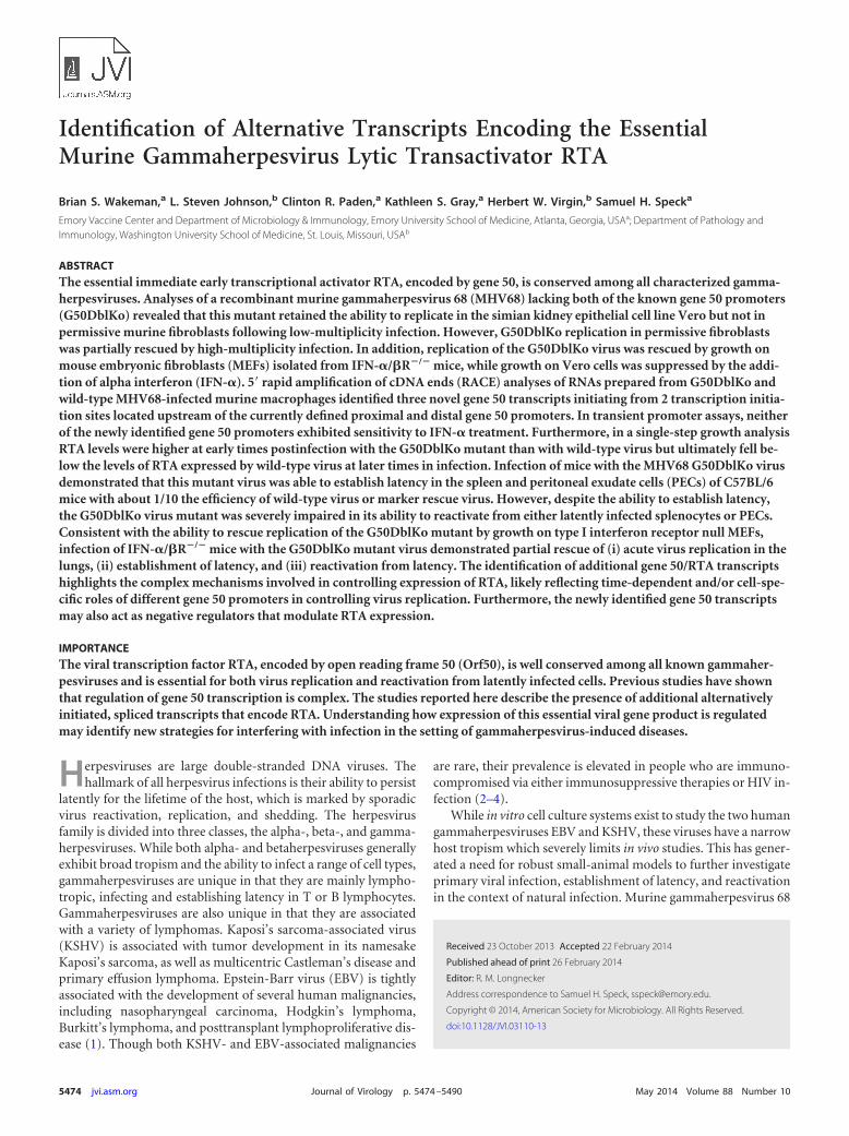
Identification of three additional gene 50 transcripts drivenby 2 promoters mapping upstream of the distal gene 50 pro-moter. We have previously shown that transcription of MHV68gene 50 is driven by two distinct promoters: (i) the proximal pro-moter, encoding a short 288-bp exon, E1, which splices to thelarge 1,800-bp exon, E2 and (ii) the distal promoter, which en-codes a short 181-bp exon, E0, which splices to E1, which in turnsplices to E2 (6, 12, 16) (Fig. 1). We further showed that thisorganization of gene 50 transcription was conserved in both EBVand KSHV (16). The unexpected ability of the G50DblKo mutantto replicate under some experimental conditions indicated thatthere must exist alternative mechanisms for driving gene 50 tran-scription, since RTA is known to be absolutely required for virusreplication (this has been shown for EBV, KSHV, and MHV68)(23, 29–32). To determine if this hypothesis was indeed correct,we performed 5= RACE analysis of RNAs from Vero-Cre, RAW264.7, and IFN-�/�R�/� MEFs infected with either wild-typeMHV68 or the G50DblKo mutant. From this analysis, we wereable to detect three previously unidentified gene 50 transcripts
FIG 4 G50DblKo virus replicates in vitro in Vero-Cre cells but fails to replicatein NIH 3T12 fibroblasts. (A) Multistep growth curve of NIH 3T12 cells in-fected with an MOI of 0.1. Cells were infected with G50DblKo, G50DblKo.MR,and WT-YFP viruses and collected for viral titer analysis at the indicated timespostinfection. (B) Multistep growth curve of Vero-Cre cells infected at an MOIof 0.1. Cells were also infected with G50DblKo, G50DblKo.MR, and WT-YFPviruses and collected for viral titer analysis at the indicated times postinfection.
Wakeman et al.
5480 jvi.asm.org Journal of Virology

which initiated from two distinct upstream transcription initia-tion sites (Fig. 7).
The three additional transcripts were identified in all three celltypes examined and also were identified in both wild-typeMHV68- and G50DblKo virus-infected cells. It is important tonote that the 5= RACE analyses conducted using the G50DblKovirus failed to detect the presence of any E1-E2 or E0-E1-E2 tran-scripts, further confirming that G50DblKo is indeed a true knock-
FIG 5 G50DblKo virus fails to replicate in vitro when IFN-� is present. (A)Multistep growth curve of C57BL/6 mouse embryonic fibroblasts (MEFs) in-fected with G50DblKo or WT-YFP virus at an MOI of 0.01. (B) Multistepgrowth curve of 129S2/SvPas.IFN-�/�R�/� mouse embryonic fibroblasts(IFN-�/�R�/� MEFs) infected with G50DblKo or WT-YFP virus at an MOI of
0.01. (C) Multistep growth curve of Vero-Cre cells infected with G50DblKo,G50DblKo.MR, or WT-YFP virus at an MOI of 0.1. (D) Multistep growthcurve of Vero-Cre cells, treated with IFN-� (10,000IU/ml) at the time of in-fection and every 24 h thereafter, infected with G50DblKo, G50DblKo.MR, orWT-YFP virus at an MOI of 0.1. All cells were collected at the times indicated,and experiments were repeated at least in triplicate.
FIG 6 Single-step growth analyses of the G50pDblKo mutant and analysis ofthe kinetics of RTA expression. (A) Single-step growth curve of G50pDblKo orWT-YFP virus in NIH 3T12 fibroblasts infected at an MOI of 10. (B) Westernblot staining for RTA, v-cyclin, and �-actin expression from the same cellsused in the single-step growth curve shown in panel A. (C) Representativesample of the small-plaque phenotype exhibited under all experimental con-ditions when using different G50pDblKo clones.
Alternative MHV68 Gene 50 Transcripts
May 2014 Volume 88 Number 10 jvi.asm.org 5481
out of the proximal and distal gene 50 promoters. The first newexon, which we have termed N3, is a 330-bp extension of thepreviously characterized E0 exon. This new N3 exon is 511 bp longand maps to coordinates bp 65579 to 66090 in the MHV68 ge-nome. The 3= end of this exon is identical to the 3= end of the E0exon. Like E0, this newly identified N3 exon splices to the E1 exon,leading to removal of a 419-bp intron, which in turn splices to theE2 exon, removing an 865-bp intron (Fig. 7). The second newexon, which we have termed N4, is a 743-bp extension of the E0exon. The N4 exon map to bp 65166 to 66090 in the MHV68genome and is 924 bp long (Fig. 7). The 3= end of this exon is alsoidentical to the 3= end of E0 and splices to the E1 exon and then tothe E2 exon. The final alternatively spliced transcript contains anew exon, which we have termed N5, which is 216 bp long andmaps to bp 65142 to 65358 in the MHV68 genome (Fig. 7). Thisnew exon consists mainly of the 5= end of the newly identified N4exon, with a 24-bp extension, indicating that the N4 and N5 exonsshare a promoter region. However, unlike N4, which is an exten-sion of E0, the N5 exon exhibits a completely unique splicing eventin which the 3= end at bp 65358 splices to E1, directly eliminatinga large 1,151-bp intron. Like the other known exons, the E1 exonthen splices to the E2 exon.
It is important to note that none of the newly identified exonshave been observed to splice directly to the E2 exon, but rather allof them splice to the E1 exon, which contains the RTA translationinitiation codon. Thus, to date, there is no evidence for alternative
RTA translation initiation sites. This, however, does not excludethe possibility of unique splicing events from the newly identifiedtranscripts to a novel position within the E2 exon or elsewherewithin the viral genome. It should be noted that there are severalshort ATG-initiated open reading frames within the newly iden-tified exons, which may play an important role (i.e., encodingnovel viral gene products and/or interfering with RTA transla-tion) (Fig. 7).
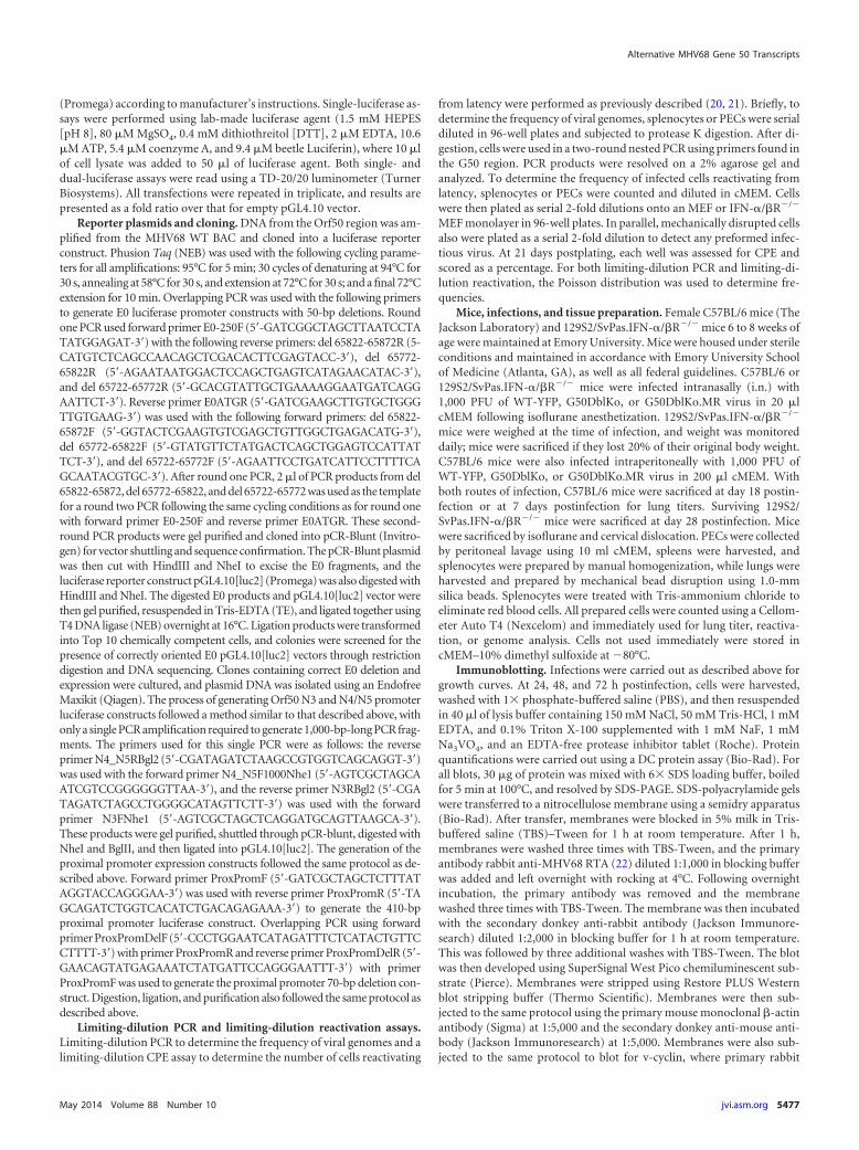
Neither the N3 promoter nor the N4/N5 promoter exhibitssensitivity to IFN-�, but RTA levels from the G50DblKo mutantare diminished in the presence of a type I IFN response at lowMOI. The identification of alternative gene 50 transcripts pro-vides a clear mechanism by which the G50DblKo virus is appar-ently able to generate sufficient levels of RTA to drive virus repli-cation, at least in the absence of a type I IFN response. However, itdoes not address the issue of why replication of the G50DblKomutant is suppressed by type I IFNs. To begin to address this issue,we cloned 1,000-bp fragments immediately upstream of the 5=ends of the N3 and N4/N5 transcripts into the pGL4.10 luciferasereporter vector (Promega Biotech) and transfected the resultingreporter constructs into Vero-Cre cells. While the N3 promoterresulted in expression �4-fold over that with empty vector, theN4/N5 promoter resulted in slightly higher levels of luciferaseexpression (�13-fold over empty vector) (Fig. 8). These datademonstrate that the region immediately upstream of the newlyidentified gene 50 transcripts exhibits modest promoter activity.
FIG 7 RACE analyses reveal three additional G50 exons upstream of E0. RACE analyses were performed using cDNAs generated from WT-YFP- and G50DblKo-infected Vero-Cre, Raw 264.7, and IFN-�/�R�/� MEFs at 24 and 48 h postinfection. 5= RACE analysis using reverse primers located in E2, E1, and E0 were usedin conjunction with the universal 5= RACE forward primer. All three experiments identified three additional exons upstream of E0: exon N3, representing a330-bp extension of E0; exon N4, representing a 743-bp extension of E0; and exon N5, representing a small novel 216-bp exon. All three new exons followcanonical splicing in which they splice to E1, which in turn splices to E2. Green arrows denote short ATG-initiated open reading frames that lie upstream of theRTA-coding sequences, which are indicated in blue. The locations of the major open reading frames antisense to gene 50 are also shown. Red arrows denoteprimers used in the 5=RACE analyses. The positions of the primers used were as follows: E2-1, bp 68095 to 68066; E2-2, bp 68051 to 68027; E1, bp 66530 to 66509;and E0, bp 65930 to 65909.
Wakeman et al.
5482 jvi.asm.org Journal of Virology
To determine if the type I IFN-sensitive replication of theG50DblKo mutant virus reflects type I IFN-mediated suppressionof gene 50 transcription from the G50DblKo mutant, we exam-ined N3 and N4/N5 promoter activity in the presence of IFN-�treatment (Fig. 8). Since Vero cells fail to produce IFN-� butretain the ability to respond to exogenously added IFN-� (Fig.5D), IFN-� was added to the transfected Vero cells and promoteractivity assessed (Fig. 8). Similar luciferase assays of previouslyidentified Orf50 transfected promoters have shown them to besensitive to IFN- (33). However, our analysis showed no evi-dence of IFN-� suppression of reporter gene activity, suggestingthat IFN-� inhibition of G50DblKo replication acts downstreamof gene 50 transcription. This is consistent with the absence of anyconsensus IFN-stimulated response elements (ISREs) in the re-gion upstream of the N3 and N4/N5 transcription initiation sites.
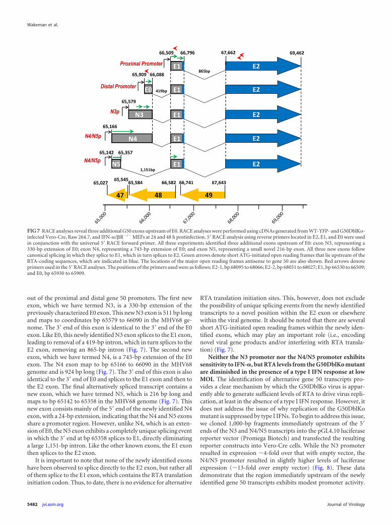
Since it appeared that the newly identified gene 50 promotersare not sensitive to IFN-�, we examined the levels of RTA proteinexpression in wild-type MHV68- and G50DblKo mutant-infectedcells. To assess RTA levels following infection, immunoblots wereprobed at several time points following low-MOI infection (Fig.9). Consistent with the observed replication defect of theG50DblKo mutant virus in either NIH 3T12 fibroblasts orC57BL/6 MEFs, RTA levels were lower at all time points assessed(Fig. 9A and B). However, equivalent levels of RTA were detectedin 129S2/SvPas.IFN-�/�R�/� MEFs infected with either wild-type MHV68 or the G50DblKo mutant virus. It is also notable thatRTA was readily detectable by 24 h postinfection of eitherC57BL/6 MEFs or 129S2/SvPas.IFN-�/�R�/� MEFs but not NIH3T12 fibroblasts (compare Fig. 9B and C with A). We have previ-ously noted that MEFs are more sensitive to MHV68 infection asdetermined by a limiting-dilution CPE analysis which can detectbetween 0.1 and 0.2 PFU of virus with titers determined on NIH3T12 fibroblasts (34), and this likely accounts for the earlier de-tection of RTA expression (i.e., effectively a higher-MOI infec-tion).
When looking at RTA levels from growth curves followinghigh-MOI infection, in which there is a partial rescue of theG50DblKo growth defect (Fig. 6A), the early kinetics and expres-sion of RTA appear to be at similar or slightly higher levels than inWT infection (Fig. 6B). RTA was detectable at significant levels at8 h postinfection in the G50DblKo virus-infected fibroblasts,while RTA was detectable at 12 h postinfection in WT virus-in-fected fibroblasts. However, the levels of RTA begin to wane by 24h postinfection with the G50DblKo mutant, while WT RTA con-tinues to increase. This may help explain the partial rescue of thegrowth phenotype at high MOI. Notably, the induction of v-cyclinexpression parallels that of RTA expression for both the WT andG50DblKo infections. These results demonstrate that despite anincreased sensitivity to type I interferons, as well as a partialgrowth defect at high MOI, the G50DblKo virus is able to generatefunctional RTA capable of driving transcription as well as launch-ing expression of downstream viral targets such as v-cyclin.
The G50DblKo mutant establishes latency in the spleen andin PECs but is severely impaired for virus reactivation. To assessinfection in mice with the G50DblKo mutant, we initially infectedC57BL/6 mice intranasally with 1,000 PFU of either theG50DblKo virus, WT MHV68, or the G50DblKo.MR virus. Wethen assessed viral latency at day 18 postinfection, the peak of
FIG 8 Promoter activity in the region immediately 5= to MHV68 N3, N4, andN5 exons. Vero cells were transfected with pGL4.10[luc] luciferase report con-structs containing either 1,000 bp upstream of N3 or 1,000 bp upstream of theN4/N5 exon. Vero cells were stimulated with IFN-� (10,000 IU/ml) at 24 hafter transfection, and at 48 h after transfection luciferase assays were per-formed. Data are presented as the fold difference of firefly luciferase activityversus the pGL4.10[luc] empty vector control. The data represent triplicates ofat least three independent transfections.
FIG 9 Immunoblot analyses of RTA protein expression levels in WT- andG50DblKo-infected NIH 3T12 fibroblasts, C57BL/6 MEFs, and 129S2/SvPas.IFN-�/�R�/� MEFs. (A) NIH 3T12 cells were infected with wild-typeMHV68 or the G50DblKo mutant at an MOI of 0.1, and cells were harvested at24, 48, and 72 h postinfection. Cells were lysed, and 30 �g of protein was usedfor the immunoblot analyses to assess RTA expression levels. (B and C) MEFsprepared from either C57BL/6 (B) or 129S2/SvPas.IFN-�/�R�/� (C) wereinfected at an MOI of 0.1, and cells were harvested at 24, 48, and 72 h postin-fection. As for panel A, cells were then lysed and 30 �g of protein used in aimmunoblot analysis to detect RTA expression levels. All immunoblots werestripped and then reprobed for �-actin levels to ensure equal protein loading.
Alternative MHV68 Gene 50 Transcripts
May 2014 Volume 88 Number 10 jvi.asm.org 5483

latency in the spleen, and determined both the frequency ofsplenocytes harboring the viral genome and the frequency ofsplenocytes able to reactivate from latency. Splenocyte reactiva-tion was determined by the previously described limiting-dilutionCPE assay (20) modified by plating splenocytes on IFN-�/�R�/�
MEF monolayers to ensure the ability to detect any reactivatedG50DblKo mutant virus. The reactivation analyses revealed thenearly complete absence of detectable reactivation of theG50DblKo virus from splenocytes (Fig. 10A). In contrast, spleno-cytes harvested from wild-type virus-infected animals reactivatedat a frequency of 1 in 7,035 cells, while the frequency of reactiva-tion from the G50DblKo.MR virus was 1 in 9,549 cells (Fig. 10Aand Table 1). No preformed infectious virus was detected in theharvested splenocyte samples, as measured by mechanical disrup-tion of the cells as previously described (20) (data not shown).
To determine if this failure to reactivate from splenocytes ob-served with the G50DblKo mutant virus was the result of a failureto establish latency, limiting-dilution PCR was performed to de-termine the frequency of splenocytes harboring viral genomes
(20). We observed that the frequency of splenocytes harboringlatent virus was roughly equivalent for the wild-type virus and theG50DblKo.MR virus at 1 in 223 and 1 in 305 cells, respectively,while the G50DblKo mutant virus was present in splenocytes at afrequency of ca. 1 in 1,941 cells (Fig. 10B and Table 1). While thefrequency of viral genome-positive splenocytes in the G50DblKovirus-infected animals was around 8.5-fold lower than that in thewild-type virus-infected animals, these analyses demonstrate thatthis mutant virus is able to get to the spleen and establish latency.Thus, the lack of detectable reactivation of the G50DblKo mutantreflects a significant defect in virus reactivation from B cells, thedominant latently infected cell population in the spleen (19, 35).
We extended these analyses to address whether the inability ofthe G50DblKo mutant virus to reactivate from splenocytes wasdependent on the route of inoculation and to assess latency andreactivation from PECs. To do this we assessed virus reactivationfrom splenocytes and PECs following intraperitoneal (i.p.) inoc-ulation. C57BL/6 mice were infected by intraperitoneal inocula-tion with 1,000 PFU of G50DblKo, wild-type, or G50DblKo.MR
FIG 10 G50DblKo virus exhibits a severe reactivation defect and a moderate latency defect in vivo. Female C57BL/6 mice were infected with 1,000 PFU i.n. (Aand B) or with 1,000 PFU i.p. (C and D). Splenocytes and PECs were harvested at 18 days postinfection and assessed for the establishment of latency andreactivation from latency by limiting-dilution CPE and PCR assays. (A) Splenocytes from i.n. infections were plated in serial dilutions onto an IFN-�/�R�/� MEFmonolayer, and at 21 days postplating wells were individually scored for CPE. The percentage of these wells was used to calculate the frequency of virallyreactivating cells. (B) Splenocytes from i.n. infections were plated in serial dilutions and subjected to nested PCR to detect gene 50 copies. The percentage ofgenome-positive cells in each dilution was used to calculate the frequency of latency. (C) PECs from i.p. infections were plated in serial dilutions onto anIFN-�/�R�/� MEF monolayer, and 21 days postplating wells were individually scored for CPE. The percentage of these wells was used to calculate the frequencyof virally reactivating cells. (D) PECs from i.p. infections were plated in serial dilutions and subjected to nested PCR to detect gene 50 copies. The percentage ofgenome-positive cells in each dilution was used to calculate the frequency of latency. For all reactivation assays (A and C), mechanically disrupted cells were platedin parallel for each virus shown to control for preformed infectious virus, and all mechanically disrupted cells were negative for reactivation (data not shown).Data are representative of at least two independent experiments consisting of 5 mice per group. Error bars indicate the standard error of the mean.
Wakeman et al.
5484 jvi.asm.org Journal of Virology
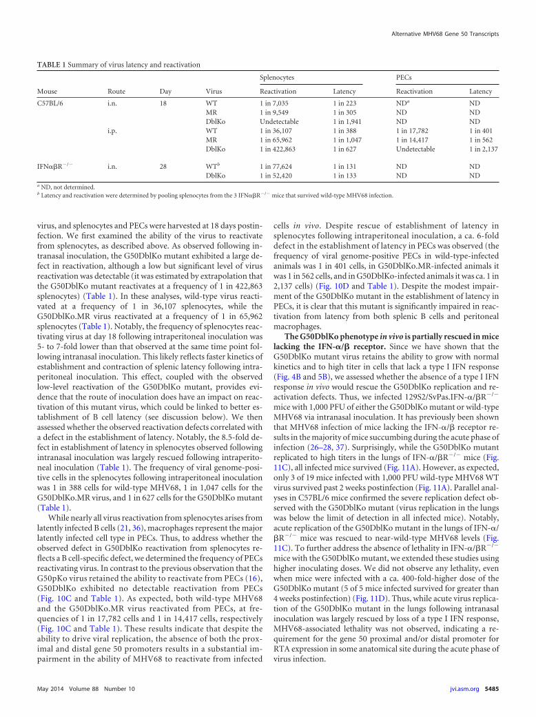
virus, and splenocytes and PECs were harvested at 18 days postin-fection. We first examined the ability of the virus to reactivatefrom splenocytes, as described above. As observed following in-tranasal inoculation, the G50DblKo mutant exhibited a large de-fect in reactivation, although a low but significant level of virusreactivation was detectable (it was estimated by extrapolation thatthe G50DblKo mutant reactivates at a frequency of 1 in 422,863splenocytes) (Table 1). In these analyses, wild-type virus reacti-vated at a frequency of 1 in 36,107 splenocytes, while theG50DblKo.MR virus reactivated at a frequency of 1 in 65,962splenocytes (Table 1). Notably, the frequency of splenocytes reac-tivating virus at day 18 following intraperitoneal inoculation was5- to 7-fold lower than that observed at the same time point fol-lowing intranasal inoculation. This likely reflects faster kinetics ofestablishment and contraction of splenic latency following intra-peritoneal inoculation. This effect, coupled with the observedlow-level reactivation of the G50DblKo mutant, provides evi-dence that the route of inoculation does have an impact on reac-tivation of this mutant virus, which could be linked to better es-tablishment of B cell latency (see discussion below). We thenassessed whether the observed reactivation defects correlated witha defect in the establishment of latency. Notably, the 8.5-fold de-fect in establishment of latency in splenocytes observed followingintranasal inoculation was largely rescued following intraperito-neal inoculation (Table 1). The frequency of viral genome-posi-tive cells in the splenocytes following intraperitoneal inoculationwas 1 in 388 cells for wild-type MHV68, 1 in 1,047 cells for theG50DblKo.MR virus, and 1 in 627 cells for the G50DblKo mutant(Table 1).
While nearly all virus reactivation from splenocytes arises fromlatently infected B cells (21, 36), macrophages represent the majorlatently infected cell type in PECs. Thus, to address whether theobserved defect in G50DblKo reactivation from splenocytes re-flects a B cell-specific defect, we determined the frequency of PECsreactivating virus. In contrast to the previous observation that theG50pKo virus retained the ability to reactivate from PECs (16),G50DblKo exhibited no detectable reactivation from PECs(Fig. 10C and Table 1). As expected, both wild-type MHV68and the G50DblKo.MR virus reactivated from PECs, at fre-quencies of 1 in 17,782 cells and 1 in 14,417 cells, respectively(Fig. 10C and Table 1). These results indicate that despite theability to drive viral replication, the absence of both the prox-imal and distal gene 50 promoters results in a substantial im-pairment in the ability of MHV68 to reactivate from infected
cells in vivo. Despite rescue of establishment of latency insplenocytes following intraperitoneal inoculation, a ca. 6-folddefect in the establishment of latency in PECs was observed (thefrequency of viral genome-positive PECs in wild-type-infectedanimals was 1 in 401 cells, in G50DblKo.MR-infected animals itwas 1 in 562 cells, and in G50DblKo-infected animals it was ca. 1 in2,137 cells) (Fig. 10D and Table 1). Despite the modest impair-ment of the G50DblKo mutant in the establishment of latency inPECs, it is clear that this mutant is significantly impaired in reac-tivation from latency from both splenic B cells and peritonealmacrophages.
The G50DblKo phenotype in vivo is partially rescued in micelacking the IFN-�/� receptor. Since we have shown that theG50DblKo mutant virus retains the ability to grow with normalkinetics and to high titer in cells that lack a type I IFN response(Fig. 4B and 5B), we assessed whether the absence of a type I IFNresponse in vivo would rescue the G50DblKo replication and re-activation defects. Thus, we infected 129S2/SvPas.IFN-�/�R�/�
mice with 1,000 PFU of either the G50DblKo mutant or wild-typeMHV68 via intranasal inoculation. It has previously been shownthat MHV68 infection of mice lacking the IFN-�/� receptor re-sults in the majority of mice succumbing during the acute phase ofinfection (26–28, 37). Surprisingly, while the G50DblKo mutantreplicated to high titers in the lungs of IFN-�/�R�/� mice (Fig.11C), all infected mice survived (Fig. 11A). However, as expected,only 3 of 19 mice infected with 1,000 PFU wild-type MHV68 WTvirus survived past 2 weeks postinfection (Fig. 11A). Parallel anal-yses in C57BL/6 mice confirmed the severe replication defect ob-served with the G50DblKo mutant (virus replication in the lungswas below the limit of detection in all infected mice). Notably,acute replication of the G50DblKo mutant in the lungs of IFN-�/�R�/� mice was rescued to near-wild-type MHV68 levels (Fig.11C). To further address the absence of lethality in IFN-�/�R�/�
mice with the G50DblKo mutant, we extended these studies usinghigher inoculating doses. We did not observe any lethality, evenwhen mice were infected with a ca. 400-fold-higher dose of theG50DblKo mutant (5 of 5 mice infected survived for greater than4 weeks postinfection) (Fig. 11D). Thus, while acute virus replica-tion of the G50DblKo mutant in the lungs following intranasalinoculation was largely rescued by loss of a type I IFN response,MHV68-associated lethality was not observed, indicating a re-quirement for the gene 50 proximal and/or distal promoter forRTA expression in some anatomical site during the acute phase ofvirus infection.
TABLE 1 Summary of virus latency and reactivation
Mouse Route Day Virus
Splenocytes PECs
Reactivation Latency Reactivation Latency
C57BL/6 i.n. 18 WT 1 in 7,035 1 in 223 NDa NDMR 1 in 9,549 1 in 305 ND NDDblKo Undetectable 1 in 1,941 ND ND
i.p. WT 1 in 36,107 1 in 388 1 in 17,782 1 in 401MR 1 in 65,962 1 in 1,047 1 in 14,417 1 in 562DblKo 1 in 422,863 1 in 627 Undetectable 1 in 2,137
IFN��R�/� i.n. 28 WTb 1 in 77,624 1 in 131 ND NDDblKo 1 in 52,420 1 in 133 ND ND
a ND, not determined.b Latency and reactivation were determined by pooling splenocytes from the 3 IFN��R�/� mice that survived wild-type MHV68 infection.
Alternative MHV68 Gene 50 Transcripts
May 2014 Volume 88 Number 10 jvi.asm.org 5485

Having shown that the severe defect in acute replication of theG50DblKo mutant observed in C57BL/6 mice was largely rescuedin mice lacking the ability to respond to type I IFNs, we assessedestablishment of latency and virus reactivation in IFN-�/�R�/�
mice. Spleens were harvested at day 28 from IFN-�/�R�/� miceintranasally infected with 1,000 PFU of either wild-type or
G50DblKo virus. It is important to note that the latency and reac-tivation analyses for wild-type virus-infected IFN-�/�R�/� micewere carried out using the subset of infected animals that survivedacute virus replication (Fig. 10A). Like the rescue of acute virusreplication in the lungs, the frequency of viral genome-positivesplenocytes went from a ca. 8-fold defect in i.n. infected C57BL/6mice (Fig. 10B) to an almost identical frequency of virus-infectedsplenocytes in IFN-�/�R�/� mice (1 in 131 cells for wild-typeMHV68 and 1 in 133 for the G50DblKo mutant) (Fig. 12A andTable 1). More importantly, the defect in virus reactivation ob-served in C57BL/6 mice was completely rescued in IFN-�/�R�/�
mice (1 in 77,624 splenocytes in WT-infected mice compared to 1in 52,420 splenocytes in G50DblKo virus-infected mice) (Fig. 12Band Table 1). The latter result argues in favor of a model in whichthe sole defect in G50DblKo virus reactivation from C57BL/6splenocytes is the inability to overcome type I IFN suppression ofvirus replication.
One possible explanation for the ability of the G50DblKo virusto replicate in the absence of type I interferons would be an in-creased expression of E1-E2 transcripts. This would make theG50DblKo virus not a true double promoter knockout virus, asE1-E2 transcripts would be responsible for the ability of the virusto replicate. However, this does not appear to be the case because(i) as shown in Fig. 6, RTA expression from the G50DblKo in
FIG 11 Infection of 129S2/SvPas.IFN-�/�R�/� mice with G50DblKo virusdoes not result in lethality as seen with WT virus, despite the presence of highviral titers in the lungs. (A) Kaplan-Meier curve depicting survival of 129S2/SvPas.IFN-�/�R�/� mice when challenged with 1,000 PFU i.n. of WT orG50DblKo virus. (B) Lung titers at day 7 from C57BL/6 mice infected with1,000 PFU i.n. of G50DblKo or G50DblKo.MR virus. (C) Lung titers at day 7from 129S2/SvPas.IFN-�/�R�/� mice infected with 1,000 PFU i.n. ofG50DblKo or G50DblKo.MR virus. (D) Kaplan-Meier curve depicting sur-vival of 129S2/SvPas.IFN-�/�R�/� mice when challenged with 440,000 PFUi.n. of G50DblKo virus compared to survival of mice when challenged with1,000 PFU G50DblKo.MR virus.
FIG 12 G50DblKo virus establishes latency and reactivates to similar levels asWT virus when used to infect 129S2/SvPas.IFN-�/�R�/� mice in vivo. Female129S2/SvPas.IFN-�/�R�/� mice were infected with 1,000 PFU of G50DblKovirus, WT virus, or G50DblKo.MR virus by intranasal injection. Mice surviv-ing at day 28 postinfection (n 20 for G50DblKo; n 3 for WT/G50DblKo.MR) were harvested for splenocytes and assessed for establishmentof latency and reactivation from latency. (A) As for Fig. 10A, splenocytes wereserially diluted and used in a CPE assay to look for the frequency of viralreactivating cells. (B) As for Fig. 10B, splenocytes were serially diluted and usedin a nested PCR assay to look for the frequency of viral genome-positive cells.
Wakeman et al.
5486 jvi.asm.org Journal of Virology

normal fibroblasts is not impaired, and (ii) analysis of 5= RACEproducts generated from Vero cells, as well as IFN-�/�R�/�
MEFs, using the G50DblKo virus failed to detect any proximalpromoter (E1-E2)- or distal promoter (E0-E1-E2)-initiated tran-scripts. However, to more directly assess potential residual prox-imal promoter activity in the absence of a type I IFN response, wecloned both the proximal promoter and the proximal promoterdeletion mutant into the pGL4.10Luc reporter plasmid. These re-porter constructs were transfected into Vero cells, in the presenceand absence of IFN-� treatment. Importantly, the proximal pro-moter does not appear to be sensitive to the presence of IFN-�(Fig. 13), which is consistent with the results shown in Fig. 5Dwhere pretreatment with IFN-� has no effect on WT MHV68growth. Furthermore, the proximal promoter deletion mutant ex-hibited significant lower promoter activity, in both the presenceand absence of added IFN-�. Thus, taken together, these resultsargue against proximal promoter-driven RTA expression ac-counting for the rescue of G50DblKo virus replication in the ab-sence of type I IFN.
DISCUSSION
Here we demonstrate that MHV68 gene 50 transcription is morecomplex than previously reported (6, 12, 16). Based on the currentanalyses, MHV68 RTA expression can be driven from four distinctpromoters, and these promoters drive expression of 5 differentspliced gene 50 transcripts. The identification of multiple promot-ers driving expression of a single gene is not a novel concept; thereare many human and viral genes whose expression has beenshown to be regulated by multiple promoters (38–42). For exam-ple, EBV has been shown to use differential splicing and from 2distinct promoters in the generation of the transcripts encodingthe six EBNA gene products (43–47). The use of multiple promot-
ers is often the result of a complex life cycle and/or, particularlyin the case of viruses, the infection of multiple cell types. We haveshown that alternatively initiated gene 50 transcripts are con-served in MHV68-, KSHV-, and EBV-infected cells, havingpreviously reported the detection of both proximal and distal pro-moter-initiated gene 50 transcripts (16). In addition, it has beenpreviously shown by others that HVS RTA expression is drivenfrom distinctly initiated transcripts expressed at different times inthe viral replication cycle (48). Here we identify 2 additional gene50 transcription initiation sites; gene 50 transcripts arising fromthese promoters could be detected in MHV68 infection of a mac-rophage cell line (RAW 264.7), a nonhuman primate epithelial cellline (Vero), and IFN-�/�R�/� mouse embryonic fibroblasts, in-dicating that these promoters appear to be widely active duringvirus replication and not restricted to a particular cell type. How-ever, we have not carried out a detailed analysis of the abundanceof specific gene 50 transcript species as a function of cell type ortime postinfection, and it is certainly possible that their activitiesin vivo are more restricted. Thus, the current analyses do not ruleout the possibility that the identified gene 50 promoters behavedifferently depending on cell type infected or address the role thateach promoter plays in different cell types during the course ofinfection.
The utility of MHV68 infection of mice is that it provides atractable small animal model to identify basic aspects of gamma-herpesvirus pathogenesis that may be relevant to the human vi-ruses EBV and KSV. Thus, we have begun to explore whether thecomplex gene 50 transcription observed during MHV68 replica-tion is conserved in the human viruses. Notably, our preliminarycharacterization of gene 50 transcription during KSHV reactiva-tion from latently infected B cells has identified multiple distinctgene 50 transcription initiation sites, as well as alternative splicingthat is very similar to that observed in MHV68-infected cells (datanot shown). Further analyses are necessary to fully characterizethese novel gene 50 transcripts expressed during KSHV reactiva-tion from B cells, as well as to characterize BRLF1 transcriptionduring EBV infection.
The data presented here are among a limited number of studies(49) showing that type I interferons play an important role insuppressing MHV68 replication in tissue culture. This is strikinglydifferent from the case for WT MHV68, where in vitro growth isnot affected by the presence of type I interferons. However, it isimportant to emphasize that in vivo type I interferons play a crit-ical role in controlling acute MHV68 replication, as mice lackingthe IFN-�/� receptor are highly susceptible to lethal MHV68 in-fection (27, 28). In addition, it has previously been shown thattype I interferons play a role in controlling viral reactivation fromlatency (27, 50).
The levels of RTA protein were greatly reduced in G50DblKo-infected cells in the presence of a type I IFN response (Fig. 9A andB), and abrogating the ability to respond to type I IFNs by disrupt-ing IFN-�/�R rescued wild-type levels of RTA expression fromthe G50DblKo mutant virus (Fig. 9C). Since RTA is the first im-mediate early gene expressed during gammaherpesvirus infection,it directly and indirectly begins the lytic replication cascade byturning on a variety of downstream genes (51–57). Many of thesedownstream genes are responsible for an anti-type I interferonresponse, such as Orf45, M2, and Orf54 (58–63). Importantly, theability of MHV68 to overcome the type I IFN response appears tobe not only essential but also immediate. Growth curves where
FIG 13 Promoter activity in the region immediately 5= to the MHV68 proxi-mal promoter and proximal promoter deletion. Vero cells were transfectedwith pGL4.10[luc] luciferase reporter constructs containing either 410 bp up-stream of exon 1 or 340 bp upstream of exon 1 containing the 70-bp deletion asshown in Fig. 3A. Vero cells were stimulated with IFN-� (10,000 IU/ml) at 24h after transfection, and at 48 h after transfection luciferase assays were per-formed. Data are presented as the fold difference of firefly luciferase activityversus the pGL4.10[luc] empty vector control. The data represent triplicates atleast three independent transfections.
Alternative MHV68 Gene 50 Transcripts
May 2014 Volume 88 Number 10 jvi.asm.org 5487
IFN-� was added prior to infection and during infection (Fig. 5D)demonstrate that the virus is able to overcome a type I IFN-in-duced antiviral cell environment; wild-type MHV68, even at a lowMOI, is unaffected by IFN-� treatment. We hypothesize, then,that this may involve three distinct mechanisms. First, the virioncontains a viral protein(s) able to suppress the type I IFN response.MHV68 proteins associated with the virion have been identified,and this includes the Orf45-encoded protein, which has alreadybeen implicated in regulating the type I IFN response (64). Sec-ond, RTA may itself be involved in mediating an anti-type I IFNresponse, but this would likely require that sufficient levels of RTAbe expressed in an appropriate time frame postinfection to beeffective. Third, RTA expression and function may be insensitiveto type I interferon effects and thus able to initiate downstreamgene signaling despite the presence of an antiviral cellular state.Future studies will need to address these possibilities.
It is notable that replication of G50DblKo is partially rescued inhigh-MOI infection. However, replication following high-MOIinfection is still severely impaired (as shown in Fig. 6, titers neverrise significantly above the input levels). Furthermore, this likelyreflects the difference between a single cycle of replication versusthe multiple rounds of replication required followed low-MOIinfection. Indeed, following high-MOI infection, initial RTA lev-els following infection with the G50DblKo virus are higher thanthose in WT virus-infected fibroblasts, but these levels decreaseover time (Fig. 6B, hours 16, 20, and 24). This indicates that RTAexpression is dysregulated in G50DblKo infected cells and thislikely impacts the tightly regulated lytic cascade ultimately leadingto diminished virus production and the observed small plaquephenotype (Fig. 6C).
Overall these studies further reveal the complex nature of gam-maherpesviruses replication. The identification of 3 additionalOrf50 transcripts was surprising but likely illustrates how the virushas evolved to carefully control expression of the essential lyticswitch gene. Though beyond the scope of this study, future workwill investigate the function of these alternative gene 50 promotersand transcripts, as well as their presence/absence in other gamma-herpesviruses. It is clear that in the absence of type I interferons,the newly identified gene 50 transcripts are able to drive viral rep-lication nearly as efficiently as wild-type MHV68. This suggeststhat these promoters may play a cell type-specific and/or time-dependent role in the viral life cycle. Indeed, independent studieshave recently revealed that the N4/N5 promoter is responsive tointerleukin-4 (IL-4) treatment in macrophages, which has beenshown to trigger MHV68 reactivation from latently infected mac-rophages (T. A. Reese et al., unpublished data). Another possibil-ity is that upstream-initiated gene 50 transcription may, undersome conditions, serve to suppress RTA expression by interferingwith transcription from the more proximal gene 50 promoters.Transcriptional interference has been shown before for some EBVtranscripts (44, 65–67). In addition, the role of the short ATG-initiated open reading frames in the 5= untranslated regions ofupstream-initiated gene 50 transcripts remains to be investigated.
In summary, the analysis of the G50DblKo mutant extends ourunderstanding of RTA expression and has revealed a hitherto-unappreciated complexity of gene 50 transcription. It is notablethat until the analysis of the G50DblKo mutant, little or no impactof type I interferons on wild-type MHV68 replication was ob-served. This was in stark contrast to the analysis of MHV68 repli-cation during the acute stages of infection in IFN-�/�R�/� mice,
where substantially higher levels of virus replication are observedin the lungs of type I interferon-unresponsive mice than in thoseof wild-type mice following i.n. virus inoculation (e.g., see Fig. 11)(28). Based on the observations with G50DblKo mutant replica-tion in vitro, it is perhaps reasonable to speculate that during acutevirus replication there may be a role for type I IFN-sensitive gene50 transcription. Future studies will address whether any of thenewly identified gene 50 transcripts play such a role during acutevirus replication in vivo.
ACKNOWLEDGMENTS
We thank members of the Speck lab for helpful comments.This research was supported by NIH R01 grant CA52004 to
S.H.S. S.H.S. was also supported by NIH R01 grants AI073830, CA095318,and AI058057.
REFERENCES1. Young LS, Rickinson AB. 2004. Epstein-Barr virus: 40 years on. Nat. Rev.
Cancer 4:757–768. http://dx.doi.org/10.1038/nrc1452.2. da Silva SR, de Oliveira DE. 2011. HIV, EBV and KSHV: viral coopera-
tion in the pathogenesis of human malignancies. Cancer Lett. 305:175–185. http://dx.doi.org/10.1016/j.canlet.2011.02.007.
3. D’Antonio A, Boscaino A, Addesso M, Piris MA, Nappi O. 2007. KSHV-and EBV-associated germinotropic lymphoproliferative disorder: a rarelymphoproliferative disease of HIV patient with plasmablastic morphol-ogy, indolent course and favourable response to therapy. Leuk. Lym-phoma 48:1444 –1447. http://dx.doi.org/10.1080/10428190701387039.
4. Pyakurel P, Pak F, Mwakigonja AR, Kaaya E, Biberfeld P. 2007. KSHV/HHV-8 and HIV infection in Kaposi’s sarcoma development. Infect.Agents Cancer 2:4. http://dx.doi.org/10.1186/1750-9378-2-4.
5. Speck SH, Virgin HW. 1999. Host and viral genetics of chronic infection:a mouse model of gamma-herpesvirus pathogenesis. Curr. Opin. Micro-biol. 2:403– 409. http://dx.doi.org/10.1016/S1369-5274(99)80071-X.
6. Virgin HW, IV, Latreille P, Wamsley P, Hallsworth K, Weck KE, DalCanto AJ, Speck SH. 1997. Complete sequence and genomic analysis ofmurine gammaherpesvirus 68. J. Virol. 71:5894 –5904.
7. Barton E, Mandal P, Speck SH. 2011. Pathogenesis and host control ofgammaherpesviruses: lessons from the mouse. Annu. Rev. Immunol. 29:351–397. http://dx.doi.org/10.1146/annurev-immunol-072710-081639.
8. Kulkarni AB, Holmes KL, Fredrickson TN, Hartley JW, Morse HC, III.1997. Characteristics of a murine gammaherpesvirus infection immuno-compromised mice. In Vivo 11:281–291.
9. Liang X, Paden CR, Morales FM, Powers RP, Jacob J, Speck SH.2011. Murine gamma-herpesvirus immortalization of fetal liver-derived B cells requires both the viral cyclin D homolog and latency-associated nuclear antigen. PLoS Pathog. 7:e1002220. http://dx.doi.org/10.1371/journal.ppat.1002220.
10. Ragoczy T, Heston L, Miller G. 1998. The Epstein-Barr virus Rta proteinactivates lytic cycle genes and can disrupt latency in B lymphocytes. J.Virol. 72:7978 –7984.
11. Sun R, Lin SF, Gradoville L, Yuan Y, Zhu F, Miller G. 1998. A viral genethat activates lytic cycle expression of Kaposi’s sarcoma-associated herpes-virus. Proc. Natl. Acad. Sci. U. S. A. 95:10866 –10871. http://dx.doi.org/10.1073/pnas.95.18.10866.
12. Liu S, Pavlova IV, Virgin HW, IV, Speck SH. 2000. Characterization ofgammaherpesvirus 68 gene 50 transcription. J. Virol. 74:2029 –2037. http://dx.doi.org/10.1128/JVI.74.4.2029-2037.2000.
13. Pavlova I, Yulin C, Speck S. 2005. Murine gammaherpesvirus 68 Rta-dependent activation of the gene 57 promoter. Virology 333:169 –179.http://dx.doi.org/10.1016/j.virol.2004.12.021.
14. Allen RD, DeZalia MN, Speck SH. 2007. Identification of an Rta respon-sive promoter involved in driving HV68 v-cyclin expression during virusreplication. Virology 365:250 –259. http://dx.doi.org/10.1016/j.virol.2007.03.021.
15. Xi X, Persson LM, O’Brien MW, Mohr I, Wilson AC. 2012. Cooperationbetween viral interferon regulatory factor 4 and RTA to activate a subset ofKaposi’s sarcoma-associated herpesvirus lytic promoters. J. Virol. 86:1021–1033. http://dx.doi.org/10.1128/JVI.00694-11.
16. Gray KS, Allen RD, Farrell ML, Forrest JC, Speck SH. 2009. Alterna-tively initiated gene 50/RTA transcripts expressed during murine and hu-
Wakeman et al.
5488 jvi.asm.org Journal of Virology

man gammaherpesvirus reactivation from latency. J. Virol. 83:314 –328.http://dx.doi.org/10.1128/JVI.01444-08.
17. Paden CR, Forrest JC, Tibbetts SA, Speck SH. 2012. Unbiased mutagen-esis of MHV68 LANA reveals a DNA-binding domain required for LANAfunction in vitro and in vivo. PLoS Pathog. 8:e1002906. http://dx.doi.org/10.1371/journal.ppat.1002906.
18. Warming S. 2005. Simple and highly efficient BAC recombineering usinggalK selection. Nucleic Acids Res. 33:e36 – e36. http://dx.doi.org/10.1093/nar/gni035.
19. Collins CM, Boss JM, Speck SH. 2009. Identification of infected B-cellpopulations by using a recombinant murine gammaherpesvirus 68 ex-pressing a fluorescent protein. J. Virol. 83:6484 – 6493. http://dx.doi.org/10.1128/JVI.00297-09.
20. Weck KE, Barkon ML, Yoo LI, Speck SH, Virgin HI. 1996. Mature Bcells are required for acute splenic infection, but not for establishment oflatency, by murine gammaherpesvirus 68. J. Virol. 70:6775– 6780.
21. Weck KE, Kim SS, Virgin HI, Speck SH. 1999. B cells regulate murinegammaherpesvirus 68 latency. J. Virol. 73:4651– 4661.
22. Dong X, He Z, Durakoglugil D, Arneson L, Shen Y, Feng P. 2012.Murine gammaherpesvirus 68 evades host cytokine production via repli-cation transactivator-induced RelA degradation. J. Virol. 86:1930 –1941.http://dx.doi.org/10.1128/JVI.06127-11.
23. Pavlova IV, Virgin HW, Speck SH. 2003. Disruption of gammaherpesvirus68 gene 50 demonstrates that Rta is essential for virus replication. J. Virol.77:5731–5739. http://dx.doi.org/10.1128/JVI.77.10.5731-5739.2003.
24. Moorman NJ, Willer DO, Speck SH. 2003. The gammaherpesvirus 68latency-associated nuclear antigen homolog is critical for the establish-ment of splenic latency. J. Virol. 77:10295–10303. http://dx.doi.org/10.1128/JVI.77.19.10295-10303.2003.
25. Adler H, Messerle M, Koszinowski UH. 2001. Virus reconstituted frominfectious bacterial artificial chromosome (BAC)-cloned murine gamma-herpesvirus 68 acquires wild-type properties in vivo only after excision ofBAC vector sequences. J. Virol. 75:5692–5696. http://dx.doi.org/10.1128/JVI.75.12.5692-5696.2001.
26. Arico E, Robertson KA, Belardelli F, Ferrantini M, Nash AA. 2004.Vaccination with inactivated murine gammaherpesvirus 68 strongly lim-its viral replication and latency and protects type I IFN receptor knockoutmice from a lethal infection. Vaccine 22:1433–1440. http://dx.doi.org/10.1016/j.vaccine.2003.10.015.
27. Barton ES, Lutzke ML, Rochford R, Virgin HW, IV. 2005. Alpha/betainterferons regulate murine gammaherpesvirus latent gene expressionand reactivation from latency. J. Virol. 79:14149 –14160. http://dx.doi.org/10.1128/JVI.79.22.14149-14160.2005.
28. Dutia BM, Allen DJ, Dyson H, Nash AA. 1999. Type I interferons andIRF-1 play a critical role in the control of a gammaherpesvirus infection.Virology 261:173–179. http://dx.doi.org/10.1006/viro.1999.9834.
29. El-Guindy A, Ghiassi-Nejad M, Golden S, Delecluse HJ, Miller G. 2013.Essential role of Rta in lytic DNA replication of Epstein-Barr virus. J. Virol.87:208 –223. http://dx.doi.org/10.1128/JVI.01995-12.
30. Guito J, Lukac DM. 2012. KSHV Rta promoter specification and viral reac-tivation. Front. Microbiol. 3:30. http://dx.doi.org/10.3389/fmicb.2012.00030.
31. Wang Y, Tang Q, Maul GG, Yuan Y. 2006. Kaposi’s sarcoma-associatedherpesvirus ori-Lyt-dependent DNA replication: dual role of replicationand transcription activator. J. Virol. 80:12171–12186. http://dx.doi.org/10.1128/JVI.00990-06.
32. Wu TT, Tong L, Rickabaugh T, Speck S, Sun R. 2001. Function of Rtais essential for lytic replication of murine gammaherpesvirus 68. J. Virol.75:9262–9273. http://dx.doi.org/10.1128/JVI.75.19.9262-9273.2001.
33. Goodwin MM, Canny S, Steed A, Virgin HW. 2010. Murine gamma-herpesvirus 68 has evolved gamma interferon and stat1-repressible pro-moters for the lytic switch gene 50. J. Virol. 84:3711–3717. http://dx.doi.org/10.1128/JVI.02099-09.
34. Krug LT, Collins CM, Gargano LM, Speck SH. 2009. NF-kappaB p50plays distinct roles in the establishment and control of murine gamma-herpesvirus 68 latency. J. Virol. 83:4732– 4748. http://dx.doi.org/10.1128/JVI.00111-09.
35. Coleman CB, Nealy MS, Tibbetts SA. 2010. Immature and transitional Bcells are latency reservoirs for a gammaherpesvirus. J. Virol. 84:13045–13052. http://dx.doi.org/10.1128/JVI.01455-10.
36. Liang X, Collins CM, Mendel JB, Iwakoshi NN, Speck SH. 2009.Gammaherpesvirus-driven plasma cell differentiation regulates virus re-activation from latently infected B lymphocytes. PLoS Pathog.5:e1000677. http://dx.doi.org/10.1371/journal.ppat.1000677.
37. Paden CR, Forrest JC, Moorman NJ, Speck SH. 2010. Murine gamma-herpesvirus 68 LANA is essential for virus reactivation from splenocytesbut not long-term carriage of viral genome. J. Virol. 84:7214 –7224. http://dx.doi.org/10.1128/JVI.00133-10.
38. Koenigsberger C, Chicca JJ, II, Amoureux MC, Edelman GM, Jones FS.2000. Differential regulation by multiple promoters of the gene encodingthe neuron-restrictive silencer factor. Proc. Natl. Acad. Sci. U. S. A. 97:2291–2296. http://dx.doi.org/10.1073/pnas.050578797.
39. Delaloy C, Lu J, Houot AM, Disse-Nicodeme S, Gasc JM, Corvol P,Jeunemaitre X. 2003. Multiple promoters in the WNK1 gene: one controlsexpression of a kidney-specific kinase-defective isoform. Mol. Cell. Biol. 23:9208–9221. http://dx.doi.org/10.1128/MCB.23.24.9208-9221.2003.
40. Ozbun MA, Meyers C. 1998. Temporal usage of multiple promotersduring the life cycle of human papillomavirus type 31b. J. Virol. 72:2715–2722.
41. Mitsui S, Nakamura T, Okui A, Kominami K, Uemura H, YamaguchiN. 2006. Multiple promoters regulate tissue-specific alternative splicing ofthe human kallikrein gene, KLK11/hippostasin. FEBS J. 273:3678 –3686.http://dx.doi.org/10.1111/j.1742-4658.2006.05372.x.
42. Kim JD, Kim CH, Kwon BS. 2011. Regulation of mouse 4-1BB expres-sion: multiple promoter usages and a splice variant. Mol. Cells 31:141–149. http://dx.doi.org/10.1007/s10059-011-0018-6.
43. Bodescot M, Perricaudet M, Farrell PJ. 1987. A promoter for the highlyspliced EBNA family of RNAs of Epstein-Barr virus. J. Virol. 61:3424 –3430.
44. Woisetschlaeger M, Strominger JL, Speck SH. 1989. Mutually exclusiveuse of viral promoters in Epstein-Barr virus latently infected lymphocytes.Proc. Natl. Acad. Sci. U. S. A. 86:6498 – 6502. http://dx.doi.org/10.1073/pnas.86.17.6498.
45. Rogers RP, Woisetschlaeger M, Speck SH. 1990. Alternative splicingdictates translational start in Epstein-Barr virus transcripts. The EMBOjournal. 9:2273–2277.
46. Speck SH, Strominger JL. 1985. Analysis of the transcript encoding thelatent Epstein-Barr virus nuclear antigen I: a potentially polycistronicmessage generated by long-range splicing of several exons. Proc. Natl.Acad. Sci. U. S. A. 82:8305– 8309. http://dx.doi.org/10.1073/pnas.82.24.8305.
47. Speck SH, Pfitzner A, Strominger JL. 1986. An Epstein-Barr virus tran-script from a latently infected, growth-transformed B-cell line encodes ahighly repetitive polypeptide. Proc. Natl. Acad. Sci. U. S. A. 83:9298 –9302.http://dx.doi.org/10.1073/pnas.83.24.9298.
48. Whitehouse A, Carr IM, Griffiths JC, Meredith DM. 1997. The herpes-virus saimiri ORF50 gene, encoding a transcriptional activator homolo-gous to the Epstein-Barr virus R protein, is transcribed from two distinctpromoters of different temporal phases. J. Virol. 71:2550 –2554.
49. Wood BM, Mboko WP, Mounce BC, Tarakanova VL. 2013. Mousegammaherpesvirus-68 infection acts as a rheostat to set the level of type Iinterferon signaling in primary macrophages. Virology 443:123–133. http://dx.doi.org/10.1016/j.virol.2013.04.036.
50. Mandal P, Krueger BE, Oldenburg D, Andry KA, Beard RS, White DW,Barton ES. 2011. A gammaherpesvirus cooperates with interferon-alpha/beta-induced IRF2 to halt viral replication, control reactivation, and min-imize host lethality. PLoS Pathog. 7:e1002371. http://dx.doi.org/10.1371/journal.ppat.1002371.
51. Chen J, Ye F, Xie J, Kuhne K, Gao SJ. 2009. Genome-wide identificationof binding sites for Kaposi’s sarcoma-associated herpesvirus lytic switchprotein, RTA. Virology. 386:290 –302. http://dx.doi.org/10.1016/j.virol.2009.01.031.
52. Liu Y, Cao Y, Liang D, Gao Y, Xia T, Robertson ES, Lan K. 2008.Kaposi’s sarcoma-associated herpesvirus RTA activates the processivityfactor ORF59 through interaction with RBP-Jkappa and a cis-acting RTAresponsive element. Virology 380:264 –275. http://dx.doi.org/10.1016/j.virol.2008.08.011.
53. Deng H, Liang Y, Sun R. 2007. Regulation of KSHV lytic gene expression.Curr. Top. Microbiol. Immunol. 312:157–183. http://dx.doi.org/10.1007/978-3-540-34344-8_6.
54. Matsumura S, Fujita Y, Gomez E, Tanese N, Wilson AC. 2005. Activa-tion of the Kaposi’s sarcoma-associated herpesvirus major latency locusby the lytic switch protein RTA (ORF50). J. Virol. 79:8493– 8505. http://dx.doi.org/10.1128/JVI.79.13.8493-8505.2005.
55. Song MJ, Deng H, Sun R. 2003. Comparative study of regulation ofRTA-responsive genes in Kaposi’s sarcoma-associated herpesvirus/
Alternative MHV68 Gene 50 Transcripts
May 2014 Volume 88 Number 10 jvi.asm.org 5489
human herpesvirus 8. J. Virol. 77:9451–9462. http://dx.doi.org/10.1128/JVI.77.17.9451-9462.2003.
56. Ragoczy T, Miller G. 1999. Role of the Epstein-Barr virus RTA protein inactivation of distinct classes of viral lytic cycle genes. J. Virol. 73:9858 –9866.
57. Chang Y, Lee HH, Chang SS, Hsu TY, Wang PW, Chang YS, Takada K,Tsai CH. 2004. Induction of Epstein-Barr virus latent membrane protein1 by a lytic transactivator Rta. J. Virol. 78:13028 –13036. http://dx.doi.org/10.1128/JVI.78.23.13028-13036.2004.
58. Zhu FX, Sathish N, Yuan Y. 2010. Antagonism of host antiviral responsesby Kaposi’s sarcoma-associated herpesvirus tegument protein ORF45.PLoS One 5:e10573. http://dx.doi.org/10.1371/journal.pone.0010573.
59. Liang X, Shin YC, Means RE, Jung JU. 2004. Inhibition of interferon-mediated antiviral activity by murine gammaherpesvirus 68 latency-associated M2 protein. J. Virol. 78:12416 –12427. http://dx.doi.org/10.1128/JVI.78.22.12416-12427.2004.
60. Liang Q, Fu B, Wu F, Li X, Yuan Y, Zhu F. 2012. ORF45 of Kaposi’ssarcoma-associated herpesvirus inhibits phosphorylation of interferonregulatory factor 7 by IKKepsilon and TBK1 as an alternative substrate. J.Virol. 86:10162–10172. http://dx.doi.org/10.1128/JVI.05224-11.
61. Sathish N, Zhu FX, Golub EE, Liang Q, Yuan Y. 2011. Mechanisms ofautoinhibition of IRF-7 and a probable model for inactivation of IRF-7 byKaposi’s sarcoma-associated herpesvirus protein ORF45. J. Biol. Chem.286:746 –756. http://dx.doi.org/10.1074/jbc.M110.150920.
62. Zhu FX, King SM, Smith EJ, Levy DE, Yuan Y. 2002. A Kaposi’ssarcoma-associated herpesviral protein inhibits virus-mediated inductionof type I interferon by blocking IRF-7 phosphorylation and nuclear accu-mulation. Proc. Natl. Acad. Sci. U. S. A. 99:5573–5578. http://dx.doi.org/10.1073/pnas.082420599.
63. Leang RS, Wu TT, Hwang S, Liang LT, Tong L, Truong JT, Sun R. 2011.The anti-interferon activity of conserved viral dUTPase ORF54 is essentialfor an effective MHV-68 infection. PLoS Pathog. 7:e1002292. http://dx.doi.org/10.1371/journal.ppat.1002292.
64. Bortz E, Whitelegge JP, Jia Q, Zhou ZH, Stewart JP, Wu TT, Sun R.2003. Identification of proteins associated with murine gammaherpesvi-rus 68 virions. J. Virol. 77:13425–13432. http://dx.doi.org/10.1128/JVI.77.24.13425-13432.2003.
65. Paulson EJ, Fingeroth JD, Yates JL, Speck SH. 2002. Methylation of theEBV genome and establishment of restricted latency in low-passage EBV-infected 293 epithelial cells. Virology 299:109 –121. http://dx.doi.org/10.1006/viro.2002.1457.
66. Schaefer BC, Strominger JL, Speck SH. 1997. Host-cell-determinedmethylation of specific Epstein-Barr virus promoters regulates the choicebetween distinct viral latency programs. Mol. Cell. Biol. 17:364 –377.
67. Woisetschlaeger M, Yandava CN, Furmanski LA, Strominger JL, SpeckSH. 1990. Promoter switching in Epstein-Barr virus during the initialstages of infection of B lymphocytes. Proc. Natl. Acad. Sci. U. S. A. 87:1725–1729. http://dx.doi.org/10.1073/pnas.87.5.1725.
Wakeman et al.
5490 jvi.asm.org Journal of Virology