Embed Size (px)
Citation preview
MOLECULAR AND CELLULAR BIOLOGY, Sept. 1992, p. 4164-41690270-7306/92/094164-06$02.00/0Copyright © 1992, American Society for Microbiology
Vol. 12, No. 9
Identification of an Early-Growth-Response Gene Encoding aNovel Putative Protein Kinase
DANIEL L. SIMMONS,`* BENJAMIN G. NEEL,2 RYAN STEVENS,' GARY EVETT,1 ANDRAYMOND L. ERIKSON3
Department of Chemistry, 226 Eyring Science Center, Brigham Young Universitz, Provo, Utah 846021;Molecular Medicine Unit, Beth Israel Hospital, Boston, Massachusetts 02215; and Department of
Cellular and Developmental Biology, Harvard University, Cambridge, Massachusetts 021383
Received 8 May 1992/Returned for modification 26 May 1992/Accepted 22 June 1992
Early-growth-response genes, also known as immediate-early genes, play important roles in regulating cellproliferation. We have identified a new type of early-growth-response gene product, a 77,811-Da putativeserine/threonine kinase, which is highly inducible by serum and phorbol ester. mRNA encoding this putativekinase is markedly elevated within 1 h after treatment with mitogen, and this induction is synergisticallyincreased by cycloheximide. Dexamethasone blocks serum induction of the kinase mRNA, as does transfor-mation by v-Ki-ras. The kinase mRNA was detected in mouse brain, lung, and heart. This new putative kinase,which we term Snk, for serum-inducible kinase, showed similarity in its proposed catalytic domain to manyother protein kinases; however, no other kinase showed enough sequence similarity with Snk to suggest theexistence of a common function. Hence, Snk represents a new type of protein kinase involved in the earlymitogenic response whose activity is transcriptionally and posttranscriptionally regulated.
More than 80 genes called early-growth-response or im-mediate-early genes are now known to be activated by awide variety of mitogens (2, 12, 20, 22, 32). The identities ofmany of these mitogen-inducible genes, such as those en-coding growth factors, transcription factors/DNA-bindingproteins, and cytoskeletal proteins, have been determinedby DNA cloning and sequencing (15). A newly discoveredisoenzyme of prostaglandin G/H synthase, which catalyzesthe rate-limiting step in the production of prostaglandins, isalso induced in an early-growth-response fashion by manymitogens, including the v-src oncogene product pp6ov-s(10, 19, 26, 33).
Signal transduction following exposure of cells to mito-gens requires messenger networks to transmit growth regu-latory signals to target sites within the cell. Key componentsin many of these signal transduction networks are kinases,which, through phosphorylation, modulate the activity ofother kinases, structural proteins, transcription factors, andsmall molecular messengers. An end result ofmany mitogen-stimulated signal transduction pathways is a rapid increasein the transcription of selected early-growth-response genes.Typically, this effect on transcription occurs within minutesof initiation of signal transduction and transpires in theabsence of protein synthesis.Here we report the identification and characterization of a
new type of mitogen-inducible early-growth-response geneproduct, which, like the prostaglandin G/H synthase, is anenzyme, in this case a putative new protein kinase, appar-ently of the serine/threonine type.
MATERIALS AND METHODSCell culture. The NIH 3T3 cell line used in these studies
and its derivative lines, F-2 and DT, were obtained fromRobert Bassin, National Institutes of Health. The DT linewas originally created by doubly transforming NIH 3T3 cellswith two copies of the v-Ki-ras oncogene (25). The F-2 cell
* Corresponding author.
line arose as a nontumorigenic, genetically dominant mor-phological revertant of DT that still contained markedlyelevated v-Ki-ras expression (25).Another cell line used in these studies, RS2, is a trans-
formed rat fibroblast line obtained from Mark Smith, Na-tional Cancer Institute, Frederick, Md. RS2 is a v-fos-transformed rat fibroblast line originating from TomCurran's laboratory (9).Mitogen stimulation was performed on 5 x 10⁶ cells per
100-mm-diameter dish cultured for 16 to 24 h under condi-tions of serum deprivation (Dulbecco's modified Eagle'smedium [DMEM] containing 0.9% fetal calf serum [FCS]) toinhibit cell proliferation. The cells were stimulated to divideby addition of FCS to 10% concentration or phorbol 12-myristate 13-acetate (PMA) to a final concentration of 75,uM. In some cases, serum or PMA was added simulta-neously with 75 ,uM cycloheximide (CHX; Sigma ChemicalCo., St. Louis, Mo.) to determine the influence of proteintranslation on induction of clone 2 mRNA. The effect of 2p,M dexamethasone (DEX; Sigma) on transcription andmRNA levels was evaluated either by adding the DEX withserum or by pretreating the cells for up to 2 h with DEX priorto addition of serum. Pretreatment of the cells with DEXappeared to be equal in effect to simultaneous addition of theDEX with serum in inhibiting induction of clone 2 mRNA.RNA isolation. RNA for gel blot analysis, library construc-
tion, and screening was obtained from cells by lithiumchloride precipitation (3). Poly(A) mRNA used for libraryconstruction was prepared by two cycles of oligo(dT) chro-matography (4). RNA from mouse tissues was prepared byhomogenizing whole organs in guanidinium isothiocyanateas described previously (7).
Library construction and screening. A cDNA library wasconstructed in XZAP, using mRNA isolated from F-2 cells tosynthesize the cDNA. The library (>106 recombinants perjig ofcDNA) was constructed with a kit made by Gibco BRL(Gaithersburg, Md.).Clone 2 was originally isolated as a sequence artifactually
ligated to another cDNA. The clone 2 chimera was obtained
4164
Snk, A NOVEL PUTATIVE PROTEIN KINASE 4165
during screening of the F-2 cDNA library for full-lengthcopies of partial-length cDNAs under characterization in ourlaboratory. Before the artifactual nature of the clone 2chimera was determined, this spurious ligation product wasused to probe a Northern (RNA) blot of RNAs isolated frommitogen-stimulated cells. This experiment detected a newmRNA which was strongly induced in serum-stimulated F-2cells. The portion of the chimera responsible for hybridizingstrongly to the serum-inducible mRNA was identified byrestriction endonuclease mapping and DNA sequencing andwas used to isolate several independent, nonchimeric iso-lates of clone 2 cDNA from approximately 105 recombi-nants.
Northern analysis. RNA was electrophoresed on denatur-ing formaldehyde gels, blotted to nylon, and probed (24).cDNA inserts, isolated by electrophoresis in low-melting-point agarose gels, were radiolabeled and hybridized for 12to 24 h to the nylon filters at 65°C in Church-Gilbert buffer(8). Hybridized filters were washed at 65°C in a solutioncontaining 0.2x SSC (lx SSC is 0.15 M sodium chlorideplus 0.015 M sodium citrate) and 0.5% sodium dodecylsulfate.DNA sequencing and computer analysis. Two separate
recombinant phage isolates containing the entire coding and3' untranslated regions of clone 2 cDNA were identified.Both of these cDNAs were 2.8 kb in size. One of the twoisolates, termed clone 2-full, was sequenced on both strandsin its entirety by the dideoxy chain termination method ofSanger et al. (29).Homology searches at the nucleic acid and protein levels
of the GenBank and National Biomedical Research Founda-tion data bases were done by using both the Intelligenetics(FASTA and FASTP) and the University of WisconsinGenetics Computer Group (WORDSEARCH and BESTFIT)programs. The BLASTN and BLASTP programs of theNational Center for Biotechnology Information were alsoused to do global searches for sequence similarity.
Transcription analyses. Isolated nuclei for transcriptionanalyses were obtained and used to measure transcription inrun-on assays as described by Linial et al. (23). Nitrocellu-lose (Schleicher & Schuell Inc., Keene, N.H.) strips con-taining filter-bound plasmids were prepared as describedpreviously (31). Hybridization of 2.8 x 10⁶ cpm/ml ofradiolabeled nascent transcripts to filter-bound plasmids wasdone in Church-Gilbert buffer at 65°C for 40 h, and the filterswere washed as described previously (31).
Nucleotide sequence accession number. The GenBank ac-cession number for clone 2 is M96163.
0.9% Serum -10% Serum
0)
3._0
Clone 2 .
x x
I ±C.) C.)
_- %- L. 28S+ +%- b- = -C Z-C " I.. _ -.S.r m N III w e s = s v I-N u:-r NLn N et etr R* co
v o-*28S
il. ffi::,
s _ ,~~~-18S
GAPDH
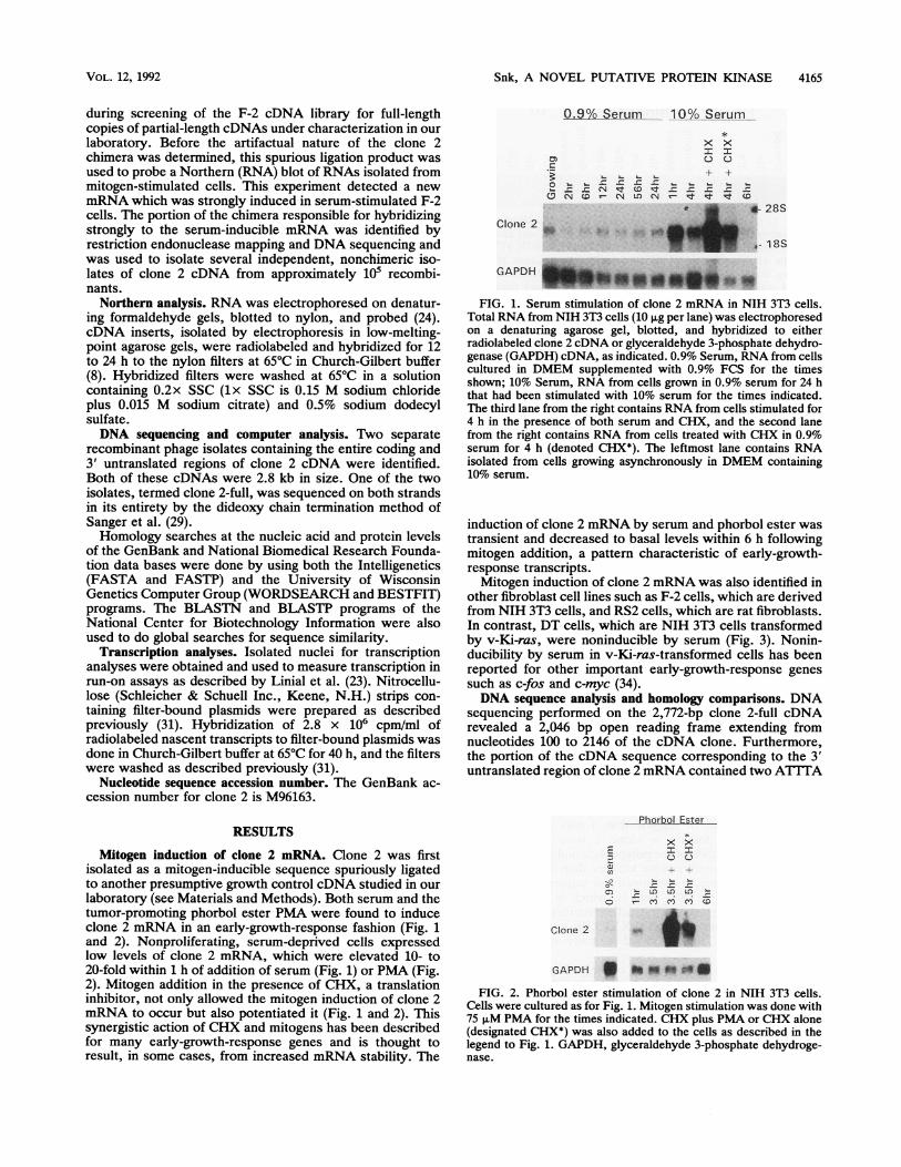
FIG. 1. Serum stimulation of clone 2 mRNA in NIH 3T3 cells.Total RNA from NIH 3T3 cells (10 pLg per lane) was electrophoresedon a denaturing agarose gel, blotted, and hybridized to eitherradiolabeled clone 2 cDNA or glyceraldehyde 3-phosphate dehydro-genase (GAPDH) cDNA, as indicated. 0.9% Serum, RNA from cellscultured in DMEM supplemented with 0.9% FCS for the timesshown; 10% Serum, RNA from cells grown in 0.9% serum for 24 hthat had been stimulated with 10% serum for the times indicated.The third lane from the right contains RNA from cells stimulated for4 h in the presence of both serum and CHX, and the second lanefrom the right contains RNA from cells treated with CHX in 0.9%serum for 4 h (denoted CHX*). The leftmost lane contains RNAisolated from cells growing asynchronously in DMEM containing10% serum.
induction of clone 2 mRNA by serum and phorbol ester wastransient and decreased to basal levels within 6 h followingmitogen addition, a pattern characteristic of early-growth-response transcripts.Mitogen induction of clone 2 mRNA was also identified in
other fibroblast cell lines such as F-2 cells, which are derivedfrom NIH 3T3 cells, and RS2 cells, which are rat fibroblasts.In contrast, DT cells, which are NIH 3T3 cells transformedby v-Ki-ras, were noninducible by serum (Fig. 3). Nonin-ducibility by serum in v-Ki-ras-transformed cells has beenreported for other important early-growth-response genessuch as c-fos and c-myc (34).DNA sequence analysis and homology comparisons. DNA
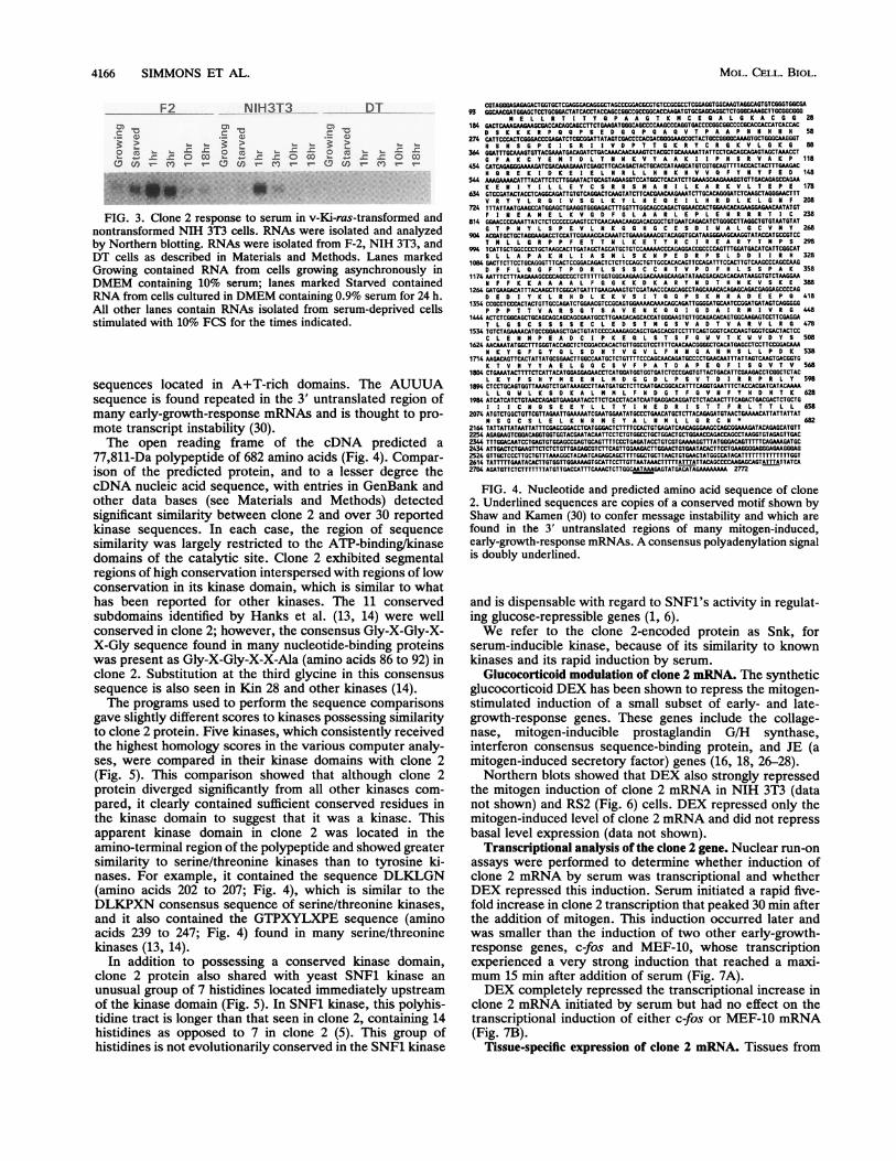
sequencing performed on the 2,772-bp clone 2-full cDNArevealed a 2,046 bp open reading frame extending fromnucleotides 100 to 2146 of the cDNA clone. Furthermore,the portion of the cDNA sequence corresponding to the 3'untranslated region of clone 2 mRNA contained two ATTTA
RESULTS
Mitogen induction of clone 2 mRNA. Clone 2 was firstisolated as a mitogen-inducible sequence spuriously ligatedto another presumptive growth control cDNA studied in ourlaboratory (see Materials and Methods). Both serum and thetumor-promoting phorbol ester PMA were found to induceclone 2 mRNA in an early-growth-response fashion (Fig. 1and 2). Nonproliferating, serum-deprived cells expressedlow levels of clone 2 mRNA, which were elevated 10- to20-fold within 1 h of addition of serum (Fig. 1) or PMA (Fig.2). Mitogen addition in the presence of CHX, a translationinhibitor, not only allowed the mitogen induction of clone 2mRNA to occur but also potentiated it (Fig. 1 and 2). Thissynergistic action of CHX and mitogens has been describedfor many early-growth-response genes and is thought toresult, in some cases, from increased mRNA stability. The
P
cJQh
Phorbol Ester_
x x
+ +
a)
0 CYLD Cr
omy Q
Clone 2
GAPDH * a Sb aFIG. 2. Phorbol ester stimulation of clone 2 in NIH 3T3 cells.
Cells were cultured as for Fig. 1. Mitogen stimulation was done with75 ,uM PMA for the times indicated. CHX plus PMA or CHX alone(designated CHX*) was also added to the cells as described in thelegend to Fig. 1. GAPDH, glyceraldehyde 3-phosphate dehydroge-nase.
VOL. 12, 1992
4166 SIMMONS ET AL.
F2 NIH3T3 DT
._ a> ._>
_
m On 0M "=-
w co (n M e n X Mn
FIG. 3. Clone 2 response to serum in v-Ki-ras-transformed andnontransformed NIH 3T3 cells. RNAs were isolated and analyzedby Northern blotting. RNAs were isolated from F-2, NIH 3T3, andDT cells as described in Materials and Methods. Lanes markedGrowing contained RNA from cells growing asynchronously inDMEM containing 10% serum; lanes marked Starved containedRNA from cells cultured in DMEM containing 0.9% serum for 24 h.All other lanes contain RNAs isolated from serum-deprived cellsstimulated with 10% FCS for the times indicated.
sequences located in A+T-rich domains. The AUUUAsequence is found repeated in the 3' untranslated region ofmany early-growth-response mRNAs and is thought to pro-mote transcript instability (30).The open reading frame of the cDNA predicted a
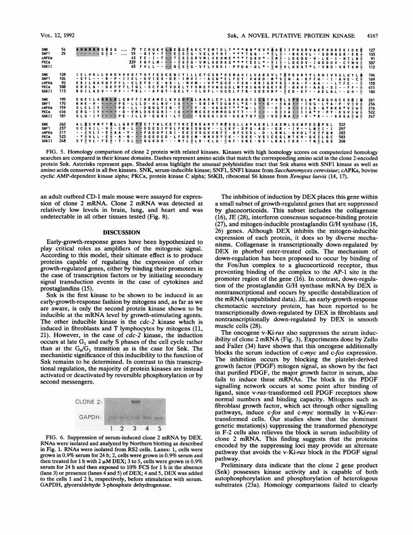
77,811-Da polypeptide of 682 amino acids (Fig. 4). Compar-ison of the predicted protein, and to a lesser degree thecDNA nucleic acid sequence, with entries in GenBank andother data bases (see Materials and Methods) detectedsignificant similarity between clone 2 and over 30 reportedkinase sequences. In each case, the region of sequencesimilarity was largely restricted to the ATP-binding/kinasedomains of the catalytic site. Clone 2 exhibited segmentalregions of high conservation interspersed with regions of lowconservation in its kinase domain, which is similar to whathas been reported for other kinases. The 11 conservedsubdomains identified by Hanks et al. (13, 14) were wellconserved in clone 2; however, the consensus Gly-X-Gly-X-X-Gly sequence found in many nucleotide-binding proteinswas present as Gly-X-Gly-X-X-Ala (amino acids 86 to 92) inclone 2. Substitution at the third glycine in this consensussequence is also seen in Kin 28 and other kinases (14).The programs used to perform the sequence comparisons
gave slightly different scores to kinases possessing similarityto clone 2 protein. Five kinases, which consistently receivedthe highest homology scores in the various computer analy-ses, were compared in their kinase domains with clone 2(Fig. 5). This comparison showed that although clone 2protein diverged significantly from all other kinases com-pared, it clearly contained sufficient conserved residues inthe kinase domain to suggest that it was a kinase. Thisapparent kinase domain in clone 2 was located in theamino-terminal region of the polypeptide and showed greatersimilarity to serine/threonine kinases than to tyrosine ki-nases. For example, it contained the sequence DLKLGN(amino acids 202 to 207; Fig. 4), which is similar to theDLKPXN consensus sequence of serine/threonine kinases,and it also contained the GTPXYLXPE sequence (aminoacids 239 to 247; Fig. 4) found in many serine/threoninekinases (13, 14).
In addition to possessing a conserved kinase domain,clone 2 protein also shared with yeast SNF1 kinase anunusual group of 7 histidines located immediately upstreamof the kinase domain (Fig. 5). In SNF1 kinase, this polyhis-tidine tract is longer than that seen in clone 2, containing 14histidines as opposed to 7 in clone 2 (5). This group ofhistidines is not evolutionarily conserved in the SNF1 kinase
93 GGC184 GAl
D274 CAT
H364 GGA
G454 CAI
H544 AAA
K634 GTI
V724 TTI
F814 GG0904 ACI
T994 TUJ
GA
'ACGAAATGACAGATCTGACAAACAACAAAGTCTACGCTGCAAAAATTATTCCTCACAGCGAAGTAGCTAAACC
1174 AATTTCTTTAAGAAAGCCGCAGCCGCTCTTTTTGGTGGCAAGAAGGACAAAGCAAGATATAACGACACACACAATAAGGTGTCTAAGGA
28
58iT
88CT
118RC
148A
178TT
208GT
238IT
268CC
298AT
328AG
358AA
38AG
418GO
448GA
478
C L E N N P E A C K E Q L S T S F Q U V T K U V D Y S 5081624 AACAAATAT&GCTTTGGGTACCAGCTCTCGGACCACACTGTTGGCGTCCTTTTCAACAACGGGGCTCACATGAGCCTCCTTCCGGACAAA
N K Y G F G Y Q L S D H T V G V L F N N A H N S L L P D K 5381714 AAGACAGTTCACTATTATGCGGAACTTGGCCAATGCTCTGTTTTCCCAGCAACAGATGCCCCTGAACAATTTATTAGTCAAGTSACGGTG
K T V H Y Y A E L G Q C S V F P A T D A P E 0 F I S Q V T V 5681804 CTGAAATACTTTTCTCATTACATGGAGGAGMACCTCATG&ATGGTGGTGATCTCCCGAGTGTTACTGACATTCGMAGACCTCGGCTCTAC
L K Y F S 8 Y N E E N L N D G G D L P S V T D I R R P R L Y 5981894 CTCCTGCAGTGGTTAAAGTCTGATAAAGCCTTAATGATGCTCTTCAATGACGGCACATTTCAGGTGAATTTCTACCACGATCATACAAAA
L L Q W L K S D K A L N M L F N D G T F 0 V N F Y 8 D H T K 6281984 ATCATCATCTGTMCCAGAGTGAAGAATACCTTCTCACCTACATCAATGAGGACAGGATCTCTACAACTTTCAGACTGACGACTCTGCTG
I I I C N 0 S E E Y L L T Y I N E D R I S T T F R L T T L L 6582074 ATGTCTGGCTGTTCGTTAGAATTGAAAAATCGAATGGAATATGCCCTGMACATGCTCTTACAGAGATGTMCTGAAAACATTATTATTAT
N S G C S L E L K N R N E Y A L N N L L Q R C N * 6822164 TATTATTATAATTATTTCGAGCGGACCTCATGGGACTCTTTTCCACTGTGAGATCAACAGGGAAGCCAGCGGAAAGATACAGAGCATGTT2254 AGAGAAGTCGGACAGGTGGTGGTACGAATACAATTCCTCTGTGGCCTGCTGGACTGCTGGAACCAGACCAGCCTAAGGTGTAGAGTTGAC2344 TTTGGACAATCCTGAGTGTGGAGCCGAGTGCAGTTTTCCCTGAGATACCTGTCGTGAAAAGGTTTATGGGACAGTTTTTCAGAAAGATGC2434 ATTGACTCTGAAGTTCTCTCTGTTGAGAGCGTCTTCAGTTGGAAGACTTGGAACTGTGAATACACTTCCTGAAGGGGAGGGAGAAGGGAG2524 OTTGCTCCCTTGCTGTTTAAAGGCTACAATCAGAGCAGCTTTTGGCTGCTTAACTGTGAACTATGGCCATACATTTTTTTTTTTTTTGGT2614 TATTTTTGAATACACTTGTGGTTGGAAAGTGCATTCCTTGTTATAAACTTTTTATATTACAGCCCCAAGAGCAGTATTTATTATCA2704 AGATGTTCTCTTTTTTTATGTTGACCATTTCAAACTCTTGOCAATAAAGAGTATGACATAGAAAAAAAA 2m
FIG. 4. Nucleotide and predicted amino acid sequence of clone2. Underlined sequences are copies of a conserved motif shown byShaw and Kamen (30) to confer message instability and which arefound in the 3' untranslated regions of many mitogen-induced,early-growth-response mRNAs. A consensus polyadenylation signalis doubly underlined.
and is dispensable with regard to SNF1's activity in regulat-ing glucose-repressible genes (1, 6).We refer to the clone 2-encoded protein as Snk, for
serum-inducible kinase, because of its similarity to knownkinases and its rapid induction by serum.
Glucocorticoid modulation of clone 2 mRNA. The syntheticglucocorticoid DEX has been shown to repress the mitogen-stimulated induction of a small subset of early- and late-growth-response genes. These genes include the collage-nase, mitogen-inducible prostaglandin G/H synthase,interferon consensus sequence-binding protein, and JE (amitogen-induced secretory factor) genes (16, 18, 26-28).Northern blots showed that DEX also strongly repressed
the mitogen induction of clone 2 mRNA in NIH 3T3 (datanot shown) and RS2 (Fig. 6) cells. DEX repressed only themitogen-induced level of clone 2 mRNA and did not repressbasal level expression (data not shown).
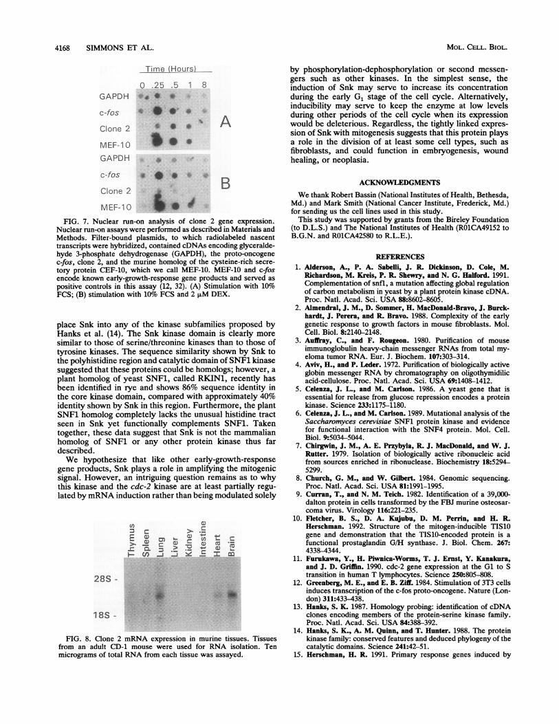
Transcriptional analysis of the clone 2 gene. Nuclear run-onassays were performed to determine whether induction ofclone 2 mRNA by serum was transcriptional and whetherDEX repressed this induction. Serum initiated a rapid five-fold increase in clone 2 transcription that peaked 30 min afterthe addition of mitogen. This induction occurred later andwas smaller than the induction of two other early-growth-response genes, c-fos and MEF-10, whose transcriptionexperienced a very strong induction that reached a maxi-mum 15 min after addition of serum (Fig. 7A).DEX completely repressed the transcriptional increase in
clone 2 mRNA initiated by serum but had no effect on thetranscriptional induction of either c-fos or MEF-10 mRNA(Fig. 7B).
Tissue-specific expression of clone 2 mRNA. Tissues from
CG1
MOL. CELL. BIOL.
VOL.12,1992~~~~~~~Snk,A NOVEL PUTATIVE PROTEIN KINASE 4167
54 S G 79
SNF1 25 ..--G-G- 55 -QI1V T ~E S G -V K LA Y H T. T G Q. L ~NK KVL- -SDMNQ GR E R
S~~~~~~~~~~.... *TLD*K Q K -V -L K 1 H T LN %n~~~~~~~~~~~~~~339F NF LM ~.~.... .G V M LADR KG T EEL*L K DV- I0D D DV -C T MV'
SW 63 FVLL Q S G V F L V R K I P P D A Q L*- N V L K K A T* L- V R D VRTK
SNK 128 E L HR LL HH K HVVQ FY H Y FED KE N IYI L LEY C SR R SM AH I LKARKVTPVyLQV
104 -SY L --R P- II KL -DV I KS -D E -I NV I .AGN *EL F D Y V QR D MN* S..
cAPKa 92 K R ILQ0AV NFP F L- K LEF S- K NS -I MV V P*GGE F SH.R R I GR F S .HA
PKCa 388 K R VLA --D KP PFL T QLSCFVRVFKS6KIl 113 R D I L A V-- P F--* R L A F 0 T K L Y- I D F L G G D L F T R S K E V N F E D- K F A E L A L D H-
SNK 195 E E I L H L G N F F I N E A E L K V G L~P AA AR L E P E H R R R T I T P N L S V L N K G H G C E S0 I
SNFI RH K -V --..'PE-LIL LD H L NV -IA S NI M T DGN F 'K.- ISG-LYA-P-V V'
PKC a 45 IYL HEEN . ....1A..PY.S :I456I18 I -.......P.. L D E G H I IS.....
K E YT F A-
PKCa S..-.VSV...KE.SIKG......'LGCSY'.VL-F~~..... ...Q.D--M..I..G-QF ...TN.I
12710391
387112
194169158455180
261236216522247
FIG. 5. Homology comparison of clone 2 protein with related kinases. Kinases with high homology scores on computerized homologysearches are compared in their kinase domains. Dashes represent amino acids that match the corresponding amino acid in the clone 2-encodedprotein Snk. Asterisks represent gaps. Shaded areas highlight the unusual polyhistidine tract that Snk shares with SNF1 kinase as well asamino acids conserved in all five kinases. SNK, serum-inducible kinase; SN-Fl, SNF1 kinase from Saccharomyces cerevisiae; cAPKa, bovinecyclic AMP-dependent kinase alpha; PKCa, protein kinase C alpha; S6KII, ribosomal S6 kinase from Xenopus laevis (14, 17).
an adult outbred CD-i male mouse were assayed for expres-sion of clone 2 mRNA. Clone 2 mRNA was detected atrelatively low levels in brain, lung, and heart and wasundetectable in all other tissues tested (Fig. 8).
DISCUSSION
Early-growth-response genes have been hypothesized toplay critical roles as amplifiers of the mitogenic signal.According to this model, their ultimate effect is to produceproteins capable of regulating the expression of othergrowth-regulated genes, either by binding their promoters inthe case of transcription factors or by initiating secondarysignal transduction events in the case of cytokines andprostaglandins (15).Snk is the first kinase to be shown to be induced in an
early-growth-response fashion by mitogens and, as far as weare aware, is only the second protein kinase shown to beinducible at the mRNA level by growth-stimulating agents.The other inducible kinase is the cdc-2 kinase which isinduced in fibroblasts and T lymphocytes by mitogens (11,21). However, in the case of cdc-2 kinase, the inductionoccurs at late G, and early S phases of the cell cycle ratherthan at the GJ/Gl transition as is the case for Snk. Themechanistic significance of this inducibility to the function ofSnk remains to be determined. In contrast to this transcrip-tional regulation, the majority of protein kinases are insteadactivated or deactivated by reversible phosphorylation or bysecond messengers.
CLONE 2-
FIG. 6. Suppression of serum-induced clone 2 mRNA by DEX.RNAs were isolated and analyzed by Northern blotting as describedin Fig. 1. RNAs were isolated from R52 cells. Lanes: 1, cells weregrown in 0.9% serum for 24 h; 2, cells were grown in 0.9% serum andthen treated for 1 h with 2 p.M DEX; 3 to 5, cells were grown in 0.9%serum for 24 h and then exposed to 10% FCS for 1 h in the absence(lane 3) or presence (lanes 4 and 5) of DEX; 4 and 5, DEX was addedto the cells 1 and 2 h, respectively, before stimulation with serum.GAPDH, glyceraldehyde 3-phosphate dehydrogenase.
The inhibition of induction by DEX places this gene withina small subset of growth-regulated genes that are suppressedby glucocorticoids. This subset includes the collagenase(16), JE (28), interferon consensus sequence-binding protein(27), and mitogen-inducible prostaglandin G/H synthase (18,26) genes. Although DEX inhibits the mitogen-inducibleexpression of each protein, it does so by diverse mecha-nisms. Collagenase is transcriptionally down-regulated byDEX in phorbol ester-treated cells. The mechanism ofdown-regulation has been proposed to occur by binding ofthe Fos/Jun complex to a glucocorticoid receptor, thuspreventing binding of the complex to the AP-1 site in thepromoter region of the gene (16). In contrast, down-regula-tion of the prostaglandin G/H synthase mRNA by DEX isnontranscriptional and occurs by specific destabilization ofthe mRNA (unpublished data). JE, an early-growth-responsechemotactic secretory protein, has been reported to betranscriptionally down-regulated by DEX in fibroblasts andnontranscriptionally down-regulated by DEX in smoothmuscle cells (28).The oncogene v-Ki-ras also suppresses the serum induc-
ibility of clone 2 mRNA (Fig. 3). Experiments done by Zulloand Faller (34) have shown that this oncogene additionallyblocks the serum induction of c-myc and c-fos expression.The inhibition occurs by blocking the platelet-derivedgrowth factor (PDGF) mitogen signal, as shown by the factthat purified PDGF, the major growth factor in serum, alsofails to induce these mRNAs. The block in the PDGFsignalling network occurs at some point after binding ofligand, since v-ras-transformed cell PDGF receptors shownormal numbers and binding capacity. Mitogens such asfibroblast growth factor, which act through other signallingpathways, induce c-fos and c-myc normally in v-Ki-ras-transformed cells. Our studies show that the dominantgenetic mutation(s) suppressing the transformed phenotypein F-2 cells also relieves the block in serum inducibility ofclone 2 mRNA. This finding suggests that the proteinsencoded by the suppressing loci may provide an alternatepathway that avoids the v-Ki-ras block in the PDGF signalpathway.
Preliminary data indicate that the clone 2 gene product(Snk) possesses kinase activity and is capable of bothautophosphorylation and phosphorylation of heterologoussubstrates (23a). Homology comparisons failed to clearly
VOL. 12, 1992
4168 SIMMONS ET AL.
Time (Hours)
0 .25 .5 1 8
GAPDH *
c-fos *- 4
Clone 2
MEF-1 0
GAPDH
c-fos
Clone 2
MEF-1 0
O., =-
* * .w
A
FIG. 7. Nuclear run-on analysis of clone 2 gene expression.Nuclear run-on assays were performed as described in Materials andMethods. Filter-bound plasmids, to which radiolabeled nascenttranscripts were hybridized, contained cDNAs encoding glyceralde-hyde 3-phosphate dehydrogenase (GAPDH), the proto-oncogenec-fos, clone 2, and the murine homolog of the cysteine-rich secre-
tory protein CEF-10, which we call MEF-10. MEF-10 and c-fosencode known early-growth-response gene products and served as
positive controls in this assay (12, 32). (A) Stimulation with 10%FCS; (B) stimulation with 10% FCS and 2 FM DEX.
place Snk into any of the kinase subfamilies proposed byHanks et al. (14). The Snk kinase domain is clearly more
similar to those of serine/threonine kinases than to those oftyrosine kinases. The sequence similarity shown by Snk tothe polyhistidine region and catalytic domain of SNF1 kinasesuggested that these proteins could be homologs; however, a
plant homolog of yeast SNF1, called RKIN1, recently hasbeen identified in rye and shows 86% sequence identity inthe core kinase domain, compared with approximately 40%identity shown by Snk in this region. Furthermore, the plantSNF1 homolog completely lacks the unusual histidine tractseen in Snk yet functionally complements SNF1. Takentogether, these data suggest that Snk is not the mammalianhomolog of SNF1 or any other protein kinase thus fardescribed.We hypothesize that like other early-growth-response
gene products, Snk plays a role in amplifying the mitogenicsignal. However, an intriguing question remains as to whythis kinase and the cdc-2 kinase are at least partially regu-
lated by mRNA induction rather than being modulated solely
a)
3L= 2 .
28S
18S
FIG. 8. Clone 2 mRNA expression in murine tissues. Tissuesfrom an adult CD-1 mouse were used for RNA isolation. Tenmicrograms of total RNA from each tissue was assayed.
by phosphorylation-dephosphorylation or second messen-gers such as other kinases. In the simplest sense, theinduction of Snk may serve to increase its concentrationduring the early G1 stage of the cell cycle. Alternatively,inducibility may serve to keep the enzyme at low levelsduring other periods of the cell cycle when its expressionwould be deleterious. Regardless, the tightly linked expres-sion of Snk with mitogenesis suggests that this protein playsa role in the division of at least some cell types, such asfibroblasts, and could function in embryogenesis, woundhealing, or neoplasia.
ACKNOWLEDGMENTSWe thank Robert Bassin (National Institutes of Health, Bethesda,
Md.) and Mark Smith (National Cancer Institute, Frederick, Md.)for sending us the cell lines used in this study.
This study was supported by grants from the Bireley Foundation(to D.L.S.) and The National Institutes of Health (ROlCA49152 toB.G.N. and R01CA42580 to R.L.E.).
REFERENCES1. Alderson, A., P. A. Sabelli, J. R. Dickinson, D. Cole, M.
Richardson, M. Kreis, P. R. Shewry, and N. G. Halford. 1991.Complementation of snfl, a mutation affecting global regulationof carbon metabolism in yeast by a plant protein kinase cDNA.Proc. Natl. Acad. Sci. USA 88:8602-8605.
2. Almendral, J. M., D. Sommer, H. MacDonald-Bravo, J. Burck-hardt, J. Perera, and R. Bravo. 1988. Complexity of the earlygenetic response to growth factors in mouse fibroblasts. Mol.Cell. Biol. 8:2140-2148.
3. Auffray, C., and F. Rougeon. 1980. Purification of mouseimmunoglobulin heavy-chain messenger RNAs from total my-eloma tumor RNA. Eur. J. Biochem. 107:303-314.
4. Aviv, H., and P. Leder. 1972. Purification of biologically activeglobin messenger RNA by chromatography on oligothymidilicacid-cellulose. Proc. Natl. Acad. Sci. USA 69:1408-1412.
5. Celenza, J. L., and M. Carlson. 1986. A yeast gene that isessential for release from glucose repression encodes a proteinkinase. Science 233:1175-1180.
6. Celenza, J. L., and M. Carlson. 1989. Mutational analysis of theSaccharomyces cerevisiae SNF1 protein kinase and evidencefor functional interaction with the SNF4 protein. Mol. Cell.Biol. 9:5034-5044.
7. Chirgwin, J. M., A. E. Przybyla, R. J. MacDonald, and W. J.Rutter. 1979. Isolation of biologically active ribonucleic acidfrom sources enriched in ribonuclease. Biochemistry 18:5294-5299.
8. Church, G. M., and W. Gilbert. 1984. Genomic sequencing.Proc. Natl. Acad. Sci. USA 81:1991-1995.
9. Curran, T., and N. M. Teich. 1982. Identification of a 39,000-dalton protein in cells transformed by the FBJ murine osteosar-coma virus. Virology 116:221-235.
10. Fletcher, B. S., D. A. Kujubu, D. M. Perrin, and H. R.Herschman. 1992. Structure of the mitogen-inducible TIS10gene and demonstration that the TIS10-encoded protein is afunctional prostaglandin G/H synthase. J. Biol. Chem. 267:4338-4344.
11. Furukawa, Y., H. Piwnica-Worms, T. J. Ernst, Y. Kanakura,and J. D. Griffin. 1990. cdc-2 gene expression at the Gl to Stransition in human T lymphocytes. Science 250:805-808.
12. Greenberg, M. E., and E. B. Ziff. 1984. Stimulation of 3T3 cellsinduces transcription of the c-fos proto-oncogene. Nature (Lon-don) 311:433-438.
13. Hanks, S. K. 1987. Homology probing: identification of cDNAclones encoding members of the protein-serine kinase family.Proc. Natl. Acad. Sci. USA 84:388-392.
14. Hanks, S. K., A. M. Quinn, and T. Hunter. 1988. The proteinkinase family: conserved features and deduced phylogeny of thecatalytic domains. Science 241:42-51.
15. Herschman, H. R. 1991. Primary response genes induced by
MOL. CELL. BIOL.
-
Snk, A NOVEL PUTATIVE PROTEIN KINASE 4169
growth factors and tumor promoters. Annu. Rev. Biochem.60:281-319.
16. Jonat, C., H. J. Rahmsdorf, K.-K. Park, A. C. B. Cato, S. Gebel,H. Ponta, and P. Herrlich. 1990. Antitumor promotion andantiinflammation: down-modulation of AP-1 (Fos/Jun) activityby glucocorticoid hormones. Cell 62:1189-1204.
17. Jones, S. W., E. Erikson, J. Blenis, J. L. Maller, and R. L.Erikson. 1988. A Xenopus ribosomal protein S6 kinase has twoapparent kinase domains that are each similar to distinct proteinkinases. Proc. Natl. Acad. Sci. USA 85:3377-3381.
18. Kujubu, D. A., B. S. Fletcher, B. C. Varnum, R. W. Urm, andH. R. Herschman. 1991. TIS 10, a phorbol ester tumor promot-er-inducible mRNA from Swiss 3T3 cells encodes a novelprostaglandin synthase/cyclooxygenase homologue. J. Biol.Chem. 20:12866-12872.
19. Kujubu, D. A., and H. R. Herschman. 1992. Dexamethasoneinhibits mitogen induction of the TIS10 prostaglandin synthase/cyclooxygenase gene. J. Biol. Chem. 267:7991-7994.
20. Lau, L. F., and D. Nathans. 1985. Identification of a set of genesexpressed during the GdG1 transition of cultured mouse cells.EMBO J. 4:3145-3151.
21. Lee, M. G., C. J. Norbury, N. K. Spurr, and P. Nurse. 1988.Regulated expression and phosphorylation of a possible mam-malian cell-cycle control protein. Nature (London) 333:676-679.
22. Lim, R. W., B. C. Varnum, and H. R. Herschman. 1987. Cloningof tetradecanoyl phorbol ester-induced "primary response"sequences and their expression in density-arrested Swiss 3T3cells and a TPA non-proliferative variant. Oncogene 1:263-270.
23. Linial, M., N. Gunderson, and M. Groudine. 1985. Enhancedtranscription of c-myc in bursal lymphoma cells requires con-tinuous protein synthesis. Science 230:1126-1131.
23a.Liu, M. A. Personal communication.24. Maniatis, T., E. F. Fritsch, and J. Sambrook. 1989. Molecular
cloning: a laboratory manual. Cold Spring Harbor LaboratoryPress, Cold Spring Harbor, N.Y.
25. Noda, M., Z. Selinger, E. M. Scolnick, and R. H. Bassin. 1983.
Flat revertants isolated from Kirsten sarcoma virus-transformedcells are resistant to the action of specific oncogenes. Proc.Natl. Acad. Sci. USA 80:5602.
26. O'Banion, M. K., H. B. Sadowski, V. Win, and D. A. Young.1991. A serum- and glucocorticoid-regulated 4-kilobase mRNAencodes a cyclooxygenase-related protein. J. Biol. Chem. 266:23261-23267.
27. Politis, A. D., J. Sivo, P. H. Driggers, K. Ozato, and S. N. Vogel.1992. Modulation of interferon consensus sequence bindingprotein mRNA in murine peritoneal macrophages: induction ofIFN-p and down-regulation by IFN-ac, dexamethasone, andprotein kinase inhibitors. J. Immunol. 148:801-807.
28. Poon, M., J. Megyesi, R. S. Green, H. Zhang, B. J. Rollins, R.Safirstein, and M. B. Taubman. 1991. In vivo and in vitroinhibition of JE gene expression by glucocorticoids. J. Biol.Chem. 266:22375-22379.
29. Sanger, F., S. Nicklen, and A. R. Coulson. 1977. DNA sequenc-ing with chain-terminating inhibitors. Proc. Natl. Acad. Sci.USA 74:5463-5467.
30. Shaw, G., and R. Kamen. 1986. A conserved AU sequence fromthe 3' untranslated region of GM-CSF mRNA mediates selec-tive mRNA degradation. Cell 46:659-667.
31. Simmons, D. L., and C. B. Kasper. 1989. Quantitation ofmRNAs specific for the mixed-function oxidase system in ratliver and extrahepatic tissues during development. Arch. Bio-chem. Biophys. 271:10-20.
32. Simmons, D. L., D. B. Levy, Y. Yannoni, and R. L. Erikson.1989. Identification of a phorbol ester-repressible v-src-induc-ible gene. Proc. Natl. Acad. Sci. USA 86:1178-1182.
33. Xie, W., J. G. Chipman, D. L. Robertson, R. L. Erikson, andD. L. Simmons. 1991. Expression of a mitogen-responsive geneencoding prostaglandin synthase is regulated by mRNA splic-ing. Proc. Natl. Acad. Sci. USA 88:2692-2696.
34. Zullo, J. N., and D. V. Faller. 1988. p21 v-ras inhibits inductionof c-myc and c-fos expression by platelet-derived growth factor.Mol. Cell. Biol. 8:5080-5085.
VOL. 12, 1992