Embed Size (px)
Citation preview
Choi et al.
1
Identification of bioactive molecules by adipogenesisprofiling of organic compounds
Yongmun Choi,* Yoshinori Kawazoe,* Koji Murakami,* Hiroyuki Misawa,
Motonari Uesugi†
The Verna and Marrs McLean Department of Biochemistry and Molecular Biology, Baylor
College of Medicine, Houston, TX 77030, USA.
* These authors contributed equally to the work.
† To whom correspondence should be addressed. E-mail: [email protected]
Copyright 2002 by The American Society for Biochemistry and Molecular Biology, Inc.
JBC Papers in Press. Published on December 19, 2002 as Manuscript M210283200 by guest on June 20, 2018
http://ww
w.jbc.org/
Dow
nloaded from
Choi et al.
2
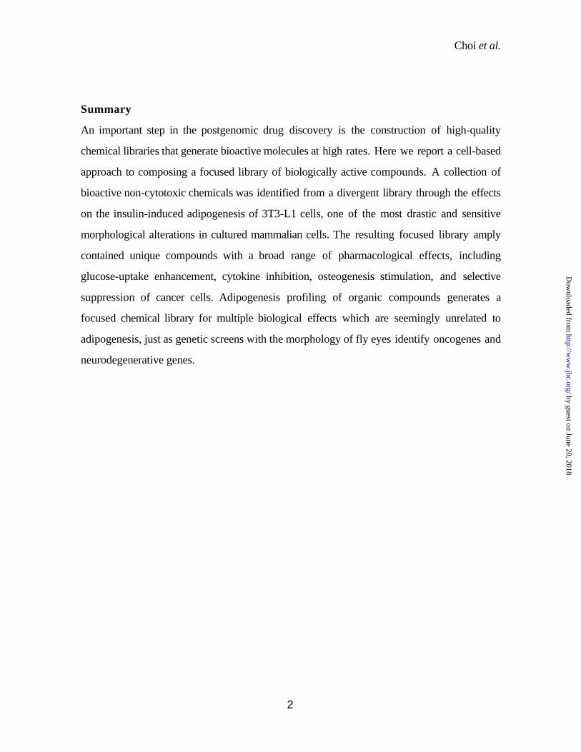
Summary
An important step in the postgenomic drug discovery is the construction of high-quality
chemical libraries that generate bioactive molecules at high rates. Here we report a cell-based
approach to composing a focused library of biologically active compounds. A collection of
bioactive non-cytotoxic chemicals was identified from a divergent library through the effects
on the insulin-induced adipogenesis of 3T3-L1 cells, one of the most drastic and sensitive
morphological alterations in cultured mammalian cells. The resulting focused library amply
contained unique compounds with a broad range of pharmacological effects, including
glucose-uptake enhancement, cytokine inhibition, osteogenesis stimulation, and selective
suppression of cancer cells. Adipogenesis profiling of organic compounds generates a
focused chemical library for multiple biological effects which are seemingly unrelated to
adipogenesis, just as genetic screens with the morphology of fly eyes identify oncogenes and
neurodegenerative genes.
by guest on June 20, 2018http://w
ww
.jbc.org/D
ownloaded from
Choi et al.
3
Complete analysis of human genome is anticipated to produce an unprecedented number of
potential drug targets. Development of high-throughput assays for these genomic
pseudotargets may be a challenging but important step for not limiting drug discovery to the
“relatively easy” targets such as G-protein-coupled receptors or particular enzymes. An
alternative or complementary effort is the construction of high-quality chemical libraries that
generate bioactive molecules at higher rates. The small size of the focused libraries would
lower the cost of screening processes and enable unique low-throughput screens, extending
the scope of assays for the genomic targets and for a given therapeutic effect.
Our approach to constructing a focused chemical library is based on the logic of
genetics. In genetic screens, clear morphological phenotypes are often used just as a sensitive
tool for discovering and analyzing genes whose primary functions are seemingly unrelated to
the morphological phenotype. A good example is the use of eye morphology in the fruit fly
Drosophila melanogaster as a genetic tool for the analysis of genes in disease-linked signaling
pathways (1). Although human diseases associated with these pathways, such as cancer and
neurodegenerative diseases, are seemingly unrelated to eye development, the use of eye
morphology as a sensitive indicator enabled a systematic understanding of the disease-linked
signaling events (2-6). We envisioned that clear morphological phenotypes of cells could
similarly be used as a sensitive indicator of the drug effects that are not associated directly
with the morphological phenotypes.
The morphological alteration we used is the differentiation of murine 3T3-L1
fibroblasts into adipocytes, one of the most drastic and sensitive morphological alterations in
cultured mammalian cells (7). In the presence of insulin, 3T3-L1 cells undergo differentiation
into adipocytes, which are visually distinct from the original cells because of the presence of
oil droplets in the cytoplasm (Fig. 1). The insulin-induced adipogenesis of 3T3-L1 cells
involves a number of disease-linked proteins such as PI3K, Ras, PPARγ, p38, or
phosphodiesterases, and known drugs for a range of diseases have been reported to have
phenotypic effects on the adipogenesis (7-12). A morphology-based adipogenesis screen of a
chemical library could identify a pool of biologically active compounds with many distinct
by guest on June 20, 2018http://w
ww
.jbc.org/D
ownloaded from
Choi et al.
4
pharmacological effects. Here we report a proof-of-principle study using a library of 10,000
divergent compounds.
by guest on June 20, 2018http://w
ww
.jbc.org/D
ownloaded from
Choi et al.
5
Experimental procedures
Adipogenesis profiling. 3T3-L1 fibroblasts were plated in 96-well plates at a density of
5X10⁴ cells /well and allowed to reach maximal confluence. The confluent cells were treated
individually with 20 ng/µL of a chemical for three days in 100 µL of DMEM containing of
insulin (5 µg/mL) and 10% fetal bovine serum. After removal of insulin and the chemical, the
cells were further maintained typically for eight days, with the replacement of media every
three days. The effects of chemicals on the adipogenesis were evaluated under microscope.
The control wells with 1% (v/v) DMSO had ~5% adipocytes. The compounds that enhanced
the adipogenesis more than five folds were scored to be adipogenesis-enhancing chemicals,
and the ones that completely inhibited adipogenesis without detectable toxicity were scored to
be adipogenesis-blocking chemicals. The effects of these chemicals were confirmed multiple
times by multiple laboratory members. Cell viability was monitored by Trypan Blue
exclusion and by counting cell numbers.
Reverse Transcriptase-PCR
Total RNA was isolated with TRI Reagent (Molecular Research Center) at day seven (aP2) or
day three (osteocalcin) . Five µg of total RNA was reverse transcribed to cDNA by using
oligo dT primer with AMV reverse transcriptase for 60 min at 42 °C. The cDNA was then
amplified by using Ex Taq (Takara) with following primer pairs: 5'-
AACACCGAGATTTCCTTCAA-3' and 5'-TCACGCCTTTCATAACACAT-3' for aP2, 5'-
TCTGACAAACCTTCATGTCC-3' and 5'-AAATAGTGATACCGTAGATGCG-3' for
osteocalcin. The amplification conditions are as follows; 95 °C (30 sec) - 60 °C (30 sec) - 72
°C (30 sec) for 23 cycles (aP2) or 30 cycles (osteocalcin).
Glucose uptake study. 3T3-L1 fibroblasts were induced to differentiate into adipocytes
by incubation in a medium containing 10% fetal bovine serum (FBS), 1 µM dexamethasone,
0.5 mM methylisobutylxanthine, and 1.7 µM insulin. After two days, the medium was
by guest on June 20, 2018http://w
ww
.jbc.org/D
ownloaded from
Choi et al.
6
switched to the one containing 10% FBS and 1.7 µM insulin for two days and then to a
normal 10% FBS medium for three days. After the total of seven days, almost 100% of 3T3-
L1 cells were differentiated into adipocytes. These fully differentiated cells were treated on
24-well plates with varied concentrations of chemicals (0.1% DMSO) for 24 hrs and then
incubated with 100 nM of insulin and [3H]-2-deoxyglucose. The cells were extensively
washed, and their radioactivity were measured by scintillation counting. All the samples were
tested in duplicate.
Cytokine production assay. For the analysis of IL-6 and TNF-α, the mouse
macrophage cell line, RAW264.7, was used. Cells were seeded onto 96-well plates, and the
cytokine production was induced by adding 10 µg/mL of lipopolysaccharide. Upon
stimulation, each one of the adipogenesis-enhancing chemicals was also added to the culture
at varied concentrations. After incubating for 48 hrs at 37 °C, the cytokine concentrations in
the culture supernatants were measured by ELISA. For the analysis of IL-2, the mouse
thymoma cell line, EL-4, was used, and the IL-2 production was induced by adding phorbol
ester and ionophore. The effects of chemicals on the IL-2 production was similarly examined
by ELISA. All the samples were tested in triplicate.
Mineralization assay. The clonal osteoblastic cell line, MC3T3-E1, clone 14, was grown
in α-MEM supplemented with 10% FBS until confluent in 96-well plates. For induction of
mineralization, the cells were further incubated with 50 µg/mL of ascorbic acid and 10 mM of
β-glycerophosphate in the presence or absence of chemicals. On day 14, the cells were
washed with phosphate buffered saline, fixed in 10% formalin, and washed with distilled
water. Bone-like mineral formation was evaluated by examining the area stained by 2%
(w/v) Alizarin Red S (pH 4.2).
Assays for IGF-activated cancer cells. The adipogenesis-blocking chemicals were
assayed for their ability to inhibit the growth of IGF-activated cancer cells. For the discovery
by guest on June 20, 2018http://w
ww
.jbc.org/D
ownloaded from
Choi et al.
7
of inhibitors of IGF2, we used five distinct human hepatocellular carcinoma cell lines, Hep-
G2, SK-Hep-1, and three lines that we recently characterized (unpublished, K. Murakami &
M. Uesugi). Three of them produce IGF2 at high levels while two express ~10 times less
amounts of IGF2 as measured by ELISA, RT-PCR, and DNA microarray experiments.
Treatment with a neutralizing antibody against IGF2 selectively inhibited the growth of the
IGF2-overexpressing cell lines, but had little effects on that of the cell lines with low levels of
IGF2. These cell lines thus served as an excellent system for discovering the chemicals that
selectively impair the growth of IGF2-overexpressing hepatocellular carcinoma cells. For
cell-viability assays, IGF2-expressing cells were plated at a density of 4X10³ onto 96-well
plates. After 24-hr incubation, the cells were treated with varied amounts of chemicals for 72
hrs. The effects of chemicals were evaluated by microscopic observation and MTT assay. All
the samples were tested at least three times. For reporter gene assays, IGF2-expressing cells
were transfected with a reporter construct in which a gene encoding secreted alkaline
phosphatase (SEAP) is controlled by the IGF2 promoter, AP-1 sites, NF-kB sites, or the
SV40 promoter. After 24 hrs, the transfected cells were treated with 94G6 (0.1 µM) for 8
hrs. SEAP activity was measured through fluorescence change of methylumbelliferyl
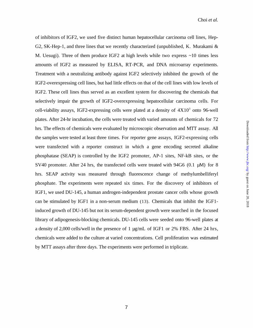
phosphate. The experiments were repeated six times. For the discovery of inhibitors of
IGF1, we used DU-145, a human androgen-independent prostate cancer cells whose growth
can be stimulated by IGF1 in a non-serum medium (13). Chemicals that inhibit the IGF1-
induced growth of DU-145 but not its serum-dependent growth were searched in the focused
library of adipogenesis-blocking chemicals. DU-145 cells were seeded onto 96-well plates at
a density of 2,000 cells/well in the presence of 1 µg/mL of IGF1 or 2% FBS. After 24 hrs,
chemicals were added to the culture at varied concentrations. Cell proliferation was estimated
by MTT assays after three days. The experiments were performed in triplicate.
by guest on June 20, 2018http://w
ww
.jbc.org/D
ownloaded from
Choi et al.
8
Results
Adipogenesis profiling of 10,000 divergent compounds. The divergent chemical
library used for our case study was a Prime-Collection 2000 Format Q (ChemBridge). In this
format, 10,000 drug-like molecules are rationally preselected to form a library that covers the
maximum pharmacore diversity with the minimum number of compounds. Two academic
groups have reported successful isolations of unique compounds from a similar chemical
library (14,15), indicating that this type of chemical libraries contains a diverse set of
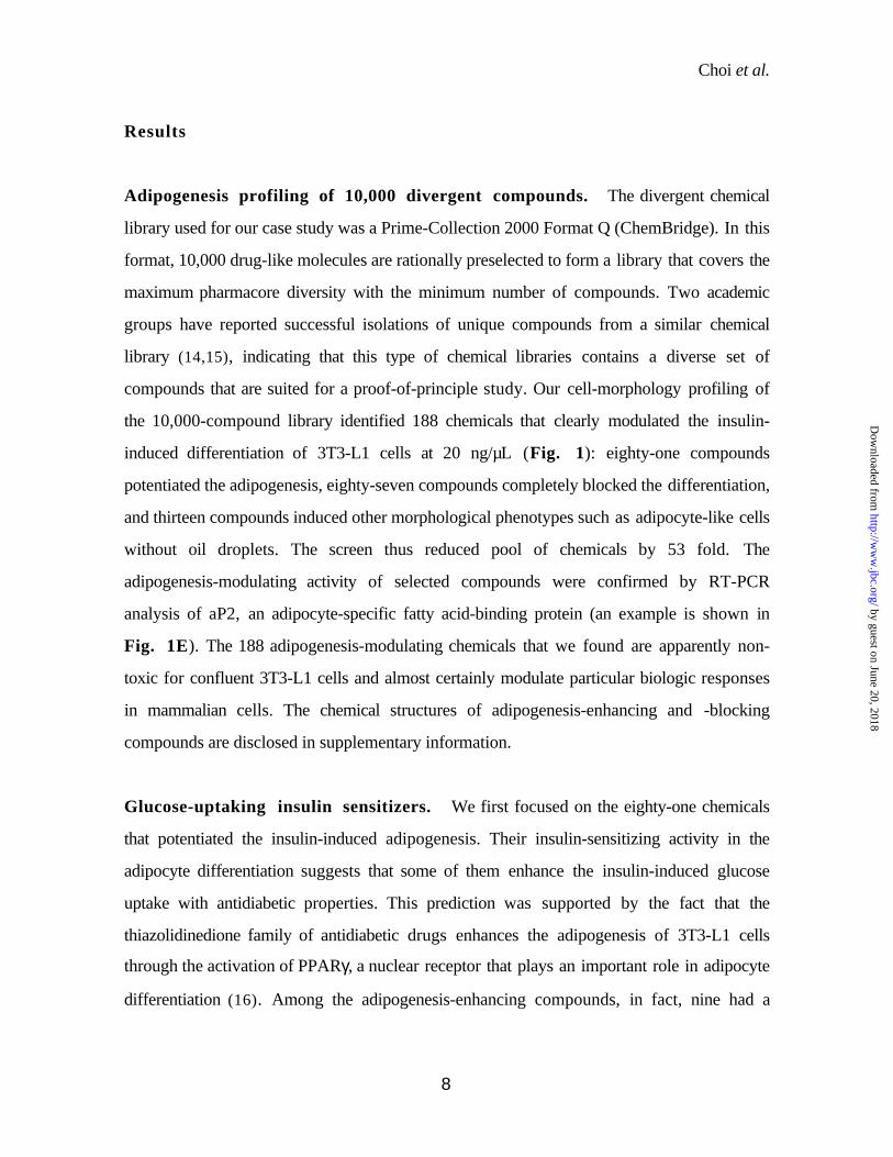
compounds that are suited for a proof-of-principle study. Our cell-morphology profiling of
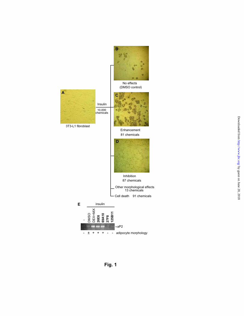
the 10,000-compound library identified 188 chemicals that clearly modulated the insulin-
induced differentiation of 3T3-L1 cells at 20 ng/µL (Fig. 1): eighty-one compounds
potentiated the adipogenesis, eighty-seven compounds completely blocked the differentiation,
and thirteen compounds induced other morphological phenotypes such as adipocyte-like cells
without oil droplets. The screen thus reduced pool of chemicals by 53 fold. The
adipogenesis-modulating activity of selected compounds were confirmed by RT-PCR
analysis of aP2, an adipocyte-specific fatty acid-binding protein (an example is shown in
Fig. 1E). The 188 adipogenesis-modulating chemicals that we found are apparently non-
toxic for confluent 3T3-L1 cells and almost certainly modulate particular biologic responses
in mammalian cells. The chemical structures of adipogenesis-enhancing and -blocking
compounds are disclosed in supplementary information.
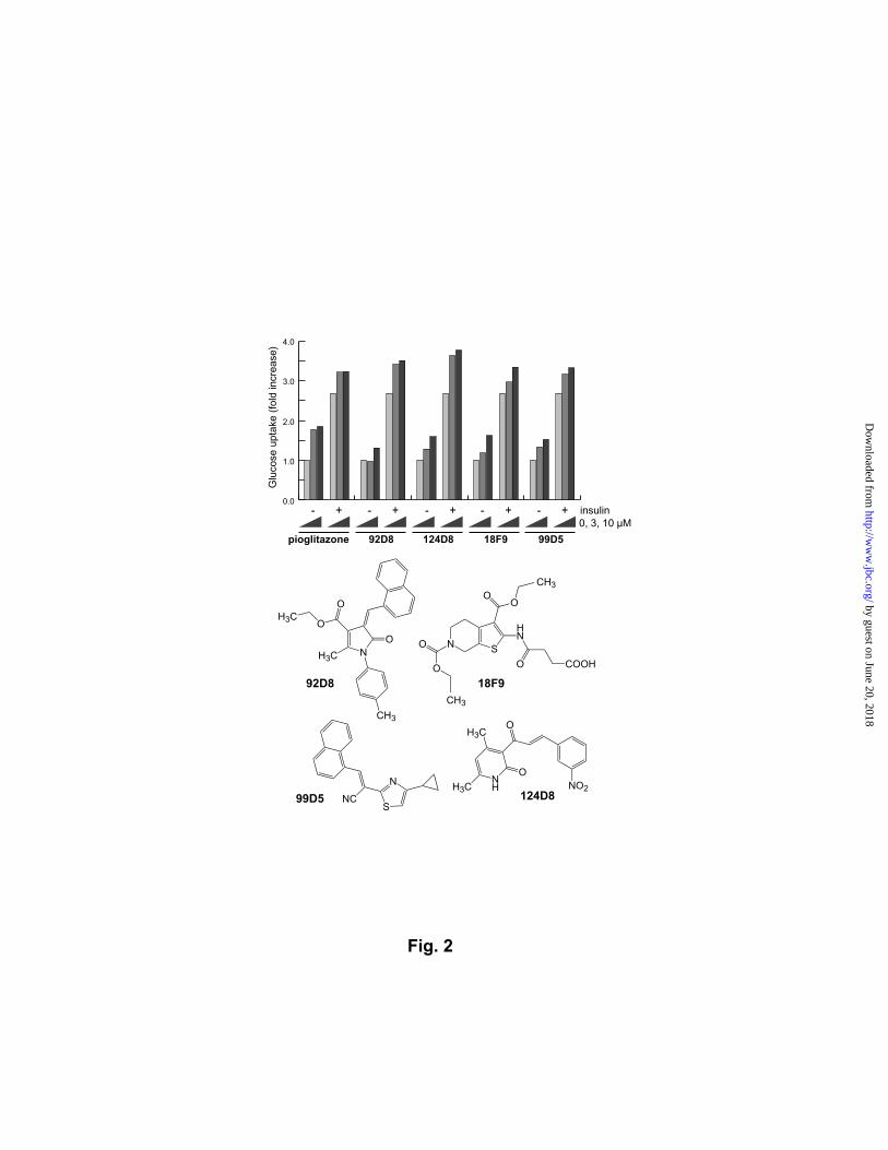
Glucose-uptaking insulin sensitizers. We first focused on the eighty-one chemicals
that potentiated the insulin-induced adipogenesis. Their insulin-sensitizing activity in the
adipocyte differentiation suggests that some of them enhance the insulin-induced glucose
uptake with antidiabetic properties. This prediction was supported by the fact that the
thiazolidinedione family of antidiabetic drugs enhances the adipogenesis of 3T3-L1 cells
through the activation of PPARγ, a nuclear receptor that plays an important role in adipocyte
differentiation (16). Among the adipogenesis-enhancing compounds, in fact, nine had a
by guest on June 20, 2018http://w
ww
.jbc.org/D
ownloaded from
Choi et al.
9
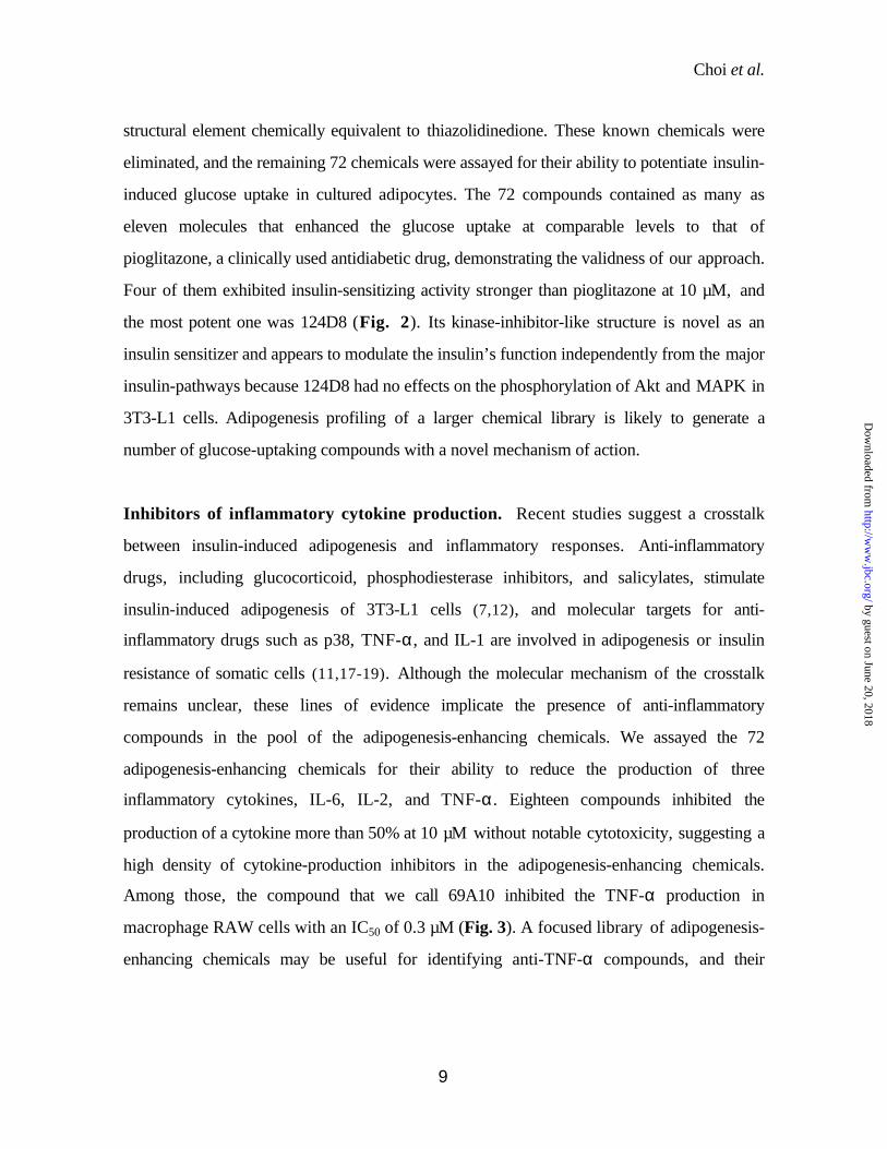
structural element chemically equivalent to thiazolidinedione. These known chemicals were
eliminated, and the remaining 72 chemicals were assayed for their ability to potentiate insulin-
induced glucose uptake in cultured adipocytes. The 72 compounds contained as many as
eleven molecules that enhanced the glucose uptake at comparable levels to that of
pioglitazone, a clinically used antidiabetic drug, demonstrating the validness of our approach.
Four of them exhibited insulin-sensitizing activity stronger than pioglitazone at 10 µM, and
the most potent one was 124D8 (Fig. 2). Its kinase-inhibitor-like structure is novel as an
insulin sensitizer and appears to modulate the insulin’s function independently from the major
insulin-pathways because 124D8 had no effects on the phosphorylation of Akt and MAPK in
3T3-L1 cells. Adipogenesis profiling of a larger chemical library is likely to generate a
number of glucose-uptaking compounds with a novel mechanism of action.
Inhibitors of inflammatory cytokine production. Recent studies suggest a crosstalk
between insulin-induced adipogenesis and inflammatory responses. Anti-inflammatory
drugs, including glucocorticoid, phosphodiesterase inhibitors, and salicylates, stimulate
insulin-induced adipogenesis of 3T3-L1 cells (7,12), and molecular targets for anti-
inflammatory drugs such as p38, TNF-α , and IL-1 are involved in adipogenesis or insulin
resistance of somatic cells (11,17-19). Although the molecular mechanism of the crosstalk
remains unclear, these lines of evidence implicate the presence of anti-inflammatory
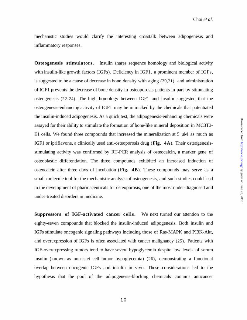
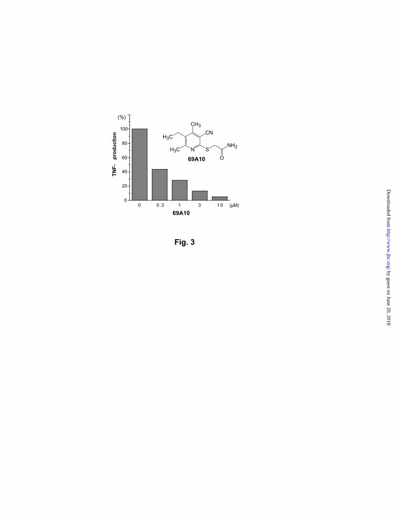
compounds in the pool of the adipogenesis-enhancing chemicals. We assayed the 72
adipogenesis-enhancing chemicals for their ability to reduce the production of three
inflammatory cytokines, IL-6, IL-2, and TNF-α . Eighteen compounds inhibited the
production of a cytokine more than 50% at 10 µM without notable cytotoxicity, suggesting a
high density of cytokine-production inhibitors in the adipogenesis-enhancing chemicals.
Among those, the compound that we call 69A10 inhibited the TNF-α production in
macrophage RAW cells with an IC50 of 0.3 µM (Fig. 3). A focused library of adipogenesis-
enhancing chemicals may be useful for identifying anti-TNF-α compounds, and their
by guest on June 20, 2018http://w
ww
.jbc.org/D
ownloaded from
Choi et al.
10
mechanistic studies would clarify the interesting crosstalk between adipogenesis and
inflammatory responses.
Osteogenesis stimulators. Insulin shares sequence homology and biological activity
with insulin-like growth factors (IGFs). Deficiency in IGF1, a prominent member of IGFs,
is suggested to be a cause of decrease in bone density with aging (20,21), and administration
of IGF1 prevents the decrease of bone density in osteoporosis patients in part by stimulating
osteogenesis (22-24). The high homology between IGF1 and insulin suggested that the
osteogenesis-enhancing activity of IGF1 may be mimicked by the chemicals that potentiated
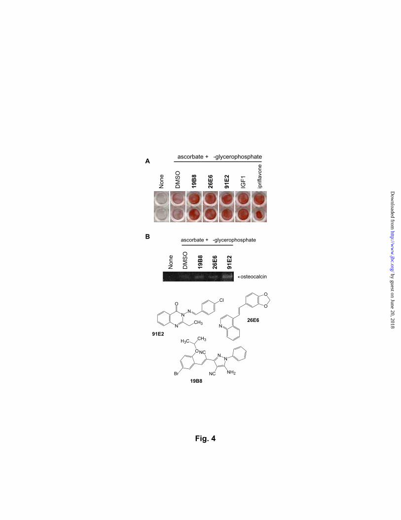
the insulin-induced adipogenesis. As a quick test, the adipogenesis-enhancing chemicals were
assayed for their ability to stimulate the formation of bone-like mineral deposition in MC3T3-
E1 cells. We found three compounds that increased the mineralization at 5 µM as much as
IGF1 or ipriflavone, a clinically used anti-osteoporosis drug (Fig. 4A). Their osteogenesis-
stimulating activity was confirmed by RT-PCR analysis of osteocalcin, a marker gene of
osteoblastic differentiation. The three compounds exhibited an increased induction of
osteocalcin after three days of incubation (Fig. 4B). These compounds may serve as a
small-molecule tool for the mechanistic analysis of osteogenesis, and such studies could lead
to the development of pharmaceuticals for osteoporosis, one of the most under-diagnosed and
under-treated disorders in medicine.
Suppressors of IGF-activated cancer cells. We next turned our attention to the
eighty-seven compounds that blocked the insulin-induced adipogenesis. Both insulin and
IGFs stimulate oncogenic signaling pathways including those of Ras-MAPK and PI3K-Akt,
and overexpression of IGFs is often associated with cancer malignancy (25). Patients with
IGF-overexpressing tumors tend to have severe hypoglycemia despite low levels of serum
insulin (known as non-islet cell tumor hypoglycemia) (26), demonstrating a functional
overlap between oncogenic IGFs and insulin in vivo. These considerations led to the
hypothesis that the pool of the adipogenesis-blocking chemicals contains anticancer
by guest on June 20, 2018http://w
ww
.jbc.org/D
ownloaded from
Choi et al.
11
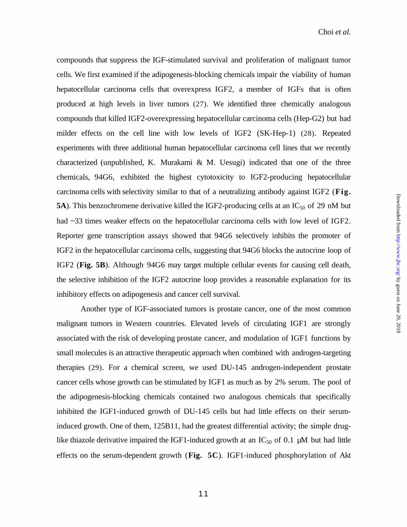
compounds that suppress the IGF-stimulated survival and proliferation of malignant tumor
cells. We first examined if the adipogenesis-blocking chemicals impair the viability of human
hepatocellular carcinoma cells that overexpress IGF2, a member of IGFs that is often
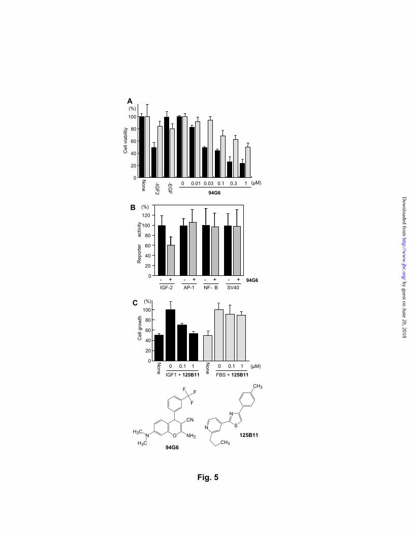
produced at high levels in liver tumors (27). We identified three chemically analogous
compounds that killed IGF2-overexpressing hepatocellular carcinoma cells (Hep-G2) but had
milder effects on the cell line with low levels of IGF2 (SK-Hep-1) (28). Repeated
experiments with three additional human hepatocellular carcinoma cell lines that we recently
characterized (unpublished, K. Murakami & M. Uesugi) indicated that one of the three
chemicals, 94G6, exhibited the highest cytotoxicity to IGF2-producing hepatocellular
carcinoma cells with selectivity similar to that of a neutralizing antibody against IGF2 (Fig.
5A). This benzochromene derivative killed the IGF2-producing cells at an IC50 of 29 nM but
had ~33 times weaker effects on the hepatocellular carcinoma cells with low level of IGF2.
Reporter gene transcription assays showed that 94G6 selectively inhibits the promoter of
IGF2 in the hepatocellular carcinoma cells, suggesting that 94G6 blocks the autocrine loop of
IGF2 (Fig. 5B). Although 94G6 may target multiple cellular events for causing cell death,
the selective inhibition of the IGF2 autocrine loop provides a reasonable explanation for its
inhibitory effects on adipogenesis and cancer cell survival.
Another type of IGF-associated tumors is prostate cancer, one of the most common
malignant tumors in Western countries. Elevated levels of circulating IGF1 are strongly
associated with the risk of developing prostate cancer, and modulation of IGF1 functions by
small molecules is an attractive therapeutic approach when combined with androgen-targeting
therapies (29). For a chemical screen, we used DU-145 androgen-independent prostate
cancer cells whose growth can be stimulated by IGF1 as much as by 2% serum. The pool of
the adipogenesis-blocking chemicals contained two analogous chemicals that specifically
inhibited the IGF1-induced growth of DU-145 cells but had little effects on their serum-
induced growth. One of them, 125B11, had the greatest differential activity; the simple drug-
like thiazole derivative impaired the IGF1-induced growth at an IC50 of 0.1 µM but had little
effects on the serum-dependent growth (Fig. 5C). IGF1-induced phosphorylation of Akt
by guest on June 20, 2018http://w
ww
.jbc.org/D
ownloaded from
Choi et al.
12
and MAPK in DU-145 cells was unaffected by 125B11, suggesting that 125B11 inhibits the
cell-proliferative function of IGF1 in a way independent of the known IGF1-signaling
pathway. Deregulation of the IGF axis is associated with the initiation and progression of
many types of human carcinoma including breast (30) and colorectal cancers (31). A focused
library of adipogenesis-blocking chemicals may serve as a source of anti-proliferative agents
against the IGF-linked cancers.
Discussion
Fat cell differentiation per se has no direct link to glucose uptake, cytokine inhibition,
osteogenesis, and selective suppression of cancer cells. Nevertheless, our proof-of-principle
study using a 10,000-compound library successfully identified non-cytotoxic bioactive
compounds for these seemingly disparate pharmacological effects, just as genetics has
identified non-lethal disease-linked genes by examining the eye morphology of fruit flies. We
randomly picked up seventy compounds that had no detectable phenotypes in the
adipogenesis profiling, and assayed for their ability to modulate glucose uptake, cytokine
production, IGF-selective cytotoxicity, and osteogenesis. As expected, no significant hits
were found in each assay, indicating that the adipogenesis profiling with 3T3-L1 cells is a
good filter at least for these pharmacological effects. A database search revealed that one of
the adipogenesis-enhancing chemicals has been patented as an inhibitor of neuropeptide Y, a
proposed attenuator of insulin and leptin that stimulates appetite (32). Neuropeptide Y
inhibitors are expected to treat feeding disorders and heart diseases (33); adipogenesis
profiling may find use in discovering chemicals with such biological effects. The insulin
family of hormones is involved in many other conditions as observed in the complications of
hyperinsulinism. The insulin-linked pharmacological effects, including wound healing and
anti-apoptosis (34), may be expected in adipogenesis-modulating compounds.
by guest on June 20, 2018http://w
ww
.jbc.org/D
ownloaded from
Choi et al.
13
One potential drawback of our approach is that the bioactive molecules from the
adipogenesis-based focused library may have side effects that are associated with
adipogenesis. However, some degree of side effects are usually expected for any
unoptimized molecules, and classical medicinal chemistry approaches have been taken for
reducing the unwanted side effects. The high sensitivity of the morphological transformation
of 3T3-L1 cells also suggests that the adipogenesis-modulating effects of chemicals may not
necessarily be reproduced in human. For instance, non-steroidal anti-inflammatory drugs and
phosphodiesterase inhibitors such as aspirin and caffeine are known to enhance adipogenesis
of 3T3-L1 cells, but have no significant effects on fat accumulation in human. The
adipogenesis profiling is perhaps a good filter for lead-like bioactive molecules that can be
used for further biological, chemical genetic, and medicinal chemical studies.
Adipogenesis-based profiling of more chemical compounds, including clinically
proven drugs, would catalog the biological activities of small organic molecules and help to
design a focused chemical library that is small enough to be screened with unique low-
throughput assays yet generates drug seeds for a broad range of disease conditions.
Systematic chemical genetic studies on morphological changes of cells provide small-
molecule tools for biological studies of human diseases, as found in the role of developmental
biology in the analysis of disease-linked genes.
Acknowledgments
We thank M. Nakatsuka, M. Taiji, F. Nishikaku, and A. Tsuchida for assistance in assays
and J. W. Harper for comments on the manuscript. Y. C. is a predoctoral fellow of US
Department of Defense.
by guest on June 20, 2018http://w
ww
.jbc.org/D
ownloaded from
Choi et al.
14
References
1. Thomas, B. J., and Wassarman, D. A. (1999) Trends Genet 15(5), 184-90.
2. Wassarman, D. A., Therrien, M., and Rubin, G. M. (1995) Curr Opin Genet Dev 5(1),
44-50.
3. Luo, H., and Dearolf, C. R. (2001) Bioessays 23(12), 1138-47.
4. McCall, K., and Steller, H. (1997) Trends Genet 13(6), 222-6.
5. Burke, R., and Basler, K. (1997) Curr Opin Neurobiol 7(1), 55-61.
6. Min, K. T., and Benzer, S. (1999) Science 284(5422), 1985-8.
7. Rosen, E. D., and Spiegelman, B. M. (2000) Annu Rev Cell Dev Biol 16, 145-71
8. Klemm, D. J., Leitner, J. W., Watson, P., Nesterova, A., Reusch, J. E., Goalstone, M.
L., and Draznin, B. (2001) J Biol Chem 276(30), 28430-5.
9. Ho, I. C., Kim, J. H., Rooney, J. W., Spiegelman, B. M., and Glimcher, L. H. (1998)
Proc Natl Acad Sci U S A 95(26), 15537-41.
10. Dowell, P., Flexner, C., Kwiterovich, P. O., and Lane, M. D. (2000) J Biol Chem
275(52), 41325-32.
11. Engelman, J. A., Lisanti, M. P., and Scherer, P. E. (1998) J Biol Chem 273(48),
32111-20.
12. Engelman, J. A., Berg, A. H., Lewis, R. Y., Lin, A., Lisanti, M. P., and Scherer, P. E.
(1999) J Biol Chem 274(50), 35630-8.
13. Iwamura, M., Sluss, P. M., Casamento, J. B., and Cockett, A. T. (1993) Prostate
22(3), 243-52
14. Komarov, P. G., Komarova, E. A., Kondratov, R. V., Christov-Tselkov, K., Coon, J .
S., Chernov, M. V., and Gudkov, A. V. (1999) Science 285(5434), 1733-7
15. Mayer, T. U., Kapoor, T. M., Haggarty, S. J., King, R. W., Schreiber, S. L., and
Mitchison, T. J. (1999) Science 286(5441), 971-4
16. Lehmann, J. M., Moore, L. B., Smith-Oliver, T. A., Wilkison, W. O., Willson, T. M.,
and Kliewer, S. A. (1995) J Biol Chem 270(22), 12953-6.
by guest on June 20, 2018http://w
ww
.jbc.org/D
ownloaded from
Choi et al.
15
17. Ohsumi, J., Sakakibara, S., Yamaguchi, J., Miyadai, K., Yoshioka, S., Fujiwara, T.,
Horikoshi, H., and Serizawa, N. (1994) Endocrinology 135(5), 2279-82.
18. Petruschke, T., and Hauner, H. (1993) J Clin Endocrinol Metab 76(3), 742-7.
19. Zick, Y. (2001) Trends Cell Biol 11(11), 437-41.
20. Rosen, C. J., and Donahue, L. R. (1998) Proc Soc Exp Biol Med 219(1), 1-7.
21. Baker, J., Liu, J. P., Robertson, E. J., and Efstratiadis, A. (1993) Cell 75(1), 73-82.
22. Bianda, T., Hussain, M. A., Glatz, Y., Bouillon, R., Froesch, E. R., and Schmid, C.
(1997) J Intern Med 241(2), 143-50.
23. Ebeling, P. R., Jones, J. D., O'Fallon, W. M., Janes, C. H., and Riggs, B. L. (1993) J
Clin Endocrinol Metab 77(5), 1384-7.
24. Grinspoon, S., Baum, H., Lee, K., Anderson, E., Herzog, D., and Klibanski, A.
(1996) J Clin Endocrinol Metab 81(11), 3864-70.
25. Yu, H., and Rohan, T. (2000) J Natl Cancer Inst 92(18), 1472-89.
26. Daughaday, W. H. (1995) Diabetes Rev 3, 62-72
27. Scharf, J. G., Dombrowski, F., and Ramadori, G. (2001) Mol Pathol 54(3), 138-44.
28. Zvibel, I., Halay, E., and Reid, L. M. (1991) Mol Cell Biol 11(1), 108-16.
29. Djavan, B., Waldert, M., Seitz, C., and Marberger, M. (2001) World J Urol 19(4),
225-33.
30. Sachdev, D., and Yee, D. (2001) Endocr Relat Cancer 8(3), 197-209.
31. Hassan, A. B., and Macaulay, V. M. (2002) Ann Oncol 13(3), 349-56.
32. Carpino, P. A., Hammond, M., and Hank, R. F. (2000) in JP2000239243
33. Balasubramaniam, A. (2002) Am J Surg 183(4), 430-4.
34. Dore, S., Kar, S., and Quirion, R. (1997) Proc Natl Acad Sci U S A 94(9), 4772-7.
by guest on June 20, 2018http://w
ww
.jbc.org/D
ownloaded from
Choi et al.
16
Figure captions
Fig. 1. Adipogenesis profiling of a library of 10,000 divergent drug-like compounds. 3T3-
L1 cells have a morphology characteristic of fibroblasts (A). After chemical treatment in the
presence of insulin, the cell morphology was examined under microscope. The control wells
that are treated with 1% (v/v) DMSO have about 5% adipocytes (B). The compounds that
enhanced adipogenesis more than five folds were scored to be adipogenesis-enhancing
chemicals (C), and the compounds that completely inhibited adipogenesis without detectable
cytotoxicity were scored to be adipogenesis-blocking chemicals (D). (E) RT-PCR analysis of
adipocyte-specific aP2. 3T3-L1 cells were treated with chemicals for three days, and total
RNA was isolated at day seven. Typical results of four representative compounds are shown
along with the positive control of 1 µM dexamethasone (DEX) and 0.5 mM
methylisobutylxanthine (MIX).
Fig. 2. Identification of glucose-uptake enhancers. Fully differentiated adipocytes were
treated with 3 or 10 µM of chemicals and [3H]-2-deoxyglucose in the presence or absence of
100 nM insulin on 24-well plates. Glucose uptake was measured by scintillation counting.
The results of the best four chemicals are shown.
Fig. 3. Inhibition of TNF-α production by 69A10. Macrophage RAW264.7 cells were
seeded onto 96-well plates, and TNF-α was induced by adding lipopolysaccharide. Upon the
stimulation, 69A10 was added to the culture. After incubating for 48 hrs, the TNF-α
concentrations were measured by ELISA.
Fig. 4. Effects of 19B8, 26E6, and 91E2 on the osteogenesis of MC3T3-E1 cells. (A)
Mineralization assay. MC3T3-E1 cells were treated with 1% (v/v) DMSO or 5 µM of 19B8,
26E6, or 91E2 for 14 days, and mineral deposits were stained by Alizarin Red. It is evident
that 19B8, 26E6, and 91E2 stimulate the formation of bone-like mineral deposits. Effects of
by guest on June 20, 2018http://w
ww
.jbc.org/D
ownloaded from
Choi et al.
17
IGF-1 (10 ng/mL) and ipriflavone (10 µM) are shown as a positive control. (B) RT-PCR
analysis of osteocalcin. MC3T3-E1 cells were treated with chemicals for three days, and total
RNA was isolated for RT-PCR analysis.
Fig. 5. Discovery of anticancer compounds from the adipogenesis-blocking chemicals. (A)
Hepatocellular carcinoma cell lines, Hep-G2 (black bars) and SK-Hep-1 (gray bars), were
treated with varied amounts of 94G6. 94G6 selectively impaired the viability of IGF2-
overexpressing Hep-G2 but had much milder effects on SK-Hep-1 with low levels of IGF2.
94G6 was as selective as a neutralizing antibody against IGF2 (100 µg/mL). The cell viability
was estimated by MTT assays in triplicate. (B) Specific inhibition of the IGF2 promoter by
94G6. Hepatocellular carcinoma cells were transiently transfected with a reporter construct in
which a gene encoding SEAP is controlled by the IGF2 promoter, AP-1 sites, κB sites, or
the SV40 promoter. The transfected cells were treated with 0.1 µM 94G6 for 8 hrs, and
SEAP activity was measured through fluorescence change of a fluorogenic substrate. (C)
125B11 inhibited the IGF1-induced growth but not the serum-induced growth. DU-145 cells
were treated with varied amounts of 125B11 in the presence of IGF1 or 2% fetal bovine
serum (FBS).
by guest on June 20, 2018http://w
ww
.jbc.org/D
ownloaded from
3T3-L1 fibroblast
No effects
Enhancement
Inhibition
AC
D
B
Other morphological effects
81 chemicals
87 chemicals
13 chemicals
Insulin
10,000chemicals
(DMSO control)
Cell death 91 chemicals
Fig. 1
-
insulin
DE
X+M
IX26
E6
69A
1027
F8
125B
11
E
aP2
adipocyte morphology- ± + + + - -
DM
SO
by guest on June 20, 2018http://w
ww
.jbc.org/D
ownloaded from
Glu
cose
upt
ake
(fol
d in
crea
se)
insulin- +0.0
1.0
2.0
3.0
4.0
99D518F9124D892D8pioglitazone
- + - + - + - +0, 3, 10 µM
NH
OH3C
H3C
NO2
O
N
OH3C
H3C
O
CH3
O
N S
O
HN
COOH
O
O
O
O
NCS
N124D8
92D8 18F9
99D5
Fig. 2
CH3
CH3
by guest on June 20, 2018http://w
ww
.jbc.org/D
ownloaded from
0 0.3 1 3 1 00
20
40
60
80
100
(µM)
TN
F-
pro
du
ctio
n(%)
69A10
N
H3C
CH3
CN
H3C SNH2
O
69A10
Fig. 3
by guest on June 20, 2018http://w
ww
.jbc.org/D
ownloaded from
ascorbate + -glycerophosphate N
one
DM
SO
iprif
lavo
ne
19B
8
26E
6
91E
2
IGF
1
N
O
O
26E6
O
CH3H3C
NN
NC NH2
NC
Br
N
N
O
N
CH3
Cl
91E2
19B8
Fig. 4
B
osteocalcin
A
ascorbate + -glycerophosphate
Non
e
DM
SO
19B
8
26E
6
91E
2
by guest on June 20, 2018http://w
ww
.jbc.org/D
ownloaded from
0
20
40
60
80
100
120
IGF-2 AP-1 NF- B SV40
Rep
orte
r ac
tivity
(%)
+- +- +- +- 94G6
A
B
C
0
20
40
60
80
100
None
-IGF
2
-EG
F
0 0.01 0.03 0.1 0.3 1 (µM)
94G6
0
20
40
60
80
100
0.1 1 (µM)0.1 100
None
None
IGF1 + 125B11 FBS + 125B11
(%)
Cel
l via
bilit
y
(%)
Cel
l gro
wth
O
CN
NH2NH3C
H3C
F F
F
N
SN
CH3
CH394G6
125B11
Fig. 5
by guest on June 20, 2018http://w
ww
.jbc.org/D
ownloaded from
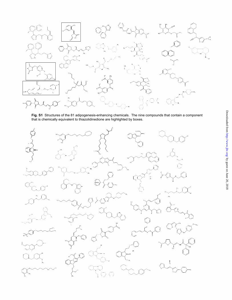
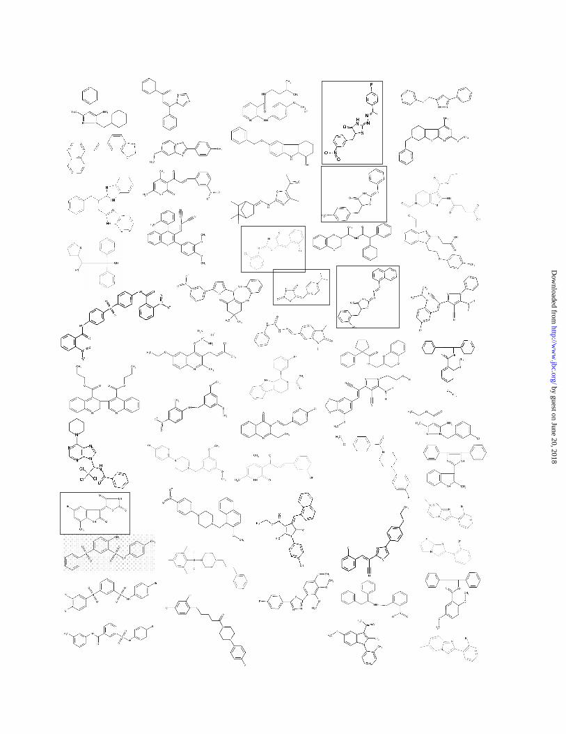
Fig. S1 Structures of the 81 adipogenesis-enhancing chemicals. The nine compounds that contain a component that is chemically equivalent to thiazolidinedione are highlighted by boxes.
by guest on June 20, 2018http://w
ww
.jbc.org/D
ownloaded from
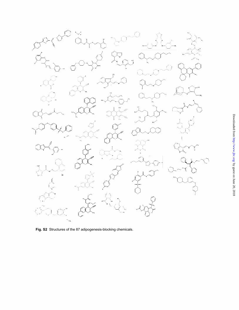
Fig. S2 Structures of the 87 adipogenesis-blocking chemicals.
by guest on June 20, 2018http://w
ww
.jbc.org/D
ownloaded from
UesugiYongmun Choi, Yoshinori Kawazoe, Koji Murakami, Hiroyuki Misawa and Motonari
Identification of bioactive molecules by adipogenesis profiling of organic compounds
published online December 19, 2002J. Biol. Chem.
10.1074/jbc.M210283200Access the most updated version of this article at doi:
Alerts:
When a correction for this article is posted•
When this article is cited•
to choose from all of JBC's e-mail alertsClick here
by guest on June 20, 2018http://w
ww
.jbc.org/D
ownloaded from