Embed Size (px)
Citation preview
Identification of Dmt-D-Lys-Phe-Phe-OH asa highly antinociceptive tetrapeptide metaboliteof the opioid-neurotensin hybrid peptide PK20
Patrycja Kleczkowska1, Engin Bojnik2, Anna LeÀniak1, Piotr Kosson1,
Isabelle Van den Eynde3, Steven Ballet3, Sandor Benyhe2, Dirk Tourwé3,
Andrzej W. Lipkowski1,4
1Mossakowski Medical Research Centre, Polish Academy of Sciences, Pawiñskiego 5,
PL 02-106 Warszawa, Poland
2Biological Research Centre, Hungarian Academy of Sciences, 6701 Szeged, Hungary
3Department of Organic Chemistry, Vrije Universiteit Brussel, Brussels, B 1050 Belgium
4Tufts University School of Medicine, Boston, MA 02111, USA
Correspondence: Andrzej W. Lipkowski, e-mail: [email protected]
Abstract:
Background: Recently, we presented a novel compound (PK20, Dmt-D-Lys-Phe-Phe-Lys-Lys-Pro-Phe-Tle-Leu-OH) that targetssingle entity opioid and neurotensin pharmacophores. This endomorphin-2-like opioid peptide was introduced as a highly active an-algesic because it elicited a strong dose- and time-dependent antinociceptive response when administered centrally and peripherally.Its pain-relieving activity was observed as rapidly as 5 min after drug injection. Such promising results led us to perform further stud-ies, such as determining the resistance to enzymatic degradation, which resulted in obtaining a very stable opioid pharmacore PK20metabolite.Methods: The synthesis of PK20 and its N-terminal tetrapeptide fragment has been accomplished using solid phase peptide chemis-try. The biological stability of peptides has been measured in human serum and analyzed by HPLC/MS. Peptides were pharmaco-logically characterized in in vitro MOP and DOP receptor binding as well as [35S]GTPgS receptor binding assays. Antinociceptiveproperties of compounds were measured by in vivo assays in C57Bl6 mice after intravenous or intrathecal applications.Results: Dmt-D-Lys-Phe-Phe-OH (PK20M), an N-terminal tetrapeptide metabolite of the opioid-neurotensin hybrid peptide PK20,is characterized by a long duration of action, as demonstrated by a preserved, long-lasting analgesic effect even 2 h post-injection(average % MPE = 69.33). In rat brain membranes, PK20M efficiently displaced both the MOP and DOP receptor selective radio-probes [3H]DAMGO and [3H]DIDI (pKi of 9.52 and 7.86, respectively) and potently stimulated [35S]GTPgS binding, proving fullagonism at both receptor types. In the [35S]GTPgS assay, which measured the agonist-mediated G protein activation, PK20M to-gether with PK20 and Met-enkephalin were potent stimulators of the regulatory G proteins. The relative affinities of PK20M for theµ and d receptor subtypes revealed µ-receptor selectivity.Conclusion: The novel MOP receptor selective metabolite has been shown to possess opioid subtype receptor selectivity, high po-tency, and effective analgesic activities as measured in various bioassays.
Key words:
in vivo analgesia, PK20 metabolite, [35S]GTPgS, antinociception, opioid receptor binding
836 Pharmacological Reports, 2013, 65, 836�846
Pharmacological Reports2013, 65, 836�846ISSN 1734-1140
Copyright © 2013by Institute of PharmacologyPolish Academy of Sciences
Introduction
The definition of pain as a complex universal experi-ence has evolved over the years. Pain can be catego-rized according to several variables, including its dura-tion (e.g., acute, convalescent, chronic), pathophysiol-ogic mechanisms (e.g., physiologic, nociceptive,neuropathic), and clinical context (e.g., postsurgical,malignancy-related, neuropathic, degenerative) [11].Acute pain follows traumatic tissue injuries, is gener-ally limited to a well-characterized duration, associatedwith a temporal reduction in intensity [21], and usuallymanaged with analgesics and anesthetics.
Opioids are the most broadly available and power-ful analgesics to date, and within the opioid family,morphine is considered to be the gold standard againstwhich all other analgesics are judged. Several bindingstudies and bioassays defined three main types ofopioid receptors in the central nervous system (CNS):µ- (MOP), d- (DOP) and k- (KOP) receptors [5, 11].Of these categories, MOP is most responsible for po-tent analgesia and is therefore considered to be essen-tial for sufficient clinical efficacy in the treatment ofmoderate to severe pain [12]. All three types of opioidreceptors are coupled to Go and Gi proteins, and theinhibitory actions of the opioids result from the clos-ing of calcium channels (in the case of k receptors) orthe opening of potassium channels (for MOP andDOP receptors) [6].
The desired duration of analgesia as well as theprevalence and severity of side effects such as toler-ance, sedation, respiratory depression and constipa-tion influence the choice of the particular opioid forpain treatment. For example, a fast onset of theantinociceptive effect is not a critical factor if the pa-tient receives a self-controlled infusion of analgesics
(e.g., via PCA pumps). In contrast, the time to analge-sic onset may be relevant in patients treated for acuteand chronic pain [13]. To reduce the typical opioid-related side effects of potent analgesics interaction be-tween opioids and synergically acting medicines havebeen proposed [2, 14] or develop medicines that arehybrids of opioid and non-opioid pharmacophores[10].
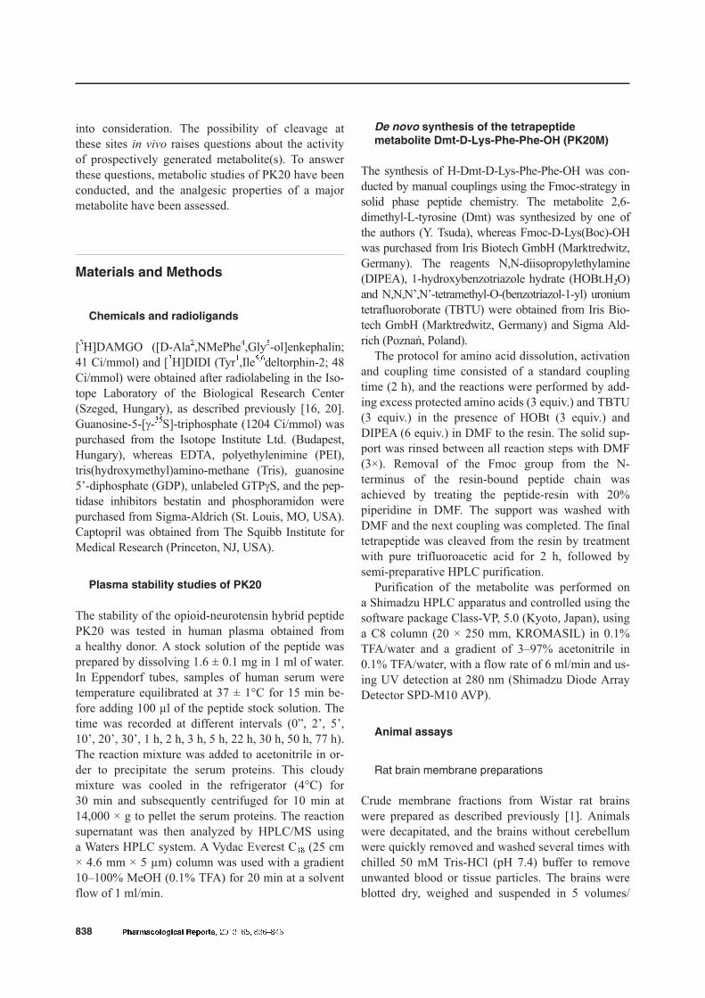
Recently, we developed a new type of analgesic inwhich opioid and neurotensin pharmacophores arehybridized into one molecule. Because opioids andneurotensin express their analgesia through differentmechanisms, a synergistic analgesic effect and de-creased side effects were expected. Indeed, PK20(Dmt-D-Lys-Phe-Phe-Lys-Lys-Pro-Phe-Tle-Leu-OH)(Fig. 1), the first reported example of this new class ofcompounds, induced strong analgesia [9]. A modifiedopioid pharmacophore has been designed by incorpo-ration of known elements that increase its resistanceto metabolic degradation; Tyr was replaced by Dmt inposition 1 and a D-amino acid residue (D-Lys) wasinserted in position 2 of the peptide sequence. On thecontrary, the metabolism of the C-terminal neuro-tensin-based pharmacophore was expected to be simi-lar to that of natural neurotensin. In neurotensin(pyrGlu-Leu-Tyr-Glu-Asn-Lys-Pro-Arg8-Arg9-Pro-Tyr-Ile-Leu-OH), one of the main cleavage bonds in themetabolic degradation is Arg8-Arg9 [4, 8]. Althoughthe dipeptide Arg8-Arg9 was replaced by Lys-Lys inthe PK20 opioid-neurotensin hybrid peptide, severalendopeptidases cleave specific peptide chains at theC-terminal side of either lysine or arginine residues[7]. Additionally, it is known that peptidyl-Lys metal-loendopeptidase (EC 3.4.24.20) cleaves specificallypeptidyl-lysine bonds (-X-Lys-) in proteins and pep-tides [7, 17]. Therefore, the metabolic release of theopioid subunit in a living organism has been taken
Pharmacological Reports, 2013, 65, 836�846 837
Analgesia of PK20 metabolitePatrycja Kleczkowska et al.
Fig. 1. Chemical structure of PK20 hy-brid compound (Dmt-D-Lys-Phe-Phe-Lys-Lys-Pro-Phe-Tle-Leu) with distinctopioid- and neurotensin-based phar-macophores
into consideration. The possibility of cleavage atthese sites in vivo raises questions about the activityof prospectively generated metabolite(s). To answerthese questions, metabolic studies of PK20 have beenconducted, and the analgesic properties of a majormetabolite have been assessed.
Materials and Methods
Chemicals and radioligands
[3H]DAMGO ([D-Ala2,NMePhe4,Gly5-ol]enkephalin;41 Ci/mmol) and [3H]DIDI (Tyr1,Ile5,6deltorphin-2; 48Ci/mmol) were obtained after radiolabeling in the Iso-tope Laboratory of the Biological Research Center(Szeged, Hungary), as described previously [16, 20].Guanosine-5-[g-35S]-triphosphate (1204 Ci/mmol) waspurchased from the Isotope Institute Ltd. (Budapest,Hungary), whereas EDTA, polyethylenimine (PEI),tris(hydroxymethyl)amino-methane (Tris), guanosine5’-diphosphate (GDP), unlabeled GTPgS, and the pep-tidase inhibitors bestatin and phosphoramidon werepurchased from Sigma-Aldrich (St. Louis, MO, USA).Captopril was obtained from The Squibb Institute forMedical Research (Princeton, NJ, USA).
Plasma stability studies of PK20
The stability of the opioid-neurotensin hybrid peptidePK20 was tested in human plasma obtained froma healthy donor. A stock solution of the peptide wasprepared by dissolving 1.6 ± 0.1 mg in 1 ml of water.In Eppendorf tubes, samples of human serum weretemperature equilibrated at 37 ± 1°C for 15 min be-fore adding 100 µl of the peptide stock solution. Thetime was recorded at different intervals (0”, 2’, 5’,10’, 20’, 30’, 1 h, 2 h, 3 h, 5 h, 22 h, 30 h, 50 h, 77 h).The reaction mixture was added to acetonitrile in or-der to precipitate the serum proteins. This cloudymixture was cooled in the refrigerator (4°C) for30 min and subsequently centrifuged for 10 min at14,000 × g to pellet the serum proteins. The reactionsupernatant was then analyzed by HPLC/MS usinga Waters HPLC system. A Vydac Everest C18 (25 cm× 4.6 mm × 5 µm) column was used with a gradient10–100% MeOH (0.1% TFA) for 20 min at a solventflow of 1 ml/min.
De novo synthesis of the tetrapeptide
metabolite Dmt-D-Lys-Phe-Phe-OH (PK20M)
The synthesis of H-Dmt-D-Lys-Phe-Phe-OH was con-ducted by manual couplings using the Fmoc-strategy insolid phase peptide chemistry. The metabolite 2,6-dimethyl-L-tyrosine (Dmt) was synthesized by one ofthe authors (Y. Tsuda), whereas Fmoc-D-Lys(Boc)-OHwas purchased from Iris Biotech GmbH (Marktredwitz,Germany). The reagents N,N-diisopropylethylamine(DIPEA), 1-hydroxybenzotriazole hydrate (HOBt.H2O)and N,N,N’,N’-tetramethyl-O-(benzotriazol-1-yl) uroniumtetrafluoroborate (TBTU) were obtained from Iris Bio-tech GmbH (Marktredwitz, Germany) and Sigma Ald-rich (Poznañ, Poland).
The protocol for amino acid dissolution, activationand coupling time consisted of a standard couplingtime (2 h), and the reactions were performed by add-ing excess protected amino acids (3 equiv.) and TBTU(3 equiv.) in the presence of HOBt (3 equiv.) andDIPEA (6 equiv.) in DMF to the resin. The solid sup-port was rinsed between all reaction steps with DMF(3×). Removal of the Fmoc group from the N-terminus of the resin-bound peptide chain wasachieved by treating the peptide-resin with 20%piperidine in DMF. The support was washed withDMF and the next coupling was completed. The finaltetrapeptide was cleaved from the resin by treatmentwith pure trifluoroacetic acid for 2 h, followed bysemi-preparative HPLC purification.
Purification of the metabolite was performed ona Shimadzu HPLC apparatus and controlled using thesoftware package Class-VP, 5.0 (Kyoto, Japan), usinga C8 column (20 × 250 mm, KROMASIL) in 0.1%TFA/water and a gradient of 3–97% acetonitrile in0.1% TFA/water, with a flow rate of 6 ml/min and us-ing UV detection at 280 nm (Shimadzu Diode ArrayDetector SPD-M10 AVP).
Animal assays
Rat brain membrane preparations
Crude membrane fractions from Wistar rat brainswere prepared as described previously [1]. Animalswere decapitated, and the brains without cerebellumwere quickly removed and washed several times withchilled 50 mM Tris-HCl (pH 7.4) buffer to removeunwanted blood or tissue particles. The brains wereblotted dry, weighed and suspended in 5 volumes/
838 Pharmacological Reports, 2013, 65, 836�846
weight of the original brain tissue with ice-cold50 mM Tris-HCl (pH 7.4) buffer. The brains werethen homogenized at 1,000 rpm with an electricallydriven Braun Teflon-glass rota-homogenizer at 4°Cusing 10 to 15 strokes of the homogenizer. The finalvolume of the homogenate was made up to 30 vol-ume/weight of the brain and filtered through four lay-ers of gauze to remove any larger aggregates. Aftercentrifugation with a Sorvall RC5C centrifuge at40,000 × g (18,000 rpm) for 20 min at 4°C, the result-ing pellet was resuspended in fresh buffer (30 vol-ume/weight) by use of a vortex. The suspension wasincubated for 30 min at 37°C to remove any endoge-nous opioids. Centrifugation was repeated under thesame conditions as described above, and the final pel-let was resuspended in 5 volumes of 50 mM Tris-HCl(pH 7.4) buffer containing 0.32 M sucrose to give a fi-nal concentration of 3–4 mg/ml protein. The presenceof sucrose is necessary for stabilization of the proteinsfor storage. The membranes were kept in 5 ml ali-quots at –70°C until used. Membranes were thawedand resuspended in 50 mM Tris-HCl (pH 7.4) bufferand centrifuged at 40,000 × g for 20 min at 4°C to re-move the sucrose. The resulting pellets were im-mersed in appropriate fresh buffer and immediatelyused in binding assays.
Receptor binding assay
The brain homogenates of Wistar rats (250–300 gbody weight) were used for further binding experi-ments. Animals were housed in the Biological Re-search Center (BRC, Szeged, Hungary) in groups offour, allowed free access to standard food and tap wa-ter, and maintained on a 12 : 12 h light/dark cycle un-til the time of sacrifice. Animals were handled ac-cording to the European Communities Council Direc-tives (86/609/ECC) and the Hungarian Act for theProtection of Animals in Research (XXVIII.tv. 32.§).
All binding assays were performed at 25°C for30 min in 50 mM Tris-HCl buffer (pH 7.4) in a finalvolume of 1 ml, containing 1 mg BSA and 0.2–0.4 mg/ml membrane protein. The samples were pre-pared in disposable plastic assay tubes (Sarstedt Co.,Nümbrecht, Germany). Rat brain membranes were in-cubated with the selective MOP receptor agonist[3H]DAMGO (0.9–1.2 nM) and the DOP receptor se-lective agonist [3H]Tyr1,Ile5,6deltorphin-2 (Tyr-D-Ala--Phe-Glu-Ile-Ile-Gly-NH2; 0.8–1.3 nM) in the pres-ence of unlabeled test ligands with concentrations
ranging from 10�5 to 10�11 M. Non-specific bindingwas determined in the presence of 10 mM naloxone.Three peptidase inhibitors (1 µM captopril, 1 µMbestatin and 1 µM phosphoramidon) were included inthe assay buffer to prevent metabolic inactivation ofthe peptides. The experiment was terminated and bothbound and free radioligands were separated by rapidfiltration under vacuum through Whatman GF/C (ra-diolabeled peptides) glass fiber filters using the Bran-del M24R Cell Harvester. Subsequently, the filterswere washed three times with 5 ml ice-cold 50 mMTris-HCl (pH 7.4) buffer. After completion of the fil-tration and separation procedure, all fiber-disks weredried under an infrared lamp and removed from thefilter-sheet by use of tweezers. Each disk was insertedinto an UltimaGold™ environmentally friendly, non-volatile, toluene-free scintillation cocktail and placedinto individual sample vials (transparent glass,Packard). The bound radioactivity was determined ina Packard Tricarb 2300TR liquid scintillation ana-lyzer. Receptor binding experiments were performedin duplicate and repeated at least three times.
Analgesia measurement
For the experiments of compound-induced antino-ciception, both male Wistar rats (225–250 g of bodyweight) and C57Bl6 mice (23–25 g of body weight)were used. Animals were housed separately at theMossakowski Medical Research Center (IMDiK,Warszawa, Poland) and maintained in plastic cageswith soft bedding, food and water available at alltimes, except during the course of the experiments.Antinociception was assessed via the tail-flick test,using the tail-flick apparatus Model 33 Tail FlickAnalgesia Meter (IITC Life Science, CA, USA), inwhich a light beam elicited a nocifensive tail re-sponse, or using the paw withdrawal test.
Central administration in rats: Groups of six ani-mals were used in each experiment, and each rat wasused only once. Surgical procedures were all per-formed under halothane anesthesia (Narkotan, LecivaA.S.) and oxygen in a concentration of 0.5 : 3.5% vol.De novo synthesized Dmt-D-Lys-Phe-Phe-OH wasadministered intrathecally (it) by means of cannulaimplanted according to the technique originally de-scribed by Yaksh and Rudy [22].
Peripheral administration in mice: C57Bl6 micewere utilized for the intravenous injection of the ex-amined drugs. Before drug or saline injections, mice
Pharmacological Reports, 2013, 65, 836�846 839
Analgesia of PK20 metabolitePatrycja Kleczkowska et al.
were warmed up under a light bulb for 10 min to di-late the tail vein. To perform the injection, animalswere subsequently placed in polyethylene restrainersand the tail was prepped with 70% ethanol. The drugor vehicle was then slowly injected into the lateralvein in a volume of 50 µl using a 29 gauge needle.
All experimental procedures used in the animaltesting were in accord with the guidelines on ethicalstandards for the investigation of experimental pain inanimals and were approved by the Animal ResearchCommittees of the Medical Research Centre, PolishAcademy of Sciences.
[35S]GTPgS binding assays
Rat brain membrane fractions (~10 µg of protein/sam-ple) were incubated at 30°C for 60 min in Tris-EGTAbuffer (50 mM Tris-HCl, 1 mM EGTA, 3 mM MgCl2,100 mM NaCl, pH 7.4) containing [35S]GTPgS(0.05 nM) and increasing concentrations (10�9 to 10�5 M)of the compounds under investigation in the presenceof 30 µM GDP in a final volume of 1 ml. The totalbinding was measured in the absence of test com-pound, whereas non-specific binding was determinedin the presence of 10 µM unlabeled GTPgS and sub-tracted from the total binding value, to determine thespecific binding. The reaction was started by adding[35S]GTPgS and terminated by filtering the samplesthrough Whatman GF/B glass fiber filters. Filterswere washed three times with ice-cold 50 mM Tris-HCl buffer (pH 7.4) using a Brandel M24R Cell Har-vester, were subsequently dried, and bound radioac-tivity was detected with the use of an UltimaGold™scintillation cocktail (Packard). Agonist-induced re-ceptor-mediated G protein stimulation is given asa percentage exceeding the basal activity as demon-strated by the specific [35S]GTPgS binding observedin the absence of receptor ligands.
[35S]GTPgS binding assays, as well as radioligandbinding experiments, were performed in duplicate andin triplicate, respectively. Experimental data wereanalyzed and graphically processed by the GraphPadPrism research software package (version 4.00 forWindows, GraphPad Software, San Diego CA, USA,www.graphpad.com) with standard office computers.Displacement curves were fitted by non-linear regres-sion using the one-site competition fitting option withno ligand depletion. The equilibrium inhibition con-stant (Ki value) was calculated from the IC50 valuesaccording to the built-in Cheng-Prusoff equation
module. Whereas the IC50 value for a compound mayvary between experiments depending on radioligandconcentration, the Ki is an absolute value. It is calcu-lated from the IC50 using the Cheng-Prusoff equation:
Ki = IC50/(1 + ([L]/Kd)
with [L] being the concentration of free radioligandused in the assay and Kd being the dissociation con-stant of the radioligand for the receptor. G proteinstimulation data were analyzed using the sigmoiddose-response curve fit option of Prism.
Measurement of pain responses
In rats: Antinociception was assessed using the tail-flick test as reported previously [9]. The measurementparameters (beam temperature, duration of exposure)were set to avoid tissue burns. The degree of antino-ciception was expressed as a percentage of the maxi-mum possible effect (% MPE) calculated as:
% MPE = [(posttreatment latency – baselinelatency)/(cutoff latency – baseline latency)] × 100
where the cut-off was 7 s in all experiments (n = 6).The nociceptive response was tested at 5, 15, 30, 60,and 120 min after drug administration.
In mice: Nociceptive responses to thermally in-duced pain was assessed in the tail flick and plantartests. In both tests, mice were wrapped in a cottontowel to minimize the stress of handling. Then, nox-ious radiant heat was applied to the plantar region ofthe hind paw or the ventral aspect of the tail. Thepositive response was either a brisk paw or tail with-drawal, which automatically stopped the built-intimer. The withdrawal latency was measured in tripli-cate in 10 s intervals between stimulations. A cut-offof 10 s was set to avoid tissue damage. The nocicep-tion measurements were performed before drug ad-ministration (baseline) and at 5, 15, 30, 60, and120 min post-injection. The intensity of radiant heatwas adjusted to elicit a response of approximately3.5 s at baseline level in the plantar test and 2.5 s inthe tail flick test. Drug-treated and saline-treated con-trol groups each consisted of 8 mice and the resultswere expressed as % MPE ± SEM.
Statistical analysis
Data on the antinociception properties of the exam-ined peptide were tested for significance with a two-
840 Pharmacological Reports, 2013, 65, 836�846
way ANOVA test followed by Tukey’s post-hoc test,and p £ 0.05, p £ 0.01 and p £ 0.001 values were con-sidered significant. In the G protein activation study,a one-way analysis of variance (ANOVA) was fol-lowed by a Tukey-Kramer multiple comparison test.All graphs were made using GraphPad Prism 5.0.
Results
Plasma stability studies of PK20
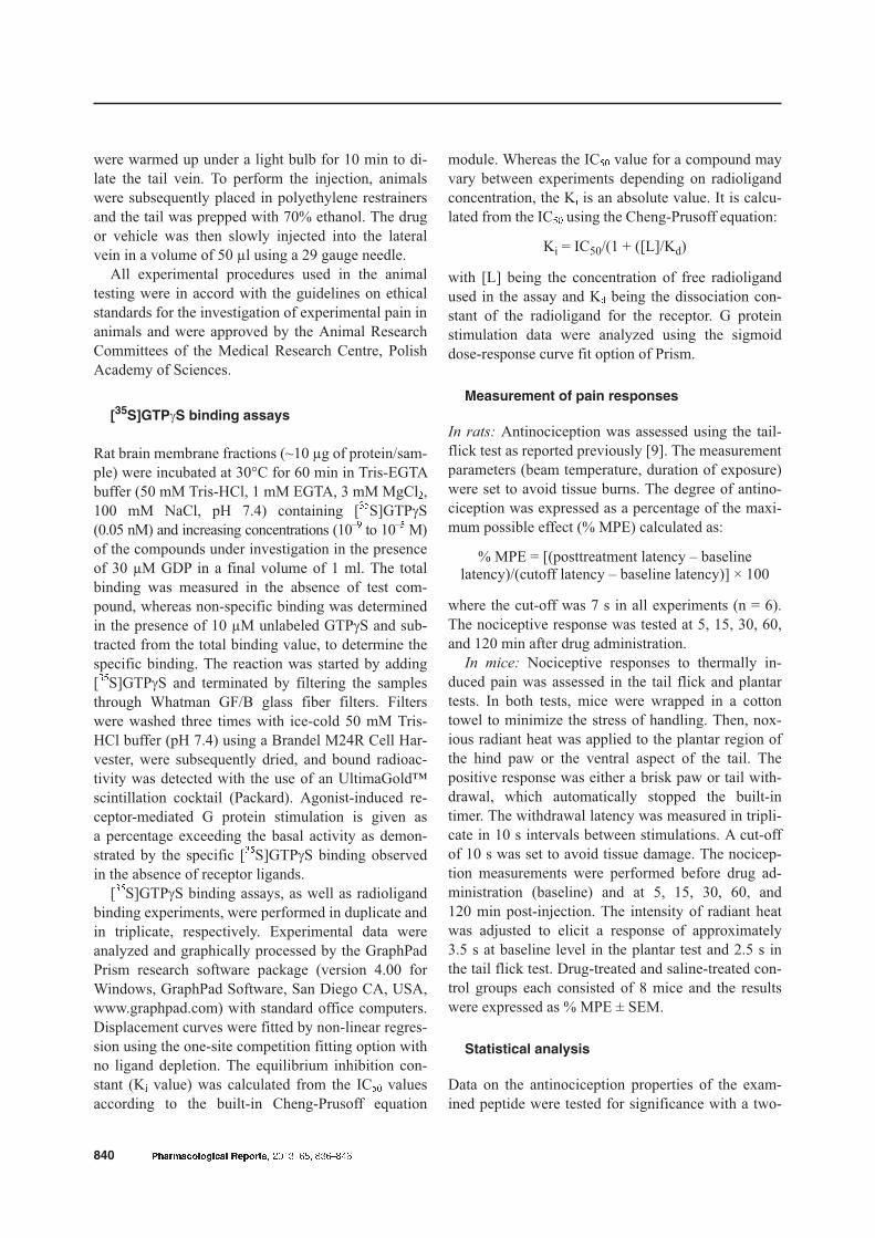
The stability of the opioid-neurotensin hybrid peptidePK20 was investigated in human plasma. As shown inFigure 2, incubation of the examined chimera PK20resulted in its enzymatic degradation, presenting thestable N-terminal tetrapeptide Dmt-D-Lys-Phe-Phe-OH (PK20M) of the parent compound. LC/MS analy-sis showed an m/z peak of 632 g/mol, correspondingto the molecular weight of PK20M. A time period ofapproximately 30 h is required for 50% degradationof PK20; consequently, the exact half-life of the pep-tide is calculated to be 31 h 45 min.
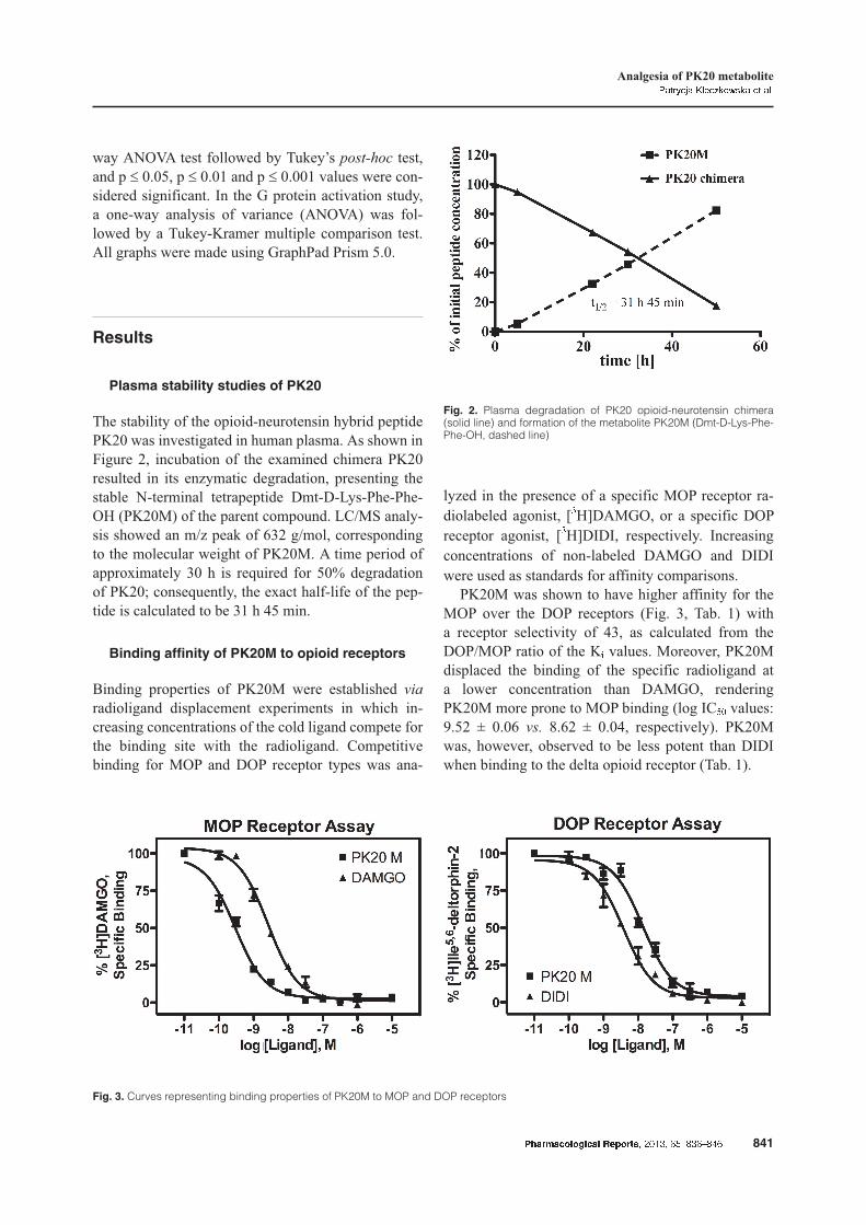
Binding affinity of PK20M to opioid receptors
Binding properties of PK20M were established via
radioligand displacement experiments in which in-creasing concentrations of the cold ligand compete forthe binding site with the radioligand. Competitivebinding for MOP and DOP receptor types was ana-
lyzed in the presence of a specific MOP receptor ra-diolabeled agonist, [3H]DAMGO, or a specific DOPreceptor agonist, [3H]DIDI, respectively. Increasingconcentrations of non-labeled DAMGO and DIDIwere used as standards for affinity comparisons.
PK20M was shown to have higher affinity for theMOP over the DOP receptors (Fig. 3, Tab. 1) witha receptor selectivity of 43, as calculated from theDOP/MOP ratio of the Ki values. Moreover, PK20Mdisplaced the binding of the specific radioligand ata lower concentration than DAMGO, renderingPK20M more prone to MOP binding (log IC50 values:9.52 ± 0.06 vs. 8.62 ± 0.04, respectively). PK20Mwas, however, observed to be less potent than DIDIwhen binding to the delta opioid receptor (Tab. 1).
Pharmacological Reports, 2013, 65, 836�846 841
Analgesia of PK20 metabolitePatrycja Kleczkowska et al.
Fig. 2. Plasma degradation of PK20 opioid-neurotensin chimera(solid line) and formation of the metabolite PK20M (Dmt-D-Lys-Phe-Phe-OH, dashed line)
Fig. 3. Curves representing binding properties of PK20M to MOP and DOP receptors
Functional activity by measurement of G
protein activation
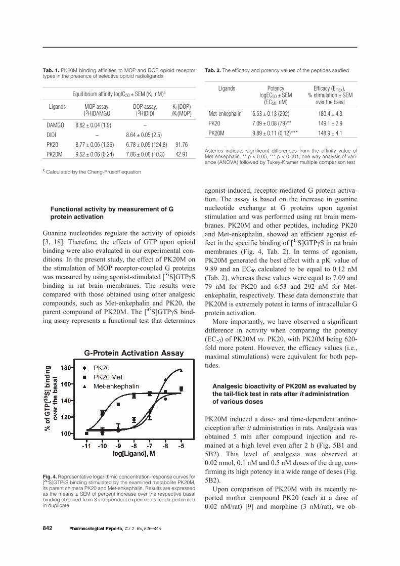
Guanine nucleotides regulate the activity of opioids[3, 18]. Therefore, the effects of GTP upon opioidbinding were also evaluated in our experimental con-ditions. In the present study, the effect of PK20M onthe stimulation of MOP receptor-coupled G proteinswas measured by using agonist-stimulated [35S]GTPgSbinding in rat brain membranes. The results werecompared with those obtained using other analgesiccompounds, such as Met-enkephalin and PK20, theparent compound of PK20M. The [35S]GTPgS bind-ing assay represents a functional test that determines
agonist-induced, receptor-mediated G protein activa-tion. The assay is based on the increase in guaninenucleotide exchange at G proteins upon agoniststimulation and was performed using rat brain mem-branes. PK20M and other peptides, including PK20and Met-enkephalin, showed an efficient agonist ef-fect in the specific binding of [35S]GTPgS in rat brainmembranes (Fig. 4, Tab. 2). In terms of agonism,PK20M generated the best effect with a pKi value of9.89 and an EC50 calculated to be equal to 0.12 nM(Tab. 2), whereas these values were equal to 7.09 and79 nM for PK20 and 6.53 and 292 nM for Met-enkephalin, respectively. These data demonstrate thatPK20M is extremely potent in terms of intracellular Gprotein activation.
More importantly, we have observed a significantdifference in activity when comparing the potency(EC50) of PK20M vs. PK20, with PK20M being 620-fold more potent. However, the efficacy values (i.e.,maximal stimulations) were equivalent for both pep-tides.
Analgesic bioactivity of PK20M as evaluated by
the tail-flick test in rats after it administration
of various doses
PK20M induced a dose- and time-dependent antino-ciception after it administration in rats. Analgesia wasobtained 5 min after compound injection and re-mained at a high level even after 2 h (Fig. 5B1 and5B2). This level of analgesia was observed at0.02 nmol, 0.1 nM and 0.5 nM doses of the drug, con-firming its high potency in a wide range of doses (Fig.5B2).
Upon comparison of PK20M with its recently re-ported mother compound PK20 (each at a dose of0.02 nM/rat) [9] and morphine (3 nM/rat), we ob-
842 Pharmacological Reports, 2013, 65, 836�846
Tab. 2. The efficacy and potency values of the peptides studied
Ligands PotencylogEC50 ± SEM
(EC50, nM)
Efficacy (Emax),% stimulation ± SEM
over the basal
Met-enkephalin 6.53 ± 0.13 (292) 180.4 ± 4.3
PK20 7.09 ± 0.08 (79)** 149.1 ± 2.9
PK20M 9.89 ± 0.11 (0.12)*** 148.9 ± 4.1
Asterics indicate significant differences from the affinity value ofMet-enkephalin. ** p < 0.05, *** p < 0.001; one-way analysis of vari-ance (ANOVA) followed by Tukey-Kramer multiple comparison test
Tab. 1. PK20M binding affinities to MOP and DOP opioid receptortypes in the presence of selective opioid radioligands
Equilibrium affinity logIC50 ± SEM (Ki, nM)a
Ligands MOP assay,[3H]DAMGO
DOP assay,[3H]DIDI
Ki (DOP)/Ki(MOP)
DAMGO 8.62 ± 0.04 (1.9) –
DIDI – 8.64 ± 0.05 (2.5)
PK20 8.77 ± 0.06 (1.36) 6.78 ± 0.05 (124.8) 91.76
PK20M 9.52 ± 0.06 (0.24) 7.86 ± 0.06 (10.3) 42.91
a Calculated by the Cheng-Prusoff equation
Fig. 4. Representative logarithmic concentration-response curves for[35S]GTPgS binding stimulated by the examined metabolite PK20M,its parent chimera PK20 and Met-enkephalin. Results are expressedas the means ± SEM of percent increase over the respective basalbinding obtained from 3 independent experiments, each performedin duplicate
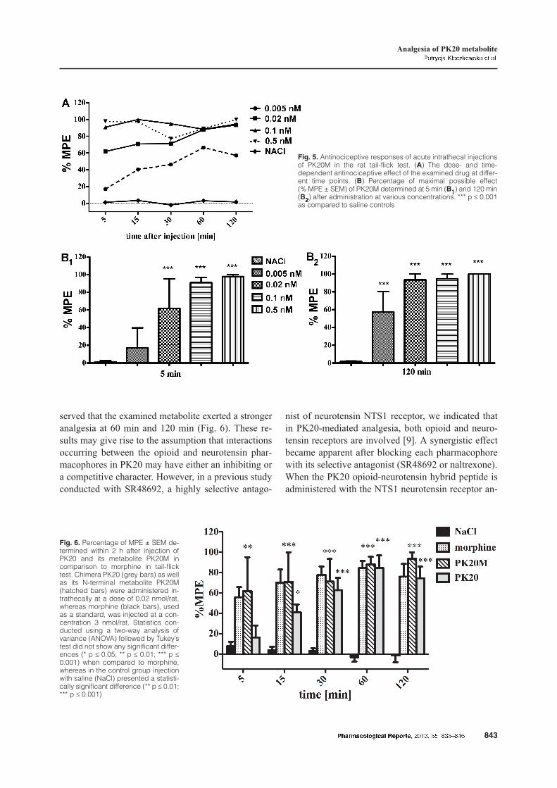
served that the examined metabolite exerted a strongeranalgesia at 60 min and 120 min (Fig. 6). These re-sults may give rise to the assumption that interactionsoccurring between the opioid and neurotensin phar-macophores in PK20 may have either an inhibiting ora competitive character. However, in a previous studyconducted with SR48692, a highly selective antago-
nist of neurotensin NTS1 receptor, we indicated thatin PK20-mediated analgesia, both opioid and neuro-tensin receptors are involved [9]. A synergistic effectbecame apparent after blocking each pharmacophorewith its selective antagonist (SR48692 or naltrexone).When the PK20 opioid-neurotensin hybrid peptide isadministered with the NTS1 neurotensin receptor an-
Pharmacological Reports, 2013, 65, 836�846 843
Analgesia of PK20 metabolitePatrycja Kleczkowska et al.
Fig. 5. Antinociceptive responses of acute intrathecal injectionsof PK20M in the rat tail-flick test. (A) The dose- and time-dependent antinociceptive effect of the examined drug at differ-ent time points. (B) Percentage of maximal possible effect(% MPE ± SEM) of PK20M determined at 5 min (B1) and 120 min(B2) after administration at various concentrations. *** p £ 0.001as compared to saline controls
Fig. 6. Percentage of MPE ± SEM de-termined within 2 h after injection ofPK20 and its metabolite PK20M incomparison to morphine in tail-flicktest. Chimera PK20 (grey bars) as wellas its N-terminal metabolite PK20M(hatched bars) were administered in-trathecally at a dose of 0.02 nmol/rat,whereas morphine (black bars), usedas a standard, was injected at a con-centration 3 nmol/rat. Statistics con-ducted using a two-way analysis ofvariance (ANOVA) followed by Tukey’stest did not show any significant differ-ences (* p £ 0.05; ** p £ 0.01; *** p £0.001) when compared to morphine,whereas in the control group injectionwith saline (NaCl) presented a statisti-cally significant difference (** p £ 0.01;*** p £ 0.001)
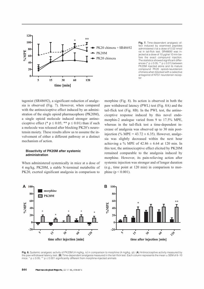
tagonist (SR48692), a significant reduction of analge-sia is observed (Fig. 7). However, when comparedwith the antinociceptive effect induced by an admini-stration of the single opioid pharmacophore (PK20M),a single opioid molecule induced stronger antino-ciceptive effect (* p £ 0.05; ** p £ 0.01) than if sucha molecule was released after blocking PK20’s neuro-tensin moiety. These results allow us to assume the in-volvement of either a different pathway or a distinctmechanism of action.
Bioactivity of PK20M after systemic
administration
When administered systemically in mice at a dose of4 mg/kg, PK20M, a stable N-terminal metabolite ofPK20, exerted significant analgesia in comparison to
morphine (Fig. 8). Its action is observed in both the
paw withdrawal latency (PWL) test (Fig. 8A) and the
tail-flick test (Fig. 8B). In the PWL test, the antino-
ciceptive response induced by this novel endo-
morphin-2 analogue varied from 9 to 17.5% MPE,
whereas in the tail-flick test a time-dependent in-
crease of analgesia was observed up to 30 min post-
injection (% MPE = 43.72 ± 6.35). However, analge-
sia was slightly decreased within the next hour
achieving a % MPE of 42.86 ± 6.64 at 120 min. In
this test, the antinociceptive effect elicited by PK20M
remained comparable to the analgesia induced by
morphine. However, its pain-relieving action after
systemic injection was stronger and of longer duration
(e.g., time point at 120 min) in comparison to mor-
phine (p < 0.001).
844 Pharmacological Reports, 2013, 65, 836�846
Fig. 7. Time-dependent analgesic ef-fect induced by examined peptidesadministered it at a dose of 0.02 nmol/rat in tail-flick test. SR48692 was in-jected at a dose of 10 µg/rat 10 min be-fore the exact compound injection.The statistics showed significant differ-ences (* p £ 0.05; ** p £ 0.01) betweenPK20M injected alone and its maturecompound PK20 opioid-neurotensinchimera when blocked with a selectiveantagonist of NTS1 neurotensin recep-tor
Fig. 8. Systemic analgesic activity of PK20M (4 mg/kg, iv) in comparison to morphine (4 mg/kg, ip). (A) Antinociceptive activity measured bythe paw withdrawal latency test. (B) Time-dependent analgesia measured in the tail-flick test. Each column represents the mean ± SEM of 8–10mice. * p £ 0.05; ** p £ 0.001 significantly different from morphine-injected animals
A B
Discussion
Incubation of PK20 with human serum resulted in al-most an exclusive release of the N-terminal tetrapep-tide H-Dmt-D-Lys-Phe-Phe-OH, abbreviated with theacronym PK20M. This tetrapeptide is similar to po-tent synthetic analogs of the N-terminal tetrapeptidefragment of dermorphin [15, 19]. The peptide wassynthesized de novo and its pharmacological activitywas tested and compared with the parent peptidePK20 and Met-enkephalin, a specific MOP receptoragonist.
PK20M expressed potent activity when adminis-tered both centrally and peripherally. Figure 5A pres-ents the time- and dose-dependent analgesia inducedby it injected PK20M in rats. PK20M, administered ata dose of 0.02 nmol/rat, induced a similar antino-ciceptive effect compared to morphine at a 150-foldhigher dose (3 nmol/rat) (Fig. 6). Additionally, PK20M’sanalgesic effect, mediated through CNS opioid recep-tors, proved to be long lasting; during a 2 h post-injection period its antinociceptive profile was pre-served for every examined dose. Similarly, sucha prolonged analgesic activity was also observed aftersystemic (iv) administration of PK20M (Fig. 8A and8B). Both the paw withdrawal latency (Fig. 8A) andthe tail-flick responses (Fig. 8B) were evidence thatPK20M effectively permeates the blood-brain barrier(BBB). Its antinociceptive action after systemic injec-tion in the tail-flick test is similar in magnitude to anidentical dose of morphine, and suggests a lower BBBpermeability for PK20M with respect to morphine.However, PK20M expressed a longer duration ofantinociceptive activity.
Regarding the G protein activation results (Fig. 4),PK20M demonstrated its extreme potency as wit-nessed by an EC50 value of 0.12 nM and was thereforeconsidered to be far more potent than PK20 itself.Furthermore, and similar to the results of the analge-sia experiments, [35S]GTPgS studies revealed thatmaximal stimulation of G proteins by PK20M occursat lower concentration (nM range), relative to PK20(µM range).
Opioids generate their effects via G protein-coupled receptor pathways with G protein activationbeing the key signaling step that results from agonistbinding to the opioid receptors. The rapidly obtainedmaximal stimulation and extreme potency of thePK20M towards the G proteins focused our attention.
The extraordinary potency of the PK20M both in re-ceptor binding and in the G protein activation assaytogether with its analgesic properties makes PK20Ma promising compound for future studies in pain man-agement.
Acknowledgments:
This work was supported by the European Grant “Normolife”
[LSHC-CT-2006-037733]; Fund for Scientific Research-Flanders
(FWO-Vlaanderen) [Grant G.000.08]; and with a scholarship from
the European Social Fund, Human Capital Operational Programme
for the execution of the project “Support for bio tech med scientists
in technology transfer” [UDA-POKL.08.02.01-14-041/09].
References:
1. Bojnik E, Magyar A, Tóth G, Bajusz S, Borsodi A,Benyhe S: Binding studies of novel non-mammalian en-kephalins, structures predicted from frog and lungfishbrain cDNA sequences. Neuroscience, 2009, 158,867–874.
2. Cegielska-Perun K, Bujalska-Zadro¿ny M, Makulska-Nowak HE, Modification of morphine analgesia by ven-lafaxine in diabetic neuropathic pain model. PharmacolRep, 2012, 64, 1267–1275.
3. Chang KJ, Blanchard SG, Cuatrecasas P: Unmasking ofmagnesium-dependent high-affinity binding sites for[DAla2, DLeu5]enkephalin after pretreatment of brainmembranes with guanine nucleotides. Proc Natl AcadSci USA, 1983, 80, 940–944.
4. Checler F, Vincent JP, Kitabgi P: Degradation of neuro-tensin by rat brain synaptic membranes: involvement ofmetalloendopeptidase (enkephalinase), angiotensin-converting enzyme, and other unidentified peptidases.J Neurochem, 1983, 41, 375–384.
5. Dhawan BN, Cesselin F, Raghubir R, Reisine T, BradleyPB, Porthogese PS, Hamon M: International Union ofPharmacology. XII. Classification of opioid receptors.Pharmacol Rev, 1996, 48, 567–592.
6. Dickenson AH: Peptides. In: Neurotransmitters, Drugsand Brain function. Ed. Webster RA. John Wiley & SonsLtd., New York, 2001, 251–264.
7. Keil B: Specificity of proteolysis. Springer-Verlag,Berlin, 1992.
8. Kitabgi P, De Nadai F, Rovère C, Bidard JN: Biosynthe-sis, maturation, release and degradation of neurotensinand neuromedin N. Ann NY Acad Sci, 1992, 668, 30–42.
9. Kleczkowska P, Kosson P, Ballet S, Van den Eynde I,Tsuda Y, Tourwé D, Lipkowski AW: PK20, a newopioid-neurotensin hybrid peptide that exhibits central andperipheral antinociceptive effects. Mol Pain, 2010, 6, 86.
10. Lipkowski AW, Cooperative reinforcement of opioidpharmacophores. Pol J Pharmacol Pharm, 1987, 39,153–164.
11. Lipkowski AW, Carr DB: Rethinking opioid equivalence.Pain-Clinical Updates, 2002, 10, 1–7.
Pharmacological Reports, 2013, 65, 836�846 845
Analgesia of PK20 metabolitePatrycja Kleczkowska et al.
12. McDonald J: Opioid receptors. Contin Educ AnaesthCrit Care Pain, 2005, 5, 22–25.
13. McQuay H: Opioids in pain management. Lancet 1999,353, 2229–2232.
14. Misterek K, Maszczynska I, Dorociak A, Gumulka SW,Carr DB, Szyfelbein SK, Lipkowski AW: Spinal co-administration of peptide substance P antagonist potenti-ates antinociceptive effect of opioid peptide. Life Sci,1994, 54, 939–944.
15. Mizoguchi H, Bagetta G, Sakurada T, Sakurada S:Dermorphin tetrapeptide analogs as potent and long-lasting analgesics with pharmacological profiles distinctfrom morphine. Peptides, 2011, 32, 421–427.
16. Nevin ST, Kabasakal L, Ötvös F, Tóth G, Borsodi A:Binding characteristics of the novel highly selective deltaagonist, [3H]Ile5,6-deltorphin II. Neuropeptides, 1994,26, 261–265.
17. Nonaka T, Dohmae N, Hashimoto Y, Takio K: Aminoacid sequences of metalloendopeptidases specific foracyl-lysine bonds from Grifola frondosa and Pleurotus
ostreatus fruiting bodies. J Biol Chem, 1997, 272,30032–30039.
18. Rosenbaum JS, Sadee W: Opiate receptor binding af-fected by guanine nucleotide. J Biol Chem, 1983, 258,1419–1422.
19. Sato T, Sakurada S, Sakurada T, Furuta S, Chaki K,Kisara K, Sasaki Y, Suzuki K: Opioid activities ofD-Arg2-substituted tetrapeptides. J Pharmacol Exp Ther,1987, 242, 654–659.
20. Tóth G, Kramer M, Sirokman F, Borsodi A, Ronai A:Preparation of [7,8,19,20-3H]naloxone of high specificactivity. J Labelled Comp Rad, 1982, 19, 1021–1030.
21. Vadivelu N, Whitney ChJ, Sinatra RS: Pain pathwaysand acute pain processing. In: Acute pain management.Eds. Sinatra RS, de Leon-Casasola OA, Viscusi ER,Ginsberg B, Cambridge University Press, New York,2009, 3–20.
22. Yaksh TL, Rudy TA: Chronic catheterization of the spi-nal subarachnoid space. Physiol Behav, 1976, 17,1031–1036.
Received: August 3, 2012; in the revised form: March 6, 2013;
accepted: March 7, 2013.
846 Pharmacological Reports, 2013, 65, 836�846