Embed Size (px)
Citation preview
Ide
ntificatth
zur
Fak
der Ludw
tion ofhrough
D
Erlangung
kultät für C
wig‐Maxim
f novel h genom
Myrt
aus Ath
Dissertatio
g des Dokt
Chemie un
milians‐Un
septatme‐wid
to Deligian
hen, Griech
2015
on
torgrades
nd Pharma
niversität M
e junctde glial
nnaki
henland
s der
azie
München
tion coscreen
mponens
ents

Diese Dissertation wurde im Sinne von §7 der Promotionsordnung vom 28.
November 2011 von Frau Prof. Dr. Ulrike Gaul betreut.
Eidesstattliche Versicherung
Diese Dissertation wurde eigenständig und ohne unerlaubte Hilfe erarbeitet.
München, 07/04/2015
......... .....
Myrto Deligiannaki
Dissertation eingereicht am: 07/04/2015
Erstgutachterin: Prof. Dr. Ulrike Gaul
Zweitgutachter: Prof. Dr. Klaus Förstemann
Tag der mündlichen Prüfung: 19/05/2015


Acknowledgements
Firstly, I would like to thank my advisor Ulrike Gaul for persistently encouraging my
research and being a challenging mentor. I appreciate all her contributions of ideas,
time and funding, as well as her support, which made my Ph.D. experience
productive and stimulating.
I would also like to thank: the members of my thesis advisory committee, Magdalena
Götz, Hiromu Tanimoto and Takashi Suzuki for the fruitful discussions and good
advice; Klaus Förstemann for being my second thesis evaluator; all members of my
defense committee: Ulrike Gaul, Klaus Förstemann, Nicolas Gompel, Magdalena
Götz, Roland Beckmann and Daniel Wilson. I am also grateful to Hans-Jörg
Schaeffer, Ingrid Wolf and everybody from the Max Planck International PhD
programme who assisted me in various ways and provided excellent workshops.
A big thank you to members of the lab that contributed with their expertise to
different aspects of my project: Abbie Casper for performing some of the
experiments in the pasiflora project; Christophe Jung for his excellent work in
developing the automated analysis of the dye assay and performing analysis of
FRAP data; Claudia Ludwig and Susi Lange for sitting with me in the flyroom for
hours counting flies during the screens - Claudia also for helping with generation of
transgenic lines; Ulrich Unnerstall for his major contribution and patient help with the
statistical analysis of the pan-glial screen.
I am particularly grateful to Sara Batelli for answering all my questions on molecular
biology, for having productive discussions with me, and mostly for being a friend who
stood all my bad moments. I am also indebted to Tina Schwabe for developing many
of the tools I used, for allowing me to use her schematics and for making useful
comments on my manuscript, as well as Xiaoling Li for our skype chats on the blood-
brain barrier and her guidance at the beginning of my project. I would also like to
thank all the members of the lab for creating a pleasant and interactive working
atmosphere.

Special thanks to everybody who provided flies and reagents: E. Arama, V. Auld,
W.Chia, L. Luo, the Vienna Drosophila Resource Center, the Bloomington
Drosophila Stock Center, the TRiP at Harvard Medical School (NIH/NIGMS R01-
GM084947), the Drosophila Genomics Resource Center and the Developmental
Studies Hybridoma Bank.
My biggest thank you goes to my parents and sister for their understanding, trust and
unquestioning support, and to Giorgos for moving to Germany, standing by me and
simplifying my efforts in so many ways. Finally, I want to thank Matilda for recently
bringing so much joy in my life.
5
Table of contents
Abbreviations
Abstract
5
8
11
Aim of the thesis 13
1. Part1: Genome-wide glial screens 15
1.1 Introduction 16
1.1.1 Glia are central players of nervous system function 16
1.1.2 Blood-brain barrier 18
1.1.3 Cellular architecture of the Drosophila blood-brain barrier 19
1.1.4 Moody signaling regulates blood-brain barrier development and
maintenance
21
1.1.5 The blood-brain barrier is more than a diffusion barrier 22
1.1.6 Insulating barriers of the mammalian nervous system 23
1.1.7 Drosophila as a genetic model to identify and characterize novel genes 24
1.1.8 Rationale 27
1.2 Genome-wide RNAi screens identify glial and subperineurial glial
genes
29
1.2.1 Overview of genome-wide RNAi screening 29
1.2.2 Primary pan-glial screen 30
1.2.3 Secondary blood-brain barrier-specific screen 37
1.2.4 Small-scale screen to identify genes required for blood-brain barrier
formation
40
1.3 Discussion 42
1.3.1 Genome-wide glial screening 42
1.3.2 Screening for blood-brain barrier genes – achievements and future
directions
43
6
2. Part 2: Pasiflora1, Pasiflora2 and Mcr are novel components of the
Drosophila septate junction
47
2.1 Introduction 48
2.1.1 Epithelia – an evolutionary novelty 48
2.1.2 Occluding junctions mediate epithelial barrier function 49
2.1.3 Overview of Drosophila septate junctions - ultrastructure and subtypes 49
2.1.4 Molecular composition and morphogenesis of Drosophila septate
junctions
50
2.1.5 Other functions of Drosophila septate junctions 54
2.1.6 Vertebrate tight and septate junctions 55
2.1.7 Claudins – determinants of barrier selectivity 56
2.2 Pasiflora proteins are novel core components of the septate
junction
58
2.2.1 The novel pasiflora genes are required for blood-brain barrier formation 58
2.2.2 pasiflora genes are required for tracheal tube size and barrier function 61
2.2.3 pasiflora genes are expressed in SJ-forming embryonic epithelia and
glia
62
2.2.4 Molecular features of Pasiflora proteins 62
2.2.5 pasiflora genes are required for localization of SJs 65
2.2.6 Pasiflora proteins localize at the SJ and their localization depends on
other complex components
67
2.2.7 Pasiflora proteins are required for SJ complex assembly 69
2.3 Mcr is a novel septate junction component 73
2.3.1 Introduction – Mcr and other thioester proteins 73
2.3.2 mcr is required for blood-brain barrier formation 74
2.3.3 mcr is required for tracheal tube size and barrier function 75
2.3.4 mcr is required for localization of SJs 77
2.3.5 Mcr is a plasma membrane protein 77
2.3.6 mcr is expressed in SJ-forming embryonic epithelia and glia 81
7
2.3.7 Mcr localizes at the SJ and its localization depends on other complex
components
81
2.4 Discussion 83
2.4.1 Pasiflora proteins and Mcr are novel SJ components 83
2.4.2 Potential roles of Pasiflora proteins 85
2.4.3 Mcr - Is there a link between epithelial barrier function and immunity? 86
2.4.4 Open questions on SJs 88
3. Materials and methods 91
3.1 Genome-wide screening for adult viability 91
3.2 Constructs 91
3.3 Fly strains 92
3.4 Immunohistochemistry 93
3.5 Live imaging of embryos and larvae 94
3.6 Confocal image acquisition and analysis 94
3.7 Dye penetration assay and quantification 94
3.8 RNA in situ hybridization 95
3.9 Embryonic viability assay 95
3.10 FRAP experiments and analysis 95
3.11 Production of antibodies 97
3.12 Cell culture and immunohistochemistry 98
3.13 Western blotting 98
References 100
8
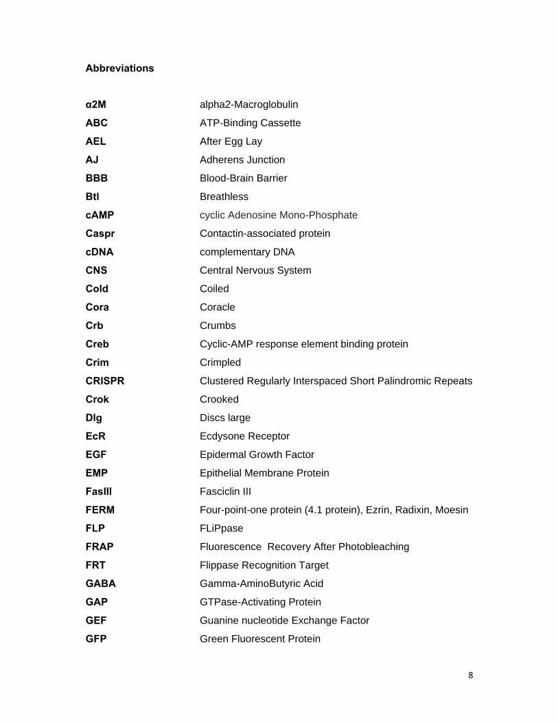
Abbreviations
α2M alpha2-Macroglobulin
ABC ATP-Binding Cassette
AEL After Egg Lay
AJ Adherens Junction
BBB Blood-Brain Barrier
Btl Breathless
cAMP cyclic Adenosine Mono-Phosphate
Caspr Contactin-associated protein
cDNA complementary DNA
CNS Central Nervous System
Cold Coiled
Cora Coracle
Crb Crumbs
Creb Cyclic-AMP response element binding protein
Crim Crimpled
CRISPR Clustered Regularly Interspaced Short Palindromic Repeats
Crok Crooked
Dlg Discs large
EcR Ecdysone Receptor
EGF Epidermal Growth Factor
EMP Epithelial Membrane Protein
FasIII Fasciclin III
FERM Four-point-one protein (4.1 protein), Ezrin, Radixin, Moesin
FLP FLiPpase
FRAP Fluorescence Recovery After Photobleaching
FRT Flippase Recognition Target
GABA Gamma-AminoButyric Acid
GAP GTPase-Activating Protein
GEF Guanine nucleotide Exchange Factor
GFP Green Fluorescent Protein
9
Gli Gliotactin
GO Gene Ontology
GPCR G-Protein Coupled Receptor
GPI GlycosylPhosphatidylInositol
GTP Guanosine TriPhosphate
IGF Insulin Growth Factor
Ilp Insulin-like peptide
Kune Kune-kune
Lac Lachesin
LDL Low Density Lipoprotein
Lgl Lethal giant larvae
Loco Locomotion defects
Ly-6 Lymphocyte antigen-6
Mcr Macroglobulin complement-related
Mdr65 Multiple drug resistance 65
Mega Megatrachea
mir microRNA
MLCK Myosin Light Chain Kinase
MP20 lens fiber Membrane intrinsic Protein
MS Mass-Spectrometry
Nrg Neuroglian
Nrv2 Nervana2
Nrx-IV Neurexin-IV
Pasi1 Pasiflora 1
Pasi2 Pasiflora 2
PBS Phosphate Buffered Saline
PDZ Post synaptic density protein (PSD95), Discs-large1 (Dlg1),
Zonula occludens-1 protein (ZO-1)
PKA Protein Kinase A
PMP22 Peripheral Myelin Protein 22
PNS Peripheral Nervous System
Prd Paired
10
Repo Reversed polarity
RISC RNA-Induced Silencing Complex
RGS Regulator of G-protein Signaling
RNAi RNA interference
RT Room Temperature
RTK Receptor Tyrosine Kinase
S2 Schneider 2
SD Standard Deviation
Sinu Sinuous
siRNA small interfering RNA
SJ Septate Junction
SPG SubPerineurial Glia
Sb Stubble
TEP ThioEster Protein
TJ Tight Junction
TMEM47 TransMEMrane protein 47
Tre1 Trapped in endoderm 1
Tub Tubulin
UAS Upstream Activating Sequence
UTR UnTranslated Region
Vari Varicose
VDRC Vienna Drosophila Resource Center
wt wild-type
11
Abstract
Epithelial barriers are central to the development of metazoans by
compartmentalizing the body in distinct chemical milieus essential for the function of
many organs. One such barrier is the blood-brain barrier, which isolates the nervous
system from the body fluid to maintain its ionic homeostasis and ensure nerve pulse
transmission. In Drosophila, the blood-brain barrier is formed late in embryogenesis
by a thin epithelium of subperineurial glia that ensheath the nervous system. Similar
to other epithelia, subperineurial glia seal the paracellular space by forming large
multiprotein complexes at the lateral membrane, the septate junctions (SJs), which
impede free diffusion and mediate barrier function.
To identify novel genes required for blood-brain barrier formation, we followed a
genome-wide in vivo RNAi approach. We initially screened almost the whole genome
for genes required in glia for adult viability and impressively identified 3679 potential
candidates. Subsequently, we tested these candidates for requirement in
subperineurial glia for adult survival and identified 383 genes. At a last step, we
directly asked if blood-brain barrier formation is compromised in the knock-down of
the genes by performing the embryonic dye penetration assay in a selection of
candidates and identified five genes that play a role during barrier development.
Three of these genes, macroglobulin complement-related (mcr) and the previously
uncharacterized pasiflora1 and pasiflora2 are further characterized in the context of
this thesis.
Here we show that all three proteins are novel components of the Drosophila SJ.
Pasiflora1 and Pasiflora2 belong to a previously uncharacterized family of tetra-
spanning membrane proteins, while Mcr was reported to be a secreted protein in S2
cells required for phagocytosis and clearance of specific pathogens. Through
detailed phenotypic analysis we demonstrate that the mutants show leaky blood-
brain and tracheal barriers, overelongated tracheal tubes and mislocalization of SJ
proteins, phenotypes that are characteristic of SJ mutants. Consistent with the
observed phenotypes, the genes are co-expressed in SJ-forming embryonic epithelia
and glia and are required cell-autonomously to exert their function. In columnar
epithelia, the proteins localize at the apicolateral membrane compartment, where
12
they colocalize with other SJ proteins, and similar to known SJ components, their
restricted localization depends on other complex members. Using fluorescence
recovery after photobleaching experiments, we demonstrate for Pasiflora proteins
that they are core SJ components, as they are required for complex formation and
themselves show restricted mobility within the membrane of wild-type epithelial cells,
but fast diffusion in cells with disrupted SJs. Taken together, our results show that
Pasiflora1 and Pasiflora2 are novel integral SJ components and implicate a new
family of tetraspan proteins in the development of cell junctions. In addition, we find a
new unexpected role for Mcr as a transmembrane SJ protein, which raises questions
about a potential intriguing link between epithelial barrier function, phagocytosis and
innate immunity and has potential implications for the function of occluding junctions.
13
Aim of the thesis
In my thesis project, I aimed at the identification and characterization of novel
genes required for blood-brain barrier formation. For this purpose, I decided to carry
out an unbiased genome-wide approach by exploiting the power of in vivo screening
in Drosophila. My goals were: first, to perform a whole-genome RNAi screen for
genes required in all glia for adult viability; second, to perform a blood-brain barrier-
specific screen; and third, to select candidates and explore their role in barrier
formation at the genetic, cellular and functional level.

14
Part 1
Genome-wide glial RNAi screens
16
1. 1 Introduction
1.1.1 Glia are central players of nervous system function
The nervous system is composed of two cell types, neurons and glia. Neurons, being
the cells firing action potentials and transmitting the information have received most
of the attention, while glia, originally thought to simply provide a static framework for
neurons are much less studied. However, in recent years the idea of such a passive
role for glia has been abandoned and an increasing repertoire of functions is being
attributed to glia themselves, which are now appreciated as critical modulators in
brain development and function. Importantly, the relative fraction of glial cells
increases with the increasing complexity of nervous systems in evolution from
around 5% in the worm to 90% in human, suggesting an important role for glia in
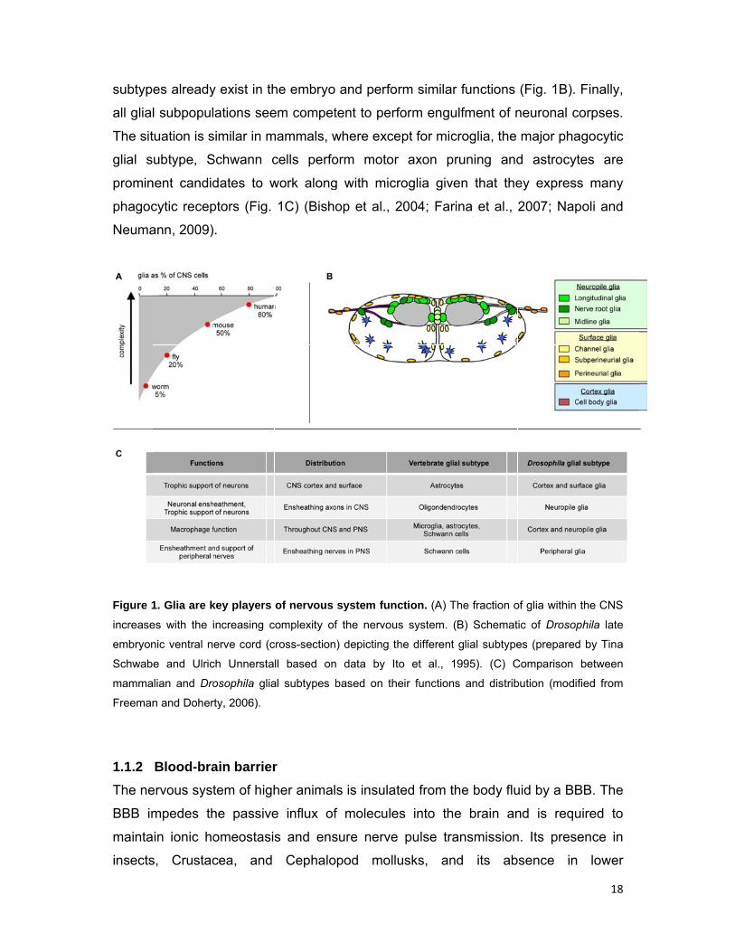
higher brain functions (Pfrieger and Barres, 1995) (Fig. 1A). Glial dysfunctions also
contribute to many neurological disorders, such as multiple sclerosis, fragile X
syndrome, brain injury, and Alzheimer’s disease and gliomas represent the majority
of malignant brain tumors (Miller, 2005; Jacobs and Doering, 2010).
In Drosophila, glia constitute 10-25% of the nervous system cells (Fig. 1A)
(Pfrieger and Barres, 1995; Kremer et al., in preparation). With the exception of
midline glia, they are derived from a small set of uniquely identifiable glioblasts and
neuro-glioblasts that delaminate from the neuroectoderm of the early embryo
(Bossing et al., 1996; Schmidt et al., 1997). One of the earliest steps in the
specification of Drosophila glia is the expression of the transcription factor Glial cells
missing (Gcm), which in turn regulates the expression of numerous glial genes. One
of its major targets is the transcription factor Reversed polarity (Repo), which is
considered a definite marker of glial cell fate (Halter et al., 1995; Hosoya et al., 1995;
Jones et al., 1995; Freeman et al., 2003). Once specified, most glia migrate
significant distances within the developing nervous system until they reach their final
sites.
Drosophila glia contribute to virtually all functions of the nervous system and
are therefore essential for survival. They play diverse roles during development and
many of these roles are recapitulated during adult life and are required for brain
homeostasis and function. In particular, glia guide neurite growth and defasciculation
by presenting growth cones with attractive and repulsive cues, and actively control
the establishment of neuronal connectivity (Hidalgo et al., 1995; Hidalgo and Booth,
17
2000; Lemke, 2001). Furthermore, they establish and maintain ionic homeostasis
and nerve pulse propagation by forming the blood-brain barrier (BBB), which
ensheaths the entire nervous system, and by insulating axon bundles, individual
axons and dendrites (Baumgartner et al., 1996; Schwabe et al., 2005; Awasaki et al.,
2008; Stork et al., 2008). Glia also provide neurons with high energy metabolic
substrates to sustain their activity and promote their survival (Buchanan and Benzer,
1993; Booth et al., 2000). Furthermore, glia protect the brain by phagocytosing
unwanted and aberrant material. They engulf and degrade excessive dying neurons
to adjust their cell number, as well as pruned axons and immature synaptic material
to eliminate exuberant connections and refine neural circuits (Sonnenfeld and
Jacobs, 1995; Awasaki and Ito, 2004; Watts et al., 2004; MacDonald et al., 2006;
Kurant et al., 2008; Fuentes-Medel et al., 2009; Tasdemir-Yilmaz and Freeman,
2014). Moreover, glia modulate synaptic activity and regulate behavior by taking up
neurotransmitters such as L-glutamate from the extracellular space (Rival et al.,
2004; Grosjean et al., 2008). Glia have also been implicated in the circadian control
of locomotor activity through the modulation of dopaminergic transmission (Suh and
Jackson, 2007). In many of these cases, a variety of reciprocal signaling interactions
between glia and neurons are necessary for proper development and function of the
nervous system.
To perform such a wide spectrum of roles, glia are present in all brain regions
and exhibit remarkable variety in morphologies. On the basis of their topology and
neurons they associate with, Drosophila adult glia fall into different subtypes with
significant morphological and functional similarities to their mammalian counterparts
(Fig. 1C). Surface glia, which are further subdivided in perineurial and subperineurial
glia (SPG), encapsulate the brain as a whole to form the BBB. These two subtypes
also insulate the nerves of the peripheral nervous system (PNS) (also called
peripheral glia), much like mammalian Schwann cells. Cortex or cell body glia are
structurally similar to astrocytes and wrap neuronal somata and neuroblasts at the
outer layer (cortex) of the central nervous system (CNS). Cortex glia make significant
physical contact with the BBB and oxygen-providing trachea, suggesting that they
might act as cellular conduits to supply gases and nutrients to target neurons.
Neuropile glia similar to oligondendrocytes, extend sheath-like membrane structures
around axons and axon tracts (ensheathing), as well as synapses (astrocyte-like
glia) (Freeman and Doherty, 2006; Kremer et al., in preparation). Most of these
subtyp
all glial
The sit
glial s
promin
phagoc
Neuma
Figure 1
increase
embryon
Schwab
mamma
Freeman
1.1.2
The ne
BBB im
mainta
insects
es already
l subpopul
tuation is s
ubtype, S
nent candid
cytic recep
ann, 2009)
1. Glia are k
es with the i
nic ventral n
e and Ulric
alian and Dro
n and Doher
Blood-bra
ervous syst
mpedes th
ain ionic ho
s, Crustac
y exist in th
ations see
similar in m
Schwann c
dates to w
ptors (Fig.
.
key players
increasing c
erve cord (c
ch Unnersta
osophila glia
rty, 2006).
ain barrier
tem of high
he passive
omeostasi
cea, and
he embryo
em compet
mammals, w
cells perfo
work along
1C) (Bish
of nervous
complexity of
cross-section
ll based on
al subtypes
r
her animal
e influx of
s and ens
Cephalo
and perfo
tent to perf
where exce
orm motor
g with mic
op et al., 2
system fun
f the nervou
n) depicting t
n data by It
based on th
s is insula
f molecule
sure nerve
opod moll
orm similar
form engu
ept for mic
r axon pr
roglia give
2004; Fari
nction. (A) T
us system. (
the different
to et al., 1
heir functions
ted from th
es into the
e pulse tra
usks, and
r functions
lfment of n
croglia, the
runing and
en that the
na et al.,
he fraction o
B) Schemat
glial subtype
995). (C) C
s and distrib
he body flu
e brain an
ansmission
d its ab
(Fig. 1B).
neuronal c
major pha
d astrocyt
ey express
2007; Nap
of glia within
ic of Drosop
es (prepared
Comparison
bution (modif
uid by a BB
nd is requ
n. Its prese
sence in
18
Finally,
corpses.
agocytic
tes are
s many
poli and
the CNS
phila late
d by Tina
between
fied from
BB. The
uired to
ence in
lower
19
invertebrates suggests that barrier function is needed to perform complex integrative
and analytical activities in the nervous system (Abbott et al., 1986). The BBB is also
of outstanding clinical relevance, not only because its dysfunction is associated with
severe pathology (e.g. multiple sclerosis, epilepsy, stroke, inflammation), but also
because it presents a major obstacle for treating neurological diseases, by
preventing the entry of possible therapeutic molecules into the brain (Zlokovic, 2008;
Abbott, 2013).
1.1.3 Cellular architecture of the Drosophila blood-brain barrier
In Drosophila the BBB serves as a shield against the extraneous concentrations of
ions and molecules of the hemolymph, such as high potassium levels and is
essential for fly development; if it is compromised, action potentials cannot be
propagated leading to paralysis and death at the end of embryonic development.
The BBB is a squamous secondary epithelium formed late in embryogenesis (20h
after egg lay, AEL) by a thin layer of SPG, which surround the CNS as a whole.
Insulation is achieved by septate junctions (SJs) that seal the paracellular space
(Schwabe et al., 2005; Schwabe et al., submitted). Like in other epithelia, SPG SJs
consist of large multi-protein complexes composed of several, mainly
transmembrane components, e.g. claudins and the cell adhesion molecules
Neuroglian, Neurexin-IV and Contactin (Izumi and Furuse, 2014). To provide a tight
barrier, SPG form deep interdigitations with their neighbors. This increases the
length of intercellular membrane juxtaposition, and thus of the SJ belt, which
ultimately determines the tightness of the seal. The barrier epithelium is attached via
Dystroglycan and integrin receptors to a basement membrane, often called the basal
lamina, which covers the outer surface of the nervous system and is secreted by
CNS associated hemocytes (Olofsson and Page, 2005; Xie and Auld, 2011) (Fig. 2).
SPG do not form a contiguous adherens junction belt, but spot adherens junctions,
and do not express apical membrane determinants like Crumbs (Crb) and Bazooka
(Fig. 2). However, SPG are clearly polarized and have distinct apical and basal
membranes. Their polarity is evidenced by the restricted localization of membrane
proteins, like Dystroglycan, which localizes at the basal, hemolymph-facing side of
the epithelium, and the receptor Moody, which localizes at the apical, brain-facing
membrane (Li et al., in preparation; Schwabe et al., submitted).
Figure 2
section)
SPG fo
Peripher
nerves a
Schwab
Furthe
Mdr65
the upt
(Mayer
the CN
SPG u
Orr-We
and th
glia. P
cells a
sheath
2. Structure
. SPG (in or
rm deep int
ral (in red) a
and CNS cha
e and Ulrich
rmore, the
at the bas
take of nu
r et al., 200
During larv
NS, while c
undergo en
eaver, 201
e SPG ep
erineurial
associated
. The fun
e of the Dro
range) form a
terdigitations
and channel
annels, resp
Unnerstall.
e existence
sal side, s
utrients and
09).
val develo
continuous
ndoreplica
2). In third
pithelium th
glia are p
with the S
ction of th
osophila BB
a thin epithe
s with their
l glia (in yel
ectively. sA
e of localiz
suggests th
d release
opment, SP
sly providin
tion and i
d-instar la
here is an
resent in t
SPG and
he perineu
BB. Schema
elium that su
neighbors a
low) are eq
AJs: spot adh
zed transp
hat SPG p
of waste p
PG need t
ng insulati
increase m
rval and a
n additiona
the embry
subsequen
urial layer
atic of 20 AE
rrounds the
and build SJ
uivalent to S
herens junctio
porters, su
perform dir
products fr
o adjust fo
ion. Thus,
massively
adult CNS,
al layer of
yo and first
ntly underg
is not kn
EL embryoni
CNS. To pro
Js along the
SPG and ins
ons. Schema
uch as the
rectional tr
rom and to
or the sub
instead o
in size (U
between
non-SJ-fo
t-instar lar
go prolifer
nown, but
c nerve cord
ovide imperm
e lateral me
sulate the p
atic prepared
ABC tran
ransport to
o the hem
stantial gro
of mitotic d
Unhavaitha
the basal
orming per
rvae as ind
ration and
it likely p
20
d (cross-
meability,
embrane.
peripheral
d by Tina
nsporter
o permit
molymph
owth of
division,
aya and
lamina
rineurial
dividual
form a
provides
21
mechanical support to the SPG epithelium and contributes to barrier selectivity
(Awasaki et al., 2008; Stork et al., 2008). A functionally and structurally equivalent
SJ-forming glial epithelium ensheaths the nerves in the PNS (blood-nerve barrier)
and the adult eye (blood-retinal barrier) (Auld et al., 1995; Baumgartner et al., 1996;
Banerjee et al., 2006; Banerjee et al., 2008).
1.1.4 Moody signaling regulates blood-brain barrier development and
maintenance
SPG are generated in the ventrolateral neuroectoderm and between 9 and 11 h AEL
they extend filopodia-like processes and migrate to the CNS surface. When they
reach their final sites, they become stationary and grow extensively. The growth of
SPG is highly synchronous and isometric, such that all cells are of similar shape and
size at any given time. By 13 h, SPG cover most of the CNS and begin contacting
their neighbors. Epithelial closure is finished between 14.5 and 15.5 h.
Subsequently, SJs start accumulating and insulation of the paracellular space is
achieved from 18.5 h onwards, indicating that a functional BBB has been established
(Fig 3B) (Schwabe et al., submitted).
BBB development is largely controlled by the Moody G-signaling pathway.
The pathway consists of the orphan G-protein coupled receptor (GPCR) Moody, two
heterotrimeric G-proteins (Gαi-βγ and Gαo-βγ), the RGS protein Loco, and cAMP-
dependent protein kinase A (PKA) as one of the downstream effectors (Fig. 3A). All
pathway components are expressed in SPG and in their loss of function, SPG SJs
are disorganized leading to a leaky BBB. Live in vivo analysis over the entire time-
course of BBB formation showed that in Moody pathway mutants, SPG growth is
retarded and asynchronous resulting in a delay in cell contact formation and
maldistribution of SJ material, which ultimately causes the impaired sealing of the
barrier (Fig. 3B). PKA acts antagonistically downstream of Gβγ and effects proper SJ
organization by regulating multiple aspects of cell behavior. It mainly controls
membrane overlap between neighboring SPG, by negatively regulating MLCK and
Rho1, and the coordinated actomyosin contractility in SPG. In addition, PKA affects
the actin and microtubule cytoskeleton, as well as vesicle transport. Except for
embryonic SPG, Moody is also expressed in other ensheathing glia of the CNS and
PNS. In addition, it is expressed throughout larval development and in the adult and
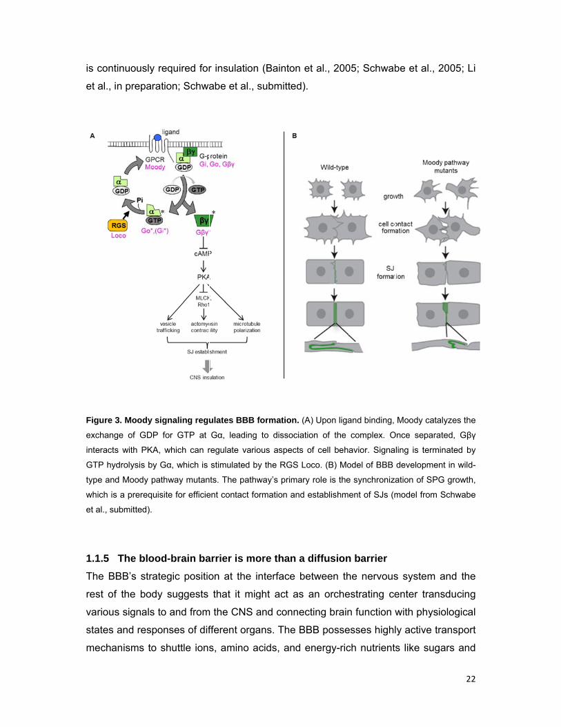
is cont
et al., i
Figure 3
exchang
interacts
GTP hyd
type and
which is
et al., su
1.1.5
The BB
rest of
various
states
mecha
inuously re
n preparat
3. Moody sig
ge of GDP f
s with PKA,
drolysis by G
d Moody pat
s a prerequis
ubmitted).
The blood
BB’s strate
f the body
s signals to
and respo
anisms to s
equired for
tion; Schwa
gnaling regu
for GTP at
which can r
Gα, which is
hway mutan
site for efficie
d-brain ba
egic positio
suggests
o and from
nses of dif
shuttle ions
r insulation
abe et al.,
ulates BBB
Gα, leading
regulate vari
stimulated b
ts. The path
ent contact fo
arrier is mo
on at the
that it mig
m the CNS
fferent orga
s, amino a
n (Bainton
submitted
formation. (
g to dissocia
ous aspects
by the RGS L
way’s prima
ormation and
ore than a
interface b
ght act as
and conne
ans. The B
acids, and
et al., 200
).
(A) Upon liga
ation of the
s of cell beh
Loco. (B) Mo
ry role is the
d establishm
a diffusion
between th
s an orche
ecting brain
BBB posse
energy-ric
05; Schwab
and binding,
complex. O
avior. Signa
odel of BBB
e synchroniz
ent of SJs (m
n barrier
he nervous
estrating ce
n function w
esses highl
ch nutrient
be et al., 2
Moody cata
Once separat
ling is termi
developmen
ation of SPG
model from S
s system a
enter trans
with physio
ly active tra
s like suga
22
2005; Li
lyzes the
ted, Gβγ
nated by
nt in wild-
G growth,
Schwabe
and the
sducing
ological
ansport
ars and
23
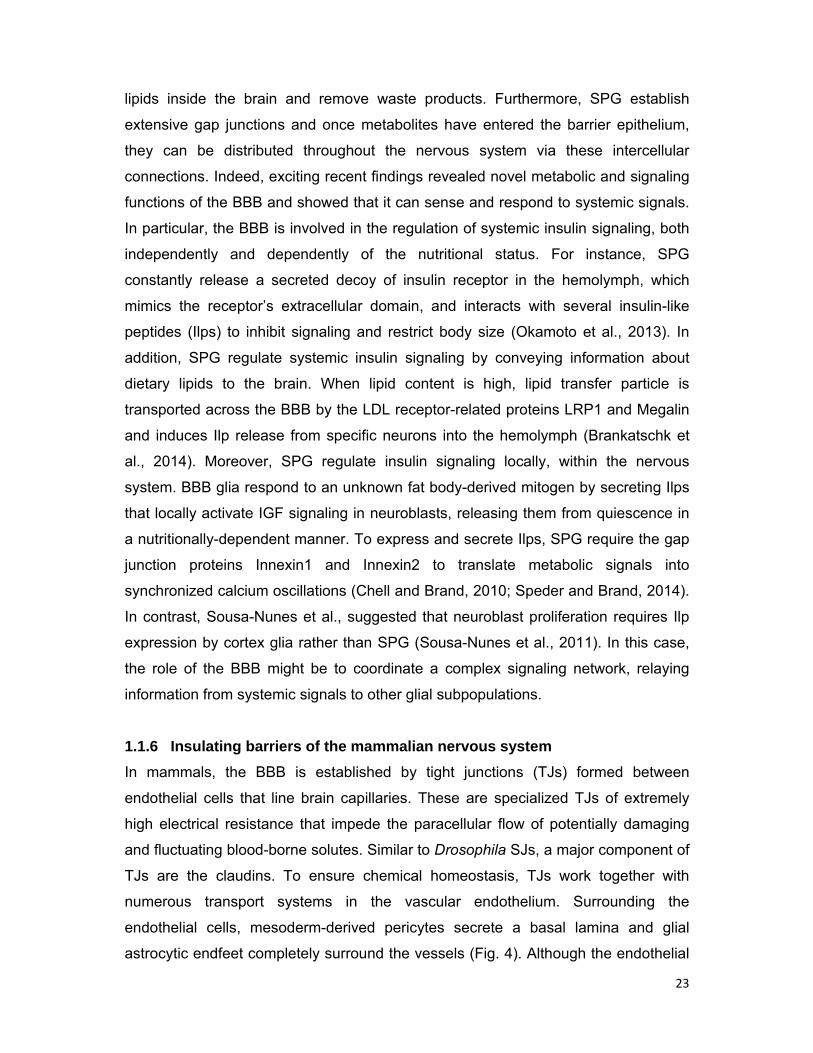
lipids inside the brain and remove waste products. Furthermore, SPG establish
extensive gap junctions and once metabolites have entered the barrier epithelium,
they can be distributed throughout the nervous system via these intercellular
connections. Indeed, exciting recent findings revealed novel metabolic and signaling
functions of the BBB and showed that it can sense and respond to systemic signals.
In particular, the BBB is involved in the regulation of systemic insulin signaling, both
independently and dependently of the nutritional status. For instance, SPG
constantly release a secreted decoy of insulin receptor in the hemolymph, which
mimics the receptor’s extracellular domain, and interacts with several insulin-like
peptides (Ilps) to inhibit signaling and restrict body size (Okamoto et al., 2013). In
addition, SPG regulate systemic insulin signaling by conveying information about
dietary lipids to the brain. When lipid content is high, lipid transfer particle is
transported across the BBB by the LDL receptor-related proteins LRP1 and Megalin
and induces Ilp release from specific neurons into the hemolymph (Brankatschk et
al., 2014). Moreover, SPG regulate insulin signaling locally, within the nervous
system. BBB glia respond to an unknown fat body-derived mitogen by secreting Ilps
that locally activate IGF signaling in neuroblasts, releasing them from quiescence in
a nutritionally-dependent manner. To express and secrete Ilps, SPG require the gap
junction proteins Innexin1 and Innexin2 to translate metabolic signals into
synchronized calcium oscillations (Chell and Brand, 2010; Speder and Brand, 2014).
In contrast, Sousa-Nunes et al., suggested that neuroblast proliferation requires Ilp
expression by cortex glia rather than SPG (Sousa-Nunes et al., 2011). In this case,
the role of the BBB might be to coordinate a complex signaling network, relaying
information from systemic signals to other glial subpopulations.
1.1.6 Insulating barriers of the mammalian nervous system
In mammals, the BBB is established by tight junctions (TJs) formed between
endothelial cells that line brain capillaries. These are specialized TJs of extremely
high electrical resistance that impede the paracellular flow of potentially damaging
and fluctuating blood-borne solutes. Similar to Drosophila SJs, a major component of
TJs are the claudins. To ensure chemical homeostasis, TJs work together with
numerous transport systems in the vascular endothelium. Surrounding the
endothelial cells, mesoderm-derived pericytes secrete a basal lamina and glial
astrocytic endfeet completely surround the vessels (Fig. 4). Although the endothelial
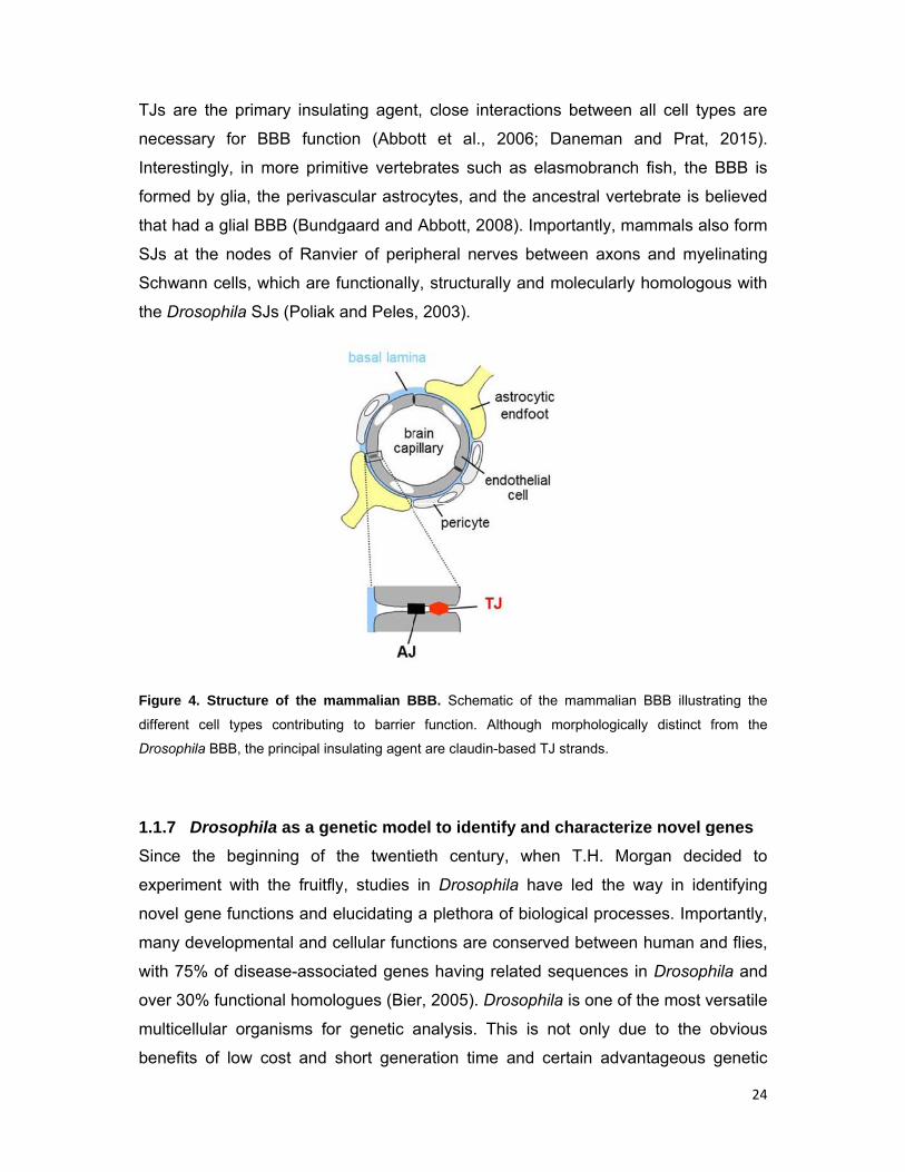
TJs ar
necess
Interes
formed
that ha
SJs at
Schwa
the Dro
Figure
different
Drosoph
1.1.7
Since
experim
novel g
many d
with 75
over 30
multice
benefit
re the prim
sary for B
stingly, in m
d by glia, th
ad a glial B
the node
ann cells, w
osophila SJ
4. Structure
t cell types
hila BBB, the
Drosophi
the begin
ment with
gene funct
developme
5% of dise
0% functio
ellular orga
ts of low c
mary insula
BBB funct
more prim
he perivas
BBB (Bundg
s of Ranv
which are f
Js (Poliak
e of the ma
contributing
e principal ins
ila as a ge
nning of t
the fruitfly
ions and e
ental and c
ease-assoc
nal homolo
anisms for
cost and s
ating agen
tion (Abbo
itive verte
scular astro
gaard and
vier of peri
functionally
and Peles
ammalian B
g to barrier
sulating agen
netic mod
the twenti
y, studies
elucidating
cellular fun
ciated gene
ogues (Bie
r genetic
short gene
t, close in
ott et al.,
brates suc
ocytes, an
Abbott, 20
pheral ner
y, structura
s, 2003).
BBB. Schem
r function. A
nt are claudin
del to iden
ieth centu
in Drosop
a plethora
ctions are
es having
er, 2005). D
analysis. T
eration tim
nteractions
2006; D
ch as elas
d the ance
008). Impo
rves betwe
ally and m
matic of the
Although m
n-based TJ s
ntify and c
ury, when
phila have
a of biolog
conserved
related se
Drosophila
This is no
me and cer
between
aneman a
smobranch
estral verte
ortantly, ma
een axons
molecularly
mammalian
orphological
strands.
characteriz
T.H. Mo
led the w
ical proces
d between
equences i
is one of t
ot only du
rtain adva
all cell typ
and Prat,
h fish, the
ebrate is b
ammals als
s and mye
homologo
BBB illustra
ly distinct f
ze novel g
organ deci
way in ide
sses. Impo
human an
n Drosoph
the most v
e to the o
ntageous
24
pes are
2015).
BBB is
believed
so form
elinating
ous with
ating the
from the
genes
ided to
entifying
ortantly,
nd flies,
hila and
versatile
obvious
genetic

25
features, but also because the early start of experimentation was built on by
succeeding generations of researchers who have developed an ever-increasing
repertoire of high-quality techniques and resources. These include excellent genome
annotation, genome-wide in vivo gene disruption (by transposable elements and
transgenic RNAi), large-scale protein trap library in which endogenously expressed
proteins are tagged with GFP, and many molecular tools for genetic manipulation
(e.g. site-directed transgenesis, homologous recombination) and spatio-temporal
control of gene expression (e.g. Gal4-UAS, FLP-FRT systems) (Morin et al., 2001; St
Johnston, 2013).
One of the most important tools Drosophila provides is the ability to carry out
large scale in vivo genetic screens (St Johnston, 2002). The simple genome
structure, the limited gene redundancy, and the fact that regulatory regions are
located near the genes they control have been proven great advantages for the
identification of novel gene functions through classical forward genetic approaches
(Nüsslein-Volhard and Wieschaus, 1980). In such screens, random mutations are
induced (e.g. by radiation, chemicals or insertional mutagenesis), mutant individuals
are recovered and the affected gene is mapped. Subsequently, the pioneering RNAi
technique and the production of transgenic UAS-RNAi libraries have facilitated the
implementation of large-scale reverse genetic screens, in which the identity of the
perturbed genes is known, and have enabled the systematic analysis of gene
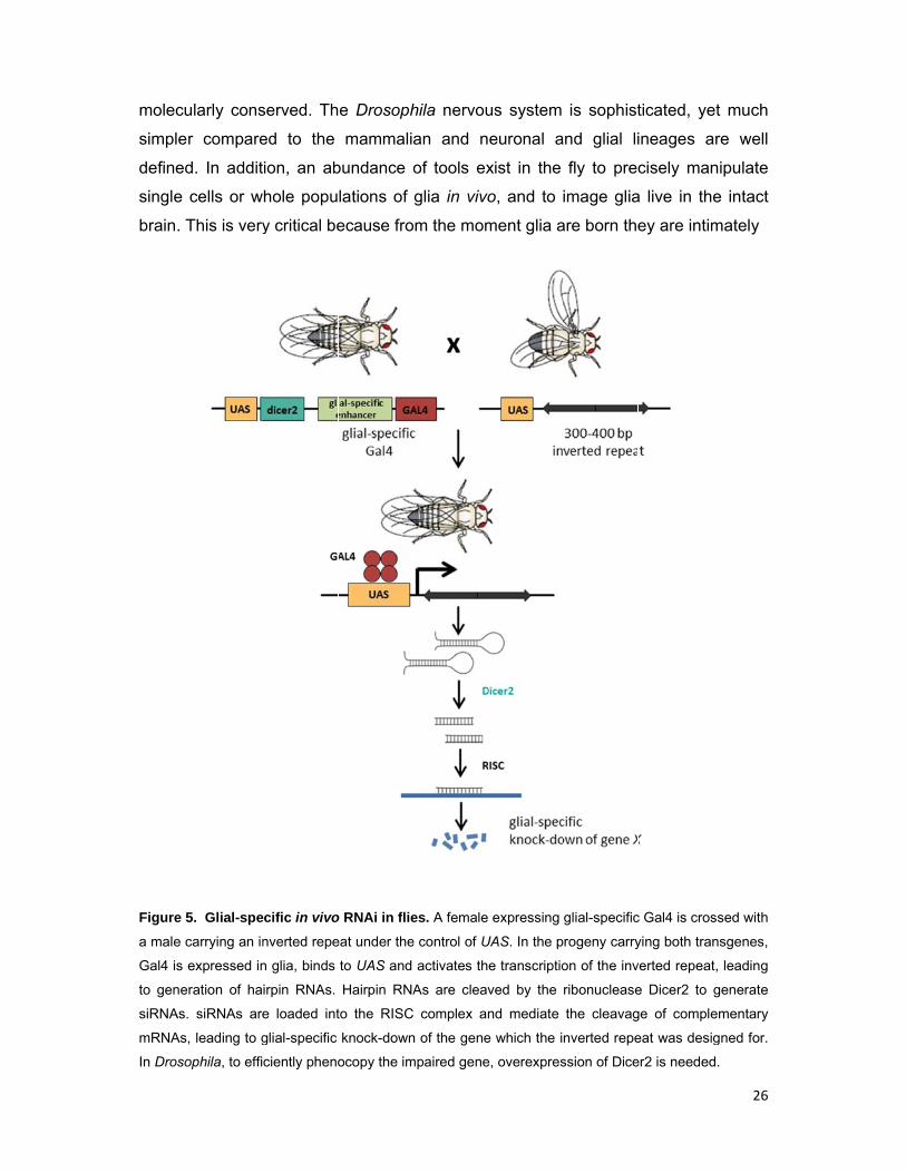
function in vivo (Dietzl et al., 2007; Ni et al., 2011). When induced with a Gal4 driver,
the UAS-RNAi transgene leads to the generation of double-stranded short interfering
RNAs (siRNAs), which mediate the degradation of complementary mRNAs.
Therefore, it results in the knock-down of the corresponding gene in a spatio-
temporal expression pattern that depends on the Gal4 enhancer (Fig. 5). Importantly,
RNAi works exceptionally well in Drosophila both in vivo and in cultured cells. After
screening, a wealth of computational and experimental data are available in
Drosophila to draw from for post-analysis. Once potential candidates are identified,
advanced genetic and molecular tools together with a large repertoire of markers
and superb imaging techniques allow for the straightforward characterization of gene
functions and for a depth of experimental scrutiny unmatched in higher organisms.
Drosophila has also emerged as a very promising model organism to study
glial biology. Drosophila glia exhibit remarkable morphological similarities to
vertebrate glia and fulfill roles that are highly analogous and in many cases
molecu
simple
defined
single
brain. T
Figure 5
a male c
Gal4 is e
to gene
siRNAs.
mRNAs,
In Droso
ularly cons
r compare
d. In addit
cells or w
This is very
5. Glial-spe
carrying an in
expressed in
ration of ha
siRNAs are
, leading to g
ophila, to effi
served. Th
ed to the
ion, an ab
hole popu
y critical be
cific in vivo
nverted repe
n glia, binds
irpin RNAs.
e loaded int
glial-specific
ciently pheno
e Drosoph
mammali
bundance
lations of
ecause fro
o RNAi in flie
eat under the
to UAS and
Hairpin RNA
to the RISC
knock-down
ocopy the im
hila nervou
an and n
of tools ex
glia in vivo
om the mom
es. A female
e control of U
activates th
As are cleav
C complex an
n of the gene
mpaired gene
us system
euronal a
xist in the
o, and to
ment glia a
expressing
UAS. In the p
e transcriptio
ved by the
nd mediate
e which the i
e, overexpres
is sophist
and glial li
fly to pre
image glia
are born the
glial-specific
rogeny carry
on of the inv
ribonuclease
the cleavag
nverted repe
ssion of Dice
ticated, ye
ineages a
cisely man
a live in the
ey are intim
c Gal4 is cros
ying both tran
verted repeat
e Dicer2 to
ge of comple
eat was desig
er2 is needed
26
et much
are well
nipulate
e intact
mately
ssed with
nsgenes,
t, leading
generate
ementary
gned for.
d.
27
associated with neurons, and these two cell types are highly interdependent for
normal development and function. Importantly, Drosophila also allows the
investigation of developmental features of glia, which in mammals can
provechallenging due to the difficulty of accessing animals in utero, where critical
glial developmental milestones occur.
1.1.8 Rationale
Although our insights on glial biology have profoundly advanced in the past years, it
seems that we are still only scratching the surface of the many glial functions.
Moreover, although we have gained much information on the Drosophila glial BBB
and the role of Moody signaling in its formation, given its physiological importance,
many questions remain open (Fig. 6). Several features of Moody signaling make it a
rather complex pathway and suggest that is currently incomplete. Both the active G-
proteins and PKA are able to transduce the signal to multiple effectors, integrating
Moody signaling with a wide range of biological responses, such as cytoskeletal
organization, cell–cell and cell–matrix adhesion, vesicular trafficking, cell polarity and
possibly gene expression. In addition, the finding that loss of function of downstream
effectors results in more severe defects than loss of Moody strongly suggests that G-
proteins receive additional activating input. The GPCR Tre1, which is the closest
paralog of Moody and is expressed in a subset of SPG is a strong candidate,
however thus far, there is insufficient evidence to exclude or confirm this possibility
(Schwabe et al., 2005). Moreover, the pathway’s role in barrier maintenance in the
adult has not been investigated in any detail. Furthermore, although strong insulation
defects are observed in Moody signaling mutants, SPG do spread over the CNS to
form an epithelium and some pathway mutants survive until later stages. Thus, when
looked from a developmental perspective, it seems that additional independent
pathways are involved in BBB establishment. In addition, the precise mechanism by
which the barrier-forming SJs are established in the SPG epithelium and how their
highly regular alignment is achieved remain poorly understood. Finally, the BBB
except for a selective diffusion barrier appears to be a dynamic and communicative
layer between the brain and the body. The BBB might act as a key integrator of
various systemic stimuli to the brain and as a signaling center orchestrating major
developmental and physiological events. Such fascinating novel roles of the BBB in
animal physiology and behavior are expected to be unraveled in the coming years.

lab ha
approa
decide
candid
functio
Figure
compon
how the
My goal w
ad exhaus
ach; i.e. b
d to carry
ates to fur
nal level.
6. Open q
ents acting b
barrier-form
was to iden
sted the id
by screeni
out a neu
ther explor
uestions o
both upstrea
ming SJs are
ntify novel g
dentificatio
ng known
utral genom
re their role
on BBB for
am and down
established
genes req
on of BBB
n G-protei
me-wide R
e in barrier
rmation. Mo
nstream of G
is still unkno
uired for B
B genes t
n effector
RNAi scree
r formation
oody signali
G-proteins re
wn.
BBB format
through a
rs for insu
en and sub
n at the ge
ng is curre
main to be i
tion. Beca
candidate
ulation de
bsequently
netic, cellu
ntly incomp
dentified. In
28
use the
e gene
fects, I
y select
ular and
lete and
addition,
1.2 G
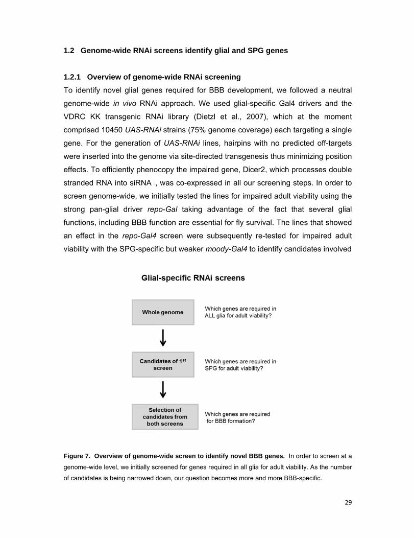
1.2.1
To ide
genom
VDRC
compri
gene.
were in
effects
strande
screen
strong
functio
an effe
viability
Figure 7
genome
of candid
enome-wi
Overview
ntify novel
me-wide in
KK trans
sed 10450
For the ge
nserted into
. To efficie
ed RNA in
genome-w
pan-glial
ns, includi
ect in the
y with the S
7. Overview
e-wide level,
dates is bein
ide RNAi s
w of genom
l glial gene
vivo RNA
sgenic RN
0 UAS-RNA
eneration o
o the geno
ently pheno
to siRNA
wide, we in
driver rep
ng BBB fu
repo-Gal4
SPG-speci
w of genome
we initially s
ng narrowed
screens id
me-wide R
es require
Ai approach
NAi library
Ai strains (
of UAS-RN
ome via site
ocopy the
s, was co-e
nitially test
po-Gal tak
unction are
4 screen w
ific but wea
e-wide scree
creened for
down, our q
dentify glia
NAi scree
d for BBB
h. We use
(Dietzl e
(75% geno
NAi lines,
e-directed
impaired g
expressed
ed the line
king adva
essential
were subse
aker mood
en to identif
genes requir
uestion beco
al and SP
ening
B developm
ed glial-sp
et al., 200
ome covera
hairpins w
transgene
gene, Dice
in all our s
es for impa
ntage of
for fly surv
equently r
dy-Gal4 to
fy novel BB
red in all glia
omes more a
G genes
ment, we fo
ecific Gal4
07), which
age) each
with no pre
esis thus m
r2, which p
screening
aired adult
the fact t
vival. The
re-tested fo
identify ca
B genes. In
a for adult via
and more BB
ollowed a
4 drivers a
at the m
targeting a
edicted off-
minimizing p
processes
steps. In o
viability us
that sever
lines that s
or impaire
ndidates in
n order to sc
ability. As the
B-specific.
29
neutral
and the
moment
a single
-targets
position
double
order to
sing the
ral glial
showed
ed adult
nvolved
reen at a
e number
30
in BBB formation and/or maintenance. In both screens, the RNAi lines targeting
housekeeping genes involved in basic cellular functions serve as internal positive
controls as they are potentially cell-lethal. Finally, to directly ask if the genes are
required for BBB formation, we performed the embryonic dye penetration assay in a
selection of candidates using repo-Gal4 (Fig. 7).
RNAi screens have the major advantage of being reverse genetic screens,
meaning that the gene whose function is disrupted is known and one looks for the
manifestation or not of the phenotype of interest. However, such screens also bear
two limitations. First, RNAi results in variable and partial loss-of-function of the
affected gene and might lead to false negatives. This is of particular concern for
proteins that are expressed in particularly high levels, such as structural components
of the cytoskeleton. Second, RNAi is prone to off-target effects due to targeting of
additional mRNAs with homology to the introduced siRNA and thus false positives.
1.2.2 Primary pan-glial screen
In our primary screen, we checked 10450 UAS-RNAi lines for impaired adult viability
using repo-Gal4, which drives strong expression in all glia except for midline glia
from embryonic stage 13 throughout the development and lifespan of the fly. We
crossed transheterozygote repo-Gal4/TM3 virgin females with UAS-RNAi males,
removed the parents after sufficient egg lay to avoid overcrowded vials and scored
the approximately 150 progeny of the first generation by counting the number of
UAS-RNAi;repo-Gal4 versus UAS-RNAi;TM3 flies. The TM3 balancer carries the
dominant marker Stubble (Sb) which causes shorter bristles compared to wild-type
(wt) allowing the easy discrimination of the two genotypes. Assuming that the
genotypes are equally viable, based on Mendelian rules they should appear in a 1:1
ratio; lower percentages in the flies with the knock-down would imply a role for the
gene in glia. In order to identify glial genes required in all developmental stages, as
well as in the adult, we counted the progeny 8-10 days after eclosion. Because after
a few rounds of screening we realized that the screen was yielding many positive
results, we continued by simply defining in which of the categories lethal, subviable,
and viable each knock-down falls and only counted the exact number of progeny for
each genotype for the class of subviable. In order to define the average percentages
with which the two genotypes appear in the case that the glial knock-down is not
affecting viability, we counted the exact number of progeny for each of the genotypes
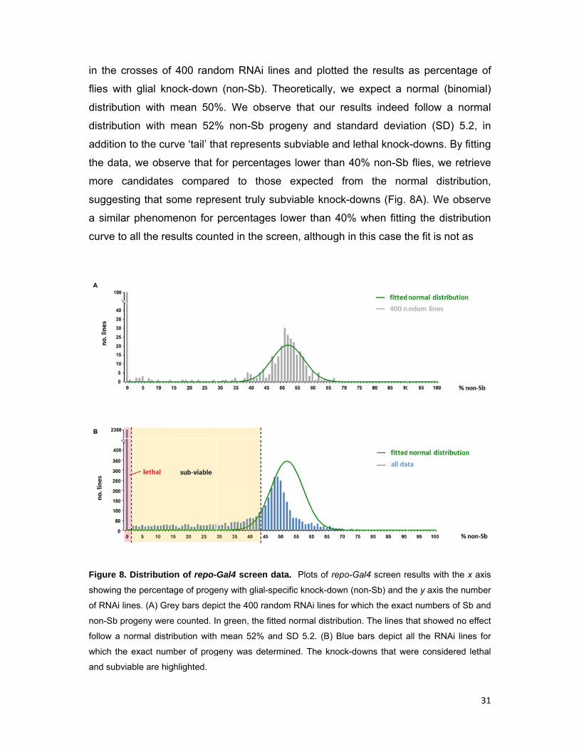
in the
flies w
distribu
distribu
additio
the dat
more
sugges
a simil
curve t
Figure 8
showing
of RNAi
non-Sb
follow a
which th
and sub
crosses of
ith glial kn
ution with
ution with
n to the cu
ta, we obs
candidate
sting that s
ar phenom
to all the re
8. Distributi
g the percent
lines. (A) Gr
progeny wer
normal dist
he exact num
viable are hi
f 400 rand
nock-down
mean 50%
mean 52%
urve ‘tail’ th
serve that f
s compar
some repre
menon for
esults coun
on of repo-
tage of proge
rey bars dep
re counted. I
ribution with
mber of prog
ghlighted.
dom RNAi
n (non-Sb)
%. We ob
% non-Sb
hat represe
for percen
red to th
esent truly
percentag
nted in the
-Gal4 screen
eny with glial
pict the 400 ra
n green, the
h mean 52%
geny was de
lines and
. Theoretic
bserve that
progeny
ents subvia
tages lowe
ose expe
y subviable
es lower t
screen, al
n data. Plot
l-specific kno
andom RNA
fitted norma
and SD 5.2
etermined. T
plotted th
cally, we e
t our resu
and stand
able and le
er than 40
ected from
e knock-do
than 40%
though in t
ts of repo-Ga
ock-down (no
Ai lines for wh
al distribution
2. (B) Blue b
he knock-do
he results
expect a n
ults indeed
dard devia
ethal knock
0% non-Sb
m the nor
owns (Fig.
when fittin
this case t
al4 screen re
on-Sb) and t
hich the exac
n. The lines t
bars depict a
owns that we
as percen
normal (bi
d follow a
ation (SD)
k-downs. B
b flies, we
rmal distr
8A). We o
ng the dist
he fit is no
esults with th
he y axis the
ct numbers o
that showed
all the RNAi
ere consider
31
ntage of
nomial)
normal
5.2, in
By fitting
retrieve
ribution,
observe
ribution
t as
he x axis
e number
of Sb and
no effect
lines for
red lethal

good b
Based
knock-
down,
library,
genes
of libra
(GO, F
name
some
unchar
predict
(Fig. 9B
mainte
transcr
mediat
Figure 9
knock-do
which re
and/or b
function
because w
on the fit
downs tha
respective
The scree
28% of g
(23.3% of
ary) adult s
Flybase), a
and biolog
prediction
racterized
ted functio
B). In this
enance of
ription, RN
ted protein
9. Results o
own resulted
esulted in let
biological fun
, two-thirds a
we were bia
tting proce
at resulted
ely (Fig. 8B
en was ve
genome) a
f library) ca
ubviability
among the
gical functi
n for the
genes w
n, 981 (36
category w
f basic c
NA splicin
degradati
of repo-Gal4
d in adult leth
thality or sub
nction (GO te
are potentiall
ased towa
edure, we
in 0-0.49%
B).
ery succes
s potentia
aused com
(Fig. 9A).
3679 can
ion assign
eir structu
ith no pre
%) are like
we include
cellular fu
ng, transla
on. After e
4 screen. (A
hality, subvia
bviability clas
erms, Flybas
ly interesting
ards counti
considere
% and 0.5-4
sful and id
l glial gene
mplete adul
Based on
didates, 16
ned, 1030
re and/or
edictions.
ely ubiquito
d all genes
nctions, s
ation, chro
excluding h
A) Chart dep
ability or had
ssified based
e). Among th
g, non-house
ng the res
ed as com
40% proge
dentified 3
es. From t
lt lethality,
classificat
696 (46.1%
(28%) are
r function
From the
ously expr
s whose p
such as
omatin as
housekeep
picting the n
no effect. (B
d on the curr
he identified
keeping gen
sults that s
mplete leth
eny with gli
3679 cand
these, kno
and of 12
tion by gen
%) are kno
e uncharac
n, and 95
e genes w
ressed hou
roducts ar
DNA rep
ssembly a
ing genes,
umber of ge
B) Chart dep
rent knowled
genes with a
nes.
showed an
hal and su
ial-specific
didates (35
ock-down o
241 genes
ne ontolog
own genes
cterized bu
53 (25.9%
with a kno
usekeeping
re required
plication, g
and protea
, the repo-
enes whose
picting the ca
dge on their
a known or p
32
n effect.
ubviable
c knock-
5.2% of
of 2438
(11.9%
y terms
s with a
ut have
%) are
own or
g genes
d for the
general
asome-
Gal4
pan-glial
andidates
structure
predicted
33
screen revealed a great number of candidates (2697) that potentially play a specific
role in glial development and/or function, and offers valuable material for studying
multiple aspects of glial biology.
Importantly, among our results we identify both known glial genes, which
serve as internal positive controls, as well as many interesting novel candidates.
These include components of numerous signaling pathways (e.g. GPCR, RTK,
Ecdysone, Hippo, JAK/ STAT, Decapentaplegic), as well as a great number of
transcription factors. We additionally find several proteins with roles in cell shape
regulation and epithelial morphogenesis (e.g. actin- and microtubule-binding
proteins, motors, RhoGEFs and RhoGAPs, components of the adherens, septate
and gap junctions) that might be of particular interest for studying BBB development.
We also identify many potential candidates with respect to Moody signalling; i.e.
Tre1 and 30 more GPCRs (without including gustatory receptors) that might be
acting together with Moody upstream of the G-proteins, as well as G- and RGS
proteins, three adenylate cyclases potentially involved in the generation of cAMP,
kinases that may act downstream of G-proteins, and several proteins that might
function in response to PKA, like components of vesicular trafficking, cytoskeletal
regulators and the transcription factors CrebA and CrebB-17A. We also find proteins
involved in circadian rhythm (e.g. Timeless, Clock, PDFR, Reg-5, Takeout),
phagocytosis (e.g. Simu, Eater, PGRPs, ABC transporters, Ced-6) and ionic and
neurotransmitter homeostasis (e.g. GABA, acetylcholine and glutamate receptors,
glutamate dehydrogenase, K+ channels, and aquaporins) and many more fascinating
molecules. Surprisingly, among our candidates we find genes that are not expected
to cause lethality, since their genomic mutants survive to adulthood (e.g. 11
gustatory receptors, phagocytosis receptors). One possible explanation for this result
is that competition for resources exists between the two genetically distinct progeny
of the first generation, leading to increased death of the flies that express the siRNA
and disrupt a given process. Another explanation might be that the siRNA targets
more than one mRNAs, e.g. multiple members of the family of gustatory receptors.
The retrieval of known glial genes suggests an efficient screening procedure.
To further check the performance of the screen, we implemented different kinds of
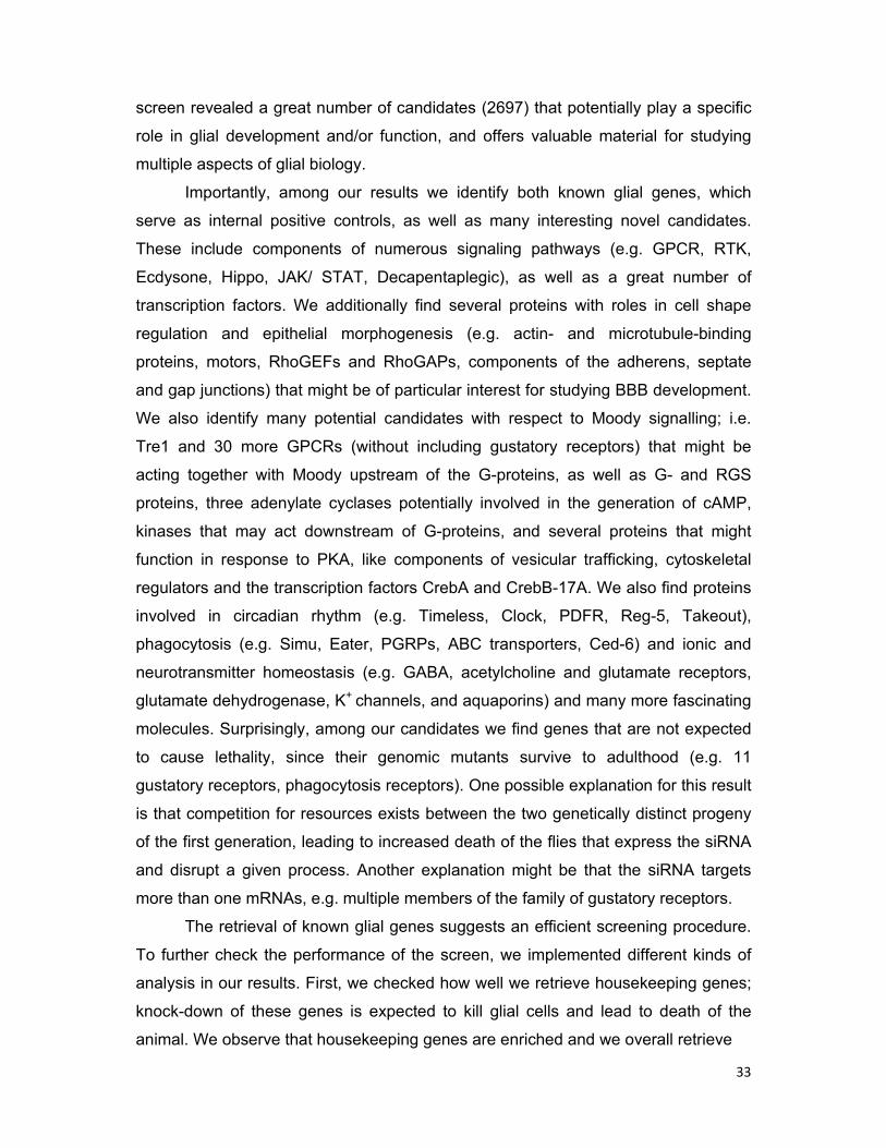
analysis in our results. First, we checked how well we retrieve housekeeping genes;
knock-down of these genes is expected to kill glial cells and lead to death of the
animal. We observe that housekeeping genes are enriched and we overall retrieve
Figure
classifie
terms, F
when co
them w
associa
fraction
assem
produc
Second
known
longitu
Neuron
10. The re
d based on
Flybase) and
ompared with
with very
ated with
n recovere
bly and D
cts and/or
d, we dete
to be req
dinal and
nally-derive
epo-Gal4 sc
their known
the fraction
h the fraction
high perce
nucleosom
ed for gen
DNA repair
result fro
ermined h
quired in
periphera
ed EGF ac
creen succe
or predicted
recovered fo
n recovered f
entages th
me and ri
nes that fa
r might re
om incomp
how well w
glia. The
l glia for t
ctivates the
essfully retr
d involvemen
or each term
for all genes
hat in som
bosome b
all in othe
flect biolo
pleteness
we retrieve
multi-com
their surviv
e EGF rece
rieves hous
nt in an esse
is shown. H
with a known
me cases
biogenesis
r GO clas
gical redu
of the RN
e compone
ponent EG
val and re
eptor on th
sekeeping
ential housek
Housekeeping
n or predicte
reach 90%
and mito
sses, such
undancy be
NAi knock
ents of sig
GF pathwa
epresents
e glial mem
genes. Ge
keeping func
g genes are
ed function (0
%, like for
osis. The
h as cytos
etween th
k-down (Fi
gnaling pa
ay is requ
a good ex
mbrane lea
34
enes are
ction (GO
enriched
0.36).
r genes
smaller
skeleton
e gene
ig. 10).
athways
uired in
xample.
ading
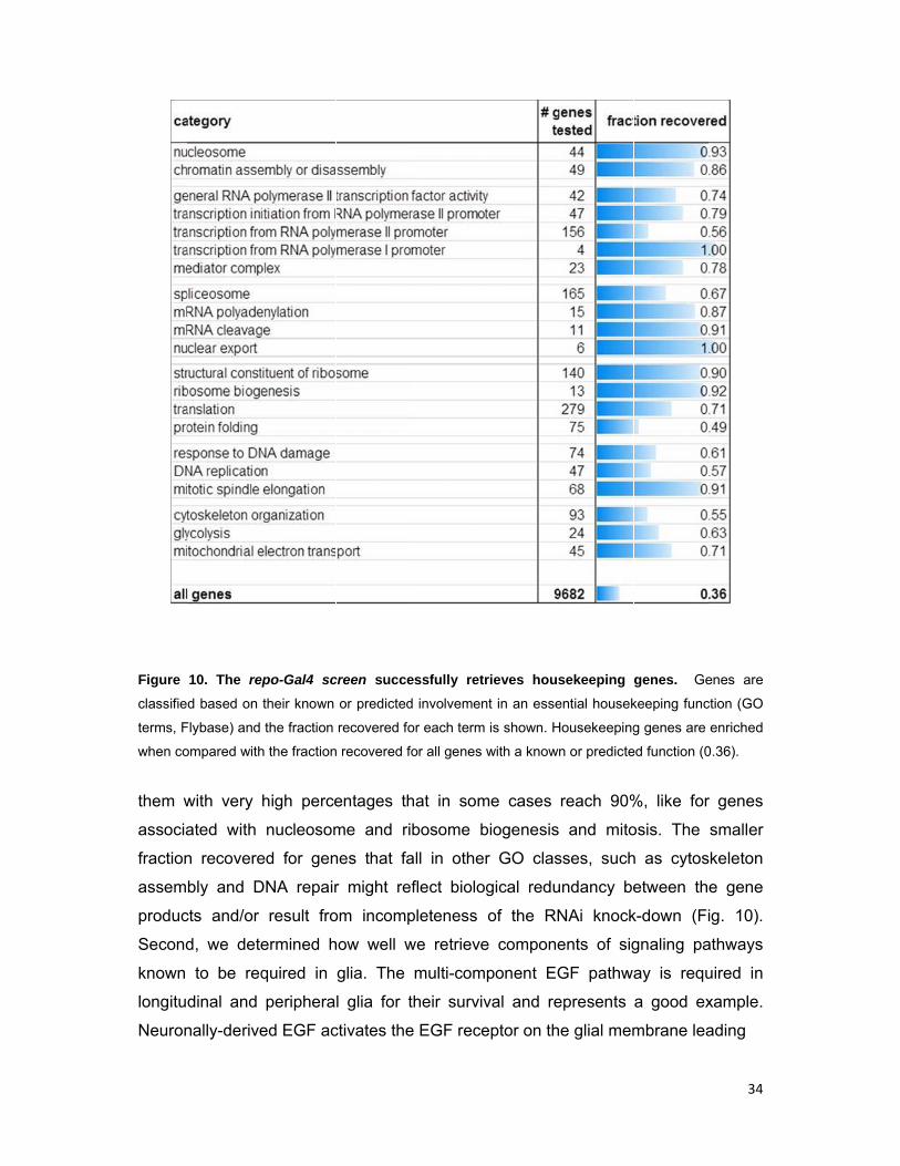
Figure 1
The mul
to activa
to a c
nuclea
regulat
Shilo, 2
11). S
in glia
the tra
respon
al., 200
profile
genes
11. The rep
lti-componen
ation of targe
cascade of
r transloca
tes a numb
2014). Imp
Similarly, w
during me
anscription
nsive gene
06). Third
of embryo
that were f
o-Gal4 scre
nt EGF pathw
et genes esse
f intracellu
ation of th
ber of targ
pressively,
we retrieve
tamorphos
n factor B
s (Fig. 12
, we comp
onic glia pre
found as d
een success
way is activa
ential for glia
ular protei
he transcr
get genes
we identif
componen
sis to prun
Broad, an
) (Awasak
pared our
eviously pe
differentially
sfully retriev
ated in glia in
al survival.
n phospho
ription fact
essential f
fy 13 of th
nts of the
e axons, s
nd many
ki and Ito,
results wi
erformed in
y expresse
ves the com
n response to
orylations
tor Pointed
for glial su
he 15 core
Ecdysone
such as the
primary a
2004; Wat
ith a micro
n the lab (
ed in glia w
mplete EGF
o neuronally-
ultimately
d. In the
rvival (Hid
pathway
pathway,
e Ecdyson
and secon
tts et al., 2
oarray-bas
U. Gaul, u
were cluste
signaling p
-derived EGF
y resulting
nucleus, P
dalgo et al.
componen
which is re
ne receptor
ndary ecd
2004; Awa
sed transc
npublished
ered in grou
35
pathway.
F leading
to the
Pointed
., 2001;
nts (Fig.
equired
r (EcR),
dysone-
asaki et
riptome
d). The
ups of
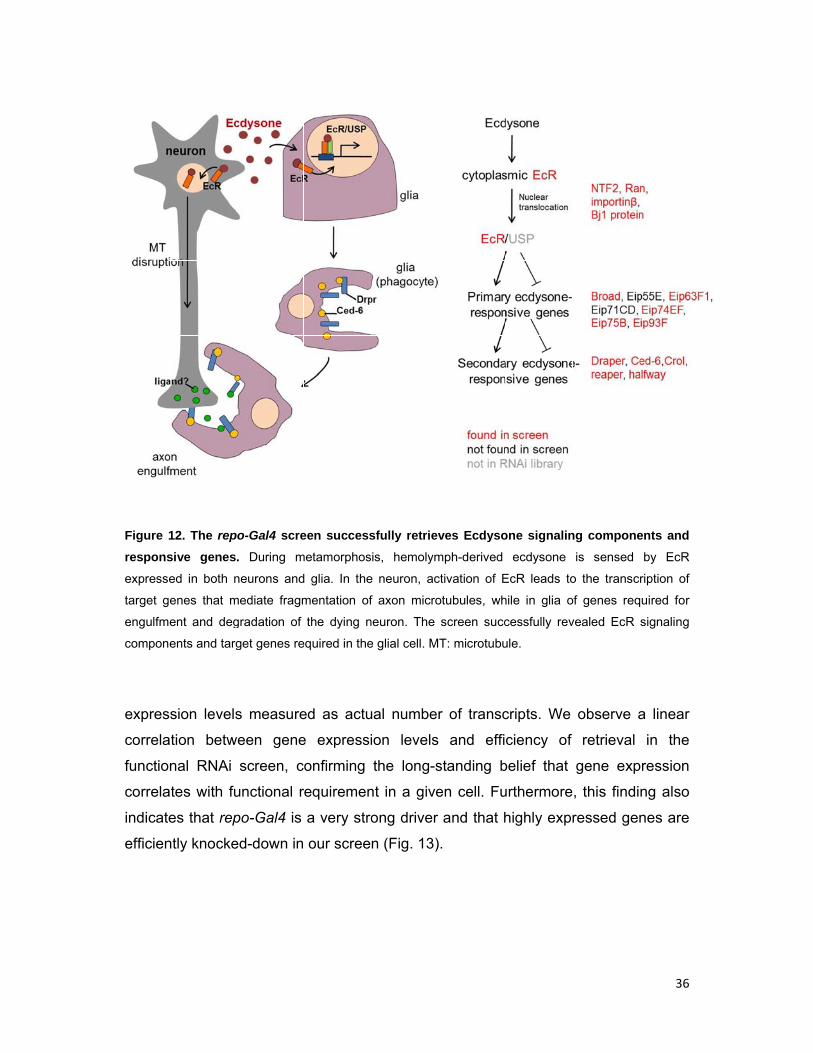
Figure 1
respons
expresse
target g
engulfm
compon
expres
correla
functio
correla
indicate
efficien
12. The rep
sive genes
ed in both n
enes that m
ent and deg
ents and targ
ssion levels
ation betw
nal RNAi
ates with fu
es that rep
ntly knocke
po-Gal4 scre
. During m
neurons and
mediate fragm
gradation of
get genes re
s measure
ween gene
screen, c
unctional r
po-Gal4 is
ed-down in
een success
metamorphos
d glia. In the
mentation of
the dying n
equired in the
ed as actu
e expressi
onfirming
requiremen
a very stro
our scree
sfully retriev
sis, hemolym
e neuron, ac
f axon micro
neuron. The
e glial cell. M
ual numbe
ion levels
the long-s
nt in a give
ong driver
n (Fig. 13)
ves Ecdyso
mph-derived
ctivation of E
otubules, wh
screen succ
T: microtubu
r of transc
s and effi
standing b
en cell. Fu
and that h
).
one signalin
ecdysone
EcR leads to
hile in glia o
cessfully rev
ule.
cripts. We
ciency of
belief that
urthermore
highly expr
ng compone
is sensed
o the transc
of genes req
vealed EcR
observe a
retrieval
gene exp
, this findi
ressed gen
36
ents and
by EcR
ription of
quired for
signaling
a linear
in the
pression
ng also
nes are
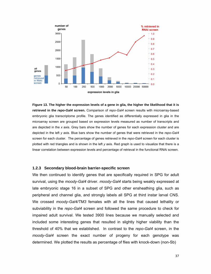
Figure 1
retrieve
embryon
microarr
are depi
depicted
screen f
plotted w
linear co
1.2.3
We the
surviva
late em
periphe
We cro
subviab
impaire
include
thresho
moody
determ
13. The high
ed in the rep
nic glia trans
ray screen a
icted in the x
d in the left y
for each clus
with red trian
orrelation bet
Secondar
en continu
al, using th
mbryonic s
eral and c
ossed mo
bility in the
ed adult s
ed some i
old of 40%
y-Gal4 scr
mined. We p
her the expr
po-Gal4 scr
scriptome pr
are grouped
x axis. Grey
y axis. Blue
ter. The per
ngles and is
tween expres
ry blood-b
ed to iden
he moody-G
stage 16 in
hannel glia
oody-Gal4/
e repo-Ga
urvival. W
nteresting
% that we
reen the
plotted the
ression leve
reen. Compa
rofile. The g
based on ex
bars show t
bars show th
rcentage of g
shown in the
ssion levels
brain barri
ntify genes
Gal4 driver
n a subse
a, and stro
/TM3 fema
l4 screen
We tested 3
genes th
establishe
exact nu
e results as
els of a gene
arison of rep
genes identif
xpression lev
the number o
he number o
genes retriev
e left y axis.
and percenta
er-specifi
s that are
r. moody-G
et of SPG
ongly labe
ales with a
and follow
3900 lines
at resulted
ed. In con
umber of
s percentag
e in glia, the
po-Gal4 scree
fied as differ
vels measur
of genes for
of genes that
ved in the rep
Red graph is
age of retriev
c screen
specifically
Gal4 starts
and other
els all SPG
all the line
wed the sa
because
d in slight
ntrast to th
progeny
ge of flies w
e higher the
en results w
rentially exp
red as numb
each expres
t were retrie
po-Gal4 scre
s used to vis
val in the fun
y required
s being wea
r ensheath
G at third i
es that ca
ame proced
we manu
tly higher
he repo-Ga
for each
with knock
e likelihood
with microarra
ressed in gl
ber of transc
ssion cluster
ved in the re
een for each
sualize that t
nctional RNA
in SPG fo
akly expre
hing glia, s
nstar larva
aused leth
dure to ch
ally select
viability th
al4 screen
h genotyp
k-down (no
37
that it is
ay-based
lia in the
cripts and
r and are
epo-Gal4
cluster is
there is a
Ai screen.
or adult
ssed at
such as
al CNS.
hality or
heck for
ted and
han the
, in the
pe was
on-Sb)
Figure
depicts
number
Based o
the num
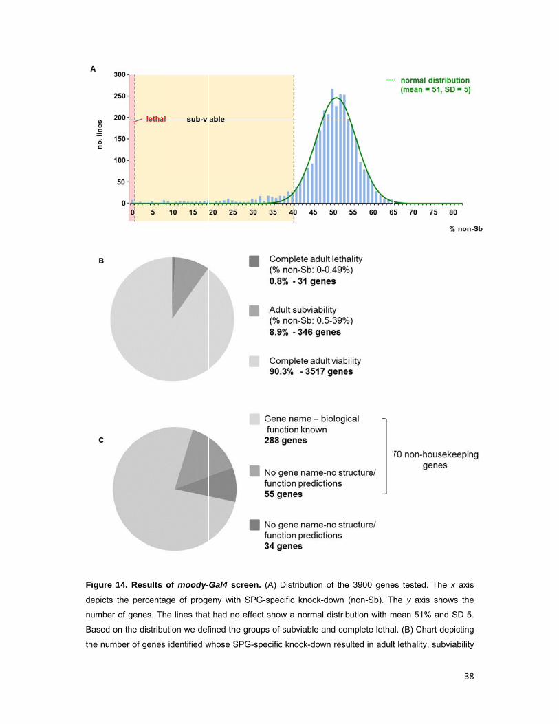
14. Results
the percenta
of genes. T
on the distrib
mber of genes
of moody-
age of proge
he lines that
ution we def
s identified w
Gal4 screen
eny with SP
t had no effe
fined the gro
whose SPG-s
n. (A) Distrib
PG-specific k
ect show a n
oups of subvi
specific knoc
bution of the
knock-down
normal distrib
iable and co
ck-down resu
e 3900 gene
(non-Sb). Th
bution with m
mplete letha
ulted in adult
es tested. Th
he y axis sh
mean 51% an
al. (B) Chart d
t lethality, su
38
he x axis
hows the
nd SD 5.
depicting
ubviability
39
or had no effect. (C) Chart depicting the candidates which resulted in lethality or subviability classified
based on the current knowledge on their structure and/or biological function (GO terms, Flybase).
and observed a normal distribution with a mean of 51% progeny with glial-specific
knock-down for most crosses that correspond to the knock-downs that had no effect.
Based on this distribution and following the same procedure as in the repo-Gal4
screen, we defined the groups of subviable and lethal (Fig.14A).
The moody-Gal4 screen resulted in the identification of significantly less
candidates. Knock-down of only 31 genes (0.8% of lines tested) caused complete
adult lethality (0-0.49% flies with SPG-specific knock-down), while 346 genes (8.9%
of lines tested) caused adult subviability (0.5-39% flies with SPG-specific knock-
down), leaving us with 377 genes potentially required in SPG (Fig. 14A,B). Among
these, 288 have a gene name and a function assigned, 55 are uncharacterized but
have predictions for their structure and/or function and 34 genes are completely
unknown and have no predictions. Furthermore, between the genes with a known or
predicted function, 273 are potentially housekeeping and 70 non-housekeeping (Fig.
14C). Among the non-housekeeping candidates, few are known SPG genes, e.g. the
G-signaling components Moody and Gβ13F, the SJ-associated proteins Nrx-IV,
ATPα, Lac, Cora, Vari and Crok, and the integrin β subunit Myospheroid (Schwabe
et al., 2005; Xie and Auld, 2011; Izumi and Furuse, 2014).
The identification of a small number of genes in the moody-Gal4 screen partly
results from the fact that the knock-down is performed in a small subset of glia and
partly from the weakness of moody-Gal4 at least during embryonic stages. Two
lines of evidence suggest that moody-Gal4 is a rather weak driver and resulted in a
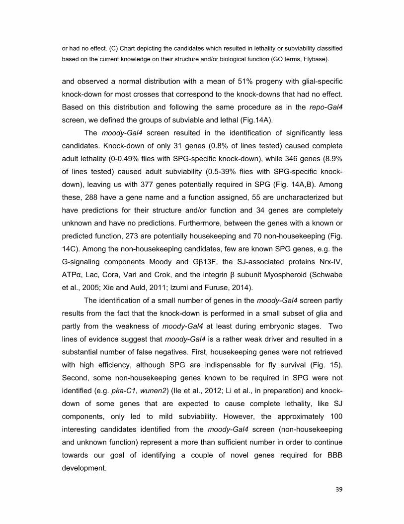
substantial number of false negatives. First, housekeeping genes were not retrieved
with high efficiency, although SPG are indispensable for fly survival (Fig. 15).
Second, some non-housekeeping genes known to be required in SPG were not
identified (e.g. pka-C1, wunen2) (Ile et al., 2012; Li et al., in preparation) and knock-
down of some genes that are expected to cause complete lethality, like SJ
components, only led to mild subviability. However, the approximately 100
interesting candidates identified from the moody-Gal4 screen (non-housekeeping
and unknown function) represent a more than sufficient number in order to continue
towards our goal of identifying a couple of novel genes required for BBB
development.
Figure
screen.
houseke
not retrie
and moo
1.2.4 S
format
To dire
selectio
injected
embryo
exclusi
nervou
defecti
(10 AE
All kno
15. Retrieva
Genes are
eeping functi
eve houseke
ody-Gal4 exp
Small-sca
tion
ectly ident
on of cand
d a 10 kD
os (Fig. 16
ion from th
us system,
ve BBB (F
EL), thus p
ock-downs
al of house
e classified
ion (GO term
eeping genes
pressing SPG
ale screen
tify novel p
didates with
Da rhodam
6A), when
he CNS 15
, but it ra
Fig. 16C).
roviding a
were chec
ekeeping ge
based on t
ms, Flybase)
s with high ef
G are indispe
n to ident
players inv
h repo-Gal
mine-conju
the barrie
min after
pidly pene
repo-Gal4
large time
cked for dy
enes in the
their known
). moody-Ga
fficiency alth
ensable for f
tify genes
volved in
l4 and perf
ugated dex
er is closed
injection. I
etrates into
4 is strong
e window f
ye penetra
moody-Ga
n or predicte
l4 is weak c
hough knock-
ly survival.
s required
BBB form
formed the
xtran in th
d, and follo
n the wt, d
o the nerv
ly express
for efficient
ation in par
al4 compare
ed involvem
compared to
-down of suc
d for blo
mation, we
e dye pene
he body c
owed its p
dye does n
ve cord o
sed from s
t knock-do
rallel to neg
ed to the re
ment in an
repo-Gal4 a
ch genes is c
od-brain
knocked-d
tration ass
cavity of 2
penetration
not diffuse
f mutants
stage 13 o
own to take
gative (w11
40
epo-Gal4
essential
and does
cell-lethal
barrier
down a
say. We
20 AEL
into or
into the
with a
onwards
e place. 118) and
positive
accum
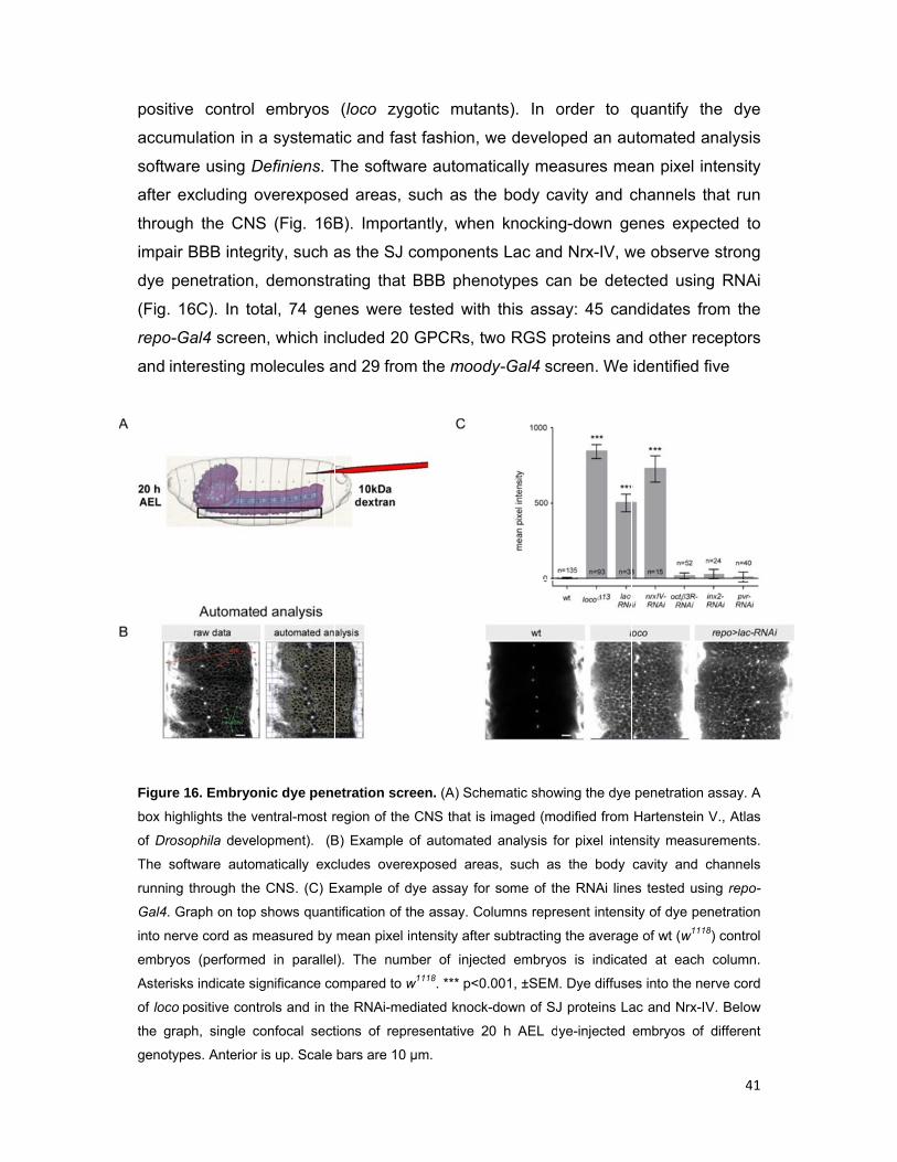
softwa
after e
through
impair
dye pe
(Fig. 1
repo-G
and int
Figure 1
box high
of Droso
The sof
running
Gal4. Gr
into nerv
embryos
Asterisk
of loco p
the grap
genotyp
e control
ulation in a
re using D
xcluding o
h the CNS
BBB integ
enetration,
6C). In tot
Gal4 screen
eresting m
16. Embryon
hlights the ve
ophila develo
ftware autom
through the
raph on top
ve cord as m
s (performed
ks indicate sig
positive contr
ph, single co
es. Anterior
embryos
a systema
Definiens. T
overexpose
S (Fig. 16B
rity, such a
demonstr
tal, 74 gen
n, which in
molecules a
nic dye pene
entral-most r
opment). (B
matically exc
CNS. (C) E
shows quant
measured by
d in parallel
gnificance co
rols and in th
onfocal sect
is up. Scale
(loco zyg
tic and fas
The softwa
ed areas, s
B). Importa
as the SJ c
rating that
nes were
ncluded 20
and 29 from
etration scr
region of the
B) Example
cludes overe
Example of d
tification of t
mean pixel i
l). The num
ompared to w
he RNAi-me
tions of repr
bars are 10
gotic muta
st fashion,
are automa
such as th
antly, whe
componen
BBB phen
tested with
GPCRs, t
m the mood
reen. (A) Sch
CNS that is
of automate
exposed are
dye assay fo
he assay. C
intensity afte
mber of injec
w1118. *** p<0
diated knock
resentative 2
µm.
ants). In o
we develo
atically me
he body ca
en knockin
nts Lac and
notypes ca
h this assa
two RGS p
dy-Gal4 sc
hematic show
s imaged (mo
ed analysis fo
eas, such as
or some of th
olumns repre
er subtracting
cted embryo
0.001, ±SEM
k-down of SJ
20 h AEL d
order to q
oped an au
asures me
avity and c
g-down ge
d Nrx-IV, w
an be dete
ay: 45 can
proteins an
creen. We
wing the dye
odified from
or pixel inte
s the body
he RNAi line
esent intens
g the average
os is indicat
. Dye diffuse
J proteins La
ye-injected
quantify th
utomated a
ean pixel in
channels t
enes expe
we observe
ected usin
ndidates fr
nd other re
identified f
penetration
Hartenstein
nsity measu
cavity and c
es tested usi
ity of dye pe
e of wt (w111
ted at each
es into the ne
ac and Nrx-IV
embryos of
41
he dye
analysis
ntensity
that run
ected to
e strong
g RNAi
rom the
ceptors
five
assay. A
V., Atlas
rements.
channels
ing repo-
enetration 8) control
column.
erve cord
V. Below
different
42
genes whose pan-glial knock-down caused BBB permeability suggesting that they
are novel genes required for BBB development. All were pursued further to
characterize their specific roles, but three (pasiflora1, pasiflora2, mcr) will be
discussed in the context of this thesis.
1.3 Discussion
1.3.1 Genome-wide glial screening
We performed a genome-wide RNAi screen for genes required in glia for adult
survival using the pan-glial repo-Gal4 driver and the VDRC KK transgenic UAS-RNAi
library. The repo-Gal4 driver is very strong and efficiently led to the identification of a
big number of potential glial genes: 3679 genes or 28% of the genome. Previously it
has been estimated that in Drosophila, disruption of approximately 30% of the
genomic loci results in lethality and 75% of lethal loci are pleiotropic, meaning that
their gene products are expressed and utilized at multiple places and times (Miklos
and Rubin, 1996). These observations further support that the 28% of the genome
we identify as required in glia is a remarkably high number. The success of the
screen is also reflected in the very high efficiency of retrieval of both housekeeping
and known glial genes. For some GO terms and signaling pathways, gene retrieval
almost reaches 100% suggesting that the screen is close to saturation. After
excluding housekeeping genes, which are needed in every cell type, the repo-Gal4
screen identified 2697 interesting molecules possibly required for glial development
and/or function. To help uncover true candidates, the results of the screen can be
integrated with those of glial transcriptome analyses by us (U. Gaul, unpublished)
and others (Egger et al., 2002; Freeman et al., 2003; Altenhein et al., 2006) and the
available information from in situ experiments (BDGP website), which have different
strengths and caveats; candidates identified on the basis of multiple lines of
evidence can be assigned to higher confidence groups.
By determining the survival rate at 10 days old adults, the identified
candidates might exert their function at any developmental stage and/or during adult
life and cover a wide repertoire of glial functions. The results of our pan-glial screen
potentially offer the whole set of essential glial genes and represent valuable
material that can contribute to many glial projects in the lab, such as BBB
development and maintenance, glial phagocytosis, and glial control of homeostasis

43
in the adult. The identified candidates also offer hints about truly novel glial functions
and can be the starting point for addressing exciting new questions on glial biology.
The lab has recently characterized 650 glial Gal4 drivers, including five drivers, each
with strong and specific expression in one glial subpopulation (perineurial, SPG,
cortex, ensheathing, astrocyte-like) (Kremer et al., in preparation). The results of the
pan-glial RNAi screen in combination with the Gal4 collection can be used to
decipher differential functions of glial subpopulations at a large scale, carry out
various more specific glial screens, and study the role of particularly appealing
candidates. In addition, by checking the RNAi lines with the strong repo-Gal4 driver,
several strains efficiently silencing housekeeping genes, such as the basal
transcriptional machinery were identified, which might also be of interest to relevant
projects in the lab.
The remarkable technique of RNAi has opened exciting new avenues for
screening. Compared to forward screens, RNAi screens have the advantages that
they can cover the whole genome and that they are overall faster because the step
of mapping the mutation, which can sometimes be labor-intensive and time-
consuming, is eliminated. However, they also bare some limitations, the most
important being the incompleteness of knock-down, which calls for strong drivers.
The recently developed CRISPR-Cas9 system provides a fast means to create
complete knock-outs and is emerging as a promising alternative to RNAi that might
allow for the realization of impressive screens under complete loss of function
conditions (Bassett et al., 2013; Kondo and Ueda, 2013). However, currently, given
the gaps in our knowledge of how the system operates in eukaryotic cells, it does not
seem that we fully understand all the potential problems to apply the technique at a
genome-wide level. CRISPR may complement RNAi approaches and can be used
for the fast validation and further characterization of individual candidates, but at
least in the near future RNAi will likely remain the method of choice for carrying out
large scale reverse genetic screens.
1.3.2 Screening for blood-brain barrier genes – achievements and future
directions
The secondary moody-Gal4 screen revealed 383 genes that when knocked-down
specifically in SPG result in reduced adult viability. Among the candidates, most are
housekeeping and some are known SPG genes, such as Moody signaling and SJ

44
components. After excluding these candidates, the screen revealed approximately
100 interesting or completely uncharacterized genes, allowing us to continue with
directly identifying novel genes required for BBB formation. In contrast to repo-Gal4,
moody-Gal4 is a rather weak driver in particular during embryonic development.
moody-Gal4 starts being weakly expressed late in embryogenesis (late stage 16-
stage 17) in only a subset of SPG, suggesting that moody-Gal4-mediated knock-
down will not capture genes required during BBB formation. However, many of the
genes involved in establishing the barrier in the embryo are also expected to be
required for its maintenance at later stages during which moody-Gal4 is strongly
expressed. The low rates of retrieval of housekeeping genes indeed suggest that the
screen resulted in a significant number of false positives. Because moody-Gal4 is
highly expressed in third instar larval SPG, this result might also indicate that after
development, cells cannot be efficiently killed by RNAi. Nevertheless, manual
exploration of the available data and specifically of the approximately 400 lines that
just escaped the threshold we established (40-44% viable flies with SPG-knock-
down) suggests that housekeeping and known or potential SPG genes (e.g. kune,
megalin, rho1, crebB-17A, tre-1) might be enriched in these groups. Therefore, a
closer look at these candidates, as well as their comparison with the RNA-seq-based
tissue-specific expression profile and microarray-based SPG transcriptome, might
help identify true and exciting SPG genes (Graveley et al., 2011; DeSalvo et al.,
2014).
To overcome the problem of the weakness of moody-Gal4 and improve the
functional RNAi screening for SPG genes, it will be important to optimize the
screening conditions to yield more positive results. For instance, pilot screens of a
selection of candidates, together with many positive controls could be carried out to
search if other SPG-specific drivers are stronger (e.g. driver with two copies of
moody-Gal4, GR54C07-Gal4). In addition, different sensitized genetic backgrounds
could be tried, i.e. the screen could be performed in flies which carry a mutation that
affects BBB formation and/or maintenance. When barrier function is already
disrupted, the residual gene activity in incomplete knock-downs that showed no
effect in the moody-Gal4 screen might no longer suffice. Suitable sensitized
backgrounds would be the removal of one copy of Gβ13F, pka-C1, or of a SJ
component since homozygous mutants of these genes have very strong insulation
defects. Except for increasing the effectiveness of the screen, selection of the
45
appropriate sensitized background may be used to bias the screen towards detection
of specific candidates. For example, loss of function of genes involved in Moody
signaling may bias the screen towards identification of additional pathway
components. Such genetic modifier screens have been proven very successful in
identifying missing pathway components; e.g. for the identification of several proteins
of the Sevenless pathway involved in R7 photoreceptor development (Simon, 1994).
In a small-scale preliminary screen, we tested 200 random RNAi lines using moody-
Gal4 in a pka-C1 heterozygous mutant background. Importantly, although loss-of-
function mutations in almost all genes are recessive, pka-C1 heterozygous mutants
show mild dye leakage into the CNS (Li et al., in preparation). Using the sensitized
background, we identify 12 additional candidates (6% more), including the known
SPG genes rho1 and kune, suggesting that a couple of hundred more genes could
be found with this strategy.
As a last step in our screening procedure, we have performed the embryonic
dye penetration assay using repo-Gal4 and identified five genes whose glial-specific
knock-down impairs BBB integrity. The genes tested included 29 candidates from
the moody-Gal4 screen and 45 candidates from the repo-Gal4 screen. Genes whose
knock-down caused adult subviability with moody-Gal4 but not a permeable
embryonic BBB might include molecules that serve functions in BBB maintenance
during later stages or that play roles in SPG other than barrier function. For instance,
the gap junction protein Innexin2 was one such candidate, which in the meantime
was shown to be required in larval SPG, together with Innexin1, for nutrient-
dependent reactivation of neuroblast proliferation (Speder and Brand, 2014).
Furthermore, because the phenotypic analysis of moody pathway mutants suggests
that there is additional activating input into the G-proteins, we tested with the dye
assay 20 GPCRs identified in the repo-Gal4 screen, including Tre1, but unfortunately
did not find any whose knock-down impairs BBB formation. Because even the
moody deletion mutant has a rather weak dye penetration phenotype (Schwabe et
al., 2005), it is possible that there is partial redundancy between the two (or more)
receptors and that RNAi-mediated knock-down is insufficient to reveal additional
GPCRs. Screen of genomic mutants or knock-down of GPCRs in a moody sensitized
background (moody-RNAi or heterozygous moody mutant) might consist more
suitable approaches in order to identify the receptors that act together with Moody to
activate the G-proteins.

46
Part 2
Pasiflora1, Pasiflora2 and Mcr are novel components
of the Drosophila septate junction
48
2.1 Introduction
2.1.1 Epithelia - an evolutionary novelty
The first epithelium was a novel structure capable of subdividing the body into
morphologically and physiologically distinct compartments and consisted an
evolutionary novelty that allowed for the development of metazoans with complex
body plans (Leys and Riesgo, 2012). Epithelial sheets surround the body as a whole
and line almost all surfaces and cavities within it. They act as selective diffusion
barriers to isolate the organs from the body fluid and maintain their homeostasis and
proper function. Given their wide distribution, epithelia also perform other functions,
including protection against mechanical stress and dehydration, absorption,
secretion, excretion, and gas exchange. To accomplish such diverse functions,
epithelia adopt different cellular arrangements. Simple squamous epithelia are single
layers of thin flattened cells, such as in the lung and blood vessels in vertebrates and
BBB in Drosophila. Simple cuboidal and columnar epithelia are also single-layered,
but consist of taller cells with expanded lateral membranes, like the epithelia of
intestine, glands, and the Drosophila trachea (Fig. 17A).
All epithelia have three common features. First, epithelial cells have a
polarized organization of the plasma membrane and underlying cytoskeleton. Their
apical and basal membranes are segregated and have unique biochemical
compositions. Within the epithelium, the component cells share an aligned polarity
with the apical membrane facing the organ lumen or the outside of the organism (St
Johnston and Ahringer, 2010). Second, their basal membrane is anchored to a
specialized dense extracellular matrix, the basement membrane (Fig. 17B). The
basement membrane not only provides mechanical support to the epithelium and
separates it from the underlying tissue, but also plays crucial roles in regulating the
growth, survival, differentiation and morphogenetic interactions of epithelial cells
(Brown, 2000). Third, epithelial cells are attached to each other with different types of
intercellular junctions. These include adhering junctions, which provide cohesion;
occluding junctions, which seal the paracellular space; and gap junctions, which
allow direct intercytoplasmic communication between the cells (Knust and Bossinger,
2002) (Fig. 17B). Many junctional proteins compose complex multi-protein families,
such as the adherens junction-associated cadherins, the occluding junction-
associated claudins, and the gap junction-associated innexins. Such large families

49
are often used in a tissue-specific manner during epithelial morphogenesis and might
explain the diversified properties of different epithelia (Franke, 2009).
Based on their mode of formation, epithelia are distinguished in primary and
secondary (Tepass, 1997; Schwabe et al., submitted). Primary epithelia, like the
Drosophila trachea, epidermis, foregut, and hindgut arise by shape changes of the
original blastoderm. In contrast, secondary epithelia, like kidney tubules and heart in
vertebrates, as well as heart, midgut and BBB in Drosophila develop through
mesenchymal intermediates in a process called mesenchymal-epithelial transition.
Primary and secondary epithelia also differ in the ways they establish apical-basal
polarity. In Drosophila primary epithelia, polarity is established by the mutually
antagonizing actions of apical determinants, like Crb and Bazooka (Par-3) and
proteins that define the basolateral domain, such as the Lgl group proteins Discs-
large (Dlg), Scribbled (Scrib) and Lethal giant larvae (Lgl) (Fig. 17B) (Laprise and
Tepass, 2011). Neither Crb nor Bazooka are found in secondary epithelia, making
more enigmatic the mechanisms by which polarity is established; however, recent
findings suggest that in the Drosophila BBB, interactions with the basement
membrane, as well as occluding junctions are involved (Schwabe et al., submitted).
2.1.2 Occluding junctions mediate epithelial barrier function
A common role shared by all epithelia is that they define compartment boundaries
and act as barriers to generate and maintain distinct chemical microenvironments.
To accomplish this role, epithelia selectively transport substances via localized
membrane channels and transporters and impede free paracellular diffusion. To
restrict diffusion, epithelial cells are densely packed and have a narrow
intermembrane space, which is sealed by specialized occluding junctions. The
sealing capacity of an epithelium is defined by its occluding junctions and can be
determined by its ability to block tracers of different sizes (Asano et al., 2003;
Schwabe et al., 2005).
2.1.3 Overview of Drosophila septate junctions - ultrastructure and subtypes
In most invertebrates, septate junctions (SJs) seal the epithelial paracellular space
and provide barrier function (Noirot-Timothee et al., 1978; Tepass and Hartenstein,
1994; Schwabe et al., 2005) (Fig. 17B). The lowest phylum known to possess SJs is
Porifera, however, it is still unclear whether sponge SJs act as occluding junctions
50
(Ledger, 1975). SJs have been described in several invertebrate groups, including
nematodes, arthropods and mollusks, as well as vertebrates (discussed in section
2.1.6, Fig. 17B) and their ultrastructure has been defined by transmission electron
microscopy. In cross-sections, SJs display a characteristic electron-dense ladder-like
appearance between adjacent cells. In tangential sections, morphologically distinct
SJs have been described in different organisms, e.g. straight, pleated, smooth, and
paired SJs (Izumi and Furuse, 2014). Drosophila possesses pleated and smooth
SJs. Pleated SJs appear as an array of regularly spaced, undulating rows of septa
that span the intermembrane space; within the SJ, the opposing membranes
maintain a constant distance of approximately 15 nm (Fig. 17C). These septa form
circumferential spirals around the cell and prevent the passive flow of solutes by
extending their travel distance through the paracellular route (Tepass and
Hartenstein, 1994; Schwabe et al., 2005). Tracer studies have indicated that
individual septa act as impartial filters; thus the more septa are arrayed, the tighter
the seal (Abbott, 1991). So far, how the highly regular septa alignment is established
remains elusive. Pleated SJs are found in the majority of Drosophila epithelia, e.g.
epidermis, trachea, foregut, hindgut, salivary glands and the glial blood-brain and
blood-nerve barriers and have been characterized in much deeper detail; for the rest
of this thesis, the term SJs will refer to pleated SJs. In contrast, smooth SJs are not
arranged as undulating rows when viewed tangentially, but as straight parallel bands
and are found in the midgut, gastric caeca and Malphigian tubules (Flower and
Filshie, 1975; Tepass and Hartenstein, 1994). So far, no functional difference
between the barrier properties of the two SJ subtypes has been described, while
their structural differences are thought to reflect variation in the molecular
architecture. In columnar epithelia, SJs localize at the apicolateral membrane just
basally of adherens junctions (Fig. 17B). Importantly, SJs are indispensable for
Drosophila development and SJ mutants die at the end of embryogenesis due to
various insulation defects.
2.1.4 Molecular composition and morphogenesis of Drosophila septate
junctions
The SJ consists of a large multiprotein complex. Although at the ultrastructural level
SJs have been described in a big number of invertebrate species, most of our
knowledge on their molecular composition comes from studies in the genetically
51
tractable Drosophila. In the fruitfly, more than 20 proteins have been identified,
mainly through genetic screens, that when missing lead to disruption of SJs and loss
of barrier integrity (Izumi and Furuse, 2014) (Fig. 17B). Most are transmembrane and
lipid-anchored proteins that localize at the SJ, such as the claudins Megatrachea
(Mega; Pickel - Flybase) (Behr et al., 2003), Sinuous (Sinu) (Wu et al., 2004), and
Kune-kune (Kune) (Nelson et al., 2010), the cell adhesion molecules Neurexin-IV
(Nrx-IV) (Baumgartner et al., 1996), Contactin (Cont) (Faivre-Sarrailh et al., 2004),
Neuroglian (Nrg) (Genova and Fehon, 2003), and Lachesin (Lac) (Llimargas et al.,
2004), the sodium pump with its two subunits ATPα and Nervana2 (Nrv2) (Genova
and Fehon, 2003; Paul et al., 2003), Melanotransferrin (Mtf; Transferrin 2 - Flybase)
(Tiklová et al., 2010) and Macroglobulin complement-related (Mcr) (Bätz et al., 2014;
Hall et al., 2014). The homophilic cell adhesion molecule Fasciclin III (FasIII) also
localizes at SJs and has been widely used as a SJ marker (Woods et al., 1997),
however, flies carrying a null mutation in fasIII are viable, suggesting that the protein
is dispensable for SJ formation (Whitlock, 1993). The complex also includes the
intracellular proteins Coracle (Cora) and Varicose (Vari) that directly interact with the
cytoplasmic tail of Nrx-IV via their FERM and PDZ domains, respectively and link the
junctions to the cytoskeleton (Fehon et al., 1994; Lamb et al., 1998; Ward et al.,
1998; Ward et al., 2001; Wu et al., 2007; Bachmann et al., 2008; Moyer and Jacobs,
2008). A hallmark of SJ proteins is that they are interdependent for localization and
removal of one component is sufficient to destabilize the whole complex and
mislocalize other SJ proteins. In addition, half of the known SJ proteins can be
readily co-immunoprecipitated from tissue extracts and detected by mass
spectrometry (MS), further suggesting that they function together in a multi-protein
complex (Genova and Fehon, 2003; Faivre-Sarrailh et al., 2004; Tiklová et al., 2010;
Jaspers et al., 2012). Fluorescence Recovery After Photobleaching (FRAP)
experiments have been instrumental in classifying most SJ proteins as core
components based on their limited mobility after photobleaching and the observation
that in their loss-of-function other SJ proteins diffuse fast into the bleached region
due to impaired complex formation (Laval et al., 2008; Oshima and Fehon, 2011)
(Fig. 2). Intriguingly, the specific roles of individual SJ proteins within the complex
remain largely unknown.
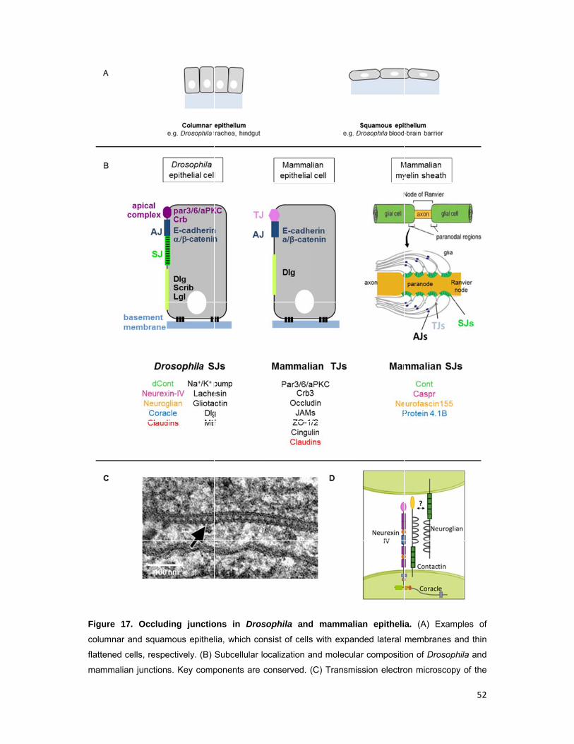
Figure
columna
flattened
mamma
17. Occlud
ar and squam
d cells, respe
alian junction
ding junctio
mous epithel
ectively. (B)
s. Key comp
ons in Dros
lia, which co
Subcellular l
ponents are
sophila and
onsist of cells
localization a
conserved.
d mammali
s with expan
and molecula
(C) Transmi
ian epitheli
nded lateral
ar compositio
ssion electro
a. (A) Exam
membranes
on of Drosop
on microsco
52
mples of
and thin
phila and
py of the
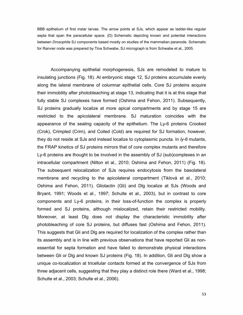
53
BBB epithelium of first instar larvae. The arrow points at SJs, which appear as ladder-like regular
septa that span the paracellular space. (D) Schematic depicting known and potential interactions
between Drosophila SJ components based mostly on studies of the mammalian paranode. Schematic
for Ranvier node was prepared by Tina Schwabe, SJ micrograph is from Schwabe et al., 2005.
Accompanying epithelial morphogenesis, SJs are remodeled to mature to
insulating junctions (Fig. 18). At embryonic stage 12, SJ proteins accumulate evenly
along the lateral membrane of columnar epithelial cells. Core SJ proteins acquire
their immobility after photobleaching at stage 13, indicating that it is at this stage that
fully stable SJ complexes have formed (Oshima and Fehon, 2011). Subsequently,
SJ proteins gradually localize at more apical compartments and by stage 15 are
restricted to the apicolateral membrane. SJ maturation coincides with the
appearance of the sealing capacity of the epithelium. The Ly-6 proteins Crooked
(Crok), Crimpled (Crim), and Coiled (Cold) are required for SJ formation, however,
they do not reside at SJs and instead localize to cytoplasmic puncta. In ly-6 mutants,
the FRAP kinetics of SJ proteins mirrors that of core complex mutants and therefore
Ly-6 proteins are thought to be involved in the assembly of SJ (sub)complexes in an
intracellular compartment (Nilton et al., 2010; Oshima and Fehon, 2011) (Fig. 18).
The subsequent relocalization of SJs requires endocytosis from the basolateral
membrane and recycling to the apicolateral compartment (Tiklová et al., 2010;
Oshima and Fehon, 2011). Gliotactin (Gli) and Dlg localize at SJs (Woods and
Bryant, 1991; Woods et al., 1997; Schulte et al., 2003), but in contrast to core
components and Ly-6 proteins, in their loss-of-function the complex is properly
formed and SJ proteins, although mislocalized, retain their restricted mobility.
Moreover, at least Dlg does not display the characteristic immobility after
photobleaching of core SJ proteins, but diffuses fast (Oshima and Fehon, 2011).
This suggests that Gli and Dlg are required for localization of the complex rather than
its assembly and is in line with previous observations that have reported Gli as non-
essential for septa formation and have failed to demonstrate physical interactions
between Gli or Dlg and known SJ proteins (Fig. 18). In addition, Gli and Dlg show a
unique co-localization at tricellular contacts formed at the convergence of SJs from
three adjacent cells, suggesting that they play a distinct role there (Ward et al., 1998;
Schulte et al., 2003; Schulte et al., 2006).
Figure
consists
stage 13
the apico
2.1.5
Except
perform
and/or
that pr
compa
part of
cell po
preced
ATPas
restrict
protein
tube le
(Wu an
secreti
elonga
2006).
18. Timeline
s of several c
3, while endo
olateral mem
Other fun
t for their
m other fun
maintenan
revents int
artments (S
a complex
larity. Duri
des the de
se and the
ting the siz
ns (Laprise
ength with
nd Beitel,
on of the
ation by a
e and playe
core compon
ocytosis and
mbrane at sta
nctions of
well esta
nctions. In
nce of cell
termixing
Schwabe e
x that plays
ng late sta
evelopment
e SJ locali
ze of the C
e et al., 200
the major
2004). SJs
chitin dea
yet unide
ers of SJ m
nents. Ly-6
the SJ prote
age 14 (mod
Drosophi
blished ro
the BBB
polarity by
of molecu
et al., subm
s a develo
ages in the
t of SJs, a
izing-prote
Crb domain
09). Finally
rity of SJ m
s control t
acetylases
entified me
orphogenes
proteins are
eins Gli and D
ified from Os
ila septate
ole as par
epithelium
y acting as
ules betwe
mitted). In
pmentally
e maturatio
a complex
ein Yurt is
n and func
y, SJs are
mutants di
tube length
Serpentin
echanism (
sis in colum
required for
Dlg are esse
shima and Fe
e junctions
acellular d
m, SJs cont
s a fence w
een the ap
addition,
earlier, ind
on of prima
x formed b
required
ctionally su
involved i
splaying o
h, at least
e and Ver
(Luschnig
mnar epithe
r assembly o
ential for com
ehon, 2011).
s
diffusion b
tribute to t
within the
pical and
some SJ
dependent
ary epitheli
by Nrx-IV,
for mainta
ubstituting
in the regu
overelonga
partly, by
rmiform th
et al., 20
elia. The SJ
of (sub)comp
mplex relocal
.
barriers, SJ
the establi
lateral mem
basal mem
proteins a
role in pro
a, which h
Cora, the
aining pola
for the Lg
ulation of t
ated dorsa
regulating
at termina
006; Wang
54
complex
plexes at
ization at
Js also
shment
mbrane
mbrane
are also
omoting
however
Na+/K+
arity by
gl group
racheal
l trunks
g apical
ate tube
g et al.,
55
2.1.6 Vertebrate tight and septate junctions
In vertebrate epithelia, the block to paracellular diffusion is mediated by tight
junctions (TJs) (Farquhar and Palade, 1963; Balda and Matter, 2008). In contrast to
SJs, TJs localize apically of the zonula adherens (Fig. 17B) and using electron
microscopy appear as a series of apparent fusions of the membranes of adjacent
cells (kissing points). These fusions are composed of strands of intramembranous
particles from opposing cells that associate to constitute a paired TJ and obliterate
the paracellular space. Similar to invertebrate SJ septa, each strand acts
independently from the others and the sealing capacity of TJs increases with the
increasing number of strands. Within paired TJs, aqueous pores are postulated to
occur, which are selectively permeable to small molecules. TJs consist of a series of
proteins embedded in the plasma membrane, such as Occludin, Tricellulin, and
junctional adhesion molecules (JAMs), which in turn are attached to cytoskeletal and
scaffold proteins, including Zonula occludens (ZO)-1/2/3 and Cingulin. TJs also
contain Crb3, Par-3, Par-6 and aPKC (atypical protein kinase C), which in Drosophila
associate with the apical and sub-apical regions. Although the set of proteins
composing the TJ is different from that of the SJ, the two complexes share a key
molecular component, the claudins (Fig. 17B).
Mammals also possess SJs at the nodes of Ranvier in the PNS, where they
form the paranodal junction between axons and myelinating glia (Fig. 17B) (Poliak
and Peles, 2003). Importantly, mammalian SJs are composed (in part) of the
homologous molecules found in Drosophila SJs (Fig. 17B). Studies of the paranodal
junction have also unraveled some of the interactions bridging together SJ proteins
in the multi-subunit complex. Caspr (Nrx-IV homologue) forms a complex with
Contactin in cis in the axonal membrane and its intracellular region, like in
Drosophila, binds the scaffold protein 4.1B (Cora homologue). Moreover,
Neurofascin 155 (Nrg homologue) has been suggested to be the glial ligand of the
Caspr/Contactin complex (Fig. 17D) (Einheber et al., 1997; Bhat et al., 2001; Boyle
et al., 2001; Pillai et al., 2009). Like Drosophila BBB SJs, paranodal SJs serve the
same dual function as a paracellular barrier impeding ion flow across the paranode
and as a fence restricting the intermixing of neuronal membrane proteins such as
Na+ and K+ ion channels. The myelin loops of Schwann cells also contain claudin-
containing TJs, which contribute to paracellular barrier function and saltatory
conduction of action potentials (Miyamoto et al., 2005).
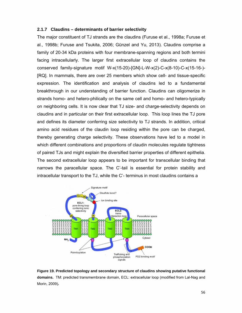
2.1.7
The ma
al., 199
family
facing
conser
[RQ]. I
expres
breakth
strands
on neig
claudin
and de
amino
thereby
which d
of paire
The se
narrow
intrace
Figure 1
domain
Morin, 2
Claudins
ajor consti
98b; Furus
of 20-34 k
intracellul
rved family
n mamma
ssion. The
hrough in
s homo- an
ghboring c
ns and in p
efines its d
acid resid
y generati
different co
ed TJs and
econd extra
ws the par
ellular trans
19. Predicte
s. TM: pred
2009).
– determi
tuent of TJ
se and Ts
kDa protein
arly. The
y-signature
ls, there a
e identifica
our under
nd hetero-
cells. It is n
particular o
diameter co
dues of the
ng charge
ombination
d might exp
acellular lo
racellular s
sport to the
d topology
icted transm
inants of b
J strands a
ukita, 2006
ns with fou
larger firs
e motif W
are over 25
ation and
rstanding o
philically o
now clear t
on their firs
onferring s
e claudin
e selectivity
ns and pro
plain the d
oop appea
space. Th
e TJ, while
and second
membrane do
barrier sel
are the clau
6; Günzel
ur membra
st extrace
W-x(15-20)-
5 members
analysis
of barrier f
on the sam
that TJ siz
st extracell
size selecti
loop resid
y. These o
portions of
iversified b
ars to be im
he C’-tail
the C’- ter
dary structu
omain, ECL:
lectivity
udins (Fur
and Yu, 2
ne-spannin
ellular loop
-[GN]-L-W-
s which sh
of claudi
function. C
me cell and
ze- and cha
ular loop.
ivity to TJ
ding within
observatio
f claudin m
barrier pro
mportant fo
is essentia
rminus in m
re of claudi
extracellular
use et al.,
2013). Cla
ng regions
p of claud
-x(2)-C-x(8
how cell- a
ins led to
Claudins ca
homo- an
arge-selec
This loop
strands. In
the pore
ons have le
molecules r
perties of d
or transce
al for pro
most claud
ns showing
r loop (modifi
1998a; Fu
udins com
s and both
dins conta
8-10)-C-x(1
nd tissue-s
o a funda
an oligome
nd hetero-t
ctivity depe
lines the T
n addition,
can be ch
ed to a m
regulate tig
different ep
llular bindi
tein stabil
ins contain
g putative fu
ied from Lal-
56
uruse et
mprise a
termini
ins the
15-16-)-
specific
amental
erize in
typically
ends on
TJ pore
, critical
harged,
model in
ghtness
pithelia.
ing that
ity and
ns a
unctional
-Nag and

PDZ-bi
protein
but the
al., 200
of tetr
periphe
membr
membe
clarins
to perf
barrier
Drosop
superfa
perform
Figure
inverteb
similar fu
inding do
ns. In Droso
eir precise
04; Nelson
Claudins a
ra-spannin
eral myelin
rane intrin
ers of the
(Adato et
form diver
formation
phila and
amily, whi
m similar fu
20. Phyl
brates. The
unctions. Fig
main (mo
ophila, thre
roles withi
et al., 201
are part of
ng membr
n protein (P
sic protein
TMEM47
al., 2002;
rse functio
n, regulati
Caenorha
ch althou
unctions w
ogenetic t
proteins are
gure is from S
ost freque
ee claudins
in the com
10) (Fig. 19
f the PMP
rane prote
PMP22), e
n (MP20),
7 family, v
Simske, 20
ons at spe
ion of ch
abditis eleg
gh highly
ith those a
tree of pfam
e highly dive
Simske, 2013
ntly VY)
s have bee
mplex rema
9).
22/EMP/M
eins (pfam
epithelial m
and more
voltage gat
013) (Fig.
ecialized c
hannel act
gans geno
divergent
assigned to
m00822 tet
ergent at the
3.
that allow
en identifie
ain unknow
MP20/Claud
m00822),
membrane
e distantly
ted calcium
20). Most s
cell-cell co
tivity and
omes also
t at the s
o their verte
traspan pro
e sequence
ws binding
ed as comp
wn (Behr e
din mamm
which in
proteins (
y related p
m channe
superfamil
ntacts, inc
protein a
o encode
sequence
ebrate cou
oteins from
level, howev
g to cytop
ponents of
t al., 2003
malian supe
ncludes cl
EMPs), len
proteins, s
el γ subun
ly member
cluding ad
aggregatio
members
level, they
unterparts.
m vertebrat
ver, they ofte
57
plasmic
the SJ,
; Wu et
erfamily
laudins,
ns fiber
such as
nits and
rs seem
hesion,
n. The
of the
y often
tes and
en serve
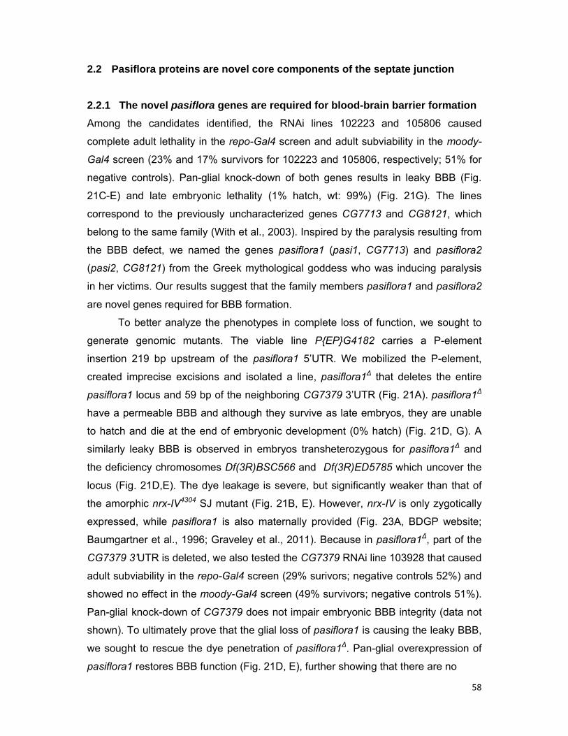
58
2.2 Pasiflora proteins are novel core components of the septate junction
2.2.1 The novel pasiflora genes are required for blood-brain barrier formation
Among the candidates identified, the RNAi lines 102223 and 105806 caused
complete adult lethality in the repo-Gal4 screen and adult subviability in the moody-
Gal4 screen (23% and 17% survivors for 102223 and 105806, respectively; 51% for
negative controls). Pan-glial knock-down of both genes results in leaky BBB (Fig.
21C-E) and late embryonic lethality (1% hatch, wt: 99%) (Fig. 21G). The lines
correspond to the previously uncharacterized genes CG7713 and CG8121, which
belong to the same family (With et al., 2003). Inspired by the paralysis resulting from
the BBB defect, we named the genes pasiflora1 (pasi1, CG7713) and pasiflora2
(pasi2, CG8121) from the Greek mythological goddess who was inducing paralysis
in her victims. Our results suggest that the family members pasiflora1 and pasiflora2
are novel genes required for BBB formation.
To better analyze the phenotypes in complete loss of function, we sought to
generate genomic mutants. The viable line P{EP}G4182 carries a P-element
insertion 219 bp upstream of the pasiflora1 5’UTR. We mobilized the P-element,
created imprecise excisions and isolated a line, pasiflora1∆ that deletes the entire
pasiflora1 locus and 59 bp of the neighboring CG7379 3’UTR (Fig. 21A). pasiflora1∆
have a permeable BBB and although they survive as late embryos, they are unable
to hatch and die at the end of embryonic development (0% hatch) (Fig. 21D, G). A
similarly leaky BBB is observed in embryos transheterozygous for pasiflora1∆ and
the deficiency chromosomes Df(3R)BSC566 and Df(3R)ED5785 which uncover the
locus (Fig. 21D,E). The dye leakage is severe, but significantly weaker than that of
the amorphic nrx-IV4304 SJ mutant (Fig. 21B, E). However, nrx-IV is only zygotically
expressed, while pasiflora1 is also maternally provided (Fig. 23A, BDGP website;
Baumgartner et al., 1996; Graveley et al., 2011). Because in pasiflora1∆, part of the
CG7379 3’UTR is deleted, we also tested the CG7379 RNAi line 103928 that caused
adult subviability in the repo-Gal4 screen (29% surivors; negative controls 52%) and
showed no effect in the moody-Gal4 screen (49% survivors; negative controls 51%).
Pan-glial knock-down of CG7379 does not impair embryonic BBB integrity (data not
shown). To ultimately prove that the glial loss of pasiflora1 is causing the leaky BBB,
we sought to rescue the dye penetration of pasiflora1∆. Pan-glial overexpression of
pasiflora1 restores BBB function (Fig. 21D, E), further showing that there are no
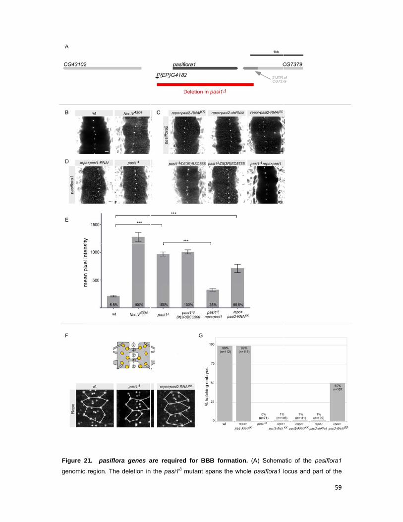
Figure
genomic
21. pasiflo
c region. The
ora genes a
e deletion in
are required
the pasi1∆
d for BBB
mutant span
formation.
ns the whole
(A) Schema
e pasiflora1 l
atic of the p
locus and pa
59
pasiflora1
art of the
60
3UTR’ of the neighboring gene CG7379. (B-D) Single confocal sections of 20 h AEL dye-injected
embryos. Dye diffuses into the nerve cord of nrx-IV positive controls (B) and in the loss of pasiflora1
(D) and pasiflora2 (C), as opposed to wt embryos (B). Pan-glial overexpression of pasiflora1 CDS
rescues the dye penetration of pasiflora1∆ (D). Anterior is up. Scale bars are 10 µm. (E) Quantification
of dye penetration assay. Raw data are plotted. Columns represent intensity of dye penetration into
nerve cord as measured by mean pixel intensity. The percentage of embryos showing penetration is
indicated at the bottom of each column. Brackets and asterisks indicate significance of pairwise
comparisons. *** p<0.001, ±SEM, n=22-107. (F) Ventral surface views of stage 16 embryonic nerve
cord stained with Repo. The full complement of SPG is detected in loss of function of pasiflora genes.
The positions of nuclei are similar between the genotypes, as visualized by overlay of connecting
lines. Schematic on top shows imaged area with SPG labeled orange. Three abdominal neuromeres
are shown. Maximum projections of 4 µm z-stacks. Anterior is up. Scale bars are 10 µm. n=8-15. (G)
Quantification of embryonic survival for different genotypes. Columns represent the percentage of
embryos that hatched. The number of aligned embryos is indicated at the top of each column.
additional mutations on the chromosome contributing to BBB breakdown and
indicating that pasiflora1 is required cell-autonomously.
In the vicinity of the pasiflora2 locus, no lines with P-element insertions were
available. Since the gene belongs to the same family, we decided to pursue
pasiflora2 using RNAi. Moreover, the KK line is very potent as it causes strong BBB
permeability and embryonic lethality with repo-Gal4 (1% hatch) (Fig. 21C, E, G). The
fact that impaired BBB is observed in the glial-specific knock-down suggests that
pasiflora2 is also cell-autonomously required. To exclude off-target effects of the line,
we tested additional RNAi strains, which target different sequences of the mRNA.
Two additional lines were used; the VDRC line GD43952 and a line we generated,
which expresses a short 21 nt siRNA (shRNAi, TRiP design, Ni et al., 2011). Both
lines show embryonic lethality and dye penetration with repo-Gal4, but the GD line
leads to milder defects (Fig. 21C, G). For all our following experiments to study
pasiflora2, we used the KK RNAi line.
To exclude that the leaky BBB is a result of earlier defects in glia specification
and/or migration, we analyzed the number and positions of SPG. To this end, we
stained stage 16 embryos with the Repo antibody, which labels all glial nuclei except
for midline glia, and focused on the ventral-most SPG. In the wt, the number of glia is
very stereotyped, and four SPG are found per abdominal neuromere in the ventral-
most 4 μm of the nerve cord (Ito et al., 1995; Beckervordersandforth et al., 2008).
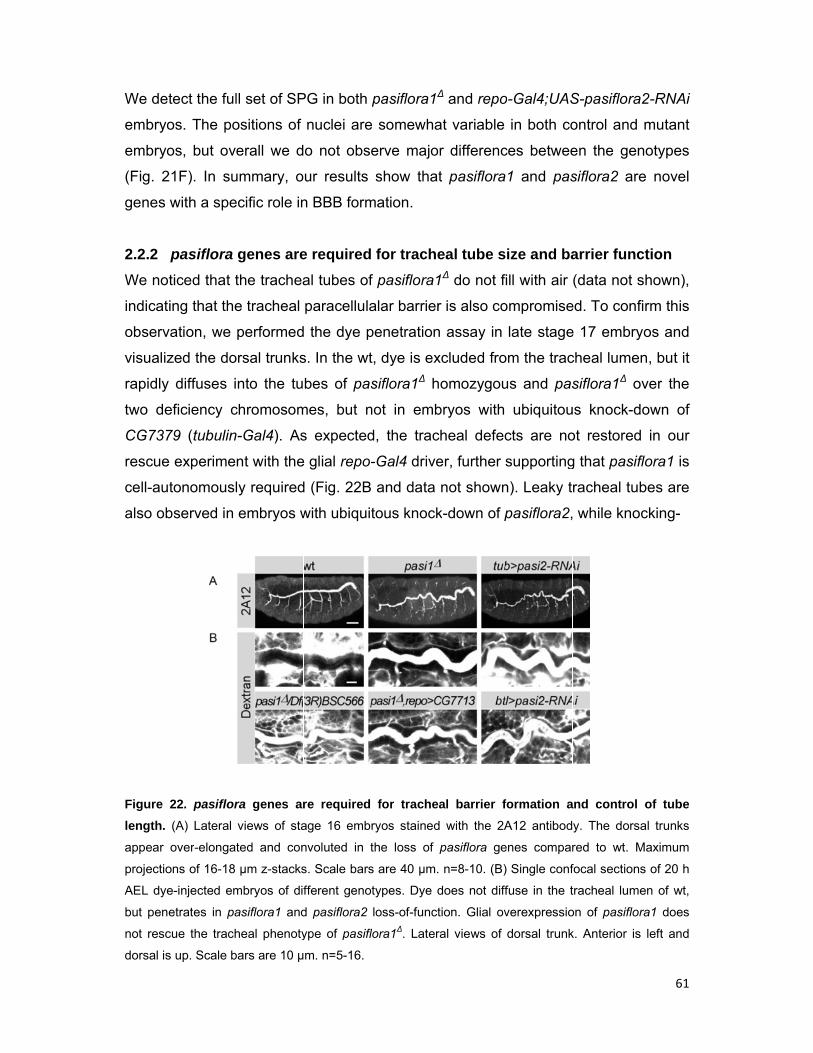
We det
embryo
embryo
(Fig. 2
genes
2.2.2
We not
indicat
observ
visualiz
rapidly
two de
CG737
rescue
cell-au
also ob
Figure
length.
appear
projectio
AEL dye
but pene
not resc
dorsal is
tect the ful
os. The po
os, but ov
21F). In su
with a spe
pasiflora
ticed that t
ing that the
vation, we
zed the do
diffuses i
eficiency c
79 (tubulin
e experime
tonomousl
bserved in
22. pasiflor
(A) Lateral
over-elongat
ons of 16-18
e-injected em
etrates in pa
cue the trach
s up. Scale b
ll set of SP
ositions of
erall we d
ummary, o
ecific role in
genes are
the trachea
e tracheal
performed
orsal trunks
nto the tu
chromosom
n-Gal4). As
nt with the
ly required
embryos w
ra genes ar
views of sta
ted and con
µm z-stacks
mbryos of dif
asiflora1 and
heal phenoty
bars are 10 µ
PG in both
nuclei are
o not obs
ur results
n BBB form
e required
al tubes of
paracellula
d the dye p
s. In the wt
bes of pas
mes, but n
s expected
e glial repo
d (Fig. 22B
with ubiqui
re required
age 16 emb
nvoluted in t
s. Scale bars
fferent geno
d pasiflora2
ype of pasifl
m. n=5-16.
pasiflora1∆
e somewha
erve majo
show tha
mation.
for trache
f pasiflora1
alar barrier
penetration
t, dye is ex
siflora1∆ h
not in emb
d, the trac
-Gal4 drive
B and data
tous knock
for trache
ryos stained
the loss of
s are 40 µm.
otypes. Dye d
loss-of-funct
lora1∆. Later
∆ and repo
at variable
r differenc
t pasiflora
eal tube s
1∆ do not f
r is also co
n assay in
xcluded fro
homozygou
bryos with
cheal defec
er, further
not shown
k-down of p
al barrier f
d with the 2A
pasiflora ge
. n=8-10. (B)
does not diff
tion. Glial ov
ral views of
o-Gal4;UAS
in both co
ces betwee
1 and pas
ize and ba
ill with air
ompromise
late stage
om the trac
us and pas
ubiquitou
cts are no
supporting
n). Leaky t
pasiflora2,
formation a
A12 antibod
enes compar
) Single conf
fuse in the t
verexpressio
dorsal trunk
S-pasiflora
ontrol and
en the gen
siflora2 are
arrier func
(data not s
ed. To conf
e 17 embry
cheal lume
siflora1∆ o
us knock-d
ot restored
g that pasif
racheal tub
while kno
and control
y. The dors
red to wt. M
focal section
tracheal lume
on of pasiflor
k. Anterior is
61
a2-RNAi
mutant
notypes
e novel
ction
shown),
firm this
yos and
n, but it
over the
down of
d in our
flora1 is
bes are
cking-
of tube
al trunks
Maximum
s of 20 h
en of wt,
ra1 does
s left and
62
down pasiflora2 with the more trachea-specific breathless-Gal4 leads to qualitatively
similar phenotypes but with lower penetrance (Fig. 22B). Both pasiflora mutants also
show excessively elongated and convoluted dorsal trunks, a result that was
confirmed by staining stage 16 embryos with the 2A12 antibody that recognizes the
luminal protein Gasp (Fig. 22A). Over-elongated dorsal trunks are observed in the
majority of SJ mutants and are believed to be, at least partly, due to the role of SJs
in apical secretion of chitin deacetylases that terminate tube elongation (Luschnig et
al., 2006; Wang et al., 2006). In summary, the pasiflora genes are required for
tracheal barrier function and control of tube size.
2.2.3 pasiflora genes are expressed in SJ-forming embryonic epithelia and glia
To characterize the expression pattern of pasiflora genes, we performed RNA in situ
hybridization in wt embryos. The genes show identical expression patterns
throughout embryogenesis (Fig. 23A). Ubiquitous weak expression is first detected
at stages 1-4, suggestive of maternal contribution. Zygotic transcripts are detected in
epithelial tissues from stage 10 onwards. The tracheal placodes are labeled at stage
10 and the anterior hindgut at stage 11; expression persists in these tissues
throughout development. During stages 14-16, trachea, foregut, hindgut, epidermis
and salivary glands are marked. At stage 16, we detect weak staining in the nervous
system and labeling of some cells that based on their position are likely to be exit
and/or peripheral glia. A clearer in situ for pasiflora1 showing similar expression is
displayed on the BDGP website (http://insitu.fruitfly.org). Therefore, both genes are
specifically expressed in embryonic epithelia and insulating glia, all tissues that form
SJs.
Several attempts to generate specific antibodies recognizing the two proteins
were unsuccessful. By conventional expression techniques, overexpression of the
highly hydrophobic Pasiflora proteins is toxic to the bacteria. We therefore raised
antibodies against a mixture of two 15-16 amino acid-long peptides (Fig. 23B,
epitopes highlighted with red asterisks). Unfortunately, neither the sera nor the
affinity purified antibodies showed specific labeling in wt embryos (data not shown).
2.2.4 Molecular features of Pasiflora proteins
Pasiflora1 and Pasiflora2 are small proteins of 19 and 29 kDa, respectively, with four
transmembrane domains but no signal peptide, and no sequence similarity to any
63
known domains or motifs. Interestingly, their predicted topology, with two intracellular
termini and a bigger first extracellular loop, strongly resembles that of claudins (Fig.
23B). To examine if the proteins localize at the plasma membrane or some
intracellular membrane compartment, we analyzed their subcellular localization in
vivo and in cell culture. Because we did not succeed in generating specific
antibodies, we C’-terminally tagged both proteins with GFP and FLAG (Fig. 23B). We
expressed the tagged proteins in Schneider cells (S2), as well as glia and SPG in
vivo, which we imaged in stage 16 embryos (repo-Gal4) and 3rd instar larval CNS
(moody-Gal4), respectively. We find that Pasiflora proteins localize at the plasma
membrane both in vivo and in S2 cells (Fig. 23C).
Pasiflora1 and Pasiflora2 were previously identified as members of a family
conserved in Drosophila and Anopheles, consisting of proteins with four
transmembrane domains and two short conserved intracellular motifs in the second
and third intracellular regions. The family includes nine members in the fly, amongst
which the founding member fire exit is expressed in a subset of exit and peripheral
glia (as demonstrated by lacZ enhancer trap pattern), without any known molecular
or biological function (With et al., 2003). Pan-glial knock-down of fire exit causes
adult subviability (19% survivors; 52% for negative control) but was not pursued
further. We did not test additional family members for BBB defects either because
their pan-glial knock-down did not impair viability (CG10311, CG12825, CG13747,
CG14767, and CG15098) or because an RNAi strain was not available in the
collection (CG13288).
Different lines of evidence indicate that pasiflora genes are co-expressed.
First, RNA in situ hybridization showed that the genes are similarly expressed in
embryonic epithelia (Fig. 23A). Second, both genes were identified as differentially
expressed in embryonic glia based on microarray transcriptome profiling (U. Gaul,
unpublished). Third, based on developmental RNA-seq, the genes group together in
a co-expression cluster; interestingly, the SJ genes kune and cold are also part of
the cluster. Pasiflora2 is also included in a second co-expression cluster together
with several SJ genes (sinu, nrx-IV, mcr, gli, crok, cold, crim) (Graveley et al., 2011).
The notion that pasiflora1 and pasiflora2 expression is tightly co-regulated is also
supported by the observation by us and others that both genes, together with more
than half of the known SJ-encoding mRNAs, are predicted targets of miR-184 (Hong
et al., 2009; Iovino et al., 2009; www.microrna.org). Based on the observed
Figure 2
embryo
pasi2 in
detected
(HG), sa
Predicte
depicted
asterisks
Single c
µm. (C.
projectio
Pasiflora
phenot
184, w
or play
protein
antibod
23 . Pasiflo
onic epitheli
n w1118 embr
d from stage
alivary gland
ed structure
d with green
s. (C) Tagge
confocal sect
2) Ventral v
ons of 7 µm
a-GFP in SP
types in pa
we hypothe
y a role i
ns was fou
dy against
ora1 and Pa
ia and glia.
ryos. Both g
10 onwards
ds (SG), cent
and topolog
n ovals. The
ed versions o
tions of S2
views of fixe
z-stacks. S
G. Maximum
asiflora mu
esized that
in complex
und a SJ
the claudi
siflora2 are
(A) RNA in
genes are ex
s in epithelia
tral (CNS) a
gy of Pasifl
e epitopes
of Pasiflora1
cells transie
ed stage 16
Scale bars a
m projections
utants, the
Pasiflora
x assemb
co-immu
in Mega fo
e tetra-span
n situ hybrid
xpressed ma
and nervous
and periphera
ora proteins
used for an
1 and Pasiflo
ently transfec
6 embryos e
re 10 µm. (
s of 10 µm z-s
expressio
proteins a
bly and/or
noprecipita
ollowed by
ning memb
ization with
aternally (sta
s system; tra
al nervous s
s. The site
ntibody prod
ora2 localize
cted with Pa
expressing P
C.3) Third in
stacks. Ante
on patterns
are either S
trafficking
ation expe
MS althou
brane protei
antisense p
age 1-4). Zy
achea (TR), f
ystem (PNS
of fusion of
duction are
e at the plasm
asiflora-FLAG
Pasiflora-GF
nstar larval
rior is up. Sc
s and the t
SJ compon
g. Howeve
eriment wi
ugh this stu
ns co-expre
probes for p
ygotic transc
foregut (FG)
S). Anterior is
f GFP/FLAG
highlighted
ma membra
G. Scale bar
FP in glia. M
CNS expres
cale bars are
targeting b
nents them
er, neither
ith a mon
udy succee
64
essed in
asi1 and
cripts are
), hindgut
s left. (B)
G tags is
with red
ne. (C.1)
rs are 10
Maximum
ssing live
e 20 µm.
by miR-
mselves
of the
noclonal
eded in
65
identifying at least 10 known SJ components (Jaspers et al., 2012). The properties of
Pasiflora proteins and in particular their small size strongly disfavor their detection by
liquid chromatography-coupled MS/MS and might explain why they escaped
identification. Only a limited number of theoretical tryptic peptides is predicted for the
proteins; three peptides for Pasiflora1 and <40 for Pasiflora2. Additionally, fully
tryptic peptides have not been observed for either of the proteins and the only
reported experimental, MS-based evidence is a single non-tryptic peptide for
Pasiflora2 (www.peptideatlas.org). However, in the study by Jaspers et al. only fully
tryptic peptides were included during the identification of MS spectra and only
proteins with at least two peptides were reported. Notably, the two other claudins,
Sinu and Kune, which have similar size and structure with the Pasiflora proteins,
were also not detected in the MS.
2.2.5 pasiflora genes are required for localization of SJs
To confirm that pasiflora genes play a role in SJ formation, we analyzed the
morphology and subcellular localization of SJs in the mutants. We first visualized the
embryonic BBB using the live endogenously expressed markers Nrg-GFP and Lac-
GFP (Morin et al., 2001). In wt late stage 17 embryos, both markers label SJs and
trace the outlines of SPG, which make continuous contacts with their neighbors to
seal the CNS. In pasiflora1∆ and repo-Gal4;UAS-pasiflora2-RNAi embryos, SJs
appear discontinuous and severely disorganized (Fig. 24A), demonstrating that both
genes are required for SJ formation in the embryonic BBB.
SPG are very large but thin cells (only 0.2 μm tall) complicating the
visualization of SJ localization along the lateral membrane during embryonic stages.
We therefore turned to examine the hindgut and tracheal columnar epithelia. In the
hindgut of wt stage 12 embryos, SJ proteins accumulate evenly along the lateral
membrane, but at stage 15 are restricted to the apicolateral membrane
compartment, as revealed by staining for Cora. In pasiflora1∆ and tubulin-Gal4;UAS-
pasiflora2-RNAi embryos, Cora localizes similarly to wt at stage 12, but fails to
restrict apicolaterally at stage 15 and remains distributed along the lateral
membrane. At stage 15 we observe similar mislocalization of additional SJ markers,
such as Nrg-GFP, ATPα-GFP, and FasIII (Fig. 24C). Notably, in the variably
penetrant phenotype of pasiflora2 RNAi, the mislocalization phenotype is more
pronounced for Cora and FasIII followed by Nrg, while ATPα is only mildly
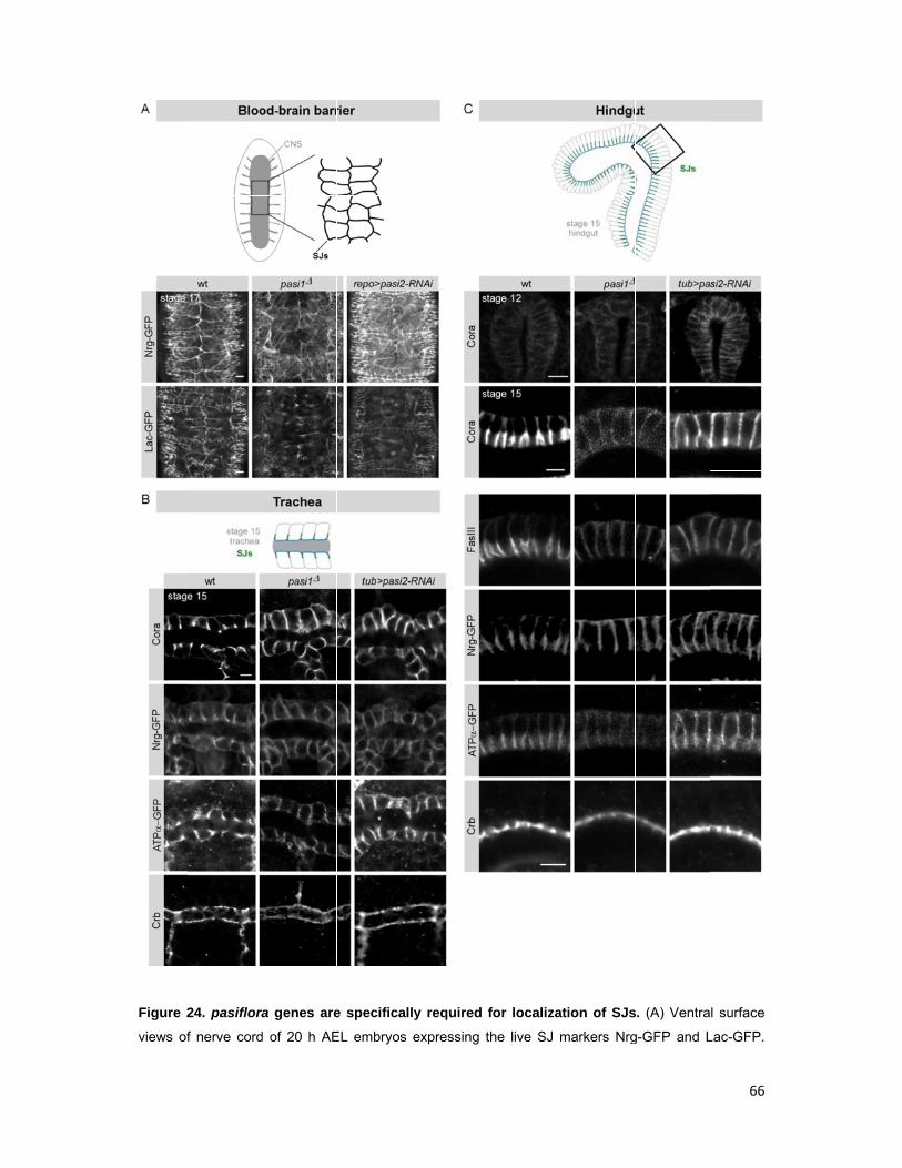
Figure 2
views of
24. pasiflora
f nerve cord
ra genes are
d of 20 h AE
e specificall
EL embryos
ly required
expressing t
for localiza
the live SJ m
ation of SJs
markers Nrg
s. (A) Ventra
g-GFP and L
66
l surface
Lac-GFP.
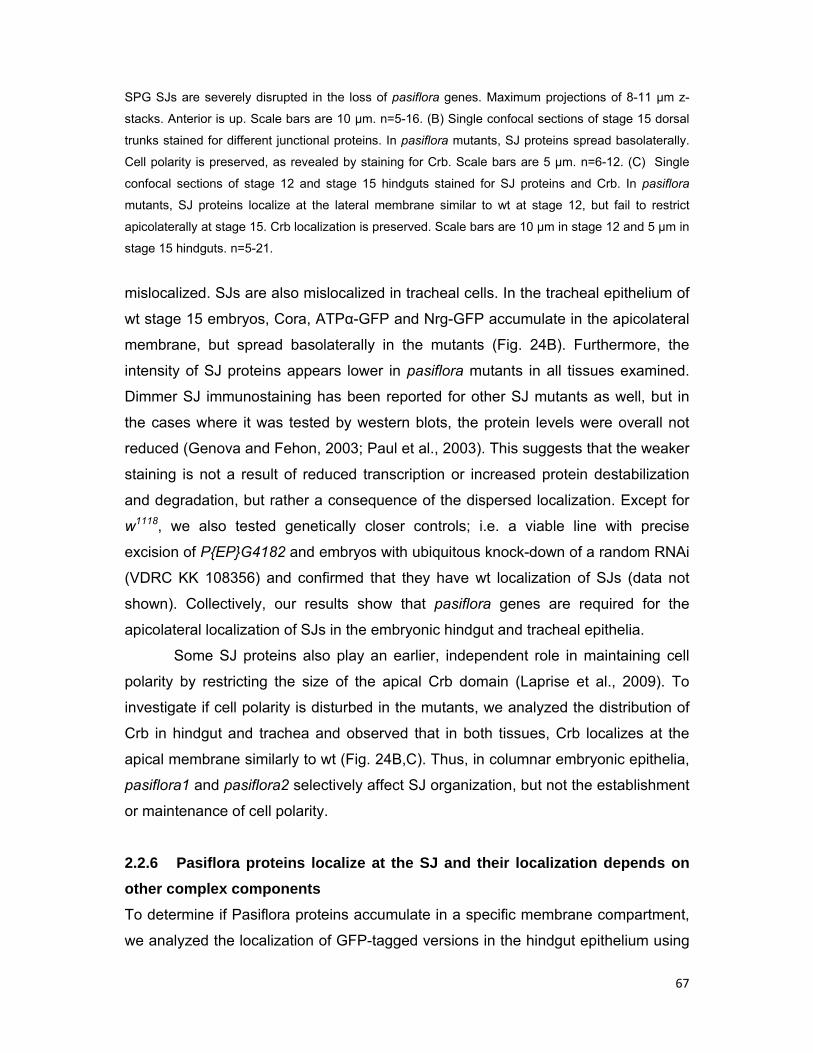
67
SPG SJs are severely disrupted in the loss of pasiflora genes. Maximum projections of 8-11 µm z-
stacks. Anterior is up. Scale bars are 10 µm. n=5-16. (B) Single confocal sections of stage 15 dorsal
trunks stained for different junctional proteins. In pasiflora mutants, SJ proteins spread basolaterally.
Cell polarity is preserved, as revealed by staining for Crb. Scale bars are 5 µm. n=6-12. (C) Single
confocal sections of stage 12 and stage 15 hindguts stained for SJ proteins and Crb. In pasiflora
mutants, SJ proteins localize at the lateral membrane similar to wt at stage 12, but fail to restrict
apicolaterally at stage 15. Crb localization is preserved. Scale bars are 10 µm in stage 12 and 5 µm in
stage 15 hindguts. n=5-21.
mislocalized. SJs are also mislocalized in tracheal cells. In the tracheal epithelium of
wt stage 15 embryos, Cora, ATPα-GFP and Nrg-GFP accumulate in the apicolateral
membrane, but spread basolaterally in the mutants (Fig. 24B). Furthermore, the
intensity of SJ proteins appears lower in pasiflora mutants in all tissues examined.
Dimmer SJ immunostaining has been reported for other SJ mutants as well, but in
the cases where it was tested by western blots, the protein levels were overall not
reduced (Genova and Fehon, 2003; Paul et al., 2003). This suggests that the weaker
staining is not a result of reduced transcription or increased protein destabilization
and degradation, but rather a consequence of the dispersed localization. Except for
w1118, we also tested genetically closer controls; i.e. a viable line with precise
excision of P{EP}G4182 and embryos with ubiquitous knock-down of a random RNAi
(VDRC KK 108356) and confirmed that they have wt localization of SJs (data not
shown). Collectively, our results show that pasiflora genes are required for the
apicolateral localization of SJs in the embryonic hindgut and tracheal epithelia.
Some SJ proteins also play an earlier, independent role in maintaining cell
polarity by restricting the size of the apical Crb domain (Laprise et al., 2009). To
investigate if cell polarity is disturbed in the mutants, we analyzed the distribution of
Crb in hindgut and trachea and observed that in both tissues, Crb localizes at the
apical membrane similarly to wt (Fig. 24B,C). Thus, in columnar embryonic epithelia,
pasiflora1 and pasiflora2 selectively affect SJ organization, but not the establishment
or maintenance of cell polarity.
2.2.6 Pasiflora proteins localize at the SJ and their localization depends on
other complex components
To determine if Pasiflora proteins accumulate in a specific membrane compartment,
we analyzed the localization of GFP-tagged versions in the hindgut epithelium using
the 69
Crb, an
they lo
apicola
protein
believe
becaus
expres
Pasiflo
Figure 2
compon
Pasiflora
embryos
and spre
B-Gal4 dri
nd display
ocalize alo
aterally (Fi
ns, but not
e that this
se we only
ssion in th
ora2 are me
Core SJ p
25. Pasiflora
nents. Singl
a2-GFP. (A)
s mutant for
ead basolate
iver. Both
the chara
ong the l
ig. 25A). O
Cora, at t
is due to o
y observe it
he given
embrane p
roteins are
a proteins l
e confocal
In the wt, b
different SJ
erally. Scale b
proteins c
acteristic d
ateral me
Occasiona
the apical
overly high
t in a mino
cell. Altog
proteins tha
e known to
localize at S
sections of
both proteins
J genes, Pas
bars are 5 µm
co-localize
ynamic ex
embrane a
ally, we de
and baso
h protein le
ority of cells
gether, ou
at localize
o be interde
SJs and the
hindguts of
s co-localize
siflora-GFP
m. n=5-11.
at the me
xpression o
and at sta
etected a
lateral me
evels unde
s and it co
ur results
at SJs.
ependent f
eir localizati
f fixed embr
e with Cora
proteins lose
embrane w
of SJ prote
age 15 b
small am
mbrane co
r Gal4-UA
orrelates w
show tha
for localiza
on depends
ryos express
at SJs, but
e their apico
with Cora,
eins: at sta
ecome re
ount of P
ompartmen
S overexp
ith the stre
at Pasiflora
tion and re
s on other c
sing Pasiflo
not with Cr
olateral accu
68
but not
age 12,
estricted
Pasiflora
nts. We
pression
ength of
a1 and
emoval
complex
ra1- and
rb. (B) In
umulation
69
of one component is sufficient to destabilize the entire complex and mislocalize other
SJ proteins. To address whether Pasiflora1 and Pasiflora2 localization is similarly
affected, we analyzed their distribution in the hindgut of stage 15 embryos
homozygous for amorphic mutations in different SJ proteins. Co-staining for Cora
served as readout of SJ integrity. In kuneC309, megaG0012, and nrgI4 core component
mutants, as well as in coldf05607 and crokKG06053a embryos that affect complex
formation, Pasiflora proteins and Cora lose their restricted localization and extend
basolaterally (Fig 25B, data for Cora not shown). In summary, our results show that
Pasiflora proteins localize at the SJ and their localization depends on other SJ
proteins, suggesting that they are novel core complex components.
2.2.7 Pasiflora proteins are required for SJ complex assembly
To further show that Pasiflora proteins are core SJ components, we performed a
series of FRAP experiments. In the epidermis of wt stage 15 embryos, when mature
SJ complexes have formed, the fluorescence of GFP-tagged core SJ proteins
exhibits slow recovery after photobleaching because the stable SJ complexes are
very large and move slowly within the membrane. In mutants of core components or
of proteins involved in complex assembly, the SJ complex is not properly formed and
the free GFP-proteins can diffuse fast to the bleached region (Oshima and Fehon,
2011).
To determine if SJ complex formation is impaired in pasiflora mutants, we
performed FRAP of Nrg-GFP in the epidermis of stage 15 embryos. For our analysis,
we calculated the percentages of mobile fractions, and more importantly the
characteristic time of diffusion, τD (see materials and methods for detailed analysis).
In wt, Nrg-GFP shows very slow recovery and even 10 min after bleaching only 10%
of the fluorescence has recovered. Recovery has not reached a plateau by this time,
but the strong movements of the embryos did not allow us to systematically perform
longer time-lapse recordings. By fitting the data, we extrapolate for Nrg-GFP in wt a
τD of 29.5 min and 29% mobile fraction. In contrast, in both pasiflora1∆ and tubulin-
Gal4;UAS-pasiflora2-RNAi embryos, Nrg-GFP recovers fast (τD=2.2 and 4.5 min,
respectively) and has a large mobile fraction (65% and 43%, respectively) (Fig.
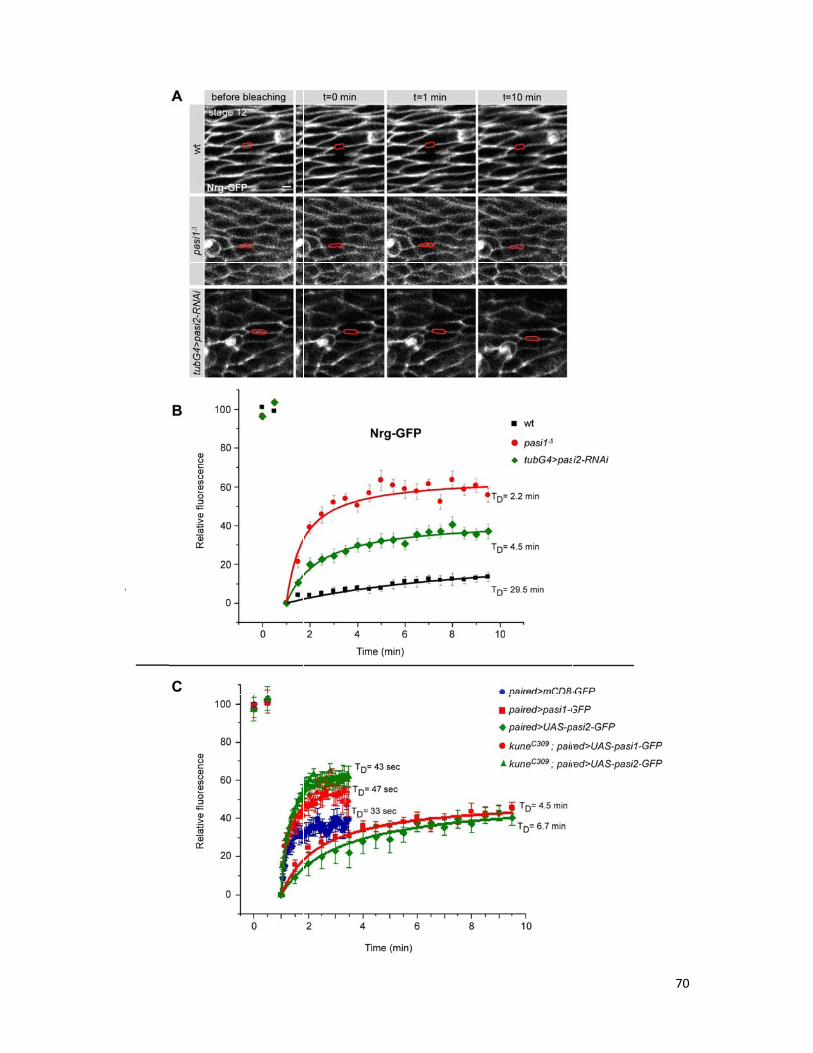
70
71
Figure 26. Pasiflora proteins are core components of the SJ. (A-B) pasiflora genes are required
for SJ complex formation. (A) Single confocal sections of lateral epidermis of stage 15 embryos
expressing live Nrg-GFP. Nrg-GFP shows restricted mobility after photobleaching in the wt, but
diffuses fast to the bleached region in pasiflora mutants. Bleached membranes are marked in red.
Scale bars are 5 µm. (B) Quantification of relative fluorescence of Nrg-GFP over time in wt and pasi
mutants, ±SEM. (C) Quantification of relative fluorescence of Pasiflora-GFP proteins over time
showing that in wt, Pasiflora proteins are more immobile compared to a membrane bound mCD8-
GFP. In kune mutants with disrupted SJs, Pasiflora proteins lose their restricted mobility. ±SEM. n=9-
17.
26A,B). Therefore, the behavior of Nrg-GFP in pasiflora mutants is similar to that
observed in mutants of core SJ components and proteins involved in complex
assembly. Together with their localization at SJ, these results argue that Pasiflora
proteins are core components required for formation of SJ complexes.
To further show that Pasiflora proteins are integral SJ components, we
analyzed their mobility within the membrane in the epidermis of stage 15 embryos.
To this end, we used paired-Gal4 and expressed GFP-tagged Pasiflora proteins in
epidermal stripes. As a control, we used membrane tagged mCD8-GFP and imaged
embryos at stage 14, at a time when SJs are not yet fully mature and diffusion within
the plasma membrane should not be impeded. mCD8-GFP recovers remarkably fast
(τD=33 sec) and its mobile fraction is 40%. In contrast, the recovery of Pasiflora1-
and Pasiflora2-GFP is significantly slower (τD=4.5 min and 6.7 min, respectively),
with their mobile fractions being 50%. The faster recovery rate of Pasiflora-GFP
proteins compared to Nrg-GFP could be due to overexpression conditions (Fig.
26C). Therefore, Pasiflora proteins are more immobile compared to other
transmembrane proteins, suggesting that they are part of a membrane complex.
Together with all our other data, this result strongly suggests that Pasiflora proteins
are core components of the SJ complex. To ultimately prove this conclusion, we
analyzed the mobility of Pasiflora-GFP proteins in epidermal cells of kuneC309
mutants that have disrupted SJs and observed that both proteins lose their restricted
mobility and diffuse very fast (Pasiflora1-GFP: τD=47 sec, mobile fraction 59%;
Pasiflora2-GFP: τD=43 sec, mobile fraction 67%) (Fig. 26C). Interestingly, their
mobile fractions are larger compared to that of mCD8-GFP in wt stage 14 epidermal
cells, further supporting the notion that SJs also serve a fence function that is likely
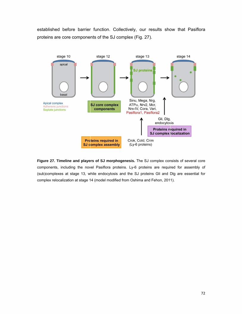
establis
protein
Figure 2
compon
(sub)com
complex
shed befo
ns are core
27. Timeline
ents, includ
mplexes at s
x relocalizatio
ore barrier
e compone
e and playe
ing the nov
stage 13, w
on at stage 1
r function.
nts of the S
rs of SJ mo
vel Pasiflora
while endocyt
14 (model mo
Collective
SJ comple
orphogenes
proteins. L
tosis and th
odified from
ely, our re
ex (Fig. 27)
sis. The SJ c
Ly-6 proteins
e SJ protein
Oshima and
esults sho
).
complex con
s are requir
ns Gli and D
Fehon, 201
ow that P
nsists of sev
red for asse
Dlg are esse
1).
72
Pasiflora
eral core
embly of
ential for
73
2.3. Mcr is a novel septate junction component
2.3.1 Introduction –Mcr and other thioester proteins
Thioester proteins (TEPs) appeared early in animal evolution and are found in such
diverse organisms as nematodes, insects, fish and mammals. In vertebrates, the
TEP family includes complement factors (C3, C4A, C4B and C5), secreted broad-
range protease inhibitors (α2-macroglobulin, PZP), and other less characterized
members (e.g. CD109, CPAMD8, Ovostatin-1 and -2). TEPs contain a hypervariable
region and most share the conserved motif GCGEQ, which allows for the formation
of a highly reactive intrachain thioester bond. The thioester binds pathogen surfaces
(in the case of complement factors) to mediate their opsonization and subsequent
phagocytosis or lysis, and to proteases (in the case of protease inhibitors) to mediate
their inhibition (Dodds and Law, 1998; Blandin and Levashina, 2004). The
mechanism of protease inhibition is mostly studied for α2-macroglobulin (α2M) and
involves steric hindrance. Upon proteolytic cleavage of a ‘’bait’’ region (that
corresponds to the hypervariable region) from the attacking protease, α2M
undergoes a conformational change that is linked to the cleavage of the thioester
bond. α2M then covalently binds the protease to the exposed thioester, collapses
about the protease and sterically shields its active site. The conformational change
also exposes a conserved C’-terminal recognition domain that mediates clearance of
the α2M-protease complex by receptor-mediated endocytosis (Borth, 1992).
Insects also encode several TEPs, which are thought to play roles in innate
immunity, however, the functions of individual members are poorly understood. The
Drosophila genome has six tep genes named tep1-tep6. Tep1-Tep4 are predicted
secreted proteins expressed in hemocytes and are thought to be involved in the
response to pathogens, while tep5 appears to be a pseudogene. Tep6 or
Macroglobulin complement-related (Mcr) is a diverged member of the family. While
Mcr has N’- and C’-terminal α2M domains, LDLα domain and N’-terminal signal
peptide also found in other TEPs, its thioester motif is mutated and it has a predicted
C’-terminal transmembrane domain (Fig. 30A), suggesting that it might play distinct
roles (Blandin and Levashina, 2004; Stroschein-Stevenson et al., 2006; Bou Aoun et
al., 2011). Nevertheless, Drosophila mcr was identified in an RNAi screen for genes
required for phagocytosis of the yeast Candida albicans and in a transcriptome
profiling of genes upregulated after infection with the alphavirus Sindbis (Stroschein-
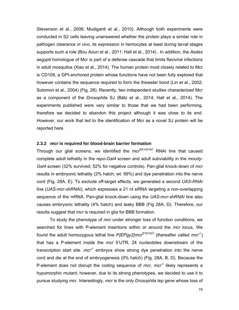
74
Stevenson et al., 2006; Mudiganti et al., 2010). Although both experiments were
conducted in S2 cells leaving unanswered whether the protein plays a similar role in
pathogen clearance in vivo, its expression in hemocytes at least during larval stages
supports such a role (Bou Aoun et al., 2011; Hall et al., 2014) . In addition, the Aedes
aegypti homologue of Mcr is part of a defense cascade that limits flaviviral infections
in adult mosquitos (Xiao et al., 2014). The human protein most closely related to Mcr
is CD109, a GPI-anchored protein whose functions have not been fully explored that
however contains the sequence required to form the thioester bond (Lin et al., 2002;
Solomon et al., 2004) (Fig. 28). Recently, two independent studies characterized Mcr
as a component of the Drosophila SJ (Bätz et al., 2014; Hall et al., 2014). The
experiments published were very similar to those that we had been performing,
therefore we decided to abandon this project although it was close to its end.
However, our work that led to the identification of Mcr as a novel SJ protein will be
reported here.
2.3.2 mcr is required for blood-brain barrier formation
Through our glial screens, we identified the mcrKK100197 RNAi line that caused
complete adult lethality in the repo-Gal4 screen and adult subviability in the moody-
Gal4 screen (32% survived; 52% for negative controls). Pan-glial knock-down of mcr
results in embryonic lethality (2% hatch; wt: 99%) and dye penetration into the nerve
cord (Fig. 28A, E). To exclude off-target effects, we generated a second UAS-RNAi
line (UAS-mcr-shRNAi), which expresses a 21 nt siRNA targeting a non-overlapping
sequence of the mRNA. Pan-glial knock-down using the UAS-mcr-shRNAi line also
causes embryonic lethality (4% hatch) and leaky BBB (Fig 28A, D). Therefore, our
results suggest that mcr is required in glia for BBB formation.
To study the phenotype of mcr under stronger loss of function conditions, we
searched for lines with P-element insertions within or around the mcr locus. We
found the adult homozygous lethal line P{EPgy2}mcrEY07421 (thereafter called mcr-/-)
that has a P-element inside the mcr 5’UTR, 24 nucleotides downstream of the
transcription start site. mcr-/- embryos show strong dye penetration into the nerve
cord and die at the end of embryogenesis (0% hatch) (Fig. 28A, B, D). Because the
P-element does not disrupt the coding sequence of mcr, mcr-/- likely represents a
hypomorphic mutant; however, due to its strong phenotypes, we decided to use it to
pursue studying mcr. Interestingly, mcr is the only Drosophila tep gene whose loss of
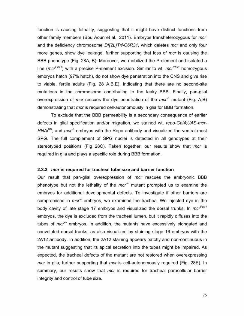
75
function is causing lethality, suggesting that it might have distinct functions from
other family members (Bou Aoun et al., 2011). Embryos transheterozygous for mcr-
and the deficiency chromosome Df(2L)Trf-C6R31, which deletes mcr and only four
more genes, show dye leakage, further supporting that loss of mcr is causing the
BBB phenotype (Fig. 28A, B). Moreover, we mobilized the P-element and isolated a
line (mcrPex1) with a precise P-element excision. Similar to wt, mcrPex1 homozygous
embryos hatch (97% hatch), do not show dye penetration into the CNS and give rise
to viable, fertile adults (Fig. 28 A,B,E), indicating that there are no second-site
mutations in the chromosome contributing to the leaky BBB. Finally, pan-glial
overexpression of mcr rescues the dye penetration of the mcr-/- mutant (Fig. A,B)
demonstrating that mcr is required cell-autonomously in glia for BBB formation.
To exclude that the BBB permeability is a secondary consequence of earlier
defects in glial specification and/or migration, we stained wt, repo-Gal4;UAS-mcr-
RNAiKK, and mcr-/- embryos with the Repo antibody and visualized the ventral-most
SPG. The full complement of SPG nuclei is detected in all genotypes at their
stereotyped positions (Fig 28C). Taken together, our results show that mcr is
required in glia and plays a specific role during BBB formation.
2.3.3 mcr is required for tracheal tube size and barrier function
Our result that pan-glial overexpression of mcr rescues the embryonic BBB
phenotype but not the lethality of the mcr-/- mutant prompted us to examine the
embryos for additional developmental defects. To investigate if other barriers are
compromised in mcr-/- embryos, we examined the trachea. We injected dye in the
body cavity of late stage 17 embryos and visualized the dorsal trunks. In mcrPex1
embryos, the dye is excluded from the tracheal lumen, but it rapidly diffuses into the
tubes of mcr-/- embryos. In addition, the mutants have excessively elongated and
convoluted dorsal trunks, as also visualized by staining stage 16 embryos with the
2A12 antibody. In addition, the 2A12 staining appears patchy and non-continuous in
the mutant suggesting that its apical secretion into the tubes might be impaired. As
expected, the tracheal defects of the mutant are not restored when overexpressing
mcr in glia, further supporting that mcr is cell-autonomously required (Fig. 28E). In
summary, our results show that mcr is required for tracheal paracellular barrier
integrity and control of tube size.
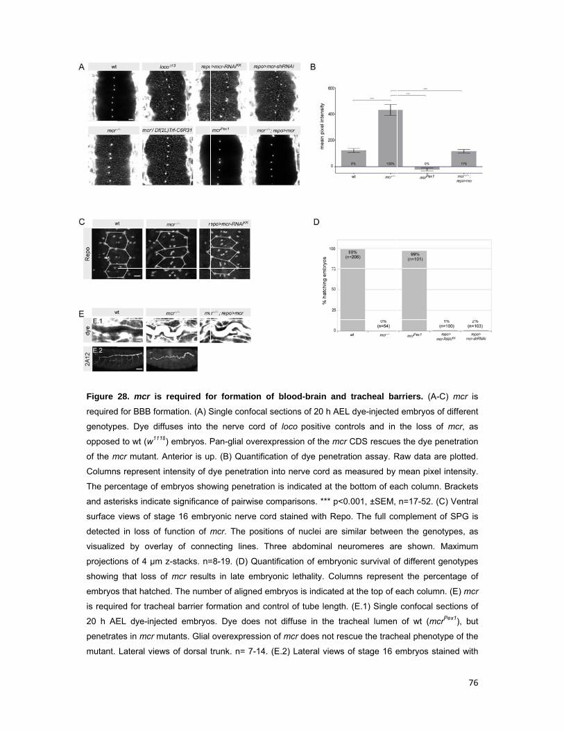
Figure
required
genotyp
opposed
of the m
Columns
The perc
and aste
surface
detected
visualize
projectio
showing
embryos
is requir
20 h AE
penetrat
mutant.
28. mcr is
d for BBB form
es. Dye diff
d to wt (w1118
mcr mutant. A
s represent i
centage of e
erisks indicat
views of sta
d in loss of
ed by overla
ons of 4 µm
g that loss o
s that hatche
red for trache
EL dye-injec
tes in mcr m
Lateral view
required fo
mation. (A) S
fuses into th8) embryos. P
Anterior is up
intensity of d
embryos sho
te significanc
age 16 embr
function of m
ay of conne
z-stacks. n=
of mcr result
ed. The numb
eal barrier fo
cted embryo
utants. Glial
ws of dorsal t
or formation
Single confoc
he nerve co
Pan-glial ove
p. (B) Quant
dye penetrat
wing penetra
ce of pairwis
ryonic nerve
mcr. The po
ecting lines.
=8-19. (D) Q
ts in late em
ber of aligne
ormation and
s. Dye does
overexpress
trunk. n= 7-1
n of blood-
cal sections
ord of loco p
erexpression
tification of d
ion into nerv
ation is indic
se compariso
cord stained
ositions of nu
. Three abd
uantification
mbryonic leth
d embryos is
d control of t
s not diffuse
sion of mcr d
14. (E.2) Lat
-brain and
of 20 h AEL
positive cont
n of the mcr C
dye penetrat
ve cord as m
cated at the b
ons. *** p<0.
d with Repo
uclei are sim
dominal neu
of embryon
hality. Colum
s indicated a
tube length.
e in the trac
does not resc
teral views o
tracheal ba
dye-injected
trols and in
CDS rescues
ion assay. R
measured by
bottom of ea
001, ±SEM,
. The full co
milar betwee
uromeres ar
nic survival o
mns represen
at the top of e
(E.1) Single
cheal lumen
cue the trach
of stage 16 e
arriers. (A-C
d embryos of
the loss of
s the dye pe
Raw data are
mean pixel
ach column.
n=17-52. (C
omplement o
en the genot
re shown. M
of different ge
nt the perce
each column
confocal se
of wt (mcrP
heal phenoty
embryos stai
76
C) mcr is
f different
mcr, as
enetration
e plotted.
intensity.
Brackets
C) Ventral
of SPG is
types, as
Maximum
enotypes
entage of
n. (E) mcr
ections of
rPex1), but
ype of the
ined with

77
2A12. mcr mutants have over-elongated and convoluted dorsal trunks, as opposed to wt (mcrPex1).
Maximum projections of 20 µm z-stacks. n=10-12. Anterior is left and dorsal is up. Scale bars are 10
µm except for images with 2A12 staining where they are 40 µm.
2.3.4 mcr is required for localization of SJs
Due to the phenotypes of permeable blood-brain and tracheal paracellular barriers,
we hypothesized that SJs are compromised in the mcr-/- mutant. To visualize SJs in
the embryonic BBB, we used the endogenously expressed live markers Nrg-, Lac-,
and ATPα-GFP. In contrast to wt, where intact SJs are formed and all markers
continuously label the periphery of SPG, in mcr-/- embryos, SJs appear severely
disorganized and in many cases almost absent (Fig. 29A). Therefore, mcr is required
for SJ formation between SPG.
To examine whether SJ localization along the lateral membrane is disturbed
in mcr-/- embryos, we analyzed the hindgut and trachea columnar epithelia. In the
hindgut of stage 15 wt embryos (mcrPex1 or w1118), the SJs markers Nrg-GFP, Cora,
ATPα and FasIII accumulate at the apicolateral membrane, while in mcr-/- embryos,
they lose their restricted localization and spread basolaterally (Fig. 29C).
Interestingly, Nrg-GFP shows a differential behaviour and also spreads at the apical
membrane (arrow in Fig.29C). In addition, ATPα is not as affected and its phenotype
is highly variable and not fully penetrant; i.e some embryos show wt localization,
some a mixture of cells with restricted and dispersed localization, and others with
mislocalization in all cells. Similarly, in tracheal cells of stage 15 mcr-/- embryos,
FasIII loses its apicolateral accumulation and spreads at the basolateral membrane
compartment (Fig. 29B). In both trachea and hindgut, the localization of the apical
membrane determinant Crb is preserved, indicating that loss of mcr does not affect
the establishment or maintenance of cell polarity (Fig. 29B,C). Moreover, the levels
of SJ proteins appear reduced in mcr-/- embryos in all tissues observed. Collectively,
our results show that mcr is specifically required for the apicolateral localization of
SJs in the columnar epithelia of hindgut and trachea.
2.3.5 Mcr is a plasma membrane protein
The suite of phenotypes observed in mcr mutants is commonly associated with
disrupted SJs, therefore, we speculated that Mcr is a SJ component itself or is
involved in the assembly and/or trafficking of SJ complexes. Furthermore, the mcr
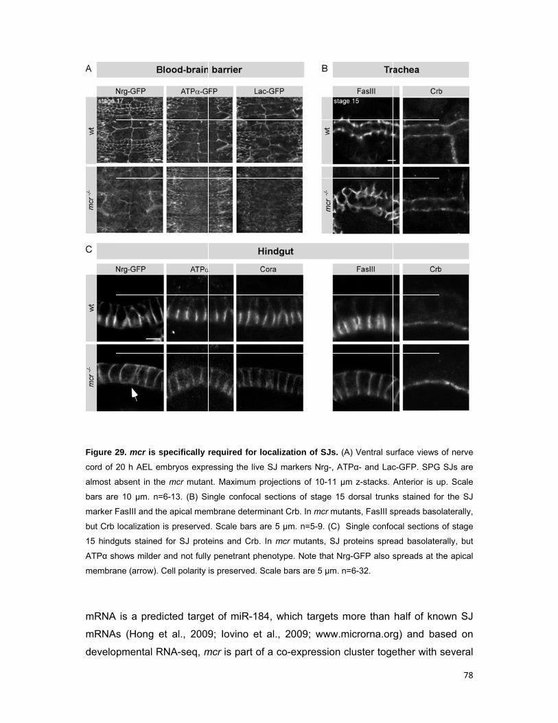
Figure 2
cord of 2
almost a
bars are
marker F
but Crb
15 hindg
ATPα sh
membra
mRNA
mRNA
develo
29. mcr is s
20 h AEL em
absent in the
e 10 µm. n=
FasIII and th
localization
guts stained
hows milder
ane (arrow). C
is a pred
s (Hong e
pmental R
specifically
mbryos expr
e mcr mutan
=6-13. (B) Si
e apical mem
is preserved
for SJ prote
and not fully
Cell polarity
icted targe
et al., 2009
RNA-seq, m
required for
ressing the li
nt. Maximum
ingle confoca
mbrane dete
d. Scale bars
eins and Cr
y penetrant p
is preserved
et of miR-1
9; Iovino e
mcr is part
r localizatio
ive SJ marke
m projections
al sections o
rminant Crb.
s are 5 µm.
b. In mcr m
phenotype. N
. Scale bars
184, which
et al., 200
of a co-ex
on of SJs. (A
ers Nrg-, AT
of 10-11 µm
of stage 15
. In mcr muta
n=5-9. (C) S
utants, SJ p
Note that Nrg
are 5 µm. n=
h targets m
09; www.m
xpression c
A) Ventral su
TPα- and Lac
m z-stacks.
dorsal trunk
ants, FasIII s
Single confo
proteins spre
g-GFP also s
=6-32.
more than
microrna.or
cluster toge
urface views
c-GFP. SPG
Anterior is u
ks stained fo
spreads baso
ocal sections
ead basolate
spreads at th
half of kno
rg) and ba
ether with
78
of nerve
G SJs are
up. Scale
or the SJ
olaterally,
of stage
rally, but
he apical
own SJ
ased on
several
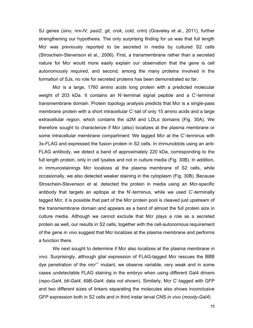
79
SJ genes (sinu, nrx-IV, pasi2, gli, crok, cold, crim) (Graveley et al., 2011), further
strengthening our hypothesis. The only surprising finding for us was that full length
Mcr was previously reported to be secreted in media by cultured S2 cells
(Stroschein-Stevenson et al., 2006). First, a transmembrane rather than a secreted
nature for Mcr would more easily explain our observation that the gene is cell
autonomously required, and second, among the many proteins involved in the
formation of SJs, no role for secreted proteins has been demonstrated so far.
Mcr is a large, 1760 amino acids long protein with a predicted molecular
weight of 203 kDa. It contains an N’-terminal signal peptide and a C’-terminal
transmembrane domain. Protein topology analysis predicts that Mcr is a single-pass
membrane protein with a short intracellular C’-tail of only 15 amino acids and a large
extracellular region, which contains the α2M and LDLα domains (Fig. 30A). We
therefore sought to characterize if Mcr (also) localizes at the plasma membrane or
some intracellular membrane compartment. We tagged Mcr at the C’-terminus with
3x-FLAG and expressed the fusion protein in S2 cells. In immunoblots using an anti-
FLAG antibody, we detect a band of approximately 220 kDa, corresponding to the
full length protein, only in cell lysates and not in culture media (Fig. 30B). In addition,
in immunostainings Mcr localizes at the plasma membrane of S2 cells, while
occasionally, we also detected weaker staining in the cytoplasm (Fig. 30B). Because
Stroschein-Stevenson et al. detected the protein in media using an Mcr-specific
antibody that targets an epitope at the N’-terminus, while we used C’-terminally
tagged Mcr, it is possible that part of the Mcr protein pool is cleaved just upstream of
the transmembrane domain and appears as a band of almost the full protein size in
culture media. Although we cannot exclude that Mcr plays a role as a secreted
protein as well, our results in S2 cells, together with the cell-autonomous requirement
of the gene in vivo suggest that Mcr localizes at the plasma membrane and performs
a function there.
We next sought to determine if Mcr also localizes at the plasma membrane in
vivo. Surprisingly, although glial expression of FLAG-tagged Mcr rescues the BBB
dye penetration of the mcr-/- mutant, we observe variable, very weak and in some
cases undetectable FLAG staining in the embryo when using different Gal4 drivers
(repo-Gal4, btl-Gal4, 69B-Gal4; data not shown). Similarly, Mcr C’-tagged with GFP
and two different sizes of linkers separating the molecules also shows inconclusive
GFP expression both in S2 cells and in third instar larval CNS in vivo (moody-Gal4)
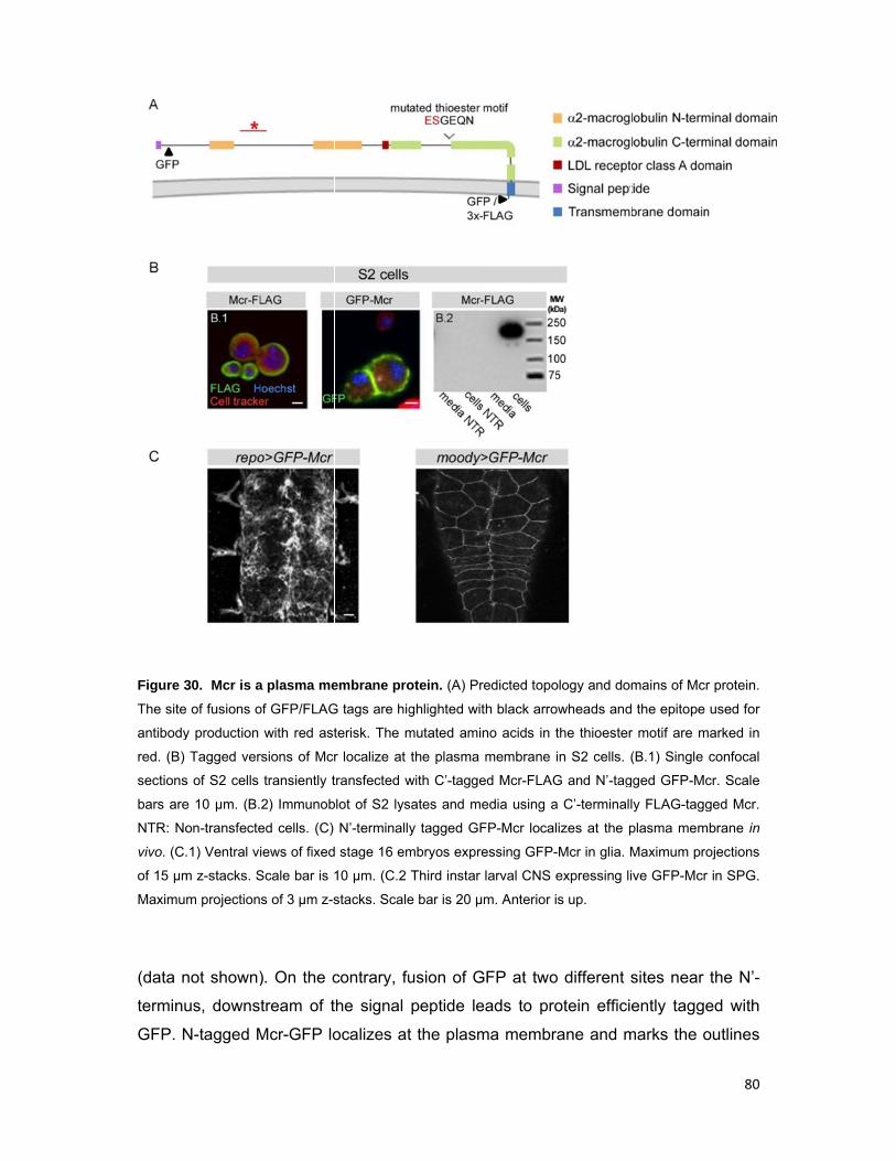
Figure 3
The site
antibody
red. (B)
sections
bars are
NTR: No
vivo. (C.
of 15 µm
Maximu
(data n
terminu
GFP. N
30. Mcr is a
of fusions o
y production
Tagged ver
s of S2 cells
e 10 µm. (B.
on-transfecte
.1) Ventral vi
m z-stacks. S
m projection
not shown)
us, downs
N-tagged M
a plasma me
of GFP/FLAG
with red ast
rsions of Mcr
transiently t
2) Immunob
ed cells. (C)
iews of fixed
Scale bar is 1
s of 3 µm z-s
). On the c
tream of t
Mcr-GFP lo
embrane pro
G tags are hig
terisk. The m
r localize at
transfected w
blot of S2 lys
N’-terminall
d stage 16 em
10 µm. (C.2
stacks. Scale
contrary, fu
the signal
ocalizes at
otein. (A) Pr
ghlighted wit
mutated amin
the plasma
with C’-tagge
sates and me
y tagged GF
mbryos expre
Third instar
e bar is 20 µ
usion of G
peptide le
t the plasm
redicted topo
th black arro
no acids in t
membrane i
ed Mcr-FLAG
edia using a
FP-Mcr local
essing GFP-
larval CNS e
m. Anterior i
GFP at two
eads to pro
ma membra
ology and do
owheads and
the thioester
in S2 cells. (
G and N’-tag
a C’-terminall
izes at the p
-Mcr in glia. M
expressing li
s up.
o different s
otein effici
ane and m
mains of Mc
d the epitope
r motif are m
(B.1) Single
gged GFP-M
ly FLAG-tagg
plasma mem
Maximum pr
ive GFP-Mcr
sites near
ently tagg
marks the o
80
r protein.
used for
marked in
confocal
cr. Scale
ged Mcr.
mbrane in
rojections
r in SPG.
the N’-
ed with
outlines
81
of glia in the embryo (repo-Gal4) and SPG in third instar larval CNS (moody-Gal4)
(Fig. 30C). Therefore, our in vivo results show that Mcr localizes at the plasma
membrane. In addition, our findings suggest that tagging of Mcr at the C’-tail might
sterically inhibit GFP from fluorescing.
2.3.6 mcr is expressed in SJ-forming epithelia and glia
To characterize the expression pattern of mcr during embryogenesis, we performed
RNA in situ hybridization in wt embryos using antisense mcr probes. Expression is
first detected at stage 10 in tracheal placodes and at stage 11 in the hindgut. During
stages 12-16 trachea, foregut, hindgut, epidermis and salivary glands are labeled,
but not the endodermally-derived midgut. At stage 16 embryos all insulating glia of
the CNS and PNS are marked, i.e. SPG, channel and peripheral glia (Fig. 31A).
Therefore mcr is expressed in SJ-forming embryonic epithelia and glia.
To study the distribution of the Mcr protein we raised a polyclonal antibody
against a non-conserved, 118 amino acids long peptide (Fig. 30A). Similar to the
published data (Bätz et al., 2014; Hall et al., 2014), on preliminary immunoblots of
stage 16 wt embryonic extracts, we detect a strong band of approximately 240 kDa
corresponding to full length Mcr, as well as bands of lower molecular weight, which
might represent degradation products or processed forms. The protein levels appear
strongly reduced and almost absent in extracts of mcr-/- and mcr-/Df(2L)Trf-C6R31
embryos, respectively and increased in embryos with pan-glial (repo-Gal4) and
ubiquitous (tub-Gal4) mcr overexpression, demonstrating that the antibody is specific
for Mcr (data not shown). To further check its specificity, we stained embryos
ectopically expressing mcr in epidermal stripes using the paired-Gal4 driver (Fig.
31B.3). In wt, embryos, the Mcr antibody stains the membranes of ectodermally-
derived epithelia and PNS glia and recapitulates most of the expression revealed by
RNA in situ (Fig. 31B.1-B.2). Collectively, our RNA and protein expression results
show that mcr is expressed in embryonic epithelia and glia that form insulating SJs.
2.3.7 Mcr localizes at the SJ and its localization depends on other complex
components
To examine if Mcr localizes at the SJ, we stained wt embryos with the Mcr and Cora
antibodies and focused on the columnar tracheal and hindgut epithelia. We observed
that Mcr colocalizes with Cora and exhibits the dynamic expression pattern of SJ
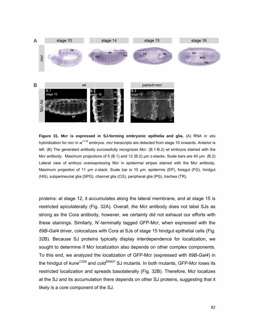
Figure
hybridiza
left. (B)
Mcr anti
Lateral
Maximu
(HG), su
protein
restrict
strong
these s
69B-Ga
32B).
sought
To this
the hin
restrict
at the S
likely is
31. Mcr is
ation for mcr
The generat
body. Maxim
view of em
m projection
ubperineurial
ns: at stage
ted apicola
as the Co
stainings.
al4 driver,
Because S
t to determ
s end, we
dgut of ku
ted localiza
SJ and its
s a core co
expressed
r in w1118 emb
ted antibody
mum project
bryo overex
n of 11 µm
glia (SPG),
e 12, it acc
aterally (Fig
ora antibod
Similarly,
colocalize
SJ protein
mine if Mcr
analyzed t
neC309 and
ation and s
accumulat
omponent o
in SJ-form
bryos. mcr tr
y successfull
ions of 5 (B.
xpressing Mc
z-stack. Sca
channel glia
cumulates
g. 32A). O
dy, howeve
N’-termina
s with Cor
ns typically
localizatio
the localiza
d coldf05607
spreads ba
tion there d
of the SJ.
ming embry
ranscripts are
y recognizes
1) and 12 (B
cr in epider
ale bar is 10
a (CG), perip
along the
Overall, the
er, we cert
ally tagged
ra at SJs of
y display
on also dep
ation of G
SJ mutant
asolaterally
depends o
yonic epithe
e detected fr
s Mcr. (B.1-B
B.2) µm z-sta
rmal stripes
0 µm. epide
heral glia (PG
lateral me
e Mcr antib
tainly did n
d GFP-Mcr
f stage 15
interdepen
pends on o
FP-Mcr (ex
ts. In both
y (Fig. 32B
on other SJ
elia and gl
rom stage 10
B.2) wt embr
acks. Scale b
stained wit
rmis (EP), f
G), trachea (
embrane, a
body does
not exhaus
r, when ex
hindgut ep
ndence for
other comp
xpressed w
mutants, G
B). Therefo
J proteins,
ia. (A) RNA
0 onwards. A
ryos stained
bars are 40 µ
h the Mcr a
foregut (FG)
(TR).
and at stag
not label
st our effo
xpressed w
pithelial ce
r localizati
plex comp
with 69B-G
GFP-Mcr lo
ore, Mcr lo
suggestin
82
A in situ
Anterior is
with the
µm. (B.2)
antibody.
, hindgut
ge 15 is
SJs as
rts with
with the
ells (Fig.
ion, we
onents.
Gal4) in
oses its
ocalizes
g that it
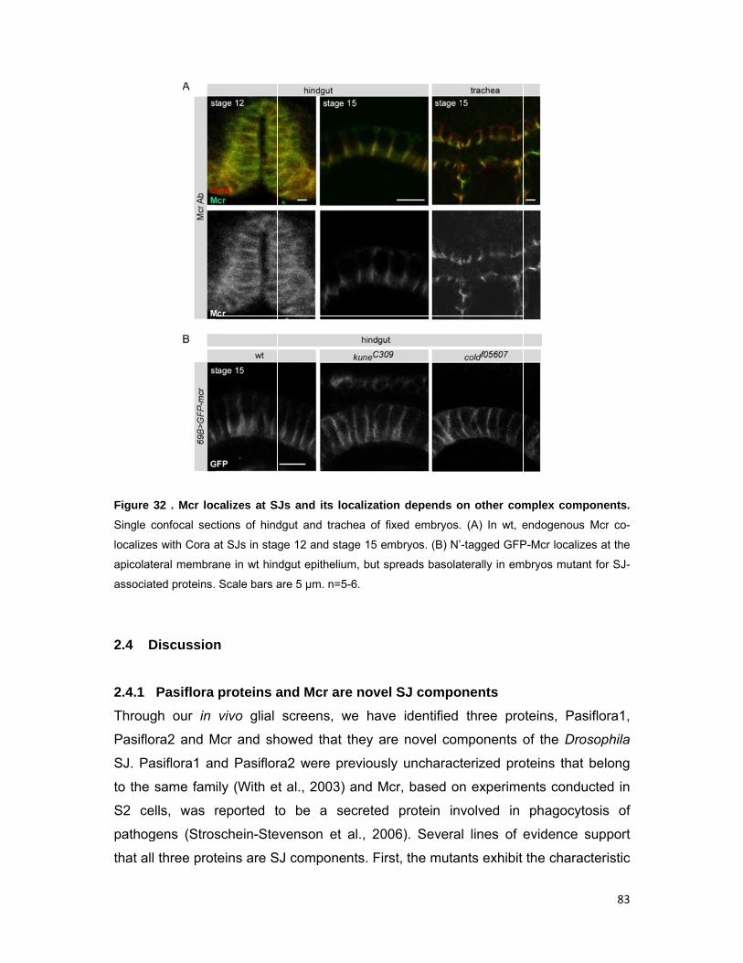
Figure 3
Single c
localizes
apicolate
associat
2.4 D
2.4.1
Throug
Pasiflo
SJ. Pa
to the
S2 ce
pathog
that all
32 . Mcr loc
confocal sec
s with Cora a
eral membra
ted proteins.
Discussion
Pasiflora
gh our in
ora2 and M
asiflora1 an
same fam
lls, was r
gens (Stros
three prot
calizes at S
ctions of hind
at SJs in sta
ane in wt hin
Scale bars a
n
proteins a
vivo glial
Mcr and sh
nd Pasiflor
ily (With e
reported t
schein-Ste
teins are S
Js and its l
dgut and tra
ge 12 and s
dgut epitheli
are 5 µm. n=
and Mcr a
screens,
howed that
ra2 were p
t al., 2003
to be a s
evenson et
SJ compon
localization
achea of fixe
tage 15 emb
ium, but spre
=5-6.
are novel S
we have
t they are
previously
3) and Mcr
secreted p
t al., 2006
ents. First,
depends o
ed embryos.
bryos. (B) N’-
eads basolat
SJ compo
identified
novel com
uncharact
r, based on
protein inv
6). Severa
, the mutan
on other com
(A) In wt, e
-tagged GFP
terally in em
nents
three pro
mponents o
terized pro
n experime
volved in
l lines of
nts exhibit
mplex comp
endogenous
P-Mcr localiz
mbryos mutan
oteins, Pas
of the Dro
oteins that
ents condu
phagocyt
evidence s
the charac
83
ponents.
Mcr co-
zes at the
nt for SJ-
siflora1,
osophila
belong
ucted in
osis of
support
cteristic
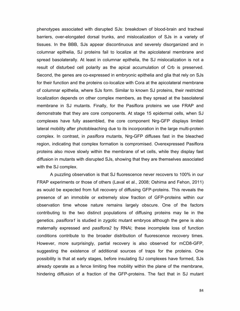
84
phenotypes associated with disrupted SJs: breakdown of blood-brain and tracheal
barriers, over-elongated dorsal trunks, and mislocalization of SJs in a variety of
tissues. In the BBB, SJs appear discontinuous and severely disorganized and in
columnar epithelia, SJ proteins fail to localize at the apicolateral membrane and
spread basolaterally. At least in columnar epithelia, the SJ mislocalization is not a
result of disturbed cell polarity as the apical accumulation of Crb is preserved.
Second, the genes are co-expressed in embryonic epithelia and glia that rely on SJs
for their function and the proteins co-localize with Cora at the apicolateral membrane
of columnar epithelia, where SJs form. Similar to known SJ proteins, their restricted
localization depends on other complex members, as they spread at the basolateral
membrane in SJ mutants. Finally, for the Pasiflora proteins we use FRAP and
demonstrate that they are core components. At stage 15 epidermal cells, when SJ
complexes have fully assembled, the core component Nrg-GFP displays limited
lateral mobility after photobleaching due to its incorporation in the large multi-protein
complex. In contrast, in pasiflora mutants, Nrg-GFP diffuses fast in the bleached
region, indicating that complex formation is compromised. Overexpressed Pasiflora
proteins also move slowly within the membrane of wt cells, while they display fast
diffusion in mutants with disrupted SJs, showing that they are themselves associated
with the SJ complex.
A puzzling observation is that SJ fluorescence never recovers to 100% in our
FRAP experiments or those of others (Laval et al., 2008; Oshima and Fehon, 2011)
as would be expected from full recovery of diffusing GFP-proteins. This reveals the
presence of an immobile or extremely slow fraction of GFP-proteins within our
observation time whose nature remains largely obscure. One of the factors
contributing to the two distinct populations of diffusing proteins may lie in the
genetics. pasiflora1 is studied in zygotic mutant embryos although the gene is also
maternally expressed and pasiflora2 by RNAi; these incomplete loss of function
conditions contribute to the broader distribution of fluorescence recovery times.
However, more surprisingly, partial recovery is also observed for mCD8-GFP,
suggesting the existence of additional sources of traps for the proteins. One
possibility is that at early stages, before insulating SJ complexes have formed, SJs
already operate as a fence limiting free mobility within the plane of the membrane,
hindering diffusion of a fraction of the GFP-proteins. The fact that in SJ mutant
85
backgrounds the mobile fractions of Pasiflora-GFP proteins are larger compared to
that of mCD8-GFP in wt epidermal cells further supports such a fence function.
2.4.2 Potential roles of Pasiflora proteins
Pasiflora1 and Pasiflora2 are predicted to be small proteins with four transmembrane
domains, two extracellular loops of which the first is bigger, and both termini located
intracellularly. Although the proteins lack putative conserved motifs that can classify
them as orthologues of any known proteins and thus define a distinct family, in a
wider context, they show structural similarity to a large superfamily of small tetra-
spanning membrane glycoproteins. The superfamily includes proteins that play roles
in specialized cell contacts, such as TJ-associated claudins, PMP22/EMP/MP20
proteins, voltage-gated calcium channel γ subunits (CACNGs), TMEM47 family
members, clarins, tetraspanins and even the larger gap junction proteins connexins
and innexins (Adato et al., 2002; Simske, 2013) . Based on structural and topological
similarity and the shared role as occluding junction components, Pasiflora proteins
mostly resemble claudins. Claudins have been shown to oligomerize homo- and
heterophilically on the same cell and homo- and hetero-typically on adjacent cells
with heterophilic oligomerization appearing to be more frequent than heterotypic
binding (Koval, 2013). In addition, in electron micrographs, both TJs and SJs appear
as a set of linear arrays, and vertebrate and invertebrate claudins may contribute to
the formation of these structures. An important question will be to determine whether
Pasiflora proteins can interact with each other in a similar fashion and together with
claudins build the stacks of regularly spaced SJ septa that span the intercellular
space. It is also tempting to speculate that similarly to vertebrate claudins, the first
extracellular loop of Pasiflora proteins participates in regulating tightness of SJs.
However, such functions have not been demonstrated for Drosophila claudins either,
and Drosophila SJs do not contain fusions of adjacent cell membranes like TJs, but
rather maintain a separation across the paracellular space at the junction.
Alternatively, Pasiflora1 and Pasiflora2 might associate with each other to organize
and build on the membrane an interacting network of SJ proteins. Tetraspanins play
such roles in organizing other proteins into multimolecular membrane microdomains,
called the tetraspanin web (Fradkin et al., 2002; Charrin et al., 2009). Finally,
Pasiflora proteins might be palmitoylated; one such site is predicted for Pasiflora1 in
position 7 (http://csspalm.biocuckoo.org). In claudins and tetraspanins, palmitoylation

86
has been shown to promote their partitioning into membrane microdomains and
assembly into higher order structures (Findley and Koval, 2009). In any case, the
highly transmembrane nature of Pasiflora1 and Pasiflora2 suggests a role for the
proteins as central structural subunits of the SJ.
Pasiflora1 and Pasiflora2 belong to a previously uncharacterized family of
four-pass membrane proteins conserved in Drosophila and Anopheles. The fly
encodes nine family members, albeit distantly related (With et al., 2003). We
demonstrated that Pasiflora proteins are expressed in embryonic epithelia and glia
and act non-redundantly in SJ formation. So far, little is known about the expression
of other family members and there are no data as to their function or subcellular
localization. Interestingly, during epithelial morphogenesis large families of junctional
proteins, such as the tetraspan claudins and innexins and the single-pass cadherins
are often used in a tissue-specific manner to generate epithelia with diversified
properties. It would therefore be interesting to examine if more family members are
required for SJ formation in the same or different epithelia. Interestingly, fire exit, the
founding member, is expressed in exit and peripheral glia but has not been assigned
a function (With et al., 2003) and CG15098 is expressed in the embryonic midgut
(BDGP), which forms a different subtype of SJs, smooth SJs. Alternatively, the other
family members might not be associated with SJs but play distinct roles at cell-cell
contacts.
2.4.3 Mcr - Is there a link between epithelial barrier function and immunity?
We have reported a novel role for the TEP member Mcr as an integral plasma
membrane protein and SJ component. This finding is further substantiated by the
recent MS-based identification of Mcr along with many membrane-associated SJ
proteins as components of the Claudin complex (Jaspers et al., 2012). Currently, the
exact role of Mcr within the complex is unknown, however, based on its large
extracellular region and its very small intracellular tail, we hypothesize that Mcr plays
roles in organizing the extracellular components of the junction. Moreover, Mcr
molecules on opposing cells might homophilically interact similar to invertebrate
soluble α2M, which acts as a homodimer (Quigley and Armstrong, 1994). To
approach elucidating the function of Mcr within SJs, deletion constructs could be
generated to analyze the role of individual domains in barrier function. For example,
the C’-terminal domain of α2M is required for receptor-mediated endocytosis of the
87
α2M/protease complex and is conserved in Mcr. Since SJ morphogenesis also
involves complex endocytosis from the basolateral membrane, it would be interesting
to examine if the Mcr C’-terminal α2M domain is involved in this process.
Many TEPs are secreted proteins that play roles in innate immunity and
similarly, full-length Mcr was shown to be secreted by S2 cells and mediate the
clearance of pathogenic C.albicans (Stroschein-Stevenson et al., 2006). In our
experiments we were not able to detect C’-terminally tagged Mcr in S2 cell media.
However, Stroschein-Stevenson et al. used an Mcr-specific antibody that targets an
N’-terminal epitope, therefore it is possible that part of the Mcr protein pool is cleaved
just upstream of the transmembrane domain and appears as a band of almost the
full size in culture media. We also did not detect secreted Mcr in the embryonic
hemocoel or tracheal lumen in vivo, either with N’-tagged Mcr or Mcr-specific
antibody, suggesting that the majority of the protein is stably associated with the
plasma membrane. However, low levels of secreted Mcr might be present that are
poorly immobilized by fixation, whereas the membrane-bound locally concentrated
pool is readily detected by immunofluorescence. The notion that Mcr is proteolytically
processed is further supported by the presence of multiple Mcr-specific bands on our
preliminary immunoblots and those of others (Bätz et al., 2014; Hall et al., 2014).
Vertebrate α2M is also cleaved by proteases, which are then inhibited through
covalent thioester bond formation with α2M. Mcr has a mutated thioester motif and
presumably does not act as a protease inhibitor, however its hypervariable region is
expected to render the protein sensitive to cleavage. The Mcr cleavage products
might include both transmembrane and secreted species, which could carry out
distinct functions in vivo. Perhaps membrane-localized isoforms are part of the SJ
complex and mediate barrier function in epithelial cells and SPG, while secreted
isoforms by mcr-expressing hemocytes (Bou Aoun et al., 2011; Hall et al., 2014) are
involved in the anti-microbial response.
Another intriguing scenario is that SJ-forming cells express both
transmembrane and secreted Mcr isoforms, which play dual roles in barrier function
and immunity or that the transmembrane Mcr species is itself also involved in the
defense mechanism. Barrier epithelia frequently come into contact with
microorganisms and thus need to fulfill the important task of preventing their
penetration. To perform this function, barrier epithelia express antimicrobial peptides
that likely create a hostile environment for the attachment of microbes (Wagner et
88
al., 2008; Davis and Engström, 2012). SJs might also be actively involved in the
antimicrobial mechanism and in preventing pathogens from crossing epithelia, with
Mcr strategically placed to provide a first line of defense. Perhaps Mcr expressed on
the apicolateral membrane of epithelial cells, such as the epidermis, trachea and
hindgut, provides an immune function in compartments that are in direct contact with
the outside world. Similarly, smooth SJs in the midgut of adult flies are components
of the gut immune barrier and are required to maintain the subtle balance between
immune response to pathogenic bacteria and immune tolerance to the endogenous
flora (Bonnay et al., 2013). SJs in the BBB-forming SPG might similarly be involved
in the clearance of pathogens approaching the nervous system. In addition, from the
time the BBB is formed, circulating macrophages cannot enter the CNS and glia are
the principal phagocytes within the nervous system. Given that different glial
subtypes are able to perform phagocytosis in Drosophila, it is plausible to suggest
that SPG also perform this function. Indeed, among the candidates identified in our
SPG-specific moody-Gal4 screen, we also find other genes likely involved in
phagocytosis, such as beta-, zeta-COP, and nucampholin. Furthermore, it is
tempting to speculate that SPG SJs are actively involved in phagocytosis;
interestingly, the major type of engulfing glia in the injured brain, ensheathing glia
also form SJs (Kremer et al., in preparation). Intriguingly, the clearance of large
pathogens from the hemocoel in insect larvae also involves the formation of SJ-like
structres. When circulating hemocytes encounter an invader that is too large to
engulf, such as a parasitic wasp egg, they spread to form a multi-layered epithelium
surrounding the pathogen and enable melanization of the capsule. This
encapsulating epithelium has ultrastructural characteristics of SJs and expresses
Nrg and Cora (Russo et al., 1996; Williams et al., 2005; Williams, 2009). These
observations, together with the identification of Mcr as both a SJ protein and
component of the antimicrobial response highlight an interesting connection between
SJs, barrier function and immunity.
2.4.4 Open questions on SJs
Over the last two decades much progress has been made in understanding the
molecular composition of SJs, and over 20 components have been identified mainly
through genetic screens. However, most likely, SJs are molecularly still partially
defined. Neutral screens for SJ phenotypes, such as the easily identifiable
89
overelongated tracheal tubes, and biased approaches (sequence similarities, miR-
184 targets, mass spectrometry of SJ complex, co-expression data) will help identify
such candidates. Although the repertoire of SJ proteins will likely continue to be
growing, the field’s focus is moving towards additional aspects of SJs, such as their
involvement in functions other than barrier establishment and the pathways and
mechanisms governing their morphogenesis and maintenance.
In addition, a comprehensive biochemical and structural characterization of
the SJ complex is missing and nothing is known about its stoichiometry due to lack
of quantitative MS data. An emerging idea is that not all SJ proteins are as
interdependent as previously thought and that distinct subcomplexes exist within the
large, highly ordered, multi-protein complex. The observations by us and others
(Nelson et al., 2010; Oshima and Fehon, 2011; Hall et al., 2014) that in SJ mutants
the localization of other complex members is differentially affected and that the
fluorescence of GFP-tagged SJ proteins does not fully recover after photobleaching
further support this notion. Therefore, it will be of particular interest to characterize
the network of interactions bridging together SJ proteins and clarify if distinct
subcomplexes exist. So far, only few direct interaction partners have been reported,
mostly derived from studies of the mammalian paranode. Finally, surprisingly little is
known about the specific roles of individual SJ proteins within the complex.
The multi-protein composition of the SJ complex suggests an elaborate
mechanism for its biogenesis. The formation of intact junctions is a multi-step
process and likely requires tight and coordinated regulation of SJ proteins at different
levels, i.e. transcriptional and post-transcriptional regulation of expression, post-
translational modification, control of trafficking and assembly into (sub)complexes
and restricted membrane localization. The notion that SJ protein expression levels
are tightly co-regulated is supported by the observation that more than half of the
known SJ-encoding mRNAs are predicted targets of miR-184 (Hong et al., 2009;
Iovino et al., 2009; www.microrna.org). Importantly, SPG-specific overexpression of
miR-184 results in dramatic disruption of SJs, suggesting a central role of the
microRNA in (fine-)tuning SJ protein levels (Iovino and Gaul, unpublished data).
Such miRNA regulons, in which one miRNA targets many functionally related
mRNAs, have also been described in other contexts, e.g. miR-19b that regulates NF-
κB signaling, miR-124 that mediates endothelial recruitment and metastasis, and
Drosophila miR-8 that regulates multiple growth factor hormones produced by the fat
90
body (Gantier et al., 2012; Png et al., 2012; Lee et al., 2014). Furthermore, there is
evidence that SJ mRNAs, such as Drosophila nrx-IV and the vertebrate homologue
of nrg are subject to cell-type specific alternative splicing that generate glial and
neuronal isoforms. In Drosophila, nrg, as well as other SJ mRNAs also have multiple
isoforms, while nrx-IV and cora splicing is co-regulated by the RNA-binding protein
Held out wings (HOW) (Hortsch et al., 1990; Genova and Fehon, 2003; Basak et al.,
2007; Rodrigues et al., 2012).
Recently, FRAP experiments have succeeded in uncoupling the processes of
complex assembly and maturation into stable junctions and have started elucidating
the dynamics of SJ morphogenesis (Oshima and Fehon, 2011). Despite the
progress, we still lack a detailed understanding of SJ development, how the proteins
cluster to form complexes and which pathways control their maturation. Finally, the
maintenance of SJs remains obscure. SJs need to be stably maintained both in
developing epithelia, where the number, morphology and arrangement of cells
change due to morphogenetic processes, and in mature epithelia, where SJ proteins
might need to be replenished. In mature dividing epithelia, where cells are actively
rearranging contacts, the immobility of SJ proteins persists, indicating that the core
components might assemble into intracellular complexes, which are slowly trafficked
to the SJ (Oshima and Fehon, 2011). Future research will help decipher the
pathways and mechanisms regulating the different steps of SJ development from
transcription to stable membrane complexes.
91
3. Materials and methods
3.1 Genome-wide screening for adult viability
Through small scale preliminary screens, the exact number of parents and the
duration of egg lay were determined to ensure that the F1 generation will grow under
non-overcrowded, non-competitive conditions. In the primary screen, 5 repo-
Gal4/TM3 virgin females were crossed with 4 UAS-RNAi males; the parents were
discarded after 4 days of egg lay. repo-Gal4 is homozygous lethal and therefore the
stock retains the TM3 balancer. moody-Gal4 is homozygous viable, therefore at a
first step moody-Gal4 females were crossed at a large scale with D/TM3 males. 4
moody-Gal4/TM3 virgin females were crossed with 4 UAS-RNAi males and
discarded after 2 days of egg lay. In both screens, approximately 150 progeny
eclosed per cross and were scored at adult days 8-10. During the whole screening,
flies were reared at optimal and strictly controlled conditions of temperature and
humidity (25°C and 60%, respectively). For analysis of the screen results, GO terms
by Flybase were used (www.flybase.org).
3.2 Constructs
pasiflora constructs
Rescue constructs were generated by PCR amplification of the coding sequence
from the full length cDNA clones RE54605 (pasiflora1), and LD42595 (pasiflora2)
(Drosophila Genomics Resource Center, DGRC, Indiana, USA) (the latter not used
in this study) with primers containing 5′ NotI and 3′ XbaI restriction sites, and cloning
into the pJFRC2 and pJFRC4 vectors that contain 10x- and 3x-UAS, respectively
(Addgene). Tagged proteins were generated by in-frame fusion of a Drosophila-
optimized GFP (pJFRC14, Addgene) or 3x-FLAG to the C’-terminus, using the
Golden Gate BsaI-based method (Engler et al., 2008; Engler et al., 2009) and cloned
into the pMT vector that contains an inducible metallothionein promoter for S2 cell
expression (U. Gaul, unpublished). To separate the protein and tag, 8-15 amino
acids-long alanine-rich linkers were used. Pasiflora1-FLAG/GFP and Pasiflora2-
FLAG/GFP were amplified from pMT with primers containing 5′ NotI and 3′ XbaI
restriction sites and cloned into pJFRC2 and pJFRC4 vectors for in vivo expression.
Pasiflora2-GFP fusions overall showed stronger expression compared to Pasiflora1-
92
GFP; the 3x-UAS-pasiflora1-GFP constructs led to almost undetectable expression
with some drivers.
mcr constructs
The mcr rescue construct was generated by PCR amplification of the coding
sequence from the full length cDNA clone LD23292 (DGRC) with primers containing
5′ and 3′ NotI restriction sites and cloning into NotI-cut dephosphorylated pJFRC2
and pJFRC4. All tagged versions of Mcr were initially cloned into the pMT vector
(BsaI) for S2 cell expression and subsequently into pJFRC2 for in vivo expression
(NotI). Mcr was tagged at the C’-terminus with FLAG (15 amino acids linker), at the
C’-terminus with GFP and two different sizes of linkers (24 and 34 amino acids), and
at two different positions at the N’ terminus (after amino acid 25 and 35) without
linker. Only N’-terminally tagged constructs showed in vivo expression of the tag. For
faster cloning of the long mcr CDS, for some of the constructs mcr was amplified as
two fragments that were ligated with the BsaI method, which does not leave extra
nucleotides. The primers were designed so that they introduce silent mutations in the
internal BsaI recognition site of mcr.
RNAi constructs
The shRNAi constructs for pasiflora2 and mcr were generated using the design by Ni
et al., 2011 by inserting an inverted repeat into NheI/EcoRI-cut pWalium20 vector
that contains 10x-UAS (Transgenic RNAi Project, TRiP, Harvard Medical School).
Transcription of the inverted repeat generates 21 nt long siRNAs. The shRNAs were
designed using the DSIR algorithm (http://biodev.cea.fr/DSIR/DSIR.html) and their
sequences were: For pasiflora2: sense strand: TACAATGTGATTATGGTGCTC,
antisense strand: GAGCACCATAATCACATTGTA; for mcr: sense strand:
TTTGACAAGAACATCACGCTA, antisense strand: TAGCGTGATGTTCTTGTCAAA.
All constructs were sequenced.
3.3 Fly strains
The following fly strains were obtained from published sources: repo-Gal4 (V. Auld),
moody-Gal4 (Schwabe et al., 2005), tubulin-Gal4 (E. Arama), GFP traps NrgG00305,
LacG00044, and ATPαG00109 (W. Chia), UAS-mCD8-GFP (L. Luo), Df(3R)BSC566,
93
Df(3R)ED5785, Df(2L)Trf-C6R31, P{EPgy2}mcrEY07421, nrx-IV4304, nrgI4, kuneC309,
megaG0012, coldf05607, crokKG06053a, 69B-Gal4, paired-Gal4 and breathless-Gal4
(Bloomington Drosophila Stock Center, BDSC, Indiana, USA). The glial screens
were performed using the KK (phi3C1) library obtained from the Vienna Drosophila
Resource Center (VDRC, Austria). The lines UAS-dicer2, pasiflora1KK102223,
pasiflora2KK105806, pasiflora2GD43952, mcrKK100197, lkb1KK108356 and CG7379KK103928,
were further used in our studies (VDRC). w1118 was used as wt except if mentioned
otherwise.
For generation of transgenic lines we used the phiC31 integrase method and
inserted constructs in attP2 and attP40 docking sites (Groth et al., 2004; Markstein et
al., 2008; Pfeiffer et al., 2010). The pasiflora1∆ genomic mutant line was generated
by imprecise excision of the P-element of the P{EP}G4182 strain (BDSC).
pasiflora1∆ was sequenced and the deletion spans the region 17794826-17796435
and removes the entire pasiflora1 locus and 59 bp of the CG7379 3’UTR. The line
mcrPex1 was sequenced and has a precise excision of the P-element of the P{EPgy2}
mcrEY07421 strain (BDSC). For genotyping, mutant and transgenic lines were
balanced with Kr::GFP (Casso et al., 2000) or Dfd-YFP (G.Beitel) obtained from
BDSC. All strains were raised at 25°C.
3.4 Immunohistochemistry
For all experiments, embryos were raised at 25°C and staged based on gut
morphology (according to Atlas of Drosophila development by Hartenstein V.) except
if mentioned otherwise.
Immunohistochemistry of embryos was performed following standard procedures:
embryos were dechorionated for 5 min in 50% commercial bleach, fixed in 4%
formaldehyde in PBS/heptane for 20 min, and devitellinized by shaking in
methanol/heptane. Washes were performed in PBTw (PBS+0.1% Tween 20);
unspecific staining was blocked with BBTw (PBT+0.1% BSA+ 5% normal serum).
Primary and secondary antibodies were diluted in BBTw and incubated overnight at
4°C and 2h at RT, respectively. Stained embryos were mounted in 80% glycerol in
PBS+ 2% DABCO antifading agent.
94
Primary antibodies used were: mouse anti-Repo (1:5, Developmental Studies
Hybridoma Bank, DSHB), rabbit anti-GFP (1:100, Molecular Probes, Life
Technologies, mouse IgM 2A12 (1:5, DSHB), mouse anti-Cora (1:5, DSHB), mouse
anti-FasIII (1:5, DSHB), mouse anti-Crb (1:5, DSHB), rabbit anti-Mcr (1:50, our
generated antibody). Alexa- (Molecular Probes) and Cy- (Jackson ImmunoResearch)
conjugated secondary antibodies were used at dilutions of 1:400 and 1:200,
respectively.
3.5 Live imaging of embryos and larvae
For live imaging of BBB SJs, 20 h AEL embryos were dechorionated, rinsed,
mounted on coverslip coated with heptane glue and covered with halocarbon oil
(Huile 10S VOLTALEF). To subdue their movement, embryos were injected with
100mM KCN (2-3% of egg volume), and imaged 45 min post-injection. Third instar
larval cephalic complexes were dissected, mounted in PBS and imaged directly.
3.6 Confocal image acquisition and analysis
All confocal images were acquired using a Zeiss LSM 710 system and the ZEN
acquisition software (Carl Zeiss). Image analysis was performed using ImageJ
(National Institutes of Health) except if mentioned otherwise.
3.7 Dye penetration assay and quantification
For the dye penetration assay, embryos were aligned on agar, transferred on glue-
coated coverslip and covered with halocarbon oil. At 20 h AEL (visibility of tracheal
tubes and/or subtle movement were used as criteria for staging), a fluorescent dye
(Texas red coupled dextran, 10 kDa, 2.5 mM, Molecular probes) dissolved in
standard injection buffer was injected from posterior into the body cavity (needle
penetration into not more than 15% of egg length to avoid hitting the nerve cord)
(Schwabe et al., 2005). Penetration into or exclusion from the CNS or dorsal trunks
was followed 15 min after injection using the confocal microscope. Every genotype
was tested in parallel to negative and positive controls.
Dye penetration in the CNS was quantified using a home-written Definiens
script that automatically measures pixel intensity after excluding overexposed areas,
such as the body cavity and channels that run through the CNS. To quantify dye
penetration, mean pixel intensity was taken as readout value and the percentage of
95
embryos showing visible dye penetration was calculated. To assess significance,
one-way Anova was performed over all groups with Student-Newman-Keuls post hoc
test.
3.8 RNA in situ hybridization
Whole-mount in situ RNA hybridization on embryos was performed as previously
described (Lehmann and Tautz, 1994) with the following modifications: post-fix step
between embryo rehydration and proteinase K treatment was removed, incubation
with anti-digoxigenin antibodies was overnight at 4°C. Anti-sense probes were
generated by in vitro transcription from the full length clones RE54605 (pasiflora1),
LD42595 (pasiflora2) and LD23292 (mcr).
3.9 Embryonic viability assay
To measure lethality, stage 15 embryos were dechorionated, rinsed, mounted on
glue coated-coverslip, covered with halocarbon oil, and placed on agar plate facing a
pile of yeast. Embryos were followed during late embryogenesis and every 24 h for
the following three days and the stage they died was scored.
3.10 FRAP experiments and analysis
FRAP experiment
Embryos were dechorionated, rinsed, mounted on coverslips with glue, and covered
with halocarbon oil. Imaging and photobleaching were performed with a c-
Apochromat 40x/1.20 W Korr M27 objective. Two images were acquired before
photobleaching and then GFP was bleached using maximal output power of 488 nm
laser. The bleached membrane was located in the lateral epidermis and had a length
of approximately 3 μm. A time series of images was started immediately after
photobleaching, with one image every 30 sec for 10 min, except for paired-
Gal4;UAS-mCD8-GFP, kuneC309; paired-Gal4, UAS-Pasiflora1-GFP, and kuneC309;
paired-Gal4, UAS-Pasiflora2-GFP for which images were captured every 4 sec for 3
min.
Image Registration and analysis
Embryo movements are unavoidable and pose severe challenges for the analysis of
time-lapse recordings. We used a home-written Definiens script to correct for lateral
96
drift and non-linear distortions of the raw confocal images due to changes of cellular
shape. In brief, for a confocal stack of n images with index 1..n, a built-in image
registration algorithm was first applied to three reference images with rounded
indexes n/6, n/2 and 5n/6, respectively (the middle image was used as reference
image for registration). The remaining images were then registered with respect to
the reference image closest in index number. Given the strong embryo movements
and drift that we observed, this strategy ensured a more robust alignment compared
to a registration procedure based on only one reference image for the whole stack.
A second Definiens script was then used to automatically extract the
fluorescence intensity trajectories of the photobleached membrane regions. To
detect the photobleached region we applied to registered images a 2D-Gaussian
filter with a kernel size of 5x5x3 pixels, followed by an edge 3D filter. This filter is
sensitive to signal variations between successive time-lapse images, and is thus
ideal to detect the photobleached region that exhibits a strong decrease in
fluorescence intensity just after the photobleaching step. The average fluorescence
intensity in the identified region can then be extracted for each time point, and
normalized with respect to its maximal and minimal values at the time points before
and immediately after photobleaching, respectively.
FRAP data analysis
In a first approximation, the diffusion in the thin photobleached membrane can be
modeled by one-dimensional free diffusion. The experimental data were fitted to the
empirical formula given in Eq. 1, which agrees within 5% with the solution of the
diffusion equation in one dimension for recovery into an interval of zero intensity
(Ellenberg et al., 1997; Ellenberg and Lippincott-Schwartz, 1999)
� 1∙
.
with I(t) = intensity as a function of time; t0= time right after photobleaching; I(final) =
final intensity reached after complete recovery; τD = characteristic time of diffusion.
The fitting procedure was performed using Origin 8.5. We kept t0 constant,
and extracted I(final) and τD from the fitted curves. Mobile fractions were calculated
as ratios of fluorescence intensity in the bleached area just before photobleaching to
fluorescence intensity in the bleached after recovery of the fluorescence signal.
97
Another common approach used to analyze FRAP recovery curves is the
calculation of half-time (t1/2) as the time required for the bleached fluorescence to
recover to half of its maximum recovery value (Yguerabide et al., 1982; Oshima and
Fehon, 2011). We extracted t1/2 from exponential fits of the recovery curves and
found for Nrg-GFP t1/2= 0.7±0.1 min, 1.1±0.1 min and 7.6±5.4 min in pasiflora1∆,
tubulin-Gal4;UAS-pasiflora2-RNAi and wt embryos, respectively. For overexpressed
Pasiflora1- and Pasiflora2-GFP, we calculated t1/2= 0.9±0.1 min and 1.9±0.2 min,
respectively, while for the control membrane-bound mCD8-GFP t1/2= 11±1 sec. For
overexpressed Pasiflora1- and Pasiflora2-GFP in the kuneC309 mutant background,
we calculated t1/2= 0.31 min and 0.26 min, respectively. All these values are in the
same order of magnitude with both the characteristic times of diffusions τD calculated
using a one-dimensional free diffusion model and the results obtained by Oshima
and Fehon. Minor quantitative differences between our results and those of Oshima
and Fehon (e.g. mobile fractions of mCD8-GFP) might result from the usage of
different drivers (paired-Gal4 vs engrailed-Gal4) and pipelines of data analysis.
3.11 Production of antibodies
Pasiflora antibodies
For each protein two 15-16 amino acids-long peptides were synthetically generated
and their mixture was injected in rabbit and guinea pig (for Pasiflora1) and hen (for
Pasiflora2) (Eurogentec, Seraing, Belgium). The epitopes were selected based on
antigenicity and were: for Pasiflora1: SPLFETDIRSSMPVA, IIWSDNVRTGSYAVA,
and for Pasiflora2: NLHSKMSRSTRSVRI, STANSLAGSRPTTPHS. The sera, as well
as affinity-purified antibodies were tested by immunostainings in wt embryos in
various concentrations (including 1:2). The full length proteins were also cloned (see
protocol for production of Mcr antibody), but their expression in E.coli following
standard procedures was toxic to the bacteria.
Mcr antibody
To produce the Mcr-specific antibody, we PCR amplified nucleotides 1096-1446 from
the LD23292 cDNA clone with primers containing 5′ EcoRI and 3′ NotI restriction
sites and cloned it into pHAT-2 vector that contains a 6x-His tag for N’-terminal
tagging (EMBL protein expression and purification facility). The sequence amplified
encodes a non-conserved 116 amino acids long sequence corresponding to the
98
hypervariable region between the two N’-terminal α2M domains of Mcr (amino acids
366-482). The plasmid was transfected into E. coli BL21 Star (DE3) cells, protein
expression was induced with IPTG at 37°C and cells were harvested 4 h after
induction (time chosen after checking IPTG time series). The His-tagged Mcr peptide
was extracted from inclusion bodies (pellets), solubilized in binding buffer (100 mM
NaH2PO4, 10 mM Tris-Cl pH 8.0, 6 M GuHCl), and purified through Ni2+ affinity
chromatography (cOmplete HIS-Tag purification resin, Roche). Protein was dialyzed
against buffer with 100 mM NaH2PO4, 10 mM Tris-Cl, 3.5 M Urea, 10% glycerol,
0.05% NP-40 and injected in two rabbits (Eurogentec, Seraing, Belgium) and their
sera were used.
3.12 Cell culture and immunohistochemistry
S2 cells were cultured at 25°C in serum-free medium (Express Five, Gibco)
supplemented with L-glutamine, penicillin and streptomycin. 50.000 cells in 100 µl
were plated in 96-well poly-L-lysine coated plates for imaging (Greiner Bio-one) and
transfected with pMT vectors using FuGene HD reagent (Roche) 24h post-plating.
Cells were induced with 0.2-0.5 mM CuSO4 24h post-transfection, and imaged live or
fixed 24h post-induction. For immunohistochemistry, cells were fixed with 2%
paraformaldehyde for 25 min, post-fixed with 4% paraformaldehyde for 25 min,
permeabilized with PBT (PBS+0.1% Triton X-100) and stained following standard
procedures. Washes were performed with PBS and blocking with PBS+2% BSA+4%
normal serum. Primary monoclonal anti-FLAG antibody (Sigma) was incubated
overnight at 4°C (1:500) and secondary Alexa488 (Molecular Probes) 1h at RT
(1:500). To stain the cytoplasm and nucleus, cells were incubated at RT with 15 µM
Cell tracker (Molecular Probes) for 20 min and Hoechst 33342 for 5 min,
respectively.
3.13 Western blotting
Polyacrylamide SDS-PAGE gel electrophoresis, immunoblotting and
chemiluminescent detection were performed as previously described (Zhang and
Ward, 2011). 7.5% polyacrylamide gels were used for the separation. For Western
blots of embryonic extracts, 40 embryos were homogenized in RIPA buffer (150 Mm
NaCl, 1% Triton X-100, 0.5% sodium deoxycholate, 0.1% SDS, 50 Mm Tric-HCl,
pH=8.0) with protease inhibitor and 30 μg were loaded per sample. For Western

99
blots from S2 cells, 500.00 cells were plated in 24-well plates, transfected 24 h post-
plating with pMT vector and induced with 0.5 mM CuSO4 24 h post-transfection.
Cells and media were collected separately and cells were lysed using cOmplete
Lysis-M, EDTA-free (Roche). Immunoblots were hybridized with mouse anti-FLAG
(1:500, Sigma), rabbit anti-Mcr (1:1500, our generated antibody) and mouse anti-
actin (1:5000, Abcam).
100
References
Abbott, N.J. (1991). Permeability and transport of glial blood-brain barriers. Annals of the New York Academy of Sciences 633, 378-394. Abbott, N.J. (2013). Blood-brain barrier structure and function and the challenges for CNS drug delivery. Journal of inherited metabolic disease 36, 437-449. Abbott, N.J., Lane, N.J., and Bundgaard, M. (1986). The blood-brain interface in invertebrates. Annals of the New York Academy of Sciences 481, 20-42. Abbott, N.J., Ronnback, L., and Hansson, E. (2006). Astrocyte-endothelial interactions at the blood-brain barrier. Nature reviews Neuroscience 7, 41-53. Adato, A., Vreugde, S., Joensuu, T., Avidan, N., Hamalainen, R., Belenkiy, O., Olender, T., Bonne-Tamir, B., Ben-Asher, E., Espinos, C., et al. (2002). USH3A transcripts encode clarin-1, a four-transmembrane-domain protein with a possible role in sensory synapses. European journal of human genetics : EJHG 10, 339-350. Altenhein, B., Becker, A., Busold, C., Beckmann, B., Hoheisel, J.D., and Technau, G.M. (2006). Expression profiling of glial genes during Drosophila embryogenesis. Developmental biology 296, 545-560. Asano, A., Asano, K., Sasaki, H., Furuse, M., and Tsukita, S. (2003). Claudins in Caenorhabditis elegans: their distribution and barrier function in the epithelium. Current biology : CB 13, 1042-1046. Auld, V.J., Fetter, R.D., Broadie, K., and Goodman, C.S. (1995). Gliotactin, a novel transmembrane protein on peripheral glia, is required to form the blood-nerve barrier in Drosophila. Cell 81, 757-767. Awasaki, T., and Ito, K. (2004). Engulfing action of glial cells is required for programmed axon pruning during Drosophila metamorphosis. Current biology : CB 14, 668-677. Awasaki, T., Lai, S.L., Ito, K., and Lee, T. (2008). Organization and postembryonic development of glial cells in the adult central brain of Drosophila. The Journal of neuroscience : the official journal of the Society for Neuroscience 28, 13742-13753. Awasaki, T., Tatsumi, R., Takahashi, K., Arai, K., Nakanishi, Y., Ueda, R., and Ito, K. (2006). Essential role of the apoptotic cell engulfment genes draper and ced-6 in programmed axon pruning during Drosophila metamorphosis. Neuron 50, 855-867. Bachmann, A., Draga, M., Grawe, F., and Knust, E. (2008). On the role of the MAGUK proteins encoded by Drosophila varicose during embryonic and postembryonic development. BMC developmental biology 8, 55. Bainton, R.J., Tsai, L.T., Schwabe, T., DeSalvo, M., Gaul, U., and Heberlein, U. (2005). moody encodes two GPCRs that regulate cocaine behaviors and blood-brain barrier permeability in Drosophila. Cell 123, 145-156. Balda, M.S., and Matter, K. (2008). Tight junctions at a glance. Journal of cell science 121, 3677-3682. Banerjee, S., Bainton, R.J., Mayer, N., Beckstead, R., and Bhat, M.A. (2008). Septate junctions are required for ommatidial integrity and blood-eye barrier function in Drosophila. Developmental biology 317, 585-599. Banerjee, S., Pillai, A.M., Paik, R., Li, J., and Bhat, M.A. (2006). Axonal ensheathment and septate junction formation in the peripheral nervous system of Drosophila. The Journal of neuroscience : the official journal of the Society for Neuroscience 26, 3319-3329.
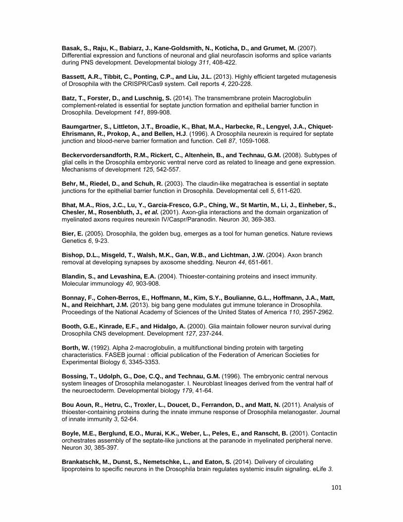
101
Basak, S., Raju, K., Babiarz, J., Kane-Goldsmith, N., Koticha, D., and Grumet, M. (2007). Differential expression and functions of neuronal and glial neurofascin isoforms and splice variants during PNS development. Developmental biology 311, 408-422. Bassett, A.R., Tibbit, C., Ponting, C.P., and Liu, J.L. (2013). Highly efficient targeted mutagenesis of Drosophila with the CRISPR/Cas9 system. Cell reports 4, 220-228. Batz, T., Forster, D., and Luschnig, S. (2014). The transmembrane protein Macroglobulin complement-related is essential for septate junction formation and epithelial barrier function in Drosophila. Development 141, 899-908. Baumgartner, S., Littleton, J.T., Broadie, K., Bhat, M.A., Harbecke, R., Lengyel, J.A., Chiquet-Ehrismann, R., Prokop, A., and Bellen, H.J. (1996). A Drosophila neurexin is required for septate junction and blood-nerve barrier formation and function. Cell 87, 1059-1068. Beckervordersandforth, R.M., Rickert, C., Altenhein, B., and Technau, G.M. (2008). Subtypes of glial cells in the Drosophila embryonic ventral nerve cord as related to lineage and gene expression. Mechanisms of development 125, 542-557. Behr, M., Riedel, D., and Schuh, R. (2003). The claudin-like megatrachea is essential in septate junctions for the epithelial barrier function in Drosophila. Developmental cell 5, 611-620. Bhat, M.A., Rios, J.C., Lu, Y., Garcia-Fresco, G.P., Ching, W., St Martin, M., Li, J., Einheber, S., Chesler, M., Rosenbluth, J., et al. (2001). Axon-glia interactions and the domain organization of myelinated axons requires neurexin IV/Caspr/Paranodin. Neuron 30, 369-383. Bier, E. (2005). Drosophila, the golden bug, emerges as a tool for human genetics. Nature reviews Genetics 6, 9-23. Bishop, D.L., Misgeld, T., Walsh, M.K., Gan, W.B., and Lichtman, J.W. (2004). Axon branch removal at developing synapses by axosome shedding. Neuron 44, 651-661. Blandin, S., and Levashina, E.A. (2004). Thioester-containing proteins and insect immunity. Molecular immunology 40, 903-908. Bonnay, F., Cohen-Berros, E., Hoffmann, M., Kim, S.Y., Boulianne, G.L., Hoffmann, J.A., Matt, N., and Reichhart, J.M. (2013). big bang gene modulates gut immune tolerance in Drosophila. Proceedings of the National Academy of Sciences of the United States of America 110, 2957-2962. Booth, G.E., Kinrade, E.F., and Hidalgo, A. (2000). Glia maintain follower neuron survival during Drosophila CNS development. Development 127, 237-244. Borth, W. (1992). Alpha 2-macroglobulin, a multifunctional binding protein with targeting characteristics. FASEB journal : official publication of the Federation of American Societies for Experimental Biology 6, 3345-3353. Bossing, T., Udolph, G., Doe, C.Q., and Technau, G.M. (1996). The embryonic central nervous system lineages of Drosophila melanogaster. I. Neuroblast lineages derived from the ventral half of the neuroectoderm. Developmental biology 179, 41-64. Bou Aoun, R., Hetru, C., Troxler, L., Doucet, D., Ferrandon, D., and Matt, N. (2011). Analysis of thioester-containing proteins during the innate immune response of Drosophila melanogaster. Journal of innate immunity 3, 52-64. Boyle, M.E., Berglund, E.O., Murai, K.K., Weber, L., Peles, E., and Ranscht, B. (2001). Contactin orchestrates assembly of the septate-like junctions at the paranode in myelinated peripheral nerve. Neuron 30, 385-397. Brankatschk, M., Dunst, S., Nemetschke, L., and Eaton, S. (2014). Delivery of circulating lipoproteins to specific neurons in the Drosophila brain regulates systemic insulin signaling. eLife 3.
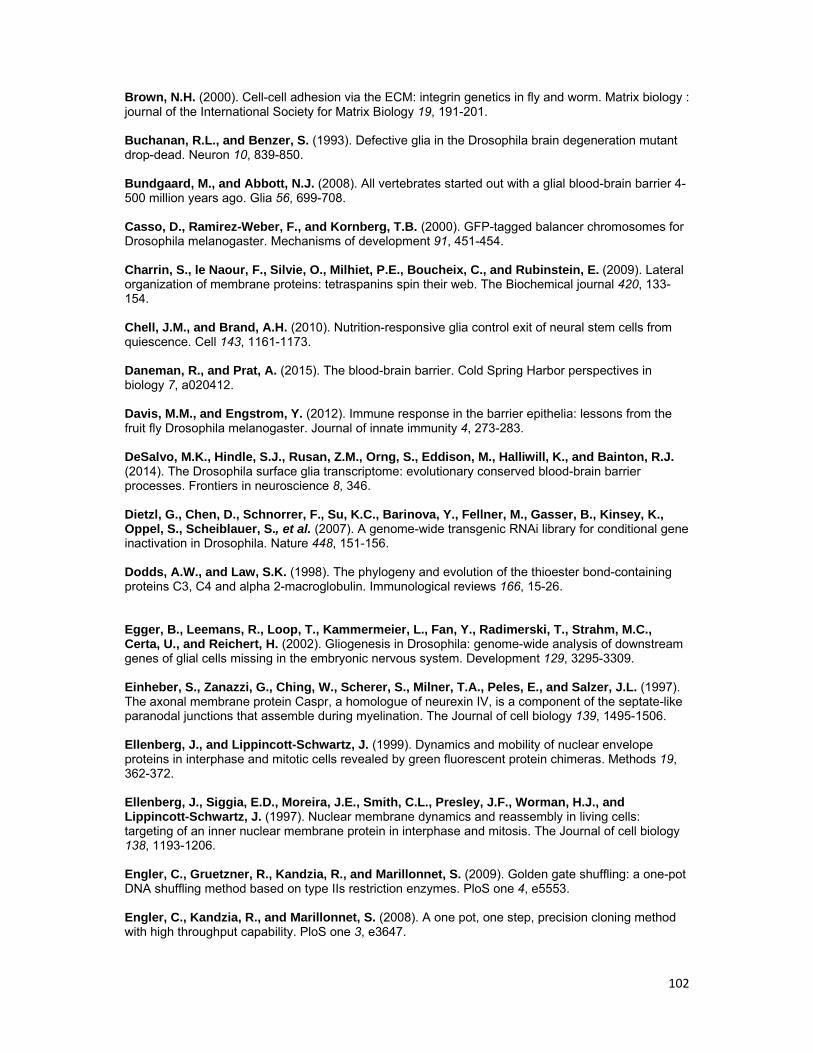
102
Brown, N.H. (2000). Cell-cell adhesion via the ECM: integrin genetics in fly and worm. Matrix biology : journal of the International Society for Matrix Biology 19, 191-201. Buchanan, R.L., and Benzer, S. (1993). Defective glia in the Drosophila brain degeneration mutant drop-dead. Neuron 10, 839-850. Bundgaard, M., and Abbott, N.J. (2008). All vertebrates started out with a glial blood-brain barrier 4-500 million years ago. Glia 56, 699-708. Casso, D., Ramirez-Weber, F., and Kornberg, T.B. (2000). GFP-tagged balancer chromosomes for Drosophila melanogaster. Mechanisms of development 91, 451-454. Charrin, S., le Naour, F., Silvie, O., Milhiet, P.E., Boucheix, C., and Rubinstein, E. (2009). Lateral organization of membrane proteins: tetraspanins spin their web. The Biochemical journal 420, 133-154. Chell, J.M., and Brand, A.H. (2010). Nutrition-responsive glia control exit of neural stem cells from quiescence. Cell 143, 1161-1173. Daneman, R., and Prat, A. (2015). The blood-brain barrier. Cold Spring Harbor perspectives in biology 7, a020412. Davis, M.M., and Engstrom, Y. (2012). Immune response in the barrier epithelia: lessons from the fruit fly Drosophila melanogaster. Journal of innate immunity 4, 273-283. DeSalvo, M.K., Hindle, S.J., Rusan, Z.M., Orng, S., Eddison, M., Halliwill, K., and Bainton, R.J. (2014). The Drosophila surface glia transcriptome: evolutionary conserved blood-brain barrier processes. Frontiers in neuroscience 8, 346. Dietzl, G., Chen, D., Schnorrer, F., Su, K.C., Barinova, Y., Fellner, M., Gasser, B., Kinsey, K., Oppel, S., Scheiblauer, S., et al. (2007). A genome-wide transgenic RNAi library for conditional gene inactivation in Drosophila. Nature 448, 151-156. Dodds, A.W., and Law, S.K. (1998). The phylogeny and evolution of the thioester bond-containing proteins C3, C4 and alpha 2-macroglobulin. Immunological reviews 166, 15-26. Egger, B., Leemans, R., Loop, T., Kammermeier, L., Fan, Y., Radimerski, T., Strahm, M.C., Certa, U., and Reichert, H. (2002). Gliogenesis in Drosophila: genome-wide analysis of downstream genes of glial cells missing in the embryonic nervous system. Development 129, 3295-3309. Einheber, S., Zanazzi, G., Ching, W., Scherer, S., Milner, T.A., Peles, E., and Salzer, J.L. (1997). The axonal membrane protein Caspr, a homologue of neurexin IV, is a component of the septate-like paranodal junctions that assemble during myelination. The Journal of cell biology 139, 1495-1506. Ellenberg, J., and Lippincott-Schwartz, J. (1999). Dynamics and mobility of nuclear envelope proteins in interphase and mitotic cells revealed by green fluorescent protein chimeras. Methods 19, 362-372. Ellenberg, J., Siggia, E.D., Moreira, J.E., Smith, C.L., Presley, J.F., Worman, H.J., and Lippincott-Schwartz, J. (1997). Nuclear membrane dynamics and reassembly in living cells: targeting of an inner nuclear membrane protein in interphase and mitosis. The Journal of cell biology 138, 1193-1206. Engler, C., Gruetzner, R., Kandzia, R., and Marillonnet, S. (2009). Golden gate shuffling: a one-pot DNA shuffling method based on type IIs restriction enzymes. PloS one 4, e5553. Engler, C., Kandzia, R., and Marillonnet, S. (2008). A one pot, one step, precision cloning method with high throughput capability. PloS one 3, e3647.
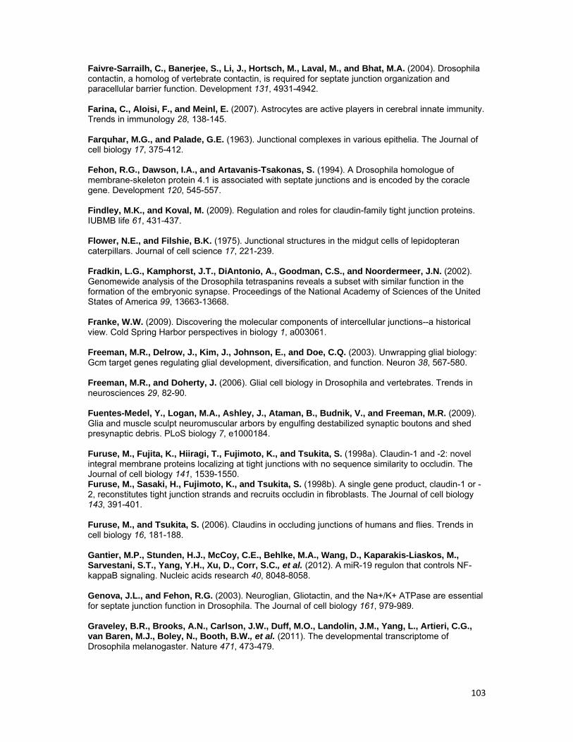
103
Faivre-Sarrailh, C., Banerjee, S., Li, J., Hortsch, M., Laval, M., and Bhat, M.A. (2004). Drosophila contactin, a homolog of vertebrate contactin, is required for septate junction organization and paracellular barrier function. Development 131, 4931-4942. Farina, C., Aloisi, F., and Meinl, E. (2007). Astrocytes are active players in cerebral innate immunity. Trends in immunology 28, 138-145. Farquhar, M.G., and Palade, G.E. (1963). Junctional complexes in various epithelia. The Journal of cell biology 17, 375-412. Fehon, R.G., Dawson, I.A., and Artavanis-Tsakonas, S. (1994). A Drosophila homologue of membrane-skeleton protein 4.1 is associated with septate junctions and is encoded by the coracle gene. Development 120, 545-557. Findley, M.K., and Koval, M. (2009). Regulation and roles for claudin-family tight junction proteins. IUBMB life 61, 431-437. Flower, N.E., and Filshie, B.K. (1975). Junctional structures in the midgut cells of lepidopteran caterpillars. Journal of cell science 17, 221-239. Fradkin, L.G., Kamphorst, J.T., DiAntonio, A., Goodman, C.S., and Noordermeer, J.N. (2002). Genomewide analysis of the Drosophila tetraspanins reveals a subset with similar function in the formation of the embryonic synapse. Proceedings of the National Academy of Sciences of the United States of America 99, 13663-13668. Franke, W.W. (2009). Discovering the molecular components of intercellular junctions--a historical view. Cold Spring Harbor perspectives in biology 1, a003061. Freeman, M.R., Delrow, J., Kim, J., Johnson, E., and Doe, C.Q. (2003). Unwrapping glial biology: Gcm target genes regulating glial development, diversification, and function. Neuron 38, 567-580. Freeman, M.R., and Doherty, J. (2006). Glial cell biology in Drosophila and vertebrates. Trends in neurosciences 29, 82-90. Fuentes-Medel, Y., Logan, M.A., Ashley, J., Ataman, B., Budnik, V., and Freeman, M.R. (2009). Glia and muscle sculpt neuromuscular arbors by engulfing destabilized synaptic boutons and shed presynaptic debris. PLoS biology 7, e1000184. Furuse, M., Fujita, K., Hiiragi, T., Fujimoto, K., and Tsukita, S. (1998a). Claudin-1 and -2: novel integral membrane proteins localizing at tight junctions with no sequence similarity to occludin. The Journal of cell biology 141, 1539-1550. Furuse, M., Sasaki, H., Fujimoto, K., and Tsukita, S. (1998b). A single gene product, claudin-1 or -2, reconstitutes tight junction strands and recruits occludin in fibroblasts. The Journal of cell biology 143, 391-401. Furuse, M., and Tsukita, S. (2006). Claudins in occluding junctions of humans and flies. Trends in cell biology 16, 181-188. Gantier, M.P., Stunden, H.J., McCoy, C.E., Behlke, M.A., Wang, D., Kaparakis-Liaskos, M., Sarvestani, S.T., Yang, Y.H., Xu, D., Corr, S.C., et al. (2012). A miR-19 regulon that controls NF-kappaB signaling. Nucleic acids research 40, 8048-8058. Genova, J.L., and Fehon, R.G. (2003). Neuroglian, Gliotactin, and the Na+/K+ ATPase are essential for septate junction function in Drosophila. The Journal of cell biology 161, 979-989. Graveley, B.R., Brooks, A.N., Carlson, J.W., Duff, M.O., Landolin, J.M., Yang, L., Artieri, C.G., van Baren, M.J., Boley, N., Booth, B.W., et al. (2011). The developmental transcriptome of Drosophila melanogaster. Nature 471, 473-479.
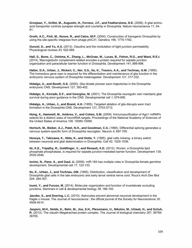
104
Grosjean, Y., Grillet, M., Augustin, H., Ferveur, J.F., and Featherstone, D.E. (2008). A glial amino-acid transporter controls synapse strength and courtship in Drosophila. Nature neuroscience 11, 54-61. Groth, A.C., Fish, M., Nusse, R., and Calos, M.P. (2004). Construction of transgenic Drosophila by using the site-specific integrase from phage phiC31. Genetics 166, 1775-1782. Gunzel, D., and Yu, A.S. (2013). Claudins and the modulation of tight junction permeability. Physiological reviews 93, 525-569. Hall, S., Bone, C., Oshima, K., Zhang, L., McGraw, M., Lucas, B., Fehon, R.G., and Ward, R.E.t. (2014). Macroglobulin complement-related encodes a protein required for septate junction organization and paracellular barrier function in Drosophila. Development 141, 889-898. Halter, D.A., Urban, J., Rickert, C., Ner, S.S., Ito, K., Travers, A.A., and Technau, G.M. (1995). The homeobox gene repo is required for the differentiation and maintenance of glia function in the embryonic nervous system of Drosophila melanogaster. Development 121, 317-332. Hidalgo, A., and Booth, G.E. (2000). Glia dictate pioneer axon trajectories in the Drosophila embryonic CNS. Development 127, 393-402. Hidalgo, A., Kinrade, E.F., and Georgiou, M. (2001). The Drosophila neuregulin vein maintains glial survival during axon guidance in the CNS. Developmental cell 1, 679-690. Hidalgo, A., Urban, J., and Brand, A.H. (1995). Targeted ablation of glia disrupts axon tract formation in the Drosophila CNS. Development 121, 3703-3712. Hong, X., Hammell, M., Ambros, V., and Cohen, S.M. (2009). Immunopurification of Ago1 miRNPs selects for a distinct class of microRNA targets. Proceedings of the National Academy of Sciences of the United States of America 106, 15085-15090. Hortsch, M., Bieber, A.J., Patel, N.H., and Goodman, C.S. (1990). Differential splicing generates a nervous system-specific form of Drosophila neuroglian. Neuron 4, 697-709. Hosoya, T., Takizawa, K., Nitta, K., and Hotta, Y. (1995). glial cells missing: a binary switch between neuronal and glial determination in Drosophila. Cell 82, 1025-1036. Ile, K.E., Tripathy, R., Goldfinger, V., and Renault, A.D. (2012). Wunen, a Drosophila lipid phosphate phosphatase, is required for septate junction-mediated barrier function. Development 139, 2535-2546. Iovino, N., Pane, A., and Gaul, U. (2009). miR-184 has multiple roles in Drosophila female germline development. Developmental cell 17, 123-133. Ito, K., Urban, J., and Technau, GM. (1995). Distribution, classification and development of Drosophila glial cells in the late embryonic and early larval ventral nerve cord. Roux's Arch Dev Biol 204: 284-307. Izumi, Y., and Furuse, M. (2014). Molecular organization and function of invertebrate occluding junctions. Seminars in cell & developmental biology 36, 186-193. Jacobs, S., and Doering, L.C. (2010). Astrocytes prevent abnormal neuronal development in the fragile x mouse. The Journal of neuroscience : the official journal of the Society for Neuroscience 30, 4508-4514. Jaspers, M.H., Nolde, K., Behr, M., Joo, S.H., Plessmann, U., Nikolov, M., Urlaub, H., and Schuh, R. (2012). The claudin Megatrachea protein complex. The Journal of biological chemistry 287, 36756-36765.
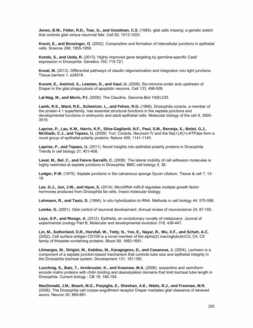
105
Jones, B.W., Fetter, R.D., Tear, G., and Goodman, C.S. (1995). glial cells missing: a genetic switch that controls glial versus neuronal fate. Cell 82, 1013-1023. Knust, E., and Bossinger, O. (2002). Composition and formation of intercellular junctions in epithelial cells. Science 298, 1955-1959. Kondo, S., and Ueda, R. (2013). Highly improved gene targeting by germline-specific Cas9 expression in Drosophila. Genetics 195, 715-721. Koval, M. (2013). Differential pathways of claudin oligomerization and integration into tight junctions. Tissue barriers 1, e24518. Kurant, E., Axelrod, S., Leaman, D., and Gaul, U. (2008). Six-microns-under acts upstream of Draper in the glial phagocytosis of apoptotic neurons. Cell 133, 498-509. Lal-Nag, M., and Morin, PJ. (2009). The Claudins. Genome Biol 10(8):235. Lamb, R.S., Ward, R.E., Schweizer, L., and Fehon, R.G. (1998). Drosophila coracle, a member of the protein 4.1 superfamily, has essential structural functions in the septate junctions and developmental functions in embryonic and adult epithelial cells. Molecular biology of the cell 9, 3505-3519. Laprise, P., Lau, K.M., Harris, K.P., Silva-Gagliardi, N.F., Paul, S.M., Beronja, S., Beitel, G.J., McGlade, C.J., and Tepass, U. (2009). Yurt, Coracle, Neurexin IV and the Na(+),K(+)-ATPase form a novel group of epithelial polarity proteins. Nature 459, 1141-1145. Laprise, P., and Tepass, U. (2011). Novel insights into epithelial polarity proteins in Drosophila. Trends in cell biology 21, 401-408. Laval, M., Bel, C., and Faivre-Sarrailh, C. (2008). The lateral mobility of cell adhesion molecules is highly restricted at septate junctions in Drosophila. BMC cell biology 9, 38. Ledger, P.W. (1975). Septate junctions in the calcareous sponge Sycon ciliatum. Tissue & cell 7, 13-18. Lee, G.J., Jun, J.W., and Hyun, S. (2014). MicroRNA miR-8 regulates multiple growth factor hormones produced from Drosophila fat cells. Insect molecular biology. Lehmann, R., and Tautz, D. (1994). In situ hybridization to RNA. Methods in cell biology 44, 575-598. Lemke, G. (2001). Glial control of neuronal development. Annual review of neuroscience 24, 87-105. Leys, S.P., and Riesgo, A. (2012). Epithelia, an evolutionary novelty of metazoans. Journal of experimental zoology Part B, Molecular and developmental evolution 318, 438-447. Lin, M., Sutherland, D.R., Horsfall, W., Totty, N., Yeo, E., Nayar, R., Wu, X.F., and Schuh, A.C. (2002). Cell surface antigen CD109 is a novel member of the alpha(2) macroglobulin/C3, C4, C5 family of thioester-containing proteins. Blood 99, 1683-1691. Llimargas, M., Strigini, M., Katidou, M., Karagogeos, D., and Casanova, J. (2004). Lachesin is a component of a septate junction-based mechanism that controls tube size and epithelial integrity in the Drosophila tracheal system. Development 131, 181-190. Luschnig, S., Batz, T., Armbruster, K., and Krasnow, M.A. (2006). serpentine and vermiform encode matrix proteins with chitin binding and deacetylation domains that limit tracheal tube length in Drosophila. Current biology : CB 16, 186-194. MacDonald, J.M., Beach, M.G., Porpiglia, E., Sheehan, A.E., Watts, R.J., and Freeman, M.R. (2006). The Drosophila cell corpse engulfment receptor Draper mediates glial clearance of severed axons. Neuron 50, 869-881.
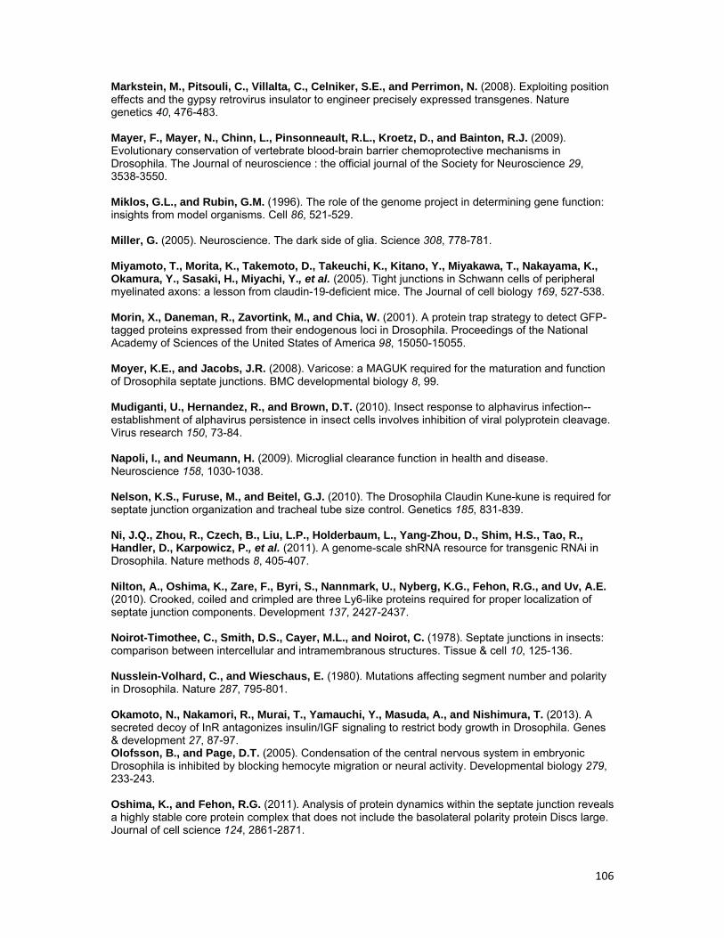
106
Markstein, M., Pitsouli, C., Villalta, C., Celniker, S.E., and Perrimon, N. (2008). Exploiting position effects and the gypsy retrovirus insulator to engineer precisely expressed transgenes. Nature genetics 40, 476-483. Mayer, F., Mayer, N., Chinn, L., Pinsonneault, R.L., Kroetz, D., and Bainton, R.J. (2009). Evolutionary conservation of vertebrate blood-brain barrier chemoprotective mechanisms in Drosophila. The Journal of neuroscience : the official journal of the Society for Neuroscience 29, 3538-3550. Miklos, G.L., and Rubin, G.M. (1996). The role of the genome project in determining gene function: insights from model organisms. Cell 86, 521-529. Miller, G. (2005). Neuroscience. The dark side of glia. Science 308, 778-781. Miyamoto, T., Morita, K., Takemoto, D., Takeuchi, K., Kitano, Y., Miyakawa, T., Nakayama, K., Okamura, Y., Sasaki, H., Miyachi, Y., et al. (2005). Tight junctions in Schwann cells of peripheral myelinated axons: a lesson from claudin-19-deficient mice. The Journal of cell biology 169, 527-538. Morin, X., Daneman, R., Zavortink, M., and Chia, W. (2001). A protein trap strategy to detect GFP-tagged proteins expressed from their endogenous loci in Drosophila. Proceedings of the National Academy of Sciences of the United States of America 98, 15050-15055. Moyer, K.E., and Jacobs, J.R. (2008). Varicose: a MAGUK required for the maturation and function of Drosophila septate junctions. BMC developmental biology 8, 99. Mudiganti, U., Hernandez, R., and Brown, D.T. (2010). Insect response to alphavirus infection--establishment of alphavirus persistence in insect cells involves inhibition of viral polyprotein cleavage. Virus research 150, 73-84. Napoli, I., and Neumann, H. (2009). Microglial clearance function in health and disease. Neuroscience 158, 1030-1038. Nelson, K.S., Furuse, M., and Beitel, G.J. (2010). The Drosophila Claudin Kune-kune is required for septate junction organization and tracheal tube size control. Genetics 185, 831-839. Ni, J.Q., Zhou, R., Czech, B., Liu, L.P., Holderbaum, L., Yang-Zhou, D., Shim, H.S., Tao, R., Handler, D., Karpowicz, P., et al. (2011). A genome-scale shRNA resource for transgenic RNAi in Drosophila. Nature methods 8, 405-407. Nilton, A., Oshima, K., Zare, F., Byri, S., Nannmark, U., Nyberg, K.G., Fehon, R.G., and Uv, A.E. (2010). Crooked, coiled and crimpled are three Ly6-like proteins required for proper localization of septate junction components. Development 137, 2427-2437. Noirot-Timothee, C., Smith, D.S., Cayer, M.L., and Noirot, C. (1978). Septate junctions in insects: comparison between intercellular and intramembranous structures. Tissue & cell 10, 125-136. Nusslein-Volhard, C., and Wieschaus, E. (1980). Mutations affecting segment number and polarity in Drosophila. Nature 287, 795-801. Okamoto, N., Nakamori, R., Murai, T., Yamauchi, Y., Masuda, A., and Nishimura, T. (2013). A secreted decoy of InR antagonizes insulin/IGF signaling to restrict body growth in Drosophila. Genes & development 27, 87-97. Olofsson, B., and Page, D.T. (2005). Condensation of the central nervous system in embryonic Drosophila is inhibited by blocking hemocyte migration or neural activity. Developmental biology 279, 233-243. Oshima, K., and Fehon, R.G. (2011). Analysis of protein dynamics within the septate junction reveals a highly stable core protein complex that does not include the basolateral polarity protein Discs large. Journal of cell science 124, 2861-2871.
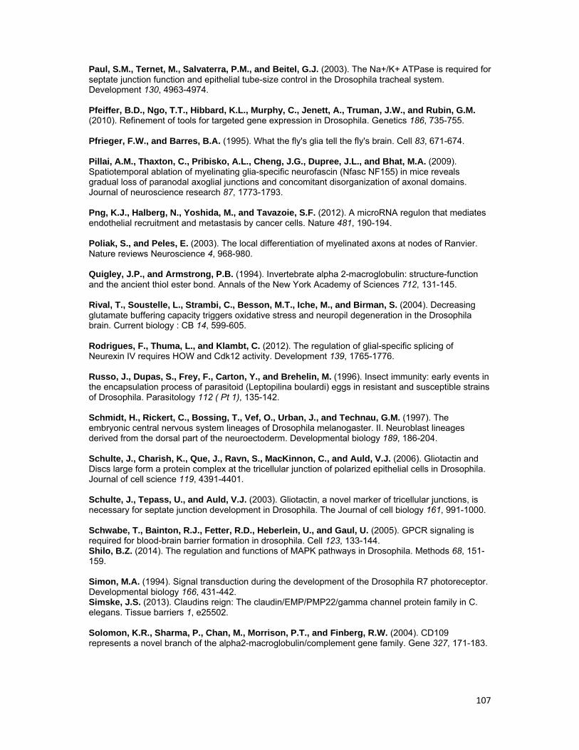
107
Paul, S.M., Ternet, M., Salvaterra, P.M., and Beitel, G.J. (2003). The Na+/K+ ATPase is required for septate junction function and epithelial tube-size control in the Drosophila tracheal system. Development 130, 4963-4974. Pfeiffer, B.D., Ngo, T.T., Hibbard, K.L., Murphy, C., Jenett, A., Truman, J.W., and Rubin, G.M. (2010). Refinement of tools for targeted gene expression in Drosophila. Genetics 186, 735-755. Pfrieger, F.W., and Barres, B.A. (1995). What the fly's glia tell the fly's brain. Cell 83, 671-674. Pillai, A.M., Thaxton, C., Pribisko, A.L., Cheng, J.G., Dupree, J.L., and Bhat, M.A. (2009). Spatiotemporal ablation of myelinating glia-specific neurofascin (Nfasc NF155) in mice reveals gradual loss of paranodal axoglial junctions and concomitant disorganization of axonal domains. Journal of neuroscience research 87, 1773-1793. Png, K.J., Halberg, N., Yoshida, M., and Tavazoie, S.F. (2012). A microRNA regulon that mediates endothelial recruitment and metastasis by cancer cells. Nature 481, 190-194. Poliak, S., and Peles, E. (2003). The local differentiation of myelinated axons at nodes of Ranvier. Nature reviews Neuroscience 4, 968-980. Quigley, J.P., and Armstrong, P.B. (1994). Invertebrate alpha 2-macroglobulin: structure-function and the ancient thiol ester bond. Annals of the New York Academy of Sciences 712, 131-145. Rival, T., Soustelle, L., Strambi, C., Besson, M.T., Iche, M., and Birman, S. (2004). Decreasing glutamate buffering capacity triggers oxidative stress and neuropil degeneration in the Drosophila brain. Current biology : CB 14, 599-605. Rodrigues, F., Thuma, L., and Klambt, C. (2012). The regulation of glial-specific splicing of Neurexin IV requires HOW and Cdk12 activity. Development 139, 1765-1776. Russo, J., Dupas, S., Frey, F., Carton, Y., and Brehelin, M. (1996). Insect immunity: early events in the encapsulation process of parasitoid (Leptopilina boulardi) eggs in resistant and susceptible strains of Drosophila. Parasitology 112 ( Pt 1), 135-142. Schmidt, H., Rickert, C., Bossing, T., Vef, O., Urban, J., and Technau, G.M. (1997). The embryonic central nervous system lineages of Drosophila melanogaster. II. Neuroblast lineages derived from the dorsal part of the neuroectoderm. Developmental biology 189, 186-204. Schulte, J., Charish, K., Que, J., Ravn, S., MacKinnon, C., and Auld, V.J. (2006). Gliotactin and Discs large form a protein complex at the tricellular junction of polarized epithelial cells in Drosophila. Journal of cell science 119, 4391-4401. Schulte, J., Tepass, U., and Auld, V.J. (2003). Gliotactin, a novel marker of tricellular junctions, is necessary for septate junction development in Drosophila. The Journal of cell biology 161, 991-1000. Schwabe, T., Bainton, R.J., Fetter, R.D., Heberlein, U., and Gaul, U. (2005). GPCR signaling is required for blood-brain barrier formation in drosophila. Cell 123, 133-144. Shilo, B.Z. (2014). The regulation and functions of MAPK pathways in Drosophila. Methods 68, 151-159. Simon, M.A. (1994). Signal transduction during the development of the Drosophila R7 photoreceptor. Developmental biology 166, 431-442. Simske, J.S. (2013). Claudins reign: The claudin/EMP/PMP22/gamma channel protein family in C. elegans. Tissue barriers 1, e25502. Solomon, K.R., Sharma, P., Chan, M., Morrison, P.T., and Finberg, R.W. (2004). CD109 represents a novel branch of the alpha2-macroglobulin/complement gene family. Gene 327, 171-183.
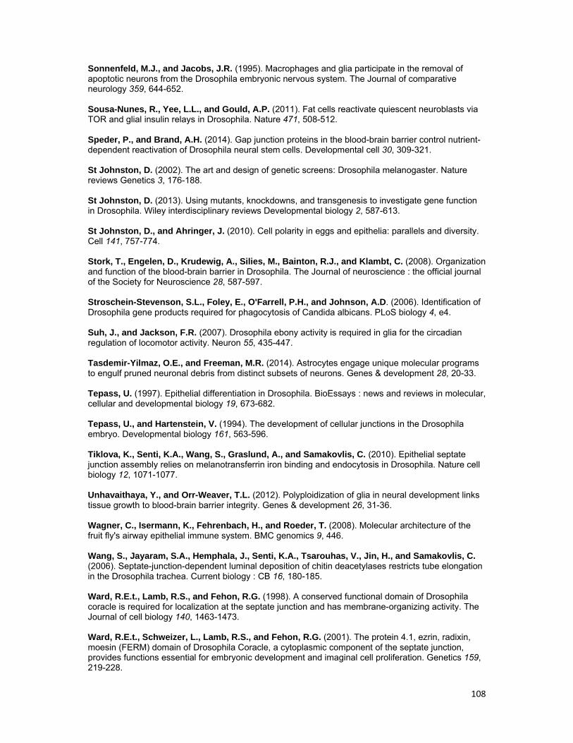
108
Sonnenfeld, M.J., and Jacobs, J.R. (1995). Macrophages and glia participate in the removal of apoptotic neurons from the Drosophila embryonic nervous system. The Journal of comparative neurology 359, 644-652. Sousa-Nunes, R., Yee, L.L., and Gould, A.P. (2011). Fat cells reactivate quiescent neuroblasts via TOR and glial insulin relays in Drosophila. Nature 471, 508-512. Speder, P., and Brand, A.H. (2014). Gap junction proteins in the blood-brain barrier control nutrient-dependent reactivation of Drosophila neural stem cells. Developmental cell 30, 309-321. St Johnston, D. (2002). The art and design of genetic screens: Drosophila melanogaster. Nature reviews Genetics 3, 176-188. St Johnston, D. (2013). Using mutants, knockdowns, and transgenesis to investigate gene function in Drosophila. Wiley interdisciplinary reviews Developmental biology 2, 587-613. St Johnston, D., and Ahringer, J. (2010). Cell polarity in eggs and epithelia: parallels and diversity. Cell 141, 757-774. Stork, T., Engelen, D., Krudewig, A., Silies, M., Bainton, R.J., and Klambt, C. (2008). Organization and function of the blood-brain barrier in Drosophila. The Journal of neuroscience : the official journal of the Society for Neuroscience 28, 587-597. Stroschein-Stevenson, S.L., Foley, E., O'Farrell, P.H., and Johnson, A.D. (2006). Identification of Drosophila gene products required for phagocytosis of Candida albicans. PLoS biology 4, e4. Suh, J., and Jackson, F.R. (2007). Drosophila ebony activity is required in glia for the circadian regulation of locomotor activity. Neuron 55, 435-447. Tasdemir-Yilmaz, O.E., and Freeman, M.R. (2014). Astrocytes engage unique molecular programs to engulf pruned neuronal debris from distinct subsets of neurons. Genes & development 28, 20-33. Tepass, U. (1997). Epithelial differentiation in Drosophila. BioEssays : news and reviews in molecular, cellular and developmental biology 19, 673-682. Tepass, U., and Hartenstein, V. (1994). The development of cellular junctions in the Drosophila embryo. Developmental biology 161, 563-596. Tiklova, K., Senti, K.A., Wang, S., Graslund, A., and Samakovlis, C. (2010). Epithelial septate junction assembly relies on melanotransferrin iron binding and endocytosis in Drosophila. Nature cell biology 12, 1071-1077. Unhavaithaya, Y., and Orr-Weaver, T.L. (2012). Polyploidization of glia in neural development links tissue growth to blood-brain barrier integrity. Genes & development 26, 31-36. Wagner, C., Isermann, K., Fehrenbach, H., and Roeder, T. (2008). Molecular architecture of the fruit fly's airway epithelial immune system. BMC genomics 9, 446. Wang, S., Jayaram, S.A., Hemphala, J., Senti, K.A., Tsarouhas, V., Jin, H., and Samakovlis, C. (2006). Septate-junction-dependent luminal deposition of chitin deacetylases restricts tube elongation in the Drosophila trachea. Current biology : CB 16, 180-185. Ward, R.E.t., Lamb, R.S., and Fehon, R.G. (1998). A conserved functional domain of Drosophila coracle is required for localization at the septate junction and has membrane-organizing activity. The Journal of cell biology 140, 1463-1473. Ward, R.E.t., Schweizer, L., Lamb, R.S., and Fehon, R.G. (2001). The protein 4.1, ezrin, radixin, moesin (FERM) domain of Drosophila Coracle, a cytoplasmic component of the septate junction, provides functions essential for embryonic development and imaginal cell proliferation. Genetics 159, 219-228.
109
Watts, R.J., Schuldiner, O., Perrino, J., Larsen, C., and Luo, L. (2004). Glia engulf degenerating axons during developmental axon pruning. Current biology : CB 14, 678-684. Whitlock, K.E. (1993). Development of Drosophila wing sensory neurons in mutants with missing or modified cell surface molecules. Development 117, 1251-1260. Williams, M.J. (2009). The Drosophila cell adhesion molecule Neuroglian regulates Lissencephaly-1 localisation in circulating immunosurveillance cells. BMC immunology 10, 17. Williams, M.J., Ando, I., and Hultmark, D. (2005). Drosophila melanogaster Rac2 is necessary for a proper cellular immune response. Genes to cells : devoted to molecular & cellular mechanisms 10, 813-823. With, S., Rice, T., Salinas, C., and Auld, V. (2003). Fire exit is a potential four transmembrane protein expressed in developing Drosophila glia. Genesis 35, 143-152. Woods, D.F., and Bryant, P.J. (1991). The discs-large tumor suppressor gene of Drosophila encodes a guanylate kinase homolog localized at septate junctions. Cell 66, 451-464. Woods, D.F., Wu, J.W., and Bryant, P.J. (1997). Localization of proteins to the apico-lateral junctions of Drosophila epithelia. Developmental genetics 20, 111-118. Wu, V.M., and Beitel, G.J. (2004). A junctional problem of apical proportions: epithelial tube-size control by septate junctions in the Drosophila tracheal system. Current opinion in cell biology 16, 493-499. Wu, V.M., Schulte, J., Hirschi, A., Tepass, U., and Beitel, G.J. (2004). Sinuous is a Drosophila claudin required for septate junction organization and epithelial tube size control. The Journal of cell biology 164, 313-323. Wu, V.M., Yu, M.H., Paik, R., Banerjee, S., Liang, Z., Paul, S.M., Bhat, M.A., and Beitel, G.J. (2007). Drosophila Varicose, a member of a new subgroup of basolateral MAGUKs, is required for septate junctions and tracheal morphogenesis. Development 134, 999-1009. Xiao, X., Liu, Y., Zhang, X., Wang, J., Li, Z., Pang, X., Wang, P., and Cheng, G. (2014). Complement-related proteins control the flavivirus infection of Aedes aegypti by inducing antimicrobial peptides. PLoS pathogens 10, e1004027. Xie, X., and Auld, V.J. (2011). Integrins are necessary for the development and maintenance of the glial layers in the Drosophila peripheral nerve. Development 138, 3813-3822. Yguerabide, J., Schmidt, J.A., and Yguerabide, E.E. (1982). Lateral mobility in membranes as detected by fluorescence recovery after photobleaching. Biophysical journal 40, 69-75. Yi, P., Johnson, A.N., Han, Z., Wu, J., and Olson, E.N. (2008). Heterotrimeric G proteins regulate a noncanonical function of septate junction proteins to maintain cardiac integrity in Drosophila. Developmental cell 15, 704-713. Zhang, L., and Ward, R.E.t. (2011). Distinct tissue distributions and subcellular localizations of differently phosphorylated forms of the myosin regulatory light chain in Drosophila. Gene expression patterns : GEP 11, 93-104. Zlokovic, B.V. (2008). The blood-brain barrier in health and chronic neurodegenerative disorders. Neuron 57, 178-201.