Embed Size (px)
Citation preview
Vol.:(0123456789)1 3
Int J Pept Res Ther DOI 10.1007/s10989-017-9633-1
SHORT COMMUNICATION
Identification of PP2A/Set Binding Sites and Design of Interacting Peptides with Potential Clinical Applications
Lu Tian1 · Xiguang Zhang1 · Dorien Haesen2 · Jeronimo Bravo3 · Jesus Fominaya1 · Sylvain Choquet4 · Jean Marc Zini5 · Severine Loisel6 · Etienne Waelkens2 · Veerle Janssens2 · Angelita Rebollo1
Accepted: 3 October 2017 © Springer Science+Business Media, LLC 2017
SET sides. The amino acid sequences of the binding sites were coupled to an optimized penetrating peptide in order to generate chimeras (Mut3DPT-PP2A and Mut3DPT-SET) able to target the PP2A/SET interaction. We demonstrate that these peptides have an in vitro apoptotic effect on breast and lung cancer cell lines, as well as an antitumoral effect on CLL and lymphoma xenograft models. The new generated chimeric peptides allow the modulation of the PP2Ac/SET interaction and might have a potential as a new therapeutic approach for cancer treatment.
Keywords Cell penetrating peptides · PP2A · SET · Apoptosis · CLL
Introduction
Protein phosphatase 2A (PP2A) belongs to the family of serine/threonine phosphatases that reverse the action of sev-eral protein kinases by removing the phosphate group from serine and threonine residues of proteins. PP2A regulates various cellular processes, including protein synthesis, cel-lular signalling, cell cycle, apoptosis, metabolism and stress responses (Westermarck and Hahn 2008). PP2A is widely described as a tumor suppressor since its pharmacologic inhibitor, okadaic acid, is a tumor promoter (Janssens et al. 2005) and inhibition of PP2A is one of five minimal prereq-uisites to fully transform human epithelial cells of various origins (Hahn and Weinberg 2002). Moreover, PP2A is a critical negative regulator of several oncoproteins, including the transcription factor c-Myc (Sablina et al. 2010). Inacti-vating mutations or decreased expression of PP2A subunits have been detected in a large variety of human malignancies (Sangodkar et al. 2016). Collectively, these findings have led to the discovery and development of several new compounds
Abstract Protein phosphatase 2A (PP2A) is known to be a negative regulator of several survival and proliferat-ing pathways that are frequently altered in cancer. In addi-tion to chemical enzymatic inhibitors of the PP2A activity, the oncoprotein SET has been described as a physiological PP2A inhibitor by forming a complex with PP2A catalytic subunit (PP2Ac). Increased SET protein levels therefore directly reduce the tumor suppressor function of PP2A and promote tumor progression. We have used the PEP-Scan approach to identify the binding site between the serine/threonine phosphatase PP2A and the oncoprotein SET. For in vivo validation of the peptides, we have used chronic lym-phocytic leukemia (CLL) xenograft models. In this manu-script we describe the identification of amino acid sequences involved in the complex formation, both at the PP2Ac and
Lu Tian and Xiguang Zhang have contributed equally to this work.
* Angelita Rebollo [email protected]
1 CIMI Paris, UPMC UMR CR7, Inserm U1135, CNRS ERL 8255, 91, bd de l’Hôpital, 75013 Paris, France
2 Laboratory of Protein Phosphorylation and Proteomics, Department Cellular and Molecular Medicine, University of Leuven & Leuven Cancer Institute (LKI), 3000 Leuven, Belgium
3 IBV-CSIC, Instituto de Biomedicina de Valencia, c/ Jaime Roig 11, 46010 Valencia, Spain
4 AH-HP, Hôpital La Pitié Salpêtrière, 43, bd de l’Hôpital, 75013 Paris, France
5 AP-HP, Hôpital Saint Louis, 1 Avenue Claude Vellefaux, 75010 Paris, France
6 Animalerie de l’UBO, UFR Médecine, 22 Avenue Camille Desmoulins, 29238 Brest, France
Int J Pept Res Ther
1 3
that are capable of restoring PP2A activity, although some-times by poorly understood mechanisms (Kalev and Sablina 2011). Therefore, the tumor suppressing function of PP2A makes it a possible target for novel anti-cancer therapies (Haesen et al. 2014; Janssens and Rebollo 2012; Lambrecht et al. 2013).
The SET protein, also known as PP2A Inhibitor 2 (I2PP2A), is an oncoprotein that belongs to a family of multi-tasking proteins involved in apoptosis, transcription, nucleo-some assembly and histone binding. SET is localized in the nucleus and cytoplasm and has a critical role in the regula-tion of normal and cancerous signal transduction. Elevated expression of SET has been linked to cell growth and trans-formation in several tumor types, particularly in hemato-logic malignancies (Ciccone et al. 2015). Moreover, as SET inhibits PP2A by forming a complex with PP2Ac (Farrell et al. 2014; Leopoldino et al. 2012; Switzer et al. 2011), it is assumed that SET oncogenic properties are partially medi-ated by its ability to inhibit PP2A. Therefore, many efforts trying to restore PP2A activity have focused on interfering with the PP2A/SET interaction (Neviani et al. 2013; Swit-zer et al. 2011). It has been shown that an apolipoprotein E-based peptide can bind to SET, inhibiting the SET/PP2A interaction and leading to increased PP2A activity (Chris-tensen et al. 2011a; Vitek et al. 2012). The pharmacologic PP2A activator FTY720 binds to a ceramide-binding domain of SET, resulting in SET dissociation from PP2A (Mukho-padhyay et al. 2009) (Pippa et al. 2014). Also, the 2-phe-nyloxypyrimidine compound TGI1002 has been shown to disrupt SET–PP2A interaction with important therapeutic potential for the treatment of drug-resistant Chronic Myeloid Leukemia (CML). Based on these findings, it is clear that the complex SET/PP2A is an interesting target for anti-cancer development strategies.
Cell penetrating peptides (CPPs) have emerged as a new class of vectors that allow the delivery of biomolecules across biological barriers. They are widely used for intra-cellular delivery of various cargos, including small molecule drugs, peptides, proteins, genes and nanoparticles. A stable CPP with additional specific targeting capabilities, such as in a combination with an interfering peptide that modulates an adequate protein/protein interaction (chimeric peptide) is an exciting targeted approach for therapy.
Using this strategy, we have recently demonstrated the use of the bi-functional peptide DPT-C9h, consisting of a penetrating sequence (Guergnon et al. 2006a, b), associated to the residues 213–230 from human caspase 9 to specifi-cally target the PP2A/caspase 9 interaction. This bi-func-tional peptide, as well as the optimized variant Mut3-DPT-C9h, induce apoptosis in vitro and have an anti-tumor effect in vivo (Arrouss et al. 2013).
Here, we have taken advantage of our knowledge on the design of peptides for targeting protein/protein interactions
in order to identify the amino acids in SET and PP2Ac involved in the PP2A/SET complex formation to gener-ate new tools for controlled manipulation of this interac-tion. We describe the identification of the binding sites, as well as the generation of chimeric peptides, with apoptotic effects on human cancer cell lines and anti-tumor effects in human CLL and lymphoma xenograft models. Hence, these chimeras can be used as therapeutic tools in tumor cells as a potential clinical approach.
Methods
Cell Lines
H1975 and H1299 are human non-small cell lung car-cinoma cell lines. MDA-MB231 is a human breast can-cer cell line. These cell lines were cultured in DMEM medium supplemented with 10% FCS. Human lymphoma cell lines Daudi and Raji (ATCC CL-213), and the human chronic lymphocytic leukemia Jok1 cell line were cultured in RPMI supplemented with 10% FBS, l-glutamine and penicillin/streptomycin.
Peptides Synthesis and Sequences
Peptides were synthesized in an automated multiple pep-tide synthesizer with solid phase procedure and stand-ard Fmoc chemistry by the company GL Biochem Ltd, Shanghai, China. The purity and composition of the pep-tides were confirmed by reverse phase HPLC and by mass spectrometry.
Purification of Recombinant SET Protein
The coding sequence of human SET isoform β (also called isoform 2) was cloned into bacterial expression vector pET15b (Ndel/BamH1), in frame with an N-ter-minal 6xHis-tag. Production of recombinant protein was achieved in 100 mL culture of E.coli BL21-pLys cells at 30 °C for 3 h under moderate shaking conditions. 0.4 mM IPTG was used for induction whtn the culture reached an OD600nm of 0.6. His-tagged SET was soluble, and was puri-fied from the bacterial lysates using nickel(II) Pentaden-tate chelatorR-SepharoseTMCL-4B beads (Affiland) using standard protocols. Elution was achieved in 50 mM Tris HCl buffer (pH 8), 300 mM NaCl and 250 mM imidazole. Purity and yield were verified by SDS-PAGE. The purified protein was stocked at a concentration of 1 mg/ml.
Int J Pept Res Ther
1 3
SET‑Binding Assays on Cellulose‑Bound Peptides Containing PP2A Sequences
Overlapping dodecapeptides with two amino acid shift, spanning the complete PP2Ac sequence (UniPROT P67775) were prepared by automated spot synthesis (Abimed, Langerfeld, Germany) onto an amino-derived cellulose membrane, as described (Frank and Overwin 1996; Gause-pohl et al. 1992). The membrane with 133 spots of dodeca-peptides was saturated using 3% non-fat dry milk/3% BSA (2 h room temperature), incubated with purified SET protein (4 μg/ml, overnight, 4 °C) and after several washing steps, incubated with polyclonal anti-SET antibody 2 h at room temperature (1:500 dilution, Santa Cruz Biotechnology), followed by HRP-conjugated secondary antibody for 1 h at room temperature (1:1000 dilution, Dako). Positive spots were visualized using the ECL system (Bio-Rad).
PP2A‑Binding Assays on Cellulose‑Bound Peptides Containing SET Sequences
Overlapping dodecapeptides spanning the complete SET sequence (UniPROT Q01105) were prepared and spotted on solid support as above (membrane contains 150 dodeca-peptides spots). The membrane was hybridizedwith purified PP2A (4 μg/ml, overnight, 4 °C), isolated from red blood cells (Calbiochem), and developed using an anti-PP2Ac spe-cific monoclonal antibody (Millipore) as above.
Apoptosis Assay on Cell Lines
We used an Annexin-V-FITC conjugated kit (eBioscience) for the detection of outer leaflet exposure of phosphatidylser-ine (PS) in the plasma membrane of apoptotic cells. Staining was performed according to the manufacturer’s instructions. Each condition was analyzed in triplicate. A total of 105 cells were analyzed by flow cytometry. The effect of peptide treat-ment was compared to untreated control cells.
Isolation and Culture of Primary Human Cells
Fresh blood from healthy donors (HD) was obtained from the Etablissement Français du Sang. Chronic lymphocytic leukemia (CLL) patient samples were obtained from the Department of Hematology. Peripheral blood mononuclear cells (PBMC) from HD and CLL patients were prepared by Ficoll gradient centrifugation. Cells were maintained in RPMI 1640 supplemented with 10% FCS, 1% non-essen-tial amino acids, 1% Hepes, 1% sodium pyruvate and 1% glutamine.
Detection of Apoptosis by Annexin‑V‑FITC Staining on Primary Cells
Human lymphocytes isolated from HD or CLL patients were stained with anti-hCD19 (APC fluorochrome from BD bio-sciences) and early apoptotic events were determined using Annexin-V FITC (eBiosciences) as described by the manu-facturer. Briefly, cells were washed in 1x binding buffer, cen-trifuged and resuspended in 100 µl of 1x binding buffer con-taining Annexin-V FITC (0.1 µg/ml) and propidium iodide (PI) (0.5 µg/ml). After incubation for 15 min, cells were analyzed by flow cytometry. Data acquired by FACS Canto (BD biosciences) were analyzed with Diva 60 software.
Animals
6–8 week old SCID CB-17 mice (female, 20–23 g) and BALB/c nude mice (male, 26–30 g) were purchased (Ele-vage Janvier, Le Genest Saint-Ile, France). All mice were maintained under conditions and protocols in accordance with the Directive 2010/63/UE of the Council of Europe on Animal Welfare. The studies (number authorization 5220) were approved by the French Ethics Committee for ani-mal experimentation number 74 and all experiments were conducted following the guidelines of the aforementioned committee.
In Vivo Activity of the Chimeric Peptides
To establish the xenograft lymphoma model, 10 × 106 Daudi cells in 100 µl of PBS were subcutaneously injected into the flank of male BALB/c nude mice. For the CLL xenograft model, 2.5 × 10⁶ Jok1 or Jok5.3 cells in 100 µl of PBS were injected into the tail vein of SCID mice.
Mice (6–16 mice per group) were then injected with saline or with Mut3DPT-PP2Ac-S2(5 or 20 mg/kg) intra-peritoneally (I.P.) at days 3 after cells transplantation or when palpable tumors were apparent (mean 150–200 mm3) for 5 consecutive days during 4 weeks. For the intravenous model, mice were monitored daily for the presence of hind-leg paralysis and in that case sacrificed. For the subcuta-neous model, tumor growth rate was recorded every 2–3 days by measuring the major and minor axes of the tumors formed with a digital caliper. Measurements were trans-formed into tumor volume using the formula: tumor volume (mm3) = major axis × minor axis2 × 0.5.
Statistical Analyses
For in vivo data, statistical comparisons between groups were performed using Factorial analysis of variance ANOVA. Normality and homogeneity were evaluated by Shapiro-Wilk test and the data were found valid.
Int J Pept Res Ther
1 3
Results
In Vitro Identification of PP2Ac Sequences Involved in SET Interaction
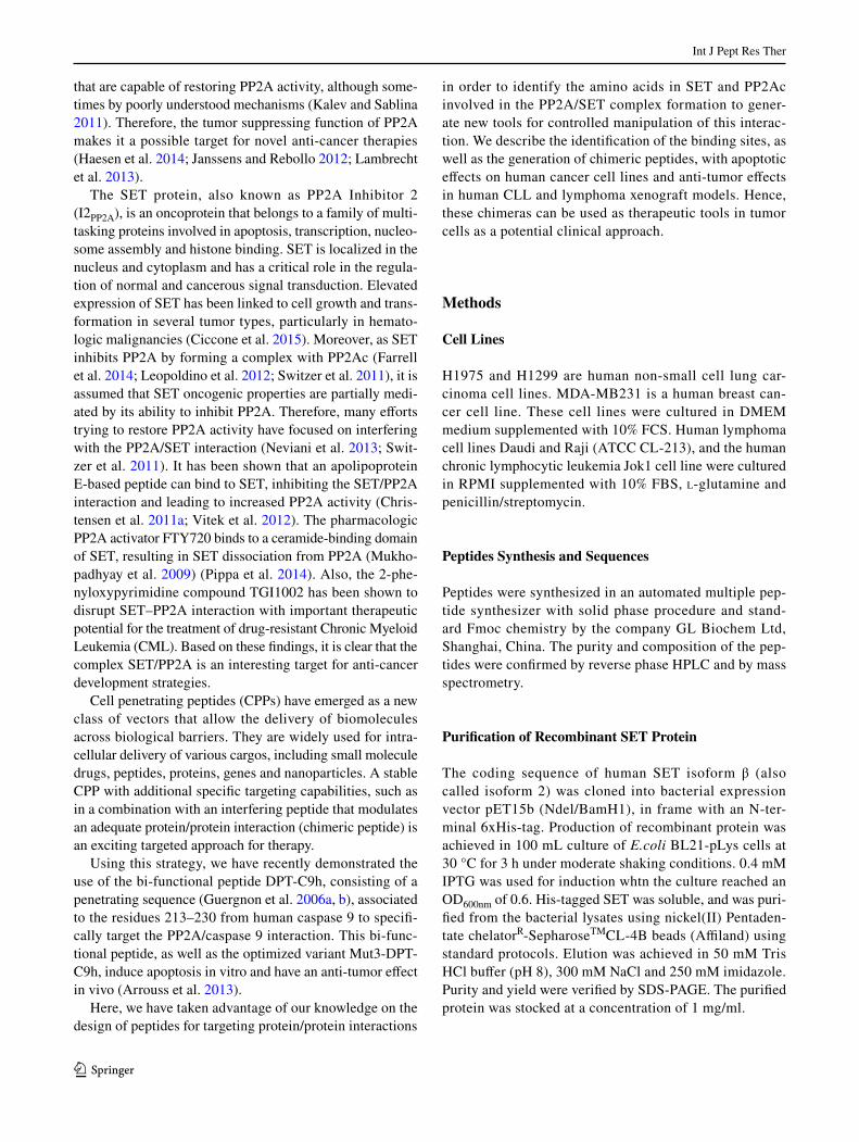
To determine the residues of PP2Ac that interact with SET protein, we generated overlapping peptides from PP2A, which were immobilized on a cellulose mem-brane. Figure 1a shows the entire human PP2Ac amino acid sequence upon hybridization with SET protein. Two different sets of spots allowed the identification of two linear interacting areas, one spanning residues 95–112 (PP2Ac-S2) and a second one comprising residues 133–146 (PP2Ac-S3) and forming an α-helical structure. Both sequences correspond to solvent-exposed areas located in close spatial proximity at the same side of PP2Ac structure, apart from the contacts between PP2Ac catalytic subunit and other members of the PP2A complex (A and B subunits) in a good agreement with a putative protein interacting area (Fig. 1a). Chimeric peptides con-taining Mut3DPT-Sh1 (VKKKKIKAEIKI), an optimized
cell penetrating sequence previously developed in our lab, followed by the selected PP2A sequences were chemi-cally synthesized and further used for functional assays (Fig. 1b).
Effect of Mut3DPT‑PP2A Peptide on Apoptosis
Given that the complex PP2A/SET plays an important role in the control of critical cell functions, we analyzed the abil-ity of peptides Mut3DPT-PP2A-S2 and Mut3DPT-PP2A-S3 to induce apoptosis in human cancer cell lines. As shown in Fig. 1c, upon 24 h of treatment with 100 μM of peptide, only Mut3DPT-PP2A-S2 induced a clear and strong apop-totic effect, as detected by Annexin-V FITC staining in two human non-small lung tumor cell lines, while control non-treated cells did not show apoptosis. The apoptotic induction effect was weaker, more variable and less consistent for pep-tide Mut3DPT-PP2A-S3, while remained at higher values for peptide Mut3DPT-PP2A-S2, showing a functional effect that could be mediated by disruption of the SET-PP2Ac interaction. Therefore, we selected Mut3DPT-PP2A-S2
Membrane PP2Ac
E95TVTLLVALKVRYRERIT112C133LRKYGNANVWKYF146
A
A
CB
PP2A
1 306090120150
B
Mut3DPT-PP2Ac-S2: VKKKKIKAEIKIETVTLLVALKVRYRERIT
Mut3DPT: VKKKKIKAEIKI
Mut3DPT-PP2Ac-S3: VKKKKIKAEIKICLRKYGNANVWKYF
C
D
0
20
40
60
80
100
H1975 H1299
Apop
tosis(%)
Control
Mut3-DPT-PP2A-S2
Mut3-DPT-PP2A-S3
0
25
50
75
100
Apop
tosis(%)
Mut3DPT-PP2Ac-S2
MDA-MB-231
Fig. 1 Identification of the binding site of PP2Ac to SET protein. a SET binding assay on cellulose-bound PP2Ac peptides. The sequence of PP2Ac was covered as a series of 150 overlapping dodecapetides. Spots were detected using a primary SET antibody, HRP-coupled secondary antibody and the ECL system. PP2A peptides that inter-act with SET are boxed and the sequences located within the three-dimensional PP2A structure. PP2Ac catalytic subunit is shown in white, while A and B subunits appear in grey. PP2A-S2 is labelled in dark grey and PP2A-S3 in black. b Sequence of the shuttle pep-
tide and the new generated peptides containing the binding sequences of PP2Ac to SET. c Apoptotic effect of Mut3DPT-PP2A-S2 and S3. H1975, H1298 cell lines were cultured in the presence or the absence (control) of the peptide at a concentration of 100 μM for 24 h. Apop-tosis was estimated by Annexin V-FITC staining and analyzed by flow cytometry. d Apoptotic effect of the peptides Mut3DPT-PP2A-S2, Mut3DPT-Sh1 and PP2A-S2 at different concentrations on breast cancer cell line MDA-MB231 estimated by Annexin V-FITC stain-ing. Non treated cells were used as negative control
Int J Pept Res Ther
1 3
(VKKKKIKAEIKI ETVTLLVALKVRYRERIT) for fur-ther studies. Figure 1d shows the apoptotic effect of Mut-3DPT-PP2Ac-S2 peptide on the breast cancer cell line MDA-MB231.
In Vitro Identification of SET Sequences Involved in PP2A Interaction
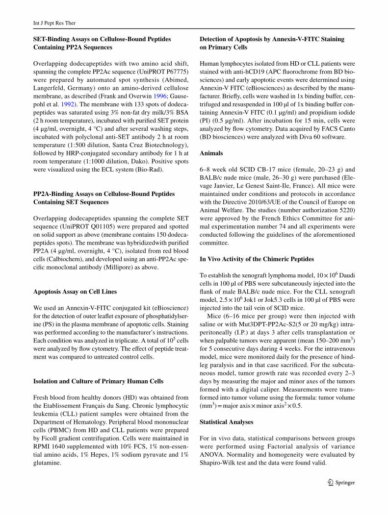
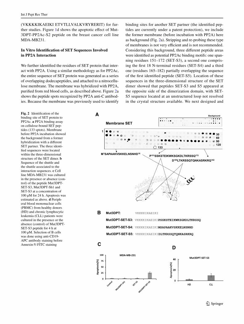
We further identified the residues of SET protein that inter-act with PP2A. Using a similar methodology as for PP2Ac, the entire sequence of SET protein was generated as a series of overlapping dodecapeptides, and attached to a nitrocellu-lose membrane. The membrane was hybridized with PP2A, purified from red blood cells, as described above. Figure 2a shows the peptide spots recognized by PP2A anti-C antibod-ies. Because the membrane was previously used to identify
binding sites for another SET partner (the identified pep-tides are currently under a patent protection), we include the former membrane (before incubation with PP2A) here as background (Fig. 2a). Stripping and re-probing these type of membranes is not very efficient and is not recommended. Considering this background, three different peptide areas were identified as potential PP2Ac binding motifs: one span-ning residues 151–172 (SET-S3), a second one compris-ing the first 18 N-terminal residues (SET-S4) and a third one (residues 165–182) partially overlapping the sequence of the first identified peptide (SET-S5). Location of these sequences in the three-dimensional structure of the SET dimer showed that peptides SET-S3 and S5 appeared at the opposite side of the dimerization domain, with SET-S5 sequence located at an unstructured loop not resolved in the crystal structure available. We next designed and
Fig. 2 Identification of the binding site of SET protein to PP2Ac. a PP2A binding assay on cellulose-bound SET pep-tides (133 sptots). Membrane before PP2A incubation showed the background from a former hybridization with a different SET partner. The three identi-fied sequences were located within the three-dimensional structure of the SET dimer. b Sequence of the shuttle and the shuttle associated to the interaction sequences. c Cell line MDA-MB231 was cultured in the presence or absence (con-trol) of the peptide Mut3DPT-SET-S3, Mut3DPT-Sh1 and SET-S3 at a concentration of 100 μM for 24 h. Apoptosis was estimated as above. d Periph-eral blood mononuclear cells (PBMC) from healthy donors (HD) and chronic lymphocytic leukemia (CLL) patients were cultured in the presence or the absence (control) of Mut3DPT-SET-S3 peptide for 4 h at 100 μM. Selection of B cells was done using anti-CD19-APC antibody staining before Annexin-V-FITC staining
A
Membrane SET
P151SSKSTEIKWKSGKDLTKRSSQ172M1SAPAAKVSKKELNSNHD18
D165LTKRSSQTQNKASRKRQ182
Background
1306090120
133
BMut3DPT-SET-S3: VKKKKIKAEIKIPSSKSTEIKWKSGKDLTKRSSQ
Mut3DPT: VKKKKIKAEIKI
CD
Mut3DPT-SET-S3
Mut3DPT-SET-S4: VKKKKIKAEIKIMSAPAAKVSKKELNSNHD
Mut3DPT-SET-S5: VKKKKIKAEIKIDLTKRSSQTQNKASRKRQ
0
25
50
75
100
)%(sisotpopA
MDA-MB-231
Int J Pept Res Ther
1 3
synthesized chimeric peptides, encompassing the shuttle Mut3DPT-Sh1 (VKKKKIKAEIKI), followed by the iden-tified SET sequences specifically recognized by PP2Ac (Fig. 2b). All chimeras were further analyzed in functional cell assays.
Effect of Mut3DPT‑SET Peptide on Apoptosis
First, we tested whether any of the Mut3DPT-SET-derived peptides were able to induce apoptosis in cancer cell lines and primary cells. Only Mut3DPT-SET-S3 (VKKKKI-KAEIKI PSSKSTEIKWKSGKDLTKRSSQ) appeared to
A
0
25
50
75
100
10 11 12 13 14 15 16 17 18 19 20
lavivrus fo egatnecreP
Days a�er inocula on tumor cells
JOK5.3-Mut3DPT-PP2Ac-S2
Control
PP2A/SET- 20mgMut3DPT-PP2Ac-S2 20 mg
B
0
500
1000
1500
2000
2500
3000
15 25 35 45 55 65
Tum
or V
olum
e (m
m3 )
Days aer inocula�on tumor cells
Daudi-Mut3DPT-PP2Ac-S2
Mut3DPT-PP2ASET-20mg
Mut3DPT-PP2ASET-temoin
Mut3DPT-PP2Ac-S2 20 mg
Control
C
0
25
50
75
100
10 11 12 13 14 15 16 17 18 19 20
Perc
enta
ge o
f sur
viva
l
Days aer inocula�on tumor cells
Raji-Mut3DPT-PP2Ac-S2
Contrôle
PP2A/SET-20mg
Control
Mut3DPT-PP2Ac-S2 20mg
D
Int J Pept Res Ther
1 3
induce a very weak apoptotic effect, in the breast cancer cell line MDA-MB231, as detected by Annexin-V-FITC stain-ing upon 24 h of culture at 100 μM peptide (Fig. 2c). The level of apoptosis detected with this peptide is far lower than the level detected with the mirror peptide Mut3DPT-PP2A-S2 at similar concentration. We also analyzed the ability of the peptide to induce apoptosis in primary cells from HD and CLL patients. PBMC from HD and CLL patients were treated 4 h at 100 μM with Mut3DPT-SET-S3 peptide and apoptosis was analyzed by Annexin-V-FITC staining. As observed in the MDA-MB231 cell line, the peptide has a very weak apoptotic effect on B cells from CLL patients, but no effect on B cells from HD (Fig. 2d).
Mut3DPT‑PP2A‑S2 has Anti‑Tumor Effects in Human Xenograft Models of Chronic Lymphocytic Leukemia and Lymphoma
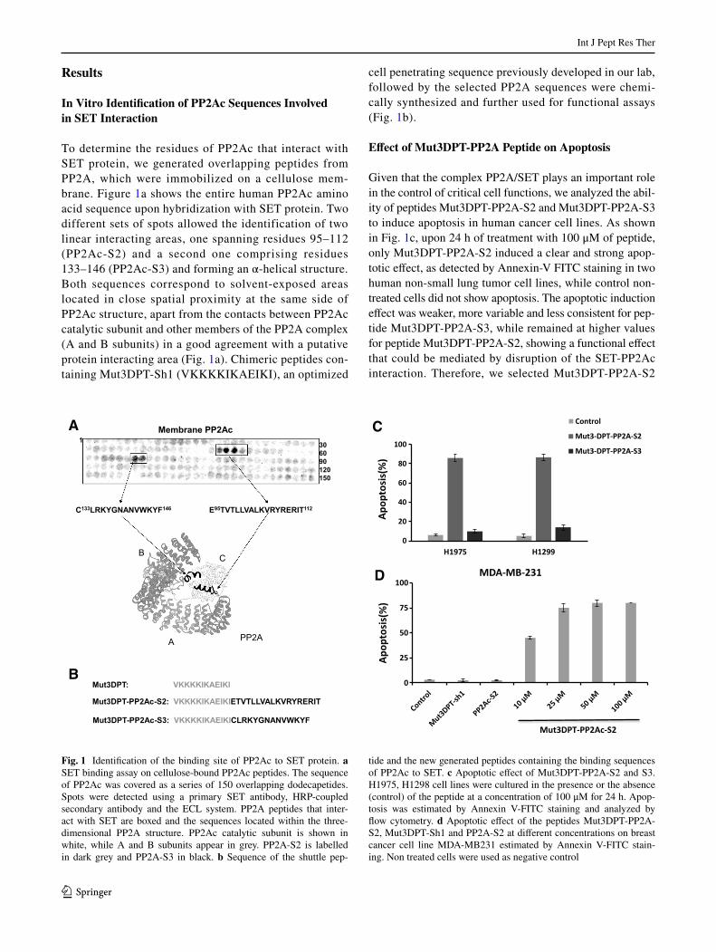
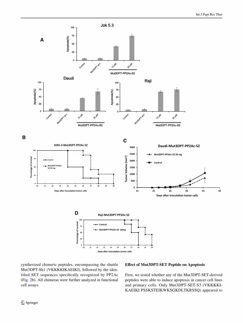
Before starting the in vivo validation of the peptide, we first analyzed its effect on two lymphoma B cell lines, Daudi and Raji, and in the chronic lymphocytic leukemia-like cell line Jok 1 (Fig. 3a). In all three cell lines, the peptide induces apoptosis, which is concentration-dependent for Jock 1 and Daudi. The non-treated cell lines were used as negative control.
The in vivo efficacy of the peptide was evaluated in three human xenograft models, using two lymphoma (Raji and Daudi cell lines) and one chronic lymphocytic leukemia cell line (CLL, Jok1). The peptide treatment was initiated 3 days after injection of the cells (Jok1 or Raji) into xenografted SCID mice. Treatment of Jok1 tumors with the peptide at 20 mg/kg/5 days per week yielded a moderate survival ben-efit, as shown by an increase in median survival (Fig. 3b). Control mice died of disease manifested with central nerv-ous system paralysis, with a median survival of 18 days after Jock 1 inoculation, while median survival was extended to
20 days in treated mice (p < 0.05). In addition, the tumor size in Daudi-inoculated mice treated with Mut3DPT-PP2A-S2 (20 mg/kg, 5 days per week) was lower, as compared to control (mice receiving NaCl) (Fig. 3c). This tumor reduc-tion was around 38%, compared to controls and is statisti-cally significant compared to the control (p < 0.05). Finally, the effect of the peptide on the lymphoma xenograft model made with Raji cells is shown in Fig. 3d. In this case, treat-ment with the peptide (20 mg/kg, 5 days per week) started 3 days after the injection of the cells into SCID mice. Again, the mice treated with the peptide show a survival benefit (p < 0.05), compared to control mice treated with vehicle (NaCl). Non-treated control mice died upon 18 days with the same symptoms described for the CLL xenograft model Jok1, while medium survival extended to 20 days in treated mice (Fig. 3d).
Discussion
Protein–protein interactions are an emerging class of molecular targets for many therapeutic areas. However, their relatively large interaction interface has traditionally been considered as a complication, since it can be difficult to modulate or disrupt using small molecules. This obstacle is even more prominent when the identification of hot spots on the interacting partners is not obvious. New approaches, exploring larger surfaces, such as the use of peptides, repre-sent a valuable alternative for the modulation of protein–pro-tein interactions.
Cell penetrating peptides (CPPs) have emerged as a new class of vectors that allow the delivery of biomolecules across biological barriers overcoming the internalization hurdle and delivering cargo into the cells. They are widely used for intracellular delivery of various cargos, including peptides (Arrouss et al. 2013, 2015; de Figueiredo et al. 2014; Huang 2014). An exciting targeted approach for therapy is the use of bi-functional peptides with penetra-tion capability and protein/protein interfering properties. We have previously published the use of one bi-functional peptide, DPT-C9h, consisting of a penetrating sequence cou-pled to the binding site of caspase-9 to PP2A (Arrouss et al. 2013). This bi-functional peptide, as well as the optimized protease-resistant variant, induces apoptosis in vitro and has an anti-tumoral effect in vivo.
PP2A has been extensively characterised as a tumor sup-pressor and inhibition of PP2A activity increases tumori-genesis. Recent publications demonstrate that modulation of PP2A activity can be beneficial for cancer treatment, including haematological malignancies (Eichhorn et al. 2009; Kalev and Sablina 2011), suggesting that this protein can be considered as a therapeutic target. Preclinical studies showed that pharmacological restoration of PP2A activity
Fig. 3 Mut3DPT-PP2A-S2 peptide has in vivo antitumor activity in human chronic lymphocytic leukemia and lymphoma xenograft mod-els. a Apoptotic effect of Mut3PP2Ac-S2, Mut3DPT-Sh1 and PP2Ac-S2 was estimated in Jok1 Daudi and Raji cell lines. Cells were treated with different concentrations of peptide for 24 h. Apoptosis was esti-mated by Annexin-V-FITC staining and analyzed by flow cytometry. Non-treated cells were used as control. b SCID mice were inoculated with 2.5 × 10⁶ Jok 5.3 cells with tail vein injection, and intraperito-neally (IP) treated 3 days after the injection of the cells with 20 mg/kg of the peptide 5 days per week. Control mice were injected with NaCl. Survival of the animals was monitored over time. c Balb/c nude mice were subcutaneously inoculated with 10 × 106 Daudi cells and IP treated with 20 mg/kg of the peptide Mut3DPT-PP2A-S2 5 days per week. Control mice received NaCl. The average tumor vol-ume of each group (10 mice per group) with SD is shown d SCID mice were inoculated with 2.5 × 10⁶ Raji cells and then, IP treated with the peptide (20 mg/kg, 5 days per week) 3 days after the injec-tion. Control mice received NaCl. The survival was monitored over time
◂
Int J Pept Res Ther
1 3
by specific drugs such as FTY720, a sphingosine analogue, selectively killed leukemic cells (Matsuoka et al. 2003; Yang et al. 2012). To date, several cellular inhibitory proteins of PP2A have been described, among them, the oncoprotein SET. This protein contributes to tumorigenesis, in part, by forming an inhibitory protein complex with PP2A (Chris-tensen et al. 2011a; Li et al. 1995, 1996). Due to the diverse roles that SET plays in multiple pathways leading to cancer progression and metastasis, the pharmacological targeting of the SET oncoprotein could provide a novel approach to anti-cancer therapy. SET is reported to be upregulated in multiple cancer types including chronic myelogenous leu-kemia, malignant brain tumors and testicular cancers (Swit-zer et al. 2011). SET expression levels have been correlated with more aggressive disease in ovarian cancer and chronic lymphocytic leukemia (Agarwal et al. 2014; Christensen et al. 2011a; Cristobal et al. 2012). Several efforts trying to restore PP2A activity are focused towards interfering with the PP2A/SET interaction (Neviani et al. 2013; Neviani and Perrotti 2014). PP2A/SET interaction is therefore a promis-ing protein–protein interaction to be modulated as a thera-peutic target.
Here, we have mapped the mutual binding sites between the proteins PP2A and SET. A Blast sequence compari-son has shown that all five identified peptides were highly conserved among species, including mammals, birds, fish, amphibians, insects and even plants. This observation indi-cates that these sequences should probably have an impor-tant functional role, in agreement with their involvement with the PP2A-SET interaction. Peptides derived from the interacting domains of both proteins were coupled to an optimized shuttle and evaluated in cellular assays for their effects. Among them, Mut3DPT-PP2A-S2 peptide showed a strong apoptotic activity, compared to the Mut3DPT-SET mirror peptides. Induction of apoptosis may result from SET-PP2A interference and releasing PP2A activ-ity from the inhibitory action of SET, even if we do not exclude that other mechanisms could be involved. How-ever, failure of apoptosis induction may occur from inef-ficient dissociation of SET-PP2A complex or by a putative inhibitory activity of the SET-derived peptide when bound to the phosphatase. While PP2Ac-derived peptides might bind to SET and release an active free phosphatase, SET-derived peptides might lead to peptide-bound PP2Ac that may remain inactive (partial or totally) if peptides behave as SET mimetics. Examples of peptides that resemble the activity of the parental proteins have been reported before, and used instead of the whole protein for poten-tial therapeutic applications. This is the case of p1684–103, a p16 mimetic that interacts with cdk4/6, inhibits pRb phosphorylation and blocks entry in S phase (Fahraeus et al. 1996). Similarly, the N-terminal peptide of p14ARF (residues 1–22) was able to replace the entire protein and
induced apoptosis by binding to partners as HDM2, Myc or E2F1 (Johansson et al. 2008). Cytochrome c derived peptides (Cyt c77–101 and Cyt c86–101) were able to mimic the parental protein and induced apoptosis (Jones et al. 2010). We have obtained preliminary results showing that Mut3DPT-SET-S3 was able to dissociate SET from an immunoprecipitated PP2A complex, but fails to efficiently activate PP2A, pointing to the SET mimetic possibility (data not shown).
Recently, the molecule OP449 (COG112), a chimeric peptide corresponding to a CPP coupled to residues 133–149 from the receptor binding region of apolipoprotein E was reported to bind SET as an apoE-mimetic, preventing SET/PP2A interaction and leading to an increase in PP2A activ-ity (Christensen et al. 2011b). As a result of PP2A activa-tion, the peptide OP449 decreases Akt signalling and cell proliferation resulting in a strong proapoptotic activity with a desirable nontoxic profile (Neviani and Perrotti 2014). Here we presented a similar chimeric peptide strategy with some differences. First, we used a proprietary CPP that has been specifically designed to provide stability for in vivo applications (Fominaya et al. 2015), and most importantly, the interfering domain of the bifunctional peptide has been designed to specifically target the interaction between SET and PP2Ac. With this direct way of targeting, we might not affect other SET functionalities, avoiding possible unwanted side effects. Another reported inhibitor of SET is FTY720 which appears to bind SET in the region responsible for histidine chaperone activity, at the bottom surface of the SET earmuff domain (Chen et al. 2014). Mut3DPT-PP2A-S2 maps to a different area of the SET dimer and should not have other effects but affecting the binding to PP2A, since the peptide specifically targets the SET-PP2A interaction.
We have previously used this strategy of cell penetrating peptides together with interfering peptides to provide tools that might be useful for targeted treatment of cancer both from efficacy and safety perspectives (Arrouss et al. 2013, 2015). Our current data suggest that the Mut3DPT-PP2A-S2 chimera could also share these favourable properties, as shown in the in vivo results. In summary, the approach of cell penetrating and interfering peptides specifically directed against a protein/protein interaction involved in cell transformation remains as a promising strategy for the future development of anti-cancer therapeutic agents.
Acknowledgements We thank Valerie Friser for help in the collection of the CLL samples. This work was supported by Inserm and SATT Lutech, the Belgian Foundation against Cancer (FAF-F/2016/822), the Fund for Scientific Research-Flanders (G.0B01.16N), the Belgian IAP Program (P7/13) and the KU Leuven (OT/13/094).
Compliance with Ethical Standards
Conflict of interest Authors declare no conflict of interest.

Int J Pept Res Ther
1 3
Ethical Approval All procedures performed in studies involving human participants were in accordance with the ethical standards of the National Ministry of Health and APHP committee and with the 1964 Helsinki declaration and its later amendments or comparable ethical standards. Informed consent was obtained from all individual participants included in the study.
Research Involving Human and Animal Participants All mice were maintained under conditions and protocols in accordance with the Directive 2010/63/UE of the Council of Europe on Animal Wel-fare. The studies (number authorization 5220) were approved by the French Ethics Committee for animal experimentation number 74 and all experiments were conducted following the guidelines of the afore-mentioned committee.
References
Agarwal A, MacKenzie RJ, Pippa R, Eide CA, Oddo J, Tyner JW, Sears R, Vitek MP, Odero MD, Christensen DJ, Druker BJ (2014) Antagonism of SET using OP449 enhances the efficacy of tyros-ine kinase inhibitors and overcomes drug resistance in myeloid leukemia. Clin Cancer Res 20:2092–2103
Arrouss I, Nemati F, Roncal F, Wislez M, Dorgham K, Vallerand D, Rabbe N, Karboul N, Carlotti F, Bravo J et al (2013) Specific tar-geting of caspase-9/PP2A interaction as potential new anti-cancer therapy. PloS ONE 8:e60816
Arrouss I, Decaudin D, Choquet S, Azar N, Parizot C, Zini JM, Nemati F, Rebollo A (2015) Cell penetrating peptides as a therapeutic strategy in chronic lymphocytic leukemia. Protein Pept Lett 22(6):539–546
Chen L, Luo LF, Lu J, Li L, Liu YF, Wang J, Liu H, Song H, Jiang H, Chen SJ et al (2014) FTY720 induces apoptosis of M2 subtype acute myeloid leukemia cells by targeting sphingolipid metabo-lism and increasing endogenous ceramide levels. PloS ONE 9:e103033
Christensen DJ, Chen Y, Oddo J, Matta KM, Neil J, Davis ED, Volkheimer AD, Lanasa MC, Friedman DR, Goodman BK et al (2011a) SET oncoprotein overexpression in B-cell chronic lymphocytic leukemia and non-Hodgkin lymphoma: a predic-tor of aggressive disease and a new treatment target. Blood 118:4150–4158
Christensen DJ, Ohkubo N, Oddo J, Van Kanegan MJ, Neil J, Li F, Col-ton CA, Vitek MP (2011b) Apolipoprotein E and peptide mimetics modulate inflammation by binding the SET protein and activating protein phosphatase 2A. J Immunol 186:2535–2542
Ciccone M, Calin GA, Perrotti D (2015). From the biology of PP2A to the PADs for therapy of hematologic malignancies. Front Oncol 5:21
Cristobal I, Garcia-Orti L, Cirauqui C, Cortes-Lavaud X, Garcia-Sanchez MA, Calasanz MJ, Odero MD (2012) Overexpression of SET is a recurrent event associated with poor outcome and contributes to protein phosphatase 2A inhibition in acute myeloid leukemia. Haematologica 97:543–550
de Figueiredo IR, Freire JM, Flores L, Veiga AS, Castanho MA (2014) Cell-penetrating peptides: a tool for effective delivery in gene-targeted therapies. IUBMB Life. doi:10.1002/iub.1257
Eichhorn PJ, Creyghton MP, Bernards R (2009) Protein phosphatase 2A regulatory subunits and cancer. Biochim Biophys Acta 1795:1–15
Fahraeus R, Paramio JM, Ball KL, Lain S, Lane DP (1996) Inhibition of pRb phosphorylation and cell-cycle progression by a 20-resi-due peptide derived from p16CDKN2/INK4A. Curr Biol 6:84–91
Farrell AS, Allen-Petersen B, Daniel CJ, Wang X, Wang Z, Rodriguez S, Impey S, Oddo J, Vitek MP, Lopez C et al. (2014) Targeting inhibitors of the tumor suppressor PP2A for the treatment of pan-creatic cancer. Mol Cancer Res 12:924–939
Fominaya J, Bravo J, Decaudin D, Brossa JY, Nemati F, Rebollo A (2015) Enhanced serum proteolysis resistance of cell-penetrating peptides. Ther Deliv 6:139–147
Frank R, Overwin H (1996). SPOT synthesis. Epitope analysis with arrays of synthetic peptides prepared on cellulose membranes. Methods Mol Biol 66:149–169
Gausepohl H, Boulin C, Kraft M, Frank RW (1992) Automated multi-ple peptide synthesis. Pept Res 5:315–320
Guergnon J, Dessauge F, Dominguez V, Viallet J, Bonnefoy S, Yuste VJ, Mercereau-Puijalon O, Cayla X, Rebollo A, Susin SA et al. (2006a) Use of penetrating peptides interacting with PP1/PP2A proteins as a general approach for a drug phosphatase technology. Mol Pharmacol 69:1115–1124
Guergnon J, Dessauge F, Traincard F, Cayla X, Rebollo A, Bost PE, Langsley G, Garcia A (2006b). A PKA survival pathway inhib-ited by DPT-PKI, a new specific cell permeable PKA inhibitor, is induced by T. annulata in parasitized B-lymphocytes. Apoptosis 11:1263–1273
Haesen D, Sents W, Lemaire K, Hoorne Y, Janssens V (2014). The basic biology of PP2A in hematologic cells and malignancies. Front Oncol 4:347
Hahn WC, Weinberg RA (2002) Rules for making human tumor cells. N Engl J Med 347:1593–1603
Huang Y (2014) Cell-penetrating peptides and drug delivery. Curr Pharm Biotechnol 15:191
Janssens V, Rebollo A (2012) The role and therapeutic potential of Ser/Thr phosphatase PP2A in apoptotic signalling networks in human cancer cells. Curr Mol Med 12:268–287
Janssens V, Goris J, Van Hoof C (2005) PP2A: the expected tumor suppressor. Curr Opin Genet Dev 15:34–41
Johansson HJ, El-Andaloussi S, Holm T, Mae M, Janes J, Maimets T, Langel U (2008). Characterization of a novel cytotoxic cell-penetrating peptide derived from p14ARF protein. Mol Ther 16:115–123
Jones S, Holm T, Mager I, Langel U, Howl J (2010) Characterization of bioactive cell penetrating peptides from human cytochrome c: protein mimicry and the development of a novel apoptogenic agent. Chem Biol 17:735–744
Kalev P, Sablina AA (2011) Protein phosphatase 2A as a potential target for anticancer therapy. Anticancer Agents Med Chem 11:38–46
Lambrecht C, Haesen D, Sents W, Ivanova E, Janssens V (2013) Struc-ture, regulation, and pharmacological modulation of PP2A phos-phatases. Methods Mol Biol 1053:283–305
Leopoldino AM, Squarize CH, Garcia CB, Almeida LO, Pestana CR, Sobral LM, Uyemura SA, Tajara EH, Silvio Gutkind J, Curti C (2012) SET protein accumulates in HNSCC and contributes to cell survival: antioxidant defense, Akt phosphorylation and AVOs acidification. Oral Oncol 48:1106–1113
Li M, Guo H, Damuni Z (1995). Purification and characterization of two potent heat-stable protein inhibitors of protein phosphatase 2A from bovine kidney. Biochemistry 34:1988–1996
Li M, Makkinje A, Damuni Z (1996) The myeloid leukemia-associated protein SET is a potent inhibitor of protein phosphatase 2A. J Biol Chem 271:11059–11062
Matsuoka Y, Nagahara Y, Ikekita M, Shinomiya T (2003) A novel immunosuppressive agent FTY720 induced Akt dephosphoryla-tion in leukemia cells. Br J Pharmacol 138:1303–1312
Mukhopadhyay A, Saddoughi SA, Song P, Sultan I, Ponnusamy S, Senkal CE, Snook CF, Arnold HK, Sears RC, Hannun YA, Ogretmen B (2009). Direct interaction between the inhibitor 2 and ceramide via sphingolipid-protein binding is involved in

Int J Pept Res Ther
1 3
the regulation of protein phosphatase 2A activity and signaling. FASEB J 23:751–763
Neviani P, Perrotti D (2014). SETting OP449 into the PP2A-activating drug family. Clin Cancer Res 20:2026–2028
Neviani P, Harb JG, Oaks JJ, Santhanam R, Walker CJ, Ellis JJ, Fer-enchak G, Dorrance AM, Paisie CA, Eiring AM et al (2013) PP2A-activating drugs selectively eradicate TKI-resistant chronic myeloid leukemic stem cells. J Clin Invest 123:4144–4157
Pippa R, Dominguez A, Christensen DJ, Moreno-Miralles I, Blanco-Prieto MJ, Vitek MP, Odero MD (2014) Effect of FTY720 on the SET-PP2A complex in acute myeloid leukemia; SET binding drugs have antagonistic activity. Leukemia 28:1915–1918
Sablina AA, Hector M, Colpaert N, Hahn WC (2010) Identification of PP2A complexes and pathways involved in cell transformation. Cancer Res 70:10474–10484
Sangodkar J, Farrington CC, McClinch K, Galsky MD, Kastrinsky DB, Narla G (2016) All roads lead to PP2A: exploiting the therapeutic potential of this phosphatase. FEBS J 283:1004–1024
Switzer CH, Cheng RY, Vitek TM, Christensen DJ, Wink DA, Vitek MP (2011) Targeting SET/I(2)PP2A oncoprotein func-tions as a multi-pathway strategy for cancer therapy. Oncogene 30:2504–2513
Vitek MP, Christensen DJ, Wilcock D, Davis J, Van Nostrand WE, Li FQ, Colton CA (2012) APOE-mimetic peptides reduce behavioral deficits, plaques and tangles in Alzheimer’s disease transgenics. Neuro-degenerative Dis 10:122–126
Westermarck J, Hahn WC (2008) Multiple pathways regulated by the tumor suppressor PP2A in transformation. Trends Mol Med 14:152–160
Yang Y, Huang Q, Lu Y, Li X, Huang S (2012) Reactivating PP2A by FTY720 as a novel therapy for AML with C-KIT tyrosine kinase domain mutation. J Cell Biochem 113:1314–1322