Embed Size (px)
Citation preview
Khong et al. BMC Cancer 2013, 13:518http://www.biomedcentral.com/1471-2407/13/518
RESEARCH ARTICLE Open Access
Identification of the angiogenic gene signatureinduced by EGF and hypoxia in colorectal cancerTak L Khong1,2, Ngayu Thairu1,2, Helene Larsen1,3, Peter M Dawson2, Serafim Kiriakidis1,3 and Ewa M Paleolog1,3*
Abstract
Background: Colorectal cancer (CRC) is characterised by hypoxia, which activates gene transcription throughhypoxia-inducible factors (HIF), as well as by expression of epidermal growth factor (EGF) and EGF receptors,targeting of which has been demonstrated to provide therapeutic benefit in CRC. Although EGF has beendemonstrated to induce expression of angiogenic mediators, potential interactions in CRC between EGF-mediatedsignalling and the hypoxia/HIF pathway remain uncharacterised.
Methods: PCR-based profiling was applied to identify angiogenic genes in Caco-2 CRC cells regulated by hypoxia,the hypoxia mimetic dimethyloxallylglycine (DMOG) and/or EGF. Western blotting was used to determine the roleof HIF-1alpha, HIF-2alpha and MAPK cell signalling in mediating the angiogenic responses.
Results: We identified a total of 9 angiogenic genes, including angiopoietin-like (ANGPTL) 4, ephrin (EFNA) 3,transforming growth factor (TGF) β1 and vascular endothelial growth factor (VEGF), to be upregulated in a HIFdependent manner in Caco-2 CRC cells in response to both hypoxia and the hypoxia mimetic dimethyloxallylglycine(DMOG). Stimulation with EGF resulted in EGFR tyrosine autophosphorylation, activation of p42/p44 MAP kinases andstabilisation of HIF-1α and HIF-2α proteins. However, expression of 84 angiogenic genes remained unchanged inresponse to EGF alone. Crucially, addition of DMOG in combination with EGF significantly increased expression of afurther 11 genes (in addition to the 9 genes upregulated in response to either DMOG alone or hypoxia alone). Theseadditional genes included chemokines (CCL-11/eotaxin-1 and interleukin-8), collagen type IV α3 chain, integrin β3chain, TGFα and VEGF receptor KDR.
Conclusion: These findings suggest that although EGFR phosphorylation activates the MAP kinase signalling andpromotes HIF stabilisation in CRC, this alone is not sufficient to induce angiogenic gene expression. In contrast, HIFactivation downstream of hypoxia/DMOG drives expression of genes such as ANGPTL4, EFNA3, TGFβ1 and VEGF.Finally, HIF activation synergises with EGF-mediated signalling to additionally induce a unique sub-group of candidateangiogenic genes. Our data highlight the complex interrelationship between tumour hypoxia, EGF and angiogenesis inthe pathogenesis of CRC.
Keywords: Colorectal cancer, Angiogenesis, Hypoxia, EGF
* Correspondence: [email protected] Institute of Rheumatology, Faculty of Medicine, Imperial College,London, UK3Present address: Kennedy Institute of Rheumatology, Nuffield Department ofOrthopaedics, Rheumatology and Musculoskeletal Sciences, University ofOxford, 65 Aspenlea Road, London W6 8LH, UKFull list of author information is available at the end of the article
© 2013 Khong et al.; licensee BioMed CentralCommons Attribution License (http://creativecreproduction in any medium, provided the or
Ltd. This is an open access article distributed under the terms of the Creativeommons.org/licenses/by/2.0), which permits unrestricted use, distribution, andiginal work is properly cited.
Khong et al. BMC Cancer 2013, 13:518 Page 2 of 17http://www.biomedcentral.com/1471-2407/13/518
BackgroundColorectal cancer (CRC) is the third most common cancerworldwide, with an estimated 530,000 patients dying fromthe condition each year [1]. Biological changes underlyingmalignant transformation are complex, but key eventssuch as angiogenesis, induced in part by alterations inoxygen tension and growth factors, represent criticalmilestones in tumour progression, self-preservation andsurvival [2,3]. Low oxygen tension (hypoxia) plays apivotal role in cancer, and low intra-tumoural oxygentensions (below 30 mmHg, approximately 4% O2) havebeen demonstrated in many solid tumours, including CRC[4,5]. The Hypoxia Inducible Factor (HIF) family of tran-scription factors is central to the homeostatic mechanismsinvolved in the cellular response to hypoxic stress, regula-ting genes involved in nutritional stress, tumour metabo-lism, invasion, cell death and angiogenesis, including thekey angiogenic molecule vascular endothelial growthfactor (VEGF) [6,7]. Levels of HIF proteins increase inhypoxic conditions (generally at below 5% O2) due toincreased stability, as a consequence of the inactivity ofoxygen-dependent HIF hydroxylase enzymes [8-10]. InCRC, increased HIF expression correlates with carcino-genesis [11,12], tumour and lymphovascular invasion, livermetastasis [13] and VEGF expression [14], as well as withmore advanced tumour stage at diagnosis and poorerprognosis [15]. Furthermore, Imamura et al. reported astatistically significant correlation between HIF-1α expres-sion and both VEGF and microvessel density [16], whileboth Yoshimura et al. and Cleven et al. found poor prog-nosis to correlate with increased HIF-2α [17,18].In addition to the important role of hypoxia/HIF in
CRC, over-expression of epidermal growth factor (EGF)receptor (EGFR/HER-1) has been demonstrated in ap-proximately 70-75% of CRC [19]. EGF signalling isnot only capable of potent mitogenic and tumourigeniceffects, but also stimulates angiogenesis in human solidtumours [20], through direct effects upon the endothe-lium of new vessels [21], or indirectly by altering expres-sion of positive and negative regulators of angiogenesisby tumours. For example, studies with glioma, gastricand prostate cancer cells demonstrated increased VEGFexpression following EGFR stimulation [20,22,23]. Con-versely, inhibition of EGFR with antibodies or tyrosinekinase inhibitors resulted in abrogation of neovasculari-sation by downregulating VEGF and interleukin-8 (IL8)through repression of phosphoinositide 3-kinase (PI3K)/Akt signalling [23-25]. Furthermore, animal models haveconfirmed the inhibitory effects of EGFR antagonists,and these favourable results have been translated to theclinical application in metastatic CRC of therapies tar-geting EGFR, namely the monoclonal antibodies cetu-ximab [26,27] and panitumumab [28]. Crucially, HIFsare also regulated by growth factor signalling, for
example EGF, suggesting that signalling cascades whichplay key roles in CRC – namely EGFR activation andHIFs – may converge. Increased HIF-1α protein andtranscriptional activity following EGFR stimulation invarious cell lines [29,30] was shown to be dependentupon activation of receptor tyrosine kinases and down-stream PI3K/Akt/MTOR [31-33]. However, the regula-tion of HIFs by EGFR signalling in CRC, and the relativeimportance of the contributions of HIFs towards a globalangiogenic response following EGFR activation, remainunexplored. Furthermore, given that EGFR over-activityand hypoxia are common features of solid tumours[19,34], it is conceivable that they may interact to modu-late expression of HIFs and thus affect angiogenic generesponses in CRC.In this study, we investigated whether EGF activated
HIF signalling in Caco-2 CRC cells. Caco-2 CRC cells arean adherent cell line isolated from a patient with colo-rectal adenocarcinoma. These cells express functionalwild-type EGFR [35], demonstrate responses to hypoxiathrough HIF-1 and HIF-2 regulation [10], and arefrequently used as an in vitro model of CRC [36]. Further-more, we examined the expression of a panel of angio-genic genes following EGFR activation, to elucidate theimportance of HIF recruitment in mediating angiogenicresponses following EGFR activation. We found that theHIF pathway was activated in Caco-2 CRC cells followingexposure to EGF, and in response to hypoxia and thehypoxia mimetic dimethyloxalylglycine (DMOG). PCRarray profiling generated a distinctive angiogenic gene sig-nature in response to hypoxia alone or DMOG alone, withinduction of angiopoietin (ANGPT) 1, angiopoietin like(ANGPTL) 3, ANGPTL4, ephrin (EFN) A1, EFNA3,FLT1, matrixmetalloprotease (MMP) 9, transforminggrowth factor (TGF) β1 and VEGF. No difference wasobserved between gene profiles induced by hypoxia versusthe hypoxia mimetic DMOG. We further characterisedthe 4 candidate genes which were upregulated to thegreatest extent by hypoxia/DMOG – namely ANGPTL4,EFNA3, TGF β1 and VEGF - to be hypoxia-regulated inCaco-2 through the HIF-1α isoform. However, despite ourobservation that EGF activated receptor autophosphory-lation, HIF stabilisation and p42/p44 MAPK signalling,angiogenic genes were unaltered by addition of EGF alone.In contrast, addition of a combination of DMOG and EGFdid not further affect expression of the hypoxia/DMOG-regulated angiogenic gene signature, but these combinedstimuli significantly upregulated expression of 11 ad-ditional angiogenic genes. These findings suggest thatalthough EGF promotes HIF stabilisation in CRC, this isnot sufficient to induce angiogenic gene responses. In con-trast, hypoxia and EGF synergise to additionally induce aunique sub-group of candidate angiogenic genes, high-lighting the complexity of the angiogenic process in CRC.
Khong et al. BMC Cancer 2013, 13:518 Page 3 of 17http://www.biomedcentral.com/1471-2407/13/518
MethodsExperimental protocolsCaco-2, a moderately differentiated adherent CRC cell line(ATCC; Rockville, MD, USA) known to have non-transformed EGFR [35] and HIF pathways [10], werecultured in Eagle’s Minimum Essential Medium (EMEM)(Biowhittaker, Lonza, Switzerland) containing non-essen-tial amino acids and 1 mM sodium pyruvate. Medium wassupplemented with 1 mM Glutamine, 10% foetal bovineserum (FBS), 100 U/mL streptomycin and 1.1 μg/mLpenicillin. For the experiments, Caco-2 cells were platedin the above medium until cells achieved 50% confluence.Cells were cultured for 24 hours in hypoxia (1% oxygen)using a Galaxy R Incubator (Wolf Laboratories, York, UK)or exposed to DMOG (dimethyloxaloylglycine; Biomol,Plymouth Meeting, PA, USA), a cell-permeable PHDinhibitor. Recombinant human EGF was purchased fromPeprotech, Rocky Hill, NJ, USA.For transfection studies, Caco-2 cells (50% confluence)
were exposed to Lipofectamine and siRNA diluted inOpti-MEM (Invitrogen, Carlsbad, CA, USA) for 6 hoursin serum-free EMEM. Subsequently, cells were supple-mented with FBS, Glutamine and streptomycin/penicillin.After a further 18 hours, cells were exposed to either 1%O2 or 1 mM DMOG for 24 hours. siRNA sequences werepurchased from MWG (Ebersberg, Germany) and siLucwas used as an irrelevant control: siHIF-1α 5′-[agcaguaggaauuggaacauu]RNA [tt]DNA 3′, siHIF-2α 5′-[gcgacagcuggaguaugaauu]RNA [tt]DNA 3′, siLuc 5′-[cguacgcggaauacuucga]RNA [tt]DNA 3′.
Analysis of gene expression by quantitative polymerasechain reaction (Q-PCR)RNA was extracted using the QIAamp RNA blood minikit (QIAGEN, GmbH, Germany) according to the manu-facturer’s protocol, followed by Turbo DNAse treatment(Ambion, Austin, USA). cDNA was synthesised usingMMLV reverse transcriptase, RNase H Minus, PointMutant (M-MLV RT (H-) and OligoDT primers (Promega,Madison, USA). Subsequently, PCR was performedusing deoxynucleotide triphosphates (dNTPs), forwardand reverse primers and SYBR® Green JumpStart™ TaqReadyMix™ (Sigma-Aldrich, St Louis, MO, USA). Theprimers were manufactured by MWG Biotech (Ebersberg,Germany): acidic ribosomal phosphoprotein (ARP) Fwd:5′-cgacctggaagtccaactac-3′, Rev: 5′-atctgctgcatctgcttg-3′;HIF-1α: Fwd: 5′-cacctctggacttgcctttc-3′, Rev: 5′-ggctgcatctcgagactttt-3; HIF-2α: Fwd: 5′-ccttcaagacaaggtctgca-3′, Rev: 5′-ttcatccgtttccacatcaa-3′; VEGF: Fwd: 5′-cttgccttgctgctctacct-3′, Rev: 5′-ctgcatggtgatgttggact-3′; ANGPTL4: Fwd: 5′-ccacttgggaccaggatcac-3′, Rev: 5′-cggaagtactggccgttgag-3′; EFNA3: Fwd: 5′-cactctcccccagttcaccat-3,Rev: 5′-cgctgatgctcttctcaagct-3′; TGFβ1: Fwd: 5′-gcaacaattcctggcgatac-3, Rev: 5′-aagccctcaatttcccctc-3′; 18S
Fwd: 5′-gtaacccgttgaacccca-3′, Rev: 5′-ccatccaatcggtagtagcg-3′.The amplification, detection and quantification steps
were carried out using the Rotor-Gene 6000 centrifugalthermal cycler (Corbett Research Mortlake, Sydney,Australia). Gene expression was quantified using cyclethreshold (Ct) values by the comparative 2-ΔΔCt method[37], normalised to the housekeeping gene (HKG) 18S.Comparable data were obtained when ARP was used asHKG (not shown).
Analysis of gene expression by PCR-based angiogenesisarraysThe Human Angiogenesis RT2 Profiler™ PCR Array(SABiosciences, Frederick, MD, USA) was used to pro-file the expression of 84 key genes involved in angioge-nesis, with cDNA synthesised using the RT2 First StrandKit (SABiosciences, Frederick, MD, USA) according tothe manufacturer’s instructions. RNA from 3 experi-ments was reverse transcribed and equal quantities ofthe generated cDNA were pooled. Each experimentalcondition was tested on duplicate PCR arrays using theABI PRISM 7700 Sequence Detector (Foster City, CA,USA). Relative expression of various genes was calcu-lated by the 2-ΔΔCt comparative method. Data werenormalised against the following HKG: 18S ribosomalRNA, 60S ribosomal protein L13a (RPLP13A), β-actin(ActB) and hypoxanthine phosphoribosyltransferase 1(HPRT1). A gene expression fold-change threshold of2.0 was applied, as previously described by our labo-ratory [38]. Arrays were performed in duplicate on2 separate occasions using pooled cDNA. To assessthe agreement between arrays, Bland-Altman statisticaltests were applied. No significant differences (p > 0.50 inall cases) were observed when arrays performed ondifferent occasions were analysed. Furthermore, changesin gene expression observed when arrays were per-formed on 2 separate occasions correlated significantly:DMOG-treated Caco-2 Spearman correlation co-efficient0.42, p < 0.01, hypoxia-treated Caco-2 Spearman correl-ation co-efficient 0.29, p < 0.05, DMOG plus EGF-treatedCaco-2 Spearman correlation co-efficient 0.49, p < 0.001.
Analysis of protein expressionFor the HIF-1α ELISA, cells were harvested and lysed in50 mM TRIS, 300 mM NaCl, 3 mM EDTA, 1 mM MgCl2,25 mM NaF, 20 mM β-glycerophosphate, 1% Triton-X,10% glycerol and protease inhibitor cocktail P-8340(Sigma, St Louis, MO, USA). Total protein was quantifiedby the Bicinchoninic assay (BCA) (Pierce, Rockford, USA).The HIF-1α Duoset IC (R&D Systems, Minneapolis, USA)was used to measure HIF-1α protein in total protein ly-sates. Results were analysed using Ascent Version 2.6 soft-ware (Thermo Fisher Scientific, Waltham, MA, USA).
(a)
(b)
10 -3 10 -2 10 -1 10 0 10 110 -3
10 -2
10 -1
10 0
10 1
ANGPT1
ANGPTL3
ANGPTL4
EFNA1
EFNA3
FLT1 MMP9
TGFB1
VEGFA
Untreated (2- Ct)
Hyp
oxia
(2-
Ct )
10 -3 10 -2 10 -1 10 0 10 110 -3
10 -2
10 -1
10 0
10 1
ANGPT1
ANGPTL3
ANGPTL4
EFNA1
EFNA3
FLT1
MMP9
TGFB1 VEGFA
Untreated (2 - Ct)
DM
OG
(2-
Ct )
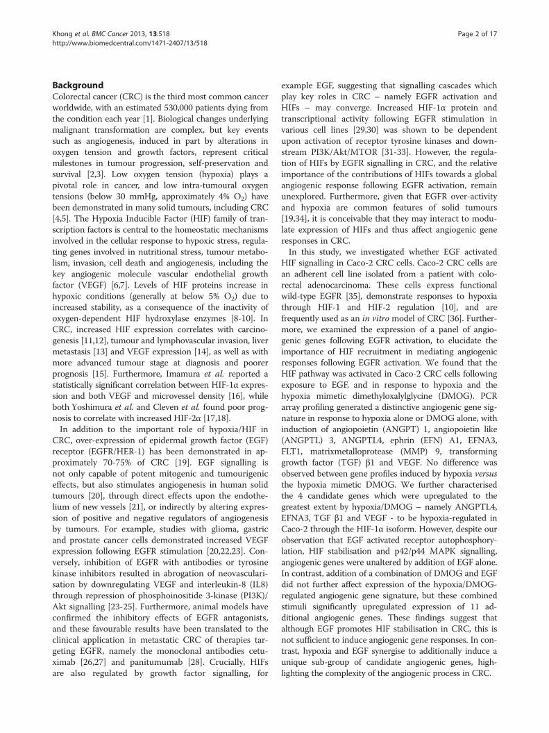
Figure 1 Scatter plot of PCR angiogenesis array analysis ofCaco-2 cells exposed to hypoxia and the hypoxia mimeticDMOG. Caco-2 cells were exposed to either (a) hypoxia (1% O2) or(b) DMOG (1 mM) for 24 hours. Scatter plot graphs are 2-ΔCt valuesfor genes expressed by Caco-2 and normalised against HKG ActB(β-actin), 18S rRNA, HPRT1 (hypoxanthine phosphoribosyltransferase1) and RPL13A (60S ribosomal protein L13a). Solid lines show nochange, dashed lines show ≥2-fold increase and decrease versusuntreated cells. Genes whose expression in both treated anduntreated samples was below detection limits of the array are notincluded. Only genes whose expression changed ≥2-fold are anno-tated. Data are from a representative array performed in duplicateusing cDNA pooled from 3 different replicate experiments.
Khong et al. BMC Cancer 2013, 13:518 Page 4 of 17http://www.biomedcentral.com/1471-2407/13/518
Western blotting was performed using total proteinlysates from cells harvested and lysed with urea buffer(8 M urea, 1% Sodium Dodecyl Sulphate, 1% glycerol and10 mM Tris (pH6.8), 0.5 mM protease inhibitor cocktail(Sigma-Aldrich, Poole, UK), 1 mM dithiothreitol) forHIFs, or RIPA buffer (50 mM Tris pH 7.4, 150 mM NaCl,1% Triton X, 0.1% SDS, 5 mM MgCl2, 50 mM NaF,50 mM DTT, 2 mM orthovanadate, 5 mg/mL sodiumdeoxycholate, 10 mM sodium pyrophosphate, 25 mMβ-glycerophosphate, 2 mM EDTA, 2 mM PMSF and pro-tease inhibitor cocktail P-8340 (Sigma, St Louis, MO,USA) for signalling studies. Samples were resolved onSDS-polyacrylamide gels, where a 3-8% Tris-AcetateNuPAGE® Novex gel (Invitrogen, Carlsbad, CA, USA) wasused for EGFR signalling studies, and a 4-12% Bis-TrisNuPAGE® Novex gel (Invitrogen, Carlsbad, CA, USA) wasused for signalling and HIF-α protein studies. Rabbitanti-human phospho EGFR (Tyr 1068), phospho EGFR(Tyr 845), phospho p38 MAP Kinase (Thr 180/Tyr 182),phospho p44/42 MAP Kinase (Thr 202/Tyr 204),phospho-Akt (Ser 473), total EGFR, total p38 MAPK andtotal p44/42 MAPK were from Cell Signaling Technology(Danvers, MA, USA). Mouse anti-human HIF-1α andHIF-2α (EPAS) were from Becton Dickinson (FranklinLakes, NJ, USA) and Santa Cruz Biotechnology (SantaCruz, CA, USA) respectively. Secondary anti-rabbit andmouse HRP-conjugated antibodies were from Dako-Cytomation (Glostrup, Denmark). Whole cell lysate ofEGF-treated A431 epithelial carcinoma cells used as posi-tive control was from Santa Cruz Biotechnology (SantaCruz, CA, USA). Densitometry was performed usingPhoretix 1D analysis software (TotalLab Ltd, Newcastleupon Tyne, UK).
Statistical analysesStatistical significance was evaluated with 1-way ANOVAwith Dunnett’s post-hoc test to compare selected groupsof data. The ΔCt values were used to determine the sta-tistical significance of differences between groups forPCR-based studies. 2-way ANOVA with Bonferroni cor-rection was used to compare selected groups of data withrespect to time.
ResultsHIF-dependent induction of angiogenic genes in Caco-2cells in response to hypoxia and the hypoxia mimeticDMOGSince hypoxia is likely to be a key stimulus for angioge-nesis in CRC, we first investigated the angiogenic geneprofile of Caco-2 cells exposed to either hypoxia or thehypoxia mimetic DMOG. Figure 1 and Table 1 illustratethe Human Angiogenesis RT2 Profiler™ PCR array dataas scatter plots, and show that 9 pro-angiogenic geneswere significantly changed by a factor of at least 2.0-fold
in response to either hypoxia (Figure 1a) or DMOG(Figure 1b), including VEGF-A, known to be highly regu-lated by hypoxia in various cell types (fold increase 3.1and 3.4 in response to hypoxia and DMOG respectively).Furthermore, 8 hypoxia-regulated genes were identified
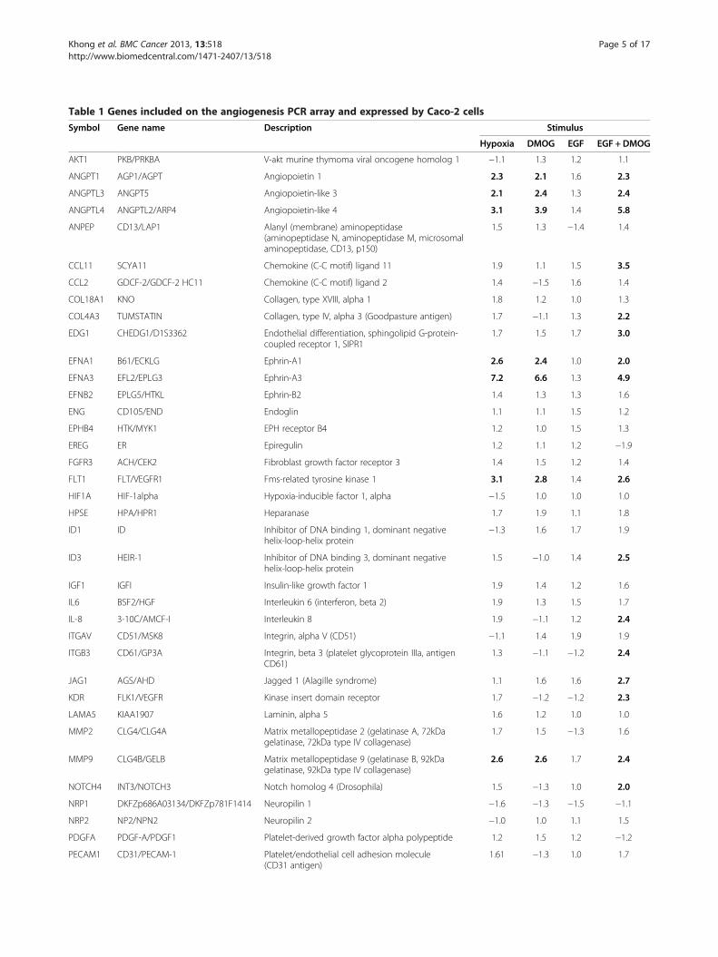
Table 1 Genes included on the angiogenesis PCR array and expressed by Caco-2 cells
Symbol Gene name Description Stimulus
Hypoxia DMOG EGF EGF + DMOG
AKT1 PKB/PRKBA V-akt murine thymoma viral oncogene homolog 1 −1.1 1.3 1.2 1.1
ANGPT1 AGP1/AGPT Angiopoietin 1 2.3 2.1 1.6 2.3
ANGPTL3 ANGPT5 Angiopoietin-like 3 2.1 2.4 1.3 2.4
ANGPTL4 ANGPTL2/ARP4 Angiopoietin-like 4 3.1 3.9 1.4 5.8
ANPEP CD13/LAP1 Alanyl (membrane) aminopeptidase(aminopeptidase N, aminopeptidase M, microsomalaminopeptidase, CD13, p150)
1.5 1.3 −1.4 1.4
CCL11 SCYA11 Chemokine (C-C motif) ligand 11 1.9 1.1 1.5 3.5
CCL2 GDCF-2/GDCF-2 HC11 Chemokine (C-C motif) ligand 2 1.4 −1.5 1.6 1.4
COL18A1 KNO Collagen, type XVIII, alpha 1 1.8 1.2 1.0 1.3
COL4A3 TUMSTATIN Collagen, type IV, alpha 3 (Goodpasture antigen) 1.7 −1.1 1.3 2.2
EDG1 CHEDG1/D1S3362 Endothelial differentiation, sphingolipid G-protein-coupled receptor 1, SIPR1
1.7 1.5 1.7 3.0
EFNA1 B61/ECKLG Ephrin-A1 2.6 2.4 1.0 2.0
EFNA3 EFL2/EPLG3 Ephrin-A3 7.2 6.6 1.3 4.9
EFNB2 EPLG5/HTKL Ephrin-B2 1.4 1.3 1.3 1.6
ENG CD105/END Endoglin 1.1 1.1 1.5 1.2
EPHB4 HTK/MYK1 EPH receptor B4 1.2 1.0 1.5 1.3
EREG ER Epiregulin 1.2 1.1 1.2 −1.9
FGFR3 ACH/CEK2 Fibroblast growth factor receptor 3 1.4 1.5 1.2 1.4
FLT1 FLT/VEGFR1 Fms-related tyrosine kinase 1 3.1 2.8 1.4 2.6
HIF1A HIF-1alpha Hypoxia-inducible factor 1, alpha −1.5 1.0 1.0 1.0
HPSE HPA/HPR1 Heparanase 1.7 1.9 1.1 1.8
ID1 ID Inhibitor of DNA binding 1, dominant negativehelix-loop-helix protein
−1.3 1.6 1.7 1.9
ID3 HEIR-1 Inhibitor of DNA binding 3, dominant negativehelix-loop-helix protein
1.5 −1.0 1.4 2.5
IGF1 IGFI Insulin-like growth factor 1 1.9 1.4 1.2 1.6
IL6 BSF2/HGF Interleukin 6 (interferon, beta 2) 1.9 1.3 1.5 1.7
IL-8 3-10C/AMCF-I Interleukin 8 1.9 −1.1 1.2 2.4
ITGAV CD51/MSK8 Integrin, alpha V (CD51) −1.1 1.4 1.9 1.9
ITGB3 CD61/GP3A Integrin, beta 3 (platelet glycoprotein IIIa, antigenCD61)
1.3 −1.1 −1.2 2.4
JAG1 AGS/AHD Jagged 1 (Alagille syndrome) 1.1 1.6 1.6 2.7
KDR FLK1/VEGFR Kinase insert domain receptor 1.7 −1.2 −1.2 2.3
LAMA5 KIAA1907 Laminin, alpha 5 1.6 1.2 1.0 1.0
MMP2 CLG4/CLG4A Matrix metallopeptidase 2 (gelatinase A, 72kDagelatinase, 72kDa type IV collagenase)
1.7 1.5 −1.3 1.6
MMP9 CLG4B/GELB Matrix metallopeptidase 9 (gelatinase B, 92kDagelatinase, 92kDa type IV collagenase)
2.6 2.6 1.7 2.4
NOTCH4 INT3/NOTCH3 Notch homolog 4 (Drosophila) 1.5 −1.3 1.0 2.0
NRP1 DKFZp686A03134/DKFZp781F1414 Neuropilin 1 −1.6 −1.3 −1.5 −1.1
NRP2 NP2/NPN2 Neuropilin 2 −1.0 1.0 1.1 1.5
PDGFA PDGF-A/PDGF1 Platelet-derived growth factor alpha polypeptide 1.2 1.5 1.2 −1.2
PECAM1 CD31/PECAM-1 Platelet/endothelial cell adhesion molecule(CD31 antigen)
1.61 −1.3 1.0 1.7
Khong et al. BMC Cancer 2013, 13:518 Page 5 of 17http://www.biomedcentral.com/1471-2407/13/518
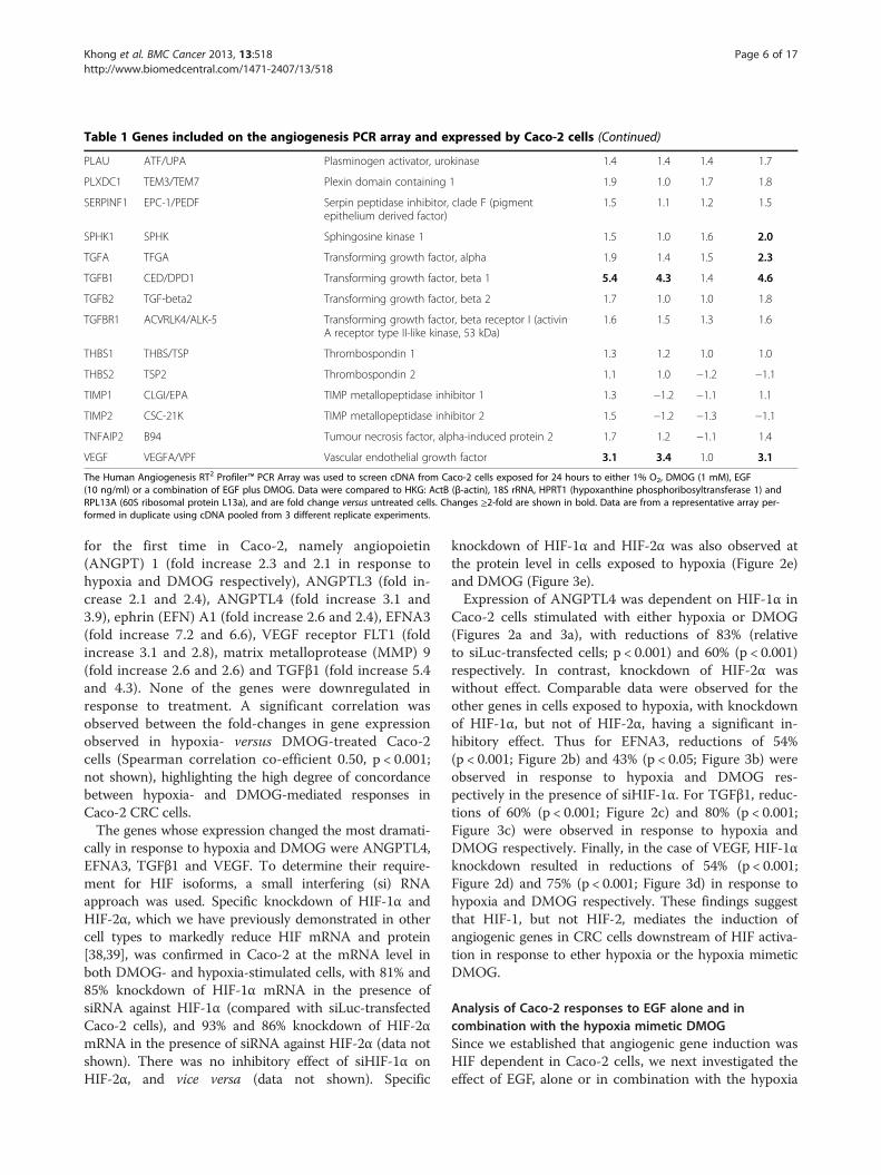
Table 1 Genes included on the angiogenesis PCR array and expressed by Caco-2 cells (Continued)
PLAU ATF/UPA Plasminogen activator, urokinase 1.4 1.4 1.4 1.7
PLXDC1 TEM3/TEM7 Plexin domain containing 1 1.9 1.0 1.7 1.8
SERPINF1 EPC-1/PEDF Serpin peptidase inhibitor, clade F (pigmentepithelium derived factor)
1.5 1.1 1.2 1.5
SPHK1 SPHK Sphingosine kinase 1 1.5 1.0 1.6 2.0
TGFA TFGA Transforming growth factor, alpha 1.9 1.4 1.5 2.3
TGFB1 CED/DPD1 Transforming growth factor, beta 1 5.4 4.3 1.4 4.6
TGFB2 TGF-beta2 Transforming growth factor, beta 2 1.7 1.0 1.0 1.8
TGFBR1 ACVRLK4/ALK-5 Transforming growth factor, beta receptor I (activinA receptor type II-like kinase, 53 kDa)
1.6 1.5 1.3 1.6
THBS1 THBS/TSP Thrombospondin 1 1.3 1.2 1.0 1.0
THBS2 TSP2 Thrombospondin 2 1.1 1.0 −1.2 −1.1
TIMP1 CLGI/EPA TIMP metallopeptidase inhibitor 1 1.3 −1.2 −1.1 1.1
TIMP2 CSC-21K TIMP metallopeptidase inhibitor 2 1.5 −1.2 −1.3 −1.1
TNFAIP2 B94 Tumour necrosis factor, alpha-induced protein 2 1.7 1.2 −1.1 1.4
VEGF VEGFA/VPF Vascular endothelial growth factor 3.1 3.4 1.0 3.1
The Human Angiogenesis RT2 Profiler™ PCR Array was used to screen cDNA from Caco-2 cells exposed for 24 hours to either 1% O2, DMOG (1 mM), EGF(10 ng/ml) or a combination of EGF plus DMOG. Data were compared to HKG: ActB (β-actin), 18S rRNA, HPRT1 (hypoxanthine phosphoribosyltransferase 1) andRPL13A (60S ribosomal protein L13a), and are fold change versus untreated cells. Changes ≥2-fold are shown in bold. Data are from a representative array per-formed in duplicate using cDNA pooled from 3 different replicate experiments.
Khong et al. BMC Cancer 2013, 13:518 Page 6 of 17http://www.biomedcentral.com/1471-2407/13/518
for the first time in Caco-2, namely angiopoietin(ANGPT) 1 (fold increase 2.3 and 2.1 in response tohypoxia and DMOG respectively), ANGPTL3 (fold in-crease 2.1 and 2.4), ANGPTL4 (fold increase 3.1 and3.9), ephrin (EFN) A1 (fold increase 2.6 and 2.4), EFNA3(fold increase 7.2 and 6.6), VEGF receptor FLT1 (foldincrease 3.1 and 2.8), matrix metalloprotease (MMP) 9(fold increase 2.6 and 2.6) and TGFβ1 (fold increase 5.4and 4.3). None of the genes were downregulated inresponse to treatment. A significant correlation wasobserved between the fold-changes in gene expressionobserved in hypoxia- versus DMOG-treated Caco-2cells (Spearman correlation co-efficient 0.50, p < 0.001;not shown), highlighting the high degree of concordancebetween hypoxia- and DMOG-mediated responses inCaco-2 CRC cells.The genes whose expression changed the most dramati-
cally in response to hypoxia and DMOG were ANGPTL4,EFNA3, TGFβ1 and VEGF. To determine their require-ment for HIF isoforms, a small interfering (si) RNAapproach was used. Specific knockdown of HIF-1α andHIF-2α, which we have previously demonstrated in othercell types to markedly reduce HIF mRNA and protein[38,39], was confirmed in Caco-2 at the mRNA level inboth DMOG- and hypoxia-stimulated cells, with 81% and85% knockdown of HIF-1α mRNA in the presence ofsiRNA against HIF-1α (compared with siLuc-transfectedCaco-2 cells), and 93% and 86% knockdown of HIF-2αmRNA in the presence of siRNA against HIF-2α (data notshown). There was no inhibitory effect of siHIF-1α onHIF-2α, and vice versa (data not shown). Specific
knockdown of HIF-1α and HIF-2α was also observed atthe protein level in cells exposed to hypoxia (Figure 2e)and DMOG (Figure 3e).Expression of ANGPTL4 was dependent on HIF-1α in
Caco-2 cells stimulated with either hypoxia or DMOG(Figures 2a and 3a), with reductions of 83% (relativeto siLuc-transfected cells; p < 0.001) and 60% (p < 0.001)respectively. In contrast, knockdown of HIF-2α waswithout effect. Comparable data were observed for theother genes in cells exposed to hypoxia, with knockdownof HIF-1α, but not of HIF-2α, having a significant in-hibitory effect. Thus for EFNA3, reductions of 54%(p < 0.001; Figure 2b) and 43% (p < 0.05; Figure 3b) wereobserved in response to hypoxia and DMOG res-pectively in the presence of siHIF-1α. For TGFβ1, reduc-tions of 60% (p < 0.001; Figure 2c) and 80% (p < 0.001;Figure 3c) were observed in response to hypoxia andDMOG respectively. Finally, in the case of VEGF, HIF-1αknockdown resulted in reductions of 54% (p < 0.001;Figure 2d) and 75% (p < 0.001; Figure 3d) in response tohypoxia and DMOG respectively. These findings suggestthat HIF-1, but not HIF-2, mediates the induction ofangiogenic genes in CRC cells downstream of HIF activa-tion in response to ether hypoxia or the hypoxia mimeticDMOG.
Analysis of Caco-2 responses to EGF alone and incombination with the hypoxia mimetic DMOGSince we established that angiogenic gene induction wasHIF dependent in Caco-2 cells, we next investigated theeffect of EGF, alone or in combination with the hypoxia
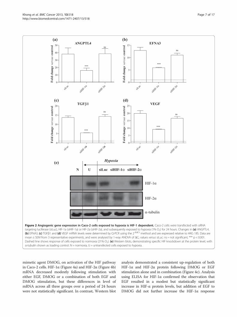
Figure 2 Angiogenic gene expression in Caco-2 cells exposed to hypoxia is HIF-1 dependent. Caco-2 cells were transfected with siRNAtargeting luciferase (siLuc), HIF-1a (siHIF-1a) or HIF-2a (siHIF-2a), and subsequently exposed to hypoxia (1% O2) for 24 hours. Changes in (a) ANGPTL4,(b) EFNA3, (c) TGFβ1 and (d) VEGF mRNA levels were determined by Q-PCR using the 2-ΔΔCt method and are expressed relative to HKG 18S. Data aremean ± SEM from 3 representative experiments, and were analysed by 1-way ANOVA of ΔCt values versus siLuc: ns = not significant, *** p < 0.001.Dashed line shows response of cells exposed to normoxia (21% O2). (e) Western blots, demonstrating specific HIF knockdown at the protein level, withα-tubulin shown as loading control. N = normoxia, U = untransfected cells exposed to hypoxia.
Khong et al. BMC Cancer 2013, 13:518 Page 7 of 17http://www.biomedcentral.com/1471-2407/13/518
mimetic agent DMOG, on activation of the HIF pathwayin Caco-2 cells. HIF-1α (Figure 4a) and HIF-2α (Figure 4b)mRNA decreased modestly following stimulation witheither EGF, DMOG or a combination of both EGF andDMOG stimulation, but these differences in level ofmRNA across all three groups over a period of 24 hourswere not statistically significant. In contrast, Western blot
analysis demonstrated a consistent up-regulation of bothHIF-1α and HIF-2α protein following DMOG or EGFstimulation alone and in combination (Figure 4c). Analysisusing ELISA for HIF-1α confirmed the observation thatEGF resulted in a modest but statistically significantincrease in HIF-α protein levels, but addition of EGF toDMOG did not further increase the HIF-1α response
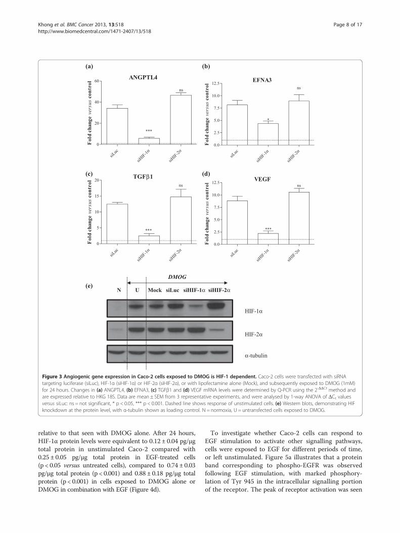
Figure 3 Angiogenic gene expression in Caco-2 cells exposed to DMOG is HIF-1 dependent. Caco-2 cells were transfected with siRNAtargeting luciferase (siLuc), HIF-1α (siHIF-1α) or HIF-2α (siHIF-2α), or with lipofectamine alone (Mock), and subsequently exposed to DMOG (1mM)for 24 hours. Changes in (a) ANGPTL4, (b) EFNA3, (c) TGFβ1 and (d) VEGF mRNA levels were determined by Q-PCR using the 2-ΔΔCt method andare expressed relative to HKG 18S. Data are mean ± SEM from 3 representative experiments, and were analysed by 1-way ANOVA of ΔCt valuesversus siLuc: ns = not significant, * p < 0.05, *** p < 0.001. Dashed line shows response of unstimulated cells. (e) Western blots, demonstrating HIFknockdown at the protein level, with α-tubulin shown as loading control. N = normoxia, U = untransfected cells exposed to DMOG.
Khong et al. BMC Cancer 2013, 13:518 Page 8 of 17http://www.biomedcentral.com/1471-2407/13/518
relative to that seen with DMOG alone. After 24 hours,HIF-1α protein levels were equivalent to 0.12 ± 0.04 pg/μgtotal protein in unstimulated Caco-2 compared with0.25 ± 0.05 pg/μg total protein in EGF-treated cells(p < 0.05 versus untreated cells), compared to 0.74 ± 0.03pg/μg total protein (p < 0.001) and 0.88 ± 0.18 pg/μg totalprotein (p < 0.001) in cells exposed to DMOG alone orDMOG in combination with EGF (Figure 4d).
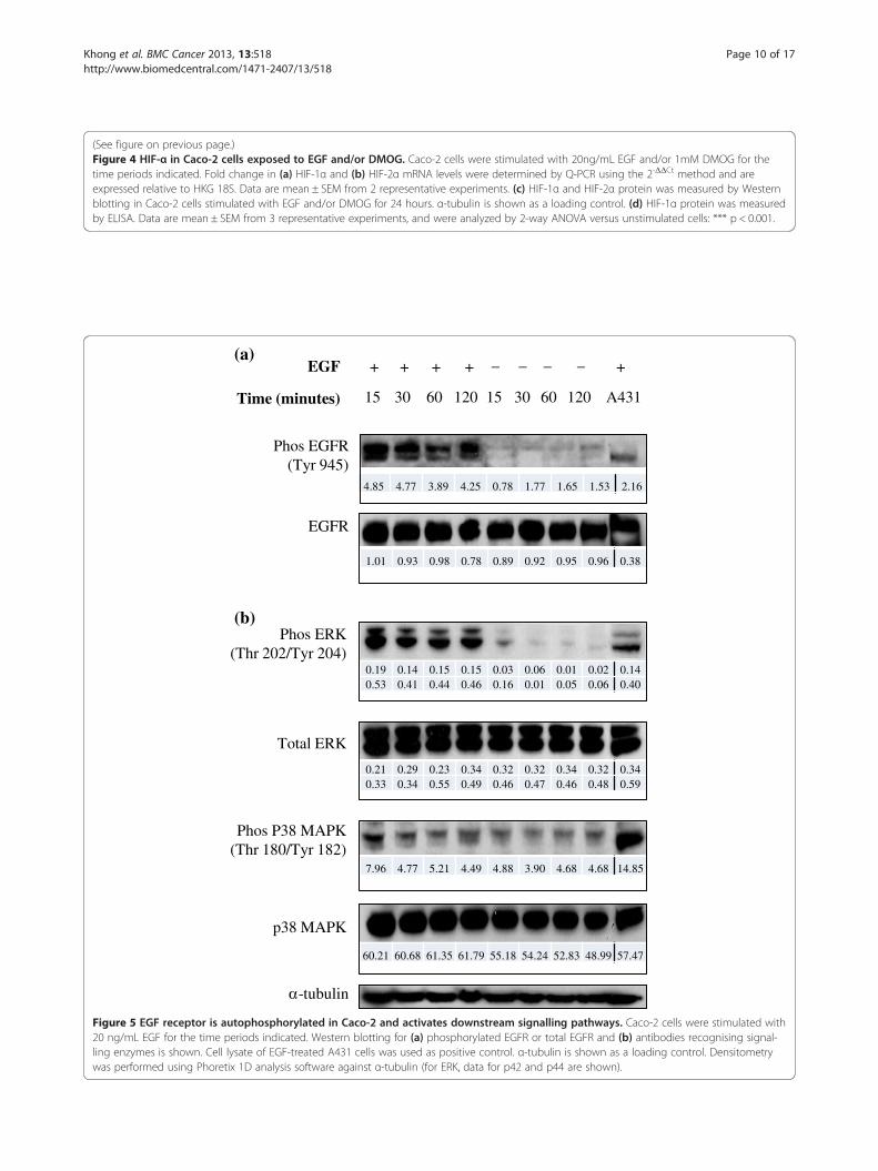
To investigate whether Caco-2 cells can respond toEGF stimulation to activate other signalling pathways,cells were exposed to EGF for different periods of time,or left unstimulated. Figure 5a illustrates that a proteinband corresponding to phospho-EGFR was observedfollowing EGF stimulation, with marked phosphory-lation of Tyr 945 in the intracellular signalling portionof the receptor. The peak of receptor activation was seen
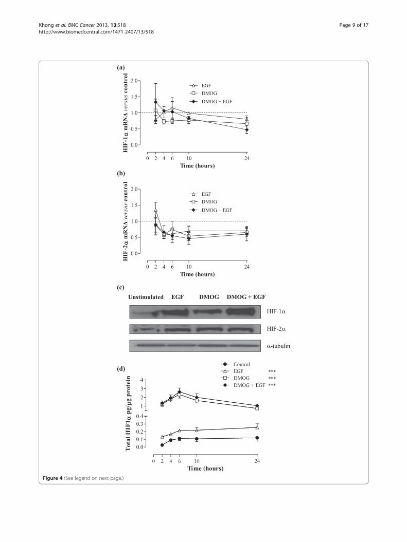
Figure 4 (See legend on next page.)
Khong et al. BMC Cancer 2013, 13:518 Page 9 of 17http://www.biomedcentral.com/1471-2407/13/518
(See figure on previous page.)Figure 4 HIF-α in Caco-2 cells exposed to EGF and/or DMOG. Caco-2 cells were stimulated with 20ng/mL EGF and/or 1mM DMOG for thetime periods indicated. Fold change in (a) HIF-1α and (b) HIF-2α mRNA levels were determined by Q-PCR using the 2-ΔΔCt method and areexpressed relative to HKG 18S. Data are mean ± SEM from 2 representative experiments. (c) HIF-1α and HIF-2α protein was measured by Westernblotting in Caco-2 cells stimulated with EGF and/or DMOG for 24 hours. α-tubulin is shown as a loading control. (d) HIF-1α protein was measuredby ELISA. Data are mean ± SEM from 3 representative experiments, and were analyzed by 2-way ANOVA versus unstimulated cells: *** p < 0.001.
15 30 60 120 15 30 60 120 A431
+ + + + – – – – +
Time (minutes)
EGF
Phos EGFR(Tyr 945)
EGFR
-tubulin
p38 MAPK
Phos P38 MAPK(Thr 180/Tyr 182)
Phos ERK(Thr 202/Tyr 204)
Total ERK
(a)
(b)
1.01 0.93 0.98 0.78 0.89 0.92 0.95 0.96 0.38
0.19 0.14 0.15 0.15 0.03 0.06 0.01 0.02 0.140.53 0.41 0.44 0.46 0.16 0.01 0.05 0.06 0.40
0.21 0.29 0.23 0.34 0.32 0.32 0.34 0.32 0.340.33 0.34 0.55 0.49 0.46 0.47 0.46 0.48 0.59
7.96 4.77 5.21 4.49 4.88 3.90 4.68 4.68 14.85
60.21 60.68 61.35 61.79 55.18 54.24 52.83 48.99 57.47
4.85 4.77 3.89 4.25 0.78 1.77 1.65 1.53 2.16
Figure 5 EGF receptor is autophosphorylated in Caco-2 and activates downstream signalling pathways. Caco-2 cells were stimulated with20 ng/mL EGF for the time periods indicated. Western blotting for (a) phosphorylated EGFR or total EGFR and (b) antibodies recognising signal-ling enzymes is shown. Cell lysate of EGF-treated A431 cells was used as positive control. α-tubulin is shown as a loading control. Densitometrywas performed using Phoretix 1D analysis software against α-tubulin (for ERK, data for p42 and p44 are shown).
Khong et al. BMC Cancer 2013, 13:518 Page 10 of 17http://www.biomedcentral.com/1471-2407/13/518

Khong et al. BMC Cancer 2013, 13:518 Page 11 of 17http://www.biomedcentral.com/1471-2407/13/518
15–30 minutes following stimulation, and progressivelydeclined over the course of 60–120 minutes. Modest auto-phosphorylation of Tyr 1068 following EGF stimulationwas also observed (data not shown).Downstream signalling pathways known to play a role
in Caco-2 cells [40,41] were investigated as potentialsignal transducers involved in initiating various intracel-lular activities resulting from EGF-induced EGFR auto-phosphorylation. Figure 5b confirms markedly higherexpression of phosphorylated p44 MAPK (ERK1) at Thr202 and p42 MAPK (ERK2) at Tyr 204 in EGF-stimulated versus control cells, which was maintainedeven 2 hours after stimulation. The presence of anti-phospho-p38 MAPK protein bands in both stimulatedand unstimulated cells suggests basal activation of p38MAPK in Caco-2, which is not further increased by EGF(although a very modest increase of less than 2-fold wasobserved 15 minutes after EGF addition). Akt phos-phorylation in Caco-2 cells was analysed and found tobe constitutively activated in Caco-2 cells (data notshown).
Figure 6 Angiogenic gene expression in Caco-2 cells exposed to EGFand/or 1 mM DMOG for 24 hours. Changes in (a) ANGPTL4, (b) EFNA3, (c)the 2-ΔΔCt method and are expressed relative to HKG 18S. Data are mean ±ANOVA of ΔCt values versus normoxia (unless otherwise indicated): ns = nounstimulated cells.
Angiogenic gene profiling of Caco-2 cells following EGFRactivationThe above cell signalling studies clearly demonstrate thatEGF is capable of activating downstream signalling inCaco-2 cells, inducing rapid phosphorylation of tyrosineresidues in EGFR, activation of ERK1/2 and stabilisationof HIF proteins. However, in spite of the observedchanges, and in particular despite stabilisation of HIF-1α,expression of the 4 angiogenic HIF-1 target genes, namelyANGPTL4 (Figure 6a), EFNA3 (Figure 6b), TGFβ1(Figure 6c) and VEGF (Figure 6d), was unaffected byaddition of EGF alone. Furthermore, responses induced byDMOG alone were not further altered by addition of EGF(p > 0.05 versus DMOG alone) specifically for these 4angiogenic genes.The Human Angiogenesis RT2 Profiler™ PCR Array
was used to examine the expression of a panel 84 esta-blished angiogenic genes in cells exposed to either EGFalone or in combination with DMOG. None of the geneswhich were detected on the array demonstrated sig-nificant change in expression (either upregulation or
and/or DMOG. Caco-2 cells were stimulated with 20 ng/mL EGFTGFβ1 and (d) VEGF mRNA levels were determined by Q-PCR usingSEM from 3 representative experiments, and were analysed by 1-wayt significant, *** p < 0.001. Dashed line shows response of
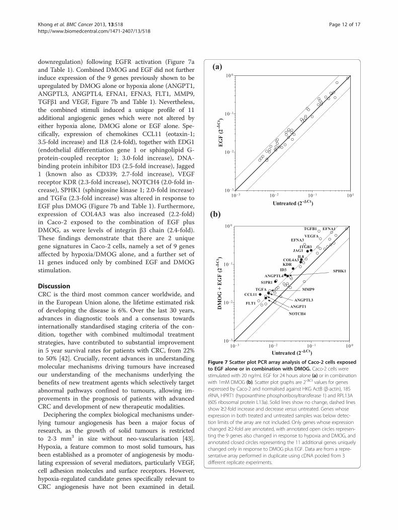
Figure 7 Scatter plot PCR array analysis of Caco-2 cells exposedto EGF alone or in combination with DMOG. Caco-2 cells werestimulated with 20 ng/mL EGF for 24 hours alone (a) or in combinationwith 1mM DMOG (b). Scatter plot graphs are 2-ΔCt values for genesexpressed by Caco-2 and normalised against HKG ActB (β-actin), 18SrRNA, HPRT1 (hypoxanthine phosphoribosyltransferase 1) and RPL13A(60S ribosomal protein L13a). Solid lines show no change, dashed linesshow ≥2-fold increase and decrease versus untreated. Genes whoseexpression in both treated and untreated samples was below detec-tion limits of the array are not included. Only genes whose expressionchanged ≥2-fold are annotated, with annotated open circles represen-ting the 9 genes also changed in response to hypoxia and DMOG, andannotated closed circles representing the 11 additional genes uniquelychanged only in response to DMOG plus EGF. Data are from a repre-sentative array performed in duplicate using cDNA pooled from 3different replicate experiments.
Khong et al. BMC Cancer 2013, 13:518 Page 12 of 17http://www.biomedcentral.com/1471-2407/13/518
downregulation) following EGFR activation (Figure 7aand Table 1). Combined DMOG and EGF did not furtherinduce expression of the 9 genes previously shown to beupregulated by DMOG alone or hypoxia alone (ANGPT1,ANGPTL3, ANGPTL4, EFNA1, EFNA3, FLT1, MMP9,TGFβ1 and VEGF, Figure 7b and Table 1). Nevertheless,the combined stimuli induced a unique profile of 11additional angiogenic genes which were not altered byeither hypoxia alone, DMOG alone or EGF alone. Spe-cifically, expression of chemokines CCL11 (eotaxin-1;3.5-fold increase) and IL8 (2.4-fold), together with EDG1(endothelial differentiation gene 1 or sphingolipid G-protein-coupled receptor 1; 3.0-fold increase), DNA-binding protein inhibitor ID3 (2.5-fold increase), Jagged1 (known also as CD339; 2.7-fold increase), VEGFreceptor KDR (2.3-fold increase), NOTCH4 (2.0-fold in-crease), SPHK1 (sphingosine kinase 1; 2.0-fold increase)and TGFα (2.3-fold increase) was altered in response toEGF plus DMOG (Figure 7b and Table 1). Furthermore,expression of COL4A3 was also increased (2.2-fold)in Caco-2 exposed to the combination of EGF plusDMOG, as were levels of integrin β3 chain (2.4-fold).These findings demonstrate that there are 2 uniquegene signatures in Caco-2 cells, namely a set of 9 genesaffected by hypoxia/DMOG alone, and a further set of11 genes induced only by combined EGF and DMOGstimulation.
DiscussionCRC is the third most common cancer worldwide, andin the European Union alone, the lifetime estimated riskof developing the disease is 6%. Over the last 30 years,advances in diagnostic tools and a consensus towardsinternationally standardised staging criteria of the con-dition, together with combined multimodal treatmentstrategies, have contributed to substantial improvementin 5 year survival rates for patients with CRC, from 22%to 50% [42]. Crucially, recent advances in understandingmolecular mechanisms driving tumours have increasedour understanding of the mechanisms underlying thebenefits of new treatment agents which selectively targetabnormal pathways confined to tumours, allowing im-provements in the prognosis of patients with advancedCRC and development of new therapeutic modalities.Deciphering the complex biological mechanisms under-
lying tumour angiogenesis has been a major focus ofresearch, as the growth of solid tumours is restrictedto 2-3 mm3 in size without neo-vascularisation [43].Hypoxia, a feature common to most solid tumours, hasbeen established as a promoter of angiogenesis by modu-lating expression of several mediators, particularly VEGF,cell adhesion molecules and surface receptors. However,hypoxia-regulated candidate genes specifically relevant toCRC angiogenesis have not been examined in detail.
Khong et al. BMC Cancer 2013, 13:518 Page 13 of 17http://www.biomedcentral.com/1471-2407/13/518
Caco-2 CRC cells are an adherent cell line isolated from apatient with colorectal adenocarcinoma. Their capacity todifferentiate into a polarised monolayer of matureenterocyte-like cells on reaching confluence, which hasled to their adoption as a standard model for in vitrostudies of enteric drug absorption and transport [44],and their widespread used as an in vitro model of CRC[36,41,45,46]. In common with approximately 50% ofcolorectal tumours, Caco-2 cells have a mutant p53 onco-gene, which is known to be associated with increasedVEGF production [47]. Caco-2 cells contain the wild-typeof two other oncogenes, K-ras and BRAF [48,49], muta-tions of which are present in 45% and 15% of colorectaltumours respectively [49,50]. Furthermore, Caco-2 expressreceptors for EGF and release VEGF in response to num-ber of stimuli including hypoxia and K-ras [14,51-53].Inappropriate mucin gene expression is also relatedto CRC development, invasiveness and prognosis, andmucin-5AC, which is expressed in large amounts inCaco-2 cells, has been observed in the early stages ofthe colorectal adenoma-carcinoma sequence [49,54]. Inaddition, Claudin-2, a unique member of the claudinfamily of transmembrane proteins which is significantlyincreased in CRC and correlates with cancer progressionand tumour growth, is regulated in Caco-2 via EGF [55].Caco-2 tumourigenicity has been demonstrated by thedevelopment of moderately-well differentiated adenocar-cinoma in vivo following inoculation into mice [56]. Useof Caco-2 cells thus allows elucidation of mechanisms ofdisease pathogenesis, including angiogenesis [57,58], withpathway-based analysis likely to yield valuable informationat the molecular level that would contribute to our under-standing of the development of CRC.The present study identified VEGF-A, known to be
regulated by hypoxia in other cell types, as a hypoxia-responsive gene in CRC cells, together with 8 additionalhypoxia-regulated genes namely ANGPT1, ANGPTL3,ANGPTL4, EFNA1, EFNA3, VEGF receptor FLT1, MMP9and TGFβ1. An identical angiogenic gene signature rele-vant to CRC was elicited following treatment of Caco-2with the pan-specific HIF hydroxylase inhibitor and HIFactivator DMOG. Genes with the highest change in ex-pression following hypoxia or DMOG stimulation, namelyANGPTL4, EFNA3, TGFβ1 and VEGF, were selected forstudies using RNA knockdown. Previous studies havedemonstrated that hypoxic induction of VEGF in Caco-2cells was in part due to HIF-1α, but this study did notdetect significant levels of HIF-2α [14]. A study byZgouras et al. showing that HIF-1α regulates butyrate-induced normoxic VEGF expression in Caco-2 cells didnot investigate the possible involvement of HIF-2α [57],and while studies have linked HIF-1α expression withapoptosis in Caco-2, none examined the role of HIF-2α[17,59]. In our study, the increase in ANGPTL4,
EFNA3, TGFβ1 and VEGF expression by hypoxia wassignificantly inhibited following knockdown of HIF-1α,with little or no contribution of HIF-2α. Thus, we haveestablished a unique set of angiogenic genes which werehypoxia-regulated in CRC Caco-2 cells, and confirmedan identical expression profile with DMOG stimulation,as well as the dependence of angiogenic responses onHIF-1 by RNA knockdown studies.In addition to the oxygen-dependent regulation of HIF-α
by hypoxia and hypoxia mimetics such as DMOG, sig-nalling by growth factors including EGFR activation hasbeen shown to induce HIF-1α expression in other celltypes under normoxic conditions [60]. The key roleof EGF/EGFR in CRC has been demonstrated by thesuccessful development of EGFR-targeted therapies cetu-ximab and panitumumab. Our study confirmed that EGFRautophosphorylation is associated with HIF-1α and HIF-2αprotein stabilisation under normoxia in Caco-2 cells.Unlike the effect of hypoxia on protein stability due to theinactivity of oxygen-dependent HIF hydroxylases, theobserved increase in HIF-α protein is most probablyattributed to post-transcriptional responses, such as in-creased stability or post-translational modifications, sincemRNA levels of HIF-1α and HIF-2α were not increasedby EGF. A study on breast cancer cells where HER2 sig-nalling specifically induced HIF-1α protein expressionwithout affecting HIF-1α mRNA showed the responsewas dependent upon activation of the PI3K/Akt/FRAPthus increasing rate of protein synthesis [31]. Other stu-dies have also reported increased HIF-1α translation me-diated through PI3K/Akt [33,61]. In order to investigatethe involvement of a similar signalling pathway, we exa-mined activation of EGFR, ERK and p38 MAPK and Akt.Our study on Caco-2 cells illustrated selective activationof MAPK ERK1/2 signalling, in contrast to PI3K/Akt andP38 MAPK which remained constitutively active irrespec-tive of exogenous EGFR stimulation.Since EGFR activation led to HIF upregulation in
Caco-2 cells, a response analogous to that observed withhypoxia or DMOG, we predicted that EGFR-inducedangiogenic gene profile would parallel that induced byhypoxia or DMOG. Such findings would lend furtherimpetus towards developing novel anti-EGFR agentssuch as the monoclonal antibodies cetuximab and pani-tumumab [26,28]. The next part of our study thereforeaimed to decipher the global involvement of known an-giogenic genes in modulating the tumour microenviron-ment. Unexpectedly, our data showed that none of the84 angiogenic genes were affected by EGFR activation,in spite of induction of downstream ERK MAPK signal-ling and stabilisation of HIF-α. The absence of effect ofEGF alone was also validated by Q-PCR for ANGPTL4,EFNA3, TGFβ1 and VEGF, genes which demonstratedsignificant upregulation in a HIF-1-dependent manner
Khong et al. BMC Cancer 2013, 13:518 Page 14 of 17http://www.biomedcentral.com/1471-2407/13/518
following exposure of Caco-2 to DMOG or hypoxia. How-ever, both EGFR over-activation and hypoxia typicallyco-exist within the tumour microenvironment and bothmay impact upon the differential modulation of angio-genic responses induced by either stimulus. We thereforeexamined the effect of simultaneous stimulation of Caco-2CRC cells using EGF and the HIF activator DMOG.Our data demonstrated that the previously establishedhypoxia-regulated angiogenic genes (ANGPT1, ANGPTL3,ANGPTL4, EFNA1, EFNA3, FLT1, MMP9, TGFβ1 andVEGF) were not further affected by addition of EGF. Im-portantly, we have instead identified an additional sub-setof genes which were only expressed following combinedEGF and DMOG, and not with either EGF alone orDMOG/hypoxia alone. The unique profile of 11 additionalangiogenic genes which were only expressed with com-bined EGF and DMOG includes chemokines CCL11(eotaxin-1) and IL8, EDG1 (endothelial differentiationgene 1 or sphingolipid G-protein-coupled receptor 1),DNA-binding protein inhibitor ID3, Jagged 1 (JAG1known also as CD339), VEGF receptor KDR, NOTCH4,SPHK1 (sphingosine kinase 1, which extracellularly acts asa ligand for EDG1) and TGFα. Furthermore, expression ofCOL4A3 (tumstatin, an angiogenesis inhibitor which is acleavage fragment of collagen IV α3 NC1 domain) wasalso increased in Caco-2 exposed to the combination ofEGF plus DMOG, as were levels of integrin β3 chain,which together with αV integrin binds tumstatin via anRGD-independent mechanism. As both EGFR [20] andhypoxia [6] are inducers of angiogenesis, these results sug-gest a novel and previously unreported synergistic rela-tionship which culminates in a downstream response thatsupersedes the angiogenic effect exerted by either of thestimuli in isolation. This synergistic effect may be ex-plained by the positive influence of activated ERK MAPKdownstream of EGFR on the activity of HIF complexes byenhancing recruitment of p300/CREB-binding protein(CBP), thus completing the formation of functionallyactive transcription complexes to transactivate hypoxiaresponse elements of select genes [62]. However it re-mains unclear why a similar response is not elicited inCaco-2 following EGFR activation alone, given that HIFexpression was significantly upregulated (paralleling thatfollowing DMOG treatment) and downstream ERKMAPK signalling was activated. It is conceivable thatdespite activated EGFR increasing expression of HIF, thistranscription factor is functionally inactive due to theactivity of HIF hydroxylase enzymes such as factor inhibi-ting HIF-1 (FIH-1), which interferes with the ability ofHIF to initiate transcription. Under normoxic conditions,hydroxylation of the asparagine residue 803 in thecarboxyl-terminal transcriptional activation domain ofHIF abrogates interactions with the transcriptional co-activators p300 and CBP [63]. Translation of results
from our study to the clinical setting suggests thatinhibition of angiogenesis with EGFR antagonists maybe better targeted at select tumours which are particu-larly hypoxic.The precise roles of ANGPTL4, EFNA3 and TGFβ1,
and the 11 unique genes induced by EGF plus DMOGwhich are not induced by DMOG or hypoxia alone, inregulating CRC angiogenesis remain unknown. ANGPTL4is a member of a family of seven molecules bearing struc-tural homology to angiopoietins [64], and appears tomediate both pro- and anti-angiogenic effects, with theeventual outcome determined by cell-specific contextsand interactions with other angiogenic factors [65-67]. Ofrelevance, a recent study has reported that expression ofANGPTL4 correlates with the depth of tumour invasion,venous invasion and Duke’s classification in CRC [68].EFNA3 was another novel gene identified as being upre-gulated by DMOG and hypoxia in Caco-2 cells. Ephrinsand their cognate receptor tyrosine kinases regulate cellmigration and adhesion, and thereby influence cell lineage,morphogenesis and organogenesis [69,70]. In adult life,ephrin upregulation, particularly of ephrin B, has beencorrelated to vascular invasion, blood vessel formationand sprouting by tumours, and soluble Eph A receptorshave been shown to inhibit tumour angiogenesis [71]. Therole of EFNA3 in CRC angiogenesis remains unproven,although ephrin and Eph receptor over-expression hasbeen reported in a variety of human cancers includingCRC [72,73]. TGFβ has a multifaceted homeostatic role inregulating cell growth and differentiation, angiogenesis,immune function and extracellular matrix formation [74].Overexpression of TGFβ1 in primary CRC is a poorprognostic predictor and correlated with advanced stageof disease, increased risk of recurrence, shorter post-operative survival, particularly in early tumours and de-creased overall survival [75,76]. Regulation of TGFβ1expression by tissue oxygenation remains unstudied inCRC, although HIF-1α has been shown to increase TGFβexpression in prostate cancer cells [77]. Immunohisto-chemical studies have demonstrated a correlation bet-ween TGFβ and VEGF expression, where CRC tissueswith the highest microvessel density expressed bothgrowth factors [78].Although the focus of the study was to investigate the
angiogenic responses induced by EGFR, the receptor,being a member of the ErbB family of receptor tyrosinekinases, also has influence over numerous cellular pro-cesses by triggering multiple signalling cascades. EGFRsignalling promotes DNA synthesis and cell cycle pro-gression by recruiting downstream MAPK, STAT pro-teins, SRC family and Akt protein kinases, which caninduce transcription of genes involved in cell growth,division, differentiation and survival [79-82]. Pre-clinicaland clinical data show that aberrant EGFR and
Khong et al. BMC Cancer 2013, 13:518 Page 15 of 17http://www.biomedcentral.com/1471-2407/13/518
downstream signalling results in cellular transformationwhich can lead to sustained proliferation of abnormal ma-lignant cells [82-84]. Furthermore, stimulation of EGFRpathways has been shown to promote tumour cell inva-sion, motility, adhesion and metastasis [85,86]. Despite theinability to demonstrate angiogenic gene responses follo-wing EGFR activation in our study, EGFR remains animportant feature as preclinical and clinical studies havedemonstrated efficacy of EGFR inhibitors in advancedCRC, particularly in combination with chemo- and radio-therapy [87,88].
ConclusionIn summary, we have identified three novel HIF-1α-regulated angiogenic genes in Caco-2 cells, of which two,ANGPTL4 and TGFβ1, are associated with worse out-come in patients with CRC. In this regard, it is relevantthat we have recently observed that primary cells isolatedenzymatically from tumour resections obtained from pa-tients with CRC also upregulate expression of VEGF,EFNA3, TGFβ1 and ANGPTL4 when exposed to hypoxia,supporting the relevance of studies using Caco-2 cells tounderstand the mechanisms underlying CRC progressionand underlining the potential importance of these angio-genic genes in CRC [89-91]. We subsequently studiedCaco-2 responses to EGF, the action of which is inhibitedby successful CRC treatments, that is anti-EGFR anti-bodies cetuximab and panitumumab. However, despiteour finding that EGFR autophosphorylation led to select-ive downstream activation of p42/p44MAPK and HIF pro-tein stabilisation, this was not sufficient to induceangiogenic gene responses in CRC cells. In contrast, EGFsynergised with the hypoxia mimetic DMOG to inducethe expression of a unique subset of angiogenic genes.Our findings support a key role for tissue hypoxia in eli-citing angiogenic gene responses in CRC cells, also incombination with EGF, and highlight the complex inter-relationship between tumour hypoxia, EGF and angio-genesis in the pathogenesis of CRC.
AbbreviationsANGPT1: Angiopoietin 1; ANGPTL: Angiopoietin like; COL4A3: Tumstatin,cleavage fragment of collagen IV α3 NC1 domain; CRC: Colorectal cancer;DMOG: Dimethyloxalylglycine; EFN: Ephrin; EGF(R): Epidermal growth factor(receptor); FLT1: Vascular endothelial growth factor receptor 1; HER: Humanepidermal receptor; HIF: Hypoxia inducible factor; HKG: House keeping gene;IL8: Interleukin 8; MMP: Matrixmetalloprotease; TGF: Transforming growthfactor; VEGF: Vascular endothelial growth factor.
Competing interestsThe authors declare that they have no competing interests.
Authors’ contributionsTK, PD and EP conceived and designed the experiments. The experimentswere performed by TK, SK, NT and HL. Paper was written by TK and EP. Allauthors read and approved the final manuscript.
AcknowledgementsThe Kennedy Institute of Rheumatology is supported by Arthritis Research UK.
Author details1Kennedy Institute of Rheumatology, Faculty of Medicine, Imperial College,London, UK. 2Department of Surgery and Cancer, Faculty of Medicine,Imperial College, London, UK. 3Present address: Kennedy Institute ofRheumatology, Nuffield Department of Orthopaedics, Rheumatology andMusculoskeletal Sciences, University of Oxford, 65 Aspenlea Road, LondonW6 8LH, UK.
Received: 15 January 2013 Accepted: 23 October 2013Published: 2 November 2013
References1. Parkin DM, Bray F, Ferlay J, Pisani P: Global cancer statistics, 2002.
CA Cancer J Clin 2005, 55:74–108.2. Khong TL, Larsen H, Raatz Y, Paleolog E: Angiogenesis as a therapeutic
target in arthritis: learning the lessons of the colorectal cancerexperience. Angiogenesis 2007, 10:243–258.
3. Thairu N, Kiriakidis S, Dawson P, Paleolog E: Angiogenesis as a therapeutictarget in arthritis in 2011: learning the lessons of the colorectal cancerexperience. Angiogenesis 2011. Epub ahead of print.
4. Mandriota SJ, Turner KJ, Davies DR, Murray PG, Morgan NV, Sowter HM,Wykoff CC, Maher ER, Harris AL, Ratcliffe PJ, Maxwell PH: HIF activationidentifies early lesions in VHL kidneys: evidence for site-specific tumorsuppressor function in the nephron. Cancer Cell 2002, 1:459–468.
5. Goethals L, Debucquoy A, Perneel C, Geboes K, Ectors N, De Schutter H,Penninckx F, McBride WH, Begg AC, Haustermans KM: Hypoxia in humancolorectal adenocarcinoma: comparison between extrinsic and potentialintrinsic hypoxia markers. Int J Radiat Oncol Biol Phys 2006, 65:246–254.
6. Semenza G: Signal transduction to hypoxia-inducible factor 1. BiochemPharmacol 2002, 64:993–998.
7. Lee JW, Bae SH, Jeong JW, Kim SH, Kim KW: Hypoxia-inducible factor(HIF-1)alpha: its protein stability and biological functions. Exp Mol Med2004, 36:1–12.
8. Epstein AC, Gleadle JM, McNeill LA, Hewitson KS, O'Rourke J, Mole DR,Mukherji M, Metzen E, Wilson MI, Dhanda A, et al: C. elegans EGL-9 andmammalian homologs define a family of dioxygenases that regulate HIFby prolyl hydroxylation. Cell 2001, 107:43–54.
9. Ivan M, Haberberger T, Gervasi DC, Michelson KS, Gunzler V, Kondo K,Yang H, Sorokina I, Conaway RC, Conaway JW, Kaelin WG Jr: Biochemicalpurification and pharmacological inhibition of a mammalian prolylhydroxylase acting on hypoxia-inducible factor. Proc Natl Acad Sci USA2002, 99:13459–13464.
10. Bracken CP, Fedele AO, Linke S, Balrak W, Lisy K, Whitelaw ML, Peet DJ:Cell-specific regulation of hypoxia-inducible factor (HIF)-1alpha andHIF-2alpha stabilization and transactivation in a graded oxygenenvironment. J Biol Chem 2006, 281:22575–22585.
11. Giles RH, Lolkema MP, Snijckers CM, Belderbos M, van der Groep P,Mans DA, van Beest M, van Noort M, Goldschmeding R, van Diest PJ, et al:Interplay between VHL/HIF1alpha and Wnt/beta-catenin pathwaysduring colorectal tumorigenesis. Oncogene 2006, 25:3065–3070.
12. Simiantonaki N, Taxeidis M, Jayasinghe C, Kurzik-Dumke U, Kirkpatrick CJ:Hypoxia-inducible factor 1 alpha expression increases during colorectalcarcinogenesis and tumor progression. BMC Cancer 2008, 8:320.
13. Kuwai T, Kitadai Y, Tanaka S, Onogawa S, Matsutani N, Kaio E, Ito M,Chayama K: Expression of hypoxia-inducible factor-1alpha is associatedwith tumor vascularization in human colorectal carcinoma. Int J Cancer2003, 105:176–181.
14. Mizukami Y, Li J, Zhang X, Zimmer MA, Iliopoulos O, Chung DC:Hypoxia-inducible factor-1-independent regulation of vascularendothelial growth factor by hypoxia in colon cancer. Cancer Res2004, 64:1765–1772.
15. Rasheed S, Harris AL, Tekkis PP, Turley H, Silver A, McDonald PJ,Talbot IC, Glynne-Jones R, Northover JM, Guenther T: Hypoxia-induciblefactor-1alpha and -2alpha are expressed in most rectal cancers but onlyhypoxia-inducible factor-1alpha is associated with prognosis. Br J Cancer2009, 100:1666–1673.
16. Imamura T, Kikuchi H, Herraiz MT, Park DY, Mizukami Y, Mino-Kenduson M,Lynch MP, Rueda BR, Benita Y, Xavier RJ, Chung DC: HIF-1alpha andHIF-2alpha have divergent roles in colon cancer. Int J Cancer 2009,124:763–771.
Khong et al. BMC Cancer 2013, 13:518 Page 16 of 17http://www.biomedcentral.com/1471-2407/13/518
17. Yoshimura H, Dhar DK, Kohno H, Kubota H, Fujii T, Ueda S, Kinugasa S,Tachibana M, Nagasue N: Prognostic impact of hypoxia-inducible factors1alpha and 2alpha in colorectal cancer patients: correlation with tumorangiogenesis and cyclooxygenase-2 expression. Clin Cancer Res 2004,10:8554–8560.
18. Cleven AH, van Engeland M, Wouters BG, de Bruine AP: Stromal expressionof hypoxia regulated proteins is an adverse prognostic factor incolorectal carcinomas. Cell Oncol 2007, 29:229–240.
19. Lockhart AC, Berlin JD: The epidermal growth factor receptor as a targetfor colorectal cancer therapy. Semin Oncol 2005, 32:52–60.
20. Akagi M, Kawaguchi M, Liu W, McCarty MF, Takeda A, Fan F, Stoeltzing O,Parikh AA, Jung YD, Bucana CD, et al: Induction of neuropilin-1 andvascular endothelial growth factor by epidermal growth factor in humangastric cancer cells. Br J Cancer 2003, 88:796–802.
21. Okamura K, Morimoto A, Hamanaka R, Ono M, Kohno K, Uchida Y, Kuwano M:A model system for tumor angiogenesis: involvement of transforminggrowth factor-alpha in tube formation of human microvascular endothelialcells induced by esophageal cancer cells. Biochem Biophys Res Commun1992, 186:1471–1479.
22. Goldman CK, Kim J, Wong WL, King V, Brock T, Gillespie GY: Epidermalgrowth factor stimulates vascular endothelial growth factor productionby human malignant glioma cells: a model of glioblastoma multiformepathophysiology. Mol Biol Cell 1993, 4:121–133.
23. Perrotte P, Matsumoto T, Inoue K, Kuniyasu H, Eve BY, Hicklin DJ, Radinsky R,Dinney CP: Anti-epidermal growth factor receptor antibody C225 inhibitsangiogenesis in human transitional cell carcinoma growingorthotopically in nude mice. Clin Cancer Res 1999, 5:257–265.
24. Petit AM, Rak J, Hung MC, Rockwell P, Goldstein N, Fendly B, Kerbel RS:Neutralizing antibodies against epidermal growth factor and ErbB-2/neureceptor tyrosine kinases down-regulate vascular endothelial growthfactor production by tumor cells in vitro and in vivo: angiogenicimplications for signal transduction therapy of solid tumors. Am J Pathol1997, 151:1523–1530.
25. Ciardiello F, Caputo R, Bianco R, Damiano V, Fontanini G, Cuccato S,De Placido S, Bianco AR, Tortora G: Inhibition of growth factor productionand angiogenesis in human cancer cells by ZD1839 (Iressa), a selectiveepidermal growth factor receptor tyrosine kinase inhibitor. Clin CancerRes 2001, 7:1459–1465.
26. Francoual M, Etienne-Grimaldi MC, Formento JL, Benchimol D, Bourgeon A,Chazal M, Letoublon C, Andre T, Gilly N, Delpero JR, et al: EGFR in colorectalcancer: more than a simple receptor. Ann Oncol 2006, 17:962–967.
27. Johnston JB, Navaratnam S, Pitz MW, Maniate JM, Wiechec E, Baust H,Gingerich J, Skliris GP, Murphy LC, Los M: Targeting the EGFR pathway forcancer therapy. Curr Med Chem 2006, 13:3483–3492.
28. Van Cutsem E, Peeters M, Siena S, Humblet Y, Hendlisz A, Neyns B,Canon JL, Van Laethem JL, Maurel J, Richardson G, et al: Open-labelphase III trial of panitumumab plus best supportive care compared withbest supportive care alone in patients with chemotherapy-refractorymetastatic colorectal cancer. J Clin Oncol 2007, 25:1658–1664.
29. Richard DE, Berra E, Gothie E, Roux D, Pouyssegur J: p42/p44 mitogen-activatedprotein kinases phosphorylate hypoxia-inducible factor 1alpha (HIF-1alpha)and enhance the transcriptional activity of HIF-1. J Biol Chem 1999,274:32631–32637.
30. Shafee N, Kaluz S, Ru N, Stanbridge EJ: PI3K/Akt activity has variablecell-specific effects on expression of HIF target genes, CA9 and VEGF, inhuman cancer cell lines. Cancer Lett 2009, 282:109–115.
31. Laughner E, Taghavi P, Chiles K, Mahon PC, Semenza GL: HER2 (neu)signaling increases the rate of hypoxia-inducible factor 1alpha(HIF-1alpha) synthesis: novel mechanism for HIF-1-mediated vascularendothelial growth factor expression. Mol Cell Biol 2001, 21:3995–4004.
32. Peng XH, Karna P, Cao Z, Jiang BH, Zhou M, Yang L: Cross-talk betweenepidermal growth factor receptor and hypoxia-inducible factor-1alphasignal pathways increases resistance to apoptosis by up-regulatingsurvivin gene expression. J Biol Chem 2006, 281:25903–25914.
33. Zhong H, Chiles K, Feldser D, Laughner E, Hanrahan C, Georgescu MM,Simons JW, Semenza GL: Modulation of hypoxia-inducible factor 1alphaexpression by the epidermal growth factor/phosphatidylinositol 3-kinase/PTEN/AKT/FRAP pathway in human prostate cancer cells: implications fortumor angiogenesis and therapeutics. Cancer Res 2000, 60:1541–1545.
34. Denekamp J: Vascular attack as a therapeutic strategy for cancer.Cancer Metastasis Rev 1990, 9:267–282.
35. Keese M, Magdeburg RJ, Herzog T, Hasenberg T, Offterdinger M,Pepperkok R, Sturm JW, Bastiaens PI: Imaging epidermal growth factorreceptor phosphorylation in human colorectal cancer cells and humantissues. J Biol Chem 2005, 280:27826–27831.
36. Fogh J, Fogh JM, Orfeo T: One hundred and twenty-seven culturedhuman tumor cell lines producing tumors in nude mice. J Natl CancerInst 1977, 59:221–226.
37. Livak KJ, Schmittgen TD: Analysis of relative gene expression data usingreal-time quantitative PCR and the 2(−Delta Delta C(T)) Method.Methods 2001, 25:402–408.
38. Muz B, Larsen H, Madden L, Kiriakidis S, Paleolog EM: Prolyl hydroxylasedomain enzyme 2 is the major player in regulating hypoxic responses inrheumatoid arthritis. Arthritis Rheum 2012, 64:2856–2867.
39. Larsen H, Muz B, Khong TL, Feldmann M, Paleolog EM: Differential effectsof Th1 versus Th2 cytokines in combination with hypoxia on HIFs andangiogenesis in RA. Arthritis Res Ther 2012, 14:R180.
40. Buzzi N, Colicheo A, Boland R, de Boland AR: MAP kinases in proliferatinghuman colon cancer Caco-2 cells. Mol Cell Biochem 2009, 328:201–208.
41. Laprise P, Chailler P, Houde M, Beaulieu JF, Boucher MJ, Rivard N:Phosphatidylinositol 3-kinase controls human intestinal epithelial celldifferentiation by promoting adherens junction assembly and p38 MAPKactivation. J Biol Chem 2002, 277:8226–8234.
42. CRUK: (Cancer Research UK) Cancer Stats: Colorectal Cancer; 2006.43. Folkman J: Tumor angiogenesis: therapeutic implications. N Engl J Med
1971, 285:1182–1186.44. Caro I, Boulenc X, Rousset M, Meunier V, Bourrié M, Julian B, Joyeux H,
Roques C, Berger Y, Zweibaum A, Fabre G: Characterisation of a newlyisolated Caco-2 clone (TC-7), as a model of transport processes andbiotransformation of drugs. Int J Pharm 1995, 116:147–158.
45. Bockmann S, Nebe B: The in vitro effects of H-89, a specific inhibitor ofprotein kinase A, in the human colonic carcinoma cell line Caco-2.Eur J Cancer Prev 2003, 12:469–478.
46. Wang S, Basson MD: Identification of functional domains in AKTresponsible for distinct roles of AKT isoforms in pressure-stimulatedcancer cell adhesion. Exp Cell Res 2008, 314:286–296.
47. Liu Y, Bodmer WF: Analysis of P53 mutations and their expression in56 colorectal cancer cell lines. Proc Natl Acad Sci USA 2006,103:976–981.
48. Brink M, de Goeij AF, Weijenberg MP, Roemen GM, Lentjes MH, Pachen MM,Smits KM, de Bruine AP, Goldbohm RA, van den Brandt PA: K-ras oncogenemutations in sporadic colorectal cancer in The Netherlands CohortStudy. Carcinogenesis 2003, 24:703–710.
49. Kikuchi H, Pino MS, Zeng M, Shirasawa S, Chung DC: Oncogenic KRAS andBRAF differentially regulate hypoxia-inducible factor-1alpha and -2alphain colon cancer. Cancer Res 2009, 69:8499–8506.
50. Baba Y, Huttenhower C, Nosho K, Tanaka N, Shima K, Hazra A, Schernhammer ES,Hunter DJ, Giovannucci EL, Fuchs CS, Ogino S: Epigenomic diversity ofcolorectal cancer indicated by LINE-1 methylation in a database of 869tumors. Mol Cancer 2010, 9:125.
51. Damstrup L, Kuwada SK, Dempsey PJ, Brown CL, Hawkey CJ, Poulsen HS,Wiley HS, Coffey RJ Jr: Amphiregulin acts as an autocrine growth factor intwo human polarizing colon cancer lines that exhibit domain selectiveEGF receptor mitogenesis. Br J Cancer 1999, 80:1012–1019.
52. Yonezawa M, Wada K, Tatsuguchi A, Akamatsu T, Gudis K, Seo T, Mitsui K,Nagata K, Tanaka S, Fujimori S, Sakamoto C: Heregulin-induced VEGFexpression via the ErbB3 signaling pathway in colon cancer.Digestion 2009, 80:215–225.
53. Gentile LB, Piva B, Diaz BL: Hypertonic stress induces VEGF production inhuman colon cancer cell line Caco-2: inhibitory role of autocrine PGE(2).PLoS One 2011, 6:e25193.
54. Bu XD, Li N, Tian XQ, Huang PL: Caco-2 and LS174T cell lines providedifferent models for studying mucin expression in colon cancer.Tissue Cell 2011, 43:201–206.
55. Dhawan P, Ahmad R, Chaturvedi R, Smith JJ, Midha R, Mittal MK, Krishnan M,Chen X, Eschrich S, Yeatman TJ, et al: Claudin-2 expression increasestumorigenicity of colon cancer cells: role of epidermal growth factorreceptor activation. Oncogene 2011, 30:3234–3247.
56. de Bruine AP, de Vries JE, Dinjens WN, Moerkerk PT, van der Linden EP,Pijls MM, ten Kate J, Bosman FT: Human Caco-2 cells transfected withc-Ha-Ras as a model for endocrine differentiation in the large intestine.Differentiation 1993, 53:51–60.
Khong et al. BMC Cancer 2013, 13:518 Page 17 of 17http://www.biomedcentral.com/1471-2407/13/518
57. Zgouras D, Wachtershauser A, Frings D, Stein J: Butyrate impairs intestinaltumor cell-induced angiogenesis by inhibiting HIF-1alpha nucleartranslocation. Biochem Biophys Res Commun 2003, 300:832–838.
58. Matsuo Y, Sawai H, Ma J, Xu D, Ochi N, Yasuda A, Takahashi H, Funahashi H,Takeyama H: IL-1alpha secreted by colon cancer cells enhancesangiogenesis: the relationship between IL-1alpha release and tumorcells’ potential for liver metastasis. J Surg Oncol 2009, 99:361–367.
59. Franovic A, Holterman CE, Payette J, Lee S: Human cancers converge atthe HIF-2alpha oncogenic axis. Proc Natl Acad Sci USA 2009,106:21306–21311.
60. Richard DE, Berra E, Pouyssegur J: Nonhypoxic pathway mediates theinduction of hypoxia-inducible factor 1alpha in vascular smooth musclecells. J Biol Chem 2000, 275:26765–26771.
61. Pore N, Jiang Z, Gupta A, Cerniglia G, Kao GD, Maity A: EGFR tyrosinekinase inhibitors decrease VEGF expression by both hypoxia-induciblefactor (HIF)-1-independent and HIF-1-dependent mechanisms.Cancer Res 2006, 66:3197–3204.
62. Sang N, Stiehl DP, Bohensky J, Leshchinsky I, Srinivas V, Caro J: MAPKsignaling up-regulates the activity of hypoxia-inducible factors by itseffects on p300. J Biol Chem 2003, 278:14013–14019.
63. Mahon PC, Hirota K, Semenza GL: FIH-1: a novel protein that interactswith HIF-1alpha and VHL to mediate repression of HIF-1 transcriptionalactivity. Genes Dev 2001, 15:2675–2686.
64. Katoh Y, Katoh M: Comparative integromics on Angiopoietin familymembers. Int J Mol Med 2006, 17:1145–1149.
65. Galaup A, Cazes A, Le Jan S, Philippe J, Connault E, Le Coz E, Mekid H,Mir LM, Opolon P, Corvol P, et al: Angiopoietin-like 4 prevents metastasisthrough inhibition of vascular permeability and tumor cell motility andinvasiveness. Proc Natl Acad Sci USA 2006, 103:18721–18726.
66. Le Jan S, Amy C, Cazes A, Monnot C, Lamande N, Favier J, Philippe J,Sibony M, Gasc JM, Corvol P, Germain S: Angiopoietin-like 4 is aproangiogenic factor produced during ischemia and in conventionalrenal cell carcinoma. Am J Pathol 2003, 162:1521–1528.
67. Oike Y, Ito Y, Maekawa H, Morisada T, Kubota Y, Akao M, Urano T,Yasunaga K, Suda T: Angiopoietin-related growth factor (AGF) promotesangiogenesis. Blood 2004, 103:3760–3765.
68. Nakayama T, Hirakawa H, Shibata K, Nazneen A, Abe K, Nagayasu T,Taguchi T: Expression of angiopoietin-like 4 (ANGPTL4) in humancolorectal cancer: ANGPTL4 promotes venous invasion and distantmetastasis. Oncol Rep 2011, 25:929–935.
69. Merlos-Suarez A, Batlle E: Eph-ephrin signalling in adult tissues andcancer. Curr Opin Cell Biol 2008, 20:194–200.
70. Surawska H, Ma PC, Salgia R: The role of ephrins and Eph receptors incancer. Cytokine Growth Factor Rev 2004, 15:419–433.
71. Brantley DM, Cheng N, Thompson EJ, Lin Q, Brekken RA, Thorpe PE,Muraoka RS, Cerretti DP, Pozzi A, Jackson D, et al: Soluble Eph A receptorsinhibit tumor angiogenesis and progression in vivo. Oncogene 2002,21:7011–7026.
72. Hafner C, Schmitz G, Meyer S, Bataille F, Hau P, Langmann T, Dietmaier W,Landthaler M, Vogt T: Differential gene expression of Eph receptors andephrins in benign human tissues and cancers. Clin Chem 2004,50:490–499.
73. Batlle E, Bacani J, Begthel H, Jonkheer S, Gregorieff A, van de Born M,Malats N, Sancho E, Boon E, Pawson T, et al: EphB receptor activitysuppresses colorectal cancer progression. Nature 2005, 435:1126–1130.
74. Javelaud D, Mauviel A: Mammalian transforming growth factor-betas:Smad signaling and physio-pathological roles. Int J Biochem Cell Biol 2004,36:1161–1165.
75. Robson H, Anderson E, James RD, Schofield PF: Transforming growthfactor beta 1 expression in human colorectal tumours: an independentprognostic marker in a subgroup of poor prognosis patients. Br J Cancer1996, 74:753–758.
76. Gulubova M, Manolova I, Ananiev J, Julianov A, Yovchev Y, Peeva K: Role ofTGF-beta1, its receptor TGFbetaRII, and Smad proteins in the progressionof colorectal cancer. Int J Colorectal Dis 2010, 25:591–599.
77. Berger AP, Kofler K, Bektic J, Rogatsch H, Steiner H, Bartsch G, Klocker H:Increased growth factor production in a human prostatic stromal cellculture model caused by hypoxia. Prostate 2003, 57:57–65.
78. Xiong B, Gong LL, Zhang F, Hu MB, Yuan HY: TGF beta1 expression andangiogenesis in colorectal cancer tissue. World J Gastroenterol 2002,8:496–498.
79. Ono M, Kuwano M: Molecular mechanisms of epidermal growth factorreceptor (EGFR) activation and response to gefitinib and otherEGFR-targeting drugs. Clin Cancer Res 2006, 12:7242–7251.
80. Fujimoto K, Sheng H, Shao J, Beauchamp RD: Transforming growthfactor-beta1 promotes invasiveness after cellular transformation withactivated Ras in intestinal epithelial cells. Exp Cell Res 2001, 266:239–249.
81. Bowman T, Garcia R, Turkson J, Jove R: STATs in oncogenesis.Oncogene 2000, 19:2474–2488.
82. Testa JR, Bellacosa A: AKT plays a central role in tumorigenesis.Proc Natl Acad Sci USA 2001, 98:10983–10985.
83. Zhou BP, Liao Y, Xia W, Spohn B, Lee MH, Hung MC: Cytoplasmiclocalization of p21Cip1/WAF1 by Akt-induced phosphorylation inHER-2/neu-overexpressing cells. Nat Cell Biol 2001, 3:245–252.
84. Chan TO, Rittenhouse SE, Tsichlis PN: AKT/PKB and other D3phosphoinositide-regulated kinases: kinase activation byphosphoinositide-dependent phosphorylation. Annu Rev Biochem 1999,68:965–1014.
85. Thant AA, Nawa A, Kikkawa F, Ichigotani Y, Zhang Y, Sein TT, Amin AR,Hamaguchi M: Fibronectin activates matrix metalloproteinase-9 secretionvia the MEK1-MAPK and the PI3K-Akt pathways in ovarian cancer cells.Clin Exp Metastasis 2000, 18:423–428.
86. Engebraaten O, Bjerkvig R, Pedersen PH, Laerum OD: Effects of EGF, bFGF,NGF and PDGF(bb) on cell proliferative, migratory and invasivecapacities of human brain-tumour biopsies in vitro. Int J Cancer 1993,53:209–214.
87. Bokemeyer C, Van Cutsem E, Rougier P, Ciardiello F, Heeger S, Schlichting M,Celik I, Kohne CH: Addition of cetuximab to chemotherapy as first-linetreatment for KRAS wild-type metastatic colorectal cancer: pooled analysisof the CRYSTAL and OPUS randomised clinical trials. Eur J Cancer 2012,48:1466–1475.
88. Van Cutsem E, Kohne CH, Hitre E, Zaluski J, Chang Chien CR, Makhson A,D'Haens G, Pinter T, Lim R, Bodoky G, et al: Cetuximab and chemotherapyas initial treatment for metastatic colorectal cancer. N Engl J Med 2009,360:1408–1417.
89. Thairu N, Kiriakidis S, Dawson P, Paleolog E: HIF-Isoforms have divergentroles in the angiogenesis of colorectal cancer. Colorectal Dis 2011,13:20 (P034).
90. Thairu N, Kiriakidis S, Dawson P, Paleolog E: Short-term cultures oftumour-derived colorectal cancer cells – a novel in vitro model for theevaluation of angiogenesis in colorectal cancer. Br J Surg 2012,99:7 (abstract O637).
91. Thairu N, Kiriakidis S, Dawson P, Paleolog E: Short-term cultures oftumour-derived colorectal cancer cells – a novel in vitro model for theevaluation of angiogenesis in colorectal cancer. Colorectal Dis 2012,14:16 (P027).
doi:10.1186/1471-2407-13-518Cite this article as: Khong et al.: Identification of the angiogenic genesignature induced by EGF and hypoxia in colorectal cancer. BMC Cancer2013 13:518.
Submit your next manuscript to BioMed Centraland take full advantage of:
• Convenient online submission
• Thorough peer review
• No space constraints or color figure charges
• Immediate publication on acceptance
• Inclusion in PubMed, CAS, Scopus and Google Scholar
• Research which is freely available for redistribution
Submit your manuscript at www.biomedcentral.com/submit