Embed Size (px)
Citation preview
University of Kentucky University of Kentucky
UKnowledge UKnowledge
University of Kentucky Doctoral Dissertations Graduate School
2006
IDENTIFICATION OF VIRAL AND HOST FACTORS INVOLVED IN IDENTIFICATION OF VIRAL AND HOST FACTORS INVOLVED IN
TOMBUSVIRUS REPLICATION AND RECOMBINATION TOMBUSVIRUS REPLICATION AND RECOMBINATION
Natalia Shapka University of Kentucky
Right click to open a feedback form in a new tab to let us know how this document benefits you. Right click to open a feedback form in a new tab to let us know how this document benefits you.
Recommended Citation Recommended Citation Shapka, Natalia, "IDENTIFICATION OF VIRAL AND HOST FACTORS INVOLVED IN TOMBUSVIRUS REPLICATION AND RECOMBINATION" (2006). University of Kentucky Doctoral Dissertations. 449. https://uknowledge.uky.edu/gradschool_diss/449
This Dissertation is brought to you for free and open access by the Graduate School at UKnowledge. It has been accepted for inclusion in University of Kentucky Doctoral Dissertations by an authorized administrator of UKnowledge. For more information, please contact [email protected].
ABSTRACT OF DISSERTATION
Natalia Shapka
The Graduate School University of Kentucky
2006
IDENTIFICATION OF VIRAL AND HOST FACTORS INVOLVED IN TOMBUSVIRUS REPLICATION AND RECOMBINATION
___________________________________________
ABSTRACT OF DISSERTATION ___________________________________________
A dissertation submitted in partial fulfillment of the
requirements for the degree of Doctor of Philosophy in the College of Agriculture
at the University of Kentucky
By Natalia Shapka
Lexington, Kentucky
Director: Dr. Peter D. Nagy, Associate Professor of Plant Pathology Lexington, Kentucky
2006
ABSTRACT OF DISSERTATION
IDENTIFICATION OF VIRAL AND HOST FACTORS INVOLVED IN TOMBUSVIRUS REPLICATION AND RECOMBINATION
Rapid evolution of RNA viruses with mRNA-sense genomes is a major concern
to health and economic welfare due to the devastating diseases these viruses inflict on
humans, animals and plants. Rapid viral RNA evolution is frequently due to RNA
recombination, which can be facilitated by recombination signals present in viral RNAs.
Among such signals are short sequences with high AU contents that constitute
recombination hot spots in Brome mosaic virus (BMV) and retroviruses. We have
demonstrated that a defective interfering (DI) RNA, a model template associated with
Tomato bushy stunt virus (TBSV), a tombusvirus, undergoes frequent recombination in
plants and protoplast cells when it carries the AU-rich hot spot sequence from BMV.
Similar to the situation with BMV, most of the recombination junction sites in the DI
RNA recombinants were found within the AU-rich region. Our results support the idea
that common AU-rich recombination signals might promote interviral recombination
between unrelated viruses.
To test if host genes can affect the evolution of RNA viruses, we used a
Saccharomyces cerevisiae single-gene deletion library, which includes ~80% of yeast
genes, in RNA recombination studies based on a small viral replicon RNA derived from
TBSV. The genome-wide screen led to the identification of five host genes, whose
absence resulted in rapid generation of novel viral RNA recombinants. Thus, these genes
normally suppress viral RNA recombination, but in their absence hosts become viral
recombination “hotbeds”. Four of the five recombination suppressor genes are likely
involved in RNA degradation, suggesting that RNA degradation could play a role in viral
RNA recombination. Overall, our results demonstrate for the first time that a set of host
genes have major effect on RNA virus recombination and evolution.
Replication of the non-segmented, plus-stranded RNA genome of Cucumber necrosis
tombusvirus (CNV) requires two essential overlapping viral-coded replication proteins,
the p33 replication co-factor and the p92 RNA-dependent RNA polymerase. We have
demonstrated that p33 is phosphorylated in vivo and in vitro by a membrane-bound plant
kinase. Based on in vitro studies with purified recombinant p33, we show evidence for
phosphorylation of threonine and serine residues adjacent to the essential RNA-binding
site in p33. Our findings suggest that phosphorylation of threonine/serine residues
adjacent to the essential RNA-binding site in the auxiliary p33 protein likely plays a role
in viral RNA replication and subgenomic RNA synthesis during tombusvirus infections.
Key words: Tomato bushy stunt virus, RNA-dependent RNA polymerase, Defective
interfering RNA, Yeast, Recombination
___________________________________________
IDENTIFICATION OF VIRAL AND HOST FACTORS INVOLVED IN TOMBUSVIRUS REPLICATION AND RECOMBINATION
By
Natalia Shapka
_________________________________________ Director of Dissertation
_________________________________________ Director of Graduate Studies
_________________________________________
Copyright © Natalia Shapka 2006.
RULES FOR THE USE OF DISSERTATIONS
Unpublished dissertations submitted for the Doctors degree and deposited in the University of Kentucky Library are as a rule open for inspection, but are used only with
due regard to the rights of the authors. Bibliographical references may be noted, but quotations or summaries of parts may be published only with permission of the author,
and with the usual scholarly acknowledgments.
Extensive copying or publication of the dissertation in whole or in part also requires the consent of the Dean of the Graduate School of the University of Kentucky.
A library that borrows this dissertation for use by its patrons is expected to secure the signature of each user.
Name Date
__________________________________________________________________
__________________________________________________________________
__________________________________________________________________
__________________________________________________________________
__________________________________________________________________
__________________________________________________________________
__________________________________________________________________
__________________________________________________________________
__________________________________________________________________
__________________________________________________________________
DISSERTATION
Natalia Shapka
The Graduate School University of Kentucky
2006
IDENTIFICATION OF VIRAL AND HOST FACTORS INVOLVED IN TOMBUSVIRUS REPLICATION AND RECOMBINATION
___________________________________________
DISSERTATION
___________________________________________
A dissertation submitted in partial fulfillment of the requirements for the degree of Doctor of Philosophy in the
College of Agriculture at the University of Kentucky
By
Natalia Shapka
Lexington, Kentucky
Director: Dr. Peter D. Nagy, Associate Professor of Plant Pathology
Lexington, Kentucky
2006
Copyright © Natalia Shapka 2006.
i
TABLE OF CONTENTS List of Tables .........................................................................................................ii List of Figures .......................................................................................................iii List of Files………………………………………………………………………..v Chapter I: Introduction...........................................................................................1 RNA recombination in model virus systems….....................................................2 Role of host factors in RNA recombination ..........................................................2 Tombusviruses are ideal model plus-stranded RNA viruses .................................3 What the thesis will show.......................................................................................5 Chapter II: The AU-rich RNA recombination hot spot sequence of Brome mosaic virus is functional in tombusviruses: Implications for the mechanism of RNA recombination Introduction ...........................................................................................................7 Materials and Methods...........................................................................................10 Results...................................................................................................................14 Discussion .............................................................................................................25 Chapter III: Genome-wide screen identifies host genes affecting viral RNA recombination…………………………………………………………………….49 Introduction ...........................................................................................................49 Materials and Methods...........................................................................................51 Results...................................................................................................................54 Discussion .............................................................................................................60 Chapter IV: Phosphorylation of the p33 replication protein of Cucumber necrosis tombusvirus adjacent to the RNA binding site affects viral RNA replication……77 Introduction ...........................................................................................................77 Materials and Methods...........................................................................................80 Results....................................................................................................................85 Discussion .............................................................................................................95 Chapter V: Summary……………………………………………………………..113 References..............................................................................................................116 Vita ........................................................................................................................132
ii
LIST OF TABLES TABLE 2.1. Primers used for PCR…………………...................………..47 TABLE 3.1. Names and functions of the identified host genes……………75
iii
LIST OF FIGURES
Fig. 2.1. The AU-rich hot spot sequence of BMV promotes recombination in DI RNA of TBSV…………………………………………………………………………………….33
Fig. 2.2. Sequences of DI RNA recombinants around the junction sites……………………36 Fig. 2.3. The role of the RII sequence in DI RNA recombination…………………………..37 Fig. 2.4. Sequences of DI RNA recombinants obtained with RII deletion mutants…………38 Fig. 2.5. The effect of short AU-rich and GC-rich sequences on DI RNA recombination….40 Figure 2.6. Efficient binding of RII(-) to the recombinant p33 and p92 replicase proteins of
TBSV in vitro…………………………………………………………………………….42 Fig. 2.7. Comparison of the level of primer extension obtained with RII(-) and GFP-derived
sequences……………………………………………………………………………… ..44 Fig. 2.8. Models of AU-rich sequence-promoted RNA recombination in tombusviruses…..46 Fig. 3.1. Absence of CTL1, MET22/HAL2, HUR1, XRN1 and UBP3 host genes leads to
enhanced recombination of TBSV DI-72 RNA replicon in yeast……………………….65 Fig. 3.2. Schematic presentation of the DI-72 replicon with four regions and the
recombinants with duplicated 3’ sequences (3’ part of RII, RIII and RIV) and 5’ deletions (RI and 5’ part of RII)………………………………………………………………….. 66
Fig. 3.3. (A) Time-course experiment with hur1Δ and xrn1Δ co-expressing p33 and p92 reveals rapid generation of recombinants (probed with RIII(-) after induction of DI-72 RNA transcription from plasmid pYC/DI-72…………………………………………...67
Fig. 3.4. Deletion of Xrn1p increases the stability of recombinant RNAs and DI-72 replicon RNA……………………………………………………………………………………..68
Fig. 3.5. (A) Absence of PEP7, IPK1, CHO2 and DCI1 host genes leads to low frequency of recombination of TBSV DI-72 RNA replicon in yeast co-expressing p33 and p92 replicase proteins………………………………………………………………………..69
Fig. 3.6. Absence of SPE3 and SPT3 genes results in altered recombination profile with (A) DI-72 RNA replicon and (B) DI-AU-FP replicon………………………………………71
Fig. 3.7. Comparison of recombination activity of DI-AU-FP replicon in xrn1Δ, pep7Δ, and parental yeast strains……………………………………………………………………72
Fig. 3.8. Schematic presentation of the DI-72 replicon-derived recombinants generated in hur1Δ strain expressing the DI-72 RNA replicon………………………………………73
Fig. 3.9. Absence of viral recombinants in yeast DNA and RNA transcripts………………74 Fig. 4.1. Location of the predicted phosphorylation sites in the CNV replication proteins..100 Fig. 4.2. Detection of in vivo phosphorylated replication proteins of CNV and TCV……..101 Fig. 4.3. In vitro phosphorylation of p33 by a plant kinase………………………………..102 Fig. 4.4. In vitro phosphorylation at S210 and T211 residues of p33 by PKC…………….103 Fig. 4.5. Comparison of accumulation of CNV gRNA carrying phosphorylation-mimicking
mutations adjacent to the RPR-motif in p33/p92 gene…………………………………104 Fig. 4.6. Effect of phosphorylation-mimicking mutations on accumulation of (A) CNV (+)
RNAs and (B) p33 replication protein………………………………………………….106 Fig. 4.7. Effect of phosphorylation-mimicking mutations on accumulation of DI-72 RNA in
trans…………………………………………………………………………………….107 Fig. 4.8. Phosphorylation-mimicking mutations affect the function of p33, and a lesser
extent, p92………………………………………………………………………………109 Fig. 4.9. Effect of phosphorylation-mimicking mutations on DI RNA replication in yeast.110
iv
Fig. 4.10. Nonphosphorylation-mimicking mutant of CNV shows delay in gRNA accumulation and symptom formation in N. benthamiana plants……………………111
v
LIST OF FILES
Shapka-Ph.D-dissertation.pdf
1
Chapter I
INTRODUCTION
Due to high frequency mutations and RNA recombination, RNA viruses can change their
genomes frequently to develop new strains or viruses (Aaziz and Tepfer, 1999; Lai, 1992;
Nagy and Simon, 1997; Worobey and Holmes, 1999). Therefore, it is not surprising that
RNA viruses are widespread in nature and that they cause many diseases of humans, animals
and plants. RNA recombination is especially powerful tool for viruses, because it can rapidly
lead to dramatic changes in virus genomes by recombining or rearranging “battle-tested” (i.e.,
evolutionarily successful) sequences. Accordingly, the significant role of RNA recombination
in emergence of new viruses or virus strains is well documented for numerous human, plant,
animal, bacterial, insect and fungal viruses (Aaziz and Tepfer, 1999; Lai, 1992; Nagy and
Simon, 1997; Worobey and Holmes, 1999). A recombinant virus may “jump species”. RNA
recombination can also occur between viral and host sequences, thus leading to the
emergence of recombinant viruses carrying novel host genes or gaining new functions. The
best example is the incorporation of ubiquitin gene into the pestivirus genome that intensifies
the disease symptoms and often leads to the death of the animal (Becher, Orlich, and Thiel,
2001). The increased pathogenicity of an influenza A virus hybrid was possibly due to
recombination with a cellular RNA (Khatchikian, Orlich, and Rott, 1989). Understanding of
the mechanism of RNA recombination is expected to help the development of safer vaccine
strains of human viruses and more effective viral-based gene-delivery vectors in plants and
animals.
2
RNA recombination in model virus systems
The major challenge in studying RNA recombination is that recombination is a chance
event. In contrast to replication, RNA recombination probably does not need to occur in each
virus-infected cell. Due to the complex nature of RNA recombination, experiments require
careful design to bring together the necessary components of RNA recombination. Therefore,
progress in this research area greatly benefits from studies with model viruses. Accordingly,
studies on poliovirus, Brome mosaic virus (BMV), Carmoviruses, and Tombusviruses
(Cascone, Haydar, and Simon, 1993; Jarvis and Kirkegaard, 1991; Kim and Kao, 2001; Lai,
1992; Nagy and Bujarski, 1993; Nagy and Simon, 1997; Pilipenko, Gmyl, and Agol, 1995;
White and Morris, 1994c; Worobey and Holmes, 1999) have contributed greatly to our
understanding of RNA recombination in general. Based on current models, the most frequent
RNA recombination events are driven by the viral replicase, which is proposed to “jump”
from one site or from one RNA molecule to another during RNA synthesis (Jarvis and
Kirkegaard, 1991; Nagy and Simon, 1997). Accordingly, the viral replicase-driven template-
switching mechanism has been demonstrated for Tombus- and Carmoviruses, and for BMV
in vitro (Cheng and Nagy, 2003; Cheng, Pogany, and Nagy, 2002; Kim and Kao, 2001;
Nagy, Zhang, and Simon, 1998). In spite of the above studies, our understanding of RNA
recombination is incomplete and it is not yet known whether host factors are involved in viral
RNA recombination.
Role of host factors in RNA recombination
3
Unfortunately, there is no published information on direct roles of host genes in RNA
recombination. Yet, indirect observations, such as the variable frequency of viral RNA
recombination in different hosts, suggest that the host influences viral recombination. Thus, it
is feasible to assume that host genes have significant roles in RNA recombination. Indeed,
viruses rely very much on their hosts for enzymes, metabolities, energy sources and
membrane surfaces for replication and other processes. For example, replication of RNA
viruses, which is performed by the viral replicase, requires not only viral-coded replicase
proteins, but host proteins, too. Accordingly, the role of host proteins in RNA replication has
been demonstrated for an increasing number of RNA viruses (Ahlquist et al., 2003; Nagy and
Pogany, 2006). In spite of the intensive studies on viral replication, most of the host factors
are still unidentified and uncharacterized, whereas the role of host factors in viral RNA
recombination is completely unknown.
Tombusviruses are ideal model plus-stranded RNA viruses
Tombusviruses, such as Tomato bushy stunt virus (TBSV) and Cucumber necrosis virus
(CNV), are important and emerging plant pathogens that are among the best-characterized
viruses (Nagy and Pogany, 2006; Panavas et al., 2005d; Rajendran and Nagy, 2006; Serviene
et al., 2006; Serviene et al., 2005). They are spherical viruses with monopartite (+)RNA
genomes of ~4.8 kb (Russo, Burgyan, and Martelli, 1994; White and Nagy, 2004). They
belong to supergroup 2 viruses that include important human and animal pathogens (e.g.,
HCV, flaviviruses, pestiviruses), and plant pathogens (luteoviruses, carmoviruses, and
others). Tombusviruses code for five proteins including two replication proteins, termed p33
4
and p92 (Fig. 1.1), which are essential for replication (Oster, Wu, and White, 1998a;
Panaviene, Baker, and Nagy, 2003; Scholthof, Scholthof, and Jackson, 1995b) and
recombination (Panaviene and Nagy, 2003). Both p33 and p92 are translated from the
genomic RNA and p92 is the result of translational readthrough of the p33 stop codon (Fig.
1.1) (Scholthof, Scholthof, and Jackson, 1995b; White and Nagy, 2004). Therefore, the N-
terminal portion of p92 overlaps with p33. p92 is the RdRp (Panaviene, Panavas, and Nagy,
2005; Panaviene et al., 2004), while p33 plays a role in RNA template selection/recruitment
and in the assembly of the viral RC (Monkewich et al., 2005; Panavas et al., 2005a; Pogany,
White, and Nagy, 2005).
Due to recent major advances, tombusviruses are among the most advanced model
viruses (Nagy and Pogany, 2006; Panavas et al., 2005d; Rajendran and Nagy, 2006; Serviene
et al., 2006; Serviene et al., 2005; White and Nagy, 2004). Powerful in vitro assays based on
highly-purified CNV (Panaviene, Panavas, and Nagy, 2005; Panaviene et al., 2004) and
TBSV replicases are available (Nagy and Pogany, 2000a). In addition, tombusvirus
replication can be studied in single plant cells (protoplasts), in whole plants and in yeast, an
excellent model host (Panavas and Nagy, 2003b; Pantaleo, Rubino, and Russo, 2003).
Tombusviruses are frequently associated with defective interfering (DI) RNAs that are
derived entirely from the genomic (g)RNA (Hillman, Carrington, and Morris, 1987). The
most frequently occurring DI RNAs (~400-800 nt) contain four short noncontiguous
segments of the gRNA without coding for functional genes (Fig. 1.1) (Hillman, Carrington,
and Morris, 1987; Law and Morris, 1994; White and Morris, 1999). DI RNAs are excellent
model templates for replication and they are used frequently in in vivo (Park, Desvoyes, and
Scholthof, 2002; Qiu et al., 2001; Ray and White, 1999; Ray and White, 2003; Ray, Wu, and
5
White, 2003; White and Morris, 1999) with helper viruses and in in vitro studies (Panavas
and Nagy, 2003a; Panavas et al., 2003; Pogany et al., 2003).
Altogether, the available in vitro replication assay based on purified tombusvirus RdRp
(Nagy and Pogany, 2000a) and development of yeast for efficient replication of a
tombusvirus replicon (Panavas and Nagy, 2003b; Pantaleo, Rubino, and Russo, 2003) makes
tombusviruses ideal to study the RNA and protein factors involved in viral RNA replication.
What the thesis will show
The objectives of this work were to study RNA and protein, both viral and host origin,
factors that affect viral RNA recombination and replication using tombusviruses as model
viruses.
Chapter 2 will provide information on the roles of RNA sequences, namely AU-rich
sequences, in promoting RNA recombination. Chapter 3 describes a high throughput screen
performed in yeast to identify host factors affecting virus recombination. This work has led to
the identification of host proteins that could suppress viral RNA recombination for the first
time. Chapter 4 focuses on the postranslational modification of the viral replicase proteins
and the role of this modification in virus replication. At the end of the thesis, I will discuss
the impact of these studies on revealing the roles of RNA elements, viral replication proteins
and host factors in tombusvirus replication and recombination.
6
aaaa
I II III IVDI-72 621 nt
Tomato Bushy Stunt Virus (TBSV) gRNA
p33 P92
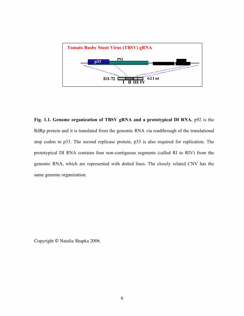
Fig. 1.1. Genome organization of TBSV gRNA and a prototypical DI RNA. p92 is the
RdRp protein and it is translated from the genomic RNA via readthrough of the translational
stop codon in p33. The second replicase protein, p33 is also required for replication. The
prototypical DI RNA contains four non-contiguous segments (called RI to RIV) from the
genomic RNA, which are represented with dotted lines. The closely related CNV has the
same genome organization.
Copyright © Natalia Shapka 2006.
7
Chapter II
The AU-rich RNA recombination hot spot sequence of Brome mosaic virus
is functional in tombusviruses: Implications for the mechanism of RNA
recombination
INTRODUCTION
RNA recombination plays a major role during the evolution of plus-strand RNA
viruses (Aranda et al., 1997; Fernandez-Cuartero et al., 1994; Strauss and Strauss, 1988;
Worobey and Holmes, 1999). New viruses or strains may emerge via recombination between
different viral RNAs or between viral and host RNAs (Strauss and Strauss, 1988;
Wierzchoslawski et al., 2003; Worobey and Holmes, 1999). In addition to contributing to the
genetic variability, RNA recombination is also proposed to function as a repair mechanism,
which can salvage damaged or mutated viral RNAs and use them to generate infectious viral
RNAs (Nagy and Simon, 1997). RNA recombination also plays a role in the formation of
defective interfering (DI) RNAs associated with many animal and plant viruses (White and
Morris, 1999). The first hallmark feature of DI RNAs is that they are derived from the parent
(helper) virus via sequence deletion(s). The second is that DI RNAs are dependent on the
helper virus for their replication, survival and/or spread. The best-known DI RNAs among
plant viruses are those associated with tombusvirus infections (White and Morris, 1999).
8
The most popular model of RNA recombination is the template switching (copy choice)
mechanism, which suggests that the viral RNA-dependent RNA polymerase (RdRp) switches
templates during complementary RNA synthesis (Jarvis and Kirkegaard, 1991; Lai, 1992;
Nagy and Simon, 1997). After the jump from the donor to the acceptor RNA, the RdRp
resumes RNA synthesis using the nascent RNA (which has been made on the donor RNA) as
a primer. Based on the role of base-pairing between the acceptor RNA and the primer
(nascent RNA), RNA recombination events are divided into three categories: base-pairing
dependent (similarity-essential), base-pairing assisted or base-pairing independent
(similarity-nonessential) (Jarvis and Kirkegaard, 1991; Lai, 1992; Nagy and Simon, 1997).
RNA recombination is probably a chance event, thus each nucleotide in an RNA molecule
may serve as a target for recombination. Most experimental data demonstrate that not all
regions within an RNA are equally recombinogenic. The sequences that participate in RNA
recombination at higher and lower frequencies are called hot and cold spots. Various models
have been proposed to explain the occurrence of the observed hot and cold spots for different
viruses (Jarvis and Kirkegaard, 1991; Lai, 1992; Nagy and Simon, 1997).
Tombusviruses are single-component plus-stranded RNA viruses of plants and they
are known to support RNA recombination at high frequency (Borja et al., 1999; White and
Morris, 1994b; White and Morris, 1994d; White and Morris, 1995). Although the
involvement of the genomic RNAs in recombination is well documented for tombusviruses,
DI RNAs associated with these viruses are the most popular templates in studies on RNA
recombination. This is because (i) they are involved in RNA recombination with high
frequencies (Borja et al., 1999; White and Morris, 1994b; White and Morris, 1994d; White
and Morris, 1995); (ii) they do not contribute essential protein factors to replication; and thus
9
(iii) have higher genetic plasticity than the viral genomic RNA. Importantly, recombination
in DI RNAs is thought to occur by using the same mechanism as recombination involving the
viral genomic RNA (White and Morris, 1999). The replication process of tombusviruses and
the associated DI RNAs is carried out by the replicase complex, which includes two viral
proteins and unknown host factors (Nagy, Pogany, and Simon, 2001; Oster, Wu, and White,
1998b; Scholthof, Scholthof, and Jackson, 1995a). The tombusvirus DI RNAs, such as the
Tomato bushy stunt virus (TBSV)-associated DI-73 (Fig. 2.1A), contain three or four
noncontiguous genomic segments (White and Morris, 1999). The two or three sequence
deletions leading to DI RNA formation are thought to be the consequence of viral replicase
jumping on the genomic template and the deletions may occur in a step-wise manner (White
and Morris, 1999).
Since it is possible that various RNA viruses may utilize similar RNA recombination
pathways or mechanism, we wished to compare how different viruses recognize and use
similar recombination promoting signals. This is not only important from mechanistic point
of view, but it can also give practical observations about the possibility of interviral
recombination, which could be facilitated by recognition of the same recombination
promoting signals by different viruses. To initiate these studies, in this paper, we analyzed
the effect of a recombination-promoting signal (a short AU-rich sequence), which is well
defined for Brome mosaic virus [BMV, ref. (Nagy and Bujarski, 1997)], on tombusvirus
recombination. The model recombination template was based on a DI RNA associated with
TBSV. We observed that the AU-rich sequence of BMV could indeed promote RNA
recombination in the model tombusvirus. The obtained data supports that viruses belonging
to different supergroups, such as BMV and TBSV, can recognize the same recombination
10
promoting signals. However, the distribution of recombination sites was different between
BMV and TBSV recombinants. We propose that, in addition to the role of the AU-rich
sequence, a putative cis-acting replication element might also affect the selection of the
recombination sites in the tombusvirus DI RNA.
MATERIALS AND METHODS
Construction of cDNA clones for TBSV DI RNAs. DI-FP recombination vector was
constructed by deleting the N-terminal segment from dEGFP (BDBiosciences) open reading
frame (ORF) using NcoI and construct DI-73dEGFP (J. Pogany and Nagy, unpublished),
leaving only a 117 bp segment (termed FP, Fig. 2.1A) from dEGFP ORF (Fig. 2.1A). The
resulting DI-FP contains, in addition to the truncated FP sequence between RI and RII in DI-
73 (White and Morris, 1994b), the following unique restriction sites at the 5’ side of the FP
sequence: XbaI, XhoI and NcoI; and at the 3’ side: MluI and BamHI.
All constructs that contained AU, GC2, R’ or AUs sequences (Fig. 2.1A) were
obtained by inserting the PCR-amplified (using the primers listed in Table 2.1) AU sequence
[including a segment from the 3’ end of BMV RNA3, between positions 197-242, (Nagy and
Bujarski, 1997)], GC2 (Nagy and Bujarski, 1998), R’ (Nagy and Bujarski, 1996) or the
artificial AUs (Nagy and Bujarski, 1997) segments into DI-FP either at the 5’ side of the FP
sequence (at the unique XbaI and NcoI restriction sites) or at the 3’ side (at the unique MluI
and BamHI restriction sites). The RII deletion constructs (derivatives of DI-AU1-FP-AU2)
were obtained by amplifying of the RII and RIII/IV sequences of DI-FP with the primer sets
shown in Table 2.1, followed by treatment with BamHI and SalI, and cloning to the similarly
11
treated DI-AU1-FP-AU2 or DI-FP (Fig. 2.1A) constructs. All the clones were sequenced to
confirm the desired changes.
Construct DI-152Bar* was made by replacing the FP region in DI-FP (Fig. 2.1A)
with the PCR-amplified 152 bp C-terminal segment of the barstar ORF (Hartley, 2001) using
the unique XbaI and BamHI sites. The PCR primers used are presented in Table 2.1.
Preparation of CNV gRNA and DI RNA transcripts in vitro. To generate the genomic
(g)RNA of Cucumber necrosis virus (CNV) and the DI RNA transcripts, we linearized
pK2/M5p20STOP and all the DI RNA clones, respectively, with SmaI, followed by in vitro
transcription with T7 RNA polymerase (Nagy, Pogany, and Simon, 1999; Nagy, Pogany, and
Simon, 2001). In vitro generated transcripts of DI RNAs were purified from 1% agarose gel,
followed by phenol/chloroform extraction, precipitation in 95% ethanol and washed three
times with 70% ethanol to remove residual salts. The in vitro RNA transcripts were analyzed
in 1% agarose gels and quantified by a UV spectrophotometer (Beckman).
For the in vitro RdRp experiments, RNA templates were obtained by in vitro
transcription reaction with T7 RNA polymerase [see above and ref. (Cheng, Pogany, and
Nagy, 2002; Nagy and Pogany, 2000b)]. The templates and primers used for PCR are listed
in Table 2.1. The unincorporated nucleotides were removed by phenol/chloroform extraction
and repeated ammonium-acetate/isopropanol precipitation (Cheng, Pogany, and Nagy, 2002;
Nagy and Pogany, 2000b). The T7 transcription products were analyzed by 5% denaturing
PAGE and the amounts of RNA were measured by UV spectrophotometer.
12
Preparation and electroporation of protoplasts. Nicotiana benthamiana protoplasts were
prepared as described before (Panaviene, Baker, and Nagy, 2003). Briefly, N. benthamiana
callus was treated with 0.5 g cellulysin and 0.1g macerase (Calbiochem) for 4.5h in
protoplast incubation medium (Nagy, Pogany, and Simon, 2001) at 25 0C, and then washed
twice with 0.5M mannitol and once with the electroporation buffer (10 mM HEPES, 10 mM
NaCl, 120 mM KCl, 4 mM CaCl2, 200 mM mannitol). For electroporation, we used 5×10⁵
protoplasts and 5 µg of CNV gRNA and 1 µg of DI-RNA. Electroporation was performed
with Gene pulser II (Biorad), under the following settings: 0.2kV voltage and 0.5µF capacity.
After electroporation, the samples were left on ice for 30min, followed by adding 1.8ml of
protoplast culture medium (Nagy, Pogany, and Simon, 2001). Protoplasts were incubated in
35×10 mm petri dishes in the dark for 24-48h at 22 0C.
N. benthamiana plants were inoculated with 3 µg of CNV gRNA and 1 µg of DI-
RNA (prepared as described above), using rub-inoculation as described earlier (Nagy,
Pogany, and Simon, 2001). Plants were incubated for 10 days in a temperature-controlled
room (~22 0C).
Total RNA extraction from protoplasts and plants and RNA analysis. Total RNA was
extracted from protoplast using phenol/chloroform method (Nagy, Pogany, and Simon,
2001). Aliquots of total RNA were analyzed on 1.2% agarose gels. RNA samples were
treated with formamide at 85 0C before loading on the gel. For Northern blot analysis, RNA
was transferred to Hybond XL membrane (Amersham-Pharmacia) by electro-transfer and
hybridized with DI-72-specific probes [RI(-), ref. (Panaviene, Baker, and Nagy, 2003)].
Hybridization with 32P-labeled RNA probes was performed in ULTRAhyb hybridization
13
buffer at 680C using the recommended conditions (Ambion). The 32P-labeled RNA probes
were made in an in vitro transcription reaction with T7 RNA polymerase in the presence of a
[α-32P] UTP and a PCR template obtained with primers #15 (5’-
GTAATACGACTCACTATAGGGCATGTCGCTTGTTTGTTG-3’) and #20 (5’-
GGAAATTCTCCAGGATTTCTC-3’) (Panaviene, Baker, and Nagy, 2003).
RT-PCR analysis, cloning and sequencing of recombinant DI RNAs. To obtain
recombinant DI RNA clones, reverse transcriptase (RT) reaction was done using primer #106
(5’-ACCTGGAAGCTTATGCCAGATTTACACTCATC-3’) and 2µl of total RNA
(Panaviene, Baker, and Nagy, 2003). This was followed by amplification using PCR with
primers #106 and #380 (5’-GGACGAATTCCATAATTATTATCTTTAFTTG-3’).
The RT-PCR products were digested with EcoRI and HindIII followed by gel
isolation and ligation into similarly treated pUC19 vector. The clones for sequencing were
selected after restriction digestion with EcoRI and HindIII. The sequencing was done using
CEQ Cycle Sequencing Kit (Beckman) and primer #553 (5’-GTAAAACGACGGCCAGT-
3’).
In vitro RNA binding studies. The RNA probe in the gel shift experiments was RIII(-
)/cPR11 (Panavas and Nagy, 2003a) containing the 82 nt minus-stranded RIII replication
enhancer and the 11 nt minimal promoter for plus-strand synthesis (23). The labeling was
done with 32P UTP using T7 RNA polymerase (Panavas and Nagy, 2003a). Competitor
RNAs (Fig. 2.6) were prepared with T7 polymerase on PCR-amplified templates using
primers described in Table 2.1. The gel shift experiments were performed according to
Rajendran and Nagy (Rajendran and Nagy, 2003). Briefly, various amounts of competitor
14
RNAs (used in 5x, 15x and 45x excess over the constant amount of 32P UTP-labeled RNA
probe) were mixed with 1 µM of recombinant p33 and p92 preparations in the presence of 50
mM Tris-HCl, pH 8.2, 10 mM MgCl2, and 10 mM DTT, 10% glycerol, 2.4U RNase inhibitor
and 100 ng tRNA. After 10 min pre-incubation at 250C, ~2 ng of 32P UTP-labeled RNA
probe was added to each RNA-binding reactions, followed by further incubation for 25 min.
Then, the samples were analyzed by electrophoresis on native 4% polyacrylamide gels run at
200V for 60 min at 4 0C in Tris-Glycine buffer (25mM Tris, 190 mM Glycine, 5 mM EDTA,
pH 8.5). Dried gels were analyzed using a phosphorimager. The recombinant proteins were
purified from E. coli as described earlier (Rajendran and Nagy, 2003; Rajendran, Pogany,
and Nagy, 2002).
RdRp assay. The CNV RdRp preparations were obtained from systemically infected N.
benthamiana leaves as described by Nagy and Pogany (Nagy and Pogany, 2000b). The RdRp
reactions contained ~1 µg of RNA transcripts (the amounts of RNA templates were adjusted
based on their sizes to have similar molar amounts of templates in each reaction). RdRp
reactions were carried out as previously described (Nagy and Pogany, 2000b). The reaction
was terminated by adding 5 µl 10% SDS followed by phenol/chloroform extraction and
ammonium-acetate/isopropanol precipitation (Nagy and Pogany, 2000b). The RdRp products
were analyzed by 5% denaturing PAGE in the presence of 8M urea, followed by
Phosphorimager analysis. The data for each sample were normalized based on the number of
templated UTP incorporation (Nagy and Pogany, 2000b).
RESULTS
15
Rationale: To test if different viruses could recognize the same recombination signal, we
chose an AU-rich sequence, which is a well-defined recombination-promoting signal in
BMV (Nagy and Bujarski, 1997). This AU-rich sequence was tested in a recombination
system based on DI RNA associated with TBSV, a tombusvirus. BMV and TBSV belong to
different supergroups of viruses and their RdRps are only distantly related. In addition,
tombusviruses do not code for an RNA helicase-like protein, which was shown to participate
in RNA recombination in BMV (Nagy et al., 1995).
Previous works defined BMV-derived and artificial AU-rich sequences that served as
recombination-promoting signals (Fig. 2.1) during BMV replication in vivo (Nagy and
Bujarski, 1996; Nagy and Bujarski, 1997; Nagy and Bujarski, 1998; Nagy, Ogiela, and
Bujarski, 1999). Frequent generation of recombinants was only observed when the two
recombining RNA templates carried the same or similar AU-rich sequences. Because
recombination sites in BMV were located within the AU-rich regions, it was proposed that
the BMV replicase recognized the AU-rich signals during the recombination events (Nagy
and Bujarski, 1996; Nagy and Bujarski, 1997; Nagy and Bujarski, 1998; Nagy, Ogiela, and
Bujarski, 1999). We wanted to test if these AU-rich sequences could also promote RNA
recombination in tombusviruses.
The control TBSV DI RNA-based recombination vector does not support RNA
recombination. To study RNA recombination in tombusviruses, first we developed a
recombination vector, which facilitated the construction and testing of additional parental DI
RNAs (Fig. 2.1A). The recombination vector, named DI-FP, is based on TBSV DI-73 RNA
16
carrying a 117 nt insert of nonviral origin (derived from dEGFP, BDBioscience) between
regions I and II (RI and RII; Fig. 2.1A). To avoid possible artifactual RNAs (i.e., unwanted
recombinant-like RNAs) made during plasmid propagation in E. coli and/or during RNA
transcription with T7 polymerase, we gel-isolated DI-FP RNA transcripts prior to their use.
In addition, to exclude those DI RNAs that might be generated spontaneously from the helper
genomic RNA (gRNA) during the infection, a process known as de novo DI RNA formation
(Rochon, 1991; White and Morris, 1999), we chose the heterologous Cucumber necrosis
virus (CNV, closely related to TBSV) as the helper virus. CNV can support efficiently the
replication of TBSV DI-73 RNA and DI-FP vector (ref. (Panaviene, Baker, and Nagy, 2003)
and Fig. 2.1C).
The gel-isolated DI-FP transcripts were co-electroporated with CNV gRNA
transcripts into N. benthamiana protoplasts as described earlier (Panaviene, Baker, and Nagy,
2003). We followed the accumulation of DI-FP RNA in protoplast (termed “zero passage”,
Fig. 2.1C) by using Northern blotting with a DI RNA-specific probe [RI(-), Fig. 2.1A] and
RT-PCR (primers of #160 and #380, Fig. 2.1A). These experiments detected only the
parental DI-FP RNAs in the zero passage protoplast samples, suggesting the lack of
recombinant accumulation (Fig. 2.1C). The total RNA obtained after 48 hours of incubation
in zero passage protoplasts was used for electroporation into a new batch of protoplasts
(termed “first passage”). Northern blot and RT-PCR analyses revealed that the DI-FP RNA
recombination vector replicated efficiently and stably in the first passage protoplasts (Fig.
2.1D, E and F, lanes 1-4). Note that we made sequential passages of the progeny DI RNAs
from one batch of protoplasts to another, since there is no cell-to-cell spread of
17
tombusviruses in protoplasts. Altogether, we performed three sequential passages, yet the DI-
FP vector did not appear to support recombinant DI RNA accumulation (not shown).
An AU-rich sequence supports RNA recombination efficiently in TBSV-associated DI
RNA. To test the effect of an AU-rich sequence on tombusvirus recombination, we inserted
the 69 nt long AU sequence (Fig. 2.1B; this sequence is identical with AU1), which had been
tested previously for recombination in BMV, into DI-FP in such a way that an identical copy
of the AU sequence flanked the dEGFP-derived FP region at the 5’ and 3’ sides, respectively
(termed AU1 and AU2 in construct DI-AU1-FP-AU2, Fig. 2.1A), resulting in a repeated
sequence. It was predicted, based on the BMV results (Nagy and Bujarski, 1997) that
recombination might take place between the repeated copies of the AU sequence, thus
resulting in deletion of one of the AU copies plus the FP sequence (Fig. 2.1A) located in
between the repeated AU sequences.
Incubation of the protoplast cells after co-electroporation of the gel-purified DI-AU1-
FP-AU2 RNA with the CNV gRNA resulted in efficient amplification of the parental-sized
DI RNA, suggesting that the insertions did not debilitate DI RNA replication (Fig. 2.1C). No
recombinants were detected in these protoplasts by Northern blotting, while RT-PCR
analysis did show the occurrence of novel, ~100-250 bp shorter than input RNAs (not
shown). This indicated that the putative recombinant DI RNAs were present in the zero
passage protoplasts at low levels that could only be detected by the more sensitive RT-PCR.
However, a passage of the total RNA to a new batch of protoplasts followed by incubation
resulted in recombinant-like DI RNAs in 100% (15 out of 15) of the experiments based on
total RNA (Fig. 2.1D, lanes 6-9), Northern blot (Fig. 2.1E) and RT-PCR analyses (Fig. 2.1F).
Interestingly, the sizes of the novel DI RNA recombinants were variable, suggesting that the
18
recombination events were “imprecise” in nature. Indeed, cloning and sequencing of these
putative recombinants confirmed that (i) they derived from DI-AU1-FP-AU2 RNA via
deletions; and (ii) the recombination junction sites were different in many of the
recombinants (Fig. 2.2A). All of the 5’ deletion sites in the 15 recombinants sequenced were
located within the AU sequence (i.e., AU1), while the 3’ deletion sites were clustered mostly
within RII, which flanks the inserted sequences in DI-AU1-FP-AU2 (Fig. 2.2A). Overall, we
did not find the generation of precise (homologous) recombinants between the duplicated AU
sequences, which were the most common recombinants in the BMV system (Nagy and
Bujarski, 1997).
Based on the distribution of the recombination junctions, the AU2 sequence did not
seem to influence the selection of recombination sites in DI-AU1-FP-AU2. Therefore, we
deleted AU2 to generate construct DI-AU1-FP (Fig. 2.1A). Testing the recombination activity
of DI-AU1-FP, in a way similar to that described above for DI-AU1-FP-AU2, revealed that
DI-AU1-FP supported RNA recombination as efficiently as DI-AU1-FP-AU2 did (Fig. 2.1D-
F, lanes 11-14). Even more importantly, the distribution of recombination sites for DI-AU1-
FP was comparable to that described for DI-AU1-FP-AU2 (Fig. 2.2C), confirming that the
recombination hot spots include the AU1 sequence and RII. This data supports that one copy
of the AU sequence plays an important role in recombination, while the role of the second
copy is less obvious.
The isolated DI RNA recombinants from protoplasts appear to be true recombinants,
since they can be detected by gel analysis of the total RNA (Fig. 2.1D) and by Northern
blotting (Fig. 2.1E). In addition, control RT-PCR performed on gel isolated DI RNA
transcripts (the same DI RNA transcripts that were used for electroporation to protoplasts)
19
did not detect recombinant-sized DI RNAs for these constructs (Fig. 2.1F, lanes 5, 10 and
15).
To test if the length of the inserts is an important factor in recombination events, we
generated a DI RNA carrying a ~152 nt long sequence from the barstar gene (Fig. 2.1A) (7).
The resulting DI-152Bar* RNA was stable in protoplasts after one (Fig. 2.1D-F, lanes 16-19)
or two passages (not shown). This result, together with that of DI-FP, supports that DI RNAs
with short inserts can be stable in protoplasts (under the conditions used) if they lack
recombination-promoting signals.
To examine if recombination might occur in whole plants as well, we tested the
accumulation of DI-FP, DI-AU1-FP-AU2 and DI-AU1-FP RNAs (in the presence of the CNV
gRNA) in N. benthamiana plants ~10 days after inoculation with gel-purified transcripts.
Northern blot (Fig. 2.1G) and RT-PCR (not shown) analyses demonstrated that the
systemically-infected (uninoculated) leaves contained the parental-sized DI RNAs for each
DI RNA tested. In contrast, recombinant-like DI RNAs appeared only in DI-AU1-FP-AU2
and DI-AU1-FP RNA-containing plants (Fig. 2.1G, lanes 5-12), but not in DI-FP-containing
plants (Fig. 2.1G, lanes 1-4). Cloning and sequencing of a representative number of
recombinants confirmed that the accumulating recombinants in plants are similar to those
observed in protoplasts with most of the junction sites located within the 5’ AU1 sequence
and RII (Fig. 2.2B and D).
RII sequence affects the distribution of recombination sites. Since many recombinants
generated with DI-AU1-FP-AU2 and DI-AU1-FP RNAs had the recombination sites within a
40 nt stretch close to the 5’ end of RII(+) (we term this region hs40 hot spot), it is possible
20
that sequences around hs40 might influence the selection of recombination sites. To test if
hs40 is required for replication of DI RNA, we generated a series of DI constructs based on
various 5’ deletions in RII. First, we tested construct DI-FP-RII/Δ40 (Fig. 2.3A), which has
the same sequence as the DI-FP vector (Fig. 2.1A), except lacking the 5’ 40 nt from RII (i.e.,
hs40). DI-FP-RII/Δ40 RNA accumulated efficiently in protoplasts and no recombinants were
detected in protoplasts after the first passage (Fig. 2.3B-C, lanes 1-5). This data suggests that
hs40 sequence is not essential for DI RNA accumulation in protoplasts.
To test if hs40 is important for RNA recombination, we tested derivatives of DI-AU1-
FP-AU2 with various 5’ deletions in RII. Construct DI-RII/Δ20, which lacked 20 nt from the
5’ end of RII(+) in DI-AU1-FP-AU2, generated recombinants as efficiently as DI-AU1-FP-
AU2 did after the first passage (Fig. 2.3B-C, lanes 7-11). Interestingly, the distribution of
recombination sites in the recombinants obtained with DI-AU1-FP-AU2 (Fig. 2.2A) and DI-
RII/Δ20 (Fig. 2.4A) was similar, except ~50% (7 out of 15) of recombinants obtained with
DI-RII/Δ20contained precise recombination sites between the duplicated AU sequences. The
second construct tested, DI-RII/Δ40, which lacked 40 nt from the 5’ end of RII(+) in DI-
AU1-FP-AU2, also generated recombinants in 100% of samples (Fig. 2.3B-C, lanes 13-17).
Interestingly, 87% (26/30) of the 3’ junctions were within the AU2 copy and only 13% within
the RII sequence. We also observed precise recombinants between the repeated AU regions
in ~36% (11/30) of recombinants (Fig. 2.4B). The occurrence of precise recombinants may
suggest that primer re-alignment (base-pairing between the the primer and the acceptor
region) assisted by the presence of duplicated sequences may take place during these
recombination events (see Discussion).
21
Construct DI-RII/Δ60, which lacked 60 nt from the 5’ end of RII(+) in DI-AU1-FP-
AU2, supported recombination as efficiently as DI-AU1-FP-AU2 (Fig. 2.3B-C, lanes 19-23).
Cloning and sequencing revealed that all 15 recombinants, each derived from separate
samples, were precise recombinants (Fig. 2.4C) with junctions between the duplicated AU
sequences. In contrast to the above DI RNA constructs, DI-RII/Δ100, which lacked 100 nt
from the 5’ end of RII(+), did not support the generation of recombinants in protoplasts (Fig.
2.3B-C, lanes 25-29). Moreover, DI-RII/Δ100 accumulated only inefficiently (tested after the
first passage) in protoplast, suggesting that an important cis-acting signal may have been
deleted from this construct (see Discussion). Overall, these results suggest that the RII
sequence may have two important effects on RNA recombination: first, it could affect the
frequency of recombinant formation and, second, it could also influence the selection of
recombination sites. In addition, we conclude that hs40 is not required for recombination to
take place within the RII sequence.
Short AU-rich sequences can also support RNA recombination in DI RNA. To test if
shorter AU-rich sequences could also support RNA recombination in tombusviruses, first we
deleted the BMV-derived R’ region (40 nt long, Fig. 2.1B) from the 69 nt AU1 sequence in
DI-RII/Δ40. The resulting construct DI-AUs-FP-AU2 contained only the 29 nt long artificial
AU-rich sequence (termed AUs, with 76% AU content), yet it still supported recombination
efficiently with most of the junction sites located within the 5’ AUs and 3’ AU2 regions (Fig.
2.5A). Note that we found that the distribution of the recombination sites was similar to that
observed for DI-RII/Δ40 recombinants, with almost 50% (7/17) of recombinants having
precise junctions between the repeated AU-rich sequences (Fig. 2.5A).
22
Second, we deleted the AUs portion from the 5’ copy of the AU sequence (Fig. 2.1B)
in construct DI-R’-FP-AU2 (Fig. 2.5B), which left the 44 nt long BMV-derived R’ region at
the 5’ location. Although the R’ sequence contained only a 21 nt AU-rich stretch (with ~75%
AU content, Fig. 2.1B), it was still active in recombination (Fig. 2.5B). Most of the
recombinants obtained with DI-R’-FP-AU2 (Fig. 2.5B) contained precise junctions between
the repeated AU-rich sequences. Overall, this data suggests that both the BMV-derived R’
and the artificial AUs portions of the original AU sequence are capable of supporting RNA
recombination efficiently in TBSV. We conclude that as short as 21 to 29 nt AU-rich
sequence can promote recombination in TBSV DI RNA.
GC-rich sequences cannot “silence” RNA recombination in TBSV. Since RNA
recombination promoted by the AU sequence could efficiently be inhibited by the presence
of GC-rich sequences located 3’ of the AU sequence (referred here in plus-strand orientation)
in the BMV RNAs (Nagy and Bujarski, 1997), we also tested the effect of one of these well-
characterized sequences [termed GC2 with 55% GC-content (Nagy and Bujarski, 1997)] on
DI RNA recombination. First, the GC2 sequence was inserted 3’ of the R’ sequence in DI-
R’-FP-AU2 (Fig. 2.5B). The resulting construct, DI-R’/GC2-FP-AU2 (Fig. 2.5C) supported
RNA recombination efficiently. Most of the junctions were located within the R’ sequence
and the 3’ AU2 sequence, including more than 60% (9/15) with precise junctions between the
repeated AU-rich sequences (Fig. 2.5C). Second, placing the GC2 sequence behind the R’
sequence within the 3’ repeat (construct DI-AU1-FP-R’/GC2) did not inhibit recombination
that took place between the 5’ AU1 sequence and either R’, GC2 or the RII sequences (Fig.
2.5C). Interestingly, none of the isolated recombinants contained precise junctions between
23
the repeated AU-rich sequences, suggesting the GC2 sequence interfered with precise
recombination, but not with imprecise recombination.
The minus-stranded RII binds efficiently to the TBSV RdRp. Previous in vitro studies
with the CNV RdRp (Cheng and Nagy, 2003; Cheng, Pogany, and Nagy, 2002) predicted
that the same AU sequence tested above is a good donor, while it is a poor acceptor during
template switching events. Based on these observations, it is possible that RNA
recombination might occur during plus-strand synthesis when the RdRp would “jump” from
the AU1(-) region to the RII(-) region (around hs40) (see Discussion). Thus, this model
predicts that RII(-) should contain a binding site(s) for the tombusvirus replicase proteins. In
contrast, the heterologous dEGFP-derived FP sequence is expected to lack high-affinity
binding site(s), thus serving as a cold spot during the recombination events. This model was
tested below in two different in vitro assays.
First, we have tested the ability of the purified recombinant TBSV replicase proteins
(i.e., p33 and p92 expressed and purified from E. coli)(Rajendran and Nagy, 2003;
Rajendran, Pogany, and Nagy, 2002) to bind to RII(-) sequences or the FP sequence (also
tested in the complementary orientation). To obtain quantitative results, we used template
competition in a gel mobility shift assay (Rajendran and Nagy, 2003; Rajendran, Pogany, and
Nagy, 2002). The experiments included the same amount of 32P-labeled template [derived
from the RIII(-) replication enhancer] and the same amount of purified recombinant p92 (Fig.
2.6B) or p33 proteins (Fig. 2.6C). The amounts of unlabeled competitors were used in 5x,
15x and 45x excess over the labeled RNA for each competitor RNA. These experiments
revealed that the two competitor RNAs that contained overlapping portions of the RII(-)
24
sequence (namely construct #7 and #18, Fig. 2.6A) were 2 to 3-fold better competitors for
binding to p92 or p33 (Fig. 2.6B-C, lanes 4-9) than the FP control sequence (lanes 1-3).
Therefore, this data supports the model that RII(-) is a hot spot region due to its increased
binding to the replicase proteins (see Discussion).
Since binding to a template does not necessarily result in productive interaction
between the particular sequence and the RdRp, we also tested the ability of the partially
purified CNV RdRp to use templates containing various portions of RII(-) sequence and of
the FP(-) sequence in the presence of a short primer in an in vitro primer extension assay
(Fig. 2.7)(Cheng and Nagy, 2003; Cheng, Pogany, and Nagy, 2002). As shown earlier,
primer extension by the CNV RdRp in the in vitro assay depends on (i) the ability of the
template region to interact with the CNV RdRp, and (ii) on the number of base-pairs formed
between the primer and the template (Cheng and Nagy, 2003; Cheng, Pogany, and Nagy,
2002). Since all the constructs tested in this work contained the same 5 base-pair
primer/template region (termed art5, which supported self-priming of RNA synthesis on the
template by the CNV RdRp)(Cheng and Nagy, 2003; Cheng, Pogany, and Nagy, 2002), we
predicted that the activity of a particular template in the CNV RdRp reaction should depend
on the ability of the template region to interact with the CNV RdRp. These experiments
revealed that primer extension was ~3 to 9-fold more efficient when it started within the
RII(-) sequence than the primer extension from the FP sequence (compare constructs #16,
#17, and #18 with #20 and #21 in Fig. 2.7A-C), while primer extension from the 3’ end of
RII(-) was almost ~2-fold more efficient (construct #11). Interestingly, the most efficient
constructs were those that contained the primer within the hs40 hot spot region (see
constructs #16 and #17) or in the middle of RII(-) (construct #17, Fig. 2.7), suggesting that
25
this region contains a putative cis-acting element (see Discussion). Overall, data from the
primer extension experiments does support the model that RII(-) might be active in
recombination due to its enhanced binding to the RdRp.
DISCUSSION
Comparison of the role of AU-rich sequences in BMV, retrovirus and tombusvirus
recombination. The emerging picture in RNA recombination is that the nonrandom
distribution of RNA recombination sites observed with several plus-strand RNA viruses
[reviewed in (Nagy and Simon, 1997)] are due to the presence of recombination promoting
signals in the RNA templates. The viral RdRp is postulated to recognize these recombination
signals, which then leads to recombination (template switching) events with high frequencies
(i.e., forming recombination hot spots) (Nagy and Simon, 1997). It is currently unknown if
recombination signals, which have been described for a particular RNA virus, could also be
recognized by another RNA virus. We were particularly interested in AU-rich sequences,
which are not only common in many RNA viruses, but they are known to promote
recombination in BMV (Nagy and Bujarski, 1995; Nagy and Bujarski, 1996; Nagy and
Bujarski, 1997; Nagy and Simon, 1997) and retroviruses (DeStefano, Bambara, and Fay,
1994; Wu et al., 1995) and possibly in poliovirus (Pilipenko, Gmyl, and Agol, 1995). Current
models on the role of AU-rich sequences in promoting recombination is based on the
assumption that the viral RdRp might pause during RNA synthesis within the AU-rich stretch
of the template due to the weak A-U base pairing between the growing nascent strand and the
donor RNA template inside the RdRp (Nagy and Bujarski, 1996; Nagy and Bujarski, 1997;
26
Nagy and Bujarski, 1998; Nagy and Simon, 1997). Moreover, the weak base pairing between
the nascent strand and the donor RNA within the AU-rich region might also facilitate
dissociation of the 3’ end of the nascent strand from the complementary donor strand. The
free 3’ end of the nascent strand may then anneal to the acceptor RNA at some frequencies,
followed by resumption of the RNA synthesis by the viral RdRp. If the weak base pairing
between the growing nascent strand and the donor RNA within the AU-rich region is indeed
“forcing” RNA recombination, then we predict that different viral RdRps should recognize
this type of signal, albeit with various frequencies depending on the processivity of different
viral RdRps.
Indeed, we found that an AU-rich sequence, which promoted RNA recombination in
BMV, also facilitated RNA recombination in a TBSV-associated DI RNA. The supporting
evidence includes the following: (i) the recombination sites were frequently located within
the AU-rich region; (ii) recombination frequency of the DI RNAs containing AU-rich
sequences were higher than the control DI RNAs carrying dEGFP-derived or barstar
sequences (Fig. 2.1). We also obtained data that support a different role for the AU-rich
sequence in tombusvirus recombination than in case of BMV. These include: (i) only one
copy of the AU-rich sequence was enough for promoting tombusvirus recombination, while
two copies were needed for efficient recombination in BMV; (ii) most of the recombination
events occurred precisely or semi-precisely between the two copies of AU-rich sequences in
BMV, while this type of recombinants were less frequent in case of TBSV DI RNA; (iii) an
additional viral sequence (i.e, RII) was also involved in RNA recombination in TBSV DI
RNA, while it is currently unknown if sequences other than the AU-rich regions are involved
in facilitating recombination to take place between the AU-rich sequences in BMV. Overall,
27
the above similarities and differences between the two recombination systems suggest that
the AU-rich sequences can promote recombination in both viruses, but the selection of
recombination sites (which are probably determined during resolution of the putative
recombination intermediates) is different for the majority of recombinants (mostly precise for
BMV and largely imprecise for TBSV recombinants). This observation is also supported by
the different effect of GC-rich sequences on recombination in the two viral systems. While
GC-rich sequences located downstream on the acceptor template (based on the progress of
the viral replicase on the template) inhibited or silenced recombination in BMV (Nagy and
Bujarski, 1996; Nagy and Bujarski, 1997; Nagy and Bujarski, 1998; Nagy and Simon, 1997),
we did not observe similar effects by the same GC-rich sequence in case of TBSV DI RNA
(Fig. 2.5C-D).
In addition to the in vivo data discussed above, in vitro data obtained with the purified
BMV and CNV (Cheng and Nagy, 2003; Cheng, Pogany, and Nagy, 2002; Kim and Kao,
2001) replicases also support the role of AU-rich sequences when present in the donor RNA.
For example, the end-to-end template switching assay developed by Kim and Kao (Kim and
Kao, 2001) demonstrated that the AU-rich stretches when present at the 5’ end of the donor
RNA, where the nascent strand must be released from the donor RNA before the template
switching occurs, facilitated end-to-end recombination events. Interestingly, the AU-rich
sequence tested in this work also supported template switching by the partially purified CNV
RdRp in vitro (Cheng and Nagy, 2003; Cheng, Pogany, and Nagy, 2002). The template RNA
containing the AU-rich sequence served primarily as a donor RNA in the in vitro CNV RdRp
assay (Cheng and Nagy, 2003; Cheng, Pogany, and Nagy, 2002), giving valuable insight into
the significance of sequence context in RNA recombination in vitro, and possibly in vivo.
28
Further evidence on the possibility that AU-rich sequences could serve as common
recombination signals comes from studies with retroviruses. Using an in vitro template
switching assay based on purified reverse transcriptase (RT) from Human immunodeficiency
virus, it has been demonstrated that AU-rich sequences promoted RNA recombination
(DeStefano, Bambara, and Fay, 1994). Since the RT favors base pairing between the nascent
strand and the acceptor strand prior to resumption of cDNA synthesis, most of the in vitro
recombinants were precise (i.e., occurred precisely within homologous regions), although
recombinants with extra nucleotides, mismatched nucleotides, or short deleted regions at the
recombination sites were also isolated (DeStefano, Bambara, and Fay, 1994). The
observation that AU-rich sequences form hot spots in recombinants obtained with BMV,
retrovirus and TBSV suggests that the induction of recombination by AU-rich sequences is
similar for these viruses. Moreover, the resolution of recombination intermediates may use
somewhat similar mechanism for BMV and retroviruses, but it is different in tombusviruses.
We propose that the difference is due to two factors: (i) the BMV RdRp and the retrovirus
RT favor a somewhat precise annealing step between the nascent strand (primer) and the
acceptor strand prior to the resumption of the RNA/DNA synthesis, while the tombusvirus
RdRp does not seem to favor this step. Indeed, we have proposed that the CNV RdRp can
easily resume RNA synthesis (primer extension) without the need of extensive base pairing
between the primer and the acceptor strand (Cheng and Nagy, 2003; Cheng, Pogany, and
Nagy, 2002). (ii) There might be differences among these viruses in using cis-acting
elements to guide the jumping viral replicase to a new acceptor site before resumption of
primer extension, resulting in recombination hot spots (see below). It is also important to
note that we cannot completely rule out that selection for the best fit recombinants might also
29
affect the types of recombinants isolated in these virus infections. In summary, in vitro and in
vivo data suggests that AU-rich sequences might serve as common recombination signals.
However, the likely differences in recombination between viruses are caused by (i)
differences in template/sequence recognition during the template switching event; and (ii)
various roles for base-pairing between the primer and template during the template switching
events.
Model of AU-rich sequence-driven recombination in tombusviruses. Since both plus and
minus-strand synthesis take place during DI RNA replication in protoplasts, it is difficult to
establish whether plus or minus-stranded RNAs are used as templates for recombination.
However, based on previous in vitro experiments with the partially-purified CNV RdRp, the
likely role of the AU-rich sequence is to promote recombination at the donor sites (Cheng
and Nagy, 2003; Cheng, Pogany, and Nagy, 2002). Moreover, the same AU sequence used in
this study was found to be a relatively poor template (in comparison with known cis-acting
elements of tombusviruses) in in vitro primer extension and template switching experiments
with the CNV RdRp (Cheng and Nagy, 2003; Cheng, Pogany, and Nagy, 2002), suggesting
that the AU sequence is unlikely to form a recombination hot spot as an acceptor site. If this
is the case in protoplast as well, then most of recombination events might involve minus-
stranded templates and, thus, template switching would occur during plus-strand synthesis
(Fig. 2.8A). This is because the first copy of the repeated AU regions (i.e. AU1), which is
observed as a hot spot with DI-AU1-FP-AU2 (Fig. 2.2A), would be at the 3’ proximal
location (relative to the deletion junctions) in the minus-stranded DI-AU1-FP-AU2 RNA (Fig.
2.8A). Thus, the AU1 repeat is favorably positioned to serve as a donor site for promoting
30
jumping events by the tombusvirus replicase to a new location [which is hs40 within RII(-),
see below]. The above model (Fig. 2.8A-B) also predicts that the second AU repeat (i.e.,
AU2), located at a more 5’ position in the minus-stranded DI-AU1-FP-AU2 RNA should be
less favorable as a donor site, because this region could only be copied by the RdRp after the
first AU1 repeat had already been copied. Accordingly, the lack of AU2 repeat in DI-AU1-FP
RNA did not significantly alter the recombination sites or the frequency of the recombination
events (Figs. 2.1 and 2.2). Interestingly, the AU sequences have also been predicted to
support recombination during plus-strand synthesis when they were present in the minus-
stranded BMV RNAs (Nagy and Bujarski, 1997).
In contrast to the proposed primary role in promoting replicase jump, the AU-rich
sequence may have no or only limited role during RdRp landing (i.e., at the acceptor sites) if
it is located “far” from the putative RII(-) cis-acting element. This model is supported by (i)
primer extension experiments with CNV RdRp, which demonstrated that the AU sequence is
a poor template (Cheng and Nagy, 2003; Cheng, Pogany, and Nagy, 2002); and (ii) the lack
of effect on distribution of recombination sites in the absence of the AU2 repeat (compare
data obtained with DI-AU1-FP-AU2 and DI-AU1-FP RNAs, Fig. 2.2A and C). Interestingly,
the AU2 sequence became a recombination hot spot, when it was located in the vicinity of the
putative RII(-) cis-acting element (such as in constructs DI-RII/Δ40 and DI-RII/Δ60, Fig.
2.4). To explain the formation of these precise recombinants, we propose that the RdRp
might still bind to the RII(-) region during the jumping event, but annealing (base-pairing)
between the primer and the template might take place within the AU2 repeat. This annealing
step might then promote primer extension to initiate from the AU2 repeat, therefore resulting
in precise or semi-precise recombination (Fig. 2.8C-D). We propose that similar annealing
31
step between the primer and the acceptor region might be inhibited in the presence of extra
sequences between AU2 and RII(-) (see construct DI-AU1-FP-AU2, Fig. 2.1), thus favoring
recombination to take place within the hs40 region that flanks the putative cis-element in
RII(-) (Fig. 2.8A-B).
A surprising observation in this work is the discovery of the major role of RII in
TBSV recombination. The in vitro binding studies with recombinant TBSV p33 and p92
replicase proteins and the primer extension studies with the CNV RdRp preparation suggest
that there is a putative cis-acting element in RII(-) located 5’ of the hs40 sequence. Indeed,
construct 18 that contained the 5’ half of RII(-) (between position 100-239, Figs. 2.6-7)
competed efficiently for binding to p33/p92 (Fig. 2.6) and it was an efficient template for
primer extension in the in vitro CNV RdRp assay (Fig. 2.7). We propose (Fig. 2.8B) that the
jumping viral replicase is likely guided by the RII(-) cis-acting element before resumption of
primer extension, resulting in recombination hot spots. The role of this cis-element is likely
more important than the effect of base-pairing between the primer and the template during
tombusvirus recombination events. Accordingly, we observed earlier that the CNV RdRp
could perform RNA synthesis (primer extension and template switching) without the need of
extensive base pairing between the nascent strand and the acceptor strand (Cheng and Nagy,
2003; Cheng, Pogany, and Nagy, 2002). This observation can explain why there are not long
stretches of sequence identity around the recombination sites generated in infections with DI-
AU1-FP-AU2 (Fig. 2.2A).
The role of cis-acting replication elements in recombination has been proposed before
in several viral systems [reviewed in (Nagy and Simon, 1997)]. For example, a known
replication enhancer element (Nagy, Pogany, and Simon, 1999) in a satellite RNA (termed
32
satC), which is associated with Turnip crinkle virus (TCV) infections, promoted
recombination between the mutated satC and satD, another satellite RNA (Cascone, Haydar,
and Simon, 1993; Nagy, Pogany, and Simon, 1999). It has been proposed that the replication
enhancer of satC is involved in binding to the jumping TCV replicase (Nagy, Pogany, and
Simon, 1999; Nagy, Pogany, and Simon, 2001), a process similar to the role that was
proposed above for the RII(-) in TBSV DI RNA (Fig. 2.8). A different class of cis-acting
element, namely the subgenomic promoter region has also been proposed to promote RNA
recombination in luteoviruses and BMV (Wierzchoslawski et al., 2003). In summary, the
discovery of the role of the RII(-) element in RNA recombination, in combination with
published data cited above, suggests that cis-acting elements might play much wider roles in
viral RNA recombination than previously anticipated.
33
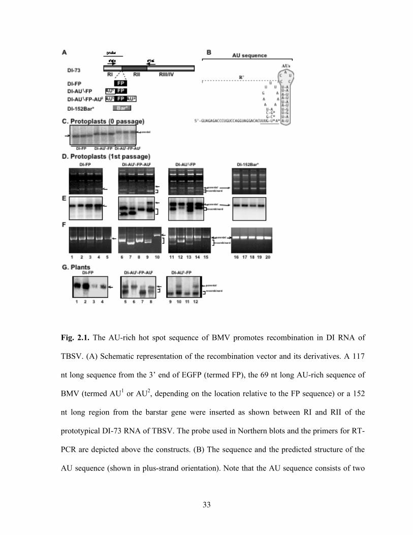
Fig. 2.1. The AU-rich hot spot sequence of BMV promotes recombination in DI RNA of
TBSV. (A) Schematic representation of the recombination vector and its derivatives. A 117
nt long sequence from the 3’ end of EGFP (termed FP), the 69 nt long AU-rich sequence of
BMV (termed AU1 or AU2, depending on the location relative to the FP sequence) or a 152
nt long region from the barstar gene were inserted as shown between RI and RII of the
prototypical DI-73 RNA of TBSV. The probe used in Northern blots and the primers for RT-
PCR are depicted above the constructs. (B) The sequence and the predicted structure of the
AU sequence (shown in plus-strand orientation). Note that the AU sequence consists of two
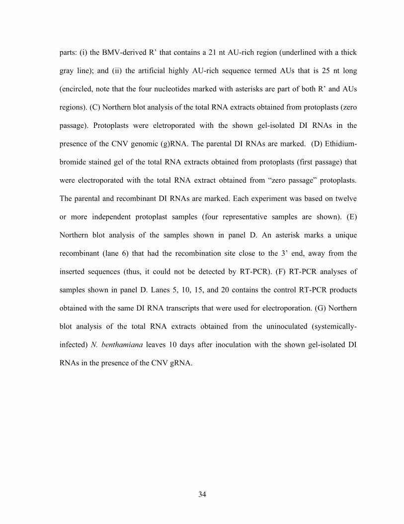
34
parts: (i) the BMV-derived R’ that contains a 21 nt AU-rich region (underlined with a thick
gray line); and (ii) the artificial highly AU-rich sequence termed AUs that is 25 nt long
(encircled, note that the four nucleotides marked with asterisks are part of both R’ and AUs
regions). (C) Northern blot analysis of the total RNA extracts obtained from protoplasts (zero
passage). Protoplasts were eletroporated with the shown gel-isolated DI RNAs in the
presence of the CNV genomic (g)RNA. The parental DI RNAs are marked. (D) Ethidium-
bromide stained gel of the total RNA extracts obtained from protoplasts (first passage) that
were electroporated with the total RNA extract obtained from “zero passage” protoplasts.
The parental and recombinant DI RNAs are marked. Each experiment was based on twelve
or more independent protoplast samples (four representative samples are shown). (E)
Northern blot analysis of the samples shown in panel D. An asterisk marks a unique
recombinant (lane 6) that had the recombination site close to the 3’ end, away from the
inserted sequences (thus, it could not be detected by RT-PCR). (F) RT-PCR analyses of
samples shown in panel D. Lanes 5, 10, 15, and 20 contains the control RT-PCR products
obtained with the same DI RNA transcripts that were used for electroporation. (G) Northern
blot analysis of the total RNA extracts obtained from the uninoculated (systemically-
infected) N. benthamiana leaves 10 days after inoculation with the shown gel-isolated DI
RNAs in the presence of the CNV gRNA.
35
36
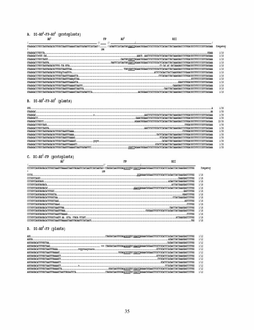
Fig. 2.2. Sequences of DI RNA recombinants around the junction sites. A representative number
of recombinants obtained with construct DI-AU1-FP-AU2 (panels A-B) or with DI-AU1-FP (panels
C-D) were amplified by RT-PCR (see Fig. 2.1), cloned and sequenced around the junction sites.
The top sequence represents the sequence of the parental DI RNAs. The regions are shown
schematically on the top. The underlined nucleotides represent marker mutations. Double slashes
indicate not shown sequences, while the lengths of the not shown sequences are indicated with
numbers below the parental sequences. The recombinants are shown below the asterisk. The
deleted sequences in the recombinants are marked with dotted lines, while nontemplated
nucleotides are shown with small letters. +++ indicates the following not shown sequence:
UGGACCGUCACCCUGCAGCCUGUGCUUCUG. Note that positive strand sequences are
shown in 5’ to 3’ orientation. Two cDNA clones were sequenced/per protoplast samples, but we
counted only one recombinant/per protoplast sample in the frequency column.
37
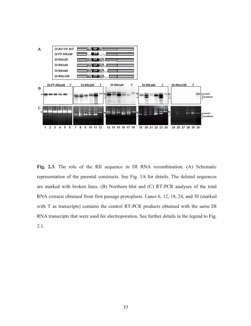
Fig. 2.3. The role of the RII sequence in DI RNA recombination. (A) Schematic
representation of the parental constructs. See Fig. 1A for details. The deleted sequences
are marked with broken lines. (B) Northern blot and (C) RT-PCR analyses of the total
RNA extracts obtained from first passage protoplasts. Lanes 6, 12, 18, 24, and 30 (marked
with T as transcripts) contains the control RT-PCR products obtained with the same DI
RNA transcripts that were used for electroporation. See further details in the legend to Fig.
2.1.
38
39
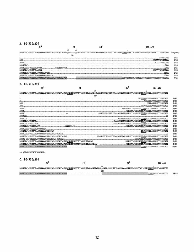
Fig. 2.4. Sequences of DI RNA recombinants obtained with RII deletion mutants. A representative
number of recombinants (from first passage protoplast) were amplified by RT-PCR (see Fig. 2.3),
cloned and sequenced. The duplicated regions that participated in precise recombination are boxed.
Note that due to sequence identity in the duplicated regions, the actual recombination sites cannot be
determined in these precise recombinants. The actual sequence of the region marked with +++ is
shown at the bottom of panel B. See further details in the legend to Fig. 2.2.
40
41
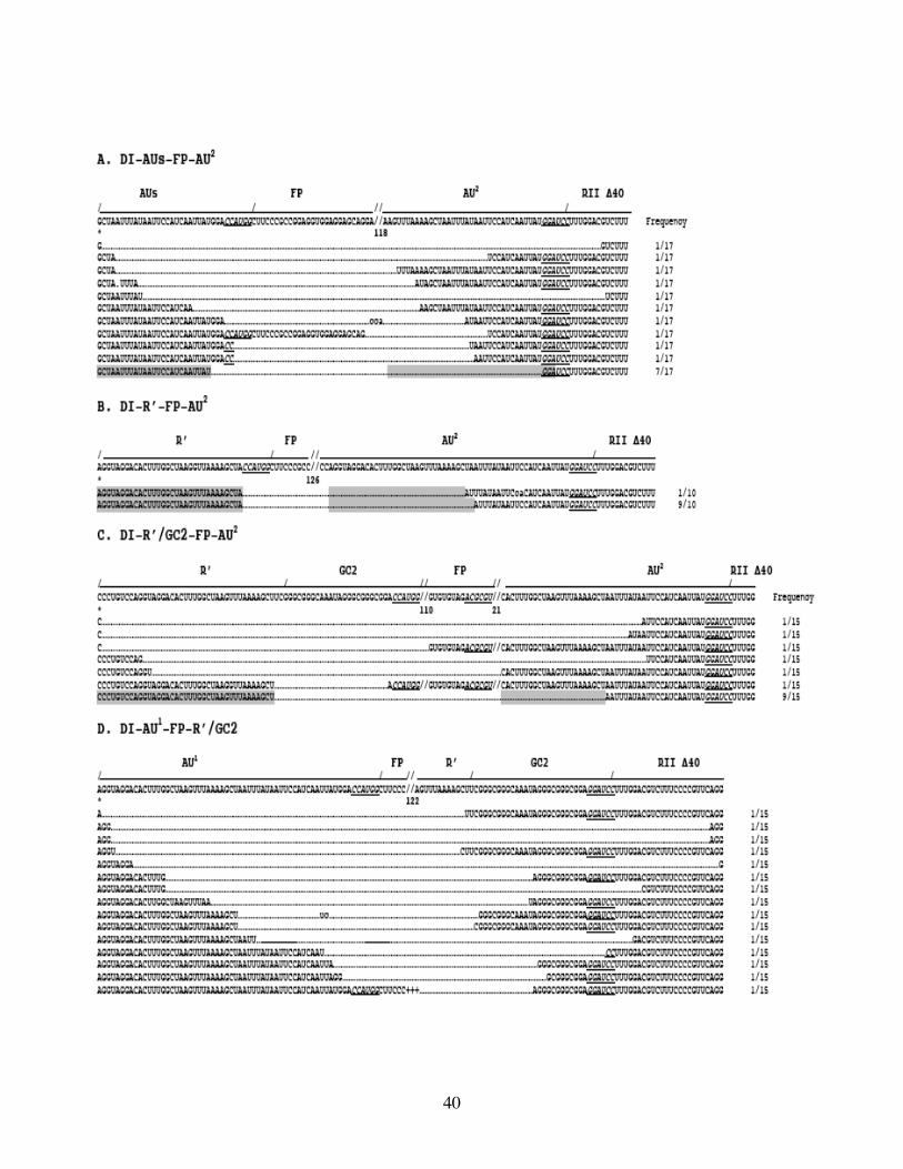
Fig. 2.5. The effect of short AU-rich and GC-rich sequences on DI RNA recombination. All these
constructs are derivatives of DI-RII/Δ40 (Fig. 2.3A). The sequences of AUs and R’ are shown in
Fig. 2.1B, while the complete GC2 sequence is shown in panels C and D. A representative number
of recombinants were amplified by RT-PCR (not shown) from first passage protoplast samples,
cloned and sequenced around the junction sites. The duplicated regions that participated in precise
recombination are boxed. +++ indicates the following not shown sequence: 5’-
GCCGGAGGUGGAGGAGCAGGAUGAU. See further details in the legend to Figs. 2.2 and 2.4.
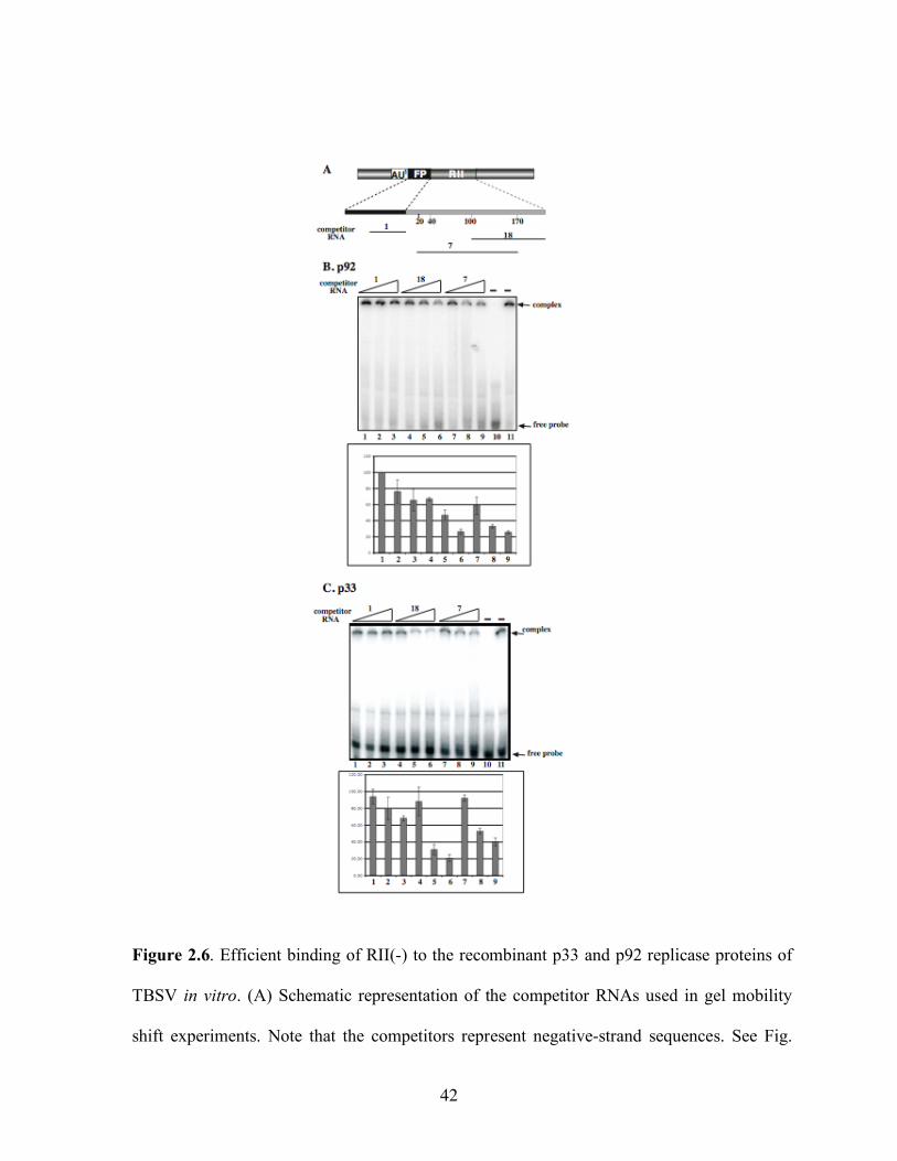
42
Figure 2.6. Efficient binding of RII(-) to the recombinant p33 and p92 replicase proteins of
TBSV in vitro. (A) Schematic representation of the competitor RNAs used in gel mobility
shift experiments. Note that the competitors represent negative-strand sequences. See Fig.
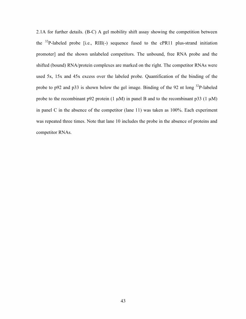
43
2.1A for further details. (B-C) A gel mobility shift assay showing the competition between
the 32P-labeled probe [i.e., RIII(-) sequence fused to the cPR11 plus-strand initiation
promoter] and the shown unlabeled competitors. The unbound, free RNA probe and the
shifted (bound) RNA/protein complexes are marked on the right. The competitor RNAs were
used 5x, 15x and 45x excess over the labeled probe. Quantification of the binding of the
probe to p92 and p33 is shown below the gel image. Binding of the 92 nt long 32P-labeled
probe to the recombinant p92 protein (1 µM) in panel B and to the recombinant p33 (1 µM)
in panel C in the absence of the competitor (lane 11) was taken as 100%. Each experiment
was repeated three times. Note that lane 10 includes the probe in the absence of proteins and
competitor RNAs.
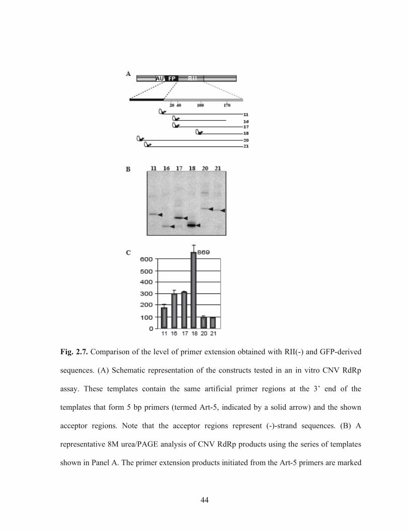
44
Fig. 2.7. Comparison of the level of primer extension obtained with RII(-) and GFP-derived
sequences. (A) Schematic representation of the constructs tested in an in vitro CNV RdRp
assay. These templates contain the same artificial primer regions at the 3’ end of the
templates that form 5 bp primers (termed Art-5, indicated by a solid arrow) and the shown
acceptor regions. Note that the acceptor regions represent (-)-strand sequences. (B) A
representative 8M urea/PAGE analysis of CNV RdRp products using the series of templates
shown in Panel A. The primer extension products initiated from the Art-5 primers are marked
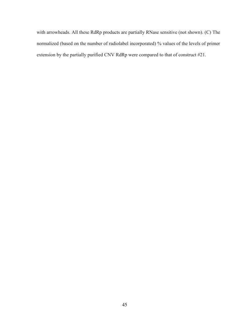
45
with arrowheads. All these RdRp products are partially RNase sensitive (not shown). (C) The
normalized (based on the number of radiolabel incorporated) % values of the levels of primer
extension by the partially purified CNV RdRp were compared to that of construct #21.
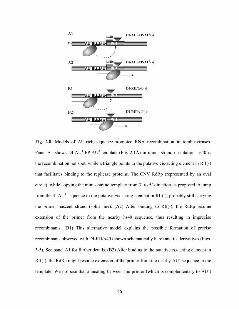
46
Fig. 2.8. Models of AU-rich sequence-promoted RNA recombination in tombusviruses.
Panel A1 shows DI-AU1-FP-AU2 template (Fig. 2.1A) in minus-strand orientation. hs40 is
the recombination hot spot, while a triangle points to the putative cis-acting element in RII(-)
that facilitates binding to the replicase proteins. The CNV RdRp (represented by an oval
circle), while copying the minus-strand template from 3’ to 5’ direction, is proposed to jump
from the 3’ AU1 sequence to the putative cis-acting element in RII(-), probably still carrying
the primer nascent strand (solid line). (A2) After binding to RII(-), the RdRp resume
extension of the primer from the nearby hs40 sequence, thus resulting in imprecise
recombinants. (B1) This alternative model explains the possible formation of precise
recombinants observed with DI-RII/Δ40 (shown schematically here) and its derivatives (Figs.
3-5). See panel A1 for further details. (B2) After binding to the putative cis-acting element in
RII(-), the RdRp might resume extension of the primer from the nearby AU2 sequence in the
template. We propose that annealing between the primer (which is complementary to AU1)

47
and the AU2 sequence in the template (represented by five short lines) prior to primer
extension might facilitate the formation of precise recombinants.
TABLE 2.1. Primers used for PCR
_________________________________________________________________________________________
Construct Primer Sequence Template
_________________________________________________________________________________________
DI-AU1-FP-AU2 114 GCAGTCTAGAGGACGCGTGTAGAGACCCTGTCCAG AU1,
115 GCAGCTCGAGCCATGGATCCATAATTGATGG
DI-AU1-FP 114 GCAGTCTAGAGGACGCGTGTAGAGACCCTGTCCAG AU1,
115 GCAGCTCGAGCCATGGATCCATAATTGATGG
DI-Bar 922 GGAGTCTAGATTGTCTGACCGGATGGGT DI-73-BARS
923 GGAGGGATCCGAAAGTATGATGGTGA
DI-FP-RII/Δ40 751 GGCGGATCCTTTGGACGTCTTTCC DI-FP
21 CCGCCATGGTCCATAATTGATGGA
DI-RII/Δ40 751 GGCGGATCCTTTGGACGTCTTTCC DI-FP
21 CCGCCATGGTCCATAATTGATGGA
DI-RII/Δ20 933 CCGGGATCCACAACCTACCAAAG DI-FP
21 CCGCCATGGTCCATAATTGATGGA
DI- RII/Δ60 934 CCGGGATCCCCGTTCAGGAAAGC DI-FP
21 CCGCCATGGTCCATAATTGATGGA
DI- RII/Δ 100 935 CCGGGATCCCCGTTCAGGAAAGC DI-FP
21 CCGCCATGGTCCATAATTGATGGA
DI-AUs-FP- AU2 860 CGACTCTAGAGCTAATTTATAATTCCATCA AU1,
753 CCGCCATGGTCCATAATTGATGGA
DI-R’/GC2-FP-AU2 752 CGACTCTAGACCCTGTCCAGGTAGG GC2,
794 CCGCCATGGTCCGCCCGCCCTATTTG
DI-AU1-FP-R’/GC2 792 CGACACGCGTCCCTGTCCAGGTAGG GC2,
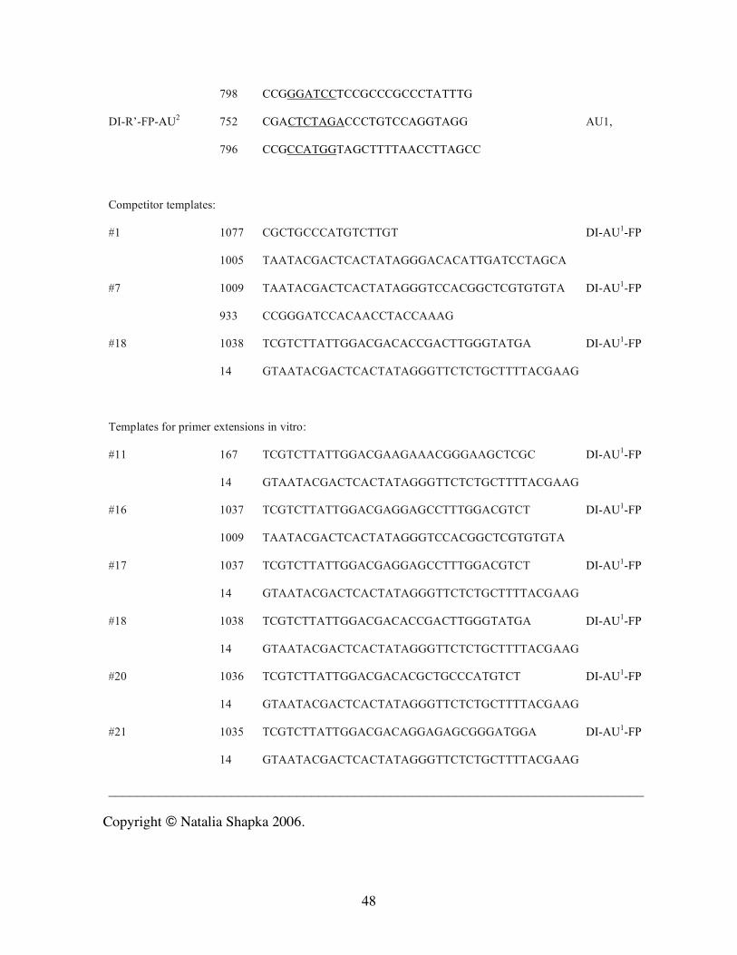
48
798 CCGGGATCCTCCGCCCGCCCTATTTG
DI-R’-FP-AU2 752 CGACTCTAGACCCTGTCCAGGTAGG AU1,
796 CCGCCATGGTAGCTTTTAACCTTAGCC
Competitor templates:
#1 1077 CGCTGCCCATGTCTTGT DI-AU1-FP
1005 TAATACGACTCACTATAGGGACACATTGATCCTAGCA
#7 1009 TAATACGACTCACTATAGGGTCCACGGCTCGTGTGTA DI-AU1-FP
933 CCGGGATCCACAACCTACCAAAG
#18 1038 TCGTCTTATTGGACGACACCGACTTGGGTATGA DI-AU1-FP
14 GTAATACGACTCACTATAGGGTTCTCTGCTTTTACGAAG
Templates for primer extensions in vitro:
#11 167 TCGTCTTATTGGACGAAGAAACGGGAAGCTCGC DI-AU1-FP
14 GTAATACGACTCACTATAGGGTTCTCTGCTTTTACGAAG
#16 1037 TCGTCTTATTGGACGAGGAGCCTTTGGACGTCT DI-AU1-FP
1009 TAATACGACTCACTATAGGGTCCACGGCTCGTGTGTA
#17 1037 TCGTCTTATTGGACGAGGAGCCTTTGGACGTCT DI-AU1-FP
14 GTAATACGACTCACTATAGGGTTCTCTGCTTTTACGAAG
#18 1038 TCGTCTTATTGGACGACACCGACTTGGGTATGA DI-AU1-FP
14 GTAATACGACTCACTATAGGGTTCTCTGCTTTTACGAAG
#20 1036 TCGTCTTATTGGACGACACGCTGCCCATGTCT DI-AU1-FP
14 GTAATACGACTCACTATAGGGTTCTCTGCTTTTACGAAG
#21 1035 TCGTCTTATTGGACGACAGGAGAGCGGGATGGA DI-AU1-FP
14 GTAATACGACTCACTATAGGGTTCTCTGCTTTTACGAAG
__________________________________________________________________________
Copyright © Natalia Shapka 2006.
49
Chapter III
Genome-wide screen identifies host genes affecting viral RNA
recombination
INTRODUCTION
Rapid evolution of RNA viruses with mRNA-sense genomes, which include SARS
coronavirus, hepatitis C virus, and West Nile virus, makes controlling RNA viruses a
difficult task. Emergence of new pathogenic RNA viruses is frequently due to RNA
recombination (Lai, 1992; Worobey and Holmes, 1999), which can lead to dramatic changes
in viral genomes by creating novel combinations of genes, motifs or regulatory RNA
sequences. Thus, RNA recombination can change the infectious properties of RNA viruses
and render vaccines and other antiviral methods ineffective (Worobey and Holmes, 1999).
RNA recombination likely contributed to outbreaks with dengue- (Holmes, Worobey, and
Rambaut, 1999; Worobey, Rambaut, and Holmes, 1999), polio- (Marturano and Fiore, 2002),
calici- (Jiang et al., 1999), astro- (Walter et al., 2001), entero- (Lukashev et al., 2004;
Oprisan et al., 2002), influenza- (Khatchikian, Orlich, and Rott, 1989), pestiviruses (Becher,
Orlich, and Thiel, 2001; Fricke, Gunn, and Meyers, 2001), and SARS coronavirus, a newly-
emerged viral pathogen of humans (Bosch, 2004; Rest and Mindell, 2003; Stavrinides and
Guttman, 2004). RNA recombination is also important in viral RNA repair, which likely
50
increases the fitness of RNA viruses that lack proofreading polymerases (Allison, Thompson,
and Ahlquist, 1990; Guan and Simon, 2000; Lai, 1992; Nagy and Simon, 1997).
Current models of RNA recombination are based on template-switching mechanism
driven by the viral replicase (Lai, 1992; Nagy and Simon, 1997) or RNA-breakage and
ligation (Chetverin et al., 1997). The more common template-switching RNA recombination
is thought to occur as an error during the replication process (Lai, 1992; Nagy and Simon,
1997). Because viral RNA replication depends not only on viral proteins, but on host factors
as well (Ahlquist et al., 2003), it is likely that host factors could affect the recombination
process, too. However, despite the significance of RNA recombination in viral evolution, the
possible roles of host genes in the viral RNA recombination process are currently unknown.
Tombusviruses, including Tomato bushy stunt virus (TBSV) and Cucumber necrosis
virus (CNV), are non-segmented, small model positive-strand RNA viruses (White and
Nagy, 2004). Due to their robust replication and the ability to generate novel RNA
recombinants in whole plants and single cells, tombusviruses are used extensively to dissect
the roles of cis-acting RNA elements during virus infections (White and Nagy, 2004). In vivo
and in vitro replication/recombination studies with a small replicon RNA, termed defective
interfering (DI-72) RNA (White and Morris, 1994b; White and Nagy, 2004), established a
role for RNA sequences/structures and viral replicase proteins in RNA recombination. Co-
expression of the replicon RNA with the two essential tombusviral replicase proteins (Fig.
3.1A) resulted in robust DI RNA replication in Saccharomyces cerevisiae (Panavas and
Nagy, 2003b; Pantaleo, Rubino, and Russo, 2003), which is a model eukaryotic host. Yeast
also supported viral RNA recombination, giving rise to recombinants similar to those in
51
plants and plant protoplasts (Panavas and Nagy, 2003b). Therefore, yeast could be a useful
host to study viral RNA recombination and to identify host proteins involved in this process.
In this paper, we have tested the effect of ~80% of all yeast genes on TBSV
recombination based on screening the entire yeast single-gene knockout (YKO) library for
the occurrence of viral RNA recombinants. Using the TBSV derived replicon RNA, we
identified five YKO strains that supported unusually high levels of new recombinant RNAs.
We also identified four yeast deletion strains that showed reduced viral recombinant
accumulation. Therefore, selected set of host genes could either suppress or accelerate viral
RNA recombination, demonstrating for the first time that host genes play significant roles in
virus recombination and evolution.
MATERIALS AND METHODS
Yeast strains and expression plasmids:
Saccharomyces cerevisiae strain BY4741 (MATa his3∆1 leu2∆0 met15∆0 ura3∆0) and the
haploid deletion series (BY4741 strain background) were from Open Biosystems (Huntville,
AL). The expression plasmids pGBK-His33 (carrying CNV p33 gene behind the ADH1
promoter), pGAD-His92 (containing CNV p92 gene behind the ADH1 promoter), and
pYC/DI-72 (expressing TBSV DI-72 RNA under the control of GAL1 promoter) have been
previously described (Panavas and Nagy, 2003b; Panaviene et al., 2004). Each yeast strain
was co-transformed with all three plasmids using LiAc/ssDNA/PEG method (Gietz and
Woods, 2002) and transformants were selected by complementation of auxotrophic markers.
Out of 4848 strains, we found that 71 were not transformable and 229 strains did not grow on
52
galactose-containing medium. Therefore, total of 4548 strains were tested for RNA
recombination below.
Yeast cultivation:
Each transformed yeast strains from the YKO library were cultured under two different
conditions during the genome-wide screen for RNA recombinants. The first screen included
yeast strains grown in 96-deep-well plates at 23oC in selective media (SC-ULH-) with 2%
galactose until reaching cell density of 0.8-1.0 (OD600). For the second screen, the yeast
strains were grown in 96-deep-well plates at 23oC for 6h in selective media (SC-ULH-) with
2% galactose, followed by 1:10 dilution with SC-ULH- medium containing 5% glucose.
Then, the cells were grown for 24h at 23oC, followed by additional dilution (1:10) and
subsequent culturing until cell density reached 0.8-1.0 (OD600). Yeast cells were harvested by
centrifugation at 1,100g for 5 min.
High-throughput RNA analysis:
We performed two separate genome-wide screens of the YKO library that included total of 4-
6 independent samples per each strain. Total RNA isolation and Northern blot analysis were
done as previously described (Panavas and Nagy, 2003b), except using a high throughput
approach. Briefly, yeast cells in 96- deep-well plates were resuspended in RNA extraction
buffer (50mM sodium acetate, pH 5.2, 10mM EDTA, 1% SDS) and phenol, followed by
incubation for 4 min at 65oC. After removal of phenol, the RNA was recovered by ethanol
precipitation. Agarose gel electrophoresis (1.5%) and Northern blotting were done as
described (Panavas and Nagy, 2003b; Shapka and Nagy, 2004). For negative-strand
53
detection, total yeast RNA obtained from selected strains was separated in denaturing 5%
polyacrylamide/8M urea gel as described previously (Panavas and Nagy, 2003b). The RNA
was quantified using a phosphorImager as described (Panavas, Pogany, and Nagy, 2002).
RT-PCR analysis of the junction sites in the recombinants:
We have used both total yeast RNA extracts and gel-isolated recombinants for reverse
transcription (RT-)PCR reactions to specifically amplify regions covering junction sites.
First, the RT reaction included primer #14
(GTAATACGACTCACTATAGGGTTCTCTGCTTTTACGAAG) for cDNA synthesis,
followed by PCR with primers #168
(TCGTCTTATTGGACGAATTCCTGTTTACGAAAG) and #270
(TTGGAAATTCTCCTTCAGTCTGAGTTTGTGGA). The PCR products were cloned into
pGEM-T Easy vector (Promega) and sequenced using M13 Reverse Primer (Cheng and
Nagy, 2003).
5’RACE and 3’RACE of recombinants:
The 5’ and 3’ sequences of recombinants were determined by using 5’RACE (rapid
amplification of complementary ends) and 3’RACE, respectively, as described (Cheng and
Nagy, 2003). To enrich for recombinants, RNA bands were gel-isolated as described
previously (Cheng and Nagy, 2003). The resulting products were cloned and sequenced.
In vitro Tombusvirus replicase assay: The in vitro replicase assay was performed with the
co-purified (endogenous) RNA as described (Panavas and Nagy, 2003b).
54
RESULTS
Systematic analysis of yeast single-gene deletion strains for enhanced level of viral RNA
recombination.
To facilitate identification of host genes involved in RNA virus evolution/recombination,
we took advantage of the advanced genomics tools available for yeast and the ability of yeast
to support TBSV recombination (Panavas and Nagy, 2003b). The recombination assay was
based on a replication-competent TBSV DI-72 RNA replicon, which, when co-expressed
with the two essential tombusviral replicase proteins (p33 and p92, Fig. 3.1A), undergoes
robust replication and it also generates small amount of RNA recombinants (Panavas and
Nagy, 2003b). The 621 nt DI-72 RNA replicon contains four noncontiguous segments (Fig.
3.1A), including the cis-acting replication elements, derived from the full-length genomic
RNA (White and Nagy, 2004). It is important to note that in this assay replication/evolution
of DI-72 RNA and the de novo generated recombinant RNAs takes place in the absence of
artificial selection markers in all viral RNAs.
To systemically test the effect of each host gene on viral RNA recombination, we have
developed a high throughput method based on the available yeast single-gene deletion
(YKO) library. Briefly, the collection of 4,848 YKO yeast strains representing ~80% of yeast
genes (those which are nonessential for yeast growth) was co-transformed with three
plasmids expressing DI-72 RNA replicon in addition to p33 and p92 replicase proteins (Fig.
3.1A). We successfully transformed 4548 strains that grew on galactose-containing media
(see M&M), and cultured them in 96-deepwell plates, followed by total RNA extraction and
55
agarose gel electrophoresis. Under these conditions, the replication-competent DI-72 RNA is
easily detectable in yeast cells and its amount is similar to the yeast ribosomal RNAs (Fig.
3.1B). In contrast, recombinant RNAs carrying rearranged RNA sequences accumulate
inefficiently in the parental yeast strain as demonstrated by Northern blotting (~1-2% of the
level of the replicating DI-72 RNA) (Fig. 3.1C). Thus, under the above conditions the
screening approach is expected to favor the identification of those YKO strains, which
support increased levels of viral RNA recombinants when compared to the parental strain.
Identification of five host genes whose absence leads to increased frequency of viral
RNA recombination.
Using the above high-throughput genome-wide screen, we identified total of five YKO
strains that generated 10-50-fold higher levels of recombinant viral RNAs than did the
parental yeast strain (Fig. 3.1B-C). Four of these deletion strains, ctl1Δ, met22/hal1Δ, xrn1Δ
and ubp3Δ accumulated one major type of recombinant RNA at levels comparable to that of
the wild-type viral replicon (Fig. 3.1B-C), whereas hur1Δ generated four recombinant RNAs,
which were ~10-50-fold more abundant than the wt replicon (Fig. 3.1B-C). Northern blot
analysis with a probe specific for an internal RIII sequence in the DI-72 RNA replicon
demonstrated the viral origin of these novel recombinant RNAs (Fig. 3.1C). On the contrary,
a probe specific for the 5’ RI sequence, only detected the wt DI-72 replicon in total RNA
samples from all five strains, but not the recombinant-like RNAs (Fig. 3.1D), suggesting that
RNA recombination might have led to dramatic rearrangement of the viral RNA. These
recombinant viral RNAs accumulated in the presence of the wt DI-72 replicon, suggesting
56
that they were generated efficiently and/or competed efficiently with the wt DI-72 RNA
replicon.
To determine the sequence of the novel recombinant-like RNAs, we gel-isolated them
followed by RT-PCR, 3’RACE, 5’RACE, cloning and sequencing (Cheng and Nagy, 2003;
Shapka and Nagy, 2004). We found that the most common recombinant RNAs obtained from
xrn1Δ, ctl1Δ, met22Δ and ubp3Δ strains were similar, partially dimeric RNAs (Fig. 3.2A and
not shown). They contained various duplicated 3’ sequences (part of RII, and complete RIII
and RIV) and had deletions of 5’ DI-72 RNA sequences (i.e., RI and part of RII, Fig. 3.2A).
Most recombinants differed slightly in their junction sequences, a feature shared with TBSV
recombinants arising in planta (5). Recombinants in hur1Δ contained 2-to-5 incomplete
copies of DI-72 RNA sequences with highly variable 5’-truncations (Supplement Fig. 3.8).
The origin of the 1-to-13 extra nucleotides at the 5’ end or at the junctions is currently
unknown. Extra nucleotides are also frequently detected at the junctions in tombusvirus
recombinants in plant protoplasts (Shapka and Nagy, 2004; White and Morris, 1994b) and in
vitro with purified tombusvirus replicase (Cheng and Nagy, 2003), supporting the model that
the tombusviral replicase adds extra sequences to the ends of viral RNAs (Nagy and Simon,
1997).
To gain insights into the dynamics of recombinant formation, we performed time-course
experiments by analyzing total RNA samples at given time points after induction of RNA
transcription from the GAL1 promoter in hur1Δ and xrn1Δ strains. We found that the
recombinants emerged as early as 2 hours after induction (Fig. 3.3A) in the absence of
artificial selection to facilitate their appearance, suggesting that their formation is an efficient
process. The amount of recombinants increased over time due to either new recombination
57
events and/or replication of the recombinant RNAs (Fig. 3.3A). Moreover, we found that the
recombinant RNAs replicated and evolved further in yeast cells over ten serial dilutions in
suppressive media (Fig. 3.3B).
To demonstrate that the recombinant DI RNAs are replication-competent, we isolated
membrane fractions containing tombusvirus replicase/viral RNA complexes from xrn1Δ and
hur1Δ cells. This was followed by in vitro replicase assays in the presence of added
ribonucleotides, including 32P-labeled UTP. These experiments led to in vitro labeling of the
recombinant-sized RNAs in the replicase assay, suggesting that the recombinant RNAs were
part of the replicase complexes (Fig. 3.3C). Their replication competence was also confirmed
by detection of minus-stranded replication intermediates for the recombinant RNAs (Fig.
3.3C). Altogether, we conclude that the recombinant RNAs, similar to the wt DI-72 RNA
replicon, are replication-competent and they are maintained on suppressive media for
extended period of time in yeast.
To test if the deletion of the host gene altered recombination frequency versus
recombinant selection, we analyzed the stability of four cloned recombinants and the wt DI-
72 replicon RNA in the parental and xrn1Δ strain. Fig. 3.4 demonstrates that the stability of
the recombinants and wt replicon was comparable in the parental strain, whereas the
recombinants and the wt replicon RNA showed 2-to-3-fold increased stability in xrn1Δ strain
(Fig. 3.4). This suggests that viral RNA degradation is hindered in xrn1Δ strain. Importantly,
however, all the recombinants and the wt replicon RNA showed similar level of increase in
stability in xrn1Δ strain, suggesting that these RNAs have comparable stability. Overall,
selective RNA degradation of wt replicon versus recombinants cannot explain the increased
accumulation of recombinants over the wt replicon in xrn1Δ strain.
58
Systematic analysis of yeast single-gene deletion strains for decreased level of viral
RNA recombination. Because the above genome-wide screen was only suitable for testing
for increased levels of TBSV recombination, we modified the screening approach to allow
the identification of YKO strains supporting reduced levels of virus recombinants when
compared with the parental strain. To this end, we induced DI-72 RNA transcription in all
4548 YKO strains (also co-expressing p33/p92) for 6 hours, followed by growing them in
glucose-containing medium prior to total RNA extraction and analysis by Northern blotting.
Under these conditions, detectable amounts of recombinant RNAs accumulated in the
parental strain (10-18% of the standard wt DI-72 RNA, Fig. 3.5A). These recombinant RNAs
included complete dimers (two copies of full-length DI-72 replicons joined head-to-tail) and
incomplete dimeric DI-RNAs (5’ truncated monomers joined head-to-tail, see Fig. 3.5A).
Northern-blot analysis of total RNA extracts from all transformants (two-to-four samples per
strain) revealed that the ratio of recombinant RNA versus wt DI-72 RNA was three-to-five-
fold lower only in four YKO strains (Fig. 3.5A). Note that we did not measure the absolute
amounts of RNA recombinants, but instead, estimated the ratio of recombinants versus non-
recombinant DI-72 RNA. This is because DI-72 RNA accumulation level could be different
in various YKO strains (Panavas et al., 2005c), which could affect the amount of RNA
substrates available for recombination.
Characterization of four host genes whose absence leads to decreased frequency of
viral RNA recombination.
59
To further test the recombination deficiency of the above identified 4 YKO strains, we
examined if they could support RNA recombination with a modified viral replicon, DI-AU-
FP. This replicon contains a 186 nt long heterologous sequence including a 46 nt-long AU-
rich stretch (Fig. 3.5B). Previous work in plant protoplasts demonstrated that DI-AU-FP
induced recombination with high efficiency in the presence of the wt helper virus (Shapka
and Nagy, 2004). Northern blot analysis of total RNA obtained from the four YKO strains
co-expressing DI-AU-FP and p33/p92 proteins revealed that RNA recombinants accumulated
poorly (3-5-fold decrease) in these strains when compared to the parental strain (Fig. 3.5B).
The sequences of the generated recombinants isolated from the parental and the selected
YKO strains were comparable (shown schematically in Fig. 3.5B), indicating that the
mechanism of their generation was likely similar. Overall, we conclude that the four
identified YKO strains supported recombination with reduced frequency and/or accumulation
rate of recombinants was lower in these strains than in the parental yeast.
It is worth noting that the above genome-wide screen also identified 5 additional YKO
strains that supported two-fold reduced level of recombinant accumulation in comparison
with the parental strain (see “weak accelerators” in Table 3.1). In addition, we found that
spe3Δ and spt3Δ generated different recombinant profile from the parental strain (Fig. 3.6A).
The notable difference was the accumulation of a single dominant recombinant RNA in these
strains (see recM, Fig. 3.6A). To confirm that the lack of SPE3 and SPT3 genes indeed
affected RNA recombination, we analyzed recombinant formation during DI-AU-FP RNA
replication. This experiment demonstrated that (i) spe3Δ and spt3Δ showed 2-4-fold
increased levels of recombinants and (ii) the profile of recombinants generated were
somewhat different from that observed with the parental strain (Fig. 3.6B).
60
Major contribution of host genes to viral RNA recombination
To demonstrate the full extent of contribution by host genes to viral RNA recombination,
we compared recombinant accumulation in xrn1Δ and pep7Δ strains carrying DI-AU-FP and
p33/p92 expression plasmids. These experiments revealed that xrn1Δ strain accumulated
viral RNA recombinants up to 80-fold higher level than pep7Δ strain did (Fig. 3.7). Because
these yeast strains differed only in the two deleted genes, the above experiment demonstrated
that host genes could play major roles in viral RNA recombination.
DISCUSSION
Viruses are known to evolve rapidly in selected hosts, yet the roles of host genes in RNA
virus recombination/evolution are currently unknown. This work, based on high throughput
genetic screen in yeast, a model host, has led to the identification of 11 host genes that
significantly affected tombusvirus recombination. We found that single deletion of the
identified genes had three types of effect on tombusvirus recombination: (i) five genes
increased, while (ii) four genes decreased recombinant accumulation, and (iii) two genes
changed the profile of recombinants. Additional five genes had lesser effect (~2-fold) on
RNA recombination.
Suppressors of RNA virus recombination
The observation that the accumulation of viral RNA recombinants increased 10-50-fold in
the absence of five host genes (Table 3.1) suggests that these genes, when present, can
suppress RNA virus recombination. Interestingly, three of the identified genes, namely
XRN1, CTL1 and MET22/HAL2, are involved in RNA metabolism/degradation. It is plausible
61
that these genes could affect viral recombination by influencing the 5’-to-3’ RNA
degradation pathway (Parker and Song, 2004). The proposed connection between RNA
degradation and viral RNA recombination is supported by the following findings: (i) the
recombinants had deletions within their 5’ sequences (Fig. 3.2); (ii) 5’ truncated viral RNAs
accumulated in these yeast strains (Fig. 3.1B-C); and (iii) identification of Xrn1p, which is
the key enzyme in the 5’-to-3’ RNA degradation pathway (Parker and Song, 2004; Sheth and
Parker, 2003), as one of the viral recombination affecting proteins; (iv) the increased stability
of both recombinant and DI-72 RNA replicon in xrn1Δ strain (Fig. 3.4). Moreover, three of
the five identified host genes are predicted and/or known to affect the activity of Xrn1p. For
example, Met22p/Hal2p has been shown to affect the activity of Xrn1p via regulating the
level of pAp, an inhibitor of Xrn1p (Dichtl, Stevens, and Tollervey, 1997). Also, Ctl1p is
known to modify the 5’ end of the RNA by removing a phosphate group that could
potentially facilitate Xrn1p-driven 5’-to-3’ RNA degradation (Rodriguez et al., 1999). In
addition, Ubp3p has been shown to increase stability of Xrn1p in cells (Brew and Huffaker,
2002). Altogether, the 5’-to-3’ exoribonuclease activity of Xrn1p could be inhibited in the
absence of one of these genes. On the contrary, the role/function of Hur1p is currently
unknown. The profile of recombinants generated in hur1Δ, however, is different (Supplement
Fig. 3.8) from the recombinants identified in the other four YKO strains, indicating that it
might use a different mechanism during viral RNA recombination. Overall, this genome-
wide screen indicates close connection between viral RNA recombination and RNA
metabolism/RNA degradation. Interestingly, a 5’-to-3’ exoribonuclease, similar to Xrn1p, is
present in Arabidopsis (Kastenmayer and Green, 2000), and Hal2p homolog has been cloned
from rice (Peng and Verma, 1995), suggesting that similar genes are functional in plants, too.
62
Proposed mechanism of suppression of viral RNA recombination:
Based on the identified host genes and the profile of generated viral RNA recombinants,
we propose that four out of five host genes, including XRN1, CTL1, MET22/HAL2 and
UBP3, could suppress viral recombination via affecting the Xrn1p-dependent rapid and
complete degradation of viral RNA. However, in the absence of Xrn1p, or due to inhibition
of Xrn1p activity in the absence of Ctl1p, Met22p or Ubp3p, degradation of the viral RNA
gets slower (Fig. 3.4). The resulting incompletely degraded viral RNA could then participate
in RNA recombination efficiently, facilitating the accumulation of partly dimeric
recombinant RNAs. The generated recombinants are also more stable in xrn1Δ strain, further
facilitating the accumulation of recombinants. Moreover, abundance of 5’ truncated RNA
species in these strains supports the model that these RNAs are intermediates (substrates) in
the RNA recombination process. In addition, efficient recombination is likely due to
“exposure” of the highly recombinogenic RII sequences (Shapka and Nagy, 2004) at the ends
of the viral RNAs after their partial degradation. On the contrary, the parental yeast cells
could efficiently and completely degrade viral RNAs, thus reducing the chance for partly
degraded RNAs to participate in RNA recombination.
Protein accelerators of viral RNA recombination:
The other set of host genes identified during this genome-wide screen includes four genes,
PEP7, IPK1, CHO2 and DCI1, whose deletion resulted in reduced level of viral RNA
recombination (Fig. 3.5). The viral replicon RNA, either DI-72 or DI-AU-FP, replicates
efficiently in these strains, whereas the dimeric recombinant RNAs accumulate 3-to-5-fold
less than in the parental strain. Therefore, these genes might directly influence the frequency
63
of recombination. Based on the known functions of these genes (Table 3.1), we suggest that
(i) intracellular transport of viral and/or host proteins (or possibly protein-viral RNA
complexes) to the site of recombination (see genes PEP7 and DCI1), and/or (ii) the lipid
content/structure of the membranous-compartment, which contains the virus-replicase could
be altered in the absence of these genes (IPK1, CHO2 and DCI1), resulting in reduced RNA
recombination efficiency.
Although the current work has not addressed the mechanism of RNA recombination in the
selected strains, comparison of sequences at the recombination junctions suggests that the
recombinants represent similarity-nonessential (nonhomologous) recombinants (Nagy and
Simon, 1997). The observed recombinants are likely generated via viral replicase-driven
template-switching mechanism, which has been shown for tombusviruses before (Cheng and
Nagy, 2003; White and Morris, 1994b). Also, data presented in the on line material exclude
that DNA recombination or RNA recombination during pol II-driven RNA transcription are
the mechanisms of viral RNA recombination (Fig. 3.9).
General conclusions:
This genome-wide screen of yeast for host genes affecting viral RNA recombination
demonstrates for the first time that selected set of host genes can accelerate or suppress viral
RNA recombination. We found that the majority of yeast single-deletion strains showed low
level of virus recombination, whereas five strains with particular genetic backgrounds were
“hotbeds” for recombination, accelerating virus evolution. This implies that mutation(s) in
host genes involved in suppression of virus recombination create “favorable” genetic
backgrounds for virus RNA recombination, suggesting that such an individual(s) might
64
contribute to RNA recombination and virus evolution more significantly than other
individuals of the same species with less favorable genetic backgrounds. Altogether, our
discovery promises to have a major influence on future thinking about the contribution of
particular host genes and individual organisms to virus recombination and evolution.
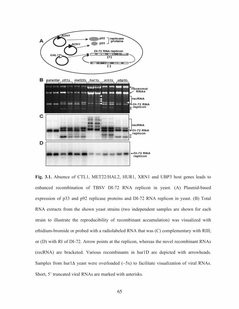
65
Fig. 3.1. Absence of CTL1, MET22/HAL2, HUR1, XRN1 and UBP3 host genes leads to
enhanced recombination of TBSV DI-72 RNA replicon in yeast. (A) Plasmid-based
expression of p33 and p92 replicase proteins and DI-72 RNA replicon in yeast. (B) Total
RNA extracts from the shown yeast strains (two independent samples are shown for each
strain to illustrate the reproducibility of recombinant accumulation) was visualized with
ethidium-bromide or probed with a radiolabeled RNA that was (C) complementary with RIII;
or (D) with RI of DI-72. Arrow points at the replicon, whereas the novel recombinant RNAs
(recRNA) are bracketed. Various recombinants in hur1D are depicted with arrowheads.
Samples from hur1Δ yeast were overloaded (~5x) to facilitate visualization of viral RNAs.
Short, 5’ truncated viral RNAs are marked with asterisks.
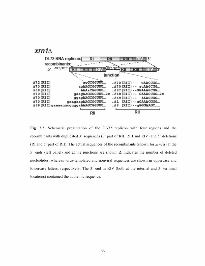
66
Fig. 3.2. Schematic presentation of the DI-72 replicon with four regions and the
recombinants with duplicated 3’ sequences (3’ part of RII, RIII and RIV) and 5’ deletions
(RI and 5’ part of RII). The actual sequences of the recombinants (shown for xrn1Δ) at the
5’ ends (left panel) and at the junctions are shown. Δ indicates the number of deleted
nucleotides, whereas virus-templated and nonviral sequences are shown in uppercase and
lowercase letters, respectively. The 3’ end in RIV (both at the internal and 3’ terminal
locations) contained the authentic sequence.
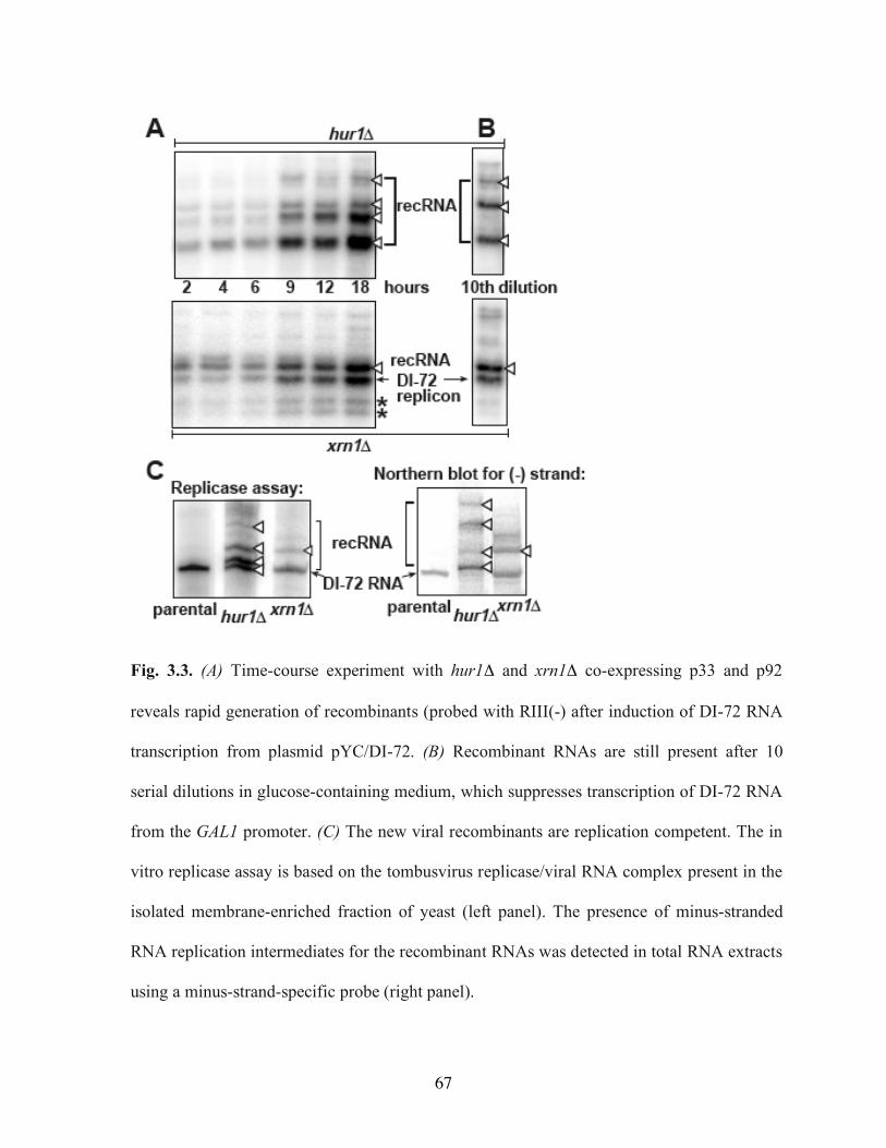
67
Fig. 3.3. (A) Time-course experiment with hur1Δ and xrn1Δ co-expressing p33 and p92
reveals rapid generation of recombinants (probed with RIII(-) after induction of DI-72 RNA
transcription from plasmid pYC/DI-72. (B) Recombinant RNAs are still present after 10
serial dilutions in glucose-containing medium, which suppresses transcription of DI-72 RNA
from the GAL1 promoter. (C) The new viral recombinants are replication competent. The in
vitro replicase assay is based on the tombusvirus replicase/viral RNA complex present in the
isolated membrane-enriched fraction of yeast (left panel). The presence of minus-stranded
RNA replication intermediates for the recombinant RNAs was detected in total RNA extracts
using a minus-strand-specific probe (right panel).
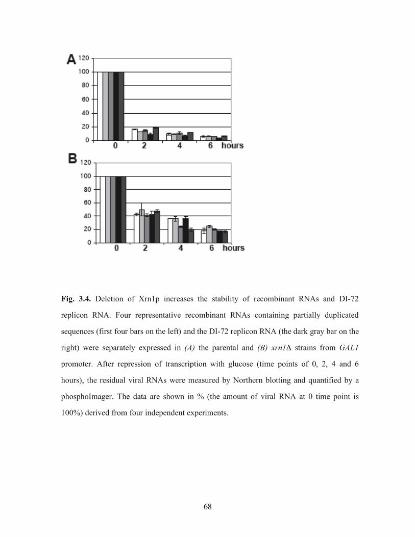
68
Fig. 3.4. Deletion of Xrn1p increases the stability of recombinant RNAs and DI-72
replicon RNA. Four representative recombinant RNAs containing partially duplicated
sequences (first four bars on the left) and the DI-72 replicon RNA (the dark gray bar on the
right) were separately expressed in (A) the parental and (B) xrn1Δ strains from GAL1
promoter. After repression of transcription with glucose (time points of 0, 2, 4 and 6
hours), the residual viral RNAs were measured by Northern blotting and quantified by a
phosphoImager. The data are shown in % (the amount of viral RNA at 0 time point is
100%) derived from four independent experiments.
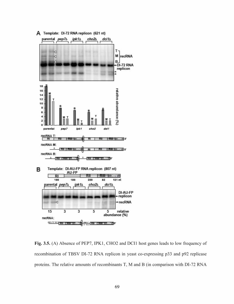
69
Fig. 3.5. (A) Absence of PEP7, IPK1, CHO2 and DCI1 host genes leads to low frequency of
recombination of TBSV DI-72 RNA replicon in yeast co-expressing p33 and p92 replicase
proteins. The relative amounts of recombinants T, M and B (in comparison with DI-72 RNA
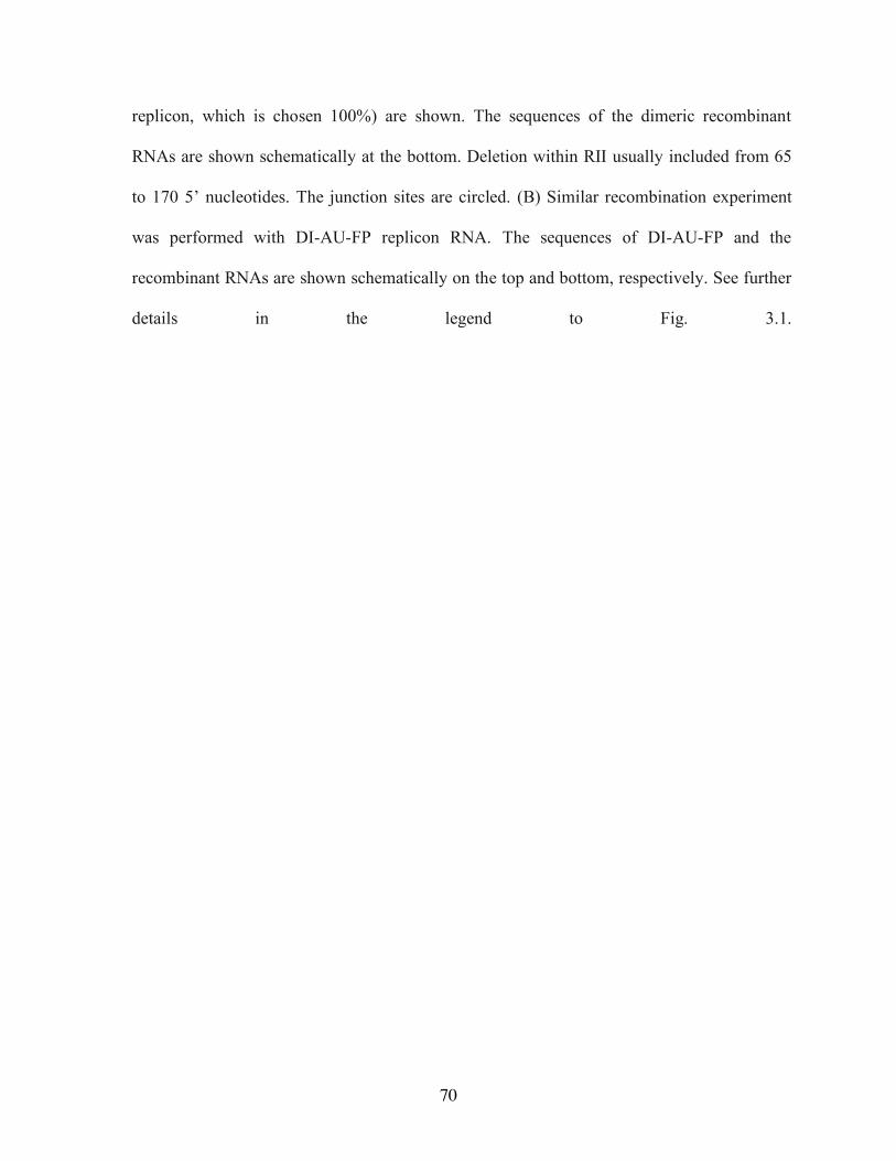
70
replicon, which is chosen 100%) are shown. The sequences of the dimeric recombinant
RNAs are shown schematically at the bottom. Deletion within RII usually included from 65
to 170 5’ nucleotides. The junction sites are circled. (B) Similar recombination experiment
was performed with DI-AU-FP replicon RNA. The sequences of DI-AU-FP and the
recombinant RNAs are shown schematically on the top and bottom, respectively. See further
details in the legend to Fig. 3.1.
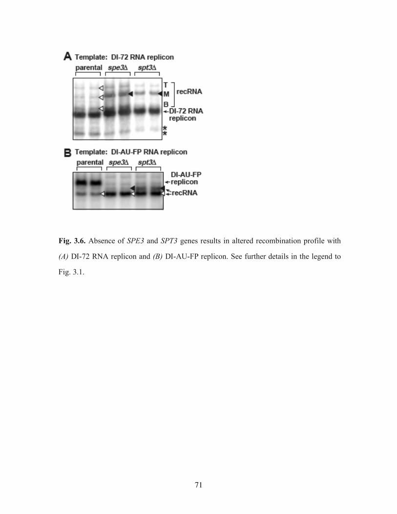
71
Fig. 3.6. Absence of SPE3 and SPT3 genes results in altered recombination profile with
(A) DI-72 RNA replicon and (B) DI-AU-FP replicon. See further details in the legend to
Fig. 3.1.
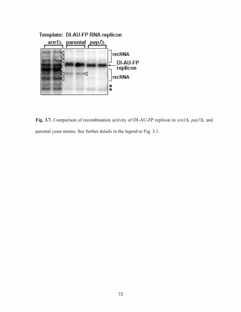
72
Fig. 3.7. Comparison of recombination activity of DI-AU-FP replicon in xrn1Δ, pep7Δ, and
parental yeast strains. See further details in the legend to Fig. 3.1.
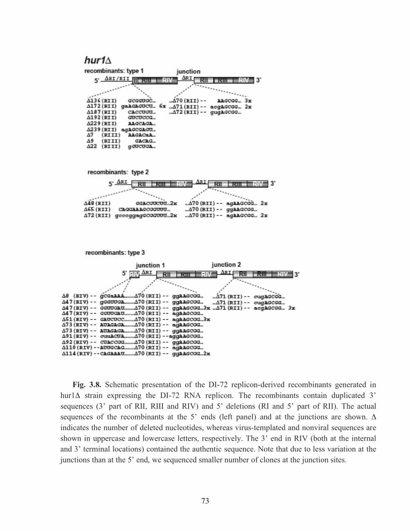
73
Fig. 3.8. Schematic presentation of the DI-72 replicon-derived recombinants generated in hur1Δ strain expressing the DI-72 RNA replicon. The recombinants contain duplicated 3’ sequences (3’ part of RII, RIII and RIV) and 5’ deletions (RI and 5’ part of RII). The actual sequences of the recombinants at the 5’ ends (left panel) and at the junctions are shown. Δ indicates the number of deleted nucleotides, whereas virus-templated and nonviral sequences are shown in uppercase and lowercase letters, respectively. The 3’ end in RIV (both at the internal and 3’ terminal locations) contained the authentic sequence. Note that due to less variation at the junctions than at the 5’ end, we sequenced smaller number of clones at the junction sites.
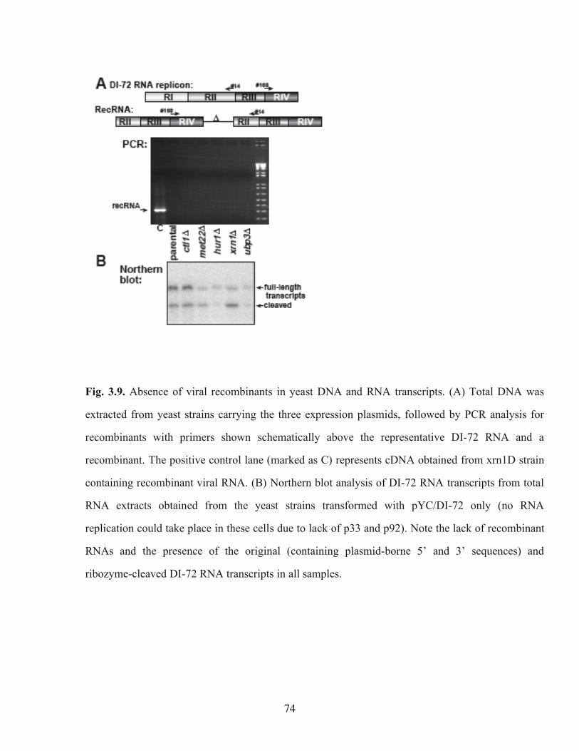
74
Fig. 3.9. Absence of viral recombinants in yeast DNA and RNA transcripts. (A) Total DNA was
extracted from yeast strains carrying the three expression plasmids, followed by PCR analysis for
recombinants with primers shown schematically above the representative DI-72 RNA and a
recombinant. The positive control lane (marked as C) represents cDNA obtained from xrn1D strain
containing recombinant viral RNA. (B) Northern blot analysis of DI-72 RNA transcripts from total
RNA extracts obtained from the yeast strains transformed with pYC/DI-72 only (no RNA
replication could take place in these cells due to lack of p33 and p92). Note the lack of recombinant
RNAs and the presence of the original (containing plasmid-borne 5’ and 3’ sequences) and
ribozyme-cleaved DI-72 RNA transcripts in all samples.
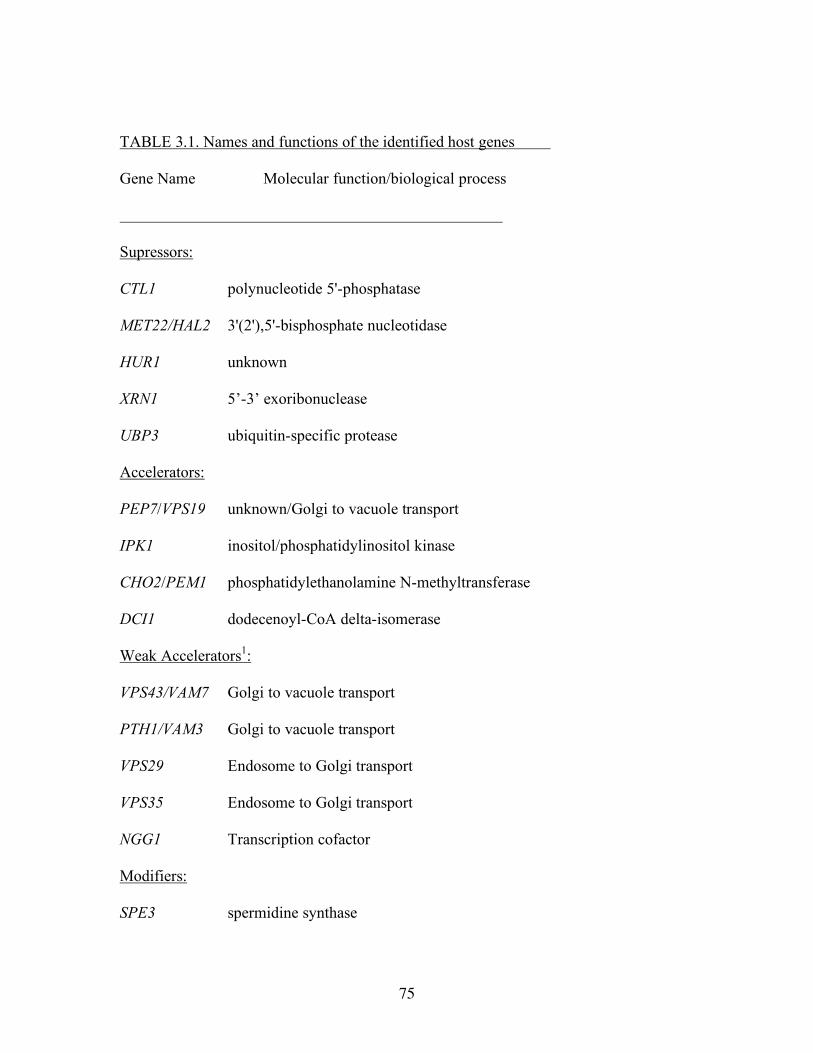
75
TABLE 3.1. Names and functions of the identified host genes
Gene Name Molecular function/biological process
Supressors:
CTL1 polynucleotide 5'-phosphatase
MET22/HAL2 3'(2'),5'-bisphosphate nucleotidase
HUR1 unknown
XRN1 5’-3’ exoribonuclease
UBP3 ubiquitin-specific protease
Accelerators:
PEP7/VPS19 unknown/Golgi to vacuole transport
IPK1 inositol/phosphatidylinositol kinase
CHO2/PEM1 phosphatidylethanolamine N-methyltransferase
DCI1 dodecenoyl-CoA delta-isomerase
Weak Accelerators1:
VPS43/VAM7 Golgi to vacuole transport
PTH1/VAM3 Golgi to vacuole transport
VPS29 Endosome to Golgi transport
VPS35 Endosome to Golgi transport
NGG1 Transcription cofactor
Modifiers:
SPE3 spermidine synthase
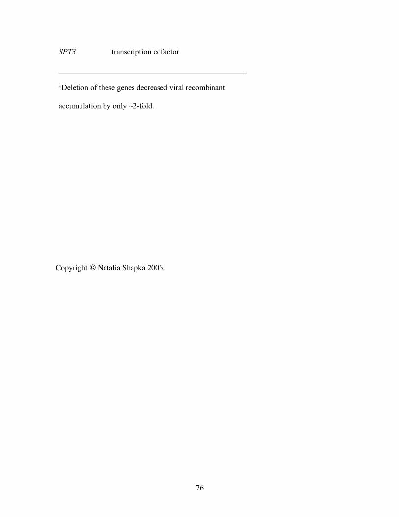
76
SPT3 transcription cofactor
1Deletion of these genes decreased viral recombinant
accumulation by only ~2-fold.
Copyright © Natalia Shapka 2006.
77
Chapter IV
Phosphorylation of the p33 replication protein of Cucumber necrosis
tombusvirus adjacent to the RNA binding site affects viral RNA replication
INTRODUCTION
Replication of plus-strand RNA viruses, which are the most abundant among plant, animal
and human viruses, is performed by viral replicases (Ahlquist, 2002; Ahlquist et al., 2003;
Buck, 1996). The viral replicase consists of viral proteins, including the RNA-dependent
RNA polymerase (RdRp), and putative host factors. In spite of their significance in virus
replication, our current understanding of the roles of viral- and host proteins is incomplete.
These proteins are likely subject to posttranslational modification(s), which could
affect/regulate their functions in viral replication. Indeed, reversible phosphorylation and
other post-translational modifications are important for the functions of many cellular and
viral proteins. For example, protein phosphorylation/dephosphorylation is frequently used as
an “on-off switch”, allowing regulation of protein function. While considerable information
is available for the role of phosphorylation of replication proteins in minus-strand RNA
viruses, which code for P replication protein, a phosphoprotein (Barik and Banerjee, 1992;
Lenard, 1999; Takacs et al., 1992), the role and significance of phosphorylation of replication
proteins in plus-strand RNA viruses is currently poorly understood. The examples include
phosphorylation and ubiquitinylation of Turnip yellow mosaic virus RNA-dependent RNA
polymerase (RdRp) (Hericourt et al., 2000) and the Vpg of potyviruses (Puustinen et al.,
78
2002). Also, phosphorylation of CMV 2a RdRp protein was shown to occur late in infection,
affecting its interaction with the 1a replication protein (a methyltransferase and helicase-like
protein) (Kim, Palukaitis, and Park, 2002). Phosphorylation of the NS5A replication protein
of hepatitis C virus (HCV) has been indicated to affect HCV replication and cellular
signaling, likely facilitating establishment of chronic infections (Huang et al., 2004;
Macdonald et al., 2004; Street et al., 2004). Phosphorylation of the NS5B RdRp protein of
HCV has been proposed to play critical role in HCV replication, based on enhanced HCV
replication in cells over-expressing protein kinase C-related kinase 2 (PRK2, a host kinase),
which is known to interact with NS5B (Kim et al., 2004). In addition to the proposed role of
phosphorylation in virus replication, posttranslational modification also affects the functions
of other viral proteins, as demonstrated for movement (MP) and coat proteins (CP) of plus-
stranded RNA viruses of plants (Ivanov et al., 2003; Waigmann et al., 2000).
Tombusviruses are important and emerging plant pathogens that are among the best
characterized plant viruses (White and Nagy, 2004). They have monopartite, single-stranded
RNA genomes of ~4.8 kb with mRNA-sense polarity. They belong to supergroup 2 viruses
that include important plant pathogens (luteoviruses, carmoviruses, and others), human and
animal pathogens (hepatitis C virus, flaviviruses, pestiviruses), and bacteriophages (Qbeta).
Two of the five Tombusvirus-coded proteins, namely p33 and p92 (Fig. 4.1), were shown to
be essential for tombusvirus replication (Oster, Wu, and White, 1998a; Panaviene, Baker,
and Nagy, 2003; Panaviene, Panavas, and Nagy, 2005; Scholthof, Scholthof, and Jackson,
1995a) and recombination (Panaviene and Nagy, 2003). Both p33 and p92 are translated
from the genomic RNA and p92 is the result of translational readthrough of the translation
stop codon of p33 (Fig. 4.1). Due to the protein expression strategy of tombusviruses, the N-
79
terminal portion of p92 overlaps with p33 (Scholthof, Scholthof, and Jackson, 1995a). p92
contains the signature RdRp motifs and together with p33 is part of the tombusvirus replicase
(Nagy and Pogany, 2000b; Panaviene et al., 2004; Scholthof, Scholthof, and Jackson, 1995a).
The essential p33 replication protein is proposed to function in template selection by
specifically recognizing a conserved internally located stem-loop structure within the p92
ORF (Pogany, White, and Nagy, 2005). Altogether, the p33 is likely involved in many
essential replication functions, such as template RNA binding (Monkewich et al., 2005;
Panavas et al., 2005b; Pogany, White, and Nagy, 2005), assembly/disassembly of the viral
replicase complex (Panaviene, Panavas, and Nagy, 2005; Panaviene et al., 2004), targeting
and/or attaching the replicase complexes to subcellular membranes (Navarro, Rubino, and
Russo, 2004; Panavas et al., 2005b), complementary RNA synthesis, release of the newly
made viral RNA progeny from the site of replication, and so on. Because posttranscriptional
modifications of the replication proteins might play roles as molecular switches in the
replication process by altering the properties of the replication proteins, we tested if the
tombusvirus p33 is phosphorylated in infected plants. This study establishes that
phosphorylation of the p33 replication protein takes place both in vivo and in vitro. Using
kinase-containing plant extract, the phosphorylation sites were mapped to threonine/serine
(T/S) residues located in the vicinity of the RNA-binding site, suggesting the possible role of
phosphorylation in modulating the RNA-binding activity of p33. Using phosphorylation-
mimicking aspartic acid (D) mutants of p33, we demonstrate that phosphorylation of the T/S
residues in p33 could influence subgenomic versus genomic RNA synthesis and the ratio
between plus- and minus-stranded RNAs. Phosphorylation incompetent alanine (A) mutants
of CNV replicated slower than wild type in plant protoplast and showed less severe
80
symptoms in Nicotiana benthamiana plants. In summary, our data promote a regulatory role
for phosphorylation of p33 in tombusvirus replication.
MATERIALS AND METHODS
Detection of phosphorylated p33 and p28 replication proteins by Western blotting using
phosphothreonine specific antibody or by staining with phosphoprotein-specific dye.
Leaves (2 g) of N. benthamiana plants systemically infected with CNV and TCV,
respectively, were used to obtain the membrane-enriched and soluble fractions as described
(Panaviene et al., 2004), except we included phosphatase inhibitor mix containing 25 mM
NaF, 10 mM benzamidine, 10 mM sodium orthovanadate. 1/10th of the obtained membrane-
enriched fraction was loaded on a 10% SDS-PAGE gels, electrophoresed, transferred to
PVDF membranes, followed by using anti-phosphothreonine and anti-phosphoserine
antibodies as recommended by the manufacturer (Research Diagnostic, Inc).
The second method to detect phosphorylated p33 in CNV-infected N. benthamiana
was based on using the phosphoprotein specific Pro-Q Diamond dye from Molecular Probes.
First, we prepared the membrane-enriched fraction (see above), followed by solubilization of
membrane-bound proteins with 2% Triton X-100 as described (Nagy and Pogany, 2000b;
Panaviene et al., 2004). Anti-p33 antibody (prepared against CVGYLKNTPENRLIY
peptide, Sigma-Genosys) was then added to the sample, followed by incubation in the cold
room and then addition of protein A (Sigma) and further incubation for 60 min. The pellet
was recovered after centrifugation, washed three times and then loaded on an SDS-PAGE
gel. The gel was then fixed overnight with 200 ml 10% acetic acid (glacial) and 50%
81
methanol, followed by three time wash with 200 ml deionized water for 15 min. Staining of
the gel was done overnight in the dark with 50 ml Pro-Q Diamond dye as recommended by
the manufacturer (Molecular Probes). De-staining was done with 200 ml 10% methanol and
7% acetic acid for 30 min. Images of gels were taken when illuminated with 302 nm UV
light.
Preparation of host kinase-containing fraction from N. benthamiana and in vitro
phosphorylation of p33
Young leaves (5 g) of uninfected N. benthamiana were used to obtain the membrane-
enriched fraction (see above), followed by solubilization with 4% Triton X-100 exactly as
described for preparation of CNV replicase (Panaviene et al., 2004). The solubilized
membrane-enriched preparation was passed through Sephadex G-25 resin (Amersham). To
reduce the background in subsequent assays, the obtained host kinase preparation was mixed
with phosphorylation buffer (0.1M HEPES pH 7.4, 10 mM EDTA, 5 mM DTT, 50 mM
MgCl2) in the presence of 1mM ATP to allow phosphorylation of any phosphorylatable
proteins that might be present in the preparation for 20 min at room temperature (RT). This
was followed by removal of ATP on a Sephadex G-25 column.
To test phosphorylation of CNV p33 in vitro by the above kinase-containing plant
membrane preparation, we selected two purified recombinant p33-derived peptides, termed
C10 (p33181-240) and C11 (p33211-240) (Rajendran and Nagy, 2003), expressed in E. coli. The
cleavage of the proteins was done with Factor Xa protease (NEB) as described (Rajendran
and Nagy, 2003). The in vitro phosphorylation reaction was made in 20 µl volume in the
phosphorylation buffer (see above), plus 1 µl γ32P-ATP (7000 Ci/mmol, 0.05 mCi), 2 µl
82
kinase-containing plant membrane fraction and 3 µg of p33-derived peptide. After incubation
for 20 min, the samples were run in SDS PAGE gels, stained with coomassie blue, dried and
exposed to a phospho-storage screen and imaged with a PhosphorImager.
In vitro phosphorylation of p33 with PKC
The affinity-purified p33 mutants (200 µg/ml, expressed in E. coli) (Rajendran, Pogany and
Nagy 2002), carrying or lacking the T205, S210, and T211 positions, were phosphorylated in 20
µl volume containing PKC-phosphorylation buffer (20mM HEPES, 0.03% triton X-100,
4mM MOPS, 5mM µ glycerol phosphate, 1 mM EGTA, 0.2 mM Na-orthovanadate, 0.2 mM
DTT), 0.1 mM ATP (unlabeled), 5 µl PKC lipid activator (Upstate), 1 µl 2mM CaCl, 2.5 µl
γ32P-ATP (7000 Ci/mmol, 0.125 mCi), and 0.5 µl PKC (Sigma), for 15 min at room
temperature. Afterwards, the samples were boiled in SDS-PAGE running buffer for three min
and loaded onto a large 10% SDS-PAGE gel (Bio-Rad protean II xi). The electrophoresis
was performed at 200 volts for 6 hours, followed by staining with coomassie blue, drying and
then scanning by a PhosphorImager.
Construction and testing of the phosphorylation-mimicking mutants of CNV p33 in N.
benthamiana protoplasts and plants
To make site-directed mutations that replaced S/T residues with A/D residues, we used
standard Quick Change XL Site-Directed Mutagenesis Kit (Stratagene) as described before
(Panaviene, Baker, and Nagy, 2003) based on an infectious CNV clone (Rochon, 1991). The
mutant clones were confirmed by sequencing, using Beckman Coulter DTCS Quickstart Kit
and primer #958 (5’-CCGGCACGGGAGCTCAAGGGTAAGGA).
83
Full-length RNA transcripts of CNV (Rochon, 1991), DI-72 and DI-73 (White and
Morris, 1994a; White and Morris, 1994e) were prepared using standard in vitro transcription
reaction with T7 RNA polymerase on SmaI linearized DNA templates. N. benthamiana
protoplasts were prepared as described before (Panaviene, Baker, and Nagy, 2003).
Protoplasts were divided into aliquots (5 x10⁵ protoplasts) and electroporated with 5 µg of wt
or mutated CNV gRNA and 1 µg of DI RNA (when the goal was to test DI RNA replication).
After electroporation, protoplasts were incubated in the dark at 22°C.
N. benthamiana plants were rub inoculated with 3 µg of wt or mutated CNV gRNA.
Inoculated plants were kept in the green house. Samples were taken after 5, 7 and 14 dpi.
CNV RNA extraction and Northern blotting
Total RNA was prepared from protoplast or plants using a standard phenol/chloroform
method (Kong, Wang, and Simon, 1997; Nagy, Pogany, and Simon, 2001) and the samples
were electrophoresed in 1.2% agarose (in case of gRNA) or 4% polyacrylamide/8 M urea
gels (for DI-72 RNA). The RNA was then transferred from the gels to Hybond XL
membrane (Amersham-Pharmacia) by electrotransfer and hybridized with CNV-specific
probes for the 3’ noncoding region as described (Panaviene, Baker, and Nagy, 2003).
Hybridization was done in ULTRAhyb hybridization buffer at 68°C as recommended by the
supplier (Ambion). The riboprobes were made in an in vitro transcription reaction with T7
RNA polymerase in the presence of [32P]UTP as described. The CNV DNA template was
obtained using PCR with pair of primers #16
(GTAATACGACTCACTATAGGGCTGCATTTCTGCAATGTTC) and #312
(GCTGTCAGTCTAGTGGA) for detection of plus-strands, and with #31
84
(GTAATACGACTCACTATAGGAAATTCTCCAGGATTTC) and #806
(CCGTCTAGAGTCGTCGTTTACTGGAAGTTAC) for detection of minus-strands. The
template for detection of DI-72 RNA (+) and (-) strands (corresponding to RIII/RIV) was
obtained with PCR using primers #1165 (AGCGAGTAAGACCAGACTCTTCA) and #22
(GTAATACGACTCACTATAGGGCTGCATTTCTGCAATGTTCC) versus #157
(GGGTTTCATAGAAAAGAAAACAAAACC) and #18
(GTAATACGACTCACTATAGGAGAAAGCGAGTAAGACAG), respectively.
The two component, trans-complementation assay was performed in N. benthamiana
protoplasts with selected mutants as described earlier (Panaviene, Baker, and Nagy, 2003).
The mutant clones were confirmed by sequencing, as described above.
Western blot analysis
Total proteins from N.bentamiana plant or protoplasts infected with CNV were prepared as
described (Panaviene et al., 2004). The samples were run in 10% SDS-PAGE, followed by
transfer onto PVDF membrane (Bio-Rad). Immunodetection of p33 protein in the Western
blot was performed by using anti-p33 antibody (generous gift of H. Scholthof).
Analysis of replicon RNA accumulation in yeast
S. cerevisiae strain INVSc1 (Invitrogen) transformed with three plasmids [i.e., pGAD-
His92, pGBK-His33 and pYC-DI-72(+)Rz (Panaviene et al., 2004)] was grown in SC-ULT
medium containing 2% galactose for 24 h at 30°C (Panavas and Nagy, 2003b). To extract
total RNA from yeast, equal volumes of RNA extraction buffer (50 mM sodium acetate, pH
5.3, 10 mM EDTA, 1% SDS) and water-saturated phenol were added to the pelleted cells
85
(Panavas and Nagy, 2003b). Agarose gel electrophoresis and Northern blotting was as
described above (Pogany, White, and Nagy, 2005).
Western blot to detect accumulation levels of His-tagged p33 was performed as
described earlier (Pogany, White, and Nagy, 2005) using monoclonal anti-His antibodies
(Amersham) and secondary alkaline phosphatase-conjugated anti-mouse antibody (Sigma).
Western blots were developed using BCIP and NBT (Sigma).
RESULTS
Rationale:
Sequence comparison of p33 replication proteins of tombusviruses (Fig. 4.1A) revealed the
presence of conserved S and T residues (i.e., T205, S210 and T211) adjacent to the previously
identified arginine (R) and proline (P)-rich RNA-binding site (termed RPR-motif,
R213P214R215R216R217P218)(Fig. 4.1B). These S and T residues are likely phosphorylated in
cells based on NetPhos analysis (i.e., they showed the probability of 0.8-0.9 relative value on
a scale of 0.0-1.0 for phosphorylation) (software: NetPhos, version 2.0, Technical University
of Denmark) (Fig. 4.1B). Possible phosphorylation of these S and T residues in p33 would
introduce negative charge in the vicinity of the positively-charged RPR-motif, thus likely
decreasing the RNA-binding capacity of p33. In support of this model, substitutions of A for
arginines (R), which reduced the overall positive charge of the RPR motif, inhibited the
ability of p33 to bind to the viral RNA as well as decreased tombusvirus replication in plant
protoplasts (Panaviene, Baker, and Nagy, 2003). Based on these observations, we predicted
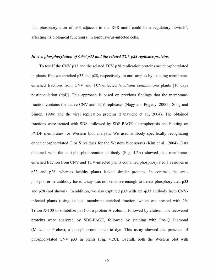
86
that phosphorylation of p33 adjacent to the RPR-motif could be a regulatory “switch”,
affecting its biological function(s) in tombusvirus-infected cells.
In vivo phosphorylation of CNV p33 and the related TCV p28 replicase proteins.
To test if the CNV p33 and the related TCV p28 replication proteins are phosphorylated
in plants, first we enriched p33 and p28, respectively, in our samples by isolating membrane-
enriched fractions from CNV and TCV-infected Nicotiana benthamiana plants [10 days
postinoculation (dpi)]. This approach is based on previous findings that the membrane-
fraction contains the active CNV and TCV replicases (Nagy and Pogany, 2000b; Song and
Simon, 1994) and the viral replication proteins (Panaviene et al., 2004). The obtained
fractions were treated with SDS, followed by SDS-PAGE electrophoresis and blotting on
PVDF membranes for Western blot analysis. We used antibody specifically recognizing
either phosphorylated T or S residues for the Western blot assays (Kim et al., 2004). Data
obtained with the anti-phosphothreonine antibody (Fig. 4.2A) showed that membrane-
enriched fraction from CNV and TCV-infected plants contained phosphorylated T residues in
p33 and p28, whereas healthy plants lacked similar proteins. In contrast, the anti-
phosphoserine antibody based assay was not sensitive enough to detect phosphorylated p33
and p28 (not shown). In addition, we also captured p33 with anti-p33 antibody from CNV-
infected plants (using isolated membrane-enriched fraction, which was treated with 2%
Triton X-100 to solubilize p33) on a protein A column, followed by elution. The recovered
proteins were analyzed by SDS-PAGE, followed by staining with Pro-Q Diamond
(Molecular Probes), a phosphoprotein-specific dye. This assay showed the presence of
phosphorylated CNV p33 in plants (Fig. 4.2C). Overall, both the Western blot with
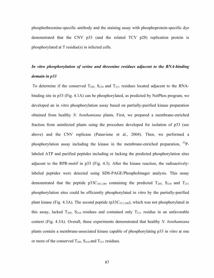
87
phosphothreonine-specific antibody and the staining assay with phosphoprotein-specific dye
demonstrated that the CNV p33 (and the related TCV p28) replication protein is
phosphorylated at T residue(s) in infected cells.
In vitro phosphorylation of serine and threonine residues adjacent to the RNA-binding
domain in p33
To determine if the conserved T205, S210 and T211 residues located adjacent to the RNA-
binding site in p33 (Fig. 4.1A) can be phosphorylated, as predicted by NetPhos program, we
developed an in vitro phosphorylation assay based on partially-purified kinase preparation
obtained from healthy N. benthamiana plants. First, we prepared a membrane-enriched
fraction from uninfected plants using the procedure developed for isolation of p33 (see
above) and the CNV replicase (Panaviene et al., 2004). Then, we performed a
phosphorylation assay including the kinase in the membrane-enriched preparation, 32P-
labeled ATP and purified peptides including or lacking the predicted phosphorylation sites
adjacent to the RPR-motif in p33 (Fig. 4.3). After the kinase reaction, the radioactively-
labeled peptides were detected using SDS-PAGE/PhosphoImager analysis. This assay
demonstrated that the peptide p33C181-240 containing the predicted T205, S210 and T211
phosphorylation sites could be efficiently phosphorylated in vitro by the partially-purified
plant kinase (Fig. 4.3A). The second peptide (p33C211-240), which was not phosphorylated in
this assay, lacked T205, S210 residues and contained only T211 residue in an unfavorable
context (Fig. 4.3A). Overall, these experiments demonstrated that healthy N. benthamiana
plants contain a membrane-associated kinase capable of phosphorylating p33 in vitro at one
or more of the conserved T205, S210 and T211 residues.
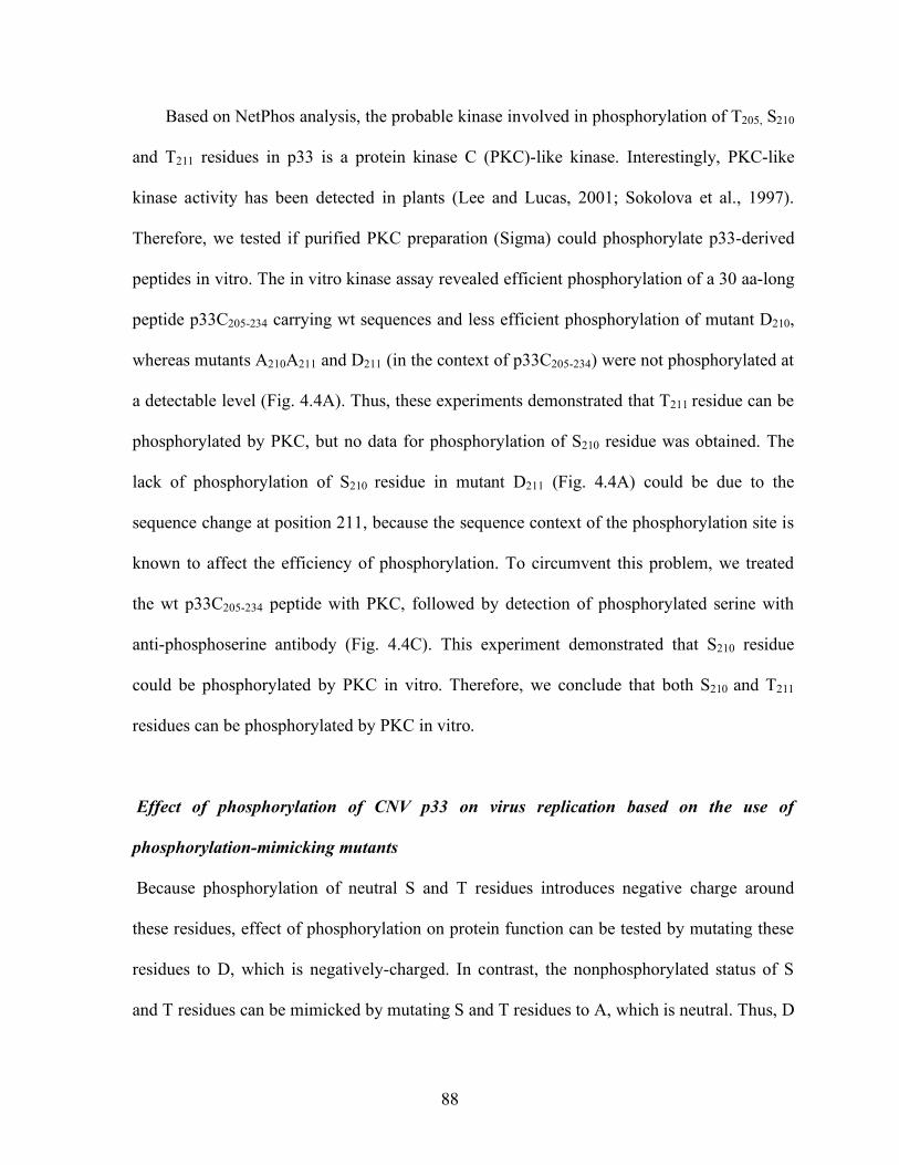
88
Based on NetPhos analysis, the probable kinase involved in phosphorylation of T205, S210
and T211 residues in p33 is a protein kinase C (PKC)-like kinase. Interestingly, PKC-like
kinase activity has been detected in plants (Lee and Lucas, 2001; Sokolova et al., 1997).
Therefore, we tested if purified PKC preparation (Sigma) could phosphorylate p33-derived
peptides in vitro. The in vitro kinase assay revealed efficient phosphorylation of a 30 aa-long
peptide p33C205-234 carrying wt sequences and less efficient phosphorylation of mutant D210,
whereas mutants A210A211 and D211 (in the context of p33C205-234) were not phosphorylated at
a detectable level (Fig. 4.4A). Thus, these experiments demonstrated that T211 residue can be
phosphorylated by PKC, but no data for phosphorylation of S210 residue was obtained. The
lack of phosphorylation of S210 residue in mutant D211 (Fig. 4.4A) could be due to the
sequence change at position 211, because the sequence context of the phosphorylation site is
known to affect the efficiency of phosphorylation. To circumvent this problem, we treated
the wt p33C205-234 peptide with PKC, followed by detection of phosphorylated serine with
anti-phosphoserine antibody (Fig. 4.4C). This experiment demonstrated that S210 residue
could be phosphorylated by PKC in vitro. Therefore, we conclude that both S210 and T211
residues can be phosphorylated by PKC in vitro.
Effect of phosphorylation of CNV p33 on virus replication based on the use of
phosphorylation-mimicking mutants
Because phosphorylation of neutral S and T residues introduces negative charge around
these residues, effect of phosphorylation on protein function can be tested by mutating these
residues to D, which is negatively-charged. In contrast, the nonphosphorylated status of S
and T residues can be mimicked by mutating S and T residues to A, which is neutral. Thus, D
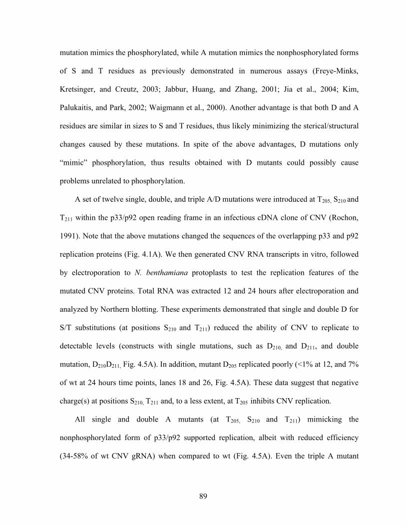
89
mutation mimics the phosphorylated, while A mutation mimics the nonphosphorylated forms
of S and T residues as previously demonstrated in numerous assays (Freye-Minks,
Kretsinger, and Creutz, 2003; Jabbur, Huang, and Zhang, 2001; Jia et al., 2004; Kim,
Palukaitis, and Park, 2002; Waigmann et al., 2000). Another advantage is that both D and A
residues are similar in sizes to S and T residues, thus likely minimizing the sterical/structural
changes caused by these mutations. In spite of the above advantages, D mutations only
“mimic” phosphorylation, thus results obtained with D mutants could possibly cause
problems unrelated to phosphorylation.
A set of twelve single, double, and triple A/D mutations were introduced at T205, S210 and
T211 within the p33/p92 open reading frame in an infectious cDNA clone of CNV (Rochon,
1991). Note that the above mutations changed the sequences of the overlapping p33 and p92
replication proteins (Fig. 4.1A). We then generated CNV RNA transcripts in vitro, followed
by electroporation to N. benthamiana protoplasts to test the replication features of the
mutated CNV proteins. Total RNA was extracted 12 and 24 hours after electroporation and
analyzed by Northern blotting. These experiments demonstrated that single and double D for
S/T substitutions (at positions S210 and T211) reduced the ability of CNV to replicate to
detectable levels (constructs with single mutations, such as D210, and D211, and double
mutation, D210D211, Fig. 4.5A). In addition, mutant D205 replicated poorly (<1% at 12, and 7%
of wt at 24 hours time points, lanes 18 and 26, Fig. 4.5A). These data suggest that negative
charge(s) at positions S210, T211 and, to a less extent, at T205 inhibits CNV replication.
All single and double A mutants (at T205, S210 and T211) mimicking the
nonphosphorylated form of p33/p92 supported replication, albeit with reduced efficiency
(34-58% of wt CNV gRNA) when compared to wt (Fig. 4.5A). Even the triple A mutant
90
(construct A205A210A211, accumulation level of 13-21% of wt, lanes 19 and 27, Fig. 4.5A)
was replication competent, suggesting that nonphosphorylated residues at positions 205, 210
and 211 are important for the function(s) of replicase proteins in vivo. However, because all
these A mutants replicated less efficiently than wt, it is likely that phosphorylation of
p33/p92 plays a regulatory role during virus replication (see below).
To demonstrate that the above mutations at T205, S210 and T211 in CNV affected
replication due to changes in the p33/p92 replication proteins, but not due to altered CNV
RNA structure, we tested the ability of the mutated CNV replication proteins to support
replication in trans, using a model template, DI-73 RNA (Fig. 4.5B). Co-electroporation of
DI-73 RNA with the CNV helper (wt or D210D211) into plant protoplasts demonstrated that
CNV D210D211 did not support replication of DI-73 RNA in trans, indicating that the
phosphorylation-mimicking replication protein(s) is not functional in vivo.
To test if the above CNV mutants expressed p33 in comparable amounts, we performed
Western blotting on total protein extracts obtained from protoplasts 12, 18 and 24 hpi with an
antibody specific for the C-terminal portion of p33 (outside the mutagenized domain of p33).
We found that D210D211 p33 was not detectable (Fig. 4.5C), whereas the amount of D210A211
was only ~10% of wt p33 (Fig. 4.6B). The accumulation of A210A211 p33 was comparable to
wt p33. The reduced accumulation of phosphorylation-mimicking p33 mutants in plant
protoplasts can be explained by several alternative models including decreased stability
and/or production of these p33 forms.
Hypophosphorylation-mimicking p33 mutants affected plus-strand RNA and subgenomic
RNA synthesis in vivo
91
To test for possible regulatory roles of phosphorylation of p33/p92 in replication, we
performed time-course experiments in protoplasts with a viable nonphosphorylation-
mimicking mutant, a hypophosphorylation mutant (i.e., D210A211 carrying only a single D
mutation) and the wt (Fig. 4.6). Total RNA samples were prepared from protoplasts and
analyzed with Northern blotting as described above. These experiments revealed that
nonphosphorylation-mimicking A210A211 and the partial (hypo)-phosphorylation -mimicking
D210A211 mutants generated 5-6 times more subgenomic (sg) RNA2 than sgRNA1 at early
time points, whereas sgRNA2 was only ~3-fold more abundant than sgRNA1 in the wt CNV
infections (Fig. 4.6A). Altogether, sgRNA2 was the most abundant viral RNA species in
A210A211 and D210A211 infections, in contrast to wt CNV, which produced the gRNA in the
largest amount (except at the early time point, Fig. 4.6A). These data suggest that
phosphorylation status of S210 and T211 in p33/p92 could affect the level and ratio of sgRNA1
and sgRNA2 synthesis in CNV infections.
To test if mimicking mutations could also affect minus- versus plus-strand synthesis
during CNV replication, we took advantage of DI-72 RNA replicon, which produces minus-
strand RNA at easily detectable levels (Monkewich et al., 2005). In this assay, N.
benthamiana protoplasts were co-electroporated with wt or mutated (A210A211, A205A210A211
and D210A211) CNV RNA and DI-72 RNA, followed by detection of plus- and minus-strand
DI-72 RNA levels in total RNA samples by Northern blotting (Fig. 4.7). We observed that
DI-72 minus-strand synthesis took place at low levels (10-20%) between 4-12 hours post
infection (hpi), followed by increased synthesis between 12-24 hours in the presence of wt or
any of the mutated helper CNV. On the contrary, plus-strand RNA synthesis showed
remarkable differences depending on the wt and mutated helper CNV (Fig. 4.7A). For
92
example, detectable level of DI-72 plus-strands was observed as early as 6 and 8 hpi in the
presence of wt CNV helper, whereas A205A210A211 and D210A211 mutants of CNV
accumulated DI-72 plus-strands at a detectable level only at 12 hpi (Fig. 4.7A). Interestingly,
the relative level of minus-strand accumulation (when compared to wt level of minus strands
representing 100%) supported by A205A210A211 and D210A211 mutants was ~20% higher than
the relative level of plus-strand accumulation (Fig. 4.7C). The altered ratio between minus
and plus-strand accumulation means that the RNA synthesis was less asymmetrical with the
above mutants than in the case of wt CNV replicase. Overall, these observations indicate that
the phosphorylation status of p33 at positions 205, 210 and 211 might affect the level of
asymmetry between plus- versus minus-strand synthesis.
Phosphorylation-mimicking mutations inhibit the function of p33 more than p92 in
protoplasts
To test if phosphorylation-mimicking mutations inhibit p33 and/or p92 functions in plant
cells, we utilized a two-component complementation-based replication assay developed by
Oster et al. (Oster, Wu, and White, 1998a), which allows separate mutagenesis of p33 and
p92. Expression of p33 and p92 (the p92 ORF contains a tyrosine mutation to suppress
termination at the end of p33 ORF, Fig. 4.8A) from two separate RNAs leads to efficient
replication of DI-p33, whereas CNV-Y replicates poorly in N. benthamiana protoplasts [Fig
4.8B-C and (Panaviene, Baker, and Nagy, 2003; Panaviene and Nagy, 2003)]. We found that
co-expression of the unmodified p92Y with D210D211 mutation in p33 led to undetectable
level of DI-p33 replication (Fig. 4.8B), suggesting that phosphorylation renders p33
nonfunctional. On the contrary, the D210D211 mutation in p92Y inhibited wt DI-p33
93
replication by only 50% (Fig. 4.8C), suggesting that phosphorylation of p92 RdRp protein at
210-211 positions interfered with its replication function to a lesser extent than
phosphorylation of p33.
Yeast based tombusvirus replication assay confirms a role for the phosphorylation status
of p33 replication protein in tombusvirus replication
To test if the phosphorylation status of p33 affects its stability and/or functions, we took
advantage of the availability of TBSV replicon-based replication assay in yeast expressing
the CNV p33 and p92 replication proteins, which supports efficient replication of DI-72
RNA (Panavas and Nagy, 2003b). In the yeast system, the expression of p33 and p92 is
driven from separate expression vectors via the constitutive ADH1 promoter. The advantage
of the yeast replication system is that we can study the replication of the DI-72 RNA without
the need of replication and gene expression of the helper virus. This allowed us to re-test if
several mimicking mutants of p33 could support tombusvirus replication in yeast.
We found that co-expression of wt p92 together with p33 carrying D for T211 mutation
(see constructs D211, D210D211, A210D211, D205D210D211, Fig. 4.9A-C) did not result in DI-72
RNA replication, based on undetectable levels of DI-72 plus- and minus-strands in yeast.
Similar mutations at T205 and S210, (see constructs D205 and D210, Fig. 4.9A-C) also reduced
replication of DI-72 RNA (down to 12 and 8% of wt levels), but to a lesser extent than
observed in plant cells. Western-blot analysis of total protein extracts of yeast strains
revealed that, unlike in the plant protoplast-based replication assay, the p33 levels in yeast
were similar for wt and the mutated p33 (Fig. 4.9D), suggesting that the stability of p33 is not
altered by these mutations. Instead, these data indicate that the phosphorylation-mimicking D
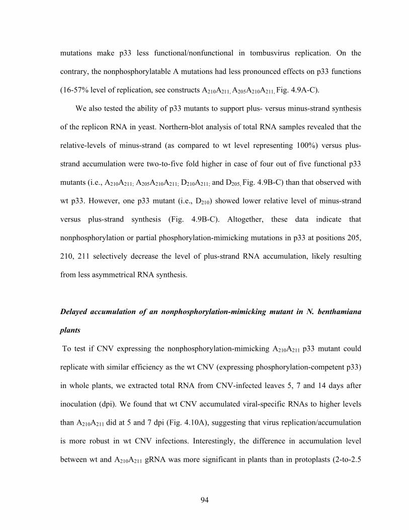
94
mutations make p33 less functional/nonfunctional in tombusvirus replication. On the
contrary, the nonphosphorylatable A mutations had less pronounced effects on p33 functions
(16-57% level of replication, see constructs A210A211, A205A210A211, Fig. 4.9A-C).
We also tested the ability of p33 mutants to support plus- versus minus-strand synthesis
of the replicon RNA in yeast. Northern-blot analysis of total RNA samples revealed that the
relative-levels of minus-strand (as compared to wt level representing 100%) versus plus-
strand accumulation were two-to-five fold higher in case of four out of five functional p33
mutants (i.e., A210A211; A205A210A211; D210A211; and D205, Fig. 4.9B-C) than that observed with
wt p33. However, one p33 mutant (i.e., D210) showed lower relative level of minus-strand
versus plus-strand synthesis (Fig. 4.9B-C). Altogether, these data indicate that
nonphosphorylation or partial phosphorylation-mimicking mutations in p33 at positions 205,
210, 211 selectively decrease the level of plus-strand RNA accumulation, likely resulting
from less asymmetrical RNA synthesis.
Delayed accumulation of an nonphosphorylation-mimicking mutant in N. benthamiana
plants
To test if CNV expressing the nonphosphorylation-mimicking A210A211 p33 mutant could
replicate with similar efficiency as the wt CNV (expressing phosphorylation-competent p33)
in whole plants, we extracted total RNA from CNV-infected leaves 5, 7 and 14 days after
inoculation (dpi). We found that wt CNV accumulated viral-specific RNAs to higher levels
than A210A211 did at 5 and 7 dpi (Fig. 4.10A), suggesting that virus replication/accumulation
is more robust in wt CNV infections. Interestingly, the difference in accumulation level
between wt and A210A211 gRNA was more significant in plants than in protoplasts (2-to-2.5
95
fold difference in plants at 5 and 7 dpi versus 5% in protoplast at 24 hpi). This might be due
to an indirect effect of phosphorylation of wt p33 on cell-to-cell movement and/or stability of
gRNA. The symptoms caused by the wt CNV infection were also more intensive than in
A210A211 infections (Fig. 4.10B), and led to the death of most leaves by 14 dpi. On the
contrary, symptoms caused by A210A211 were mild and did not lead to necrosis. Interestingly,
viral RNA accumulation was three-fold higher in A210A211 -infected than in wt CNV-infected
plants at 14 dpi, likely due to the sickness of wt CNV-infected plants (Fig. 4.10A).
Altogether, these data demonstrate that the nonphosphorylatable A mutations at positions 210
and 211 in p33 have pronounced effects on the pathogenecity of CNV in N. benthamiana.
DISCUSSION
Detection of phosphorylated replication proteins in plus-strand RNA virus-infected
hosts is a challenging task due to the low abundance of viral replication proteins, their
localization to less amenable cellular membranes, the dynamic and reversible nature of
phosphorylation and temporally existence of some phosphorylated proteins. By using both
anti-phosphothreonine antibody-based Western blot and phosphoprotein-specific staining
assays, we demonstrated the accumulation of phosphorylated CNV p33 (and the related TCV
p28) replication protein in plants, while we could not detect phosphorylated CNV p92,
possibly due to its very low abundance [~20-fold less than p33, (Scholthof, Scholthof, and
Jackson, 1995a)]. In addition, we obtained a partially-purified kinase preparation from
solubilized membrane-enriched fraction of N. benthamiana, which was able to phosphorylate
96
the CNV p33 replication protein at conserved S/T residues (likely at T205, S210 and T211)
adjacent to the RPR motif, an RNA-binding site (Panaviene, Baker, and Nagy, 2003;
Rajendran and Nagy, 2003). On the contrary, the solubile (cytoplasmic) fraction of cells did
not contain kinases capable of phosphorylation of the same region in p33 in vitro (not
shown). Because p33 is membrane-associated in cells and tombusvirus replication occurs in
membrane-containing compartments (Navarro, Rubino, and Russo, 2004; Panavas et al.,
2005b; Panaviene et al., 2004; Rubino and Russo, 1998; Scholthof, Scholthof, and Jackson,
1995a) it is likely that phosphorylation of p33 takes place in cellular membranes
[peroxisomal membrane for CNV (Panavas et al., 2005b)] in infected cells. Thus, the kinase
present in the membrane-enriched fraction is likely responsible for in vivo phosphorylation
of p33. This host kinase is functionally similar to PKC, based on phosphorylation site
prediction and demonstration that purified PKC preparation could also phosphorylate
recombinant p33 at residues S210 and T211 in vitro (Fig. 4.4). Altogether, we conclude that
p33 gets phosphorylated in CNV-infected plants by a membrane-associated host kinase.
The effects of p33 phosphorylation during CNV infection were studied using
phosphorylation-mimicking mutants (see rationale in the results section). The advantage of
using mimicking mutants is that it simplifies the interpretation of data due to the presence of
p33 representing either the “nonphosphorylated” (i.e, alanine mutants) or “phosphorylated”
(aspartic acid mutants) forms of p33 during the entire infection. Disadvantage is that the
dynamics of phophorylation cannot be fully examined with these mutants. Nevertheless,
based on data obtained with the mimicking mutants, the model predicting that the
nonphosphorylated form of p33 (we refer only to phosphorylation of T205, S210, and T211 in the
text below) is the functional form is supported, while the fully phosphorylated form of p33
97
seems to be nonfunctional in replication. Accordingly, in vitro phosphorylation of p33 and
phosphorylation mimicking mutations in p33 (at S210 and T211 positions) both inhibited the
ability of p33 to bind to the viral RNA in vitro (see Stork et al., accompanying paper),
suggesting that phosphorylation and mimicking mutations had similar effect on p33 function.
However, we cannot completely exclude that results obtained with D mutants could possibly
cause changes in the structure of p33 unrelated to phosphorylation.
The mimicking mutants were also useful to demonstrate that similar phosphorylation of
p92 RdRp protein (at T205, S210, and T211), which overlaps with p33 within its N-terminus, had
only limited effect on its activity in protoplasts. Thus, we suggest that phosphorylation at
T205, S210, and T211 mainly affects p33 function(s) during CNV infections. Phosphorylation of
p33 and p92 at additional amino acid residues was not addressed in this paper.
In addition to the nonphosphorylated and fully phosphorylated forms of p33, we suggest
that hypophosphorylation of p33 at either T205, S210, or T211 positions might affect its
activity/function. For example, we observed changes in the ratio of sgRNA1 versus sgRNA2
and of plus- versus minus-strand levels (i.e., strand asymmetry was reduced) with mimicking
mutants affecting only one or two of the predicted phosphorylation sites. These observations
open up the possibility that hypophosphorylation of p33 might be involved in (i) timing
and/or fine tuning of subgenomic RNA synthesis (sgRNA1 versus sgRNA2) and (ii)
controlling the timing and/or ratio of plus- versus minus-strand synthesis. For example,
hypophosphorylation of p33 might modify the activity of the CNV replicase, affecting the
timing of production of different viral RNA species and the ratio of replication products. In
contrast, full phosphorylation of p33 (especially phosphorylation of T211 seems to be the most
critical) might shut down replication, which could be beneficial for viral RNA encapsidation,
98
cell-to-cell movement and other processes by releasing the viral RNA from replication (see
the accompanying paper by Stork et al.). Indeed, the nonphosphorylated A210A211 mutant
showed delay in gRNA accumulation in systemically-infected leaves when compared to the
wt CNV, suggesting less efficient virus spread in plants and/or decreased stability of mutant
A210A211 gRNA.
The amount of p33 replication protein was not the same in infections with wt and
phosphorylation-mimicking CNV mutants in N. benthamiana protoplasts (Fig. 4.5C and
4.6B). For example, protoplasts containing a replication incompetent mutant (D210D211) and
an inefficiently replicating mutant (D210A211) accumulated p33 at undetectable and reduced
levels, respectively (Fig. 4.5C and 4.6B). This suggests that phosphorylation-mimicking
aspartic acid mutations (and analogously phosphorylation) might promote rapid p33
degradation, thus affecting the stability of p33. Although we cannot fully exclude that the
above D mutations facilitate p33 degradation, this model is not supported by the observation
that the amounts and stability of wt and phosphorylation-mimicking D mutants of p33 were
comparable in yeast, expressing these proteins constitutively from plasmids (Fig. 4.9D).
Alternatively, it is possible that reduced accumulation of p33 carrying the phosphorylation-
mimicking mutations is due to the production of limited amount of new CNV RNA progeny
in cells. We propose that some of the new CNV RNA progeny could participate in new
rounds of translation to boost p33/p92 production, and thus replication, in infected cells. The
proposed translational boost might not take place for CNV RNA expressing p33 with the
phosphorylation-mimicking mutations that produce only limited amount of new progeny
plus-strands.
99
The phosphorylation sites studied in this paper are located adjacent to the positively
charged RNA-binding domain in p33 (Fig. 4.1B). Therefore, it is possible that the observed
effects of the phosphorylation-mimicking mutants on CNV replication are due to altered
RNA-binding capacity of p33. Accordingly, we show in the accompanying paper (Stork et
al.) the inhibitory effect of phosphorylation of these sites on RNA-binding by the
recombinant p33. Thus, phosphorylation of p33 is likely an important step in CNV
replication by affecting replicase and template interactions.
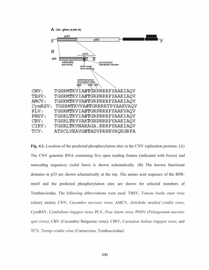
100
Fig. 4.1. Location of the predicted phosphorylation sites in the CNV replication proteins. (A)
The CNV genomic RNA containing five open reading frames (indicated with boxes) and
noncoding sequences (solid lines) is shown schematically. (B) The known functional
domains in p33 are shown schematically at the top. The amino acid sequence of the RPR-
motif and the predicted phosphorylation sites are shown for selected members of
Tombusviridae. The following abbreviations were used: TBSV, Tomato bushy stunt virus
(cherry strain); CNV, Cucumber necrosis virus; AMCV, Artichoke mottled crinkle virus;
CymRSV, Cymbidium ringspot virus; PLV, Pear latent virus; PNSV (Pelargonium necrotic
spot virus); CBV (Cucumber Bulgarian virus); CIRV, Carnation Italian ringspot virus; and
TCV, Turnip crinkle virus (Carmovirus, Tombusviridae).

101
Fig. 4.2. Detection of in vivo phosphorylated replication proteins of CNV and TCV. (A)
Detection of CNV p33 in the enriched membrane fraction by using anti-p33 antibody. (B)
Detection of phosphorylated CNV p33 and TCV p28 proteins in membrane-enriched fractions
prepared from mock-, CNV and TCV-infected N. benthamiana plants by Western blotting
with anti-phosphothreonine antibody. Replication proteins p33 of CNV and p28 of TCV are
indicated by arrowheads, whereas a band of unknown origin (present only in samples from
CNV-infected plants) is marked with an asterisk. (C) Detection of CNV p33 in membrane-
enriched fraction of N. benthamiana by staining with phosphoprotein-specific Pro-Q Diamond
dye. The CNV p33 was captured on protein A column with anti-p33 antibody from the
membrane-enriched fraction prior to loading to SDS-PAGE.
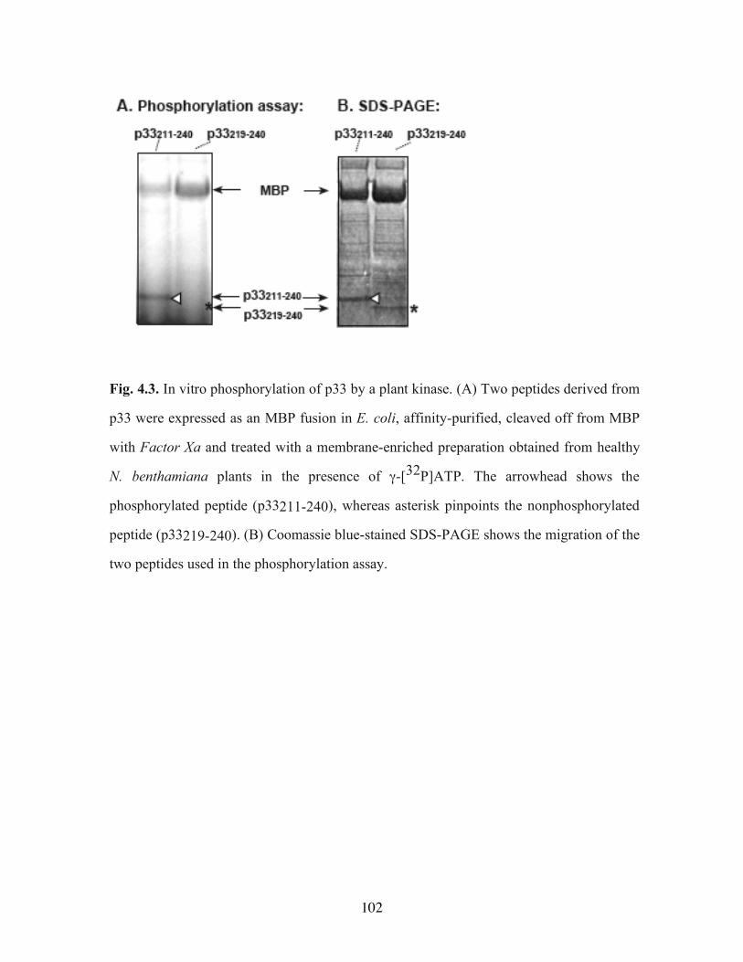
102
Fig. 4.3. In vitro phosphorylation of p33 by a plant kinase. (A) Two peptides derived from
p33 were expressed as an MBP fusion in E. coli, affinity-purified, cleaved off from MBP
with Factor Xa and treated with a membrane-enriched preparation obtained from healthy
N. benthamiana plants in the presence of γ-[32P]ATP. The arrowhead shows the
phosphorylated peptide (p33211-240), whereas asterisk pinpoints the nonphosphorylated
peptide (p33219-240). (B) Coomassie blue-stained SDS-PAGE shows the migration of the
two peptides used in the phosphorylation assay.
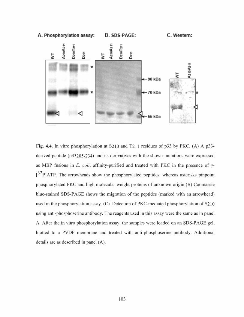
103
Fig. 4.4. In vitro phosphorylation at S210 and T211 residues of p33 by PKC. (A) A p33-
derived peptide (p33205-234) and its derivatives with the shown mutations were expressed
as MBP fusions in E. coli, affinity-purified and treated with PKC in the presence of γ-
[32P]ATP. The arrowheads show the phosphorylated peptides, whereas asterisks pinpoint
phosphorylated PKC and high molecular weight proteins of unknown origin (B) Coomassie
blue-stained SDS-PAGE shows the migration of the peptides (marked with an arrowhead)
used in the phosphorylation assay. (C). Detection of PKC-mediated phosphorylation of S210
using anti-phosphoserine antibody. The reagents used in this assay were the same as in panel
A. After the in vitro phosphorylation assay, the samples were loaded on an SDS-PAGE gel,
blotted to a PVDF membrane and treated with anti-phosphoserine antibody. Additional
details are as described in panel (A).
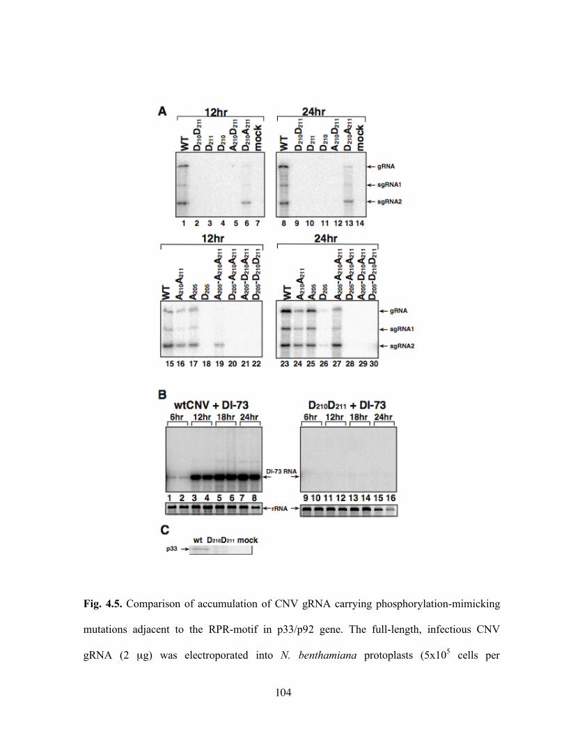
104
Fig. 4.5. Comparison of accumulation of CNV gRNA carrying phosphorylation-mimicking
mutations adjacent to the RPR-motif in p33/p92 gene. The full-length, infectious CNV
gRNA (2 µg) was electroporated into N. benthamiana protoplasts (5x10⁵ cells per
105
experiment). Total RNA was isolated, electrophoresed and blotted onto a nylon membrane,
followed by probing with 32P-labeled RNA specific for gCNV (see Materials and Methods).
The same amount (5 µg) of total RNA was used for loading onto the gels (based on the
estimation of the host ribosomal RNA in ethidium bromide-stained gels). The positions of the
CNV gRNA, sgRNA1 and sgRNA2 are depicted on the right. The names of the constructs
are shown on the top. The samples were taken 12 and 24 hours after electroporation. The
experiment was repeated three times. (B) The ability of CNV mutants to support DI-73 RNA
replication in trans. The full-length DI-73 RNA (1 µg) was co-electroporated with the
infectious CNV gRNA (2 µg, mutated or wt) into N. benthamiana protoplasts (5x10⁵ cells
per experiment). Sample preparation and Northern-blotting was done as described in the
legend to panel A, except the probing was done with 32P-labeled RNA specific for DI-73
RNA (see Materials and Methods). (C) Accumulation of CNV p33 at 24 hpi. Western blot of
total protein extracts was done with anti-p33 antibody.
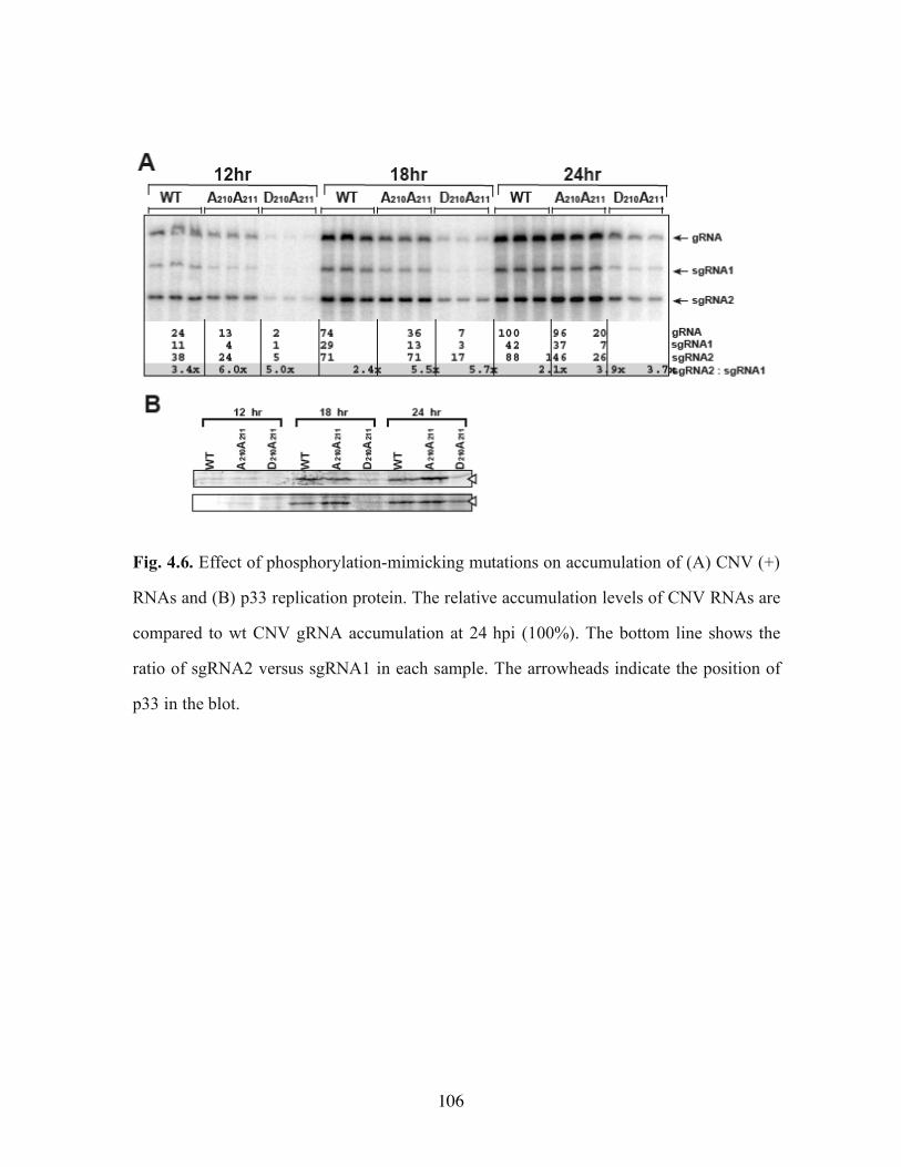
106
Fig. 4.6. Effect of phosphorylation-mimicking mutations on accumulation of (A) CNV (+)
RNAs and (B) p33 replication protein. The relative accumulation levels of CNV RNAs are
compared to wt CNV gRNA accumulation at 24 hpi (100%). The bottom line shows the
ratio of sgRNA2 versus sgRNA1 in each sample. The arrowheads indicate the position of
p33 in the blot.
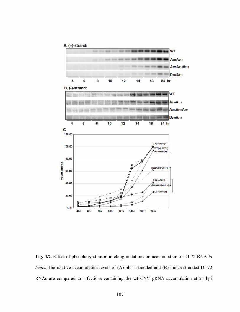
107
Fig. 4.7. Effect of phosphorylation-mimicking mutations on accumulation of DI-72 RNA in
trans. The relative accumulation levels of (A) plus- stranded and (B) minus-stranded DI-72
RNAs are compared to infections containing the wt CNV gRNA accumulation at 24 hpi
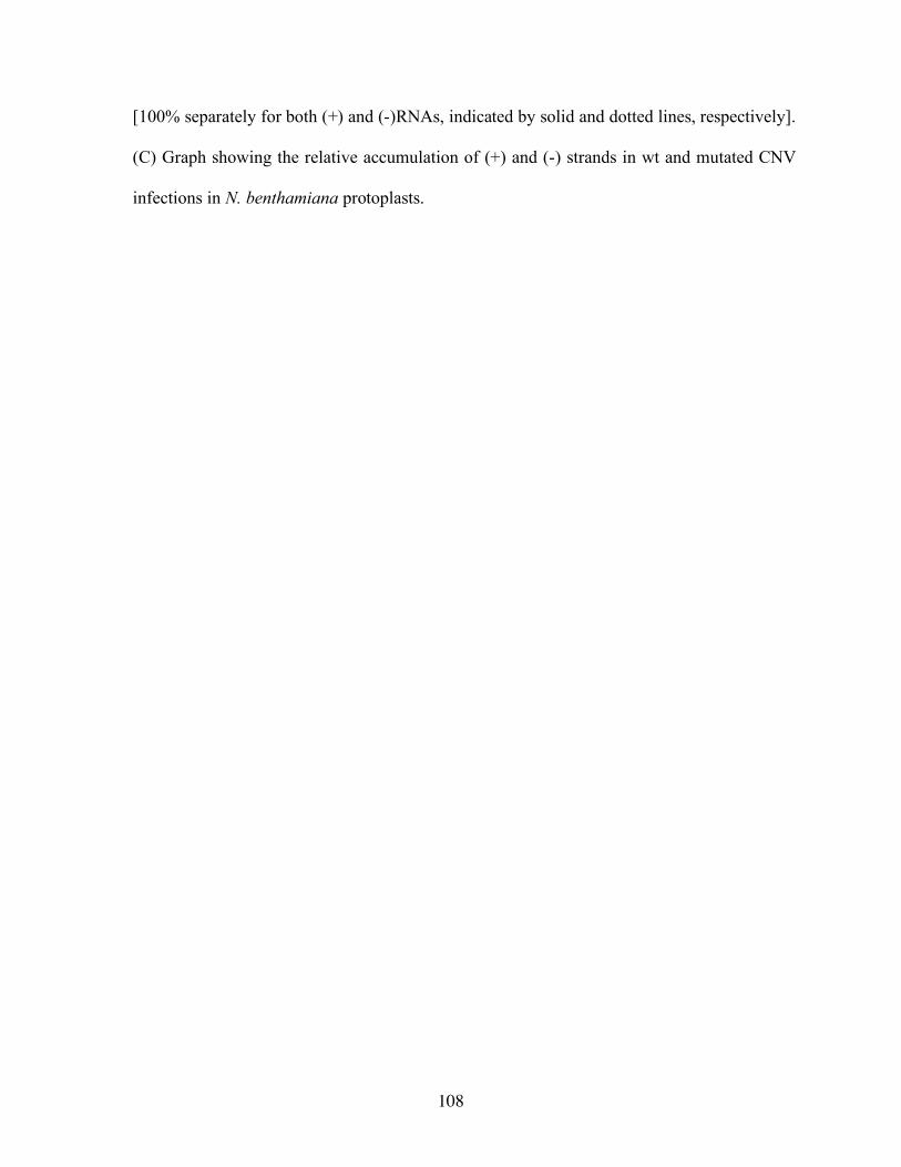
108
[100% separately for both (+) and (-)RNAs, indicated by solid and dotted lines, respectively].
(C) Graph showing the relative accumulation of (+) and (-) strands in wt and mutated CNV
infections in N. benthamiana protoplasts.
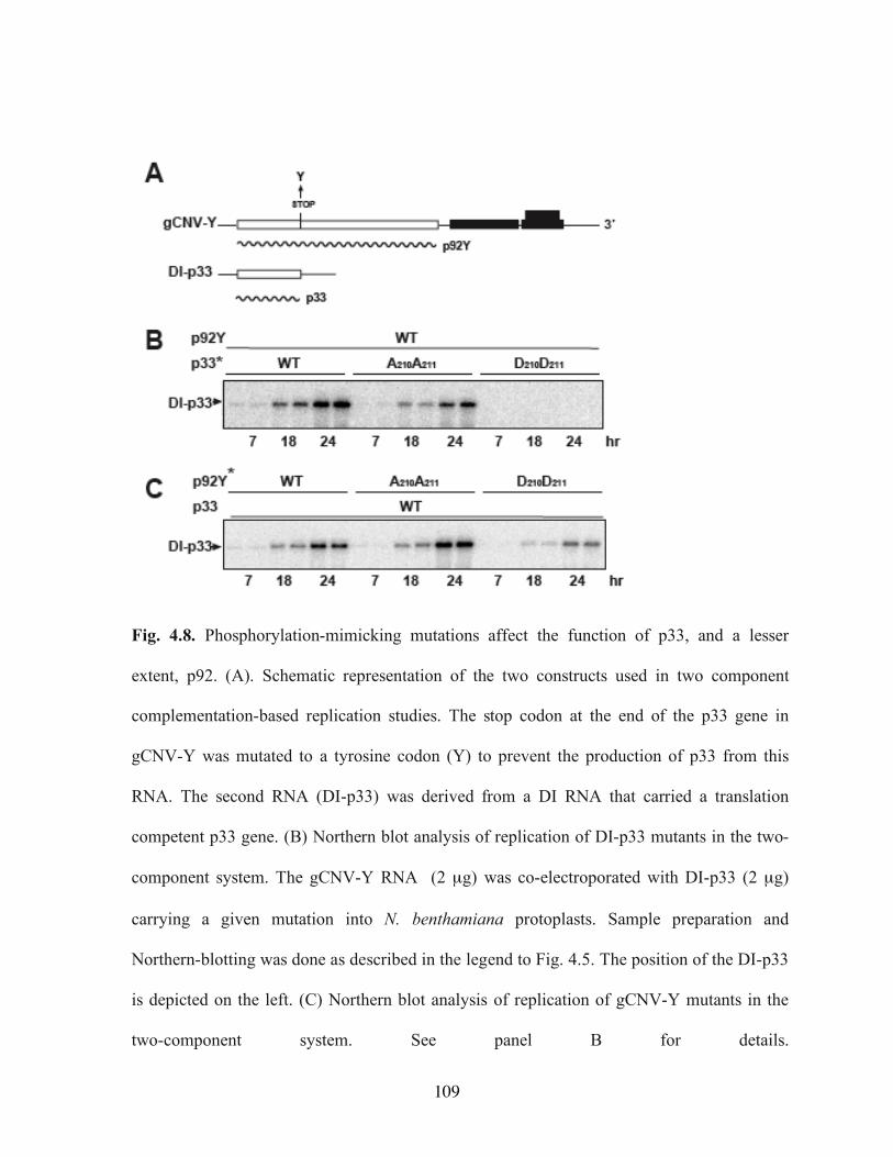
109
Fig. 4.8. Phosphorylation-mimicking mutations affect the function of p33, and a lesser
extent, p92. (A). Schematic representation of the two constructs used in two component
complementation-based replication studies. The stop codon at the end of the p33 gene in
gCNV-Y was mutated to a tyrosine codon (Y) to prevent the production of p33 from this
RNA. The second RNA (DI-p33) was derived from a DI RNA that carried a translation
competent p33 gene. (B) Northern blot analysis of replication of DI-p33 mutants in the two-
component system. The gCNV-Y RNA (2 µg) was co-electroporated with DI-p33 (2 µg)
carrying a given mutation into N. benthamiana protoplasts. Sample preparation and
Northern-blotting was done as described in the legend to Fig. 4.5. The position of the DI-p33
is depicted on the left. (C) Northern blot analysis of replication of gCNV-Y mutants in the
two-component system. See panel B for details.
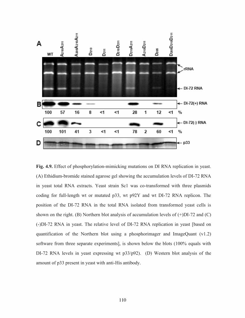
110
Fig. 4.9. Effect of phosphorylation-mimicking mutations on DI RNA replication in yeast.
(A) Ethidium-bromide stained agarose gel showing the accumulation levels of DI-72 RNA
in yeast total RNA extracts. Yeast strain Sc1 was co-transformed with three plasmids
coding for full-length wt or mutated p33, wt p92Y and wt DI-72 RNA replicon. The
position of the DI-72 RNA in the total RNA isolated from transformed yeast cells is
shown on the right. (B) Northern blot analysis of accumulation levels of (+)DI-72 and (C)
(-)DI-72 RNA in yeast. The relative level of DI-72 RNA replication in yeast [based on
quantification of the Northern blot using a phosphorimager and ImageQuant (v1.2)
software from three separate experiments], is shown below the blots (100% equals with
DI-72 RNA levels in yeast expressing wt p33/p92). (D) Western blot analysis of the
amount of p33 present in yeast with anti-His antibody.
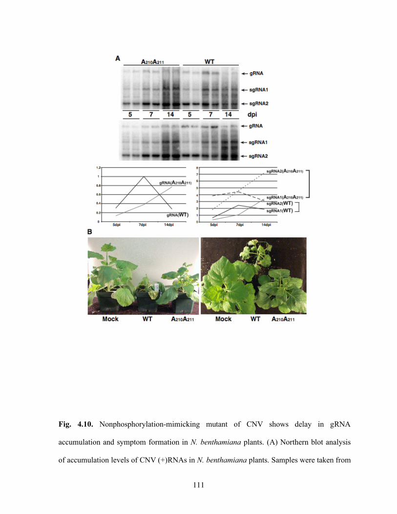
111
Fig. 4.10. Nonphosphorylation-mimicking mutant of CNV shows delay in gRNA
accumulation and symptom formation in N. benthamiana plants. (A) Northern blot analysis
of accumulation levels of CNV (+)RNAs in N. benthamiana plants. Samples were taken from
112
two systemically-infected leaves/plant and two separate plants were used in each experiment
(top and bottom panels). The relative levels of gRNA, sgRNA1 and sgRNA2 are shown
below the blots (100% equals with wt CNV gRNA level at 7 dpi). (B) Symptoms of mock-
and CNV-infected plants at 7 dpi.
Copyright © Natalia Shapka 2006.
113
Chapter V
SUMMARY
Rapid evolution of RNA viruses with mRNA-sense genomes is a major concern to
health and economic welfare due to the devastating diseases these viruses inflict on humans,
animals and plants. RNA recombination can be facilitated by recombination signals present
in viral RNAs. Among such signals are short sequences with high AU contents that constitute
recombination hot spots in Brome mosaic virus (BMV) and retroviruses. In this paper, we
demonstrate that a defective interfering (DI) RNA, a model template associated with Tomato
bushy stunt virus (TBSV), a tombusvirus, undergoes frequent recombination in plants and
protoplast cells when it carries the AU-rich hot spot sequence from BMV. Similar to the
situation with BMV, most of the recombination junction sites in the DI RNA recombinants
were found within the AU-rich region. However, unlike BMV or retroviruses, where
recombination usually occurred with precision between duplicated AU-rich sequences, the
majority of TBSV DI RNA recombinants were imprecise. In addition, only one copy of the
AU-rich sequence was essential to promote recombination in the DI RNA. The selection of
junction sites was also influenced by a putative cis-acting element present in the DI RNA.
We found that this RNA sequence bound to the TBSV replicase proteins more efficiently
than did control non-viral sequences, suggesting that it might be involved in replicase
“landing” during the template switching events. In summary, evidence is presented that a
tombusvirus can use the recombination signal of BMV. This supports the idea that common
114
AU-rich recombination signals might promote interviral recombination between unrelated
viruses.
To test if host genes can affect the evolution of RNA viruses, we used a
Saccharomyces cerevisiae single-gene deletion library, which includes ~80% of yeast genes,
in RNA recombination studies based on a small viral replicon RNA derived from Tomato
bushy stunt virus. The genome-wide screen led to the identification of five host genes, whose
absence resulted in rapid generation of novel viral RNA recombinants. Thus, these genes
normally suppress viral RNA recombination, but in their absence hosts become viral
recombination “hotbeds”. Four of the five suppressor genes are likely involved in RNA
degradation, suggesting that RNA degradation could play a role in viral RNA recombination.
On the contrary, deletion of four host genes inhibited virus recombination, indicating that
these genes normally accelerate the RNA recombination process. Comparison of deletion
strains with the lowest and the highest recombination rate revealed that host genes could
affect recombinant accumulation by up to 80-fold. Overall, our results demonstrate for the
first time that a set of host genes have major effect on RNA virus recombination and
evolution.
Replication of the non-segmented, plus-stranded RNA genome of Cucumber necrosis
tombusvirus (CNV) requires two essential overlapping viral-coded replication proteins, the
p33 replication co-factor and the p92 RNA-dependent RNA polymerase. In this paper, we
demonstrate that p33 is phosphorylated in vivo and in vitro by a membrane-bound plant
kinase. Phosphorylation of p33 was also demonstrated in vitro by using purified protein
kinase C. The related p28 replication protein of Turnip crinkle virus was also found to be
phosphorylated in vivo, suggesting that posttranslational modification of replication proteins
115
is a general feature among members of the large Tombusviridae family. Based on in vitro
studies with purified recombinant p33, we show evidence for phosphorylation of threonine
and serine residues adjacent to the essential RNA-binding site in p33. Phosphorylation-
mimicking aspartic acid mutations rendered p33 nonfunctional in plant protoplasts and in
yeast, a model host. Comparable mutations within the pre-readthrough portion of p92 did not
abolish replication. The nonphosphorylation-mimicking alanine mutants of CNV were able
to replicate in plant protoplasts and in yeast, albeit with reduced efficiency when compared to
the wild type. These alanine mutants also showed altered subgenomic RNA synthesis and a
reduction in the ratio between plus- and minus-strand RNAs produced during CNV infection.
These findings suggest that phosphorylation of threonine/serine residues adjacent to the
essential RNA-binding site in the auxiliary p33 protein likely plays a role in viral RNA
replication and subgenomic RNA synthesis during tombusvirus infections.
Copyright © Natalia Shapka 2006.
116
REFERENCES
Aaziz, R., and Tepfer, M. (1999). Recombination in RNA viruses and in virus-resistant
transgenic plants. J Gen Virol 80(Pt 6), 1339-46.
Ahlquist, P. (2002). RNA-dependent RNA polymerases, viruses, and RNA silencing.
Science 296(5571), 1270-3.
Ahlquist, P., Noueiry, A. O., Lee, W. M., Kushner, D. B., and Dye, B. T. (2003). Host
factors in positive-strand RNA virus genome replication. J Virol 77(15), 8181-6.
Allison, R., Thompson, C., and Ahlquist, P. (1990). Regeneration of a functional RNA
virus genome by recombination between deletion mutants and requirement for
cowpea chlorotic mottle virus 3a and coat genes for systemic infection. Proc Natl
Acad Sci U S A 87(5), 1820-4.
Aranda, M. A., Fraile, A., Dopazo, J., Malpica, J. M., and Garcia-Arenal, F. (1997).
Contribution of mutation and RNA recombination to the evolution of a plant
pathogenic RNA. J Mol Evol 44(1), 81-8.
Barik, S., and Banerjee, A. K. (1992). Phosphorylation by cellular casein kinase II is
essential for transcriptional activity of vesicular stomatitis virus phosphoprotein P.
Proc Natl Acad Sci U S A 89(14), 6570-4.
Becher, P., Orlich, M., and Thiel, H. J. (2001). RNA recombination between persisting
pestivirus and a vaccine strain: generation of cytopathogenic virus and induction
of lethal disease. J Virol 75(14), 6256-64.
117
Borja, M., Rubio, T., Scholthof, H. B., and Jackson, A. O. (1999). Restoration of wild-
type virus by double recombination of tombusvirus mutants with a host transgene.
Mol Plant Microbe Interact 12(2), 153-62.
Bosch, X. (2004). SARS: an amalgam of avian and mammalian viruses? Lancet Infect
Dis 4(2), 65.
Brew, C. T., and Huffaker, T. C. (2002). The yeast ubiquitin protease, Ubp3p, promotes
protein stability. Genetics 162(3), 1079-89.
Buck, K. W. (1996). Comparison of the replication of positive-stranded RNA viruses of
plants and animals. Adv Virus Res 47, 159-251.
Cascone, P. J., Haydar, T. F., and Simon, A. E. (1993). Sequences and structures required
for recombination between virus-associated RNAs. Science 260(5109), 801-5.
Cheng, C. P., and Nagy, P. D. (2003). Mechanism of RNA recombination in carmo- and
tombusviruses: evidence for template switching by the RNA-dependent RNA
polymerase in vitro. J Virol 77(22), 12033-47.
Cheng, C. P., Pogany, J., and Nagy, P. D. (2002). Mechanism of DI RNA Formation in
Tombusviruses: Dissecting the Requirement for Primer Extension by the
Tombusvirus RNA-Dependent RNA Polymerase in Vitro. Virology 304(2), 460-
73.
Chetverin, A. B., Chetverina, H. V., Demidenko, A. A., and Ugarov, V. I. (1997).
Nonhomologous RNA recombination in a cell-free system: evidence for a
transesterification mechanism guided by secondary structure. Cell 88(4), 503-13.
118
DeStefano, J. J., Bambara, R. A., and Fay, P. J. (1994). The mechanism of human
immunodeficiency virus reverse transcriptase-catalyzed strand transfer from
internal regions of heteropolymeric RNA templates. J Biol Chem 269(1), 161-8.
Dichtl, B., Stevens, A., and Tollervey, D. (1997). Lithium toxicity in yeast is due to the
inhibition of RNA processing enzymes. Embo J 16(23), 7184-95.
Fernandez-Cuartero, B., Burgyan, J., Aranda, M. A., Salanki, K., Moriones, E., and
Garcia-Arenal, F. (1994). Increase in the relative fitness of a plant virus RNA
associated with its recombinant nature. Virology 203(2), 373-7.
Freye-Minks, C., Kretsinger, R. H., and Creutz, C. E. (2003). Structural and dynamic
changes in human annexin VI induced by a phosphorylation-mimicking mutation,
T356D. Biochemistry 42(3), 620-30.
Fricke, J., Gunn, M., and Meyers, G. (2001). A family of closely related bovine viral
diarrhea virus recombinants identified in an animal suffering from mucosal
disease: new insights into the development of a lethal disease in cattle. Virology
291(1), 77-90.
Gietz, R. D., and Woods, R. A. (2002). Transformation of yeast by lithium acetate/single-
stranded carrier DNA/polyethylene glycol method. Methods Enzymol 350, 87-96.
Guan, H., and Simon, A. E. (2000). Polymerization of nontemplate bases before
transcription initiation at the 3' ends of templates by an RNA-dependent RNA
polymerase: an activity involved in 3' end repair of viral RNAs. Proc Natl Acad
Sci U S A 97(23), 12451-6.
Hartley, R. W. (2001). Barnase-barstar interaction. Methods Enzymol 341, 599-611.
119
Hericourt, F., Blanc, S., Redeker, V., and Jupin, I. (2000). Evidence for phosphorylation
and ubiquitinylation of the turnip yellow mosaic virus RNA-dependent RNA
polymerase domain expressed in a baculovirus-insect cell system. Biochem J
349(Pt 2), 417-25.
Hillman, B. I., Carrington, J. C., and Morris, T. J. (1987). A defective interfering RNA
that contains a mosaic of a plant virus genome. Cell 51(3), 427-33.
Holmes, E. C., Worobey, M., and Rambaut, A. (1999). Phylogenetic evidence for
recombination in dengue virus. Mol Biol Evol 16(3), 405-9.
Huang, L., Sineva, E. V., Hargittai, M. R., Sharma, S. D., Suthar, M., Raney, K. D., and
Cameron, C. E. (2004). Purification and characterization of hepatitis C virus non-
structural protein 5A expressed in Escherichia coli. Protein Expr Purif 37(1), 144-
53.
Ivanov, K. I., Puustinen, P., Gabrenaite, R., Vihinen, H., Ronnstrand, L., Valmu, L.,
Kalkkinen, N., and Makinen, K. (2003). Phosphorylation of the potyvirus capsid
protein by protein kinase CK2 and its relevance for virus infection. Plant Cell
15(9), 2124-39.
Jabbur, J. R., Huang, P., and Zhang, W. (2001). Enhancement of the antiproliferative
function of p53 by phosphorylation at serine 20: an inference from site-directed
mutagenesis studies. Int J Mol Med 7(2), 163-8.
Jarvis, T. C., and Kirkegaard, K. (1991). The polymerase in its labyrinth: mechanisms
and implications of RNA recombination. Trends Genet 7(6), 186-91.
120
Jia, J., Tong, C., Wang, B., Luo, L., and Jiang, J. (2004). Hedgehog signalling activity of
Smoothened requires phosphorylation by protein kinase A and casein kinase I.
Nature 432(7020), 1045-50.
Jiang, X., Espul, C., Zhong, W. M., Cuello, H., and Matson, D. O. (1999).
Characterization of a novel human calicivirus that may be a naturally occurring
recombinant. Arch Virol 144(12), 2377-87.
Kastenmayer, J. P., and Green, P. J. (2000). Novel features of the XRN-family in
Arabidopsis: evidence that AtXRN4, one of several orthologs of nuclear
Xrn2p/Rat1p, functions in the cytoplasm. Proc Natl Acad Sci U S A 97(25),
13985-90.
Khatchikian, D., Orlich, M., and Rott, R. (1989). Increased viral pathogenicity after
insertion of a 28S ribosomal RNA sequence into the haemagglutinin gene of an
influenza virus. Nature 340(6229), 156-7.
Kim, M. J., and Kao, C. (2001). Factors regulating template switch in vitro by viral RNA-
dependent RNA polymerases: implications for RNA-RNA recombination. Proc
Natl Acad Sci U S A 98(9), 4972-7.
Kim, S. H., Palukaitis, P., and Park, Y. I. (2002). Phosphorylation of cucumber mosaic
virus RNA polymerase 2a protein inhibits formation of replicase complex. Embo
J 21(9), 2292-300.
Kim, S. J., Kim, J. H., Kim, Y. G., Lim, H. S., and Oh, J. W. (2004). Protein kinase C-
related kinase 2 regulates hepatitis C virus RNA polymerase function by
phosphorylation. J Biol Chem 279(48), 50031-41.
121
Kong, Q., Wang, J., and Simon, A. E. (1997). Satellite RNA-mediated resistance to
turnip crinkle virus in Arabidopsis involves a reduction in virus movement. Plant
Cell 9(11), 2051-63.
Lai, M. M. (1992). RNA recombination in animal and plant viruses. Microbiol Rev 56(1),
61-79.
Law, M. D., and Morris, T. J. (1994). De novo generation and accumulation of tomato
bushy stunt virus defective interfering RNAs without serial host passage. Virology
198(1), 377-80.
Lee, J. Y., and Lucas, W. J. (2001). Phosphorylation of viral movement proteins--
regulation of cell-to-cell trafficking. Trends Microbiol 9(1), 5-8; discussion 8.
Lenard, J. (1999). Host cell protein kinases in nonsegmented negative-strand virus
(mononegavirales) infection. Pharmacol Ther 83(1), 39-48.
Lukashev, A. N., Lashkevich, V. A., Koroleva, G. A., Ilonen, J., and Hinkkanen, A. E.
(2004). Recombination in uveitis-causing enterovirus strains. J Gen Virol 85(Pt
2), 463-70.
Macdonald, A., Crowder, K., Street, A., McCormick, C., and Harris, M. (2004). The
hepatitis C virus NS5A protein binds to members of the Src family of tyrosine
kinases and regulates kinase activity. J Gen Virol 85(Pt 3), 721-9.
Marturano, J., and Fiore, L. (2002). Investigation of the presence of recombinant
polioviruses in the hit population in Albania during the 1996 outbreak. J Clin
Microbiol 40(1), 316-7.
Monkewich, S., Lin, H. X., Fabian, M. R., Xu, W., Na, H., Ray, D., Chernysheva, O. A.,
Nagy, P. D., and White, K. A. (2005). The p92 polymerase coding region contains
122
an internal RNA element required at an early step in Tombusvirus genome
replication. J Virol 79(8), 4848-58.
Nagy, P. D., and Bujarski, J. J. (1993). Targeting the site of RNA-RNA recombination in
brome mosaic virus with antisense sequences. Proc Natl Acad Sci U S A 90(14),
6390-4.
Nagy, P. D., and Bujarski, J. J. (1995). Efficient system of homologous RNA
recombination in brome mosaic virus: sequence and structure requirements and
accuracy of crossovers. J Virol 69(1), 131-40.
Nagy, P. D., and Bujarski, J. J. (1996). Homologous RNA recombination in brome
mosaic virus: AU-rich sequences decrease the accuracy of crossovers. J Virol
70(1), 415-26.
Nagy, P. D., and Bujarski, J. J. (1997). Engineering of homologous recombination
hotspots with AU-rich sequences in brome mosaic virus. J Virol 71(5), 3799-810.
Nagy, P. D., and Bujarski, J. J. (1998). Silencing homologous RNA recombination hot
spots with GC-rich sequences in brome mosaic virus. J Virol 72(2), 1122-30.
Nagy, P. D., Dzianott, A., Ahlquist, P., and Bujarski, J. J. (1995). Mutations in the
helicase-like domain of protein 1a alter the sites of RNA-RNA recombination in
brome mosaic virus. J Virol 69(4), 2547-56.
Nagy, P. D., Ogiela, C., and Bujarski, J. J. (1999). Mapping sequences active in
homologous RNA recombination in brome mosaic virus: prediction of
recombination hot spots. Virology 254(1), 92-104.
Nagy, P. D., and Pogany, J. (2000a). Partial purification and characterization of
Cucumber necrosis virus and Tomato bushy stunt virus RNA-dependent RNA
123
polymerases: similarities and differences in template usage between tombusvirus
and carmovirus RNA-dependent RNA polymerases. Virology 276(2), 279-88.
Nagy, P. D., and Pogany, J. (2000b). Partial purification and characterization of
Cucumber necrosis virus and Tomato bushy stunt virus RNA-dependent RNA
polymerases: similarities and differences in template usage between tombusvirus
and carmovirus RNA-dependent RNA polymerases. Virology 276(2), 279-88.
Nagy, P. D., and Pogany, J. (2006). Yeast as a model host to dissect functions of viral and
host factors in tombusvirus replication. Virology 344(1), 211-20.
Nagy, P. D., Pogany, J., and Simon, A. E. (1999). RNA elements required for RNA
recombination function as replication enhancers in vitro and in vivo in a plus-
strand RNA virus. Embo J 18(20), 5653-65.
Nagy, P. D., Pogany, J., and Simon, A. E. (2001). In vivo and in vitro characterization of
an RNA replication enhancer in a satellite RNA associated with turnip crinkle
virus. Virology 288(2), 315-24.
Nagy, P. D., and Simon, A. E. (1997). New insights into the mechanisms of RNA
recombination. Virology 235(1), 1-9.
Nagy, P. D., Zhang, C., and Simon, A. E. (1998). Dissecting RNA recombination in
vitro: role of RNA sequences and the viral replicase. Embo J 17(8), 2392-403.
Navarro, B., Rubino, L., and Russo, M. (2004). Expression of the Cymbidium ringspot
virus 33-kilodalton protein in Saccharomyces cerevisiae and molecular dissection
of the peroxisomal targeting signal. J Virol 78(9), 4744-52.
124
Oprisan, G., Combiescu, M., Guillot, S., Caro, V., Combiescu, A., Delpeyroux, F., and
Crainic, R. (2002). Natural genetic recombination between co-circulating
heterotypic enteroviruses. J Gen Virol 83(Pt 9), 2193-200.
Oster, S. K., Wu, B., and White, K. A. (1998a). Uncoupled expression of p33 and p92
permits amplification of tomato bushy stunt virus RNAs. J Virol 72(7), 5845-51.
Oster, S. K., Wu, B., and White, K. A. (1998b). Uncoupled expression of p33 and p92
permits amplification of tomato bushy stunt virus RNAs. J Virol 72(7), 5845-51.
Panavas, T., Hawkins, C. M., Panaviene, Z., and Nagy, P. D. (2005a). The role of the
p33:p33/p92 interaction domain in RNA replication and intracellular localization
of p33 and p92 proteins of Cucumber necrosis tombusvirus. Virology.
Panavas, T., Hawkins, C. M., Panaviene, Z., and Nagy, P. D. (2005b). The role of the
p33:p33/p92 interaction domain in RNA replication and intracellular localization
of p33 and p92 proteins of Cucumber necrosis tombusvirus. Virology 338(1), 81-
95.
Panavas, T., and Nagy, P. D. (2003a). The RNA replication enhancer element of
tombusviruses contains two interchangeable hairpins that are functional during
plus-strand synthesis. J Virol 77(1), 258-69.
Panavas, T., and Nagy, P. D. (2003b). Yeast as a model host to study replication and
recombination of defective interfering RNA of Tomato bushy stunt virus.
Virology 314(1), 315-25.
Panavas, T., Panaviene, Z., Pogany, J., and Nagy, P. D. (2003). Enhancement of RNA
synthesis by promoter duplication in tombusviruses. Virology 310(1), 118-29.
125
Panavas, T., Pogany, J., and Nagy, P. D. (2002). Internal initiation by the cucumber
necrosis virus RNA-dependent RNA polymerase is facilitated by promoter-like
sequences. Virology 296(2), 275-87.
Panavas, T., Serviene, E., Brasher, J., and Nagy, P. D. (2005c). Yeast genome-wide
screen reveals dissimilar sets of host genes affecting replication of RNA viruses.
Proc Natl Acad Sci U S A.
Panavas, T., Serviene, E., Brasher, J., and Nagy, P. D. (2005d). Yeast genome-wide
screen reveals dissimilar sets of host genes affecting replication of RNA viruses.
Proc Natl Acad Sci U S A 102(20), 7326-31.
Panaviene, Z., Baker, J. M., and Nagy, P. D. (2003). The overlapping RNA-binding
domains of p33 and p92 replicase proteins are essential for tombusvirus
replication. Virology 308(1), 191-205.
Panaviene, Z., and Nagy, P. D. (2003). Mutations in the RNA-binding domains of
tombusvirus replicase proteins affect RNA recombination in vivo. Virology
317(2), 359-72.
Panaviene, Z., Panavas, T., and Nagy, P. D. (2005). Role of an internal and two 3'-
terminal RNA elements in assembly of tombusvirus replicase. J Virol 79(16),
10608-18.
Panaviene, Z., Panavas, T., Serva, S., and Nagy, P. D. (2004). Purification of the
cucumber necrosis virus replicase from yeast cells: role of coexpressed viral RNA
in stimulation of replicase activity. J Virol 78(15), 8254-63.
126
Pantaleo, V., Rubino, L., and Russo, M. (2003). Replication of Carnation Italian ringspot
virus defective interfering RNA in Saccharomyces cerevisiae. J Virol 77(3), 2116-
23.
Park, J. W., Desvoyes, B., and Scholthof, H. B. (2002). Tomato bushy stunt virus
genomic RNA accumulation is regulated by interdependent cis-acting elements
within the movement protein open reading frames. J Virol 76(24), 12747-57.
Parker, R., and Song, H. (2004). The enzymes and control of eukaryotic mRNA turnover.
Nat Struct Mol Biol 11(2), 121-7.
Peng, Z., and Verma, D. P. (1995). A rice HAL2-like gene encodes a Ca(2+)-sensitive
3'(2'),5'-diphosphonucleoside 3'(2')-phosphohydrolase and complements yeast
met22 and Escherichia coli cysQ mutations. J Biol Chem 270(49), 29105-10.
Pilipenko, E. V., Gmyl, A. P., and Agol, V. I. (1995). A model for rearrangements in
RNA genomes. Nucleic Acids Res 23(11), 1870-5.
Pogany, J., Fabian, M. R., White, K. A., and Nagy, P. D. (2003). A replication silencer
element in a plus-strand RNA virus. Embo J 22(20), 5602-11.
Pogany, J., White, K. A., and Nagy, P. D. (2005). Specific Binding of Tombusvirus
Replication Protein p33 to an Internal Replication Element in the Viral RNA Is
Essential for Replication. J Virol 79(8), 4859-69.
Puustinen, P., Rajamaki, M. L., Ivanov, K. I., Valkonen, J. P., and Makinen, K. (2002).
Detection of the potyviral genome-linked protein VPg in virions and its
phosphorylation by host kinases. J Virol 76(24), 12703-11.
127
Qiu, W., Park, J. W., Jackson, A. O., and Scholthof, H. B. (2001). Retention of a small
replicase gene segment in tomato bushy stunt virus defective RNAs inhibits their
helper-mediated trans-accumulation. Virology 281(1), 51-60.
Rajendran, K. S., and Nagy, P. D. (2003). Characterization of the RNA-binding domains
in the replicase proteins of tomato bushy stunt virus. J Virol 77(17), 9244-58.
Rajendran, K. S., and Nagy, P. D. (2006). Kinetics and functional studies on interaction
between the replicase proteins of Tomato Bushy Stunt Virus: Requirement of
p33:p92 interaction for replicase assembly. Virology 345(1), 270-9.
Rajendran, K. S., Pogany, J., and Nagy, P. D. (2002). Comparison of turnip crinkle virus
RNA-dependent RNA polymerase preparations expressed in Escherichia coli or
derived from infected plants. J Virol 76(4), 1707-17.
Ray, D., and White, K. A. (1999). Enhancer-like properties of an RNA element that
modulates Tombusvirus RNA accumulation. Virology 256(1), 162-71.
Ray, D., and White, K. A. (2003). An internally located RNA hairpin enhances
replication of Tomato bushy stunt virus RNAs. J Virol 77(1), 245-57.
Ray, D., Wu, B., and White, K. A. (2003). A second functional RNA domain in the 5'
UTR of the Tomato bushy stunt virus genome: intra- and interdomain interactions
mediate viral RNA replication. Rna 9(10), 1232-45.
Rest, J. S., and Mindell, D. P. (2003). SARS associated coronavirus has a recombinant
polymerase and coronaviruses have a history of host-shifting. Infect Genet Evol
3(3), 219-25.
128
Rochon, D. M. (1991). Rapid de novo generation of defective interfering RNA by
cucumber necrosis virus mutants that do not express the 20-kDa nonstructural
protein. Proc Natl Acad Sci U S A 88(24), 11153-7.
Rodriguez, C. R., Takagi, T., Cho, E. J., and Buratowski, S. (1999). A Saccharomyces
cerevisiae RNA 5'-triphosphatase related to mRNA capping enzyme. Nucleic
Acids Res 27(10), 2181-8.
Rubino, L., and Russo, M. (1998). Membrane targeting sequences in tombusvirus
infections. Virology 252(2), 431-7.
Russo, M., Burgyan, J., and Martelli, G. P. (1994). Molecular biology of tombusviridae.
Adv Virus Res 44, 381-428.
Scholthof, K. B., Scholthof, H. B., and Jackson, A. O. (1995a). The tomato bushy stunt
virus replicase proteins are coordinately expressed and membrane associated.
Virology 208(1), 365-9.
Scholthof, K. B., Scholthof, H. B., and Jackson, A. O. (1995b). The tomato bushy stunt
virus replicase proteins are coordinately expressed and membrane associated.
Virology 208(1), 365-9.
Serviene, E., Jiang, Y., Cheng, C. P., Baker, J., and Nagy, P. D. (2006). Screening of the
Yeast yTHC Collection Identifies Essential Host Factors Affecting Tombusvirus
RNA Recombination. J Virol 80(3), 1231-41.
Serviene, E., Shapka, N., Cheng, C. P., Panavas, T., Phuangrat, B., Baker, J., and Nagy,
P. D. (2005). Genome-wide screen identifies host genes affecting viral RNA
recombination. Proc Natl Acad Sci U S A 102(30), 10545-50.
129
Shapka, N., and Nagy, P. D. (2004). The AU-rich RNA recombination hot spot sequence
of Brome mosaic virus is functional in tombusviruses: implications for the
mechanism of RNA recombination. J Virol 78(5), 2288-300.
Sheth, U., and Parker, R. (2003). Decapping and decay of messenger RNA occur in
cytoplasmic processing bodies. Science 300(5620), 805-8.
Sokolova, M., Prufer, D., Tacke, E., and Rohde, W. (1997). The potato leafroll virus 17K
movement protein is phosphorylated by a membrane-associated protein kinase
from potato with biochemical features of protein kinase C. FEBS Lett 400(2),
201-5.
Song, C., and Simon, A. E. (1994). RNA-dependent RNA polymerase from plants
infected with turnip crinkle virus can transcribe (+)- and (-)-strands of virus-
associated RNAs. Proc Natl Acad Sci U S A 91(19), 8792-6.
Stavrinides, J., and Guttman, D. S. (2004). Mosaic evolution of the severe acute
respiratory syndrome coronavirus. J Virol 78(1), 76-82.
Strauss, J. H., and Strauss, E. G. (1988). Evolution of RNA viruses. Annu Rev Microbiol
42, 657-83.
Street, A., Macdonald, A., Crowder, K., and Harris, M. (2004). The Hepatitis C virus
NS5A protein activates a phosphoinositide 3-kinase-dependent survival signaling
cascade. J Biol Chem 279(13), 12232-41.
Takacs, A. M., Barik, S., Das, T., and Banerjee, A. K. (1992). Phosphorylation of specific
serine residues within the acidic domain of the phosphoprotein of vesicular
stomatitis virus regulates transcription in vitro. J Virol 66(10), 5842-8.
130
Waigmann, E., Chen, M. H., Bachmaier, R., Ghoshroy, S., and Citovsky, V. (2000).
Regulation of plasmodesmal transport by phosphorylation of tobacco mosaic
virus cell-to-cell movement protein. Embo J 19(18), 4875-84.
Walter, J. E., Briggs, J., Guerrero, M. L., Matson, D. O., Pickering, L. K., Ruiz-Palacios,
G., Berke, T., and Mitchell, D. K. (2001). Molecular characterization of a novel
recombinant strain of human astrovirus associated with gastroenteritis in children.
Arch Virol 146(12), 2357-67.
White, K. A., and Morris, T. J. (1994a). Enhanced competitiveness of tomato bushy stunt
virus defective interfering RNAs by segment duplication or nucleotide insertion. J
Virol 68(9), 6092-6.
White, K. A., and Morris, T. J. (1994b). Nonhomologous RNA recombination in
tombusviruses: generation and evolution of defective interfering RNAs by
stepwise deletions. J Virol 68(1), 14-24.
White, K. A., and Morris, T. J. (1994c). Nonhomologous RNA recombination in
tombusviruses: generation and evolution of defective interfering RNAs by
stepwise deletions. J Virol 68(1), 14-24.
White, K. A., and Morris, T. J. (1994d). Recombination between defective tombusvirus
RNAs generates functional hybrid genomes. Proc Natl Acad Sci U S A 91(9),
3642-6.
White, K. A., and Morris, T. J. (1994e). Recombination between defective tombusvirus
RNAs generates functional hybrid genomes. Proc Natl Acad Sci U S A 91(9),
3642-6.
131
White, K. A., and Morris, T. J. (1995). RNA determinants of junction site selection in
RNA virus recombinants and defective interfering RNAs. Rna 1(10), 1029-40.
White, K. A., and Morris, T. J. (1999). Defective and defective interfering RNAs of
monopartite plus-strand RNA plant viruses. Curr Top Microbiol Immunol 239, 1-
17.
White, K. A., and Nagy, P. D. (2004). Advances in the molecular biology of
tombusviruses: gene expression, genome replication, and recombination. Prog
Nucleic Acid Res Mol Biol 78, 187-226.
Wierzchoslawski, R., Dzianott, A., Kunimalayan, S., and Bujarski, J. J. (2003). A
transcriptionally active subgenomic promoter supports homologous crossovers in
a plus-strand RNA virus. J Virol 77(12), 6769-76.
Worobey, M., and Holmes, E. C. (1999). Evolutionary aspects of recombination in RNA
viruses. J Gen Virol 80(Pt 10), 2535-43.
Worobey, M., Rambaut, A., and Holmes, E. C. (1999). Widespread intra-serotype
recombination in natural populations of dengue virus. Proc Natl Acad Sci U S A
96(13), 7352-7.
Wu, W., Blumberg, B. M., Fay, P. J., and Bambara, R. A. (1995). Strand transfer
mediated by human immunodeficiency virus reverse transcriptase in vitro is
promoted by pausing and results in misincorporation. J Biol Chem 270(1), 325-
32.
132
VITA
Born: Makajeva, Ukraine, 1966, December 1.
Education:
Kiev State University, department of Virology. 1987-1992
Master degree in Virology
Kiev State University, 1987-1990
Bachelor degree
Professional positions:
Belozersky Institute of Physico-Chemical Biology, Moscow State University. 1996-1998
Position: Research assistant
Zlabolotny Institute of Microbiology and Virology, Kiev 1992-1999
Position: Research assistant
Publications:
Shapka, N., and Nagy, P. D. (2004). The AU-rich RNA recombination hot spot sequence
of Brome mosaic virus is functional in tombusviruses: implications for the
mechanism of RNA recombination. J Virol 78(5), 2288-300.
133
Serviene, E., Shapka, N., Cheng, C. P., Panavas, T., Phuangrat, B., Baker, J., and Nagy,
P. D. (2005). Genome-wide screen identifies host genes affecting viral RNA
recombination. Proc Natl Acad Sci U S A 102(30), 10545-50.
Shapka, N., Stork, J., and Nagy, P. D. (2005). Phosphorylation of the p33 replication
protein of Cucumber necrosis tombusvirus adjacent to the RNA binding site
affects viral RNA replication. Virology 343(1), 65-78.
Stork, J., Panaviene, Z., and Nagy, P. D. (2005). Inhibition of in vitro RNA binding and
replicase activity by phosphorylation of the p33 replication protein of Cucumber
necrosis tombusvirus. Virology 343(1), 79-92.


![· Web viewBy targeting cellular or viral genes, these miRNAs are involved in the regulation of multiple cellular responses such as host cell proliferation, apoptosis [12-15], and](https://img.pdfslide.net/doc/110x75/5f0b48857e708231d42fc04c/web-view-by-targeting-cellular-or-viral-genes-these-mirnas-are-involved-in-the.jpg)