Embed Size (px)
Citation preview
ONCOLOGY LETTERS 16: 5375-5382, 2018
Abstract. Major depression disorder (MDD) has become increasingly common in patients with ovarian cancer, which complicates the treatment course. The microRNA (miRNA)-mRNA regulation network may help elucidate the potential mechanism of MDD in ovarian cancer. The differentially expressed microRNAs (DEmiRs) and mRNAs (DEmRNAs) were therefore identified from the GSE61741, GSE58105 and GSE9116 ovarian cancer datasets using GEO2R. The target genes of the DEmiRs were then obtained using the TargetScan, microRNAorg, microT-CDS, miRDB and miRTarBase prediction tools. The DAVID program was used to identify the KEGG pathways of target genes, and the core genes of major depressive disorder (MDD) were identified using the Kaplan-Meier Plotter for ovarian cancer. A total of 5 DEmiRs (miR-23b-3p, miR-33b-3p, miR-1265, miR-933 and miR‑629‑5p) were obtained from GSE61741 and GSE58105. The target genes of these DEmiRs were enriched in pathways that were considered high risk for developing MDD in ovarian cancer. A total of 11 risk genes were selected from these path-ways as the core genes in the miRNA-mRNA network of MDD in ovarian cancer, and eventually identified the following 12 miRNA-mRNAs pairs: miR-629-5p-FGF1, miR-629-5p-AKT3, miR-629-5p-MAGI2, miR-933-BDNF, miR-933-MEF2A,
miR-23b-3p-TJP1, miR-23b-3p-JMJD1, miR-23b-3p-APAF1, miR-23b-3p-CAB39, miR-1265-CDKN1B, miR-33b-3p- CDKN1B, and miR-33b-3p-F2R. These results may provide novel insights into the mechanisms of developing MDD in ovarian cancer patients.
Introduction
With continuous progress in modern medicine, the overall survival of patients with malignant tumors has improved. However, accompanying depression has become increasingly common among cancer patients. A great deal of epidemio-logical studies have shown that the incidence of depression in cancer patients is 2‑4 times higher than that in the normal population, and up to 20‑50% of the patients are afflicted (1,2). Ovarian cancer has the highest mortality rate among all gynecological malignancies, which results in extreme anxiety and depression in the patients. Bodurka reported a depression rate of 21% among the patients with ovarian cancer (3), while Price et al (4), found clinical depression in 5.9% of 798 women with ovarian cancer in a prospective cohort study.
Due to the belief that depression is a normal and universal reaction to cancer, it is often underplayed in patients with cancer. However, depression not only results in emotional trauma, but more importantly also causes pathophysiological changes in the patients (5,6). Neuroendocrine-immune modulation (NIM) negative feedback network is one of the important pathophysi-ological basis of clinical depression in patients with cancer. Depression in pancreatic cancer patients has been linked to the secretion of amines resistant to emotional excitement (7).
Various genes are involved in clinical depression, and miRNA-mRNA interactions play an important role in regu-lating its pathophysiological basis. Micro RNAs bind to the 3'-untranslated region (3'-UTR) of target genes involved in cellular processes like proliferation, differentiation, apoptosis, and immune responses (8). The miRNA-mRNA regulatory network is of great significance in identifying the mechanism of major depression disorder (MDD) in ovarian cancer. In addition, the relevant miRNAs may be potential diagnostic markers for the early detection of ovarian cancer related
Identifying miRNA‑mRNA regulation network of major depressive disorder in ovarian cancer patients
CHENGJIANG WU1,2, YANGJING ZHAO2, YUEFANG LIU2,3, XINXIN YANG2, MEINA YAN2, YUJIAO MIN2, ZIHUI PAN2, SHALI QIU2, SHENG XIA2, JUN YU4, PEIFANG YANG4, BING WAN1 and QIXIANG SHAO2
1Department of Respiratory Medicine, The Affiliated Jiangning Hospital of Nanjing Medical University, Nanjing, Jiangsu 210002; 2Department of Immunology, Key Laboratory for Laboratory Medicine of Jiangsu, Jiangsu University
Medical School, Zhenjiang, Jiangsu 212013; 3Department of Medical Genetics and Prenatal Diagnostics, Huaian Maternity and Child Health Care Hospital Affiliated to Yangzhou University, Huaian, Jiangsu 223002; 4Department of Gynecology and Obstetrics, Affiliated Hospital of Jiangsu University, Zhenjiang, Jiangsu 212001, P.R. China
Received March 29, 2018; Accepted July 26, 2018
DOI: 10.3892/ol.2018.9243
Correspondence to: Professor Qixiang Shao, Department of Immunology, Key Laboratory for Laboratory Medicine of Jiangsu, Jiangsu University Medical School, 301 Xuefu Road, Zhenjiang, Jiangsu 212013, P.R. ChinaE-mail: [email protected]
Dr Bing Wan, Department of Respiratory Medicine, The Affiliated Jiangning Hospital of Nanjing Medical University, 168 Gushan Road, Nanjing, Jiangsu 210002, P.R. ChinaE-mail: [email protected]
Key words: major depressive disorder, ovarian cancer, miRNA-mRNA regulation network, gene ontology, KEGG pathway
WU et al: miRNA-mRNA NETWORK OF MDD IN OVARIAN CANCER5376
depression, as well as prognostic indicators for treatment response. Although a large number of epidemiological studies have reported depression in cancer patients, few studies exist on the miRNA-mRNA networks related to depression in cancer patients, which can detect and diagnose MDD at early stages. Bioinformatics and expression profiling techniques can help identify such networks in various diseases.
In the present study, we analyzed the miRNA expression profiles of patients with ovarian cancer or MDD, and the mRNA expression profiles of depressed and non‑depressed patients with ovarian tumors. Using bioinformatics, we identi-fied the miRNAs and their target genes, and constructed an miRNA-mRNA-pathway regulatory network.
Materials and methods
Acquisition of microarray data. Ovarian cancer and MDD associated gene expression datasets, original data and platform records were acquired from the Gene Expression Omnibus (GEO, http://www.ncbi.nlm nih.gov/geo/) database from the National Center for Biotechnology Information (NCBI). The dataset GSE61741 (ovarian cancer) based on GPL9040 platform (febit Homo Sapiens miRBase 13.0) was submitted by Keller, and included 94 normal and 24 ovarian cancer samples. The MDD-associated dataset GSE58105 based on GPL1873 platform [Agilent-021827 Human miRNA Microarray (miRNA_107_Sep09)] was submitted by Lopez, and included 11 normal and 14 MDD samples. The GSE9116 dataset of depressed patients with ovarian tumors based on GPL96 platform [(HG-U133A) Affymetrix Human Genome U133A Array], was submitted by Cole, and included data from 5 depressed patients and non-depressed patients each, all with primary ovarian tumors.
Identification of dif ferentially expressed microRNAs (DEmiRs) or DEmRNAs and miRNA target genes. GEO2R (https://www.ncbi.nlm.nih.gov/geo/geo2r/) is a useful online microarray data analysis tool that allows users to compare two or more groups of samples in a GEO series in order to identify DEmRNAs. The miRNA or mRNA with P-value <0.05 were regarded as DEmiRs or DEmRNAs respectively. The miRNA target genes were obtained from the following 5 prediction tools: TargetScan, microRNAorg, microT-CDS, miRDB and miRTarBase. Genes overlapping in three or more prediction tools were selected as putative target genes of miRNA.
Risk pathways and survival curves of target genes involved in risk pathways. Gene ontology (GO) is a widely used method for the large-scale functional annotation of genes, and is based on certain structured, defined and controlled terms (9). Kyoto Encyclopedia of Genes and Genomes (KEGG) database is a collection of online databases of gene functions, enzymatic pathways, and helps link genomic information with higher-order functional information (10). The Database for Annotation, Visualization and Integrated Discovery (https://david.ncifcrf.gov/, DAVID) provides a comprehensive set of functional annotation tools to identify KEGG pathways and biological process (11,12). To determine the cellular pathways of target genes, the DAVID program was used to identify KEGG pathways and biological process,
with P<0.05 as the threshold value. Kaplan-Meier Plotter for ovarian cancer (13), an online analysis tool, was used to assess the effect of the selected genes on ovarian cancer. P<0.05 was considered to indicate a statistically significant difference.
Construction of miRNA‑mRNA‑pathway regulation network. Cytoscape (http://www.cytoscape.org/), an open source soft-ware platform for complex network analysis and visualization, was used to construct the miRNA-target genes-pathways network.
Results
Co‑differentially expressed miRNA and target genes in ovarian cancer and depression. After comparing 24 ovarian cancer and 94 normal samples from the GSE61741 dataset, we identified 300 DEmiRs with P<0.05. Similarly, 40 DEmiRs were identified from the GSE58105 dataset. Furthermore, overlapping DEmiRs from both datasets included 4 upregu-lated (miR-23b-3p, miR-33b-3p, miR-1265 and miR-933) and 1 downregulated (miR‑629‑5p) DEmiRs. We used five gene prediction programs on these DEmiRs, and selected the genes common to three or more prediction tools as the target genes. There were 130, 29, 41, 18 and 30 target genes of miR‑23b‑3p, miR-33b-3p, miR-1265, miR-933 and miR-33b-3p respectively, and 90, 14, 34, 12 and 22 of them respectively were also DEmRNAs in the GSE9116 dataset (Fig. 1).
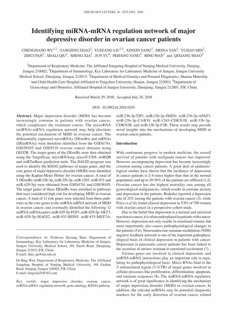
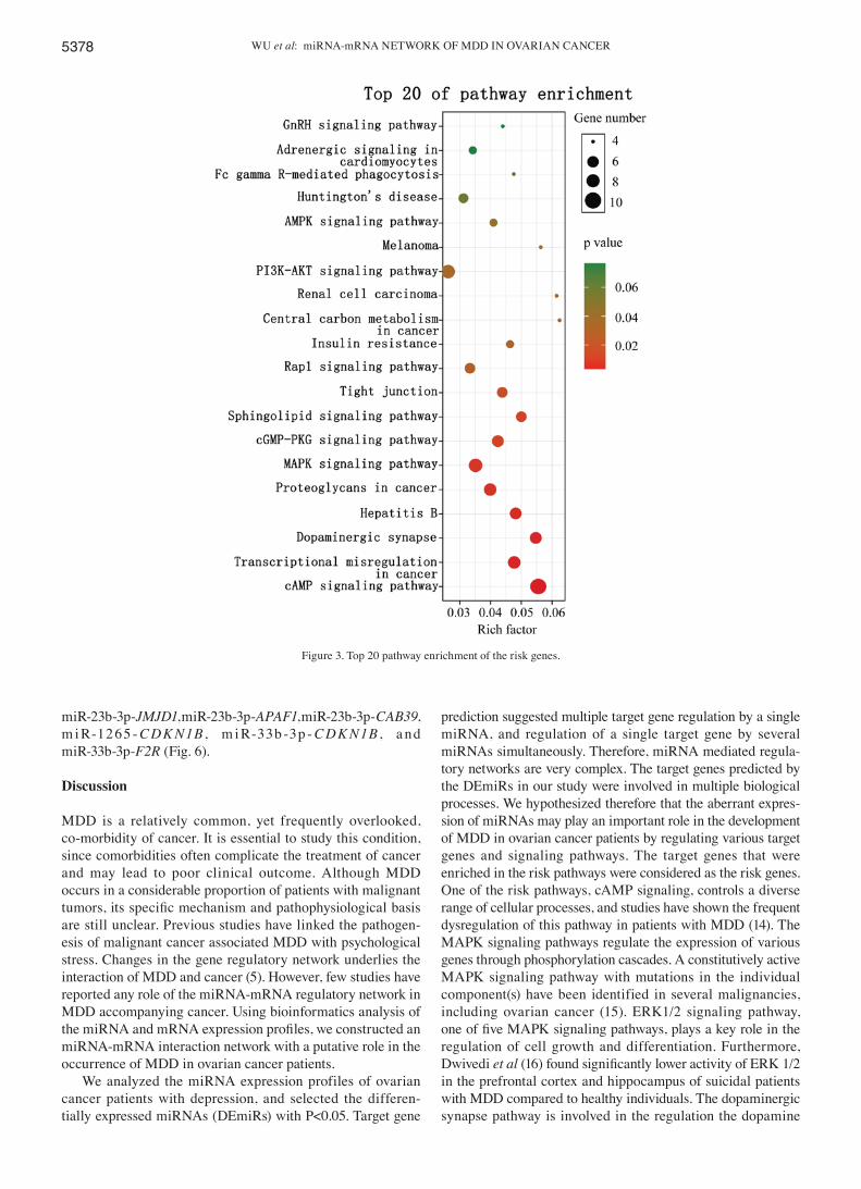
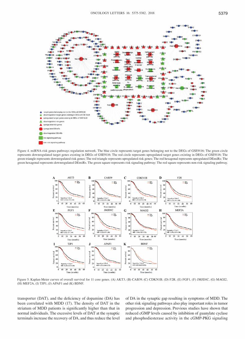
Identification of the risk pathways, risk genes and miRNA‑risk gene‑pathways regulation network. GO annotations showed that these target genes were involved in biological process including neural tube closure, negative regulation of neuron apoptotic process, nerve growth factor signaling pathway, positive regulation of cell migration, and apoptosis (Fig. 2). The most significantly enriched pathways of target genes which also exist as DEmRNAs in the GSE9116 dataset are shown in Fig. 3. A P-value<0.05 indicated statistically significant enrichment. Pathways associated with ovarian cancer and MDD, considered risk pathways, included cAMP signaling, transcriptional dysregulation in cancer, dopami-nergic synapse, hepatitis B, proteoglycans in cancer, MAPK signaling, cGMP-PKG signaling, sphingolipid signaling, tight junction, rap1 signaling, central carbon metabolism in cancer, PI3K-Akt signaling, and AMPK signaling. Genes enriched in these risk pathways, i.e. the risk genes, included 22 upregulated and 15 downregulated genes. The miRNA-mRNA regulatory network was constructed using 5 DEmiRs, 37 risk genes and 13 risk pathways (Fig. 4).
Identif ication of core genes in ovarian cancer. Upon overexpression of the 22 upregulated risk genes, 10 risk genes could significantly increase the mortality of ovarian cancer patients, while only BDNF overexpression among the 15 downregulated risk genes could significantly increase survival rate. Taken together, 11 risk genes were identified as the core genes of MDD development in ovarian cancer, which are also DEmRNAs in GSE9116. The core genes include BDNF, MEF2A, FGF1, AKT3, MAGI2, TJP1, JMJD1C, APAF1, CAB39, CDKN1B and F2R (Fig. 5).
ONCOLOGY LETTERS 16: 5375-5382, 2018 5377
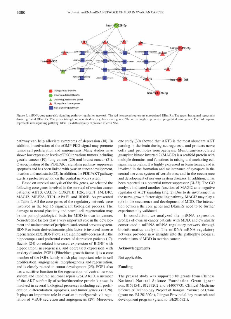
Analysis of miRNA‑core gene‑pathway regulation network. We confirmed 12 pairs of miRNA‑mRNA interaction including
miR-629-5p-FGF1, miR-629-5p-AKT3, miR-629-5p-MAGI2, miR-933-BDNF, miR-933-MEF2A, miR-23b-3p-TJP1,
Figure 1. Intersection of miRNA target genes predicted by five kinds of software. (A) miR‑23b‑3p, (B) miR‑33b‑3p, (C) miR‑629‑5p, (D) miR‑933 and (E) miR-1265.
Figure 2. Top 15 biological process of the risk genes.
WU et al: miRNA-mRNA NETWORK OF MDD IN OVARIAN CANCER5378
miR-23b-3p-JMJD1, miR-23b-3p-APAF1, miR-23b-3p-CAB39, m i R-1265 - CDK N1B , m i R-33b -3p - CDK N1B , a nd miR-33b-3p-F2R (Fig. 6).
Discussion
MDD is a relatively common, yet frequently overlooked, co-morbidity of cancer. It is essential to study this condition, since comorbidities often complicate the treatment of cancer and may lead to poor clinical outcome. Although MDD occurs in a considerable proportion of patients with malignant tumors, its specific mechanism and pathophysiological basis are still unclear. Previous studies have linked the pathogen-esis of malignant cancer associated MDD with psychological stress. Changes in the gene regulatory network underlies the interaction of MDD and cancer (5). However, few studies have reported any role of the miRNA-mRNA regulatory network in MDD accompanying cancer. Using bioinformatics analysis of the miRNA and mRNA expression profiles, we constructed an miRNA-mRNA interaction network with a putative role in the occurrence of MDD in ovarian cancer patients.
We analyzed the miRNA expression profiles of ovarian cancer patients with depression, and selected the differen-tially expressed miRNAs (DEmiRs) with P<0.05. Target gene
prediction suggested multiple target gene regulation by a single miRNA, and regulation of a single target gene by several miRNAs simultaneously. Therefore, miRNA mediated regula-tory networks are very complex. The target genes predicted by the DEmiRs in our study were involved in multiple biological processes. We hypothesized therefore that the aberrant expres-sion of miRNAs may play an important role in the development of MDD in ovarian cancer patients by regulating various target genes and signaling pathways. The target genes that were enriched in the risk pathways were considered as the risk genes. One of the risk pathways, cAMP signaling, controls a diverse range of cellular processes, and studies have shown the frequent dysregulation of this pathway in patients with MDD (14). The MAPK signaling pathways regulate the expression of various genes through phosphorylation cascades. A constitutively active MAPK signaling pathway with mutations in the individual component(s) have been identified in several malignancies, including ovarian cancer (15). ERK1/2 signaling pathway, one of five MAPK signaling pathways, plays a key role in the regulation of cell growth and differentiation. Furthermore, Dwivedi et al (16) found significantly lower activity of ERK 1/2 in the prefrontal cortex and hippocampus of suicidal patients with MDD compared to healthy individuals. The dopaminergic synapse pathway is involved in the regulation the dopamine
Figure 3. Top 20 pathway enrichment of the risk genes.
ONCOLOGY LETTERS 16: 5375-5382, 2018 5379
transporter (DAT), and the deficiency of dopamine (DA) has been correlated with MDD (17). The density of DAT in the striatum of MDD patients is significantly higher than that in normal individuals. The excessive levels of DAT at the synaptic terminals increase the recovery of DA, and thus reduce the level
of DA in the synaptic gap resulting in symptoms of MDD. The other risk signaling pathways also play important roles in tumor progression and depression. Previous studies have shown that reduced cGMP levels caused by inhibition of guanylate cyclase and phosphodiesterase activity in the cGMP-PKG signaling
Figure 4. miRNA‑risk genes‑pathways regulation network. The blue circle represents target genes belonging not to the DEGs of GSE9116; The green circle represents downregulated target genes existing in DEGs of GSE9116; The red circle represents upregulated target genes existing in DEGs of GSE9116; The green triangle represents downregulated risk genes; The red triangle represents upregulated risk genes; The red hexagonal represents upregulated DEmiRs; The green hexagonal represents downregulated DEmiRs. The green square represents risk signaling pathway; The red square represents non-risk signaling pathway.
Figure 5. Kaplan-Meier curves of overall survival for 11 core genes. (A) AKT3, (B) CAB39, (C) CDKN1B, (D) F2R, (E) FGF1, (F) JMJD1C, (G) MAGI2, (H) MEF2A, (I) TJP1, (J) APAF1 and (K) BDNF.
WU et al: miRNA-mRNA NETWORK OF MDD IN OVARIAN CANCER5380
pathway can help alleviate symptoms of depression (18). In addition, inactivation of the cGMP-PKG signal may promote tumor cell proliferation and angiogenesis. Many studies have shown low expression levels of PKG in various tumors including gastric cancer (19), lung cancer (20) and breast cancer (21). Over-activation of the PI3K/AKT signaling pathway suppresses apoptosis and has been linked with ovarian cancer development, invasion and metastasis (22). In addition, the PI3K/AKT pathway exerts a protective action on the central nervous system.
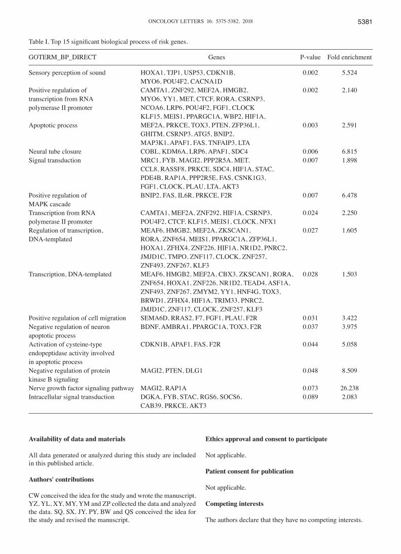
Based on survival analysis of the risk genes, we selected the following core genes involved in the survival of ovarian cancer patients: AKT3, CAB39, CDKN1B, F2R, FGF1, JMJD1C, MAGI2, MEF2A, TJP1, APAF1 and BDNF. As presented in Table I, All the core genes of the regulatory network were involved in the top 15 significant biological process. The damage to neural plasticity and neural cell regeneration may be the pathophysiological basis for MDD in ovarian cancer. Neurotrophic factors play a very important role in the develop-ment and maintenance of peripheral and central nervous system. BDNF, or brain-derived neurotrophic factor, is involved in nerve regeneration (23). BDNF levels are significantly decreased in the hippocampus and prefrontal cortex of depression patients (17). Bachis (24) correlated increased expression of BDNF with hippocampal neurogenesis, and decreased expression with anxiety disorder. FGF1 (Fibroblast growth factor 1) is a core member of the FGFs family which play important roles in cell proliferation, angiogenesis, morphogenesis and regeneration, and is closely related to tumor development (25). FGF1 also has a nutritive function in the regeneration of central nervous system and impaired neuronal repair (26). AKT3, a member of the AKT subfamily of serine/threonine protein kinases, is involved in several biological processes including cell prolif-eration, differentiation, apoptosis, and tumorigenesis (27,28). It plays an important role in ovarian tumorigenesis via regu-lation of VEGF secretion and angiogenesis (29). Moreover,
one study (30) showed that AKT3 is the most abundant AKT paralog in the brain during neurogenesis, and protects nerve cells and promotes neurogenesis. Membrane-associated guanylate kinase inverted 2 (MAGI2) is a scaffold protein with multiple domains, and functions in raising and anchoring cell signaling proteins. It is highly expressed in brain tissues, and is involved in the formation and maintenance of synapses in the central nervous system of vertebrates, and in the occurrence and development of nervous system diseases. In addition, it has been reported as a potential tumor suppressor (31-33). The GO analysis indicated another function of MAGI2 as a negative regulator of AKT signaling (Fig. 2). Due to its involvement in the nerve growth factor signaling pathway, MAGI2 may play a role in the occurrence and development of MDD. The interac-tion between the core genes and DEmiRs need to be further experimentally validated.
In conclusion, we analyzed the miRNA expression profiles of ovarian cancer patients with MDD, and eventually constructed a miRNA-mRNA regulatory network through bioinformatics analysis. The miRNA-mRNA regulatory network provides new insights into the pathophysiological mechanisms of MDD in ovarian cancer.
Acknowledgements
Not applicable.
Funding
The present study was supported by grants from Chinese National Natural Science Foundation Grant (grant nos. 81671541, 81273202 and 31400773), Clinical Medicine Science & Technology Project of Jiangsu Province of China (grant no. BL2013024), Jiangsu Provincial key research and development program (grant no. BE2016721).
Figure 6. miRNA-core gene-risk signaling pathway regulation network. The red hexagonal represents upregulated DEmiRs; The green hexagonal represents downregulated DEmiRs; The green triangle represents downregulated core genes; The red triangle represents upregulated core genes; The bule square represents risk signaling pathway. DEmiRs, differentially expressed microRNAs.
ONCOLOGY LETTERS 16: 5375-5382, 2018 5381
Availability of data and materials
All data generated or analyzed during this study are included in this published article.
Authors' contributions
CW conceived the idea for the study and wrote the manuscript. YZ, YL, XY, MY, YM and ZP collected the data and analyzed the data. SQ, SX, JY, PY, BW and QS conceived the idea for the study and revised the manuscript.
Ethics approval and consent to participate
Not applicable.
Patient consent for publication
Not applicable.
Competing interests
The authors declare that they have no competing interests.
Table I. Top 15 significant biological process of risk genes.
GOTERM_BP_DIRECT Genes P-value Fold enrichment
Sensory perception of sound HOXA1, TJP1, USP53, CDKN1B, 0.002 5.524 MYO6, POU4F2, CACNA1D Positive regulation of CAMTA1, ZNF292, MEF2A, HMGB2, 0.002 2.140transcription from RNA MYO6, YY1, MET, CTCF, RORA, CSRNP3, polymerase II promoter NCOA6, LRP6, POU4F2, FGF1, CLOCK KLF15, MEIS1, PPARGC1A, WBP2, HIF1A, Apoptotic process MEF2A, PRKCE, TOX3, PTEN, ZFP36L1, 0.003 2.591 GHITM, CSRNP3, ATG5, BNIP2, MAP3K1, APAF1, FAS, TNFAIP3, LTA Neural tube closure COBL, KDM6A, LRP6, APAF1, SDC4 0.006 6.815Signal transduction MRC1, FYB, MAGI2, PPP2R5A, MET, 0.007 1.898 CCL8, RASSF8, PRKCE, SDC4, HIF1A, STAC, PDE4B, RAP1A, PPP2R5E, FAS, CSNK1G3, FGF1, CLOCK, PLAU, LTA, AKT3 Positive regulation of BNIP2, FAS, IL6R, PRKCE, F2R 0.007 6.478MAPK cascade Transcription from RNA CAMTA1, MEF2A, ZNF292, HIF1A, CSRNP3, 0.024 2.250polymerase II promoter POU4F2, CTCF, KLF15, MEIS1, CLOCK, NFX1 Regulation of transcription, MEAF6, HMGB2, MEF2A, ZKSCAN1, 0.027 1.605DNA‑templated RORA, ZNF654, MEIS1, PPARGC1A, ZFP36L1, HOXA1, ZFHX4, ZNF226, HIF1A, NR1D2, PNRC2, JMJD1C, TMPO, ZNF117, CLOCK, ZNF257, ZNF493, ZNF267, KLF3 Transcription, DNA-templated MEAF6, HMGB2, MEF2A, CBX3, ZKSCAN1, RORA, 0.028 1.503 ZNF654, HOXA1, ZNF226, NR1D2, TEAD4, ASF1A, ZNF493, ZNF267, ZMYM2, YY1, HNF4G, TOX3, BRWD1, ZFHX4, HIF1A, TRIM33, PNRC2, JMJD1C, ZNF117, CLOCK, ZNF257, KLF3 Positive regulation of cell migration SEMA6D, RRAS2, F7, FGF1, PLAU, F2R 0.031 3.422Negative regulation of neuron BDNF, AMBRA1, PPARGC1A, TOX3, F2R 0.037 3.975apoptotic process Activation of cysteine‑type CDKN1B, APAF1, FAS, F2R 0.044 5.058endopeptidase activity involved in apoptotic process Negative regulation of protein MAGI2, PTEN, DLG1 0.048 8.509kinase B signaling Nerve growth factor signaling pathway MAGI2, RAP1A 0.073 26.238Intracellular signal transduction DGKA, FYB, STAC, RGS6, SOCS6, 0.089 2.083 CAB39, PRKCE, AKT3
WU et al: miRNA-mRNA NETWORK OF MDD IN OVARIAN CANCER5382
References
1. Massic MJ: Prevalence of depression in patients with cancer. J Natl Cancer Inst Monogr: 57‑71, 2004.
2. Bailey RK, Geyen DJ, Scott-Gurnell K, Hipolito MM, Bailey TA and Beal JM: Understanding and treating depression among cancer patients. Int J Gynecol Cancer 15: 203-208, 2005.
3. Bodurka-Bevers D, Basen-engquist K, Carmack CL, Fitzgerald MA, Wolf JK, de Moor C and Gershenson DM: Depression, anxiety, and quality of life in patients with epithelial ovarian cancer. Gynecol Oncol 78: 302-308, 2000.
4. Price MA, Butow PN, Costa DS, King MT, Aldridge LJ, Fardell JE, DeFazio A and Webb PM; Australian Ovarian Cancer Study Group; Australian Ovarian Cancer Study Group Quality of Life Study Investigators: Prevalence and predictors of anxiety and depression in women with invasive ovarian cancer and their caregivers. Med J Aust 193 (5 Suppl): S52-S57, 2010.
5. Yang Y, Cui Y, Sang K, Dong Y, Ni Z, Ma S and Hu H: Ketamine blocks bursting in the lateral habenula to rapidly relieve depres-sion. Nature 554: 317‑322, 2018.
6. Cui Y, Yang Y, Ni Z, Dong Y, Cai G, Foncelle A, Ma S, Sang K, Tang S, Li Y, et al: Astroglial Kir4.1 in the lateral habenula drives neuronal bursts in depression. Nature 554: 323‑327, 2018.
7. Lovejoy NC and Matties M: Pharmacokinetics and pharmacody-namics of mood-altering drugs in patients with cancer. Cancer Nurs 19: 407‑418, 1996.
8. Dwivedi Y, Rizavi HS, Roberts RC, Conley RC, Tamminga CA and Pandey GN: Reduced activation and expression of ERK1/2 MAP kinase in the post-mortem brain of depressed suicide subjects. J Neurochem 77: 916-928, 2001.
9. Ashburner M, Ball CA, Blake JA, Botstein D, Butler H, Cherry JM, Davis AP, Dolinski K, Dwight SS, Eppig JT, et al: Gene ontology: Tool for the unification of biology. The gene ontology consortium. Nat Genet 25: 25-29, 2000.
10. Kanehisa M and Goto S: KEGG: Kyoto encyclopedia of genes and genomes. Nucleic Acids Res 28: 27-30, 2000.
11. Huang DW, Sherman BT and Lempicki RA: Systematic and inte-grative analysis of large gene lists using DAVID bioinformatics resources. Nature Protoc 4: 44‑57, 2009.
12. Huang da W, Sherman BT and Lempicki RA: Bioinformatics enrichment tools: Paths toward the comprehensive functional analysis of large gene lists. Nucleic Acids Res 37: 1-13, 2009.
13. Gyorffy B, Lánczky A and Szállási Z: Implementing an online tool for genome-wide validation of survival-associated biomarkers in ovarian-cancer using microarray data of 1287 patients. Endocr Relat Cancer 19: 197-208, 2012.
14. Plattner F, Hayashi K, Hernández A, Benavides DR, Tassin TC, Tan C, Day J, Fina MW, Yuen EY, Yan Z, et al: The role of ventral striatal cAMP signaling in stress-induced behaviors. Nat Neurosci 18: 1094‑1100, 2015.
15. Wong KK: Recent developments in anti-cancer agents targeting the Ras/Raf/MEK/ERK pathway. Recent Pat Anticancer Drug Discov 4: 28‑35, 2009.
16. Dwivedi Y, Rizavi HS, Roberts RC, Conley RC, Tamminga CA and Pandey GN: Reduced activation and expression of ERK1/2 MAP kinase in the post-mortem brain of depressed suicide subjects. J Neurochem 77: 916-928, 2001.
17. Taliaz D, Stall N, Dar DE and Zangen A: Knockdown of brain-derived neurotrophic factor in specific brain sites precipitates behaviors associated with depression and reduces neurogenesis. Mol Psychiatry 15: 80-92, 2010.
18. Ding L, Zhang C, Masood A, Li J, Sun J, Nadeem A, Zhang HT, O' Donnell JM and Xu Y: Protective effects of phosphodiesterase 2 inhibitor on depression- and anxiety-like behaviors: involve-ment of antioxidant and anti-apoptotic mechanism. Behav Brain Res 268: 150‑158, 2014.
19. Wu M, Chen Y, Jiang L, Li Y, Lan T, Wang Y and Qian H: Type II cGMP-dependent protein kinase inhibits epidermal growth factor-induced phosphatidylinositol-3-kinase/Akt signal transduction in gastric cancer cell. Oncol Lett 6: 1723-1728, 2013.
20. Tao Y, Gu YJ, Cao ZH, Bian XJ, Lan T, Sang JR, Jiang L, Wang Y, Qian H and Chen YC: Endogenous cGMP-dependent protein kinase reverses EGF-induced MAPK/ERK signal transduction through phosphorylation of VASP at Ser239. Oncol Lett 4: 1104‑1108, 2012.
21. Karami-Tehrani F, Fallahian F and Atri M: Expression of cGMP-dependent protein kinase, PKGIα, PKGIβ, and PKGII in malignant and benign breast tumors. Tumor Biol 33: 1927-1932, 2012.
22. Li H, Zeng J and Shen K: PI3K/AKT/mTOR signaling pathway as a therapeutic target for ovarian cancer. Arch Gynecol Obstet 290: 1067‑1078, 2014.
23. Polyakova M, Stuke K, Schuemberg K, Mueller K, Schoenknecht P and Schroeter ML: BDNF as a biomarker for successful treatment of mood disorders: A systematic & quanti-tative meta‑analysis. J Affect Disord 174: 432‑440, 2015.
24. Bachis A, Mallei A, Cruz MI, Wellstein A and Mocchetti I: Chronic antidepressant treatments increase basic fibroblast growth factor and fibroblast growth factor‑binding protein in neurons. Neropharmacology 55: 1114‑1120, 2008.
25. Jiao J, Zhao X, Liang Y, Tang D and Pan C: FGF1-FGFR1 axis promotes tongue squamous cell carcinoma (TSCC) metastasis through epithelial-mesenchymal transition (EMT). Biochem Biophys Res Commun 466: 327‑332, 2015.
26. Renaud F, Desset S, Oliver L, Gimenez‑Gallego G, Van Obberghen E, Courtois Y and Laurent M: The neurotrophic activity of fibroblast growth factor 1 (FGF1) depends on endoge-nous FGF1 expression and is independent of the mitogen-activated kinase cascade pathway. J Biol Chem 271: 2801-2811, 1996.
27. Cohen MM Jr: The AKT genes and their roles in various disor-ders. Am J Med Genet A 161A: 2931-2937, 2013.
28. Madhunapantula SV and Robertson GP: Targeting protein kinase-b3 (akt3) signaling in melanoma. Expert Opin Ther Targets 21: 273-290, 2017.
29. Yeganeh PN, Richardson C, Bahrani-Mostafavi Z, Tait DL and Mostafavi MT: Dysregulation of AKT3 along with a small panel of mRNAs stratifies high-grade serous ovarian cancer from both normal epithelia and benign tumor tissue. Genes Cancer 8: 784‑798, 2017.
30. Poduri A, Evrony GD, Cai X, Elhosary PC, Beroukhim R, Lehtinen MK, Hills LB, Heinzen EL, Hill A, Hill RS, et al: Somatic activation of AKT3 causes hemispheric developmental brain malformations. Neuron 74: 41‑48, 2012.
31. Hirao K, Hata Y, Ide N, Takeuchi M, Irie M, Yao I, Deguchi M, Toyoda A, Sudhof TC and Takai Y: A novel multiple PDZ domain-containing molecule interacting with N-methyl-D-aspartate receptors and neuronal cell adhesion proteins. J Biol Chem 273: 21105-21110, 1998.
32. Hirao K, Hata Y, Yao I, Deguchi M, Kawabe H, Mizoguchi A and Takai Y: Three isoforms of synaptic scaffolding molecule and their characterization. Multimerization between the isoforms and their interaction with N-methyl-D-aspartate receptors and SAP90/PSD-95-associated protein. J Bol Chem 275: 2966-2972, 2000.
33. Hu Y, Li Z, Guo L, Wang L, Zhang L, Cai X, Zhao H and Zha X: MAGI-2 Inhibits cell migration and proliferation via PTEN in human hepatocarcinoma cells. Arch Biochem Biophys 467: 1‑9, 2007.
This work is licensed under a Creative Commons Attribution-NonCommercial-NoDerivatives 4.0 International (CC BY-NC-ND 4.0) License.


![IdentificationandAnalysisofThreeHubPrognosticGenes ...particularinterest[10–12].AmongtheceRNA,theinterplay betweenlongnoncodingRNA(lncRNA),messengerRNA (mRNA),andmicroRNA(miRNA)hasbeenwidelyin](https://img.pdfslide.net/doc/110x75/60cd4e257c8b6c5ade17aa87/identificationandanalysisofthreehubprognosticgenes-particularinterest10a12amongthecernatheinterplay.jpg)