Embed Size (px)
Citation preview

IDIOPATHIC HYDROCELES.
M. MED. (SURGERY)

' V IDIOPATHIC HYDROCELES.
An Analysis of 173 Cases of Scrotal Swellings seen at the
Kenyaita National Hospital, Nairobi over c period of
//5 years, 1973 - 1977/
BENJAM IN CHARLES A KEN G A , LRCP(lrel).
LRCS(lrel).
LM. (Dublin).
A dissertation submitted in parr fulfillment for the degree of
Master of Medicine (Surgery) University of Nairobi, 1978.
University of NAIROBI Library
lllllli.il0324744 2

This dissertation is my original work and has not been
presented for a degree in any other University.
BENJAMIN CHARLES AKENGA CANDIDATE.
This dissertation has been submitted for Examination with
my Approval, as University Supervisor.
PROFESSORAM 3 ROSE WAS UN N A .
SUPERVISOR.

CONTENTS
3.
4.
5.
6.
7.
8 .
9.
10.11.12.
13.I
14.
Table of contents
Acknowledgements
Introduction
Material and Methods
Results
Incidence of hydroceles at the Kenyatta National Hospita
and review of world liierature.
Embryology of - scrotum
- tunica vaginalis formation
- testis and its descent
Anatomy of the scrotum and contents
Pathology of - hydrocelet
- hydrocele fljid
Aetiology
Classification of hydroceles
Clinical presentation and differential diagnosis
Treatment - common forms of treatment offered.— 1 •*
Summary and ). - Aetiology
conclusion ) - Incidents
- Pathology
- Diagnosis
Treatment
15. References.

A C K N O W L E D G E M E N T
I am very much indebted to Professor F. Fasana of the
Department of Human Anatomy, University of Nairobi
whose constant and persistent help made it possible for
me to write this dissertation.
I also owe much thanks io Professor Ambrose Wasunna,
Head of the Department of Surgery, University of Nairobi
for reading the proof and supervising the writing of this
dissertation. The following also made the production of
this dissertation possible:- Mrs. Ndegvva and her members
of staff in the Rocoids Department at Kenyatta National
Hospital for helping me in collect! ng data, Mr. Mbugua
end Mr. J.Wckhisi for undertaking the photography. Last
but not least, I wish to thank sincerely KArs.Susan Thuo
end Mrs. Mory Kamau for typing the whole script.

I N T R O D U C T I O N :
Much has been written about secondary forms of hydroceles,
particularly those found to be due to filariasis .(Nos.5,6,7,8,9,
15 a, 20). Yet in this series at Kenyatta National Hospital
situated in Nairobi, which is far from the endemic filarial
areas, hydroceles are commonly seen. Hydrocelectomy is
therefore a commonly performed operation .(15c)
Pare (1510-1590) was the first to use the world "hydrocele",
though Celsus (first Century) and Galen (131 A .D .) described
the operation for hydrocele through an inguinal incision using
seton. Documented and artistic work shows that hydrocele wcs57
recognised in Greece in the second century before Christ. But
;"ispite of this, a clear definition of idiopathic hydrocele, and
indeed its aetiology, has not been agreed on.
A hydrocele is an abnormal accumulation of fluid within the two
layers of tunica vaginalis. Idiopathic hydrocele is c type of
hydrocele with chronic fluid accumulation of unknown or obscure
cetiology. It has been shown to have, as components,failure of
prompt clearance of any accumulated fluid in tunica vaginalis
and cystic cavities, or complete failure of closure of the processus
vaginalis soon after birth (52,53,51). A secondary form o(
hydroce le is one that is seconder'/ ;c a causative oi aefioicgic
factor.

- 2 -
The congenital type occurs during infancy and is usually
due to paten1 processus vaginalis.
Studies show that in East Africa and Kenya in particular, hydrocele
is mainly secondary. (5 ,6,/ ,8 ,9). In this series it was found
that over 75% of the cases had no cause for their scrotal swellings
i.e. there v/ere no physical finding s suggestive of primary or
accompanying condition p edisposing to hydrocele, and also where
hydrocele occured in children. The rest showed concomitant
presence of physcial findings that could be associated with hydrocele
formation - e.g. diabetes, congestive cardiac failure, renal
pathology, Tuberculosis, trauma to the scrotum, torsion of testis,
pyogenic infection and also testicular tumours.
In this review, many cases of congestive cardicc failure, renal
pathology and liver failure which gave rise to pitting oedema
and anasarca were found. But where the words scrotal swelling
did not appear in the diagnosis or physical finding that ccse was
left out for the purpose of this study.
The Embryology anatomy and pathology are presented first with
specia1 emphasis on the lymphatics. ThereaAer, Aeiio'ogy,
classification, diagnostic procedures and forms of management
are presented. In the discussion cr>.d conclusion, new information
is high iighted and a summary of all the chapters is cyven in c
concise form.

- 3 -
AIMS OF THE STUDY
The aims of this dissertation are:-
1. To assess the frequency of idiopathic hydrocele in
comparison with the other scrota! swellings seen at
the Kenya?ta National Hospital, Nairobi.
To compare the local frequency with those of other
countries.
To correlate the theories on the aetiology of
idiopathic hydrocele to **">e anatomy and embryology
of the scrotum and contents.
To review ond compare the various forms of surgical
management of this form of hydrocele.

- 4 -
MATERIALS A N D M ETHODS.
This being a retrospective stud/, all the materials have been collected
trom'the records of 178 patients admitted to the Kenyatta National
Hospital from January 1973 to December, 1977 inclusive with scrotal
swellings. 135 of these cases were idiopathic hydroceles - which gives
an average of 27 ca*"' rT idiopathic hydroceles admitted to the Kenyutfa
National Hospital every year. No seasonal variation was noted.
All the records in this study were obtained from the Kenyatta National
Hospital Records Department.
R E S U L T S
The records of each patient gave information about age ct the time of
•presentation; tribe; presenting sympto-n«; duration of symptoms; side of
presenting lesion (the left or right), associated illness, and the forms
of management given.
A ge distribution at the time of prcse.itat?on.
From Table 1 it will be observed that the peak incidence was at 1 - 13*
years and 20 - 69 yecrs. The youngest patient seen was 3 weeks oid
whiie the oldest was 90 years, a mean average of 45 years.
Frequency of different scrotal swellings .
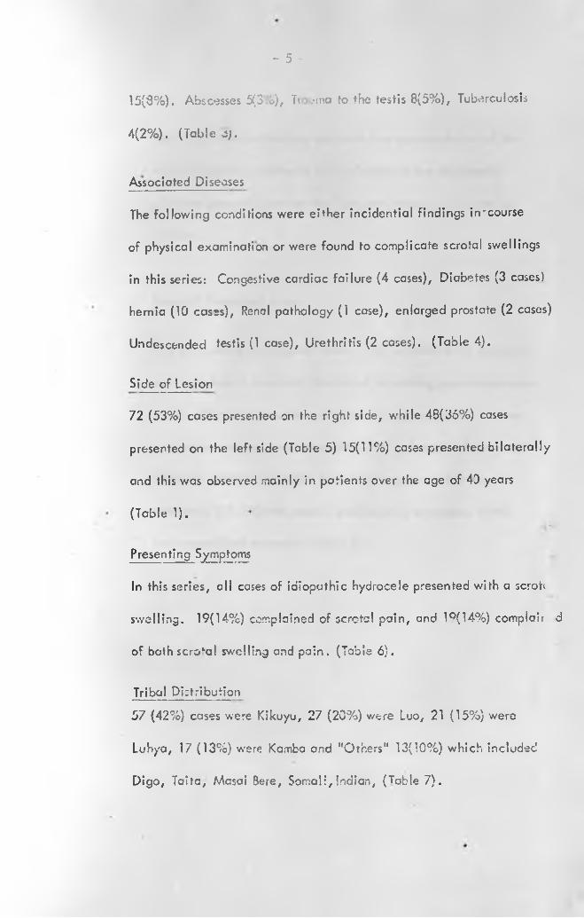
Out ot c total o* 178 cares of sc ctcri swellings seen, 135(76%) of these
were idiopathic hydrocele (lablc 2). Other causes of scrotal swellings
found in this seres a<-e: T-mours 11(6%), Torsion of *est:s
- 5 -
15(8%). Abscesses 5(3 .o), Tr -.•ino to the testis 8(5%), Tuberculosis
4(2%). (Table oj.
Associated Diseases
The following conditions were either incidential findings in'course
of physical examination or were found to complicate scrotal swellings
in this series: Congestive cardiac failure (4 cases), Diabetes (3 cases)
hernia (10 cases), Renal pathology (1 case), enlarged prostate (2 cases)
Undescended testis (1 case), Urethritis (2 cases). (Table 4).
Side of lesion
72 (53%) cases presented on the right side, while 48(36%) cases
presented on the left side (Table 5) 15(11%) cases presented bilaterally
and this was observed mainly in patients over the Gge of 40 years
(Table 1).
Presenting Symptoms
In this series, all cases of idiopathic hydrocele presented with a scroti
swelling. 19(14%) complained of scrotal pain, and 19(14%) complair d
of both scro‘al swelling and pain. (Table 6).
Tribal Di:tribution
57 (42%) cases were Kikuyu, 27 (20%) were Luo, 21 (15%) were
Luhya, 17 (13%) w-ere Kamba end "Others" 13(10%) which included
Digo, Taifc, Masai Bere, Somali,Indian, (Table 7).

- 6 -
This tribal distribution may not be a true representation of the
actual situation. Rather it is a reflexion of the distribution
of tribes generally seen at the Kenyatta National Hospital as
patients.
Forms of Treatment given.
In 8 (6%) cases nothing was done - i.e. patients were observed
over a period of time with the aim of achieving spontaneous cure
In 2(1.5%) the hydrocele was tapped or aspirated; In 117 cases
surgical operations were done: 44 (33%) had inguinal approcach
(19 were chi Idren )#74(55%) had scrotal approach (13 were
children); 1(0.5-%) had scroral and inguinal approach; 6(4%)
hod unspecified treatment (Table 8).

TABLE 1.
AGE DISTRIBUTION AT THE TIME OF PRESENTATION (AND
___________________ SIDE OF LESION). ____
—
AT BIRTH1
BILATERAL LEFT RIGHT
l
TOTAL
0 3 5 8
UP TO 1 YEAR 0 0 1 1
1 TO 13 YEARS 1 7 29 37
14 TO 20 YEARS 0 2 2 4
21 TO 40 YEARS 0 11 17 28
41 TO 60 YEARS
_ _ _ _ _ _ _ _ _ _ _ _ _ _ _
6 13 14 33
OVER 60 YEARS 8 12 4 24
TOTAL NUMBER OF HYDROCELES 15
j---------------
£ 72 135
TABLE 2
RATES OF OCCURENCE OF IDIOPATHIC HYDROCELES
v
NUMBERPERCENTAGE!
OFTOTAL
NUMBER O F IDIOPATHIC
HYDROCELES
135 76%
NUMBER O F OTHER SCROTAL
SWELLINGS 43 24%
TOTAL NUMBER OF SCROTAL
SW ELLINGS
»---- --------------------------------------------
178 100%

TABLE 3.
ANALYSIS OF SCROTAL SW ELLINGS DUE TO OTHER CAUSES.
1------------------------------------------------
CAUSESTOTAL
NUMBER
PERCENTAGE
OF TOTAL
TUMOURS 11 6%
TORSiON OF TESTIS 15 8%
ABSCESSES 5 3%
TRAUMA 8
l
5%
TUBERCULOSIS 4 2 %
TOTAL 43 24%

TABLE 4.
ASSOCIATED PRESENTING DISEASES (TOTAL 23).
ASSOCIATED PRESENTIN G DISEASE NUMBER1
CONGEST IVE CARDIAC FAILURE 4
DIABETES MELLITUS 3
HERNIA (IN G U IN AL). 13
RENAL PATHOLOGY 1
ENLARGED PROSTATE 2
UNDESCENDED TESTIS 1
URETHRITIS 2
TOTAL
1
23

TABLE 5.
SIDE OF LESION.
SIDE
—
NUMBERi
PERCENTAGE
OF TOTAL
RIGHT 72 53%
LEFT 43 36%
BILATERAL 15 11%
TOTAL 135
— — r
100%

TABLE 6.
PRESENTING SYMPTOMS IN IDIOPATHIC HYDROCELE (TOTAL 135)
I
~ T ~
SYM PTO M NUMBER
PERCENTAGE OF
TOTAL.
SCROTAL SW ELLING 135 100%
PAIN 19 14%
BOTH SCROTAL SWELLING
A N D PAIN19 14%

TABLE 7
TRI B A L DISTRIBUTION (TOTAL 135)

TABLE 8.
FORMS OF TREATMENT G IVEN . (TOTAL IDIOPATHIC HYDROCELES)135.
TOTAL NUMBER1
PERCENTAGE
FORM OF TREATMENT OF CASES OF TOTAL
CONSERVATIVE 8 6 %
ASPIRATION
!
2 1.5%
OPERATIVE - Inguinal approach 44 33%
- Scrotal approcach 74 55%
- Both inguinal and
scrotal approach 1 0 .5 %
UNSPECIFIED TREATMENT 6 4 %
TOTAL 135f
100% ! |
—

- 7 -
I N C I D E N C E
The actual incidence of idiopathic hydrocele is Kenya and
probably the whole world is not known, as evidenced by the Scanty
Literature available.
In this series a total of 178 cases of scrotal swellings were seen
ovei a period of 5 years giving an average of 36 cases per year.
O f these, 135 (76%) were idiopathic hydroceles. The frequency
■7 . .of occurence on the left or right side are set out in table 5.
Fasana '"^working in India, Uganda and Kenya (Nyeri)
from 1954 to 1972 operated on 273 hydrocele coses. Among the
children he operated on, 8 had patent processus vaginalis and 4
inguinal herniae as a concomitant finding. All the other cases
were secondary to Wucherena Bnncrofti infection. The incidence
of the right sided hydrocele was 75%, bilateral 79%, those
'associated v/ith hernia 6% . He found microfilaria in the blood of
51% cf the cdult cases, whereas only 33% of the patients hod
microfilariae in scrotai fluid.
22Barkitf working in Lango District of Uganda reported that an
average of 25% of male odults had hydroceles, and that 43% of
them were bilateral.
Ecrlier studies by Hawking^ in the coastal creas of Kenya cod
ionr.ar.ic snowed then where filariasis v'cj common, the incidence
of hyorocele was nigh viz. 32% hydrocele rate among adult me les
around Kilwa and just less at the Kenya Coast.
- 5Wijers and McMahon found hydrocele rates above 40% in males
over the age of 14 years along the Tana River and Lamu District
of Kenya. These, however, were all secondary hydroceles, mosf
probably due to filariasis and therefore gave no true picture of the
incidence of idiopathic hydrocele.
36King examining 263 American Troops in Puerto Rico found no
cause for 80% of the hydrocele cases. This is comparable to the
present series where 76% of the scrotal swellings were idiopathic
hydroceles.
9QJordon*' was the first worker to draw parallels on incidence.
Working in Mwanza Region of Tanzania he wrote: "In areas
where no Parasitological evidence of Bancroftian filariasis was
found, 1 .2% of adult males were found to have hydrocele".
Drawing evidence from this study end observation, Jordan then
undertook to determine the incidence of hydrocele in young
British Army recruits from the Far East. This he found to be 1 in
1000. He then concluded that the incidence of idiopathic
hydrccele in Tanzania is the same as In Britain.
- 1 -

_ 9 -
E M B R Y O L O G Y
The embryology of the scrotum and contents and also of the
spermatic cord is essential in the understanding of the
mechanism of the various forms of hydrocele. First I would
deal with the embryology of the descent of the testis and its
appendages. In the process, I hope to show how the various
defects or failures cause particular types of hydrocele.1/ -
Embryology of the Scrotum
In the embryos during the undifferentiated sexual stage, the
external genitalia develop in the region of the cioaccl membrane.
This region extends on the ventral aspect of the body from the
umbilical cord to the tail. At .ne cephalic end of the cloacal
membrane, by the sixth week, develops a tubercle called the
genital -tubercle. (The gejiital tubercle has been described as
unpaired, by Spaulding; 1921; paired by Felix 1921, Pallen and
Berry 1952). The margins which flank the membrane and the
tubercle ro:se upto form the genital swellings or folds. In the
male the swellings move caudal I y, fuse together and form the
scrotum.

-10 -
Embryology of '.he Descent of the Testis end its appendages, and the
Formation of Tunica Vaginalis.______________________________
At first the testis lies on the dorsal abdominal wall but as it enlarges,
its cephalad end degenerates and it therefore assumes a more caudal
position (G ray 's anatomy). The testis, during the greater part of
embryonic life ond up to the seventh month lies at the level of the
49acetabulum (Youssef & Raslan 1971)
iiThis is contrary to the normal teaching that the testis actually descends
from the level almost corresponding to that of the kidney. The testis
is never far from the groin, and there is no abdominal descent of the
testis (Loch Wood 1838, Bramenn 1884, Wells 1947 , Lemeh 1970 J
48Wyndhan 1943) .
The testis, is attached to the mesonephiic fold by the mesorchium
which contains testicular vessels and nerves. The lower border of
the testis becomes connected to the skin which is larer to form the
scrotum, by a fold of mesenchymal ceils, included in a peritonea!
fold (inguinal fold). This fold forms a cord, which later becomes a
fibromuscular bundle and is termed the Gubernaculum testis.
Gubernaculum restis is actually a condensation of ligamentum testis
ond scrotal iigamenf. From the gubernaculum testis develop fascia!
coverings of the testis and spermatic cord, including cremaster (Gray
& Skandalaiis).

In the meantirr'' a cociOwic evagination is formed in the inguinal
region of each side wl . the caudal end of the ligamentum testis
is attached. Each of these peritoneal lined extensions of the coelom
is known as the processus vaginalis (First described by Galen A .D .
176) 53. Scrotal ligament connects the processus vaginalis to the
scrotum. Ao the processus vaginalis is deepened, the scrotal ligament
becomes shortened, and broadened and ultimately blends into
2connective tissue layer of the scrotum (Patten ). The lower pole
of the testis is retained in opposition to the deep inguinal ring by
Gubernaculum testis by the 5th month. During the 7th month the
testis begins to pass through the inguinal ring and by ihe 8th month
2they have come to lie iri the scrotal pouches (Paifen ). The distal
end of the processus vaginalis into which the testis projects, forms
the tunica vaginalis testis, but the portion associated with the
spermatic cord in the scrotum ana in the inguinal canal normally
becomes obliterated. Obliteration of the processus vaginalis is
4complete at birth in 50 - 70% infants (Gray and Scandaialis) .
In its entire descent the testis moves caudally beneath the peritoneum.
It does not enter the luynen of the scrota! pouch directly but slips
down under the peritoneal lining and protrudes into the lumen,
reflecting a peritonea! layer over itself. This reflected peritoneum

-1 2 -
is known anatovicolly as the visceral layer of tunica! vaginalis
proprius. The remainder of the peritoneal sac which now lines*
the scrotal cavity is called the parietal layer of the tunica vaginalis
proprius. (See photograph I).
S c * 4 * i t r ' i in * c fbm j m tffefbH hfV & € f i i A f - v t S i h P I f 75.*
* "b th b ’6W>‘J tO * *
Of W * 7 o s t*s (hot . - • * M r r e *» ‘
U«W'«*( — .- £
4,» «-•-< /
B
- 13 -
Couses of Testiculcr Dr ,c,it
TI1-3 cause o ' testicular descent is poorly understood (Gray's
14Anatomy ) ;->ut the factors often thought to be involved ere:
1. Hormonal, and rrw«-hanical factors: Hormonal role of
the anterior pituitary, maternal chorionic gonadotroprin
which stimulates cnd.ogen production in the adrenal
cortex, and progesterone have been suggested by Shapiro
(1930), E jgle (1932), Wislock (1933), Martins (1943),
( as quoted by Gray and Shandalckis ).
2. Self propelling through testicular organisation and
mechanical factors. Although this view has been
supported by Hunter (1786) a d others, others workers
have debated it. The mechanical factors often quoted
are:
a) Intra-abdominal pressure (Gray and Shandalalis
141972, and Grays Anatomy) .
b) The contraction of the muscles around the inguinal
14canal (Gray's Anatomy).
c) The contraction of the Gubernaculum
(Gray's Anatomy).14

-14 -
Youssef and Raslan (1971) after extensive
stud/ have come to the conclusions:
a) That there is a gradual increase of the size
of the testis during its descent and its
weight plays an important role in the
process of descent.
b) Gubernaculum prepares with its morphological
and histological modifications the way to
the descent of the testis, widens the inguinal
canal and by contraction of *he stomach
muscle fibres drav/s the testis into the
scrotum.
c) Thot the weight of the viscera and the
meconium ought to be considered as an
important factor in the testicular descent.
49

- 15 -
A N A TO M Y
A simplified ana 'elevo.-it anatomical consideration will be presented
here. It is disigned to show mainly the structure one comes across at/operation. It will also act as a reminder of the structures one comes
across at operation, and those mentioned in the chapter on aetiology
of idiopathic hydrocele. Specicl mention is made o': the lymphatic
drainage which seems to be implicated all round as a major cause of
hydrocele formation. The lymphatics have thus got a mapr share of
this chapter.
The coverings and contents of the scrotum are presented in tabulated
f orm to indicate die order in which one sees them at operation,starting
with skin. A comparison is made with the abdominal wall and spermatic
cord coverings. (See Table).
A BD O M IN A L WALL SCROTAL W ALL
1. Skin
i
Skin
t2. Subcutaneous fat (Campers
end Scarper"s fascia)Dartos (Scarpas fascia)
3. Externa! oblique apuneurosis External spermatic sheath (external oblique aporenosis)
4. Cremasteric fascia (Internal ' oblique muscle)
Cremasteric fascia
5. Internal spermatic sheath (transversalis fascia)
Internal spermatic sheath
i
6. Preperi toneal fat Pre peritoneal fat
7. Hernia! Sac (Peritoneum)•
Peritoneum (Tunica Vaguialis)
— ---------------- --------- Testis.

- 16-
Blood supply scrotum one! contents
The front or the scrotum is supplied by the external pudendal
✓ arteries whereas the back is supplied by the scrotal branches of
the internal pudendal artery, H.unches of the testicular artery
and artery to the cremaster help to supply the scrotum. The
testis is supplied by the testicular artery arising from the abdominal
aorta.
The veins accompany the arteries. The testicular veins form the
pampiniform plexus of veins which form the bulk of the spermatic
cord, and eventually drains into the inferior vena cava (Gardner56
& O 'Rohily) . .
Nerve Supply to^he Scrotum and Contents
The anterior part of the scrotum is supplied by the ilio-inguinai
nerve and by the genital branch of the genito femoral nerve. Toe
posterior part is supplied by the media! end lateral scrotal branches
of the perineal nerve and by the perineal branch of the posterior
56femora! cutaneous nerve. (Gardner et al) • The testis is supplied
by the testicular p!exus of the sympathetic nerves (Gray's Anatomy)
14.
Lymphatic drunoge of the Scrotum and Con ten Is
In ‘he naimcl hydrocele sac, the subserous lymphatics consist of
superficial and deep plexuses, both being confined to the basal
fibrous layer of the parietal tunica vaginalis. The superficial
plexuses are much smaller and communicate by narrow channels
with the deeper plexuses which lie within the deeper portion53 55
of the basal fibrous layer, (L.Allen) and (Rainller and Allen) .
Each plexus is drained by channels which course around the tunica
vaginalis from the medical and lateral side and empty into marginal53
vessel which originates within the testis (L.Allen)
Some lymph from the tunica vaginalis and the tail of the epididymis
jesses direct to the external iliac glands before passing to the
para-aortic group. Other vessels drain directly into the para-
9aortics (Galloway 1954). (P.Jordan) . The testicular lyrnph also
drains into the para-aortics. The visceral leaf of tunica vaginalis
10 53has no lymphatics (S. Grafana) (Lane Allen)

/
P A T H O L O G Y
1. Pathology . T e liydrocele fluid .is usually described as straw41
coloured or amber coloured. Douth Waite nas described it as
"a golden fluid of soapy feel" . Many workers have analysed
the constituents of idiopathic hydrocele fluids, with a view to
ascertain whether this is trapped peritoneal fluid or the result
or increased capillary permeability and reduced lymph drainage.
As might be expected nearly all have come out with very
similar readings.
(1)A.F. W allace ' found the following as chemical constituents
of an idiopathic hydrocele fluid, as compaired with the
12findings of Boyce & Pollitano
. }0 _
Water Resembles plasma
Soluble and insoluble salts Few lymphocytes and epithelial cells
S.G . 1016 - 1026 S.C-. 1010 - 1025
I
Albumin Albumin 3 -6% of prolein
Globulin Protein 4-o.3gm%
Chlor'des Chlorides
Fibrin Fibrinogen
Cholesterol Cholesterol
A.F. W allace) (Boyce and Pottllcno)

, ’
C. Poppis, end his ^.racial.< ; worked out the swlt content of
hydrocele fiuia in comparison with that of serum. His findings are
presented in Toble form to include other electrolytes.
Electrolyte
i
Serum
—
Hydrocele Fluid
Sodium No (MEq/lt) 137.1 136.5
Potassium K (MEq/it) 4.4 4.3 i.
Chlorides Ci (MEq/!t) 101.4 107.2
Calcium Ca (M g%) 9.3 9.5
Urea (Mg%) 40.2 29.2
Normal constituents cf plasma have therefore been extracted from
idiopathic hydrocele fluid, bur always in lower concentration. The
large the molecular size the smaller is their concentration in hydroceie
fluid in comparison with blood. Hydrocele fluid has the composition
of a transudate (A.F. Wallace) \
The Pathology of Tunica V aginalis.
In the non-inf I amatory (Primary) cases the tunica appears pearly whitish
grey in colour and has the texture of peritoneum (especially in children).
It is usually thinner than secondary hydrocele, but this will depend
also on the size of the hydrocele and extent er stretch. The secondary
hydrocele sac is usually thick, firmer and may even be cl&cified.

A E T I O L O G Y
An idiopathic condition is one in which no precise cause is known,,
That goes for the idiopathic hydrocele, too, where routine examination
fails to reveal a cousative facto'. Many attemts have been made to
explain the causative factors of this type of hydrocele, but so far only
theories and hypothese: have been propounded some more acceptable
than others. The follo ving are the more workable theories put forward
with regarod to the aetiological consideration of idiopathic hydrocele:-
1. incomplete obliteration of processus vaginalis, which led
35Tibbs, and Miller (quoting Fowler and Burnet 1958) to
conclude that "all child hood hydroceles were due to
incomplete obliteration of processus vaginaiis". This was
as a result of studies with methylene-blue dye injected into
the sac to delineate the extent of the hydrocele at operation
2. Weakness and/or paralysis of the cremaster muscle. This
theory hinges cn the fact that the cremaster muscle helps to
suspend the spermatic vessels. The spermatic veins, having
no valves, present a long and weighty column of blood whicn
the cremaster muscle has to support. Contraction of this
muscle accelerates circulation. "If the contraction of this
muscle is week or quite lost, the spermatic vessels, now no

-21-
longer supported, will be dropp'd down by their weight and that
of the testicle ~nd their tone impaired, in concequence of which
water will at last be accumulated in the cells of the cord". This
theory was propounded by Ruvsh I7 29 and supported by and
elaborated on by Douglas 1755 (as quoted by A.F. Wallace)t
3. Rupture of the hydotid of Morgagni, which as morgagni wrote in
1761 "these hydatids burst asunder and first pour out the water
they contain and after that go on to secrete still more and more;
there is not the lea^t doubt but they must produce hydrocele"
(A.F. Wallace).
4. Nearly 2 centuries later Barjoi; arid Cade (1903) after having examined
microscopically 25.idiopathic hydrocele fluid specimens they found
samples from secondary hydroceles and found that all contained
spermatozoa. They then proposed a modification of Morgagni's
theory in that rupture of the cyst of the epididymis rather than that
of Morgagni is the cause.
5. Hunter (1786-37) made the following observations regarding
hydroceles:
a) That it occurs in people of all kind of constitution.
b) That it occurs in all countries
c) That it is most common In worm countries. This is a view2?
supported 2 centuries by Caries (1924) who quotes

- 22 -
d)
examples of Indians wearing supports for
their pendulous scrota.
They go away by themselves in the young
but never go away in the adults (A.F.
Wallace).^
6. Idiopathic hydrocele may be congenitclly inherited. This
view was originated by CadwaUnder (1895) who published
the first "hydrocele Family" over 3 generations; (A.F.Wallace)
Hydrocele

- 23 -
7. Alteration in the serosa of the tunica vaginaiis. This
view was initiated by Peyrot and Million (1901) who
listed the following conditions cs being responsible for
the alteration of the serosa! lining.
Alcoholism Rheumatism
Typhoid Arteriosclerosis
Irregular Cardiac Renal
rythm insufficiency
Bronchitis
Embhysema
Prostate
enlargement.
8. There is an ill-understood association between the benign
prostate hyperteephy and idiopathic hydrocele in old men.
This view was expressed by Peyrot and M i Ilian (1901),
Posner (1911).
9. Compression of the Lymphatics draining the parietal tunica;
23a) by a hernia (Douglas arid Thompson 1937).
b) by fibrosis (S . Gratanc ^ ) ,
c) low grade inflamatory and tra.natic lesions of the
epididymis (S . Gratcna 19,20).
5310. Ailen nas proposed that the obliteration of the proximal
processus vaginalis before the establishment cf on effective
absorptive Lymphatic system would allow serous fluid rc
accumulate i» -be s a n d result in conger;'tal hydrocele o?
the new' born. This view is supported by C.Pcppis and

- 24 •
associates ' ' a id McCrien and associates''", who
concluded that hydrocele seems to be due to entry
of fluid in the tunica vaginalis by capillary filtration
and the trapping of +U!J fluid by impaired lymphatic
drainage. Failure of lymphatic drainage or delay in
absorption as an aetiologic factor in idiopathic hydrocele
has also been reported by S. Grotana O 'crowley
and Herzlich 1944; Huggins aid Endtz^ A.F.Wallace^ \
Huggins and Endtz, in their research with phenyl
sulphonphthaleln, found that absorption of this substence
from tunica vaginalis in idiopathic hydroceles was "so
slow as to indicate that delayed absorption may be the
chief factor in the accumulation of fluids".*
‘5
It has also been proposed that high ligature of varicoceles
possibly leads to a long collumn of blood which favours
hydrocele formation (Hanley 1955, Ruysh and Douglcs) ,
This would explain the composition of the hydrocele fluid
since it has the composition of a transudate (A.F. Wallace) 1.

- 2 5 -
12. Traumata* regenerative activation of the endothelial cells
of the Tunica vaginalis can give rise to hydrocele formation.
This view was stated by Obney (1956) who said that
endothelial cells of the tunica vaginalis are capable of
regenerating and causing a hydrocele to reform after combined
operation for hernia and hydrocele.
13. To the Great majority cf idiopathic hydroceles no label or\
theory could by put up to explain their occurence (S.Grafcna
18,20,Q^d O 'Crowley and Herzlich 1944).
V*

CLASSiF ICA 'i:Q N O r HYDROCELES
Many workers have classified hydroceles in various ways i.e.:
according to aetiology (Primary (Idiopathic) or Secondary);
according to anatomic location of hydrocele; according to presence
or a'osce.nce of infection; according to shape and position of hydrocele
etc, etc. This obviously adds up io the confusion there is about
hydroceles.
The aim of this chapter is twofold:
1. To present the old classification that is generally
taught and that is usuoliy found in textbooks of
Surgery.
2. To pcesent a new classification that takes into accoun*
the rationale for surgical treatment of hydroceles.
. 12The first classification is that presented by Boyce and Politano
1. Hydrocele of the testis.
a) Simple hydrocele. This is the most common type in
which a normally formed tunica, vaginalis is distended
with fluid. It usually appears as a globular or oval
mess.
b) Infantile hydiocele. This is one in which the finger
like funicular process has failed to close. In these
c a d the processus funicuiarls remains open and

extends to various levels, even to the internal
ring; however, the upper end is closed, and there is
no communication with peritoneal cavity.
Congenital hydrocele. This is the one in which
processus funicularis has a small lumen communicating
with the abdominal cavity. In this type, the fluid in
the tunica may ascend or can be forced upward into
the peritoneal cavity-the communicating hydrocele.
Inguinal hydroce le . This is very much like a simple
hydrocele of the testis with the exception that the
testis Is underscended and its position may be within
the inguinal canal or the pubic area. (In this respect,
hydrocele has been known to occur in intra abdominal
testis).
Encysted hydrocele of the epididymis or of the testis.
These usually occur on portions of testis or epididymis
without tunica vaginalis covering (posterior surface of
testis). They appear as a collecrion of fluid between
tunica vaginalis (visceral layer) and Tunica albuginea

-28 -
2. Hernial '.-iydrocele. An accunulation of fluid wiihin the
tunica vaginalis may be associated v/ith an inguinal hernia in
several different ways. In one type there is merely a small
end limited projection of processus fursicularis into the scrotum.
This closed hernial pouch terminates above the testis and does
not communicate v/ith tunica vaginalis sorrounding the latter.
Bowel and omentum ore usually not present in this sac because
IIof its small lumen, but it communicates with the general
peritoneal cavity. Another variety i s that in which a large
communication exists between the cavity of tunica vaginalis
ar.d peritoneal cavity. This is the complete congenital hernia,
where bowel and omentun have descended partially into the
scrotum v/ith fluid present distal to the bowel in the cavity of
processus vaginalis below.
3. Hydrocele of the cord. These are usually long, oval or fusiform,
and lie in the upper portion of the scrotum or in the inguinal canal.
They are closed at each end and have no connection with tunica
vaginalis or the peritoneal cavity.
4. Combination Hydrocele. There are c number of combinations of
the above mentioned hydroceles, e.g. a simple hydrocele of
the test's associated with hydrocele of the cord without
communication between the two. Hydrocele of the cord may

- 29 -
also occur with an inguinal hernia in which the
peritoneal or hernial pouch above does not
communicate with hydrocele of the cord.
Following the criteria established by the aetiology
apd operative needs, and also ihe rationale of
the surgical treatment of hydroceles, a new
classification of hydroceles has evolved. This was
by J .G . Ross^. .McKay et al; and Fasana^0 .
Hydroceles are divided into 2 types:-
a) Communio-.ir.g hydroceles - in which the
following types belongs:
1. Congenital hydrocele
2. Hemic! hydrocele
3. Abdomino scrotal.l
t) Non-Communicating hydrocele -in which the
following types be long:-
1. Vaginal hydrocele
2. Infertile hydrocele
3. Hydrocele of ihe testis and epididymis
(without tunica covering).

-30-
CLIN ICAL PRESENTATION A N D D IA G N O S IS .
The scrotal swelling due to hydrocele is often 'la rg e , heavy,
33ovoid, tense and elastic ralher than fluctuating" (Donthwaile)
(Photograph II)
Clinically the dicgnosis of hydrocele doesn't present much difficulty
except when it is complicated and/or associated with inguinal
hernia and other problems. The following then are the major diagnostic
fectures commonly found:-
c) Scrota! swelling: All of the cases (100%) in this scries
presented with, or were found *o have scrota! swelling. It
is usually of constant or increasing in size, in the common!ce~

- S i
ting types, it may le re ported es "coming c.i,d going".
b) Pain ie the scrotum. This is usually associated with secondary
cases especially those with filariasis, funiculitis epididymo-
orchitis etc. Large idiooathic hydroceles may give rise to
traction of scrotal suspensory tissues which may be described
as painful. In this series 14% of the cases presented with
pain in the scrotum.
c) Cough impulse - This is usually absent in scrotal swelling
"per se" but present if there is herniation of gut into the
scrotum. In this series, this sign was not demonstreted.
d) Transillumination. This is nearly always a finding in small
and mederately big idiopathic hydroceles. Large ones and
secondary hydrocele: often frcnsilluminate. Note - In
children hernia often also iransilluminate.
e) Hinge sign - to delineate scroral swellingsand distinguish
them from hernia. Again in large hydroceles this mey not be
practical. In this series this sign was not demonstrated.
f) Swelling of inguinaNymph nodes. This is usually associated
36with conditions affecting the scrotal coverings (King ).
This was not a finding in any of the cases in this series.

- 32 -
12In differential diagnosis, Boyce and Polita.no have given more
fhan 55 possible conditions that may cause scrota! swellings.
50But the table below presented by Essenhigh gives a more
practical differential diagnosis.
jI n our series, the following were found:-
IIa) Tumour of testis, scrotum and the cord.
b) Infections: Pyogenic end Tuberculosis
c) Inguinal hernia (mostly indirect ones).
d) Cysts of the cord
e) Congestive cardiac failure, hypertension,
liver failure and ren ji failure.
f) Trauma and haematocele
g) Torsion of testis.

Causes of Scrotal Swellings.
Origin of Swelling Pathology
Testis Congenital: Appendix of testis
Inflamation: OrchitsGumma
Neoplasm/1
Epididymis Congenital: Appendix epididymis cysts:
containing sperm: spermatocele not
containing sperm: epididymal cyst.
Inflametion: non-specific cyst.Tuber
culosis
Neoplasm.
Cord Congenital: Hydrocele of the cord.
Torsion, Variccele,
Neoplasm: Lipoma.
Tunica Vaginalis Congenital? Primary hydrocele
Secondary hydrocele: Haemnlocele
Scrotum Sebaceous cyst: Idiopathic scrotal oedema
Scrotal oedema.
Originating outs ide
the scrotum Hernia.

-.34-
fREATMENT OP HYDROCELE
The methods that have been employed in the treatment of hydrocele
are many and varied. In the literature, and in our series, they
vary according ro age of onset, associated findings, certoinl'y of
Diagnosis, ability of patient to withstand general anaesthesia etc.•»
etc. They can be enumerated as follows:-
1. Conservative: This is commonly employed in the management
of hydrocele in very young children. Some hydroceles do
disappear spontaneously in the first two years of life. (Boyce
12 1 25 39and Politano j Wallace , quoting Hunter ; Ross ‘ ,' Tibbs
35and M iller . In our series, 6 % were treated this way, i.e.
there was no surgical intervention and the hydroceles
disappeared spontaneously, after a period of observation.
2. Tapping or needle aspiration: This is usually employed in
iadult case where the patients condition doesn’t permit surgery
e.g. the aged, and the high-risk patients. It rarely leads to
12complete cure. Boyce and Politano state that this method
is curative in young children. But it is dangerously easy to
misdiagnose a hernia and mistake it for hydrocele - as both
transillumincte in children. In our series, 2 cases were treated
in this way. Both were elderly.

3. Aspiration with injecrionof sclerosing solutions: The
solution usually consist of quinine hydrochloride, urathane
and water (Farquhason and Rintoul^). This method has had
12"many adherents and a* many opponents" Boyce and Politano
Opponents of injection stress the occasional occurence of local
reactions (epididymitis, funicuiitis) to misplaced injections
in scrotal layers, sclerosing of abdomen through patent
processus vaginalis (communicating type of hydrocele), and
inadvertently injection a hernia, There is also the danger of
overlooking scrotal pathology as being the primary cause.
"injection therapy of secondary oi complicated hydrocele is
. . 1 2contra indicated" Boyce and Politano . In our series this
method was not used.
Inguinal approach; v/'nereby through on incision of sk:n,
externa! oblique, and exposure of cremaster muscle over the
superficial inauinal ring, the testis with the hydroceie(tota!35
or partial) are delivered, (Tibbs and Miller ) . The hydrocele
is punctured and aspirated and then 2 methods are aval lable
(Farquahanson and Rinton^):
c) Eversion of the Sac (J about ay) with a few sutures to
rctoi* the sac in position behind the ^estis and surrounding
the epididymis.
l*ltVERSITv OFNW506' IBRARX

-35-
b] Lxcision of the sac especially in the large or thick-
walled hydrocele, (the writer has employed this
method in some of the inguinal approached he has
made) and haemostasis, is achieved by a running
catgut suture of the cut edge. In this series this
was the method employed when either the inguinal
hernia was the initial diagnosis, or inguinal herniaif
was an associated finding, or when the scroial
1 swelling was so big that there was no certainty as
39to diagnosis. In these situations, Ross has advocated
the method as being the usual approach in children.
This technique Was used in 33% of cases in this
series.
5. Scrota! Approach:
a) Lord^ has described an operation (in 7 stages)
whereby through the scrotum, the tunica vaginalis
is opened and the hydrocele fluid evcquated. The
testis is delivered through the wound with the tunica
vaginalis or sac turned inside out. Forceps are
applied at several places cround its cut edge, and
five to six gathering stiches are inserted into the

-37-
tunica vaginalis and when these are tied, the
tunica is plicated to form a collar around the
junction of testis and epididymis. Alfthan and
21Sivula have echoed successes similar to those of
Lord when using this method.
• / . . . .This technique was used in 55% of cases in this
series.
15cb) Fascana , after noticing that the above method
proposed by Lord is "only suitable when the tunica
vaginalis is thin and normal and can be easily
plicated", proposed a modification which he found
useful in the 273 case he operated on while working
] . in Indio, Uganda,and Kenya (materials for a later
publication). After opening the scrotum anteriorly
and tunica vaginalis incised, the fluid is aspirated.
The testis is extruded from the cavity, thereby
everting the hydrocele sac. The edges of parietal
tunica vaginalis thus incised are sutured with
perpendicular stitches with interrupted number 00
catgut. This brings the edges to close proximity
but leaving enough space for testicular vessels and
the cord. The testis and the epididymis are returned

-33-
into the scrotal incision by stretching the periscrotal tissues
and by gentle pressure. The dead space is closed with
interrupted subcutaneous number 00 catgut sutures and the
skin with Mitchell Clips. Suspensory support is provided
Iand patients discharged on the 4th day. N o post-operative
complications were noticed in the 58 cases used for this
method. The method avoids mobilisation of the hydrocele
sac and is free from complications and recurrences. This is
a new technique and was not done in any of the cases in
this series.

-oV -
C Q N C L U S I O N S
The purpose of this study wcs to analyse the pattern of
presentation, aetiology, incidence and treatment of idiopathic
hydrocele, amidst other scrotal swellings. In conclusion then,
I will summarise the major observations made:
A etiology:
1. The precise cause of idiopathic hydrocele remains
unknown.
2. The mechanism of hydrocele fluid accumulation
seems to be due to impaired fluid absorption due
to a lymphatic defect, and increased capillary
permeabi lity.
Embryology:
3. The classical teaching that the testis descends into
the scrotum from a position in the abdomen
corresponding to that of the kidney has been
refuted.
Incidence:
4. Secondary hydroceie would appecr to be commoner
than the idiopathic type.
5. idiopathic hydrocele can occur at any age: In this series
the youngest patient v/as 3 weeks while the oldest was
90 years.
o. A ll in all, the scanty literature available shows that
idiopathic hydrocele incidence varies little from continent
to continent.
7. The pattern of distribution of idiopathic hydrocele in Kenya
has yet to be worked out. From the data available at the
Kenyatta National Hospital, it is not possible to arrive at
any precise pattern to reflect the situation in the whoie country.
Fhesentation:
8. The majority of hydroceles presented on the right side in this
series; In old age group, hydrocele tends to present bilaterally.
9. Pain in rarely a presenting features of id'opathic hydrocele.
C lassification:
10. A new v/ay of classifying hydroceles has been proposed. It
takes into account the rctionale for surgical treatment of these
conditions and attempts to remove the unnecessary confusion
about classification.

Treaimeni:
11. The concervative methods of treatment are suitable in the
young, the aged, and the surgical risk cases, while
operation is the definitive cure. Aspiration and injection
of sclerosing solutions have almost lost favour with most
workers.
12. Fasana (1977) proposed a modification of Lord's operation
in which the tunica vaginalis is not plicated, and is adequate
in all cases of thickened tunica vaginalis. It has been
described and its value emphasized.
Recurrence
13. O n ly 2 cases of recurrence of hydrocele were seen.
As recommendation, I would like more information extracted
from our patients, with a view of further research in this
field, as regards the foilowing:-
a) Size and amount of hydrocele fluid
b) Histological findings of the hydrocele sac should be
reported on and recorded in all cases.
c) The fertility pattern of ell adult makes with hydrocele
2o qqshould be recorded since Jordan and England seem
to think that "males with hydroceles are less fertile
than those without. "

l :st o f r e f e r e n c e s .
Wallace, A.F. British Journal of Urology.32:--33-79
1960 Aetiology of Idiopathic
Hydrocele
Human Embryology 3rd Edition - by Pattern M.pp436
Me Grawther
Esentials of human Embryology 2nd Edition by F.Ailan
Oxford 1969.
Embryology for Surgeons - Gray and Scandalakis i?72
Wijers, D.J . B.
W.B. Sanders.
Early signs and symptoms of
of Bancroftion filariasis in males
Me Mahon, J .E. East African Medical Journal 53:
57-63 1976.
Hawkins, F. - Bulletin of W .H .O . 1957, 16,
581-592. The distribution of
Bancroftion filariasis in Africa.
Spencer, J . Journcl of Tropical,Medico! Hygiene,
65; 256-259,1962 (Lar.go-Uganda).
Some clinical aspects of Baaoftian
filariasis in the Lango District of
Uganda.

43-
S. Woodman, H. - Central African Journal of Medicine -
6;23?-294,1955.
9. Jordan, D. - Journal of Tropical Medical Hygiene,
53:113 - 118, 1955
Notes on Elephatiasis and hydrocele
due to Wuchereria Bancrofti . .
10. Lord - "A bloodless operation of the radical
cure of Idiopathic hydrcceie" 7 stages.
British Journal of Surgery, 1964,
51,914.
11. Farquliarson,E. L .) -
• ))
Rintoul,R.F. )
12. Bcyce,W.H. ) -)
Politar.o,V.A. -
Textbook of operative Surgery 5th
Edition pp932-35, Churchill Living
stone and English Book Society.
Contribution to'"Urology" V o l.l
Chapter 16
3rd Edition W.B. Saunders.
13. Albu,F.D. Essentials of Embryology. 2nd Edition
pg.179.

14. Grays Anatomy. Edited by D .V . Davies, R.E.Complan
34th Edition Page 250.
15. Fasana - a). f
The fr0picol hydrocele. Thesis for special
zation in General Surgery. University of
Pavia (1973) ITALY.
b) Personal Communication; Professor of
Human Anatomy, University of Nairobi.
c) Treatment of the tropical hydrocele (1977)
in Print. East African Medical Journal.
16. Bai ley & Love - Short prod ice of Surgery 14th edition.
Edited by Peris + Capper.H. K. Lcv/is &
Co.London.
17. S. Grafama - The pathogenesis of hydrocele in Filarial
infection pp.254 - 267. Tropical end
Geographical Medicine. Vai.21 N o .l
1969.

-45-
13. Jordan, P.
19. Gratama,S.
/
20. Jordan,P.
il
Ban cr of fra in Filoriasis in Tanganyika
Annals of Tropical Medicine and
Parasitology, 54(1960) 132.
The Aetiology of Hydrocele in 5.E.
Liberia Territory Vol .21 N o .3 Pg.269,
1969.
Observation of Filariasis. Microfilaria!
density, Genital Fi lariasis and
microfilaria rates. Annals of Tropical
NAedicine and Parasitology. V o l.54
(1960) 133.
21. A lfth a n ,0 .) - Experience of a simple operation for
Idiopathic hydrocele. Annals ClinicalSivuIa,A. ^
E.T. Gynaecological Ferr.nial, Finland.
Vol. 53,1969.25.
22. Burkiti, D.P. - Primary hydrocele and its treatment.
The Lancet V o l. 1 (1951) 1341.
23. Douglas,J . ( 1755) "A treatise - on hydrocele. pp33,
44( London).
24. Morgagni,J. B„ (1761) "The seats and couses of disease
investigated by Anatomy" Translated by
Alexander London 1769, Letters 24,4,42,
43.
I-
25. Hunter J. (1786-87) "Lectures upon the principles of Surgery"
Collected by Palmar, London, 1335. po454,
457.
26. Cadwallader, R. (1895) Medical Recoias New York, 43,383.
27. Peyrot, J . ) - 1901 Bulletin of Academic Medicine,
M illion ,G . ) Paris, 45,162.
28. Bcrjon, F. ) - 1903 Archives of General Medicine 192-2177.
Cade, A. )
29. Carless:, 1924 Manual of Surgery. Edited by Rose and
Carless. 11th Edition p 1413 London (B.T.C.)
30. Carnpheli M.F. Surgery, Gynaecology, Obstetrics, 45, 192.
31. Thompson, A.R. 1937 - Guys Hospital Gazette 51-221.
32. O ' Crowley, C.R.) - American Journal of Surgery,66.157-60
Herzelick, J . ) 1944.
Boyd, W . - Pathology for the surgeons . 6th
Edition pp.452. W.B. Saunders.
33.

~/r7_
*
34. Jachowski,L.A.)
Gonzalez-Flores B)
Lichtenbergh F)
H \ i
35. David Tibbs ) )))
Douglas Miller)
Filarial Aetiology of Tropical
Hydroceles in Puerto Rico, American
Journal of Tropical Medicine and
Hygiene 11; 220-233. 1962.
Hydrocele in Infancy and childhood
Recent advances in Surgery. Pg. 134
little Brown and Co.London.
36. King, B. Go Early Filarial diagnosis and clinical
findings; a report of 263 cases in
American Troops.
American Journal of Tropical
Medicine and Hygine, 24:285-293.
1944.
37. Pondero et a !. Clinical manifestations of Bancroftian
filariasis in a suburb of Calcutta,
India.
American Journal of Tropical
Medicine and Hygiene 25,64-/3
1976.

England, E.C. . - Evidence of Filarial infection in East
Africa. A reviev/ of the literature
from 1955.
Health and Disease in Africa-
proceeding of E.A. Medicol Council
Scientific conference 1970. East
African Literature Bureau, 1971.
Graham Ross, J . - Treatment of P rimary hydrocele in
infancy and childhood. British Journo
of Surgery 49:415-418,1962.
Huggins) - Journal of Urology 25,44.1931.
Entz )
French's Index of Differential Diagnosis 9th edition -Edited
b y A . H . Doughwaite, Bristol .John Wright & Son Cc„
W. Felix, 1912, Development of the Urogenital Organs.
Manual of Ht/r.on Enbrology.
-49-
43. Patten, B.) . - Genesis of Extroply bladder and
Berry, A .) 1952 Epispadias: American Journal of Anatomyi
90:35-57.
44. Lockwood,C.B.- 1833, Development and transition of the
testis, normal and abnormal. Journal d
Anatomy, London Vol .22:33-44.
Vo. 478:505-541.
45. Bramann,F. 1834, Britnag-Zur Lebrc.
Von dans descondans, testicuiorum and
Gene Gjber naculum, Husteri-des 1834
Meschen. Archives of Anatomy pp310-40.
46. Wells, C.J. 1949, Misconception of the Gubernaculum
testis. Surgery 22:507.
47. Lemeh, E .N . 1960, A study of the development and
structural relationship of the testis and
Gabcmaculum.
' Surgery, Gynaecology, Obstefri cs,
110:164-72.

43. Wyndhar.', 1943. A Morphological study of Testicular descent.
Journal of Anatomy, 77-179-188.
49. Youssef,E.H.) Stuc/ of the factors which affect the descent
Raslan ,N .A ,) of testis in man.
Acta anatomia, 79:422-444,1971.
50.
50. Essenhigh, D .M . 1974 Scrotal swellings. The Practitioner
212: 216-220.
51 , McBrien,M .P. ) Lymphography of the testis and its adnexa
Edwards, J . M . ) in tne normal and idiopathic hydrocele.
Kinmonth, J , B . ) Archives of surgery Vol. 104,June 1972
pg.820-825.
52. Panpis,C. ) -)
Tryfoncs, G . ))
Kloni,M )
Vreitos/ G . ^
Constanrinides)
Hydrocele fluid proteins in Infants and
children.
Acta Paediatrica Beige 29:99-102.19/6.
0F NAIROBI •-‘RRARY

•51
53. Allen, L. - The Lymphatics of the parietal Tunica
Vaginalis Propria of man. Anatomy
Records 85:427-432. 1943.
54. 5itadevi,C.) The study of Eleclrophoretic pattern
Israel, R .P , ) of hydrocele fluid in relation to
Ramaich, Y.) Pathological changes in tunica
Reddy,N .V) vaginalis. The journal of Urology vol,
Reddy, C. R. R.M ) 104:298-299.1970.
55.
56.
Robert Rinker,J) A lymphatic defect in hydrocele.
Allen, L. ) American Surgeon. V o l. 17:681-686
1951.
Gardner) Anatomy - Regional study of human structure.
Gray ) 3rd Edition W.B. Saunders Company.
O 'Rahily) (Pniladephia Torcnto-London).
57 Landes, R.R.) The history of hydrocele
Leonhardt, K.O. Urol. Survey 17:135-146. 1967.


![Idiopathic Infected Hydrocele in a Toddler: A Case Report ... · Hydrocele, the most common cause of scrotal swelling[1], is a fluid collection within the tunica vaginalis of the](https://img.pdfslide.net/doc/110x75/60bd41f77450a90b774b35f1/idiopathic-infected-hydrocele-in-a-toddler-a-case-report-hydrocele-the-most.jpg)