Embed Size (px)
Citation preview

Idiopathic Interstitial PneumoniaDo Community and Academic Physicians Agree on Diagnosis?
Kevin R. Flaherty1, Adin-Cristian Andrei2, Talmadge E. King, Jr.3, Ganesh Raghu4, Thomas V. Colby5,Athol Wells6, Nadir Bassily7, Kevin Brown8, Roland du Bois6, Andrew Flint9, Steven E. Gay1, Barry H. Gross10,Ella A. Kazerooni10, Robert Knapp11, Edmund Louvar7, David Lynch8, Andrew G. Nicholson6, John Quick12,Victor J. Thannickal1, William D. Travis12, James Vyskocil7, Frazer A. Wadenstorer7, Jeffrey Wilt11,Galen B. Toews1, Susan Murray2, and Fernando J. Martinez1
1Division of Pulmonary and Critical Care Medicine, University of Michigan Health System, Ann Arbor, Michigan; 2Department of Biostatistics,University of Michigan School of Public Health, Ann Arbor, Michigan; 3University of California, San Francisco, San Francisco, California;4University of Washington, Seattle, Washington; 5Mayo Clinic, Scottsdale, Arizona; 6Royal Brompton Hospital, London, United Kingdom;7McLaren Regional Medical Center, Flint, Michigan; 8National Jewish Medical Center, Denver, Colorado; Departments of 9Pathology and10Radiology, University of Michigan Health System, Ann Arbor, Michigan; 11Spectrum Health System, Grand Rapids, Michigan; and12Memorial Sloan Kettering, New York, New York
Rationale: Treatment and prognoses of diffuse parenchymal lungdiseases (DPLDs) varies by diagnosis. Obtaining a uniform diagnosisamong observers is difficult.Objectives: Evaluate diagnostic agreement between academic andcommunity-based physicians for patients with DPLDs, and deter-mine if an interactive approach between clinicians, radiologists,and pathologists improved diagnostic agreement in communityand academic centers.Methods: Retrospective review of 39 patients with DPLD. A total of19 participants reviewed cases at 2 community locations and 1academic location. Information from the history, physical examina-tion, pulmonary function testing, high-resolution computed tomog-raphy, and surgical lung biopsy was collected. Data were presentedin the same sequential fashion to three groups of physicians onseparate days.Measurements and Main Results: Each observer’s diagnosis was codedinto one of eight categories. A � statistic allowing for multiple raterswas used to assess agreement in diagnosis. Interactions betweenclinicians, radiologists, and pathologists improved interobserveragreement at both community and academic sites; however, finalagreement was better within academic centers (� � 0.55–0.71) thanwithin community centers (� � 0.32–0.44). Clinically significantdisagreement was present between academic and community-based physicians (� � 0.11–0.56). Community physicians were morelikely to assign a final diagnosis of idiopathic pulmonary fibrosiscompared with academic physicians.Conclusions: Significant disagreement exists in the diagnosis ofDPLD between physicians based in communities compared withthose in academic centers. Wherever possible, patients should bereferred to centers with expertise in diffuse parenchymal lung disor-ders to help clarify the diagnosis and provide suggestions regardingtreatment options.
Keywords: academic; community; diagnosis; nonspecific interstitialpneumonia; usual interstitial pneumonia
(Received in original form June 22, 2006; accepted in final form January 25, 2007)
Supported in part by National Institutes of Health, National Heart, Lung, andBlood Institute grants P50HL-56402, NHLBI 2 K24 HL04212, and 1 K 23 HL68713.
Correspondence and requests for reprints should be addressed to Kevin R. Flaherty,M.D., M.S., 3916 Taubman Center, 1500 East Medical Center Drive, Ann Arbor,MI 48109-0360. E-mail: [email protected]
This article has an online supplement, which is accessible from this issue’s tableof contents at www.atsjournals.org
Am J Respir Crit Care Med Vol 175. pp 1054–1060, 2007Originally Published in Press as DOI: 10.1164/rccm.200606-833OC on January 25, 2007Internet address: www.atsjournals.org
AT A GLANCE COMMENTARY
Scientific Knowledge on the Subject
The treatment and prognosis of idiopathic interstitial pneu-monias varies by diagnosis. An interactive clinical–radio-graphic–pathologic approach to diagnosis improves finaldiagnostic agreement.
What This Study Adds to the Field
Significant disagreement exists in the diagnosis of idiopathicinterstitial pneumonia between physicians based in commu-nity and those in academic centers. Patients with suspecteddiffuse parenchymal lung disorders should be referred tocenters with expertise in this area to help clarify the diagno-sis and for suggestions regarding treatment.
Histopathologic subsets of idiopathic interstitial pneumonia(IIP) exhibit different prognoses (1–9). Therefore, an accuratediagnosis is critical to the management of patients with IIP. Clinicalfeatures, high-resolution computed tomography (HRCT) (10–14),and surgical lung biopsy (SLB) (15) all play a role in establishinga diagnosis. The American Thoracic Society/European Respira-tory Society has recommended a dynamic, diagnostic, integratedprocess in which clinicians, radiologists, and pathologists ex-change information in the determination of a diagnosis (16). Werecently documented that such an approach among expertsleads to changes in the final diagnosis compared with individualobservers acting in isolation (17). In this study, we evaluatedthe agreement in classification of patients with suspected IIPin community and academic settings. As secondary goals, weexamined the influence of an iterative diagnostic approach ondiagnostic agreement in a community compared with an aca-demic setting, and addressed features that influenced diagnosticapproaches.
METHODS
Patient Selection
Data from patients referred to the University of Michigan SpecializedCenter of Research in the Pathobiology of Fibrotic Lung Disease be-tween August 2002 and December 2003 were used for this study. Pa-tients with suspected IIP were referred to the study center by partici-pants in the University of Michigan Fibrotic Lung Disease Network(see section before References). Through the course of evaluation, all

Flaherty, Andrei, King, et al.: Diagnosing Idiopathic Interstitial Pneumonia 1055
patients underwent a history, physical examination, complete pulmo-nary function testing, HRCT, and SLB. Patients without an HRCTscan or an SLB were excluded.
Data Collection
A standard form was used to collect clinical information, includingsymptoms, environmental exposures, comorbid illnesses, medications,smoking history, family history, physical exam findings, and serologicdata. Pulmonary function data (spirometry, lung volumes, and diffusioncapacity for carbon monoxide) and HRCT within 6 months of SLBwere reviewed. Data from bronchoscopy (transbronchial biopsy and/or bronchoalveolar lavage) were only available in a minority of patientsand were, therefore, not included in the data presented.
Study Organizational Scheme
Case information was provided to three groups (community 1, commu-nity 2, and the University of Michigan) on separate days. Participantsat the University of Michigan were expert clinicians, radiologists, andpathologists from five centers (within and outside the United States).On average, participants at the University of Michigan had been inpractice longer and spend a greater amount of time in the evaluationand treatment of patients with interstitial lung disease (see Table E1in the online supplement). The cases were presented with the sameinformation and in the same order at each institution. We providedparticipants incremental information through five stages (Figure 1), aspreviously described (17). Briefly, clinicians and radiologists indepen-dently reviewed clinical information, followed by HRCT, and thendiscussed, as a group, the clinical and HRCT features (stages 1–3). Asthis was occurring, the pathologists were independently reviewing SLBspecimens and assigning an independent histopathologic diagnosis. Thefourth step included a group (clinicians, radiologists, and pathologists)discussion of the findings. During step 5, an attempt was made to reacha consensus diagnosis.
Statistical Analysis
Each observer’s diagnosis was coded into one of eight categories: idio-pathic pulmonary fibrosis (IPF), nonspecific interstitial pneumonia (NSIP),bronchiolar/airway, hypersensitivity pneumonitis (HP), respiratorybronchiolitis interstitial lung disease/desquamative interstitial pneumo-nia (RBILD/DIP), cryptogenic organizing pneumonia (COP), intersti-tial lung disease with suspected underlying collagen vascular disease
Figure 1. Schematic representation of the information presented toeach of the participants at each step of the study. Individuals madetheir diagnostic decisions without conferring in steps 1 and 2 and indi-vidually after conferring in steps 3–5 (modified from Reference 17).HRCT � high-resolution computed tomography; SLB � surgical lungbiopsy.
(ILD/CVD), and “other.” McNemar tests were subsequently used totest whether two probabilities of agreement conducted during differentsteps or by different raters were equal. A � statistic allowing for multipleraters was also used to assess agreement in diagnosis. � Scores are ratedas almost perfect agreement (above 0.8), substantial agreement (scoresbetween 0.6 and 0.8), moderate agreement (scores between 0.4 and0.6), fair agreement (scores between 0.2 and 0.4), slight agreement(scores between 0.0 and 0.2), and poor agreement (scores below 0.0)(18). An estimating equation approach to the analysis of correlated� statistics was used in comparisons of � statistics estimated throughoutthe study and in producing confidence intervals for the � statistics (19).SAS (SAS Institute, Cary, NC) macros, developed and described byGwet (20), were used to obtain the numerical characteristics of the� statistics.
RESULTS
A total of 39 cases were evaluated by the community and aca-demic specialists. Clinically significant differences in diagnoseswere present among the study participants.
Interobserver Agreement
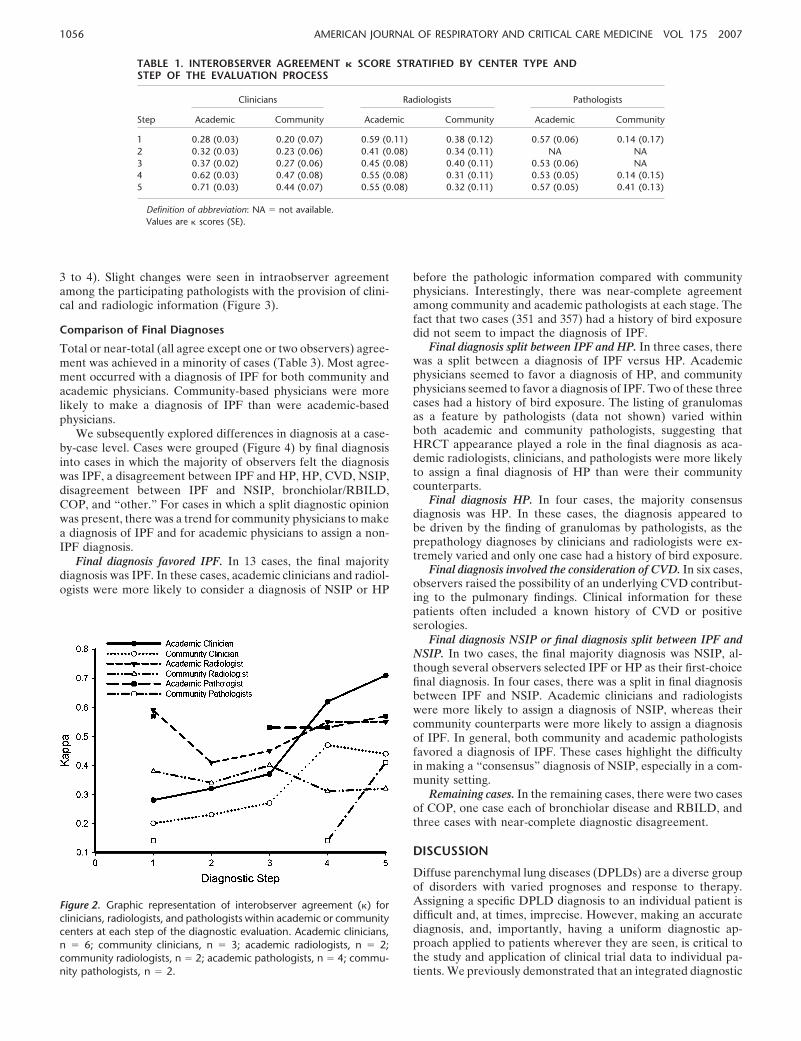
Clinicians. Academic physicians displayed better agreementcompared with community physicians (Table 1; Figure 2). Theacademic clinicians exhibited a very good agreement upon astep 5 final diagnosis (� � 0.71), as compared with the communityclinicians (� � 0.44). The same was true for all previous diagnosissteps. This improved agreement occurred despite being morenumerous than their community counterparts (n � 6 vs. n � 3)and, hence, less likely to reach agreement all else equal. � Scoresimproved for both the community clinicians and academic clini-cians as more information was provided (steps 1–5), although thiswas less impressive among the community participants (Table 1).The fias fias fias fias final diagnosis agreement between academicand community clinicians varied from 0.20 to 0.56 (Table 2)
Radiologists. There was greater interobserver agreementamong the academic radiologists than among the communityradiologists. � Scores failed to improve for both the academicradiologists and community radiologists as more informationwas provided (Table 1; Figure 2). The final diagnosis agreementbetween the academic and the community radiologists was low(range, 0–0.34 [Table 2]).
Pathologists. There was greater interobserver agreementamong the academic pathologists than among the communitypathologists. � Scores for academic pathologists were similar atall stages of evaluation, whereas community pathologists dis-played improvement in agreement after discussing the case withclinicians and radiologists (Table 1; Figure 2). The final diagnosisagreement between the academic and community pathologistswas low (range, 0.12–0.48 [Table 2]).
Intraobserver Agreement
In general, � scores for the clinicians appeared to be somewhatlower between the first and second stages than between thesecond and third stages, suggesting that clinical information al-tered HRCT interpretation more than did interaction with theradiologists (Figure 3). In addition, the � scores appeared lowerbetween the third and fourth stages than between the secondand third stages, confirming the influence of pathologic interpre-tation on changing diagnoses.
Among radiologists, the provision of clinical information atstage 2 appeared to have a greater effect on academic radiolo-gists, as suggested by lower intraobserver agreement among theseparticipants between the first and second stages (Figure 3) com-pared with community radiologists. Interaction with clinicians(between second and third stages) led to few changes. The provi-sion of pathologic information led to the greatest changes (stages
1056 AMERICAN JOURNAL OF RESPIRATORY AND CRITICAL CARE MEDICINE VOL 175 2007
TABLE 1. INTEROBSERVER AGREEMENT � SCORE STRATIFIED BY CENTER TYPE ANDSTEP OF THE EVALUATION PROCESS
Clinicians Radiologists Pathologists
Step Academic Community Academic Community Academic Community
1 0.28 (0.03) 0.20 (0.07) 0.59 (0.11) 0.38 (0.12) 0.57 (0.06) 0.14 (0.17)2 0.32 (0.03) 0.23 (0.06) 0.41 (0.08) 0.34 (0.11) NA NA3 0.37 (0.02) 0.27 (0.06) 0.45 (0.08) 0.40 (0.11) 0.53 (0.06) NA4 0.62 (0.03) 0.47 (0.08) 0.55 (0.08) 0.31 (0.11) 0.53 (0.05) 0.14 (0.15)5 0.71 (0.03) 0.44 (0.07) 0.55 (0.08) 0.32 (0.11) 0.57 (0.05) 0.41 (0.13)
Definition of abbreviation: NA � not available.Values are � scores (SE).
3 to 4). Slight changes were seen in intraobserver agreementamong the participating pathologists with the provision of clini-cal and radiologic information (Figure 3).
Comparison of Final Diagnoses
Total or near-total (all agree except one or two observers) agree-ment was achieved in a minority of cases (Table 3). Most agree-ment occurred with a diagnosis of IPF for both community andacademic physicians. Community-based physicians were morelikely to make a diagnosis of IPF than were academic-basedphysicians.
We subsequently explored differences in diagnosis at a case-by-case level. Cases were grouped (Figure 4) by final diagnosisinto cases in which the majority of observers felt the diagnosiswas IPF, a disagreement between IPF and HP, HP, CVD, NSIP,disagreement between IPF and NSIP, bronchiolar/RBILD,COP, and “other.” For cases in which a split diagnostic opinionwas present, there was a trend for community physicians to makea diagnosis of IPF and for academic physicians to assign a non-IPF diagnosis.
Final diagnosis favored IPF. In 13 cases, the final majoritydiagnosis was IPF. In these cases, academic clinicians and radiol-ogists were more likely to consider a diagnosis of NSIP or HP
Figure 2. Graphic representation of interobserver agreement (�) forclinicians, radiologists, and pathologists within academic or communitycenters at each step of the diagnostic evaluation. Academic clinicians,n � 6; community clinicians, n � 3; academic radiologists, n � 2;community radiologists, n � 2; academic pathologists, n � 4; commu-nity pathologists, n � 2.
before the pathologic information compared with communityphysicians. Interestingly, there was near-complete agreementamong community and academic pathologists at each stage. Thefact that two cases (351 and 357) had a history of bird exposuredid not seem to impact the diagnosis of IPF.
Final diagnosis split between IPF and HP. In three cases, therewas a split between a diagnosis of IPF versus HP. Academicphysicians seemed to favor a diagnosis of HP, and communityphysicians seemed to favor a diagnosis of IPF. Two of these threecases had a history of bird exposure. The listing of granulomasas a feature by pathologists (data not shown) varied withinboth academic and community pathologists, suggesting thatHRCT appearance played a role in the final diagnosis as aca-demic radiologists, clinicians, and pathologists were more likelyto assign a final diagnosis of HP than were their communitycounterparts.
Final diagnosis HP. In four cases, the majority consensusdiagnosis was HP. In these cases, the diagnosis appeared tobe driven by the finding of granulomas by pathologists, as theprepathology diagnoses by clinicians and radiologists were ex-tremely varied and only one case had a history of bird exposure.
Final diagnosis involved the consideration of CVD. In six cases,observers raised the possibility of an underlying CVD contribut-ing to the pulmonary findings. Clinical information for thesepatients often included a known history of CVD or positiveserologies.
Final diagnosis NSIP or final diagnosis split between IPF andNSIP. In two cases, the final majority diagnosis was NSIP, al-though several observers selected IPF or HP as their first-choicefinal diagnosis. In four cases, there was a split in final diagnosisbetween IPF and NSIP. Academic clinicians and radiologistswere more likely to assign a diagnosis of NSIP, whereas theircommunity counterparts were more likely to assign a diagnosisof IPF. In general, both community and academic pathologistsfavored a diagnosis of IPF. These cases highlight the difficultyin making a “consensus” diagnosis of NSIP, especially in a com-munity setting.
Remaining cases. In the remaining cases, there were two casesof COP, one case each of bronchiolar disease and RBILD, andthree cases with near-complete diagnostic disagreement.
DISCUSSION
Diffuse parenchymal lung diseases (DPLDs) are a diverse groupof disorders with varied prognoses and response to therapy.Assigning a specific DPLD diagnosis to an individual patient isdifficult and, at times, imprecise. However, making an accuratediagnosis, and, importantly, having a uniform diagnostic ap-proach applied to patients wherever they are seen, is critical tothe study and application of clinical trial data to individual pa-tients. We previously demonstrated that an integrated diagnostic

Flaherty, Andrei, King, et al.: Diagnosing Idiopathic Interstitial Pneumonia 1057
TABLE 2. INTEROBSERVER AGREEMENT � SCORE FOR THE FINAL DIAGNOSIS BETWEENACADEMIC- AND COMMUNITY-BASED CLINICIANS, RADIOLOGISTS, AND PATHOLOGISTS
Academic 1 Academic 2 Academic 3 Academic 4 Academic 5 Academic 6
CliniciansCommunity 1 0.22 (0.10) 0.28 (0.10) 0.20 (0.10) 0.21 (0.11) 0.35 (0.11) 0.21 (0.10)Community 2 0.40 (0.09) 0.39 (0.09) 0.38 (0.09) 0.40 (0.10) 0.50 (0.10) 0.25 (0,09)Community 3 0.50 (0.09) 0.50 (0.09) 0.46 (0.09) 0.55 (0.09) 0.44 (0.09) 0.56 (0.09)
RadiologistsRadiologists
Community 1 0.23 (0.08) 0.34 (0.09) — — — —Community 2 0.11 (0.09) 0.23 (0.10) — — — —
PathologistsCommunity 1 0.40 (0.12) 0.12 (0.12) 0.26 (0.13) 0.23 (0.12) — —Community 2 0.47 (0.10) 0.45 (0.10) 0.48 (0.11) 0.46 (0.10) — —
Values are � scores (SE).
approach involving expert clinicians, radiologists, and patholo-gists results in an altered diagnosis compared with that of individ-ual physicians working in isolation (17). In the current study,we expand these findings by examining the diagnostic agreementbetween community- and academic-based physicians using a dy-namic interactive process involving pulmonary clinicians, radiol-ogists, and pathologists. We demonstrate that: (1) clinically sig-nificant disagreement exists regarding the diagnosis of IIP among
Figure 3. Graphic representation of intraobserver agreement (�) forclinicians, radiologists, and pathologists within academic or communitycenters between different steps in the diagnostic process. A high-level� indicates little change in diagnosis between steps. For communitypathologists, the value at step 3/4 represents the change in diagnosisfrom their individual histopathologic interpretation compared with thediagnosis after discussing the clinical, radiographic, and histopathologicinformation as a group. For academic pathologists, the value at step2/3 represents the agreement between the individual pathologist’s in-terpretation and the group pathology discussion; step 3/4 representsthe agreement between the group pathology diagnosis before and afterdiscussing the clinical, radiographic, and histopathologic informationas a group. For all participants, step 4/5 represents the agreement indiagnosis from the group discussion (step 4) and final consensus (step5). Academic clinicians, n � 6; community clinicians, n � 3; academicradiologists, n � 2; community radiologists, n � 2; academic patholo-gists, n � 4; community pathologists, n � 2.
academic-based clinicians and between community- andacademic-based physicians, with community physicians morelikely to make a diagnosis of IPF; (2) final diagnostic agreementwas higher between academic physicians compared with commu-nity physicians; (3) most diagnostic agreement occurred for casesof IPF; (4) most diagnostic discord occurred between cases ofIPF versus HP, IPF versus NSIP, and the potential influence ofCVD, with community-based physicians more likely to rendera diagnosis of IPF. These data highlight how an individual patientwith suspected DPLD can have a significantly different diagnosisdepending on the physician and, particularly, the location ofevaluation. Although a combined clinical, radiographic, andpathologic approach improves agreement, significant disagree-ment still exists. These data highlight the need for better waysto approach and classify patients with suspected DPLD.
Recent guidelines suggest that DPLDs, including IIPs, canbe separated based on clinical, radiographic, and histopathologiccriteria (16). The importance of “splitting” versus “lumping”DPLDs stems from the varied etiologies, treatments, and prog-noses associated with different diseases. Academic physiciansused a wider array of diagnoses compared with community-based physicians who used a more consistent diagnosis of IPF.In our series, 13 (33%) cases were believed to represent IPF bya majority of both community- and academic-based physicians.Importantly, community physicians made the diagnosis of IPFin 11 additional cases, where the academic physicians believedHP (n � 3), NSIP (n � 4), or CVD-associated (n � 4) disease
TABLE 3. AGREEMENT IN FINAL DIAGNOSIS
IPF NSIP Bronchiolar HP RBILD Other COP CVD
All* 7 — — 1 — — 1 —All � 1† 5 — — 1 — — — —All � 2‡ 1 — — — — — —Community clinicians 14 2 — 2 — — 2Academic clinicians 12 1 1 4 1 — 1 3Community radiologists 16 1 — 1 — — 1 1Academic radiologists 8 4 1 6 2 1 2 3Community pathologists 19 — — 3 — — 1 2Academic pathologists 13 1 1 4 1 — 1 —
Definition of abbreviations: COP � cryptogenic organizing pneumonia; CVD �
collagen vascular disease; HP � hypersensitivity pneumonitis; IPF � idiopathicpulmonary fibrosis; NSIP � nonspecific interstitial pneumonia; RBILD � respiratorybronchiolitis interstitial lung disease.
* All observers were in agreement.† All � 1 � all observers except 1 observer that disagreed.‡ All – 2 � all observers except 2 observers that disagreed.Values are number of cases.

1058 AMERICAN JOURNAL OF RESPIRATORY AND CRITICAL CARE MEDICINE VOL 175 2007
Figure 4. Color and character represen-tation of diagnosis for each case (Pt_code)by observer. Columns headed with a “2”represent the diagnosis before pathologicinformation (for clinicians and radiolo-gists) or clinical/HRCT information (forpathologists). Columns headed with a “5”represent the final diagnosis after a clini-cal/radiographic/pathologic discussion.Each cell is letter/color coded (I/red � id-iopathic pulmonary fibrosis [IPF]; N/lightblue � nonspecific interstitial pneumonia[NSIP]; B/dark green � airway/bronchiolardisease; H/yellow � hypersensitivity pneu-monia [HP]; R/light green � respiratorybronchiolitis interstitial lung disease[RBILD]; O/orange � other; C/pink �
cryptogenic organizing pneumonia [COP];S/dark blue � systemic collagen vasculardisease–associated interstitial lung dis-ease). For example, community clinician1 (CC1) initially diagnosed case 365 asNSIP, but changed to a diagnosis of IPFafter a clinical/radiographic/pathologicdiscussion. IIP � idiopathic interstitialpneumonia.
was present. The importance of this finding is highlighted bythe different prognoses among these diagnoses, and the noveltherapeutic approaches currently under study based on biologi-cal plausibility in IPF. The relative minority of IPF cases com-pared with other IIPs in our series may reflect the requirementfor all cases to have both an HRCT and SLB; cases of IPF basedsolely on definite HRCT criteria were excluded.
The current data document that the greatest disagreementin diagnosis occurred between academic and community physi-cians. However, significant disagreement was present evenwithin academic centers. The better agreement for academicphysicians likely reflects, at least in part, that these physicianswith an interest in DPLD have collaborated on previous projects,including the generation of consensus statements. This suggeststhat more intense interaction between academic and communityphysicians could improve the diagnostic agreement betweencommunity and academic physicians, and should help standard-
ize the approach to the treatment and study of patients withDPLD. In addition, the proportion of time devoted to clinicalmanagement of DPLD is likely important, as the communityclinician who devoted the greatest time to the management ofthese disorders exhibited greater agreement with his academiccounterparts.
We previously demonstrated that a dynamic, iterative ap-proach to IIP diagnosis improves interobserver agreementamong expert clinicians, particularly in patients without IPF(17). The current data highlight that a similar iterative approachimproves diagnostic agreement within community clinicians.Two differences between community and academic physicianswere evident. Community clinicians’ agreement improved to alesser degree than that of academic clinicians, and the final diag-nosis by community pathologists was more influenced by theinteraction with clinicians/radiologists than in the academic set-ting. This suggests that the final diagnosis in an academic setting
Flaherty, Andrei, King, et al.: Diagnosing Idiopathic Interstitial Pneumonia 1059
is driven by pathology compared with the clinician/radiologistin the community setting. This latter observation could reflectthe relative expertise, and thus assertiveness, of community clini-cians/radiologists compared with community pathologists in thediagnosis of IIP. Pathology information appeared to influencethe diagnosis of some cases in both community and academiccenters, as the diagnosis of HP versus IPF seemed to correlatemore with the presence/absence of granulomas compared witha clinical history of bird exposure.
Academic clinicians and radiologists used a wider array ofdiagnoses before receiving pathology information. The findingsof subpleural, lower-lobe, honeycomb, and reticular change with-out micronodules, peribronchiolar nodules, consolidation, iso-lated cysts, or a predominance of ground glass opacity have ahigh positive predictive value for finding the histopathologicpattern of usual interstitial pneumonia (UIP) on SLB (10, 11,22). A recent, survey-based study suggested that 67% of clini-cians would accept an HRCT diagnosis of IPF, particularly ifthe observer had a higher self-rating of proficiency in readingHRCT (23). It is possible that the academic clinicians and radiol-ogists in our study were more stringent in their application ofthese findings, and thus less likely to make a diagnosis of IPFwithout a biopsy.
Our prospectively collected data suggest that pathologists willconsider clinical and radiologic data in rendering a final diagnosisand emphasizes the need for pathologists to consider these datain rendering a final diagnosis. This is supported by the decreasein intraobserver agreement between stages with and withoutclinical and radiologic information. This evaluative process wasseen among both academic and community-based pathologists.Review of the individual patient data suggests that an exposurehistory consistent with HP, or a history suggestive of a CVC,was particularly likely to alter diagnosis, including away from adiagnosis of IPF. Given the difference in survival characteristicbetween IPF and connective tissue–associated UIP and chronicHP (21), this point may have important clinical ramifications.
A limitation of this study is the lack of transbronchial biopsyand/or bronchoalveolar lavage data for the majority of patients.This absence reflects the practice pattern at the University ofMichigan and surrounding communities, where bronchoscopy isused infrequently when a diagnosis of IIP (especially UIP orNSIP) is considered. It is possible that rigorous collection ofbronchoscopy data could impact the final diagnostic impression.Additional research is required to clarify the role of bronchos-copy, relative HRCT, and SLB in the diagnostic algorithm forpatients with suspected IIP. Another limitation of this study isthe involvement of academic physicians who devote the majorityof their time to the study of interstitial lung disorders and, there-fore, might not be representative of the whole “academic” physi-cian group. This study was also mostly based in the United Statesand Europe, and might not fully represent the situation in othercountries.
Our data expand on previous literature on interobserveragreement between clinicians, radiologists, and pathologists indiagnosing IIPs. We confirm that an interactive approach be-tween clinicians, radiologists, and pathologists improves interob-server agreement. On the other hand, even with this approach,significant disagreement exists within, and particularly between,community and academic centers. The fact that community phy-sicians were more likely to render a diagnosis of IPF has impor-tant implications, as individual patients with HP, NSIP, or CVD-associated ILD are more likely to respond to immunosuppressivetreatment, whereas patients with IPF should be referred, when-ever possible, for participation in therapeutic trials. Future ef-forts are needed to bridge the gap of apparent discordance be-tween community and academic experts in their diagnostic
proficiency. It is hoped that this will be accomplished with contin-ued education, workshops, and increased interactions betweenacademic and community-based physicians. In the short-term,these data suggest that, whenever possible, patients should bereferred to centers with expertise in diffuse parenchymal lungdisorders to help clarify the diagnosis and provide suggestionsregarding treatment options.
Conflict of Interest Statement : K.R.F. does not have a financial relationship witha commercial entity that has an interest in the subject of this manuscript. A.-C.A.does not have a financial relationship with a commercial entity that has an interestin the subject of this manuscript. T.E.K. does not have a financial relationshipwith a commercial entity that has an interest in the subject of this manuscript.G.R. does not have a financial relationship with a commercial entity that has aninterest in the subject of this manuscript. T.V.C. does not have a financial relation-ship with a commercial entity that has an interest in the subject of this manuscript.A.W. does not have a financial relationship with a commercial entity that has aninterest in the subject of this manuscript. N.B. does not have a financial relationshipwith a commercial entity that has an interest in the subject of this manuscript.K.B. does not have a financial relationship with a commercial entity that has aninterest in the subject of this manuscript. R.d.B. does not have a financial relation-ship with a commercial entity that has an interest in the subject of this manuscript.A.F. does not have a financial relationship with a commercial entity that has aninterest in the subject of this manuscript. S.E.G. does not have a financial relation-ship with a commercial entity that has an interest in the subject of this manuscript.B.H.G. does not have a financial relationship with a commercial entity that hasan interest in the subject of this manuscript. E.A.K. does not have a financialrelationship with a commercial entity that has an interest in the subject of thismanuscript. R.K. does not have a financial relationship with a commercial entitythat has an interest in the subject of this manuscript. E.L. does not have a financialrelationship with a commercial entity that has an interest in the subject of thismanuscript. D.L. received less than $5,000 in 2004, 2005, and 2006 from In-termune, Inc., for interpretation of computed tomography scans, and has alsoreceived less than $5,000 from Encysive, Inc., for consultation regarding clinicaltrials; D.L. received $6,000 in 2006 for service on an advisory board for Actelion,Inc. A.G.N. received $2,500 for reviewing slides for a multicenter trial in 2005for Intermune Ltd., and £9,500 for reviewing slides for a multicenter trial forActelion Ltd. in 2006. J.Q. does not have a financial relationship with a commercialentity that has an interest in the subject of this manuscript. V.J.T. does not havea financial relationship with a commercial entity that has an interest in the subjectof this manuscript. W.D.T. does not have a financial relationship with a commercialentity that has an interest in the subject of this manuscript. J.V. does not have afinancial relationship with a commercial entity that has an interest in the subjectof this manuscript. F.A.W. does not have a financial relationship with a commercialentity that has an interest in the subject of this manuscript. J.W. does not havea financial relationship with a commercial entity that has an interest in the subjectof this manuscript. G.B.T. does not have a financial relationship with a commercialentity that has an interest in the subject of this manuscript. S.M. does not havea financial relationship with a commercial entity that has an interest in the subjectof this manuscript. F.J.M. does not have a financial relationship with a commercialentity that has an interest in the subject of this manuscript.
The University of Michigan Fibrotic Lung Disease Network includes the followingparticipants: University of Michigan, Division of Pulmonary and Critical Care, AnnArbor, MI—D. Arenberg, W. Bria, D. Dahlgren, C. Grum, R. Hyzy, V. Lama, T. Ojo,M. Peters-Golden, R. Simon, T. Sisson, T. Standiford, V. Thannickal, E. White;Internal Medicine Clinic, Alpena, MI—P. Bachwich, C. Easton, J. Mazur; The LungCenter, Battle Creek, MI—S. Chaparala, G. Harrington, N. Potempa; Bay City,MI—S. Manawar, J. Summer; Clawson, MI—P. Hukku, J. Sung; Clinton Township,MI—R. Babcock; Pulmonary and Critical Care Medicine Consultants, Commerce, MI—J.Belen, M. Dunn, D. Maxwell, R. Reagle, R. Sherman, S. Simecek; Oakwood Hospital,Dearborn, MI—L. Victor; Henry Ford Hospital, Detroit, MI—B. DiGiovine, M. Eiche-nhorn, J. Popovich, Jr., D. Spizarny; Botsford General Hospital, Farmington Hills,MI—B. Rabinowitz; Pulmonary and Critical Care Specialists, Farmington Hills, MI—G.Ferguson, P. Kaplan, S. Sklar, W. VanderRoest; Pulmonary Associates, P.C., Flint,MI—O. Filos, V. Rao, M.V. Thomas, J. Varghese, J. Vyskocil, F. Wadenstorer; GrandValley Internal Medicine, Grand Rapids, MI—J. Cantor, W. Katz, R. Johnson, Jr.,D. Listello, J. Wilt; Michigan Medical Professional Company, Grand Rapids,MI—C. Acharya, W. Couwenhoven, T. Daum, M. Harrison, M. Koets, G. Sandman,G. VanOtteren; Michigan Medical, P.C., Holland, MI—S. Kraker; Huntington Woods,MI—M. Greenberger, A. O’Neill, D. Wu; Pulmonary Clinics of Southern Michigan,Jackson, MI—R.C. Albertson, III, J. Chauncey, T. Murray, G. Patten; AssociatedPulmonary and Critical Care Specialists, P.C., Kalamazoo, MI—T. Abraham, J. Dirks,B. Dykstra, G. Grambau, J. Schoell; Pulmonary and Critical Care Associates, P.C.,Kalamazoo, MI—R. Brush, S. Jefferson, J. Miller, S. Schuldheisz, M. Warlick; Pulmo-nary and Critical Care Consultants, Lansing, MI—J. Armstrong, A. Atkinson,T. Kantra, L. Rawsthorne, D. Young; Pulmonary Services, Lansing, MI—A. Abbasi,C.M. Gera, G. Kashyap, J. Morlock; Respiratory Medicine, Marquette, MI—S. Danek,A. Saari; Midland, MI—S. Yadam; Central Michigan Healthcare System, Mt. Pleasant,MI—E. Obeid; Muskegon Pulmonary Associates, Muskegon, MI—D. Hoch,A. Kleaveland; Owosso Medical Group, Owosso, MI—A. Allam, M.A. Gad, Jr.; LungAssociates, Pontiac, MI—A. Desai U. Dhanjal, A. Sethi; St. Joseph’s Hospital, Pontiac,MI—F. Ahmad, L. Kaiser, L. Rosenthal, D. Sak; Physician HealthCare Network, PortHuron, MI—R. Ailani, M. Basha, A. Hadar, S. Holstine; Pulmonary, Critical Care,

1060 AMERICAN JOURNAL OF RESPIRATORY AND CRITICAL CARE MEDICINE VOL 175 2007
and Sleep, P.C., Rochester Hills, MI—M.W. Al-Ameri, R. Go, M. Kashlan; Rochester,MI—K. Aggarwal; Roseville, MI—W. Hanna, R. Marchese; William Beaumont Hospi-tal, Royal Oak, MI—R. Begle, D. Erb, K.P. Ravikrishnan, J. Seidman, S. Sherman;Saginaw, MI—R. Agarwal, F. Ansari, T. Damuth, C. Indira; Spring Lake, MI—M.Ivey; Lakeside Healthcare Specialists, St. Joseph, MI—S. Deskins, A. Palmer, S. Shastri;Pulmonary and Critical Care Associates, St. Clair Shores, MI and Troy, MI—R. DiLisio,S. Galens, K. Grady, D. Harrington, R. Herbert, C. Hughes, J. Lee, A. Starrico,K. Stevens, M. Trunsky, W. Ventimiglia; Taylor, MI—D. Mahajan; Pulmonary Medi-cine Associates, Warren, MI—H. Kaplan, L. Tankanow; Henry Ford Wyandotte Hospi-tal, Wyandotte, MI—M. Pensler; Toledo Pulmonary and Sleep Specialists, Toledo,OH—F.O. Horton, III, A. Nathanson, R. Wainz.
References1. Flaherty KR, Toews GB, Travis WD, Colby TV, Kazerooni EA, Gross
BH, Jain A, Strawderman RL III, Paine R III, Flint A, et al. Clinicalsignificance of histological classification of idiopathic interstitial pneu-monia. Eur Respir J 2002;19:275–283.
2. Flaherty KR, Travis WD, Colby TV, Toews GB, Kazerooni EA, GrossBH, Jain A, Strawderman RL III, Flint A, Lynch JP III, et al. Histologicvariability in usual and nonspecific interstitial pneumonias. Am JRespir Crit Care Med 2001;164:1722–1727.
3. Katzenstein ALA, Fiorelli RF. Nonspecific interstitial pneumonia/fibrosis:histologic features and clinical significance. Am J Surg Pathol 1994;18:136–147.
4. Nagai S, Kitaichi M, Itoh H, Nishimura K, Izumi T, Colby TV. Idiopathicnonspecific interstitial pneumonia/fibrosis: comparison with idiopathicpulmonary fibrosis and BOOP. Eur Respir J 1998;12:1010–1019.
5. Nicholson AG, Colby TV, DuBois RM, Hansell DM, Wells AU. Theprognostic significance of the histologic pattern of interstitial pneumo-nia in patients presenting with the clinical entity of cryptogenic fibros-ing alveolitis. Am J Respir Crit Care Med 2000;162:2213–2217.
6. Travis WD, Matsui K, Moss J, Ferrans VJ. Idiopathic nonspecific intersti-tial pneumonia: prognostic significance of cellular and fibrosing pat-terns. Am J Surg Pathol 2000;24:19–33.
7. Bjoraker JA, Ryu JH, Edwin MK, Myers JL, Tazelaar HD, SchorederDR, Offord KP. Prognostic significance of histopathologic subsets inidiopathic pulmonary fibrosis. Am J Respir Crit Care Med 1998;157:199–203.
8. Lama VN, Flaherty KR, Toews GB, Colby TV, Travis WD, Long Q,Murray S, Kazerooni EA, Gross BH, Lynch JP III, et al. Prognosticvalue of desaturation during a 6-minute walk test in idiopathic intersti-tial pneumonia. Am J Respir Crit Care Med 2003;168:1084–1090.
9. Jegal Y, Kim DS, Shim TS, Lim CM, Do Lee S, Koh Y, Kim WS, KimWD, Lee JS, Travis WD, et al. Physiology is a stronger predictor ofsurvival than pathology in fibrotic interstitial pneumonia. Am J RespirCrit Care Med 2005;171:639–644.
10. Flaherty KR, Mumford JA, Murray S, Kazerooni EA, Gross BH, ColbyTV, Travis WD, Flint A, Toews GB, Lynch JP, et al. Prognostic implica-
tions of physiologic and radiographic changes in idiopathic interstitialpneumonia. Am J Respir Crit Care Med 2003;168:543–548.
11. Hunninghake GW, Lynch DA, Galvin JR, Muller N, Schwartz D, KingTE Jr, Lynch JP III, Hegele R, Waldron JA Jr, Colby TV, et al.Radiologic findings are strongly associated with a pathologic diagnosisof usual interstitial pneumonia. Chest 2003;124:1215–1223.
12. Hunninghake GW, Zimmerman MB, Schwartz DA, King TE Jr, LynchJ, Hegele R, Waldron J, Colby T, Muller N, Lynch D, et al. Utility ofa lung biopsy for the diagnosis of idiopathic pulmonary fibrosis. AmJ Respir Crit Care Med 2001;164:193–196.
13. Raghu G. Interstitial lung disease—a diagnostic approach: are CT scanand lung biopsy indicated for every patient? Am J Respir Crit CareMed 1995;151:909–914.
14. Raghu G, Mageto YN, Lockhart D, Schmidt RA, Wood DE, GodwinJD. The accuracy of the clinical diagnosis of new-onset idiopathicpulmonary fibrosis and other interstitial lung disease: a prospectivestudy. Chest 1999;116:1168–1174.
15. Katzenstein ALA, Myers JL. Idiopathic pulmonary fibrosis: clinical rele-vance of pathologic classification. Am J Respir Crit Care Med 1998;157:1301–1315.
16. American Thoracic Society, European Respiratory Society. AmericanThoracic Society/European Respiratory Society international multidis-ciplinary consensus classification of the idiopathic interstitial pneumo-nias. Am J Respir Crit Care Med 2002;165:277–304.
17. Flaherty KR, King TE Jr, Raghu G, Lynch JP III, Colby TV, TravisWD, Gross BH, Kazerooni EA, Toews GB, Long Q, et al. Idiopathicinterstitial pneumonia: what is the effect of a multidisciplinary ap-proach to diagnosis? Am J Respir Crit Care Med 2004;170:904–910.
18. Landis JR, Koch GG. The measurement of observer agreement for cate-gorical data. Biometrics 1977;33:159–174.
19. Thompson JR. Estimating equations for � statistics. Stat Med 2001;20:2895–2906.
20. Gwet K. Computing inter-rater reliability using the SAS system. StatisticalMethods for Inter-Rater reliability Assessment, No. 3. Gaithersburg, MD:STATAXIS Consulting; 2002. Available from: http://www.stataxis.com/files/articles/inter_rater_reliability_with_sas.pdf (accessed December 13,2006).
21. Thomeer MJ, Vansteenkiste J, Verbeken EK, Demedts M. Interstitiallung diseases: characteristics at diagnosis and mortality risk assess-ment. Respir Med 2004;98:567–573.
22. Lynch DA, David Godwin J, Safrin S, Starko KM, Hormel P, BrownKK, Raghu G, King TE Jr, Bradford WZ, Schwartz DA, et al. High-resolution computed tomography in idiopathic pulmonary fibrosis:diagnosis and prognosis. Am J Respir Crit Care Med 2005;172:488–493.
23. Diette GB, Scatarige JC, Haponik EF, Merriman B, Fishman EK. Dohigh-resolution CT findings of usual interstitial pneumonitis obviatelung biopsy? Views of pulmonologists. Respiration (Herrlisheim)2005;72:134–141.