Embed Size (px)
Citation preview

Idiopathic Scoliosis
John F. Lovejoy III, M.D.Chair, Department of Orthopaedics and Sports Medicine
Nemours Children’s HospitalAssociate Professor
University of Central Florida

Disclosures
• None relevant to this talk
• Recognize different types of spinal deformity in children• Understand common presentation & physical exam findings in children
with spinal deformity• Understand radiographic assessment of Scoliosis• Recognize “atypical” presentations• Understand the concepts behind treatment of childhood spinal deformity• Understand the importance of early recognition of post-operative
complications
Learning Objectives
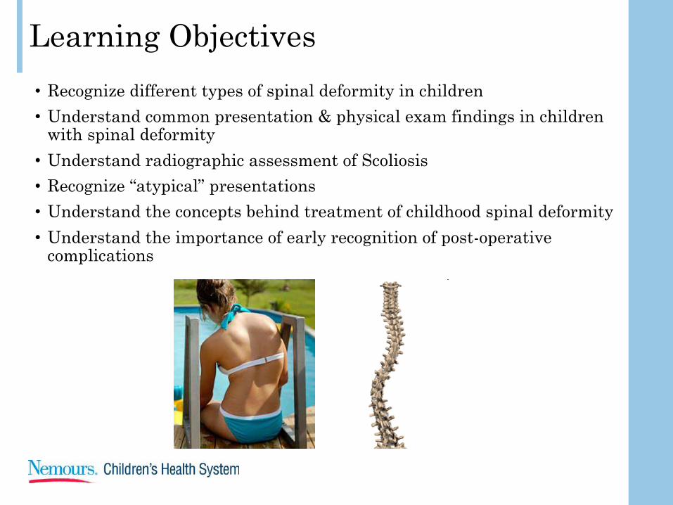
Pediatric Scoliosis
Definition:• Structural spinal deformity
characterized by decompensationof the normal vertebral alignment during rapid skeletal growth• Deformity is 3 dimensional• Coronal, sagittal, as well as
abnormal vertebral rotation
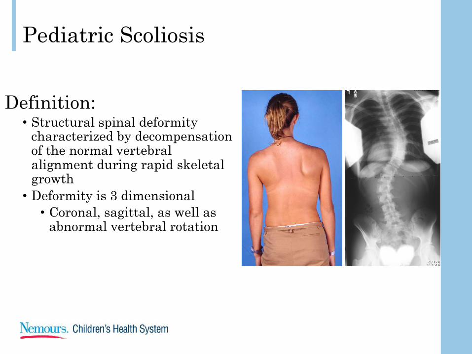
Types of Scoliosis• Congenital - due to a congenital abnormality of the vertebrae or fused
ribs• Neuromuscular - caused by problems such as poor muscle control or
muscle weakness, or paralysis due to diseases • Cerebral Palsy• Myelomeningocele• Muscular Dystrophy
• Idiopathic scoliosis - is scoliosis of unknown cause. Genetic in origin.

Idiopathic Scoliosis
•By far the most common type >90%•No definite cause•Believed to be related to
asymmetric growth of vertebral bodies• Types• Early-Onset• Infantile Scoliosis• Juvenile Scoliosis
• Late-Onset• Adolescent Scoliosis
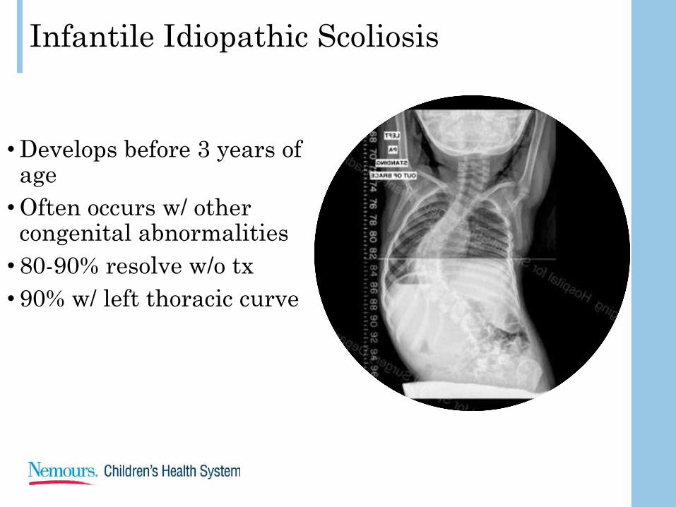
Infantile Idiopathic Scoliosis
•Develops before 3 years of age•Often occurs w/ other congenital abnormalities• 80-90% resolve w/o tx• 90% w/ left thoracic curve
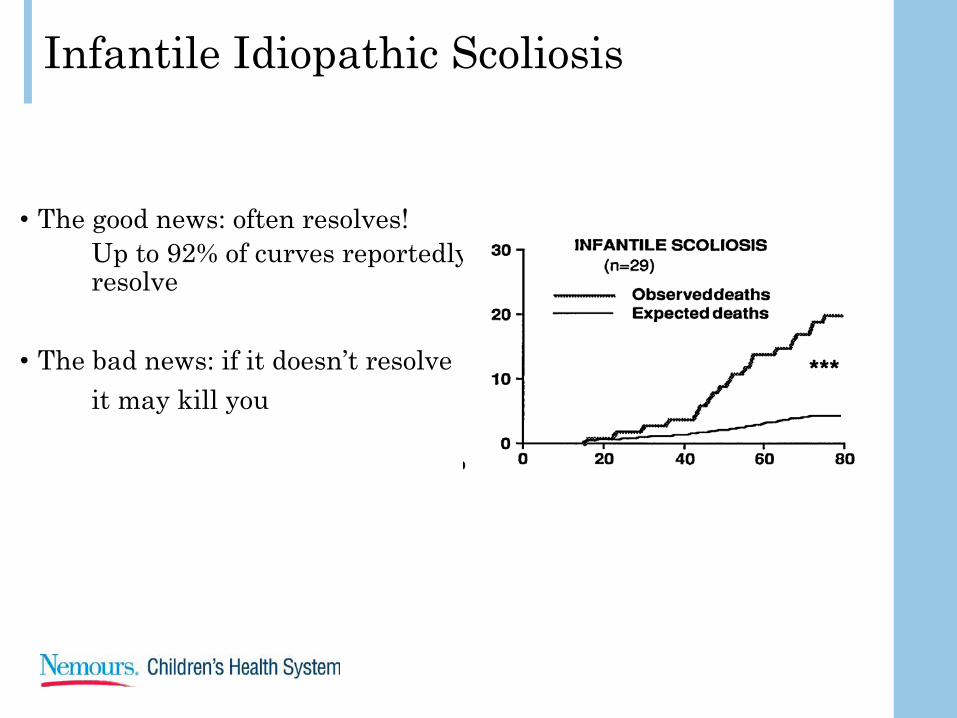
Infantile Idiopathic Scoliosis
• The good news: often resolves!Up to 92% of curves reportedly resolve
• The bad news: if it doesn’t resolveit may kill you

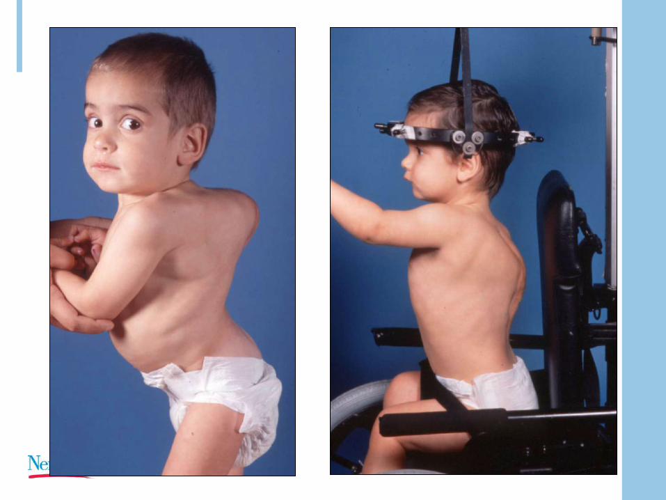
Bracing in EOS
•Advantages – removable, lighter, widely accessible•Disadvantages – removable, rib wall deformity•Contraindications (relative)• children with decreased pulmonary function
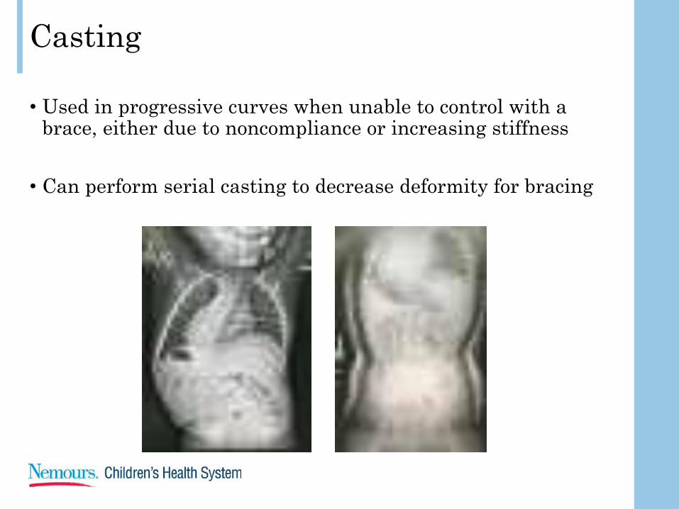
Casting
• Used in progressive curves when unable to control with a brace, either due to noncompliance or increasing stiffness
• Can perform serial casting to decrease deformity for bracing

Casting - Brief History
Risser- Introduced casting in early 1900s for AIS- Principles elongation and derotationed with IIS/EOS
Morel and Cotrel (1964)- Built on Risser’s principles but added FLEXION
Mehta (1975-2000)- Modified technique of Morel and Cotrel- Emphasized early intervention- Demonstrated ability to cure EOS especially before age 2
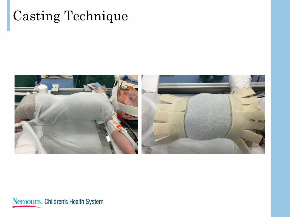
Casting Technique
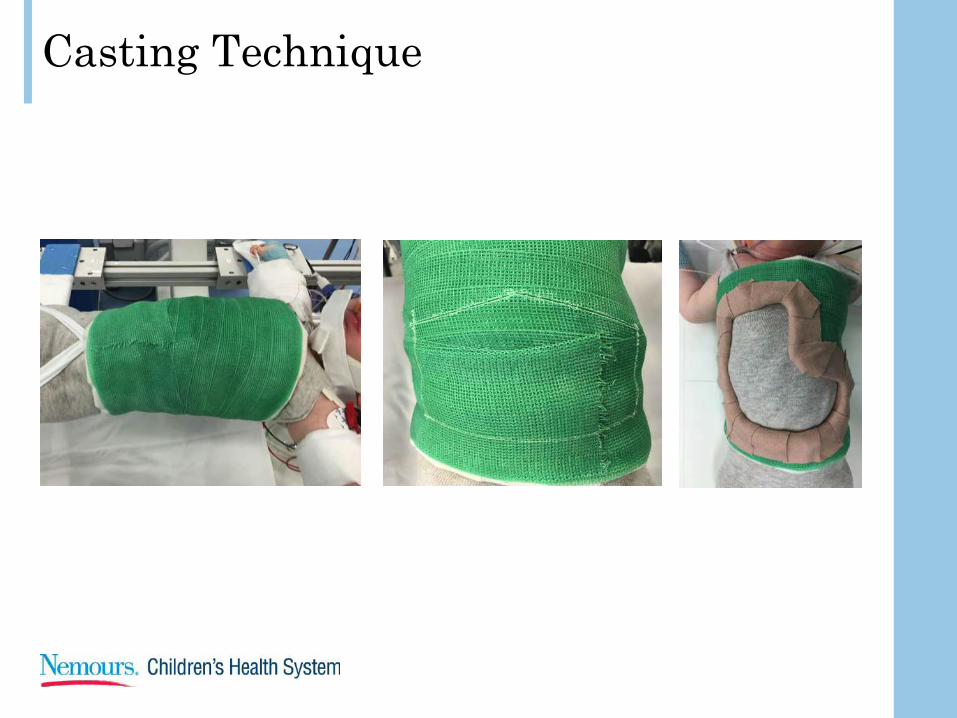
Casting Technique

Casting as definitive treatmentAge 12 months

Cast to buy time
2+6 yo boy 110 degrees
10 months later
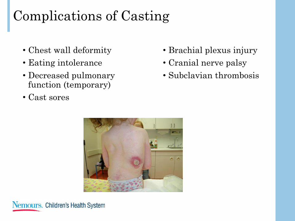
Complications of Casting
• Brachial plexus injury• Cranial nerve palsy• Subclavian thrombosis
• Chest wall deformity• Eating intolerance• Decreased pulmonary
function (temporary)• Cast sores

What about when casting fails? Traction
• Indicated in stiff curves with inability to brace or cast due to larger curves or proximal thoracic curves
• Goal is to increase traction as tolerated until:• Tiptoes if standing• Barely on buttocks if in wheelchair
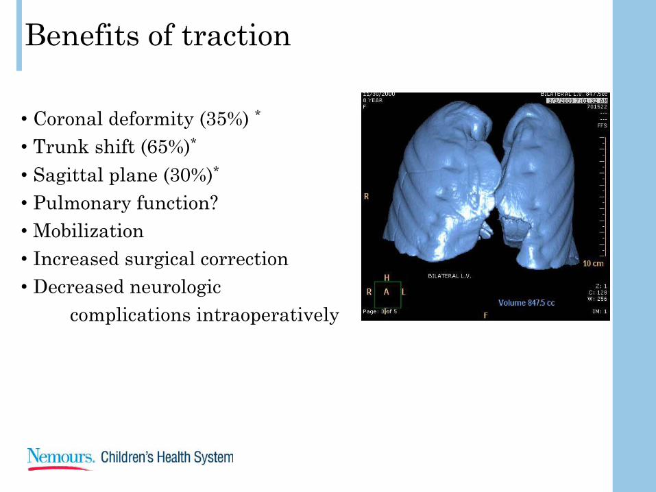
Benefits of traction
• Coronal deformity (35%) *• Trunk shift (65%)*
• Sagittal plane (30%)*
• Pulmonary function?• Mobilization• Increased surgical correction• Decreased neurologic
complications intraoperatively
*Sink 2001


Multiple Surgical Options
• Stay tuned – we are still figuring this out
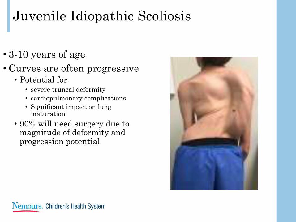
Juvenile Idiopathic Scoliosis
• 3-10 years of age•Curves are often progressive• Potential for
• severe truncal deformity• cardiopulmonary complications• Significant impact on lung
maturation• 90% will need surgery due to
magnitude of deformity and progression potential

Adolescent Idiopathic Scoliosis
• 11-18 years of age•Approximately 90% of childhood
spinal deformity• Prevalence 1-3%• Female-male ratio
• 6:1 non-operative• 7:1 operative
• > 90% w/ right thoracic curve•Majority asymptomatic• Back pain is common
• Patient/Family perception of body asymmetry
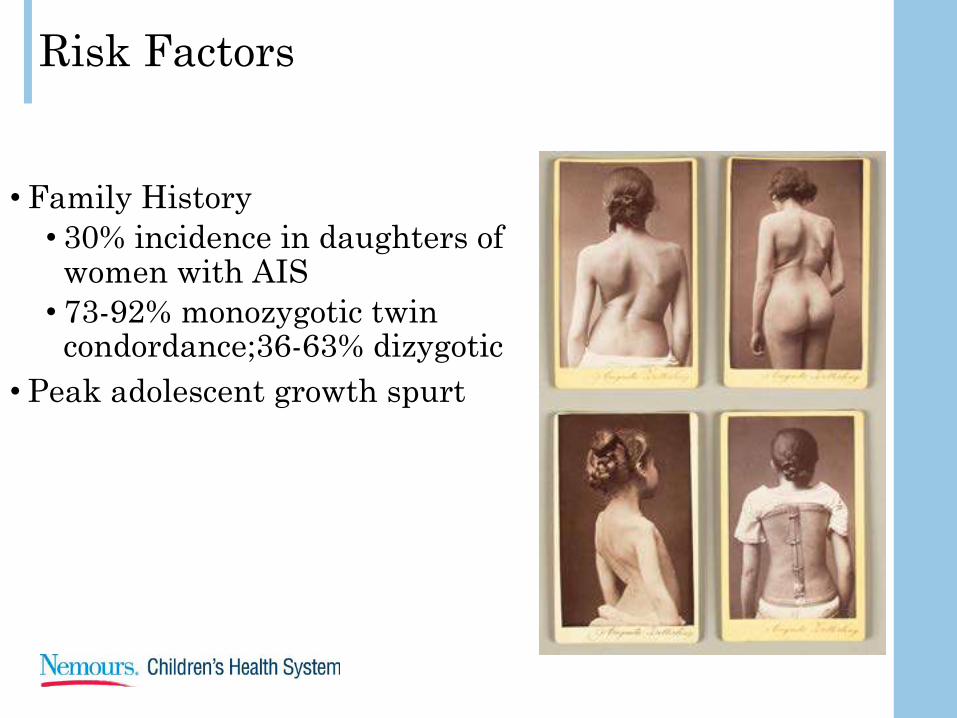
Risk Factors
• Family History• 30% incidence in daughters of
women with AIS• 73-92% monozygotic twin
condordance;36-63% dizygotic• Peak adolescent growth spurt
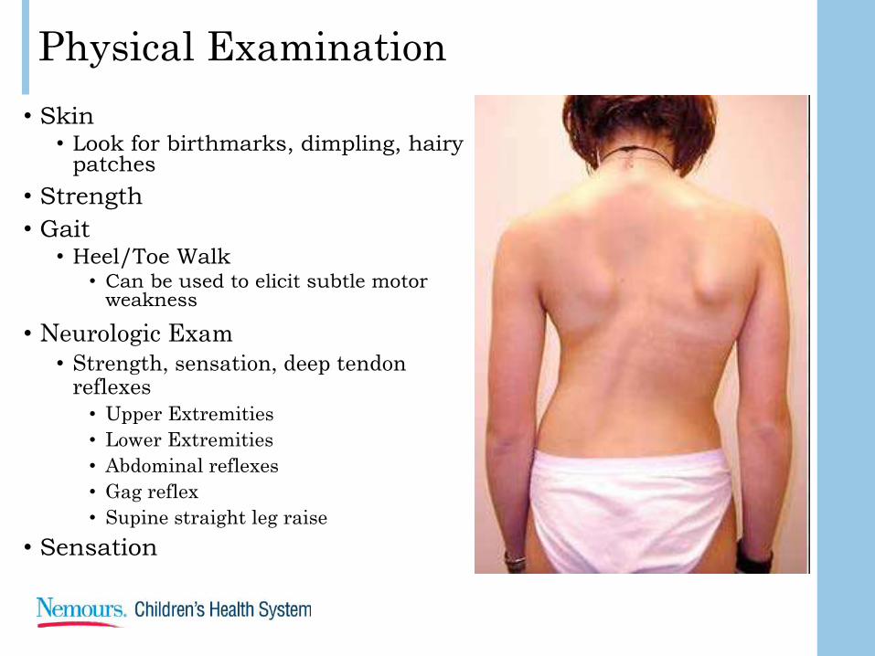
Physical Examination• Skin• Look for birthmarks, dimpling, hairy
patches• Strength• Gait• Heel/Toe Walk
• Can be used to elicit subtle motor weakness
• Neurologic Exam• Strength, sensation, deep tendon
reflexes• Upper Extremities• Lower Extremities• Abdominal reflexes• Gag reflex• Supine straight leg raise
• Sensation
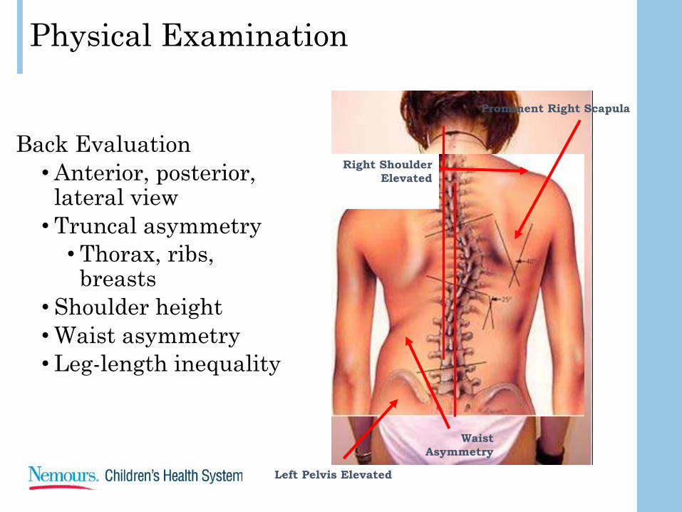
Physical Examination
Back Evaluation•Anterior, posterior,
lateral view• Truncal asymmetry• Thorax, ribs,
breasts• Shoulder height•Waist asymmetry• Leg-length inequality
Waist Asymmetry
Left Pelvis Elevated
Right Shoulder Elevated
Prominent Right Scapula
Physical Examination
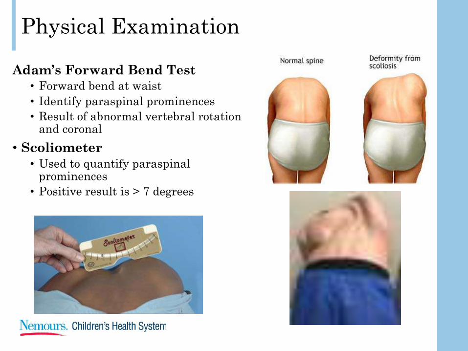
Adam’s Forward Bend Test• Forward bend at waist• Identify paraspinal prominences• Result of abnormal vertebral rotation
and coronal• Scoliometer• Used to quantify paraspinal
prominences• Positive result is > 7 degrees

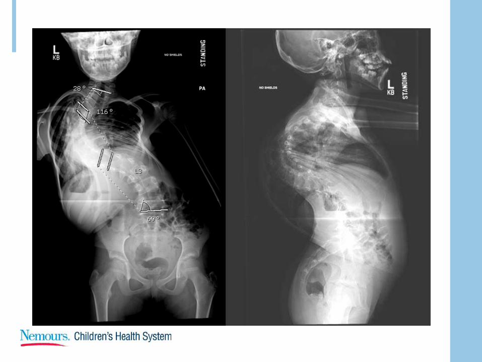
Radiographs
• PA and Lat of the entire spine• EOS

Radiographs
• Risser Sign:• Iliac apophysis develops from lateral to medial on AP view of
pelvis• Risk of progression in Risser 1 or less as high as 70%• Risser of 3 has risk of progression ~ 10%
• Triradiate Cartilage• Closure coincides w/ the end of peak adolescent growth spurt
Radiographic Examination
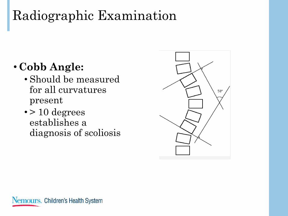
•Cobb Angle:• Should be measured
for all curvatures present• > 10 degrees
establishes a diagnosis of scoliosis

Radiographic Examination
•Kyphosis:•Assessment of
sagittal thoracic contour• Patients w/ AIS are
usually hypo-kyphotic, but convex rib prominence may give appearance of increased kyphosis
Radiographic Examination
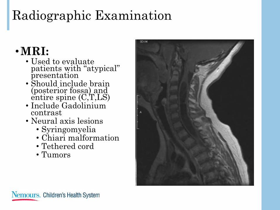
•MRI:• Used to evaluate
patients with “atypical” presentation• Should include brain
(posterior fossa) and entire spine (C,T,LS)• Include Gadolinium
contrast• Neural axis lesions• Syringomyelia• Chiari malformation• Tethered cord• Tumors

Atypical Presentation
• Signs/symptoms that may suggest non-idiopathic deformity•Rapid progression• Large curve at dx• Left sided T curve• Pain that limits
activity•ANY neurologic
symptom/finding•Early onset
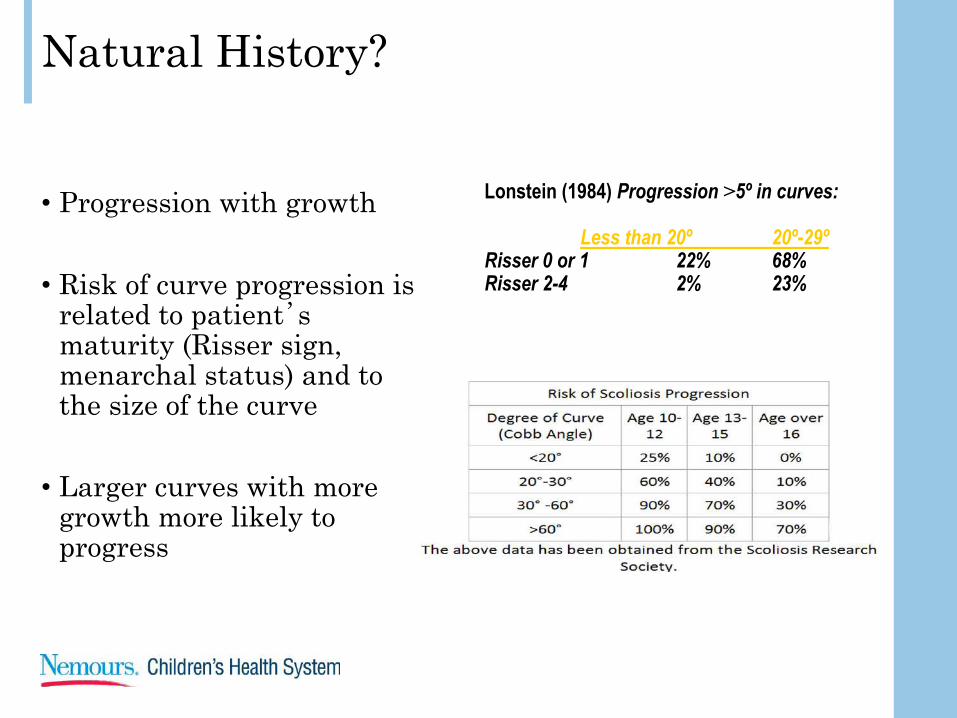
Natural History?
• Progression with growth
• Risk of curve progression is related to patient’s maturity (Risser sign, menarchal status) and to the size of the curve
• Larger curves with more growth more likely to progress
Lonstein (1984) Progression >5º in curves:
Less than 20º 20º-29ºRisser 0 or 1 22% 68%Risser 2-4 2% 23%
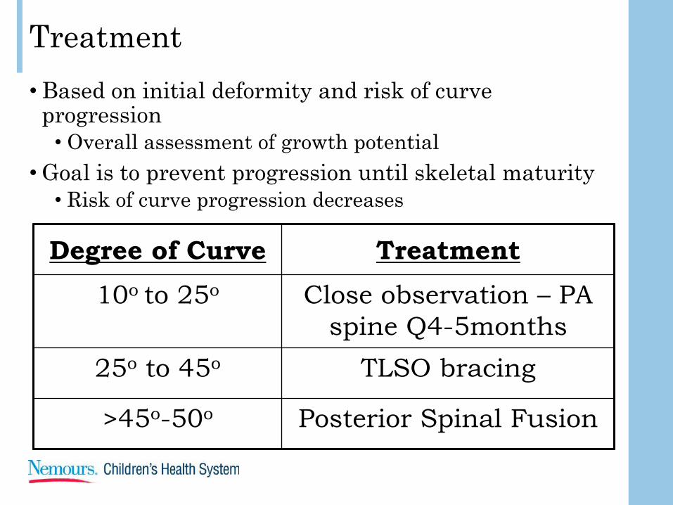
Treatment•Based on initial deformity and risk of curve
progression• Overall assessment of growth potential
•Goal is to prevent progression until skeletal maturity• Risk of curve progression decreases
Degree of Curve Treatment10o to 25o Close observation – PA
spine Q4-5months25o to 45o TLSO bracing
>45o-50o Posterior Spinal Fusion

Therapy
• Observational Monitoring• Follow-up standing PA
and lateral scoliosis x-rays at 4-12 month intervals depending on growth potential
• Core strengthening & conditioning exercises• Recommended for all
patients, especially those with pain• Physical Therapy
techniques• Yoga/Pilates

Bracing• Braist study (NEJM 2013
Weinstein et al.)• Study stopped short because of
success• Brace wear positively associated
with decreasing rate of progression to surgical grade curve• More brace wear associated with
greater success with greatest success seen in those wearing the brace >12.9 hours daily
• Cobb angle between 25 & 45 degrees who are at increased risk of progression• Premenarchal• Risser < 2
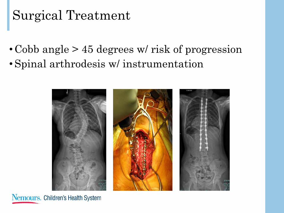
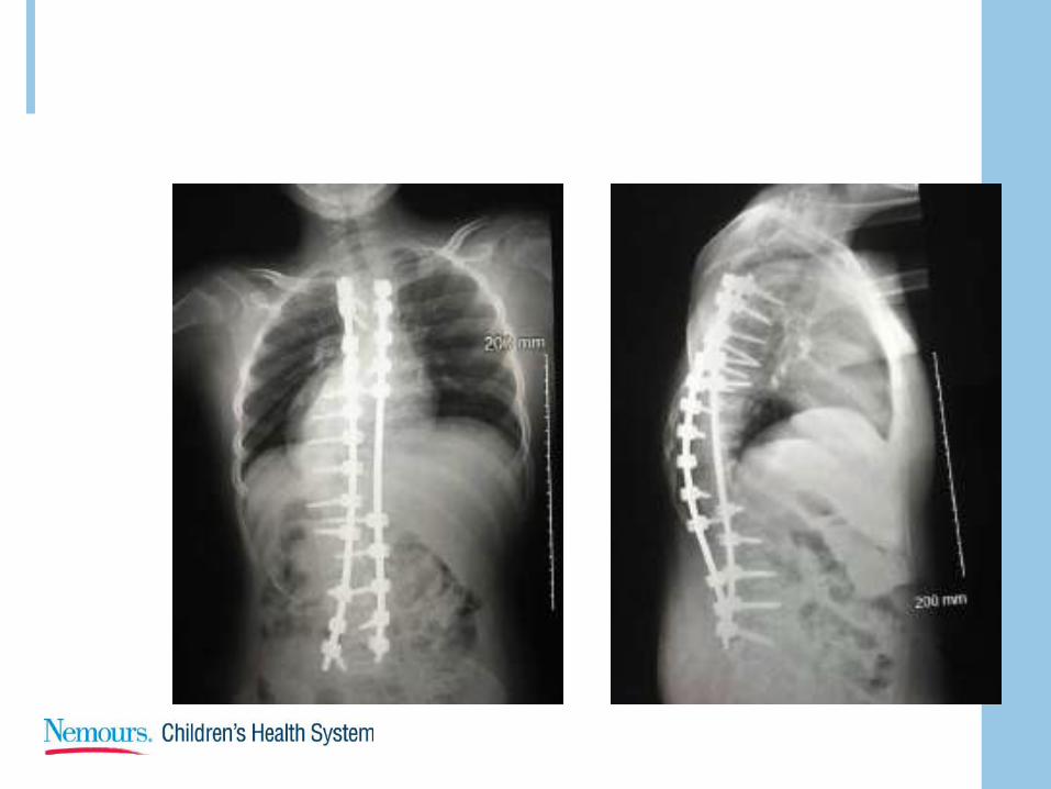
Surgical Treatment
•Cobb angle > 45 degrees w/ risk of progression•Spinal arthrodesis w/ instrumentation
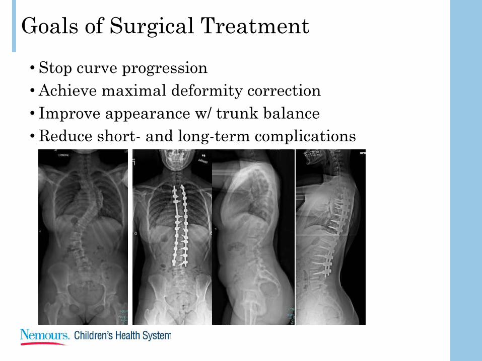
Goals of Surgical Treatment• Stop curve progression•Achieve maximal deformity correction• Improve appearance w/ trunk balance•Reduce short- and long-term complications

Surgical Treatment Technique•Choice of surgical approach and technique is dependent
on deformity, flexibility, and surgeon preference• Most treated by posterior approach• Some by anterior approach• Occasionally combined approach
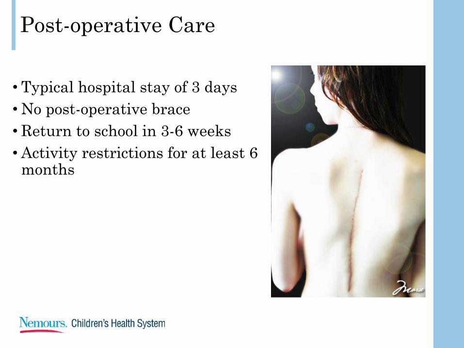
Post-operative Care
• Typical hospital stay of 3 days•No post-operative brace•Return to school in 3-6 weeks•Activity restrictions for at least 6
months
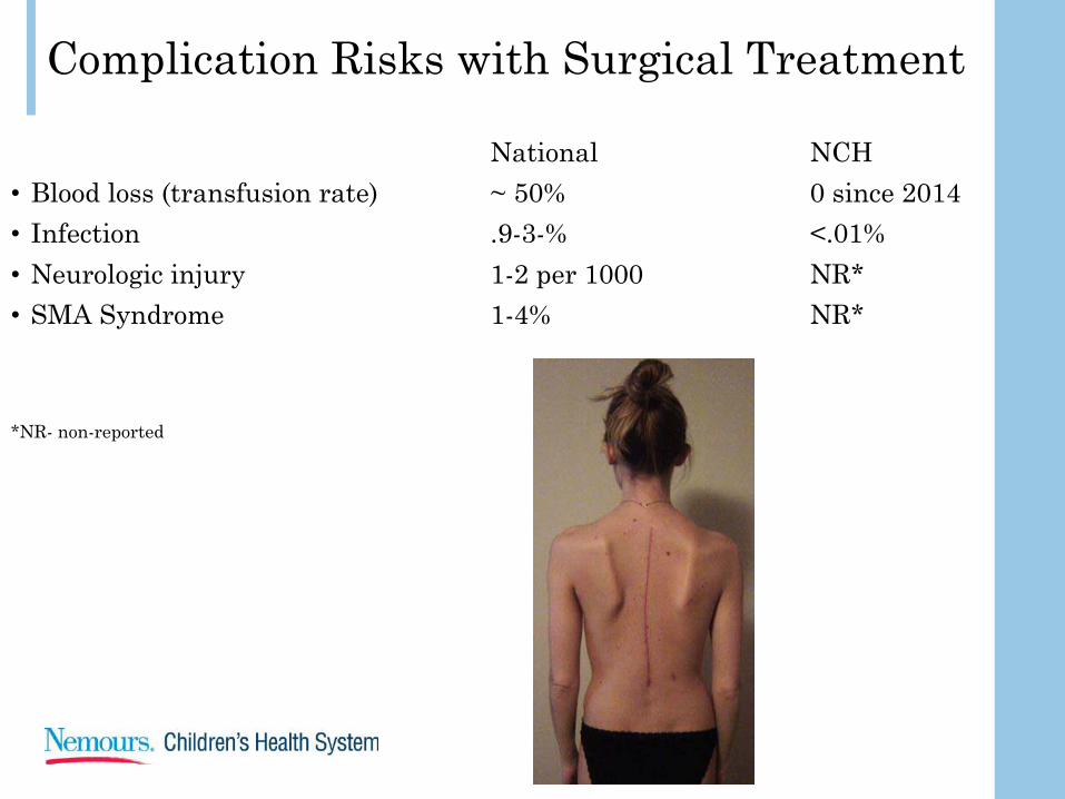
Complication Risks with Surgical Treatment
National NCH• Blood loss (transfusion rate) ~ 50% 0 since 2014• Infection .9-3-% <.01%• Neurologic injury 1-2 per 1000 NR*• SMA Syndrome 1-4% NR*
*NR- non-reported

Take Home Points
• Who• Spinal deformity in children is relatively common• Can develop at any age• Predominately effects girls
• Diagnosis • Clinical exam• Full length PA and Lat scoliosis films• Recognize “atypical” presentations• In all age groups larger curves are associated with greater risk of
progression• Significant effects on thoracic and pulmonary growth and ultimately
mortality in patients <8yrs old especially in IIS
• Treatment• Varies based on age and severity of curve
Thank YouFrom the Nemours Children’s Hospital Orthopaedic Team