Embed Size (px)
Citation preview

Idiopathic Sinus of Valsalva to Right AtrialFistula: An Unusual Origin of Right-sided
Heart FailureArun Prahash, MD, Neal Kleiman, MD, Rafael Espada, MD, and
Hisham Dokainish, MD, Houston, Texas
CASE REPORT
A 43-year-old man who was previously well presentedwith a 4-month history of increasing peripheral edema,dyspnea, and orthopnea. There was no medical history ofhypertension, congestive heart failure, coronary arterydisease, chronic pulmonary disease, or alcohol abuse.Physical examination revealed a normotensive man withmarked jugular venous distension, hepatomegaly, bilateralpitting edema to the level of the upper thigh, and markedabdominal distention. On cardiac auscultation, a 3/6 con-tinuous murmur was heard over the left second and thirdintercostal spaces. Transthoracic echocardiography re-vealed marked enlargement of all 4 cardiac chambers withpreserved ventricular systolic function. Doppler interroga-tion revealed high velocity continuous flow from theaortic root into the right atrium. Transesophageal echo-cardiography revealed a fistulous communication be-tween the right sinus of Valsalva (SOV) and the rightatrium without a SOV aneurysm (Figure 1). Aortographyconfirmed the presence of a fistula but did not reveal adilated aortic root or obstructive coronary artery disease(Figure 2). At operation, a fistula from the right SOV wasnoted communicating with the right atrium (Figure 3).There was no evidence of a SOV aneurysm. The fistula wasrepaired with a patch and the patient recovered unevent-fully.
DISCUSSION
SOV fistulae, in the absence of an aneurysm or otheridentifiable pathology, has rarely been described.Congenital fistulae have been described in an infantwith congestive heart failure, and in 4 other cases in
From the Section of Cardiology, Baylor College of Medicine, andthe Department of Cardiovascular Surgery, the Methodist Hospi-tal (R.E.).Reprint requests: Hisham Dokainish, MD, Baylor College of Med-icine, 6550 Fannin, SM-1901, Houston, TX 77030 (E-mail:[email protected]).J Am Soc Echocardiogr 2004;17:1317-8.0894-7317/$30.00Copyright 2004 by the American Society of Echocardiography.
doi:10.1016/j.echo.2004.07.007asymptomatic patients with continuous murmurs.1
There are no other reports, to our knowledge, of
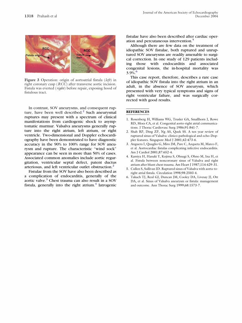
Figure 1 Transesophageal echocardiogram: short-axisview (left) shows defect (arrow) in right coronary cusp(RCC) communicating with right atrium (RA). Leftatrium (LA) is seen opposite RA. Right ventricle (RV) isseen below tricuspid valve. Color Doppler imaging (right)demonstrates high velocity jet (arrow) directed from rightsinus of Valsalva into RA.
Figure 2 Aortogram: fistula (arrow) arising from rightaortic (RA) root sinus is seen. Note runoff of contrast intoRA (curved arrow).
idiopathic SOV fistula.
1317
Journal of the American Society of Echocardiography1318 Prahash et al December 2004
In contrast, SOV aneurysms, and consequent rup-ture, have been well described.2 Such aneurysmalruptures may present with a spectrum of clinicalmanifestations from cardiogenic shock to asymp-tomatic murmur. Valsalva aneurysms generally rup-ture into the right atrium, left atrium, or rightventricle. Two-dimensional and Doppler echocardi-ography have been demonstrated to have diagnosticaccuracy in the 90% to 100% range for SOV aneu-rysm and rupture. The characteristic “wind sock”appearance can be seen in more than 50% of cases.Associated common anomalies include aortic regur-gitation, ventricular septal defect, patent ductusarteriosus, and left ventricular outlet obstruction.2
Fistulae from the SOV have also been described asa complication of endocarditis, generally of theaortic valve.3 Chest trauma can also result in a SOV
Figure 3 Operation: origin of aortoatrial fistula (left) inright coronary cusp (RCC) after transverse aortic incision.Fistula was everted (right) before repair, exposing hood offistulous tract.
fistula, generally into the right atrium.4 Iatrogenic
fistulae have also been described after cardiac oper-ation and percutaneous intervention.5
Although there are few data on the treatment ofidiopathic SOV fistulae, both ruptured and unrup-tured SOV aneurysms are readily amenable to surgi-cal correction. In one study of 129 patients includ-ing those with endocarditis and associatedcongenital lesions, the in-hospital mortality was3.9%.6
This case report, therefore, describes a rare caseof idiopathic SOV fistula into the right atrium in anadult, in the absence of SOV aneurysm, whichpresented with very typical symptoms and signs ofright ventricular failure, and was surgically cor-rected with good results.
REFERENCES
1. Rosenberg H, Williams WG, Trusler GA, Smallhorn J, RoweRD, Moes CA, et al. Congenital aorto-right atrial communica-tions. J Thorac Cardiovasc Surg 1986;91:841-7.
2. Shah RP, Ding ZP, Ng AS, Quek SS. A ten year review ofruptured sinus of Valsalva: clinico-pathological and echo-Dop-pler features. Singapore Med J 2001;42:473-6.
3. Anguera I, Quaglio G, Miro JM, Pare C, Azqueta M, Marco F,et al. Aortocardiac fistulas complicating infective endocarditis.Am J Cardiol 2001;87:652-4.
4. Kamiya H, Hanaki Y, Kojima S, Ohsugi S, Ohno M, Ina H, etal. Fistula between noncoronary sinus of Valsalva and rightatrium after blunt chest trauma. Am Heart J 1987;114:429-31.
5. Cullen S, Sullivan ID. Ruptured sinus of Valsalva with aorta-to-right atrial fistula. Circulation 1998;98:2503-4.
6. Takach TJ, Reul GJ, Duncan JM, Cooley DA, Livesay JJ, OttDA, et al. Sinus of Valsalva aneurysm or fistula: management
and outcome. Ann Thorac Surg 1999;68:1573-7.