Embed Size (px)
Citation preview
IMMUNOBIOLOGY
Idiotype Vaccination in Human Myeloma: Generation of Tumor-Specific ImmuneResponses After High-Dose Chemotherapy
By Massimo Massaia, Paolo Borrione, Silvano Battaglio, Sara Mariani, Eloise Beggiato, Patrizia Napoli, ClaudiaVoena, Alberto Bianchi, Marta Coscia, Barbara Besostri, Silvia Peola, Thomas Stiefel, Jos Even,
Domenico Novero, Mario Boccadoro, and Alessandro Pileri
Igs contain unique portions, collectively termed idiotypes
(Id), that can be recognized by the immune system. Id
expressed by tumor cells in B-cell malignancies can be
regarded as tumor-specific antigens and a target for vaccine
immunotherapy. We have started a vaccination trial in
multiple myeloma (MM) using Id-specific proteins conju-
gated to keyhole limpet hemocyanin (KLH) as immunogens
and low doses of subcutaneous granulocyte-macrophage
colony-stimulating factor (GM-CSF) or interleukin-2 (IL-2) as
immunoadjuvants. Twelve patients who had previously been
treated with high-dose chemotherapy followed by periph-
eral blood progenitor cell (PBPC) transplantation entered
this study from August 1995 to January 1998. All patients
were in first remission at the time of vaccination. They
received subcutaneous injections of Id vaccines and immuno-
adjuvants in an outpatient setting. The generation of Id-
specific T-cell proliferative responses was documented in 2
patients, whereas a positive Id-specific delayed-type hyper-
sensitivity (DTH) reaction was observed in 8 of the 10
patients studied. DTH specificity was confirmed in 1 patient
by investigating the reactivity to synthetic peptides derived
from the VDJ sequence of the tumor-specific Ig heavy chain.
None of the patients generated soluble immune responses
to Id, whereas the generation of soluble and cellular immune
responses to KLH was observed in 100% and 80%, respec-
tively. Eleven patients completed the treatment, whereas 1
patient failed to finish owing to progression of disease.
Freedom from disease progression (FFDP), measured from
the date of first Id/KLH injection to the date of first treatment
after vaccination or last follow-up, ranged from 9 to 36
months. These data indicate that the immune competence
status of MM patients is still susceptible to specific immuni-
zation after high-dose chemotherapy and PBPC transplanta-
tion. It remains to be determined whether generation of
Id-specific immune responses can reduce the relapse rate of
patients with minimal residual disease.
r 1999 by The American Society of Hematology.
M ULTIPLE MYELOMA (MM) is still a fatal B-cellneoplastic disease, with a median survival of less than 4
years.1,2 High-dose chemotherapy followed by autologous bonemarrow or peripheral blood progenitor cell (PBPC) transplanta-tion have recently increased the complete remission rate andremission duration.3-5 However, overall survival has only beenslightly prolonged, and no evidence for a cure has beenobtained.6 All patients ultimately relapse even under mainte-nance therapy with interferon-a (IFN-a) alone7 or in combina-tion with steroids.8,9 A possible strategy to improve the clinicaloutcome is to prolong the duration of the remission phase byinducing an active specific immune response. MM is character-ized by the clonal expansion of lymphoid cells with rearrangedIg genes. Igs contain unique portions, collectively termedidiotype (Id), that can be recognized by the immune system. Idexpressed by tumor cells in MM can be regarded as atumor-specific antigen and a target for active specific immuno-therapy. MM has other immunologic features that can beadvantageously exploited in the setting up of active specificimmunotherapy. First, there is a T-cell population open toexploitation as a source of specific antitumor effector cells.10-14
Second, the T-cell effector mechanisms are not exhausted bychronic tumor cell stimulation.15,16 However, despite the evi-dence of activation and immune recognition, it is clear that Tcells do not perform adequately in vivo and are unable to holdthe disease in check. Normal T cells can recognize andeliminate tumor cells, but this ability is impaired in MMpatients due to several mechanisms, including inadequate tumorantigen presentation leading to T-cell apoptosis.17 Immunothera-peutic strategies aimed at conferring immunogenicity on autolo-gous Id may achieve two goals: the first is to rescue T cells fromapoptosis; the second is to induce an active specific immuneresponse against tumor cells. Lynch and Eisen were the first to
prove in mice that Id can be rendered immunogenic.18,19 In thelight of these and other experimental data, Id-specific proteinshave come into medical use in patients with follicular lym-phoma.20,21 These pioneering studies have provided the rationale for exploring the use of autologous Id as a therapeuticvaccine in MM. Clinical results in the allogeneic transplantationsetting, including donor lymphocyte infusions, have providedproof in principle that an adequate antitumor immune response,unlike chemotherapy, may eradicate the disease.22-25 We havetherefore started a vaccination trial using Id-specific proteincoupled to keyhole limpet hemocyanin (KLH) and low doses ofsubcutaneous interleukin-2 (IL-2) or granulocyte-macrophage
From the Divisione di Ematologia and II Servizio di AnatomiaPatologica dell’Universita’ di Torino, Azienda Ospedaliera San Gio-vanni Battista di Torino, Torino, Italy; Biosyn Arzeittemel, Fellbach,Germany; and INSERM U277/Department d’Immunologie InstitutPasteur, Paris, France.
Submitted December 31, 1998; accepted March 10, 1999.Supported by AIRC (Milano, Italy), MURST 60% (Roma, Italy), and
Compagnia San Paolo di Torino (Torino, Italy). Fellowship recipientsare S.P. (Comitato Gigi Ghirotti, Torino, Italy), S.M. (AIL, Torino,Italy), and B.B. (Associazione Italiana Amici Jose Carreras, Torino,Italy). The support of FIRC (Milano, Italy) to M.M. is also acknowl-edged.
Address reprint requests to Massimo Massaia, MD, DivisioneUniversitaria di Ematologia, Via Genova 3, 10126 Torino, Italy; e-mail:[email protected].
The publication costs of this article were defrayed in part by pagecharge payment. This article must therefore be hereby marked‘‘adver-tisement’’ in accordance with 18 U.S.C. section 1734 solely to indicatethis fact.
r 1999 by The American Society of Hematology.0006-4971/99/9402-0030$3.00/0
Blood, Vol 94, No 2 (July 15), 1999: pp 673-683 673
For personal use only.on April 13, 2017. by guest www.bloodjournal.orgFrom
colony-stimulating factor (GM-CSF). Treatment of an initialseries of 8 MM patients in relapse or with resistant disease hasshown that Id vaccines are safe and can be administered in anoutpatient setting. Two patients had stable disease for 20months with no further chemotherapy. We now report the resultsof a subsequent study in which Id vaccines were administered infirst remission as a maintenance treatment after high-dosechemotherapy and PBPC transplantation.
MATERIALS AND METHODSPatients. Twelve MM patients (Table 1) entered this study from
from August 1995 to January 1998. Approval was obtained from theInstitutional Review Board for these studies. Informed consent wasprovided according to the Declaration of Helsinki. MM was diagnosedas previously reported.26 According to the Durie and Salmon stagingsystem,27 9 patients were classified as stage III and 3 were stage II; 1was substage B. Seven were IgG, 4 were IgA, and 1 was Bence Jonesmyeloma. All patients had received previous high-dose chemotherapyfollowed by PBPC transplantation according to the Italian MyelomaStudy Group regimens3,28 and were in first remission after inductionchemotherapy. Peripheral blood samples were collected from age-matched normal donors (kindly provided by the local Blood Bank) toset the reference values for some of the immunologic analyses.
Vaccine preparation. Id purification from serum and urine wasaccomplished by means of precipitation and chromatography tech-niques that exploit Id-specific molecular weight and isoelectric point.To reduce microbiological contamination, chromatography separationswere generally performed in disposable conical tubes rather than incolumns. Different strategies were used to purify IgG, IgA, and lightchains, as previously reported.29 Purified IgG were obtained by ionexchange chromatography followed by affinity chromatography. IgAwere separated by ammonium sulfate precipitation followed by gelfiltration. k or l light chains were isolated from urine by means ofammonium sulfate precipitation. The precipitate was dissolved inphysiologic saline and dialyzed versus three changes of physiologicsaline overnight. Further purification was not required, because glomeru-lar filtration itself separates molecules on the basis of their size. Whenpurified Id were reanalyzed by high-resolution agarose gel electrophore-
sis, no extra bands were observed above the nephelometry threshold.The median recovery of IgG, IgA, and free light chain from 30 sampleswas 50%, 15%, and 16%, respectively. KLH (Biosyn, ArzneimittelGmbH, Fellbach, Germany), purified from the hemolymph of thekeyhole limpet (Megathura crenulata), was conjugated to Ids aspreviously reported.20,21,29Briefly, equal amounts of Id and 1 mg/mLKLH in physiologic saline were mixed with 0.1% sterile glutaraldehyde(Sigma, Milano, Italy) for 4 hours at room temperature. Final aliquots ofId/KLH conjugates contained 0.5 mg of Id and KLH each per milliliterof physiologic saline. Id/KLH conjugates were tested for Mycoplasma,fungi, bacteria, and endotoxin contamination before vialing and storingat220°C.
On average, endotoxin contamination was about 3,000 USP-EU/mL,which is approximately 10-fold higher than the European threshold forparentalia. To decrease it, a final affinity chromatography was per-formed by mixing 2 mL of conjugate with 2 mL of polymyxin (Bio-RadLaboratories, Irvine, CA) in conical tubes. After incubation at 4°C for12 hours, tubes were centrifuged for 5 minutes at 2,000 rpm and thesupernatant was carefully collected. This procedure was repeated twiceand yielded a mean concentration of 37 USP-EU/mL (range, l to 90USP-EU/mL). Id/KLH conjugates were then sterilized by passagethrough a 0.2-µm filter and stored at220°C until use. Id/KLHconjugates were successfully manufactured in all 16 cases attempted. In2 patients, vaccine treatment was not started because early relapseoccurred. Two patients refused to be enrolled in the study, after initialinformed consent and preparation of clinical grade Id vaccines ready-for-use. Thus, the number of vaccines actually used versus the number ofpreparations manufactured was 12 to 16.
Commercially available human polyclonal Igs (IgVena; Sclavo, Pisa,Italy) were used as a control in delayed-type hypersensitivity (DTH)skin tests. They were dialyzed versus physiologic saline, incubatedtwice with polymyxin, passed through a 0.2-µm filter, and stored at220°C until use.
Treatment schedule.Patients received subcutaneous injections of0.5 mg of Id-KLH conjugates at time 0 and at 2, 6, 10, 14, 24, and 28weeks. IL-2 (Proleukin; EuroCetus, Milano, Italy) at 1.5 IU/m2/d (2patients) or GM-CSF (Leucomax; Sandoz, Milano, Italy) at 150µg/m2/d (10 patients) was administered subcutaneously close to thevaccine site for the next 5 days. The preferential use of GM-CSF wasbased on both our initial series of 8 MM patients in relapse or withresistant disease and the first 4 patients of the present series, in whomGM-CSF proved to be a better immunoadjuvant than IL-2 in terms ofanti-KLH antibody responses and DTH skin tests.
Clinical evaluation. The serum level of the tumor-related heavychain, the serumk/l light chain ratio, and Bence Jones proteinuria weredetermined by nephelometry (Sanofi Diagnostics Pasteur, Paris, France).Plasma cell infiltration was determined by microscopic evaluation ofbone marrow aspirates after May-Grunwald-Giemsa staining. In pa-tients with normal high-resolution agarose gel electrophoresis andnormal k/l ratio, the disease was detected by immunofixation or bypolymerase chain reaction (PCR) using oligonucleotide primers andprobes derived from the tumor-specific Ig heavy-chain gene se-quences.6,30,31
Freedom from disease progression (FFDP) was measured from thedate of first immunization to the date of progression or last follow-up.Survival was measured from the date of first immunization to the date ofdeath or last follow-up.
Amplification and sequencing of the tumor-specific variable heavychain. Bone marrow mononuclear cells were separated on a Ficoll-Hypaque density gradient. RNA was isolated using the RNAzol Bmethod (Biotech Laboratories, Houston, TX), and total RNA (5 µg) wasreverse-transcribed into Ig cDNA with an isotype-specific primer aspreviously described.6 Amplification and sequencing of the tumor-specific VDJ were performed as previously described.31 Briefly, 1 µL ofIg cDNA was amplified using a VH3 consensus primer derived from the
Table 1. Patients’ Characteristics
UPN Age/Gender Isotype Previous Treatments
Time OffTherapy toVaccination
(mo)
DiseaseStatus at
Vaccination
14 60/M IgAl HDS*, DEX 1 IFN-a 3 RC240 61/F IgGl HDS*, DEX 1 IFN-a 3 RC453 63/F IgGl CM*, DEX 1 IFN-a 4 1R50485 62/F IgGl CM* 8 RC510 47/F IgAk HDS*, IFN-a 2 RC522 53/M IgGl CM* 3 1R75535 57/M IgAk HDS*, IFN-a 17 RC637 54/M IgAl HDS* 4 RC734 60/F IgGk Mev*, DEX 2 1R75743 65/M k† Mev*, DEX 2 RC749 64/F IgGk Mev*, DEX 2 RC847 60/F IgGl Mev*, DEX 2 RC
Abbreviations: CM, high-dose cyclophosphamide 33, high-dosemelphalan 33; HDS, high-dose sequential chemotherapy; Mev, high-dose cyclophosphamide 31, high-dose melphalan 32/3; DEX, dexa-methasone; IFN-a, interferon-a; RC, first complete remission; 1R75,first remission (75% reduction serum M protein level); 1R50, firstpartial remission (50% reduction).
*These regimens are followed by PBPC infusion.†Light chain disease.
674 MASSAIA ET AL
For personal use only.on April 13, 2017. by guest www.bloodjournal.orgFrom
IgH framework region (FR1) and an antisense primer derived from the38 end of the JH region (JH3). The reaction was performed for 33 cycles(denaturation at 94°C for 30 seconds, annealing at 65°C for 30 seconds,and extension at 72°C for 30 seconds), with a final extension at 72°C for7 minutes. PCR products were run on a 2% preparative agarose gel. Theexpected size was excised and phenol-extracted. Direct sequencing ofthe amplified products was performed using the Promega fmol system(Promega, Madison, WI) according to the manufacturer’s instructions.Reactions were performed in a thermocycler at 68°C annealingtemperature for 15 cycles. Because the direct sequencing did not allow acomplete reading of the complementarity determining regions (CDRs),DNA was reamplified with primers containingEcoRI andHindIIIrestriction sites and cloned in a Bluescript SK vector (Stratagene, SanDiego, CA). Restriction enzyme analysis was performed on plasmidDNAs prepared by the alkaline lysis method, and miniprep plasmidDNAs were then sequenced. Sequence analysis was performed with thePC-GENE software (Intelligenetics, Inc, Mountain View, CA).
Monitoring of minimal residual disease.Bone marrow and periph-eral blood were evaluated for the presence of residual myeloma cells byPCR, using oligonucleotide primers and probes derived from thetumor-specific Ig heavy-chain gene sequences, as described above.Postswitch B cells were detected as previously described.30 Briefly, 2 µLof 50 µL of total cDNA was amplified with a 58 primer derived from theCDR2 and a 38primer from the Ca or Cg first exon sequence. Anested-PCR strategy was used to detect preswitch B cells. The firstamplification was performed with a consensus primer for the variableregion (VH.L or VH.D) and a primer from the Cµ first exon (Cµ-5). Ofthis amplification, 5 µL was reamplified using the internal primersCDR2 and Cµ-7. Twenty percent of the PCR product was analyzed byagarose gel electrophoresis, blotted overnight, and hybridized to CDR3probes end-labeled with [g-32P] (Amersham, Milano, Italy) adenosinetriphosphate (ATP). To avoid false-negatives, all of the cDNA samplesnot producing PCR products were reamplified and the cDNA qualitywas tested by amplifying the sequence of p53 exon 5 or n-ras exon 2.
Peptide synthesis.The VDJ sequence of the tumor-specific Igheavy chain of patient 510 was translated into the amino acid sequencewith the PC-GENE software (Intelligenetics, Inc). The deduced aminoacid sequence was analyzed with a program predicting immunogenicitybased on the potential beta turn formation within the sequence. Thesequences around the glycine residues in position 17, 38 (part ofCDR3), and 47 were identified as the most potentially immunogenicregions. A sequence around glycine in position 38 and including theCDR3 sequence (position 32-42) was selected as the most tumor-specific sequence, whereas a sequence corresponding to the FR3 region(position 22-31) was selected as an internal control. The sequences instandard single-letter code with the N-acetylated amino terminus on theleft are Ac-ESRHAVYYCA-OH and Ac-EAVGYGARFD-OH for theCDR3- and FR3-derived peptides, respectively.
Peptides were synthesized on polyethylene pins that had beenradiation grafted with hydroxyethylmethacrylic acid (HEMA).32 Thesecrowns were functionalized with Fmoc-protected amino acid esters of4-hydroxymethylphenoxyacetic (HMP) that yield a free acid c-terminuson cleavage. Amino acid coupling was performed in distilled N1N-dimethylformamide (DMF) at 120 mmol/L by means of HBTU/HoBT/NMM activation (120:120:180 mmol/L). After the completion of thesynthesis cycle, peptides were N-terminal–acetylated with acetic anhy-dride. They were then simultaneously side chain deprotected andcleaved using a mixture of 95% trifluoracetyl (TFA)/2.5% anisole/2.5%ethanedithiol (EDT). The peptide TFA solutions then were reducedunder vacuum and the cleaved peptide was precipitated with diethylether/petroleum ether at 40°C to 60°C (1:2 vol/vol). The precipitate waswashed twice with the ether/petrol mixture. The peptides were thenair-dried, dissolved in a solution of 10% AcOH/MeCN, and sampled foranalysis by mass spectroscopy and high-performance liquid chromatog-raphy (HPLC). The remaining solution was frozen and lyophilized.
Peptides were purified by reverse-phase chromatography with a
Vydac RP C18 2503 10 mm column (Metachem, Torrence, CA)installed in a Waters system (Cornerstone Water System, Phoenix, AZ).The chromatogram was developed at a flow rate of 4 mL/min using0.1% TFA in water and 0.1% TFA in MeCN as the limiting solvent.Analytical HPLC was performed using a Merck LiChrosphere (Merck,Whitehouse Station, NY) 100 RP C18 (2503 4 mm; flow rate, 1.5mL/min) installed in a Waters system. Solvents used were similar tothose used to purify the peptide. Ion spray mass spectral analysis wasperformed on a Perkin Elmer Sciex API 111 biomolecular massanalyzer (Perkin-Elmer, Foster City, CA). Purity was 96.5% and 98.4%for the CDR3- and the FR3-derived peptide, respectively.
Dry peptides were solubilized in 10 mmol/L phosphate buffer and100 mmol/L sodium chloride at pH 6.0 to give a final concentration of 1mg/mL. A final affinity chromatography step was performed by mixing2 mL of peptides with 2 mL of polymyxin (Affi-Prep; Bio-RadLaboratories) in conical tubes. After incubation at 4°C for 12 hours,tubes were centrifuged for 5 minutes at 2,000 rpm and the supernatantwas carefully collected. This procedure was repeated twice and yieldedan endotoxin concentration of 0.2 USP-EU/mL (CDR3-derived peptide)and 0 USP-EU/mL (FR3-derived peptide). Final aliquots of unconju-gated Id as well as CDR3- and FR3-derived peptides were tested forMycoplasma, fungi, and bacteria before vialing and storing at220°C.
Humoral responses.Microtiter plates (Costar, Milano, Italy) werecoated with 10 µg/mL KLH, incubated for 12 hours at room tempera-ture, and washed 3 times with 0.05% Triton X-100 (Sigma). Afterincubation for 1 hour at room temperature with phosphate-bufferedsaline (PBS)1 2% bovine serum albumin (BSA), they were washed,blocked with 0.2% Tween-20 in PBS for 30 minutes, and washed.Preimmune and postimmune sera were diluted in PBS1 5% fetal calfserum (FCS), dispensed into microwells, and incubated for 1 hour atroom temperature. After washing, horseradish peroxidase (HRP)-conjugated-goat antihuman IgG or IgM (Cappel; Organon TeknicaCorp, West Chester, PA) was added and incubated for 1 hour at roomtemperature. The enzyme substrate solution was added after extensivewashing and incubated for 10 minutes at 37°C. Absorption wasevaluated at an optical density of 405 nm with an enzyme-linkedimmunosorbent assay (ELISA) microtiter plate reader (Titertek Mul-tiskan Plus; Flow Laboratories, Milano, Italy).
Anti-Id antibody responses were evaluated as follows. Microtiterplates were coated with monoclonal antibodies (MoAbs) specific for thehumank or l chain present in the patient’s Id. Plates were incubated for2 hours at room temperature and then overnight at 4°C. After blockingwith 0.2% Tween-20 in PBS for 30 minutes, 10 µg/mL purified Id inPBS 12% FCS was added and incubated for 1 hour at roomtemperature. Preimmune and postimmune sera were diluted in PBS1
2% FCS, dispensed into microwells, and incubated for 1 hour at roomtemperature. After washing, HRP-conjugated goat antihumank or l
chain (opposite to the light chain of the coating Id) was added andincubated for 1 hour at room temperature. The enzyme substratesolution was added after extensive washing and incubated for 10minutes at 37°C. Absorption was evaluated at an optical density of 405nm with an ELISA microtiter plate reader.
Cellular proliferation assays. Cellular proliferations to autologousId, control Id (unrelated isotype-matched Id), and KLH were evaluatedat the end of the vaccination procedure. Briefly, 53 106 PBMC/mLwere incubated in RPMI1 20% FCS at 37°C in a humidifiedatmosphere of 5% CO2 in air. After 2 hours of incubation, nonadherentcells were recovered and T cells were isolated by rosetting with sheepred blood cells at 29°C for 1 hour to exclude the majority of CD32,CD21 rosette-forming cells.15 Adherent cells (AC) were gently recov-ered using a cell scraper. AC were incubated overnight at 13 106/mL inRPMI 1 10% pooled human AB serum supplemented with 50 ng/mLGM-CSF alone (control culture) or with GM-CSF and 100 µg/mLautologous Id, 100 µg/mL control Id, or 100 µg/mL KLH (experimentalcultures). Tumor necrosis factor-a (TNF-a; Genzyme, Cambridge, UK)at 10 ng/mL was added for the last 3 hours of incubation. AC were then
IDIOTYPE VACCINES IN HUMAN MYELOMA 675
For personal use only.on April 13, 2017. by guest www.bloodjournal.orgFrom
washed, irradiated, and cultured in flat-bottomed microtiter plates withautologous purified T cells (1:5 ratio) for 5 days in RPMI1 10% FCSand 5 U/mL IL-2. Cell proliferation was evaluated by pulsing 200 µL ofcells with 5 µCi3[H]TdR (47 MBq/mmol; Amersham, Milano, Italy)and harvesting 4 hours later with a semiautomated sample harvester.The filters were counted in a liquid scintillation counter. Controls were14 normal blood donors (kindly provided by the local Blood Bank) andwere matched for sex and age. AC derived from normal donors wereincubated with GM-CSF alone or with GM-CSF and control Id or KLH.Experiments were set up in such a way that T cells from at least onenormal donor were always studied simultaneously with T cells from thepatients. Results are expressed as the following stimulation index:(3[H]TdR incorporation in the experimental culture)/(3[H]TdR incorpo-ration in the control culture)5 Stimulation index.
The stimulation index to KLH was scored positive in MM when itwas above the mean plus 3 standard deviations (AVG1 3SD) observedin the controls. The stimulation index to autologous Id was scoredpositive if it was at least threefold higher than that observed to thecontrol Id in the same experiment.
DTH skin tests. DTH skin tests were performed in 5 patients beforevaccination and in 10 patients 1 month after the last injection of Id/KLHconjugates. Unconjugated autologous Id was prepared as describedabove. At the KLH conjugation step, one aliquot was left unconjugatedand used for DTH skin testing, whereas the remainder was used toprepare the vaccine. Unconjugated autologous Id alone (0.5 mg) wasinjected intradermally into the forearm. In parallel, an equivalentamount of human polyclonal Ig (IgVena; Sclavo) was injected into theopposite forearm as a control. DTH skin tests with the CDR3- andFR3-derived peptides were performed by injecting intradermally 0.1mg of each peptide side-by-side on the same forearm. Final aliquots ofunconjugated Id as well as CDR3- and FR3-derived peptides weretreated with polymyxin and tested for Mycoplasma, fungi, bacteria, andendotoxin content before vialing and storing at220°C.
Skin biopsies were taken with a punch from selected patients at sitesof Id and/or control injections. Formalin-fixed, paraffin-embedded skinbiopsy sections were stained with the following antibodies: polyclonalanti-CD3 (Dako, Milano, Italy; 1:100 final dilution), C8/144B MoAb(CD8, IgG1-k; Dako), OPD4 MoAb (CD4/CD45R0, IgG1-k; Dako;1:50 final dilution), L-26 MoAb (CD20, IgG2a-k; Dako), and Leu7MoAb (CD57, HNK1, IgM-k; Becton Dickinson, Milano, Italy; 1:20final dilution). Staining was performed after antigen retrieval with amicrowave. Reactions were shown by the avidin-biotin-peroxidasecomplex technique.
Analysis of TCRBV repertoire by molecular evaluation of CDR3 sizedistribution. RNA was extracted from 5 to 103 106 unfractionatedperipheral blood or bone marrow mononuclear cells using the TRIzolReagent (Life Technologies, S. Giuliano Milanese, Italy). RNA fromskin biopsies was obtained by homogenization in the presence ofguanidine isothiocyanate solution, followed by ultracentrifugation on acesium chloride discontinuous density gradient.33 Reverse transcriptionof RNA into cDNA was performed at 42°C by AMV reverse transcrip-tase (Promega kit). The CDR3 size distribution of the BV chain wasdetermined with a two-step PCR reaction.34 Briefly, the first stepconsists of 24 reactions, each containing one specific human BVsubfamily primer coupled with a consensus antisense BC primer.Sequences of BV and BC primers were derived from Genevee et al,35
whereas numbering of BV subfamilies was obtained from Wei et al.36
The PCR was performed in 96 polycarbonate microwell plates (MJResearch, Watertown, MA) using a PTC-100 thermal cycler (MJResearch). PCR conditions were as follows: denaturation at 94°C for 2minutes; 40 cycles of amplification each consisting of 20 seconds ofdenaturation at 94°C, annealing at 60°C for 20 seconds, and extension at72°C for 20 seconds; and a final extension at 72 °C for 5 minutes. Thesecond step, named run-off, was performed to obtain a readablefluorescent product. Briefly, 2 µL of the first PCR was further amplifiedfor 5 cycles in the presence of a nested consensus fluorescent BC primer
or 13 BJ-specific fluorescent primers. Sequences of BJ primers werederived from Pusieux et al.37 The run-off reaction products werevisualized on a 4.25% polyacrylamide sequencing gel in a 377 ABIDNA Sequencer (Perkin-Elmer), and the size of the fragments obtainedwas compared with a set of fluorescent size markers ranging from 35 to350 nucleotides in length (Genescan 350 Tamra; Perkin-Elmer). Auto-matic size analyses and CDR3 size distributions were determined withthe Immunoscope software package.38
RESULTS
Toxicity. All courses were delivered on an outpatient basiswithout any acute World Health Organization (WHO) gradeIII/IV toxicity. Local reactions included erythema, induration,and local discomfort without any skin breakdown at sites ofinjection. Mild axillary lymph node enlargement occurred in 2patients. Systemic toxicity was mostly associated with theinjection of cytokines and consisted in bone pain, myalgia, andarthralgia in patients receiving GM-CSF (WHO grade I/II) andfever in patients receiving IL-2 (WHO grade I/II). Thesesymptoms were easily controlled with oral acetaminophen.
Patients used a self-report diary to record side-effects andprovide an overall evaluation about toxicity, feasibility, andtolerability. Most patients stated that their quality of liferemained good or very good during the vaccine treatment.
Clinical observations. Eleven patients completed the treat-ment, whereas 1 patient failed to finish owing to progression ofdisease. The clinical impact was determined by evaluating thetumor mass, FFDP, and survival (Table 2). The serum tumor-related heavy chain level, the serumk/l ratio and Bence Jonesproteinuria (unique patient no. [UPN] 743), and bone marrowplasma cell infiltration were used to determine the tumor massbefore vaccination and 1 month after the last immunization.Immunofixation was used to detect the disease in 9 patients withnormal serum M protein level and normal serumk/l ratio(UPNs 14, 240, 453, 485, 510, 522, 535, 743, and 749). A PCRanalysis was also used in 2 patients to detect Ca- andCµ-associated tumor-specific VDJ sequences in the peripheralblood and bone marrow (UPNs 14 and 510; data not shown).Use of the C region defines the cell differentiation stage andallows discrimination between clonally related preswitch Bcells (which can be regarded as potential myeloma cell precur-sors) and postswitch tumor B cells.30 The vaccine treatment didnot reduce the tumor mass, irrespective of its magnitude.
Five of the 11 patients who completed the treatment havemaintained the remission at 9 to 30 months after the firstimmunization, whereas 6 patients relapsed at 9 to 36 months(Table 2). Ten patients are alive. At a median follow-up of 26months, the median survival was not reached and ranged from11 to 39 months (Table 2).
Humoral responses.Antibody responses to KLH wereevaluated by comparing prevaccine with postvaccine serumsamples collected 1 month after the last injection of Id/KLHconjugates (Table 3). Anti-KLH responses were observed in allpatients. On average, there was an eightfold increase inanti-KLH antibody titer (8.126 4.22; range, 3.2 to 15.7). Thekinetics of anti-KLH antibody response was analyzed in 2patients. IgM antibodies were detected after the first immuniza-tion and boosted with the second and third, after which theystarted to decline. The appearance of IgG antibodies wasslightly delayed; they were boosted by subsequent immuniza-
676 MASSAIA ET AL
For personal use only.on April 13, 2017. by guest www.bloodjournal.orgFrom
tions and remained high up to the end of the vaccinationprocedure (data not shown).
Antibody responses to Id were evaluated by comparing thereactivity of prevaccine and postvaccine serum samples withautologous and unrelated isotype-matched Id. No increase wasobserved in the titers of specific anti-Id antibodies (Table 3).
Cellular proliferation assays. T-cell proliferative responsesto KLH, control Id (unrelated isotype-matched Id), and autolo-gous Id were evaluated 1 month after the last injection ofId/KLH conjugates (Table 3). Proliferations to KLH and controlId were also determined in 14 age-matched normal donors andwere used as reference values. On average, the stimulationindexes to KLH and control Id in the normal donors were1.006 0.15 and 0.756 0.4, respectively. Nine of 11 patientstested showed a positive response to KLH, whereas only 2 of 11showed a positive response to autologous Id (Table 3).
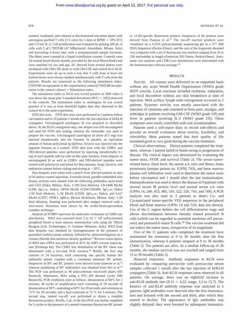
DTH skin tests. DTH skin tests were performed in 10patients 1 month after the last injection of Id/KLH conjugates. Alocal reaction characterized by erythema and induration was
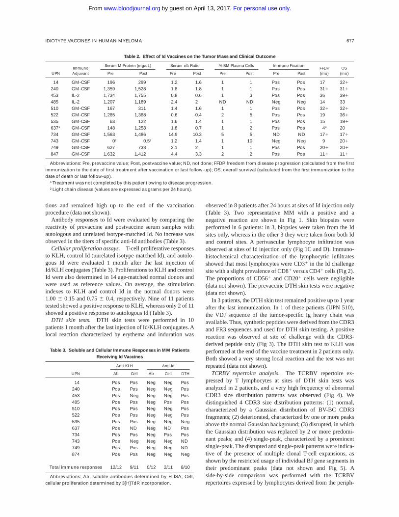
observed in 8 patients after 24 hours at sites of Id injection only(Table 3). Two representative MM with a positive and anegative reaction are shown in Fig 1. Skin biopsies wereperformed in 6 patients: in 3, biopsies were taken from the Idsites only, whereas in the other 3 they were taken from both Idand control sites. A perivascular lymphocyte infiltration wasobserved at sites of Id injection only (Fig 1C and D). Immuno-histochemical characterization of the lymphocytic infiltratesshowed that most lymphocytes were CD31 in the Id challengesite with a slight prevalence of CD81 versus CD41 cells (Fig 2).The proportions of CD561 and CD201 cells were negligible(data not shown). The prevaccine DTH skin tests were negative(data not shown).
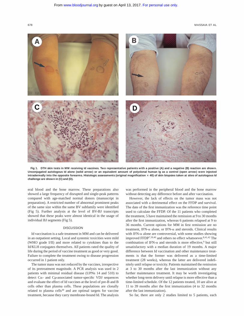
In 3 patients, the DTH skin test remained positive up to 1 yearafter the last immunization. In 1 of these patients (UPN 510),the VDJ sequence of the tumor-specific Ig heavy chain wasavailable. Thus, synthetic peptides were derived from the CDR3and FR3 sequences and used for DTH skin testing. A positivereaction was observed at site of challenge with the CDR3-derived peptide only (Fig 3). The DTH skin test to KLH wasperformed at the end of the vaccine treatment in 2 patients only.Both showed a very strong local reaction and the test was notrepeated (data not shown).
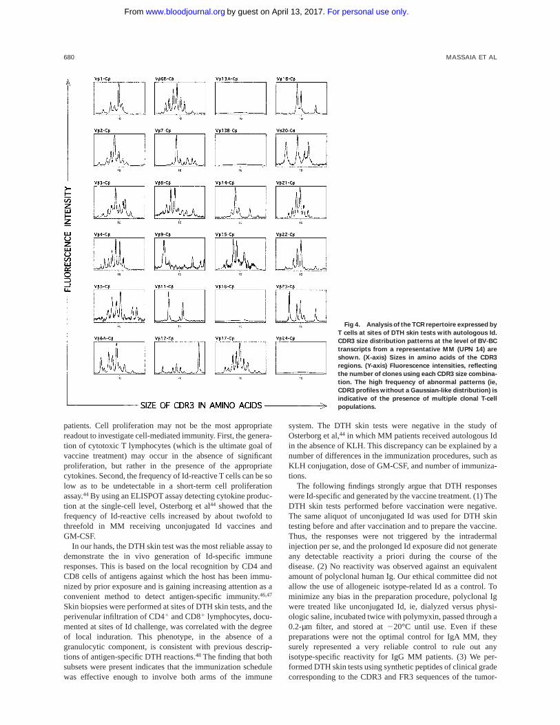
TCRBV repertoire analysis.The TCRBV repertoire ex-pressed by T lymphocytes at sites of DTH skin tests wasanalyzed in 2 patients, and a very high frequency of abnormalCDR3 size distribution patterns was observed (Fig 4). Wedistinguished 4 CDR3 size distribution patterns: (1) normal,characterized by a Gaussian distribution of BV-BC CDR3fragments; (2) deteriorated, characterized by one or more peaksabove the normal Gaussian background; (3) disrupted, in whichthe Gaussian distribution was replaced by 2 or more predomi-nant peaks; and (4) single-peak, characterized by a prominentsingle-peak. The disrupted and single-peak patterns were indica-tive of the presence of multiple clonal T-cell expansions, asshown by the restricted usage of individual BJ gene segments intheir predominant peaks (data not shown and Fig 5). Aside-by-side comparison was performed with the TCRBVrepertoires expressed by lymphocytes derived from the periph-
Table 2. Effect of Id Vaccines on the Tumor Mass and Clinical Outcome
UPNImmunoAdjuvant
Serum M Protein (mg/dL) Serum k/l Ratio % BM Plasma Cells Immuno FixationFFDP(mo)
OS(mo)Pre Post Pre Post Pre Post Pre Post
14 GM-CSF 196 299 1.2 1.6 1 1 Pos Pos 17 321
240 GM-CSF 1,359 1,528 1.8 1.8 1 1 Pos Pos 311 311
453 IL-2 1,734 1,755 0.8 0.6 1 3 Pos Pos 36 391
485 IL-2 1,207 1,189 2.4 2 ND ND Neg Neg 14 33510 GM-CSF 167 311 1.4 1.6 1 1 Pos Pos 321 321
522 GM-CSF 1,285 1,388 0.6 0.4 2 5 Pos Pos 19 361
535 GM-CSF 63 122 1.6 1.4 1 1 Pos Pos 15 191
637* GM-CSF 148 1,258 1.8 0.7 1 2 Pos Pos 4* 20734 GM-CSF 1,563 1,486 14.9 10.3 5 5 ND ND 171 171
743 GM-CSF 0† 0.5† 1.2 1.4 1 10 Neg Neg 9 201
749 GM-CSF 627 738 2.1 2 1 1 Pos Pos 201 201
847 GM-CSF 1,632 1,412 4.4 3.3 2 2 Pos Pos 111 111
Abbreviations: Pre, prevaccine value; Post, postvaccine value; ND, not done; FFDP, freedom from disease progression (calculated from the firstimmunization to the date of first treatment after vaccination or last follow-up); OS, overall survival (calculated from the first immunization to thedate of death or last follow-up).
*Treatment was not completed by this patient owing to disease progression.†Light chain disease (values are expressed as grams per 24 hours).
Table 3. Soluble and Cellular Immune Responses in MM Patients
Receiving Id Vaccines
UPN
Anti-KLH Anti-Id
Ab Cell Ab Cell DTH
14 Pos Pos Neg Neg Pos240 Pos Pos Neg Neg Pos453 Pos Neg Neg Neg Pos485 Pos Pos Neg Pos Pos510 Pos Pos Neg Neg Pos522 Pos Pos Neg Neg Pos535 Pos Pos Neg Neg Neg637 Pos ND Neg ND Pos734 Pos Pos Neg Pos Pos743 Pos Neg Neg Neg ND749 Pos Pos Neg Neg ND874 Pos Pos Neg Neg Neg
Total immune responses 12/12 9/11 0/12 2/11 8/10
Abbreviations: Ab, soluble antibodies determined by ELISA; Cell,cellular proliferation determined by 3[H]TdR incorporation.
IDIOTYPE VACCINES IN HUMAN MYELOMA 677
For personal use only.on April 13, 2017. by guest www.bloodjournal.orgFrom
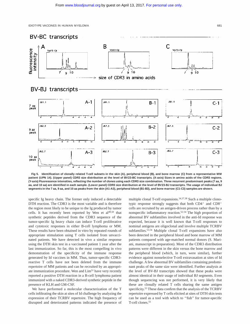
eral blood and the bone marrow. These preparations alsoshowed a large frequency of disrupted and single-peak patternscompared with age-matched normal donors (manuscript inpreparation). A restricted number of abnormal prominent peaksof the same size within the same BV subfamily were identified(Fig 5). Further analysis at the level of BV-BJ transcriptsshowed that these peaks were almost identical in the usage ofindividual BJ segments (Fig 5).
DISCUSSION
Id vaccination is a safe treatment in MM and can be deliveredin an outpatient setting. Local and systemic toxicities were mild(WHO grade I/II) and more related to cytokines than to theId/KLH conjugates themselves. All patients rated the quality oflife during the period of vaccine treatment as good or very good.Failure to complete the treatment owing to disease progressionoccurred in 1 patient only.
The tumor mass was not reduced by the vaccines, irrespectiveof its pretreatment magnitude. A PCR analysis was used in 2patients with minimal residual disease (UPNs 14 and 510) todetect Ca- and Cµ-associated tumor-specific VDJ sequencesand evaluate the effect of Id vaccines at the level of pre-B and Bcells other than plasma cells. These populations are clonallyrelated to plasma cells30 and are optimal targets for vaccinetreatment, because they carry membrane-bound Id. The analysis
was performed in the peripheral blood and the bone marrowwithout detecting any difference before and after vaccination.
However, the lack of effects on the tumor mass was notassociated with a detrimental effect on the FFDP and survival.The date of the first immunization was the reference time pointused to calculate the FFDP. Of the 11 patients who completedthe treatment, 5 have maintained the remission at 9 to 30 monthsafter the first immunization, whereas 6 patients relapsed at 9 to36 months. Current options for MM in first remission are notreatment, IFN-aalone, or IFN-aand steroids. Clinical resultswith IFN-a alone are controversial, with some studies showingimproved FFDP7,39,40and others no effect whatsoever.8,41,42Thecombination of IFN-a and steroids is more effective,9 but stillunsatisfactory with a median duration of 19 months. A majordifference between Id vaccination and other maintenance treat-ments is that the former was delivered as a time-limitedtreatment (28 weeks), whereas the latter are delivered indefi-nitely until relapse or toxicity. Patients maintained the remissionat 3 to 30 months after the last immunization without anyfurther maintenance treatment. It may be worth investigatingwhether long-term delivery until relapse is more effective than atime-limited schedule. Of the 12 patients treated, 10 are alive at11 to 39 months after the first immunization (4 to 32 monthsafter the last immunization).
So far, there are only 2 studies limited to 5 patients, each
Fig 1. DTH skin tests in MM receiving Id vaccines. Two representative patients with a positive (A) and a negative (B) reaction are shown.
Unconjugated autologous Id alone (solid arrow) or an equivalent amount of polyclonal human Ig as a control (open arrow) were injected
intradermally into the opposite forearms. Histologic assessments (original magnification 3 40) of skin biopsies taken at sites of autologous Id
challenge are shown in (C) and (D).
678 MASSAIA ET AL
For personal use only.on April 13, 2017. by guest www.bloodjournal.orgFrom
about the clinical use of Id vaccines in MM.43,44Six patients hadearly stage disease and were untreated, whereas the remainderhad received conventional chemotherapy from 2 to 5 yearsbefore vaccination. Vaccines consisted of autologous unconju-gated Id precipitated in aluminium phosphate43 or administeredin the presence of GM-CSF.44 One patient experienced a 65%decrease in the serum M protein level that lasted about 9months, but no other clinical data were reported.
Unlike chemotherapy, which requires tumor cytoreduction tobe effective, immunotherapy exploits more subtle mechanismsto achieve tumor control in the absence of tumor massreduction. In this regard, immunologic monitoring is crucial todetermine to what extent immunotherapy has acted on theimmune system and generated tumor-specific responses, if any.We initially evaluated humoral and cellular responses to KLH.KLH is commonly used as a protein carrier to make Idimmunogenic, but it can also be exploited as an internal controlto evaluate the efficacy of the immunization schedule. Asignificant production of antibodies to KLH was observed in allpatients. Cellular responses to KLH were also generated anddetected using a cell proliferation assay or the DTH skin test.Thus, the immune competence status of MM patients is notpermanently impaired by chemotherapy, even when multiplecourses of high-dose chemotherapy followed by autologousPBPC transplantation are delivered.
The detection of humoral and cellular responses to autolo-gous Id was more difficult. Id is a much weaker antigen thanKLH: it belongs to self and is under the protection ofself-tolerance mechanisms. We could not detect the presence ofantibodies to Id with an ELISA assay. By using an enzyme-linked immunospot assay (ELISPOT), Bergenbrant et al43 havedetected B cells producing anti-Id antibodies in MM patientsreceiving autologous unconjugated Id. The number of thesecells increased after immunization and then diminished by theend of the vaccine treatment. Conflicting results have also beenreported in follicular lymphomas. Antibodies to Id were docu-mented in 17 of 41 patients receiving Id vaccines consisting ofId coupled to KLH and emulsified in an immunologic adju-vant,21 but in none of 4 patients receiving a more effectivevaccine formulation consisting of dendritic cells pulsed withtumor-specific Id protein.45 Beside experimental variations, it ispossible that we could not detect antibodies to Id because theyare bound by residual tumor cells or by circulating Id that arenot eradicated, even after multiple PBPC transplantations.6
Cellular responses to Id were equally difficult to detect. A cellproliferation assay detected Id-specific responses in 2 of 11patients. Similar data have recently been reported by Osterborget al,44 who detected Id-specific T-cell responses in 1 of 5
Fig 2. Immunohistochemical characterization (original magnifica-
tion 3 20) of lymphocytes infiltrating the skin at sites of autologous Id
challenge. A representative MM with a positive DTH reaction is
shown. Formalin-fixed, paraffin-embedded skin biopsy sections were
stained with the following antibodies: (A) polyclonal anti-CD3; (B)
OPD4 MoAb (CD4/CD45R0); and (C) C8/144B MoAb (CD8).
Fig 3. DTH skin tests with synthetic peptides derived from the
VDJ sequence of the tumor-specific Ig heavy chain: CDR3-derived
peptide (solid arrow) or an equivalent amount of FR3-derived peptide
(open arrow) were injected intradermally side-by-side into the fore-
arm.
IDIOTYPE VACCINES IN HUMAN MYELOMA 679
For personal use only.on April 13, 2017. by guest www.bloodjournal.orgFrom
patients. Cell proliferation may not be the most appropriatereadout to investigate cell-mediated immunity. First, the genera-tion of cytotoxic T lymphocytes (which is the ultimate goal ofvaccine treatment) may occur in the absence of significantproliferation, but rather in the presence of the appropriatecytokines. Second, the frequency of Id-reactive T cells can be solow as to be undetectable in a short-term cell proliferationassay.44 By using an ELISPOT assay detecting cytokine produc-tion at the single-cell level, Osterborg et al44 showed that thefrequency of Id-reactive cells increased by about twofold tothreefold in MM receiving unconjugated Id vaccines andGM-CSF.
In our hands, the DTH skin test was the most reliable assay todemonstrate the in vivo generation of Id-specific immuneresponses. This is based on the local recognition by CD4 andCD8 cells of antigens against which the host has been immu-nized by prior exposure and is gaining increasing attention as aconvenient method to detect antigen-specific immunity.46,47
Skin biopsies were performed at sites of DTH skin tests, and theperivenular infiltration of CD41 and CD81 lymphocytes, docu-mented at sites of Id challenge, was correlated with the degreeof local induration. This phenotype, in the absence of agranulocytic component, is consistent with previous descrip-tions of antigen-specific DTH reactions.48 The finding that bothsubsets were present indicates that the immunization schedulewas effective enough to involve both arms of the immune
system. The DTH skin tests were negative in the study ofOsterborg et al,44 in which MM patients received autologous Idin the absence of KLH. This discrepancy can be explained by anumber of differences in the immunization procedures, such asKLH conjugation, dose of GM-CSF, and number of immuniza-tions.
The following findings strongly argue that DTH responseswere Id-specific and generated by the vaccine treatment. (1) TheDTH skin tests performed before vaccination were negative.The same aliquot of unconjugated Id was used for DTH skintesting before and after vaccination and to prepare the vaccine.Thus, the responses were not triggered by the intradermalinjection per se, and the prolonged Id exposure did not generateany detectable reactivity a priori during the course of thedisease. (2) No reactivity was observed against an equivalentamount of polyclonal human Ig. Our ethical committee did notallow the use of allogeneic isotype-related Id as a control. Tominimize any bias in the preparation procedure, polyclonal Igwere treated like unconjugated Id, ie, dialyzed versus physi-ologic saline, incubated twice with polymyxin, passed through a0.2-µm filter, and stored at220°C until use. Even if thesepreparations were not the optimal control for IgA MM, theysurely represented a very reliable control to rule out anyisotype-specific reactivity for IgG MM patients. (3) We per-formed DTH skin tests using synthetic peptides of clinical gradecorresponding to the CDR3 and FR3 sequences of the tumor-
Fig 4. Analysis of the TCR repertoire expressed by
T cells at sites of DTH skin tests with autologous Id.
CDR3 size distribution patterns at the level of BV-BC
transcripts from a representative MM (UPN 14) are
shown. (X-axis) Sizes in amino acids of the CDR3
regions. (Y-axis) Fluorescence intensities, reflecting
the number of clones using each CDR3 size combina-
tion. The high frequency of abnormal patterns (ie,
CDR3 profiles without a Gaussian-like distribution) is
indicative of the presence of multiple clonal T-cell
populations.
680 MASSAIA ET AL
For personal use only.on April 13, 2017. by guest www.bloodjournal.orgFrom
specific Ig heavy chain. The former only induced a detectableDTH reaction. The CDR3 is the most variable and is thereforethe region most likely to be unique to the Ig produced by tumorcells. It has recently been reported by Wen et al49,50 thatsynthetic peptides derived from the CDR3 sequence of thetumor-specific Ig heavy chain can induce T-cell proliferativeand cytotoxic responses in either B-cell lymphoma or MM.These results have been obtained in vitro by repeated rounds ofappropriate stimulation using T cells isolated from unvacci-nated patients. We have detected in vivo a similar responseusing the DTH skin test in a vaccinated patient 1 year after thelast immunization. So far, this is the most compelling in vivodemonstration of the specificity of the immune responsegenerated by Id vaccines in MM. Thus, tumor-specific CDR3-reactive T cells have not been deleted from the immunerepertoire of MM patients and can be recruited by an appropri-ate immunization procedure. Wen and Lim51 have very recentlyreported a positive DTH reaction in a B-cell lymphoma patientimmunized with a naked CDR3-derived synthetic peptide in thepresence of KLH and GM-CSF.
We have performed a molecular characterization of the Tcells infiltrating the skin at sites of Id challenge by analyzing theexpression of their TCRBV repertoire. The high frequency ofdisrupted and deteriorated patterns indicated the presence of
multiple clonal T-cell expansions.34,37,38Such a multiple clono-typic response strongly suggests that both CD41 and CD81
cells are recruited by an antigen-driven process rather than by anonspecific inflammatory reaction.52-54 The high proportion ofabnormal BV subfamilies involved in the anti-Id response wasexpected, because it is well known that T-cell responses tonominal antigens are oligoclonal and involve multiple TCRBVsubfamilies.53,54 Multiple clonal T-cell expansions have alsobeen detected in the peripheral blood and bone marrow of MMpatients compared with age-matched normal donors (S. Mari-ani, manuscript in preparation). Most of the CDR3 distributionpatterns were different in the skin versus the bone marrow andthe peripheral blood (which, in turn, were similar), furtherevidence against nonselective T-cell extravasation at sites of Idchallenge. A few abnormal BV subfamilies containing predomi-nant peaks of the same size were identified. Further analysis atthe level of BV-BJ transcripts showed that these peaks werealmost identical in their usage of individual BJ segments. Eventhough sequencing was not performed, it is very likely thatthese are clonally related T cells sharing the same antigenspecificity.55 These data confirm that the analysis of the TCRBVrepertoire expressed by T cells elicited at sites of DTH skin testscan be used as a tool with which to ‘‘fish’’ for tumor-specificT-cell clones.55
Fig 5. Identification of clonally related T-cell subsets in the skin (A), peripheral blood (B), and bone marrow (C) from a representative MM
patient (UPN 14). (Upper panel) CDR3 size distribution at the level of BV23-BC transcripts. (X-axis) Sizes in amino acids of the CDR3 regions.
(Y-axis) Fluorescence intensities, reflecting the number of clones using each CDR3 size combination. Three recurrent predominant peaks (7 aa, 9
aa, and 10 aa) are identified in each sample. (Lower panel) CDR3 size distribution at the level of BV23-BJ transcripts. The usage of individual BJ
segments in the 7 aa, 9 aa, and 10 aa peaks from the skin (A1-A3), peripheral blood (B1-B3), and bone marrow (C1-C3) samples are shown.
IDIOTYPE VACCINES IN HUMAN MYELOMA 681
For personal use only.on April 13, 2017. by guest www.bloodjournal.orgFrom
In conclusion, we have been able to generate specific anti-Idimmune responses in MM patients in first remission afterhigh-dose chemotherapy and PBPC transplantation. In ourhands, DTH skin tests were a convenient read-out to pick up thein vivo generation of anti-Id immune responses. However, it iscurrently unknown whether CD41 and CD81 cells specificallyelicited in the skin by Id challenge are indeed the same cells thatcan hold myeloma cells in check in the bone marrow. Identifica-tion and monitoring of clinically relevant tumor-specific im-mune responses remains a major challenge in the setting of Idvaccination. Finally, it remains to be determined whether thegeneration of such responses can provide a better outcome inthe setting of minimal residual disease compared with othermaintenance treatments.
NOTE ADDED IN PROOF
Subsequent to the submission of this work, Reichardt et al56
have reported a series of 12 MM treated with Id-pulsedautologous dendritic cells after high-dose chemotherapy andPBPC transplantation. Eleven of 12 patients made stronganti-KLH cellular responses and 2 of 12 developed cellularId-specific proliferative responses.
ACKNOWLEDGMENT
The authors thank Prof John Iliffe for editorial assistance. They alsothank Drs Henri Gilbert and Gordon Tribbick (Chiron Technologies,Clayton Victoria, Australia) for peptide synthesis.
REFERENCES
1. Alexanian R, Dimopoulos M: The treatment of multiple myeloma.N Engl J Med 330:484, 1994
2. Boccadoro M, Pileri A: Diagnosis, prognosis, and standardtreatment of multiple myeloma. Hematol Oncol Clin North Am 11:111,1997
3. Gianni AM, Tarella C, Bregni M, Siena S, Lombardi F, Gandola L,Caracciolo D, Stern A, Bonadonna G, Boccadoro M, Pileri A: High dosesequential chemoradiotherapy, a widely applicable regimen, conferssurvival benefit to patients with high-risk multiple myeloma. J ClinOncol 12:503, 1994
4. Attal M, Harousseau JL, Stoppa AM, Sotto JJ, Fuzibet JG, RossiJF, Casassus P, Maisonneuve H, Facon T, Ifrah N, Payen C, Bataille R:A prospective, randomized trial of autologous bone marrow transplanta-tion and chemotherapy in multiple myeloma. Intergroupe Francais duMyelome. N Engl J Med 335:91, 1996
5. Vesole DH, Tricot G, Jagannath S, Desikan KR, Siegel D, BracyD, Miller L, Cheson B, Crowley J, Barlogie B: Autotransplants inmultiple myeloma: What have we learned? Blood 88:838, 1996
6. Corradini P, Voena C, Astolfi M, Ladetto M, Tarella C, BoccadoroM, Pileri A: High-dose sequential chemoradiotherapy in multiplemyeloma: Residual tumor cells are detectable in bone marrow andperipheral blood cell harvests and after autografting. Blood 85:1596,1995
7. Mandelli F, Avvisati G, Amadori S, Boccadoro M, Gernone A,Lauta VM, Marmont F, Petrucci MT, Tribalto F, Vegna ML, DammaccoF, Pileri A: Maintenance treatment with alpha-2b recombinant inter-feron significantly improves response and survival duration in multiplemyeloma patients responding to conventional induction chemotherapy.N Engl J Med 322:1430, 1990
8. Salmon SE, Crowley JJ, Grogan TM, Finley P, Pugh RP, BarlogieB: Combination chemotherapy, glucocorticoids, and interferon alfa inthe treatment of multiple myeloma: A Southwest Oncology GroupStudy. J Clin Oncol 12:2405, 1994
9. Salmon SE, Crowley JJ, Balcerzak SP, Roach RW, Taylor SA,
Rivkin SE, Samlowski W: Interferon versus interferon plus prednisoneremission maintenance therapy for multiple myeloma: A SouthwestOncology Group Study. J Clin Oncol 16:890, 1998
10. Dianzani U, Pileri A, Boccadoro M, Palumbo A, Pioppo P,Bianchi A, Camponi A, Battaglio S, Massaia M: Activated idiotype-reactive cells in suppressor/cytotoxic subpopulations of monoclonalgammopathies: Correlation with diagnosis and disease status. Blood72:1064, 1988
11. Osterborg A, Masucci M, Bergenbrant S, Holm G, Lefvert AK,Mellstedt H: Generation of T cell clones binding F(ab8)2 fragments ofthe idiotypic immunoglobulin in patients with monoclonal gammopa-thy. Cancer Immunol Immunother 34:157, 1991
12. Yi Q, Osterborg A, Bergenbrant S, Mellstedt H, Holm G, LefvertAK: Idiotype-reactive T-cell subsets and tumor load in monoclonalgammopathies. Blood 86:3043, 1995
13. Yi Q, Eriksson I, He W, Holm G, Mellstedt H, Osterborg A:Idiotype-specific T lymphocytes in monoclonal gammopathies: Evi-dence for the presence of CD41 and CD81subsets. Br J Haematol96:338, 1997
14. Yi Q, Holm G, Lefvert AK: Idiotype-induced T cell stimulationrequires antigen presentation in association with HLA-DR molecules.Clin Exp Immunol 104:359, 1996
15. Massaia M, Bianchi A, Attisano C, Peola S, Redoglia V,Dianzani U, Pileri A: Detection of hyperreactive T cells in multiplemyeloma by multivalent cross-linking of the CD3/TCR complex. Blood78:1770, 1991
16. Massaia M, Attisano C, Peola S, Carlesso N, Omede P, FerreroD, Corradini P, Boccadoro M, Bianchi A, Pileri A: Rapid generation ofeffective anti-tumor T cells in the bone marrow of myeloma patients byCD3 engagement. Blood 82:1787, 1993
17. Massaia M, Borrione P, Attisano C, Barral P, Beggiato E,Montacchini L, Bianchi A, Boccadoro M, Pileri A: Dysregulated Fasand bcl-2 expression leading to enhanced apoptosis in T cells ofmultiple myeloma patients. Blood 85:3679, 1995
18. Lynch RG, Graff RJ, Sirisinha S, Simms ES, Eisen HN:Myeloma proteins as tumor-specific transplantation antigens. Proc NatlAcad Sci USA 69:1540, 1972
19. Hannestad K, Kao MS, Eisen HN: Cell-bound myeloma proteinson the surface of myeloma cells: Potential targets for the immunesystem. Proc Natl Acad Sci 69:2295, 1972
20. Kwak LW, Campbell MJ, Czerwinski DK, Hart S, Miller RA,Levy R: Induction of immune responses in patients with B-celllymphoma against the surface-immunoglobulin idiotype expressed bytheir tumors. N Engl J Med 327:1209, 1992
21. Hsu FJ, Caspar CB, Czerwinski D, Kwak LW, Liles TM,Syrengelas A, Taidi-Laskowski B, Levy R: Tumor-specific idiotypevaccines in the treatment of patients with B-cell lymphoma: Long-termresults of a clinical trial. Blood 89:3129, 1997
22. Tricot G, Vesole DH, Jagannath S, Hilton J, Munshi N, BarlogieB: Graft-versus-myeloma effect: Proof of principle. Blood 87:1196,1996
23. Bjorkstrand BB, Ljungman P, Svensson H, Hermans J, Alegre A,Apperley J, Blade J, Carlson K, Cavo M, Ferrant A, Goldstone AH, deLaurenzi A, Majolino I, Marcus R, Prentice HG, Remes K, Samson D,Sureda A, Verdonck LF, Volin L, Gahrton G: Allogeneic bone marrowtransplantation versus autologous stem cell transplantation in multiplemyeloma: A retrospective case-matched study from the EuropeanGroup for Blood and Marrow Transplantation. Blood 88:4711, 1996
24. Lokhorst HM, Schattenberg A, Cornelissen JJ, Thomas LL,Verdonck LF: Donor leukocyte infusions are effective in relapsedmultiple myeloma after allogeneic bone marrow transplantation. Blood90:4206, 1997
25. Alyea EP, Soiffer RJ, Canning C, Neuberg D, Schlossman R,Pickett C, Collins H, Wang Y, Anderson KC, Ritz J: Toxicity andefficacy of defined doses of CD41 donor lymphocytes for treatment ofrelapse after allogeneic bone marrow transplant. Blood 91:3671, 1998
682 MASSAIA ET AL
For personal use only.on April 13, 2017. by guest www.bloodjournal.orgFrom
26. Durie BGM, Salmon SE: Multiple myeloma, macroglobulinemiaand monoclonal gammopathies, in Hoffbrand AV, Brown MC, Hirsh J(eds): Recent Advances in Haematology. Edinburgh, UK, ChurchillLivingstone, 1977, p 243
27. Durie BGM, Salmon SE: A clinical staging system for multiplemyeloma: Correlation of measured myeloma cell mass with presentingfeatures, response to treatment, and survival. Cancer 36:842, 1975
28. Palumbo A, Pileri A, Triolo S, Omede P, Bruno B, Ciravegna G,Galliano M, Frieri R, Boccadoro M: Multicyclic, dose-intensivechemotherapy supported by hemopoietic progenitors in refractorymyeloma patients. Bone Marrow Transplant 19:23, 1997
29. Battaglio S, Napoli P, Beggiato E, Borrione P, Ciaiolo C, Peola S,Bianchi A, Stiefel T, Boccadoro M, Pileri A, Massaia M: Developmentof vaccines against autologous tumor-derived idiotype in humanmyeloma: Rationale and preparation. Minerva Biotec 8:1, 1996
30. Corradini P, Boccadoro M, Voena C, Pileri A: Evidence for abone marrow B cell transcribing malignant plasma cell VDJ joined toCµ sequence in immunoglobulin IgG- and IgA-secreting multiplemyelomas. J Exp Med 178:1091, 1993
31. Voena C, Ladetto M, Astolfi M, Provan D, Gribben JG,Boccadoro M, Pileri A, Corradini P: A novel nested-PCR strategy forthe detection of rearranged immunoglobulin heavy-chain genes in Bcell tumors. Leukemia 11:1793, 1997
32. Valerio RM, Bray AM, Campbell RA, DiPasquale A, MargellisC, Rodda SJ, Geysen HM, Maeji NJ: Multipin peptide synthesis at themicromole scale using 2-hydroxyethyl methacrylate grafted polyethyl-ene supports. Int J Pept Protein Res 42:1, 1993
33. Chirgwin JM, Przybyla AE, MacDonald RJ, Rutter WJ: Isolationof biologically active ribonucleic acid from sources enriched inribonuclease. Biochemistry 18:5294, 1979
34. Scwab R, Szabo P, Manavalan JS, Weksler ME, Posnett DN,Pannetier C, Kourilsky P, Even J: Expanded CD41 and CD81T cellclones in elderly humans. J Immunol 158:4493, 1997
35. Genevee C, Diu A, Nierat J, Caignard A, Dietrich PY, FerradiniL, Roman RS, Triebel F, Hercend T: An experimentally validated panelof subfamily-specific oligoucleotide primers (Valpha1-w29/Vbeta 1-w24)for the study of human T cell receptor variable V gene segment usage bypolymerase chain reaction. Eur J Immunol 22:1261, 1992
36. Wei S, Charmley P, Robinson MA, Concannon P: The extent ofthe human germline T-cell receptor V beta segment repertoire. Immuno-genetics 40:27, 1994
37. Pusieux I, Even J, Pannetier C, Jotereau F, Favrot M, KourilskyP: Oligoclonality of tumor-infiltrating lymphocytes from human mela-nomas. J Immunol 153:2807, 1994
38. Pannetier C, Even J, Kourilsky P: T-cell repertoire diversity andclonal expansions in normal and clinical samples. Immunol Today16:176, 1995
39. Browman GP, Bergsagel D, Sicheri D, O’Reilly S, Wilson KS,Rubin S, Belch A, Shustik C, Barr R, Walker I, James K, Zee B,Johnston D: Randomized trial of interferon maintenance in multiplemyeloma: A study of the National Cancer Institute of Canada ClinicalTrials Group. J Clin Oncol 13:2354, 1995
40. Ludwig H, Cohen AM, Polliack A, Huber H, Nachbaur D, SennHJ, Morant R, Eckhardt S, Gunczler P, Seewann HL: Interferon-alphafor induction and maintenance in multiple myeloma: Results of twomulticenter randomized trials and summary of others’ studies. AnnOncol 6:467, 1995
41. Abrahamson GM, Bird JM, Newland AC, Gaminara E, Giles C,Joyner M, Kelsey SM, Lewis D, McCarthy DM, Roques AW, Tew CJ,
Treacy M, van de Pette J, Samson D: A randomized study of VADtherapy with either concurrent or maintenance interferon in patientswith newly diagnosed multiple myeloma. Br J Haematol 94:659, 1996
42. Peest D, Deicher H, Coldewey R, Leo R, Bartl R, Bartels H,Braun HJ, Fett W, Fischer JT, Gobel B: A comparison of polychemo-therapy and melphalan/prednisone for primary remission induction andinterferon-alpha for maintenance treatment in multiple myeloma. Aprospective trial of the German Myeloma Treatment Group. Eur JCancer 89:328, 1995
43. Bergenbrant S, Yi Q, Osterborg A, Bjorkholm M, Osby E,Mellstedt H, Lefvert AK, Holm G: Modulation of anti-idiotypicimmune response by immunization with the autologous M-componentprotein in multiple myeloma patients. Br J Haematol 92:840, 1996
44. Osterborg A, Yi Q, Henriksson L, Fagerberg J, Bergenbrant S,Jeddi-Tehrani M, Ruden U, Lefvert AK, Holm G, Mellstedt H: Idiotypeimmunization combined with granulocyte-macrophage colony-stimulat-ing factor in myeloma patients induced type I, major histocompatibilitycomplex-restricted, CD8- and CD4-specific T-cell responses. Blood91:2459, 1998
45. Hsu FJ, Benike C, Fagnoni F, Liles TM, Czerwinski D, Taidi B,Engleman EG, Levy R: Vaccination of patients with B-cell lymphomausing autologous antigen-pulsed dendritic cells. Nat Med 2:52, 1996
46. Nestle FO, Alijagic S, Gilliet M, Sun Y, Grabbe S, Dummer R,Burg G, Schadendorf D: Vaccination of melanoma patients withpeptide- or tumor lysate-pulsed dendritic cells. Nat Med 4:328, 1998
47. Berd D, Maguire HC Jr, Schuchter LM, Hamilton R, Hauck WW,Sato T, Mastrangelo MJ: Autologous hapten-modified melanoma vac-cine as postsurgical adjuvant treatment after resection of nodal metasta-ses. J Clin Oncol 15:2359, 1997
48. Kuramoto Y, Sekita Y, Tagami H: Histoanalytical study of thecellular infiltrate in the tubercolin reaction. Clin Exp Dermatol 18:111,1993
49. Wen YJ, Lim SH: T cells recognize the VH complementarity-determining region 3 of the idiotypic protein of B cell non-Hodgkin’slymphoma. Eur J Immunol 27:1043, 1997
50. Wen YJ, Lim SH: Immunogenicity and cross-reactivity withidiotypic IgA of VH CDR3 peptide in multiple myeloma. Br J Haematol100:464, 1998
51. Wen YJ, Lim SH:In-vivo immune responses to idiotypic VHcomplementarity-determining region 3 peptide vaccination in B-cellnon-Hodgkin’s lymphoma. Br J Haematol 103:663, 1998
52. Currier JR, Deulofeut H, Barron KS, Kehn PJ, Robinson MA:Mitogens, superantigens, and nominal antigens elicit distinctive pat-terns of TCRB CDR3 diversity. Hum Immunol 48:39, 1996
53. Naumov YN, Hogan KT, Naumova EN, Pagel JT, Gorski J: Aclass I MHC-restricted recall response to a viral peptide is highlypolyclonal despite stringent CDR3 selection: Implications for establish-ing memory T cell repertoires in ‘‘real-world’’ conditions. J Immunol160:2842, 1998
54. Deulofeut H, Robinson MA: The human T cell receptor reper-toire utilized in response to HbsAg. Hum Immunol 57:54, 1997
55. Farace F, Angevin E, Poullion I, Leboullaire C, Ferir G, Elias D,Escudier B, Triebel F: T-cell receptor CDR3 size distribution analysis toevaluate specific T-cell response to cancer vaccines. Int J Cancer71:972, 1997
56. Reichardt VL, Okada CY, Liso A, Benike CJ, Stocker-GoldsteinKE, Engleman EG, Blume KG, Levy R: Idiotype vaccination usingdendritic cells after autologous peripheral blood stem cell transplanta-tion for multiple myeloma– A feasbility study. Blood 93:2411, 1999
IDIOTYPE VACCINES IN HUMAN MYELOMA 683
For personal use only.on April 13, 2017. by guest www.bloodjournal.orgFrom
1999 94: 673-683
Domenico Novero, Mario Boccadoro and Alessandro PileriClaudia Voena, Alberto Bianchi, Marta Coscia, Barbara Besostri, Silvia Peola, Thomas Stiefel, Jos Even, Massimo Massaia, Paolo Borrione, Silvano Battaglio, Sara Mariani, Eloise Beggiato, Patrizia Napoli, Immune Responses After High-Dose ChemotherapyIdiotype Vaccination in Human Myeloma: Generation of Tumor-Specific
http://www.bloodjournal.org/content/94/2/673.full.htmlUpdated information and services can be found at:
(5466 articles)Immunobiology Articles on similar topics can be found in the following Blood collections
http://www.bloodjournal.org/site/misc/rights.xhtml#repub_requestsInformation about reproducing this article in parts or in its entirety may be found online at:
http://www.bloodjournal.org/site/misc/rights.xhtml#reprintsInformation about ordering reprints may be found online at:
http://www.bloodjournal.org/site/subscriptions/index.xhtmlInformation about subscriptions and ASH membership may be found online at:
Copyright 2011 by The American Society of Hematology; all rights reserved.Hematology, 2021 L St, NW, Suite 900, Washington DC 20036.Blood (print ISSN 0006-4971, online ISSN 1528-0020), is published weekly by the American Society of
For personal use only.on April 13, 2017. by guest www.bloodjournal.orgFrom


![RESEARCH OpenAccess · PDF fileobtained by an over-segmentation method [14] ... After summarizing the related study in Section 2, we ... sage transferring and heat energy diffusion](https://img.pdfslide.net/doc/110x75/5abea74f7f8b9aa15e8d1b0a/research-openaccess-by-an-over-segmentation-method-14-after-summarizing.jpg)
![Monitoring multiple myeloma by idiotype-specific peptide ... › track...Aldrich], and analyzed by an ELISA reader at 405 nm [Labsystems multiscan MS]. In vitro cell binding Cultured](https://img.pdfslide.net/doc/110x75/60d60bc658c9b43e297a2fd8/monitoring-multiple-myeloma-by-idiotype-specific-peptide-a-track-aldrich.jpg)
![arXiv:1604.01094v1 [ ] 5 Apr 2016 · PDF fileobtained for water splitting cells of di erent con gurations are summarized [1]. ... The PV-assisted water electrolysis experiments were](https://img.pdfslide.net/doc/110x75/5aac41827f8b9a2b4c8ceec7/arxiv160401094v1-5-apr-2016-for-water-splitting-cells-of-di-erent-con-gurations.jpg)