Embed Size (px)
Citation preview
IDPH ESF-8 Plan: Burn Surge Annex 2016
ATTACHMENT 18: ADULT BURN CARE GUIDELINES Purpose: Provide guidance to practitioners caring for adult burn patients during a disaster. Instructions: These guidelines should be used as a reference by non-burn hospital providers when caring for adult burn patients for extended periods of time when the annex is activated during a burn MCI. These guidelines should be used in conjunction with medical consultation from the State Burn Coordinating Center (SBCC). Disclaimer: This guideline are not meant to be all inclusive, replace an existing policy and procedure at a health care facility or substitute for clinical judgment. These guidelines may be modified at the discretion of the health care provider.
Last updated: November 2016
96 Hour Care Guidelines for Adult Burn Patients if Transfer to a Hospital with Burn Capabilities is Not Feasible
Initial Patient Treatment
Stop the burning process.
Use universal precautions.
Remove all clothing and jewelry.
Prior to initiating care of the patient with wounds, it is critical that health care providers take measures to reduce their own risk of exposure to potentially infectious substances and/or chemical decontamination. Rinse liberally with water, according to protocol if suspected chemical exposure. Apply clean, dry dressing(s) initially to avoid hypothermia.
Apply clean DRY sheet or bedding to prevent hypothermia.
For the care of a burn patient with radiation exposure, see page 81.
Consult the State Burn Coordinating Center (SBCC) for assistance with care of the acutely and critically ill patient, to individualize patient care, if patient does not improve and needs to be transferred and as needed for further support and consult.
Palliative care/comfort care patients: During a burn MCI, resources may not be available to treat those with extensive burn injuries. There are sections within the following guidelines that provide guidance to providers in order to address their needs. Consult the SBCC for additional assistance from palliative care experts.
Primary Assessment, Monitoring, Interventions and Key Points
Assessment and Monitoring Interventions Key Points
Airway Maintenance with Cervical Spine Motion Restriction
Assess throat and nares
Signs of airway injury o Hypoxia o Facial burns o Carbonaceous sputum o Stridor o Hoarseness o Nasal singe o History of a closed space fire
Airway Maintenance with Cervical Spine Motion Restriction
Chin lift/jaw thrust with C-spine motion restriction as needed.
Place an oral pharyngeal airway or endotracheal tube (ETT) in the unconscious patient.
Intubate early.
Secure ETT with ties passed around the head; do not use tape on facial burns since it will not adhere to burned tissue.
Airway Maintenance with Cervical Spine Motion Restriction
Airway edema increases significantly after IV/IO fluids are started.
Stridor or noisy breath sounds indicate impending upper airway obstruction.
Prophylactic intubation is preferred because the ensuing edema obliterates landmarks needed for successful intubation. However, during a burn MCI, there is a need to consider resource availability (e.g. number of ventilators, number of trained staff to
IDPH ESF-8 Plan: Burn Surge Annex 2016
ATTACHMENT 18: ADULT BURN CARE GUIDELINES
64 Last updated: November 2016
Assessment and Monitoring Interventions Key Points
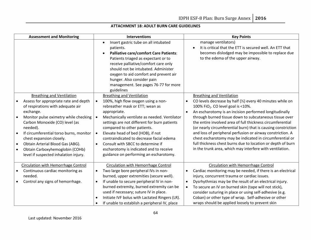
Insert gastric tube on all intubated patients.
Palliative care/comfort Care Patients: Patients triaged as expectant or to receive palliative/comfort care only should not be intubated. Administer oxygen to aid comfort and prevent air hunger. Also consider pain management. See pages 76-77 for more guidelines
manage ventilators)
It is critical that the ETT is secured well. An ETT that becomes dislodged may be impossible to replace due to the edema of the upper airway.
Breathing and Ventilation
Assess for appropriate rate and depth of respirations with adequate air exchange.
Monitor pulse oximetry while checking Carbon Monoxide (CO) level (as needed).
If circumferential torso burns, monitor chest expansion closely.
Obtain Arterial Blood Gas (ABG).
Obtain Carboxyhemoglobin (COHb) level if suspected inhalation injury.
Breathing and Ventilation
100%, high flow oxygen using a non-rebreather mask or ETT; wean as appropriate.
Mechanically ventilate as needed. Ventilator settings are not different for burn patients compared to other patients.
Elevate head of bed (HOB), if not contraindicated to decrease facial edema
Consult with SBCC to determine if escharotomy is indicated and to receive guidance on performing an escharotomy.
Breathing and Ventilation
CO levels decrease by half (½) every 40 minutes while on 100% FiO2. CO level goal is <10%.
An escharotomy is an incision performed longitudinally through burned tissue down to subcutaneous tissue over the entire involved area of full thickness circumferential (or nearly circumferential burn) that is causing constriction and loss of peripheral perfusion or airway constriction. A chest escharotomy may be indicated in circumferential or full thickness chest burns due to location or depth of burn in the trunk area, which may interfere with ventilation.
Circulation with Hemorrhage Control
Continuous cardiac monitoring as needed.
Control any signs of hemorrhage.
Circulation with Hemorrhage Control
Two large bore peripheral IVs in non-burned, upper extremities (secure well).
If unable to secure peripheral IV in non-burned extremity, burned extremity can be used if necessary; suture IV in place.
Initiate IVF bolus with Lactated Ringers (LR).
If unable to establish a peripheral IV, place
Circulation with Hemorrhage Control
Cardiac monitoring may be needed, if there is an electrical injury, concurrent trauma or cardiac issues.
Dysrhythmias may be the result of an electrical injury.
To secure an IV on burned skin (tape will not stick), consider suturing in place or using self-adhesive (e.g. Coban) or other type of wrap. Self-adhesive or other wraps should be applied loosely to prevent skin
IDPH ESF-8 Plan: Burn Surge Annex 2016
ATTACHMENT 18: ADULT BURN CARE GUIDELINES
65 Last updated: November 2016
Assessment and Monitoring Interventions Key Points
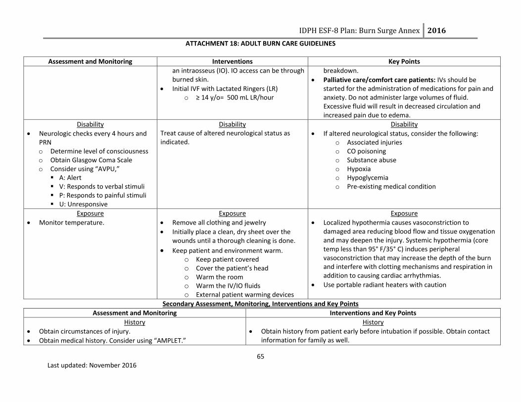
an intraosseus (IO). IO access can be through burned skin.
Initial IVF with Lactated Ringers (LR) o ≥ 14 y/o= 500 mL LR/hour
breakdown.
Palliative care/comfort care patients: IVs should be started for the administration of medications for pain and anxiety. Do not administer large volumes of fluid. Excessive fluid will result in decreased circulation and increased pain due to edema.
Disability
Neurologic checks every 4 hours and PRN o Determine level of consciousness o Obtain Glasgow Coma Scale o Consider using “AVPU,” A: Alert V: Responds to verbal stimuli P: Responds to painful stimuli U: Unresponsive
Disability Treat cause of altered neurological status as indicated.
Disability
If altered neurological status, consider the following: o Associated injuries o CO poisoning o Substance abuse o Hypoxia o Hypoglycemia o Pre-existing medical condition
Exposure
Monitor temperature.
Exposure
Remove all clothing and jewelry
Initially place a clean, dry sheet over the wounds until a thorough cleaning is done.
Keep patient and environment warm. o Keep patient covered o Cover the patient’s head o Warm the room o Warm the IV/IO fluids o External patient warming devices
Exposure
Localized hypothermia causes vasoconstriction to damaged area reducing blood flow and tissue oxygenation and may deepen the injury. Systemic hypothermia (core temp less than 95° F/35° C) induces peripheral vasoconstriction that may increase the depth of the burn and interfere with clotting mechanisms and respiration in addition to causing cardiac arrhythmias.
Use portable radiant heaters with caution
Secondary Assessment, Monitoring, Interventions and Key Points
Assessment and Monitoring Interventions and Key Points
History
Obtain circumstances of injury.
Obtain medical history. Consider using “AMPLET.”
History
Obtain history from patient early before intubation if possible. Obtain contact information for family as well.
IDPH ESF-8 Plan: Burn Surge Annex 2016
ATTACHMENT 18: ADULT BURN CARE GUIDELINES
66 Last updated: November 2016
Assessment and Monitoring Interventions and Key Points
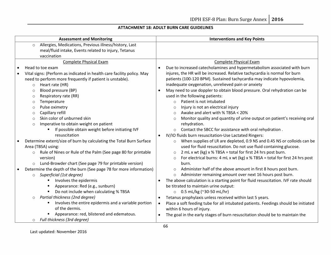
o Allergies, Medications, Previous illness/history, Last meal/fluid intake, Events related to injury, Tetanus vaccination
Complete Physical Exam
Head to toe exam
Vital signs: (Perform as indicated in health care facility policy. May need to perform more frequently if patient is unstable).
o Heart rate (HR) o Blood pressure (BP) o Respiratory rate (RR) o Temperature o Pulse oximetry o Capillary refill o Skin color of unburned skin o Imperative to obtain weight on patient
If possible obtain weight before initiating IVF resuscitation
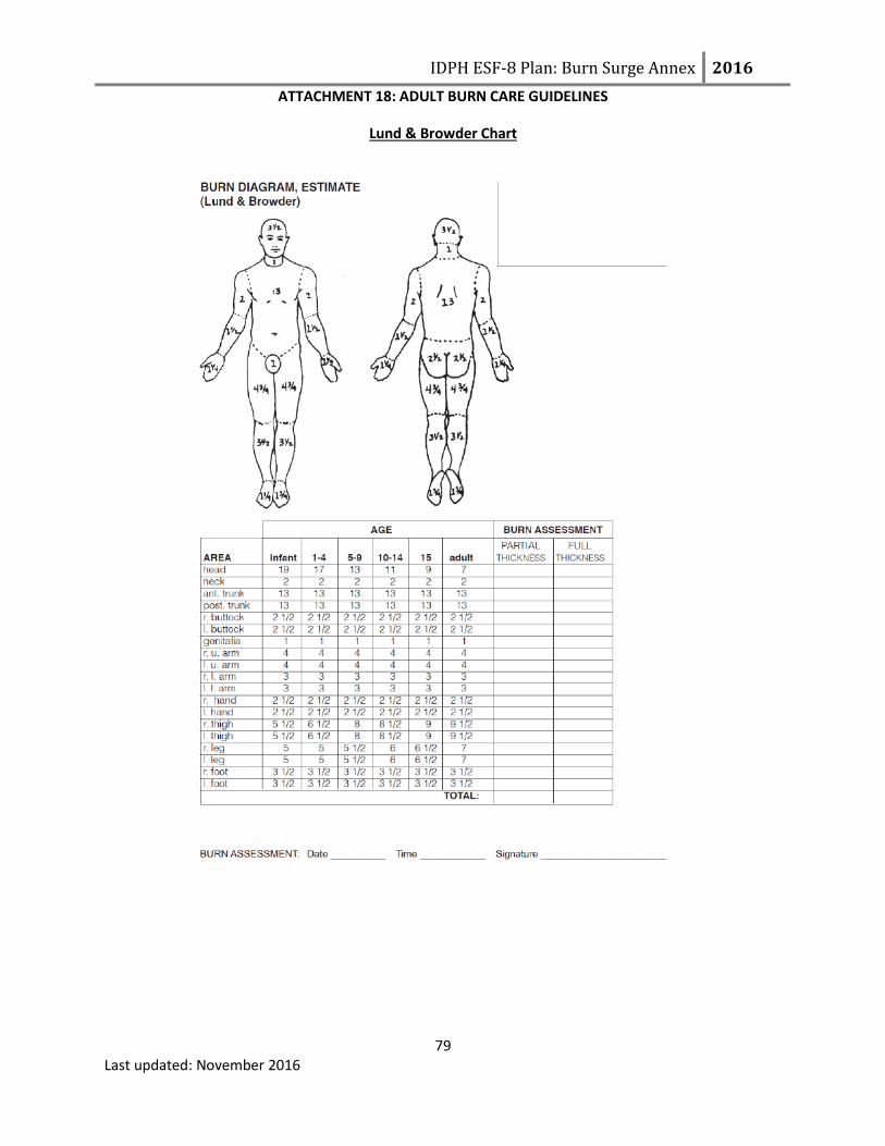
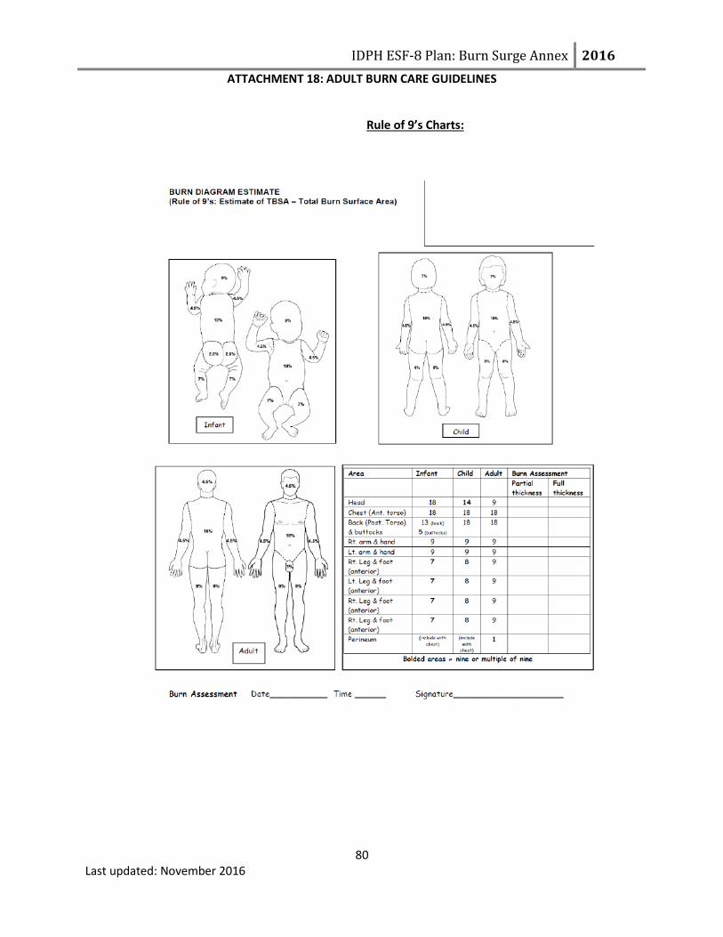
Determine extent/size of burn by calculating the Total Burn Surface Area (TBSA) using:
o Rule of Nines or Rule of the Palm (See page 80 for printable version)
o Lund-Browder chart (See page 79 for printable version)
Determine the depth of the burn (See page 78 for more information) o Superficial (1st degree)
Involves the epidermis Appearance: Red (e.g., sunburn) Do not include when calculating % TBSA
o Partial thickness (2nd degree) Involves the entire epidermis and a variable portion
of the dermis. Appearance: red, blistered and edematous.
o Full thickness (3rd degree)
Complete Physical Exam
Due to increased catecholamines and hypermetabolism associated with burn injures, the HR will be increased. Relative tachycardia is normal for burn patients (100-120 BPM). Sustained tachycardia may indicate hypovolemia, inadequate oxygenation, unrelieved pain or anxiety
May need to use doppler to obtain blood pressure. Oral rehydration can be used in the following patients:
o Patient is not intubated o Injury is not an electrical injury o Awake and alert with % TBSA < 20% o Monitor quality and quantity of urine output on patient’s receiving oral
rehydration. o Contact the SBCC for assistance with oral rehydration .
IV/IO fluids burn resuscitation-Use Lactated Ringers: o When supplies of LR are depleted, 0.9 NS and 0.45 NS or colloids can be
used for fluid resuscitation. Do not use fluid containing glucose. o 2 mL x wt (kg) x % TBSA = total for first 24 hrs post burn. o For electrical burns: 4 mL x wt (kg) x % TBSA = total for first 24 hrs post
burn. o Administer half of the above amount in first 8 hours post burn. o Administer remaining amount over next 16 hours post burn.
The above calculation is a starting point for fluid resuscitation. IVF rate should be titrated to maintain urine output:
o 0.5 mL/kg (~30-50 mL/hr)
Tetanus prophylaxis unless received within last 5 years.
Place a soft feeding tube for all intubated patients. Feedings should be initiated within 6 hours of injury.
The goal in the early stages of burn resuscitation should be to maintain the
IDPH ESF-8 Plan: Burn Surge Annex 2016
ATTACHMENT 18: ADULT BURN CARE GUIDELINES
67 Last updated: November 2016
Assessment and Monitoring Interventions and Key Points
Involves the destruction of the entire epidermis and dermis.
Appearance: white, brown, dry, leathery with possible coagulated vessels.
If camera is available, take pictures of initial burn injuries to document progression of burn injury.
Labs on admission and every day as indicated by medical condition: o Electrolyte panel o Complete blood count (CBC) o ECG for electrical injury or cardiac history o ABG with COHb o Cardiac panel for electrical injury
CXR if intubated, inhalation injury suspected or underlying pulmonary condition.
Monitor for the following signs and symptoms in full thickness, circumferential burn injuries that may indicate a circulation deficit and possible need for escharotomy: (6 P’s)
o Pallor or cyanosis of distal unburned skin on a limb o Pain o Pulselessness o Paralysis o Paresthesia o Poikilothermia o Inability to ventilate in patients with deep circumferential
burns of the chest
individual’s pre-event BP.
If signs of circulation deficit are present, contact the SBCC.
Eyes: o Remove contact lens prior to eyelid swelling, if facial involvement. o Fluorescein should be used to identify corneal injury. o If eye involvement or facial burns, consider consulting an
ophthalmologist.
Consult with SBCC to determine if escharotomy is indicated and to receive guidance on performing an escharotomy.
Finger escharotomies are rarely indicated.
Comfort
Frequent pain/sedation assessment o A minimum of every 4 hours o Before and after pain/sedation medication given
Comfort
Emotional support and education is essential.
IV/IO analgesia is preferred route during initial post injury period.
Large amounts of IV/IO analgesic may be required to attain initial pain control (e.g., Morphine 40-60 mg).
o Administer opioids in frequent (every 5 minutes) small to moderate
IDPH ESF-8 Plan: Burn Surge Annex 2016
ATTACHMENT 18: ADULT BURN CARE GUIDELINES
68 Last updated: November 2016
Assessment and Monitoring Interventions and Key Points
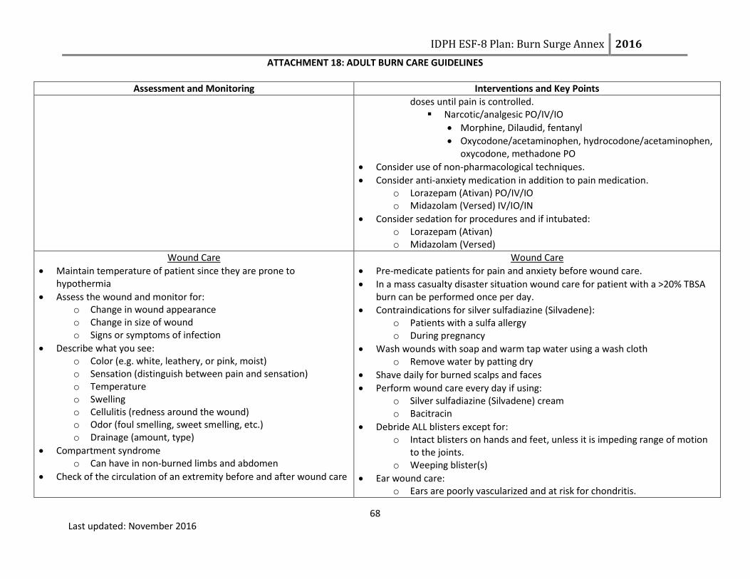
doses until pain is controlled. Narcotic/analgesic PO/IV/IO
Morphine, Dilaudid, fentanyl
Oxycodone/acetaminophen, hydrocodone/acetaminophen, oxycodone, methadone PO
Consider use of non-pharmacological techniques.
Consider anti-anxiety medication in addition to pain medication. o Lorazepam (Ativan) PO/IV/IO o Midazolam (Versed) IV/IO/IN
Consider sedation for procedures and if intubated: o Lorazepam (Ativan) o Midazolam (Versed)
Wound Care
Maintain temperature of patient since they are prone to hypothermia
Assess the wound and monitor for: o Change in wound appearance o Change in size of wound o Signs or symptoms of infection
Describe what you see: o Color (e.g. white, leathery, or pink, moist) o Sensation (distinguish between pain and sensation) o Temperature o Swelling o Cellulitis (redness around the wound) o Odor (foul smelling, sweet smelling, etc.) o Drainage (amount, type)
Compartment syndrome o Can have in non-burned limbs and abdomen
Check of the circulation of an extremity before and after wound care
Wound Care
Pre-medicate patients for pain and anxiety before wound care.
In a mass casualty disaster situation wound care for patient with a >20% TBSA burn can be performed once per day.
Contraindications for silver sulfadiazine (Silvadene): o Patients with a sulfa allergy o During pregnancy
Wash wounds with soap and warm tap water using a wash cloth o Remove water by patting dry
Shave daily for burned scalps and faces
Perform wound care every day if using: o Silver sulfadiazine (Silvadene) cream o Bacitracin
Debride ALL blisters except for: o Intact blisters on hands and feet, unless it is impeding range of motion
to the joints. o Weeping blister(s)
Ear wound care: o Ears are poorly vascularized and at risk for chondritis.
IDPH ESF-8 Plan: Burn Surge Annex 2016
ATTACHMENT 18: ADULT BURN CARE GUIDELINES
69 Last updated: November 2016
Assessment and Monitoring Interventions and Key Points
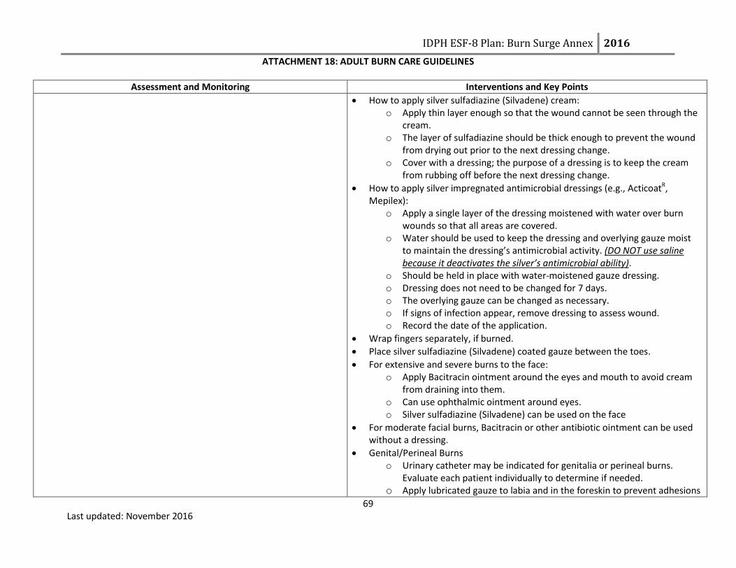
How to apply silver sulfadiazine (Silvadene) cream: o Apply thin layer enough so that the wound cannot be seen through the
cream. o The layer of sulfadiazine should be thick enough to prevent the wound
from drying out prior to the next dressing change. o Cover with a dressing; the purpose of a dressing is to keep the cream
from rubbing off before the next dressing change.
How to apply silver impregnated antimicrobial dressings (e.g., ActicoatR, Mepilex):
o Apply a single layer of the dressing moistened with water over burn wounds so that all areas are covered.
o Water should be used to keep the dressing and overlying gauze moist to maintain the dressing’s antimicrobial activity. (DO NOT use saline because it deactivates the silver’s antimicrobial ability).
o Should be held in place with water‐moistened gauze dressing. o Dressing does not need to be changed for 7 days. o The overlying gauze can be changed as necessary. o If signs of infection appear, remove dressing to assess wound. o Record the date of the application.
Wrap fingers separately, if burned.
Place silver sulfadiazine (Silvadene) coated gauze between the toes.
For extensive and severe burns to the face: o Apply Bacitracin ointment around the eyes and mouth to avoid cream
from draining into them. o Can use ophthalmic ointment around eyes. o Silver sulfadiazine (Silvadene) can be used on the face
For moderate facial burns, Bacitracin or other antibiotic ointment can be used without a dressing.
Genital/Perineal Burns o Urinary catheter may be indicated for genitalia or perineal burns.
Evaluate each patient individually to determine if needed. o Apply lubricated gauze to labia and in the foreskin to prevent adhesions
IDPH ESF-8 Plan: Burn Surge Annex 2016
ATTACHMENT 18: ADULT BURN CARE GUIDELINES
70 Last updated: November 2016
Assessment and Monitoring Interventions and Key Points
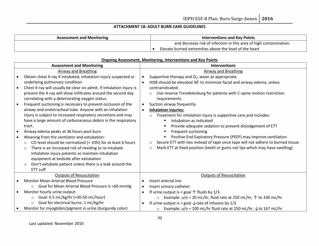
and decrease risk of infection in this area of high contamination. Elevate burned extremities above the level of the heart
Ongoing Assessment, Monitoring, Interventions and Key Points
Assessment and Monitoring Interventions
Airway and Breathing
Obtain chest X-ray if intubated, inhalation injury suspected or underlying pulmonary condition.
Chest X-ray will usually be clear on admit. If inhalation injury is present the X-ray will show infiltrates around the second day correlating with a deteriorating oxygen status.
Frequent suctioning is necessary to prevent occlusion of the airway and endotracheal tube. Anyone with an inhalation injury is subject to increased respiratory secretions and may have a large amount of carbonaceous debris in the respiratory tract.
Airway edema peaks at 36 hours post burn
Weaning from the ventilator and extubation: o CO level should be normalized (< 10%) for at least 6 hours o There is an increased risk of needing to re-intubate
inhalation injury patients so maintain intubation equipment at bedside after extubation
o Don’t extubate patient unless there is a leak around the ETT cuff
Airway and Breathing
Supportive therapy and O2; wean as appropriate.
HOB should be elevated 30 ̊ to minimize facial and airway edema, unless contraindicated. o Use reverse Trendelenburg for patients with C-spine motion restriction
requirements.
Suction airway frequently.
Inhalation Injuries: o Treatment for inhalation injury is supportive care and includes:
Intubation as indicated Provide adequate sedation to prevent dislodgement of ETT Frequent suctioning Positive End Expiratory Pressure (PEEP) may improve ventilation
o Secure ETT with ties instead of tape since tape will not adhere to burned tissue. o Mark ETT at fixed position (teeth or gums not lips which may have swelling).
Outputs of Resuscitation
Monitor Mean Arterial Blood Pressure o Goal for Mean Arterial Blood Pressure is >60 mmHg
Monitor hourly urine output: o Goal: 0.5 mL/kg/hr (≈30-50 mL/hour) o Goal for electrical burns: 1 mL/kg/hr
Monitor for myoglobin/pigment in urine (burgundy color)
Outputs of Resuscitation
Insert arterial line
Insert urinary catheter
If urine output is < goal ↑ fluids by 1/3. o Example: u/o = 20 mL/hr, fluid rate at 250 mL/hr, ↑ to 330 mL/hr
If urine output is > goal ↓rate of infusion by 1/3 o Example: u/o = 100 mL/hr fluid rate at 250 mL/hr, ↓to 167 mL/hr
IDPH ESF-8 Plan: Burn Surge Annex 2016
ATTACHMENT 18: ADULT BURN CARE GUIDELINES
71 Last updated: November 2016
Assessment and Monitoring Interventions
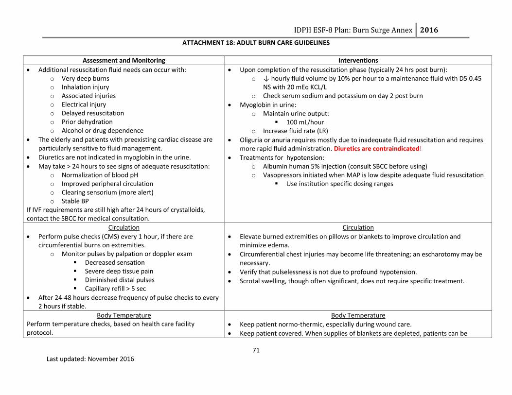
Additional resuscitation fluid needs can occur with: o Very deep burns o Inhalation injury o Associated injuries o Electrical injury o Delayed resuscitation o Prior dehydration o Alcohol or drug dependence
The elderly and patients with preexisting cardiac disease are particularly sensitive to fluid management.
Diuretics are not indicated in myoglobin in the urine.
May take > 24 hours to see signs of adequate resuscitation: o Normalization of blood pH o Improved peripheral circulation o Clearing sensorium (more alert) o Stable BP
If IVF requirements are still high after 24 hours of crystalloids, contact the SBCC for medical consultation.
Upon completion of the resuscitation phase (typically 24 hrs post burn): o ↓ hourly fluid volume by 10% per hour to a maintenance fluid with D5 0.45
NS with 20 mEq KCL/L o Check serum sodium and potassium on day 2 post burn
Myoglobin in urine: o Maintain urine output:
100 mL/hour o Increase fluid rate (LR)
Oliguria or anuria requires mostly due to inadequate fluid resuscitation and requires more rapid fluid administration. Diuretics are contraindicated!
Treatments for hypotension: o Albumin human 5% injection (consult SBCC before using) o Vasopressors initiated when MAP is low despite adequate fluid resuscitation
Use institution specific dosing ranges
Circulation
Perform pulse checks (CMS) every 1 hour, if there are circumferential burns on extremities.
o Monitor pulses by palpation or doppler exam Decreased sensation Severe deep tissue pain Diminished distal pulses Capillary refill > 5 sec
After 24-48 hours decrease frequency of pulse checks to every 2 hours if stable.
Circulation
Elevate burned extremities on pillows or blankets to improve circulation and minimize edema.
Circumferential chest injuries may become life threatening; an escharotomy may be necessary.
Verify that pulselessness is not due to profound hypotension.
Scrotal swelling, though often significant, does not require specific treatment.
Body Temperature Perform temperature checks, based on health care facility protocol.
Body Temperature
Keep patient normo-thermic, especially during wound care.
Keep patient covered. When supplies of blankets are depleted, patients can be
IDPH ESF-8 Plan: Burn Surge Annex 2016
ATTACHMENT 18: ADULT BURN CARE GUIDELINES
72 Last updated: November 2016
Assessment and Monitoring Interventions
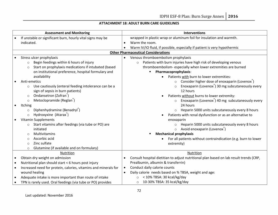
If unstable or significant burn, hourly vital signs may be indicated.
wrapped in plastic wrap or aluminum foil for insulation and warmth.
Warm the room.
Warm IV/IO fluid, if possible, especially if patient is very hypothermic
Other Pharmaceutical Considerations
Stress ulcer prophylaxis o Begin feedings within 6 hours of injury o Start on prophylaxis medications if intubated (based
on institutional preference, hospital formulary and availability
Anti-emetics o Use cautiously (enteral feeding intolerance can be a
sign of sepsis in burn patients) o Ondansetron (Zofran®) o Metoclopramide (Reglan®)
Itching o Diphenhydramine (Benadryl®) o Hydroxyxine (Atarax®)
Vitamin Supplements o Start vitamins after feedings (via tube or PO) are
initiated o Multivitamins o Ascorbic acid o Zinc sulfate o Glutamine (if available and on formulary)
Venous thromboembolism prophylaxis o Patients with burn injuries have high risk of developing venous
thromboembolism- especially when lower extremities are burned Pharmacoprophylaxis:
Patients with burn to lower extremities: o Consider higher dose of enoxaparin (Lovenox®) o Enoxaparin (Lovenox®) 30 mg subcutaneously every
12 hours
Patients without burns to lower extremity: o Enoxaparin (Lovenox®) 40 mg subcutaneously every
24 hours o Heparin 5000 units subcutaneously every 8 hours
Patients with renal dysfunction or as an alternative to enoxaparin
o Heparin 5000 units subcutaneously every 8 hours o Avoid enoxaparin (Lovenox®)
Mechanical prophylaxis
For all patients without contraindication (e.g. burn to lower extremity)
Nutrition
Obtain dry weight on admission
Nutritional plan should start < 6 hours post injury
Increased need for protein, calories, vitamins and minerals for wound healing
Adequate intake is more important than route of intake
TPN is rarely used. Oral feedings (via tube or PO) provides
Nutrition
Consult hospital dietitian to adjust nutritional plan based on lab result trends (CRP, Prealbumin, albumin & transferrin)
Conduct daily calorie counts
Daily calorie needs based on % TBSA, weight and age: o < 10% TBSA: 30 kcal/kg/day o 10-30% TBSA: 35 kcal/kg/day
IDPH ESF-8 Plan: Burn Surge Annex 2016
ATTACHMENT 18: ADULT BURN CARE GUIDELINES
73 Last updated: November 2016
Assessment and Monitoring Interventions
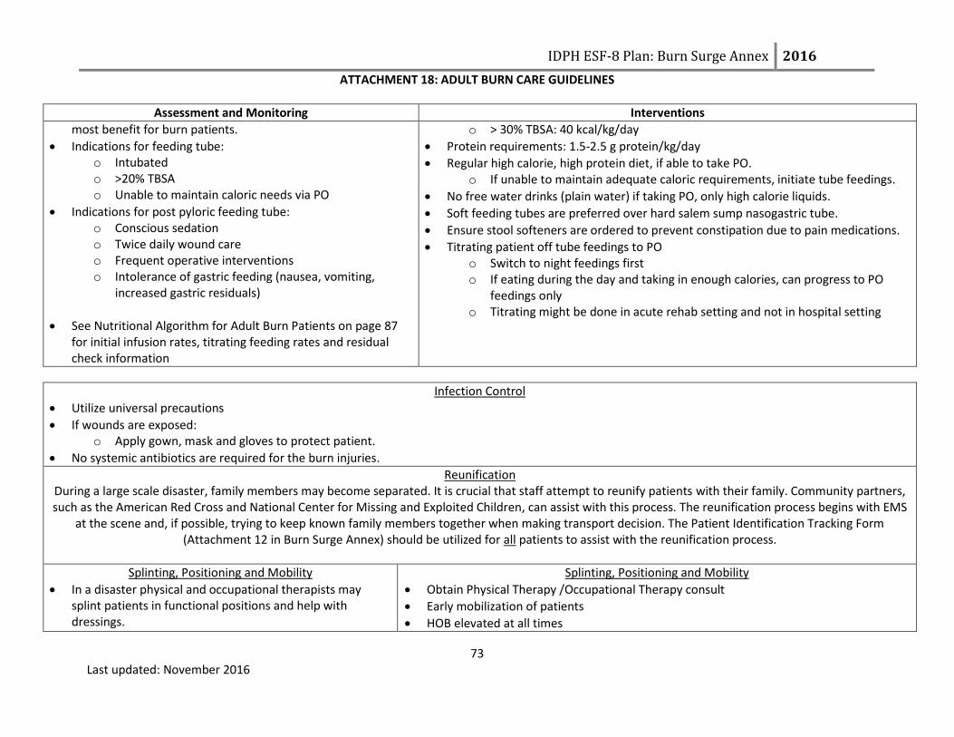
most benefit for burn patients.
Indications for feeding tube: o Intubated o >20% TBSA o Unable to maintain caloric needs via PO
Indications for post pyloric feeding tube: o Conscious sedation o Twice daily wound care o Frequent operative interventions o Intolerance of gastric feeding (nausea, vomiting,
increased gastric residuals)
See Nutritional Algorithm for Adult Burn Patients on page 87 for initial infusion rates, titrating feeding rates and residual check information
o > 30% TBSA: 40 kcal/kg/day
Protein requirements: 1.5-2.5 g protein/kg/day
Regular high calorie, high protein diet, if able to take PO. o If unable to maintain adequate caloric requirements, initiate tube feedings.
No free water drinks (plain water) if taking PO, only high calorie liquids.
Soft feeding tubes are preferred over hard salem sump nasogastric tube.
Ensure stool softeners are ordered to prevent constipation due to pain medications.
Titrating patient off tube feedings to PO o Switch to night feedings first o If eating during the day and taking in enough calories, can progress to PO
feedings only o Titrating might be done in acute rehab setting and not in hospital setting
Infection Control
Utilize universal precautions
If wounds are exposed: o Apply gown, mask and gloves to protect patient.
No systemic antibiotics are required for the burn injuries.
Reunification During a large scale disaster, family members may become separated. It is crucial that staff attempt to reunify patients with their family. Community partners, such as the American Red Cross and National Center for Missing and Exploited Children, can assist with this process. The reunification process begins with EMS
at the scene and, if possible, trying to keep known family members together when making transport decision. The Patient Identification Tracking Form (Attachment 12 in Burn Surge Annex) should be utilized for all patients to assist with the reunification process.
Splinting, Positioning and Mobility
In a disaster physical and occupational therapists may splint patients in functional positions and help with dressings.
Splinting, Positioning and Mobility
Obtain Physical Therapy /Occupational Therapy consult
Early mobilization of patients
HOB elevated at all times
IDPH ESF-8 Plan: Burn Surge Annex 2016
ATTACHMENT 18: ADULT BURN CARE GUIDELINES
74 Last updated: November 2016
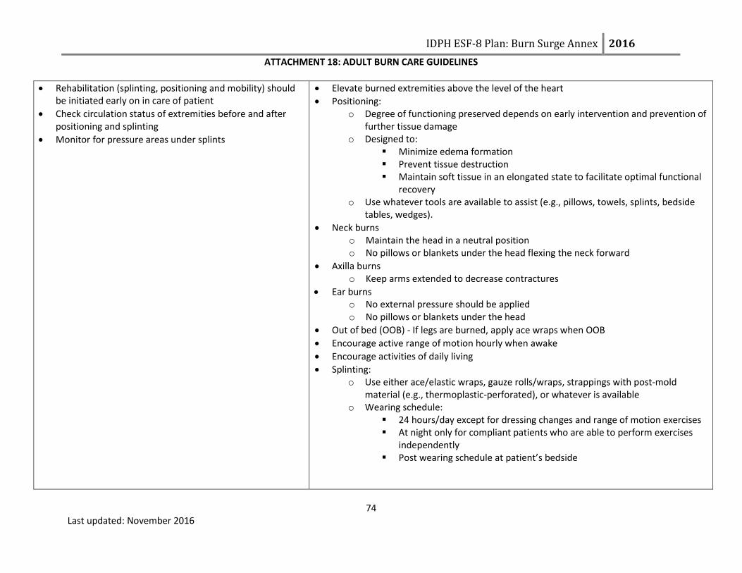
Rehabilitation (splinting, positioning and mobility) should be initiated early on in care of patient
Check circulation status of extremities before and after positioning and splinting
Monitor for pressure areas under splints
Elevate burned extremities above the level of the heart
Positioning: o Degree of functioning preserved depends on early intervention and prevention of
further tissue damage o Designed to:
Minimize edema formation Prevent tissue destruction Maintain soft tissue in an elongated state to facilitate optimal functional
recovery o Use whatever tools are available to assist (e.g., pillows, towels, splints, bedside
tables, wedges).
Neck burns o Maintain the head in a neutral position o No pillows or blankets under the head flexing the neck forward
Axilla burns o Keep arms extended to decrease contractures
Ear burns o No external pressure should be applied o No pillows or blankets under the head
Out of bed (OOB) - If legs are burned, apply ace wraps when OOB
Encourage active range of motion hourly when awake
Encourage activities of daily living
Splinting: o Use either ace/elastic wraps, gauze rolls/wraps, strappings with post-mold
material (e.g., thermoplastic-perforated), or whatever is available o Wearing schedule:
24 hours/day except for dressing changes and range of motion exercises At night only for compliant patients who are able to perform exercises
independently Post wearing schedule at patient’s bedside
IDPH ESF-8 Plan: Burn Surge Annex 2016
ATTACHMENT 18: ADULT BURN CARE GUIDELINES
75 Last updated: November 2016
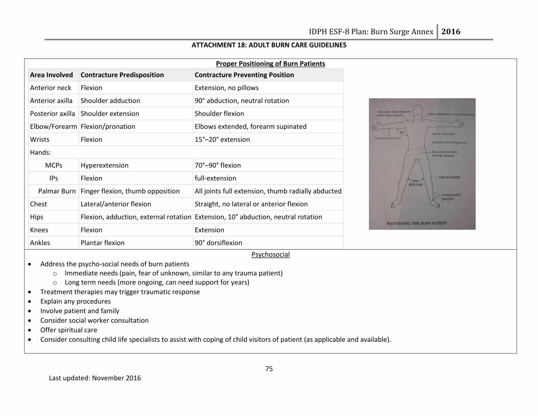
Proper Positioning of Burn Patients
Area Involved Contracture Predisposition Contracture Preventing Position
Anterior neck Flexion Extension, no pillows
Anterior axilla Shoulder adduction 90° abduction, neutral rotation
Posterior axilla Shoulder extension Shoulder flexion
Elbow/Forearm Flexion/pronation Elbows extended, forearm supinated
Wrists Flexion 15°–20° extension
Hands:
MCPs Hyperextension 70°–90° flexion
IPs Flexion full-extension
Palmar Burn Finger flexion, thumb opposition All joints full extension, thumb radially abducted
Chest Lateral/anterior flexion Straight, no lateral or anterior flexion
Hips Flexion, adduction, external rotation Extension, 10° abduction, neutral rotation
Knees Flexion Extension
Ankles Plantar flexion 90° dorsiflexion
Psychosocial
Address the psycho-social needs of burn patients o Immediate needs (pain, fear of unknown, similar to any trauma patient) o Long term needs (more ongoing, can need support for years)
Treatment therapies may trigger traumatic response
Explain any procedures
Involve patient and family
Consider social worker consultation
Offer spiritual care
Consider consulting child life specialists to assist with coping of child visitors of patient (as applicable and available).
IDPH ESF-8 Plan: Burn Surge Annex 2016
ATTACHMENT 18: ADULT BURN CARE GUIDELINES
76 Last updated: November 2016
Palliative Care/Comfort Care During disasters, patients with extensive burn injuries may be triaged as Expectant based on the Burn Triage Guidelines. Patient’s triaged as Expectant still need palliative care/comfort care provided. See the following page for additional information
IDPH ESF-8 Plan: Burn Surge Annex 2016
ATTACHMENT 18: ADULT BURN CARE GUIDELINES
77 Last updated: November 2016
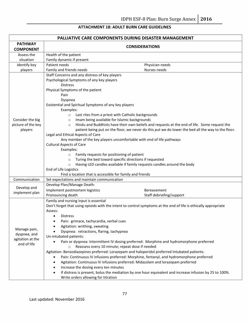
PALLIATIVE CARE COMPONENTS DURING DISASTER MANAGEMENT
PATHWAY COMPONENT
CONSIDERATIONS
Assess the situation
Health of the patient Family dynamic if present
Identify key players
Patient needs Family and friends needs
Physician needs Nurses needs
Consider the big picture of the key
players
Staff Concerns and any distress of key players Psychological Symptoms of any key players Distress Physical Symptoms of the patient Pain Dyspnea Existential and Spiritual Symptoms of any key players Examples:
o Last rites from a priest with Catholic backgrounds o Imam being available for Islamic backgrounds o Hindu and Buddhists have their own beliefs and requests at the end of life. Some request the
patient being put on the floor, we never do this put we do lower the bed all the way to the floor. Legal and Ethical Aspects of Care Any member of the key players uncomfortable with end of life pathways Cultural Aspects of Care Examples:
o Family requests for positioning of patient o Turing the bed toward specific directions if requested o Having LED candles available if family requests candles around the body
End of Life Logistics Find a location that is accessible for family and friends
Communication Set expectations and maintain communication
Develop and implement plan
Develop Plan/Manage Death:
Implement postmortem logistics Pronouncing death
Bereavement Staff debriefing/support
Manage pain, dyspnea, and
agitation at the end of life
Family and nursing input is essential Don’t forget that using opioids with the intent to control symptoms at the end of life is ethically appropriate Assess:
Distress
Pain: grimace, tachycardia, verbal cues
Agitation: writhing, sweating
Dyspnea: retractions, flaring, tachypnea Un-intubated patients:
Pain or dyspnea: Intermittent IV dosing preferred: Morphine and hydromorphone preferred o Reassess every 10 minute; repeat dose if needed
Agitation: Benzodiazepines preferred: Lorazepam and haloperidol preferred Intubated patients:
Pain: Continuous IV infusions preferred: Morphine, fentanyl, and hydromorphone preferred
Agitation: Continuous IV infusions preferred: Midazolam and lorazepam preferred
Increase the dosing every ten minutes
If distress is present, bolus the mediation by one hour equivalent and increase infusion by 25 to 100%. Write orders allowing for titration
IDPH ESF-8 Plan: Burn Surge Annex 2016
ATTACHMENT 18: ADULT BURN CARE GUIDELINES
78 Last updated: November 2016
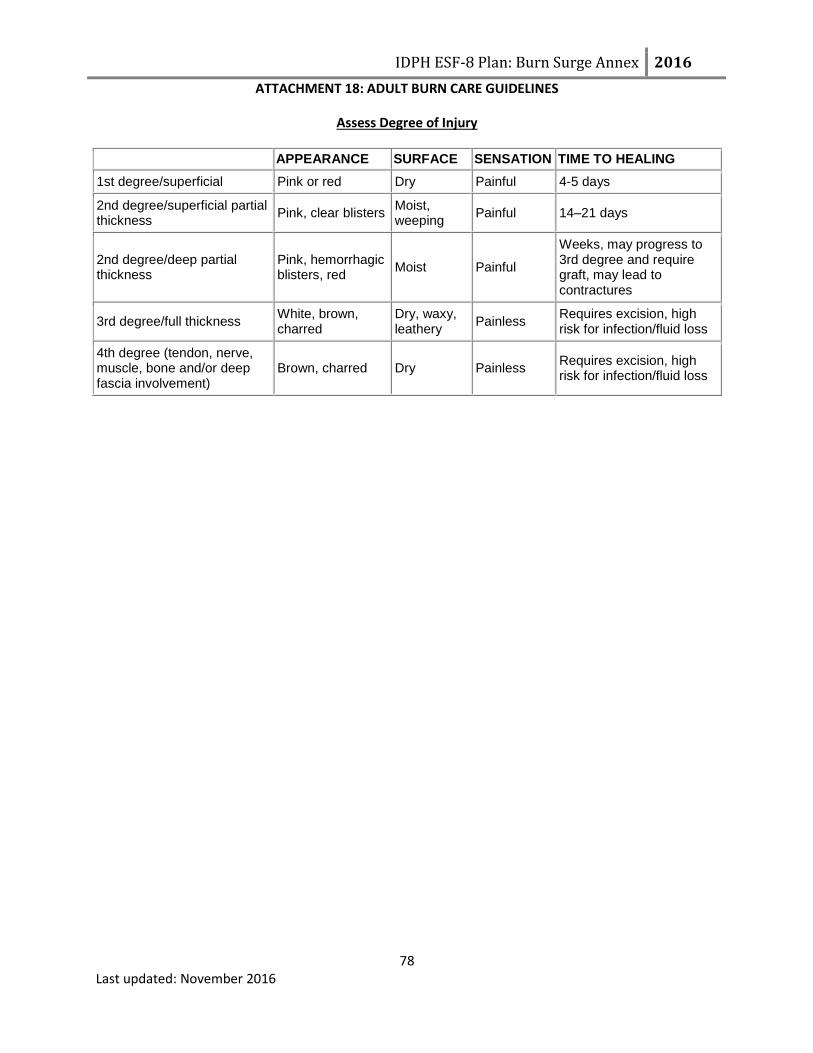
Assess Degree of Injury
APPEARANCE SURFACE SENSATION TIME TO HEALING
1st degree/superficial Pink or red Dry Painful 4-5 days
2nd degree/superficial partial thickness
Pink, clear blisters Moist, weeping
Painful 14–21 days
2nd degree/deep partial thickness
Pink, hemorrhagic blisters, red
Moist Painful
Weeks, may progress to 3rd degree and require graft, may lead to contractures
3rd degree/full thickness White, brown, charred
Dry, waxy, leathery
Painless Requires excision, high risk for infection/fluid loss
4th degree (tendon, nerve, muscle, bone and/or deep fascia involvement)
Brown, charred Dry Painless Requires excision, high risk for infection/fluid loss
IDPH ESF-8 Plan: Burn Surge Annex 2016
ATTACHMENT 18: ADULT BURN CARE GUIDELINES
79 Last updated: November 2016
Lund & Browder Chart
IDPH ESF-8 Plan: Burn Surge Annex 2016
ATTACHMENT 18: ADULT BURN CARE GUIDELINES
80 Last updated: November 2016
Rule of 9’s Charts:
IDPH ESF-8 Plan: Burn Surge Annex 2016
ATTACHMENT 18: ADULT BURN CARE GUIDELINES
81 Last updated: November 2016
Management of Burn Patients with Radiation Exposure
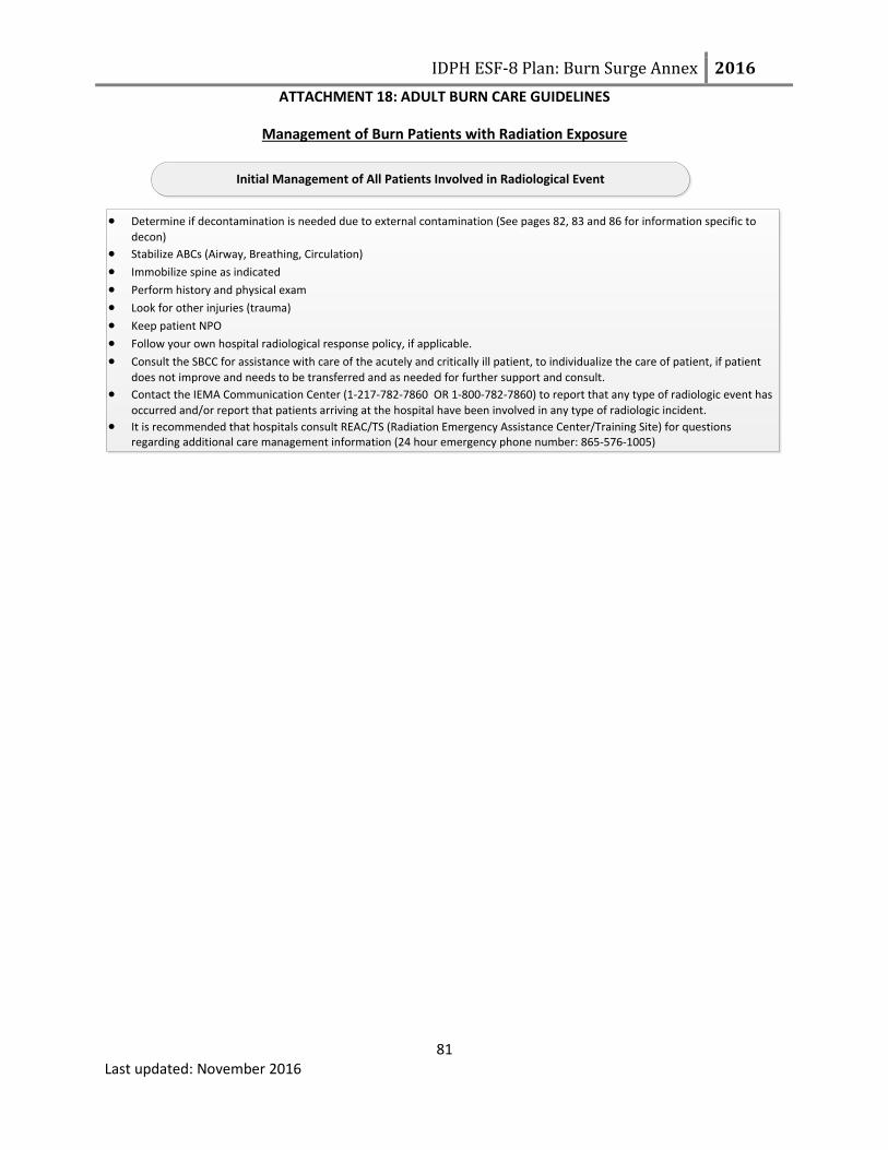
Initial Management of All Patients Involved in Radiological Event
Determine if decontamination is needed due to external contamination (See pages 82, 83 and 86 for information specific to
decon)
Stabilize ABCs (Airway, Breathing, Circulation)
Immobilize spine as indicated
Perform history and physical exam
Look for other injuries (trauma)
Keep patient NPO
Follow your own hospital radiological response policy, if applicable.
Consult the SBCC for assistance with care of the acutely and critically ill patient, to individualize the care of patient, if patient
does not improve and needs to be transferred and as needed for further support and consult.
Contact the IEMA Communication Center (1-217-782-7860 OR 1-800-782-7860) to report that any type of radiologic event has
occurred and/or report that patients arriving at the hospital have been involved in any type of radiologic incident.
It is recommended that hospitals consult REAC/TS (Radiation Emergency Assistance Center/Training Site) for questions regarding additional care management information (24 hour emergency phone number: 865-576-1005)
IDPH ESF-8 Plan: Burn Surge Annex 2016
ATTACHMENT 18: ADULT BURN CARE GUIDELINES
82 Last updated: November 2016
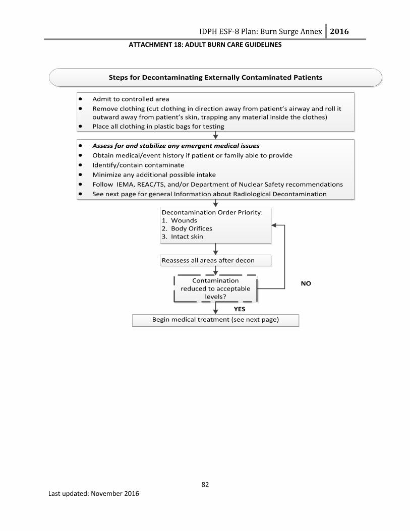
Steps for Decontaminating Externally Contaminated Patients
Admit to controlled area
Remove clothing (cut clothing in direction away from patient’s airway and roll it
outward away from patient’s skin, trapping any material inside the clothes)
Place all clothing in plastic bags for testing
Assess for and stabilize any emergent medical issues
Obtain medical/event history if patient or family able to provide
Identify/contain contaminate
Minimize any additional possible intake
Follow IEMA, REAC/TS, and/or Department of Nuclear Safety recommendations
See next page for general Information about Radiological Decontamination
Decontamination Order Priority:1. Wounds2. Body Orifices3. Intact skin
Reassess all areas after decon
Begin medical treatment (see next page)
Contamination reduced to acceptable
levels?
YES
NO
IDPH ESF-8 Plan: Burn Surge Annex 2016
ATTACHMENT 18: ADULT BURN CARE GUIDELINES
83 Last updated: November 2016
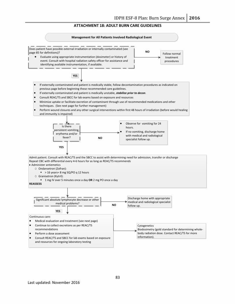
Management for All Patients Involved Radiological Event
If externally contaminated and patient is medically stable, follow decontamination procedures as indicated on
previous page before beginning these recommended care guidelines.
If externally contaminated and patient is medically unstable, stabilize prior to decon.
Consult REAC/TS and SBCC for lab exams based on exposure and resources
Minimize uptake or facilitate excretion of contaminant through use of recommended medications and other
techniques. (See next page for further management)
Perform wound closures and any other surgical interventions within first 48 hours of irradiation (before would healing
and immunity is impaired)
Follow normal treatment
procedures
Observe for vomiting for 24
hours.
If no vomiting, discharge home
with medical and radiological specialist follow up.
Admit patient. Consult with REAC/TS and the SBCC to assist with determining need for admission, transfer or discharge Repeat CBC with differential every 4-6 hours for as long as REAC/TS recommends Administer antiemetics
O Ondansetron (Zofran): > 16 years= 8 mg SQ/PO q 12 hours
O Granisetron (Kytril): 1 mg IV over 5 minutes once a day OR 2 mg PO once a day
REASSESS
NO
NO
Does patient have possible external irradiation or internally contaminated (see page 85 for definitions)?
Evaluate using appropriate instrumentation (dosimeter) or history of
event. Consult with hospital radiation safety officer for assistance and identifying available instrumentation, if available.
Is there persistent vomiting,
erythema and/or fever?
Discharge home with appropriate medical and radiological specialist follow up.
Continuous care:
Medical evaluation and treatment (see next page)
Continue to collect excretions as per REAC/TS
recommendations
Perform a dose assessment
Consult REAC/TS and SBCC for lab exams based on exposure
and resources for ongoing laboratory testing
CytogeneticsBiodosimetry (gold standard for determining whole-body radiation dose. Contact REAC/TS for more information).
Significant absolute lymphocyte decrease or other medical problems?
YES
NO
YES
YES
IDPH ESF-8 Plan: Burn Surge Annex 2016
ATTACHMENT 18: ADULT BURN CARE GUIDELINES
84 Last updated: November 2016
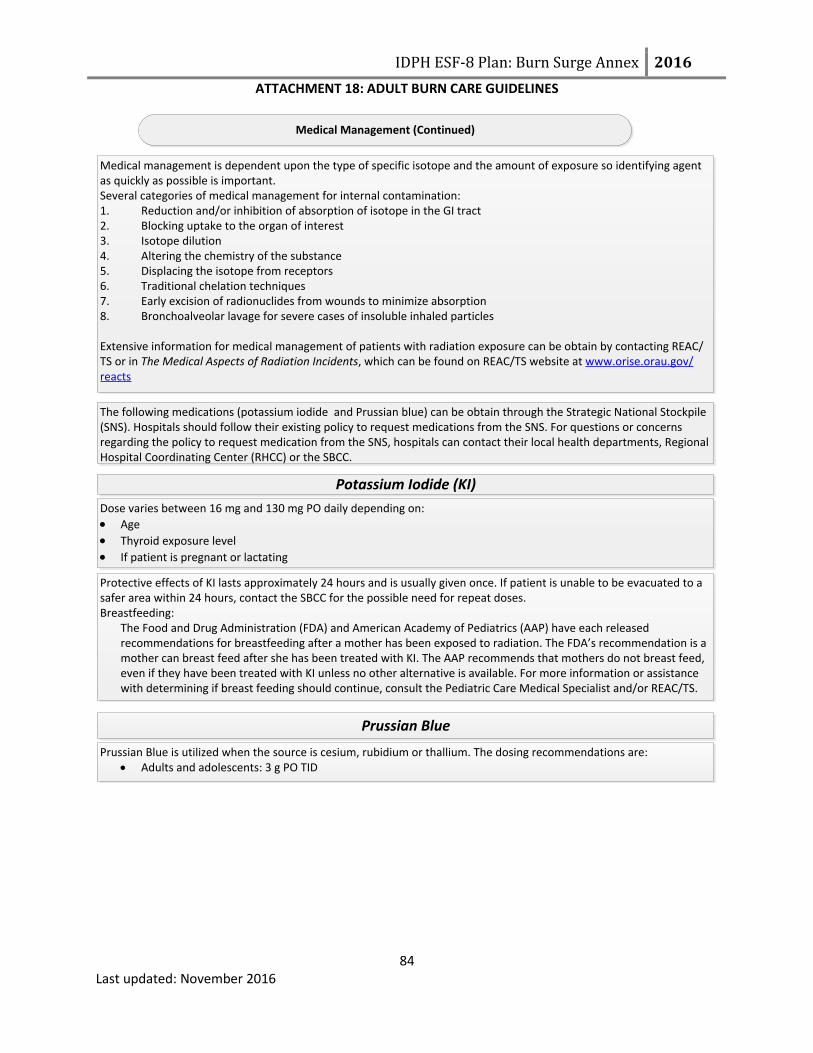
Medical Management (Continued)
Medical management is dependent upon the type of specific isotope and the amount of exposure so identifying agent as quickly as possible is important.Several categories of medical management for internal contamination:1. Reduction and/or inhibition of absorption of isotope in the GI tract2. Blocking uptake to the organ of interest3. Isotope dilution4. Altering the chemistry of the substance5. Displacing the isotope from receptors6. Traditional chelation techniques7. Early excision of radionuclides from wounds to minimize absorption8. Bronchoalveolar lavage for severe cases of insoluble inhaled particles
Extensive information for medical management of patients with radiation exposure can be obtain by contacting REAC/TS or in The Medical Aspects of Radiation Incidents, which can be found on REAC/TS website at www.orise.orau.gov/reacts
The following medications (potassium iodide and Prussian blue) can be obtain through the Strategic National Stockpile (SNS). Hospitals should follow their existing policy to request medications from the SNS. For questions or concerns regarding the policy to request medication from the SNS, hospitals can contact their local health departments, Regional Hospital Coordinating Center (RHCC) or the SBCC.
Potassium Iodide (KI)
Dose varies between 16 mg and 130 mg PO daily depending on:
Age
Thyroid exposure level
If patient is pregnant or lactating
Protective effects of KI lasts approximately 24 hours and is usually given once. If patient is unable to be evacuated to a safer area within 24 hours, contact the SBCC for the possible need for repeat doses. Breastfeeding:
The Food and Drug Administration (FDA) and American Academy of Pediatrics (AAP) have each released recommendations for breastfeeding after a mother has been exposed to radiation. The FDA’s recommendation is a mother can breast feed after she has been treated with KI. The AAP recommends that mothers do not breast feed, even if they have been treated with KI unless no other alternative is available. For more information or assistance with determining if breast feeding should continue, consult the Pediatric Care Medical Specialist and/or REAC/TS.
Prussian Blue
Prussian Blue is utilized when the source is cesium, rubidium or thallium. The dosing recommendations are: Adults and adolescents: 3 g PO TID
IDPH ESF-8 Plan: Burn Surge Annex 2016
ATTACHMENT 18: ADULT BURN CARE GUIDELINES
85 Last updated: November 2016
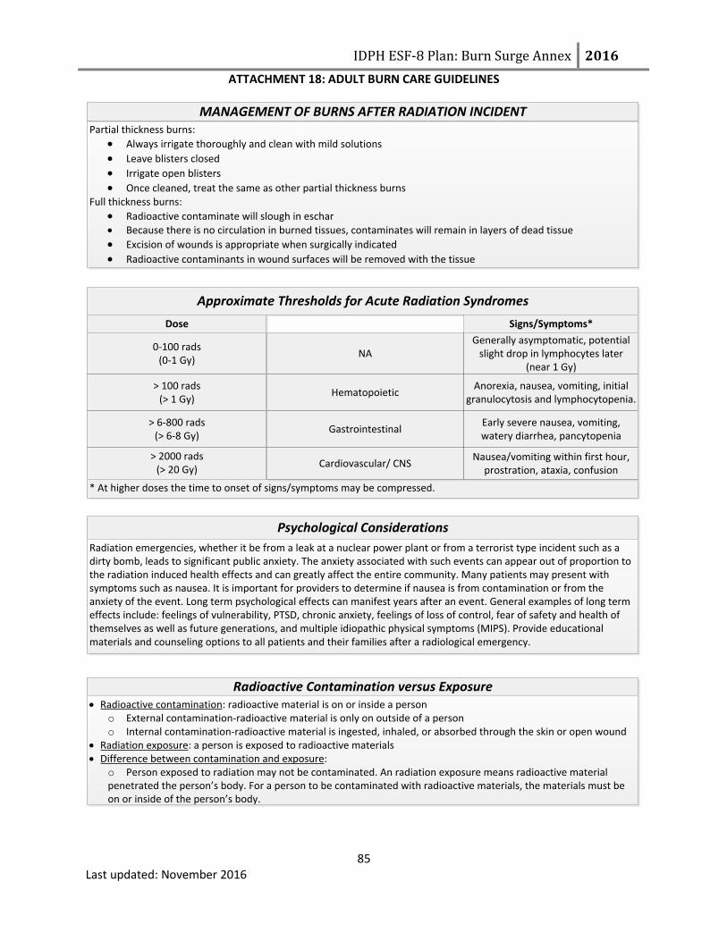
Partial thickness burns:
Always irrigate thoroughly and clean with mild solutions
Leave blisters closed
Irrigate open blisters
Once cleaned, treat the same as other partial thickness burnsFull thickness burns:
Radioactive contaminate will slough in eschar Because there is no circulation in burned tissues, contaminates will remain in layers of dead tissue
Excision of wounds is appropriate when surgically indicated
Radioactive contaminants in wound surfaces will be removed with the tissue
MANAGEMENT OF BURNS AFTER RADIATION INCIDENT
Approximate Thresholds for Acute Radiation Syndromes
Dose Signs/Symptoms*
0-100 rads(0-1 Gy)
> 100 rads(> 1 Gy)
> 6-800 rads(> 6-8 Gy)
> 2000 rads(> 20 Gy)
* At higher doses the time to onset of signs/symptoms may be compressed.
NA
Hematopoietic
Gastrointestinal
Cardiovascular/ CNS
Generally asymptomatic, potential slight drop in lymphocytes later
(near 1 Gy)
Anorexia, nausea, vomiting, initial granulocytosis and lymphocytopenia.
Early severe nausea, vomiting, watery diarrhea, pancytopenia
Nausea/vomiting within first hour, prostration, ataxia, confusion
Psychological Considerations
Radiation emergencies, whether it be from a leak at a nuclear power plant or from a terrorist type incident such as a dirty bomb, leads to significant public anxiety. The anxiety associated with such events can appear out of proportion to the radiation induced health effects and can greatly affect the entire community. Many patients may present with symptoms such as nausea. It is important for providers to determine if nausea is from contamination or from the anxiety of the event. Long term psychological effects can manifest years after an event. General examples of long term effects include: feelings of vulnerability, PTSD, chronic anxiety, feelings of loss of control, fear of safety and health of themselves as well as future generations, and multiple idiopathic physical symptoms (MIPS). Provide educational materials and counseling options to all patients and their families after a radiological emergency.
Radioactive Contamination versus Exposure Radioactive contamination: radioactive material is on or inside a person
o External contamination-radioactive material is only on outside of a person o Internal contamination-radioactive material is ingested, inhaled, or absorbed through the skin or open wound
Radiation exposure: a person is exposed to radioactive materials Difference between contamination and exposure:
o Person exposed to radiation may not be contaminated. An radiation exposure means radioactive material penetrated the person’s body. For a person to be contaminated with radioactive materials, the materials must be on or inside of the person’s body.
IDPH ESF-8 Plan: Burn Surge Annex 2016
ATTACHMENT 18: ADULT BURN CARE GUIDELINES
86 Last updated: November 2016
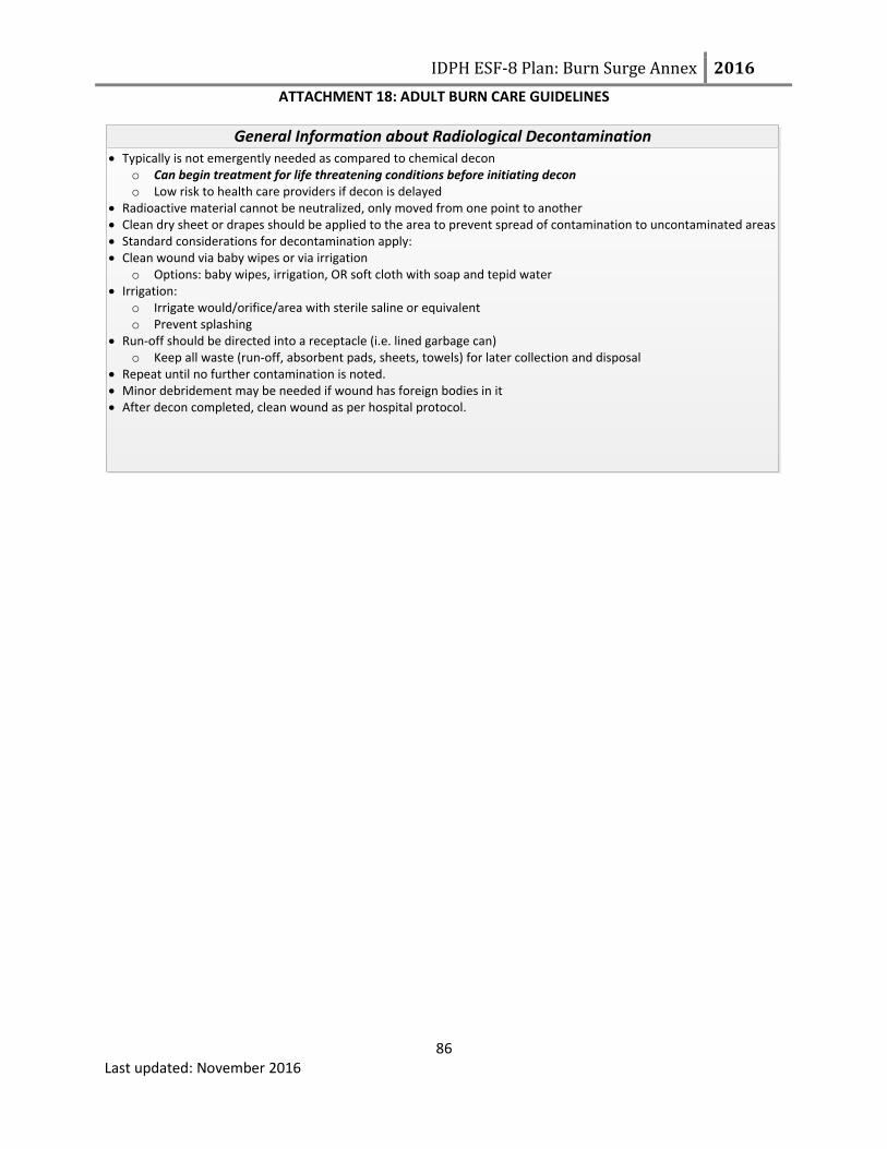
General Information about Radiological Decontamination Typically is not emergently needed as compared to chemical decon
o Can begin treatment for life threatening conditions before initiating decono Low risk to health care providers if decon is delayed
Radioactive material cannot be neutralized, only moved from one point to another Clean dry sheet or drapes should be applied to the area to prevent spread of contamination to uncontaminated areas Standard considerations for decontamination apply: Clean wound via baby wipes or via irrigation
o Options: baby wipes, irrigation, OR soft cloth with soap and tepid water Irrigation:
o Irrigate would/orifice/area with sterile saline or equivalento Prevent splashing
Run-off should be directed into a receptacle (i.e. lined garbage can)o Keep all waste (run-off, absorbent pads, sheets, towels) for later collection and disposal
Repeat until no further contamination is noted. Minor debridement may be needed if wound has foreign bodies in it After decon completed, clean wound as per hospital protocol.
IDPH ESF-8 Plan: Burn Surge Annex 2016
ATTACHMENT 18: ADULT BURN CARE GUIDELINES
87 Last updated: November 2016
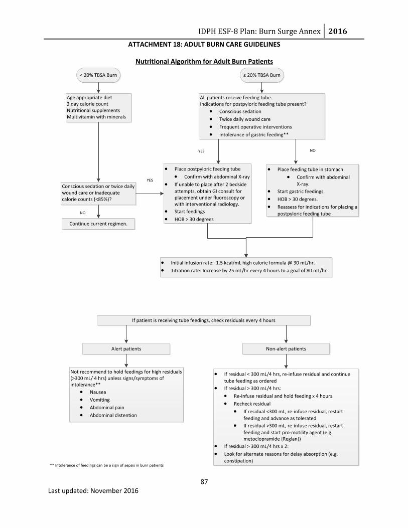
Nutritional Algorithm for Adult Burn Patients
< 20% TBSA Burn ≥ 20% TBSA Burn
Age appropriate diet2 day calorie countNutritional supplementsMultivitamin with minerals
All patients receive feeding tube. Indications for postpyloric feeding tube present?
Conscious sedation
Twice daily wound care
Frequent operative interventions
Intolerance of gastric feeding**
Conscious sedation or twice daily wound care or inadequate calorie counts (<85%)?
Continue current regimen.
Place postpyloric feeding tube
Confirm with abdominal X-ray
If unable to place after 2 bedside
attempts, obtain GI consult for placement under fluoroscopy or with interventional radiology.
Start feedings
HOB > 30 degrees
Place feeding tube in stomach
Confirm with abdominal
X-ray.
Start gastric feedings.
HOB > 30 degrees.
Reassess for indications for placing a
postpyloric feeding tube
YES
YES
Initial infusion rate: 1.5 kcal/mL high calorie formula @ 30 mL/hr.
Titration rate: Increase by 25 mL/hr every 4 hours to a goal of 80 mL/hr
If patient is receiving tube feedings, check residuals every 4 hours
Alert patients Non-alert patients
Not recommend to hold feedings for high residuals (>300 mL/ 4 hrs) unless signs/symptoms of intolerance**
Nausea
Vomiting
Abdominal pain
Abdominal distention
If residual < 300 mL/4 hrs, re-infuse residual and continue
tube feeding as ordered
If residual > 300 mL/4 hrs:
Re-infuse residual and hold feeding x 4 hours
Recheck residual
If residual <300 mL, re-infuse residual, restart
feeding and advance as tolerated
If residual >300 mL, re-infuse residual, restart
feeding and start pro-motility agent (e.g. metoclopramide {Reglan})
If residual > 300 mL/4 hrs x 2:
Look for alternate reasons for delay absorption (e.g.
constipation)** Intolerance of feedings can be a sign of sepsis in burn patients
NO
NO


![Manning, Judy [IDPH]...Manning, Judy [IDPH] From; Ashley Goddard Thursday, October 22 201, 5 2:27 PM Ashley Goddard Manning, Judy [IDPH] Re: dry needling](https://img.pdfslide.net/doc/110x75/602d98d517162d08910cf5e1/manning-judy-idph-manning-judy-idph-from-ashley-goddard-thursday-october.jpg)