Embed Size (px)
Citation preview

IEEE SENSORS JOURNAL, VOL. 15, NO. 1, JANUARY 2015 417
A Subcutaneous Biochip for Remote Monitoringof Human Metabolism: Packaging and
Biocompatibility AssessmentAndrea Cavallini, Tanja Rezzonico Jost, Seyedeh Sara Ghoreishizadeh, Jacopo Olivo, Maaike Op de Beeck,
Benjamin Gorissen, Fabio Grassi, Giovanni De Micheli, Fellow, IEEE, and Sandro Carrara, Senior Member, IEEE
Abstract— This paper represents the extended version of theconference paper “Developing highly-integrated subcutaneousbiochips for remote monitoring of human metabolism” pre-sented at the IEEE Sensors Conference 2012, and presentsdata on assembly, packaging and short term in vitro and invivo biocompatibility evaluation of a fully implantable biosensorarray. The device was realized integrating three building blocks:1) a multielectrode platform; 2) an inductive coil; and 3) anintegrated circuit. The entire system measures 2.2 mm × 2.2mm × 15 mm. Corrosion of electronic components and leakingof potentially hazardous substances in the body is preventedwith a conformal coating of Parylene C, while an outer packageof medical grade silicone was employed to create a soft shellsuitable for implantation. Biocompatibility experiments did notshow in vitro cytotoxicity in the considered period of 7 days,while comparison between 7 and 30 days in vivo implantationsshowed significant reduction of the inflammatory response intime, suggesting normal host recovery.
Index Terms— Biosensors, implantable biomedical devices,electronics packaging.
I. INTRODUCTION
CONTINUOUS monitoring of humans is already in themarket for glucose [1] and lactate [2], thanks to the
electrochemical sensing, and prototypes of fully implantable
Manuscript received May 11, 2014; accepted July 7, 2014. Date of publi-cation July 18, 2014; date of current version November 7, 2014. This workwas supported in part by the SNF Sinergia Project CRSII2 127547/1 entitledInnovative Enabling Micro- Nano-Bio-Technologies for Implantable Systemsin Molecular Medicine and Personalized Therapy, the i-IronIC project witha grant from the Swiss Nano- Tera.ch, and evaluated by the SNF and theERC project, and by the NanoSys project within the program ERC-2009-AdG- 246810. The associate editor coordinating the review of this paper andapproving it for publication was Dr. Perena I. Gouma.
A. Cavallini, S. S. Ghoreishizadeh, J. Olivo, G. De Micheli,and S. Carrara are with the Integrated System Laboratory, ÉcolePolytechnique Fédérale de Lausanne, Lausanne CH-1015, Switzerland(e-mail: [email protected]: [email protected];[email protected]; [email protected]; [email protected]).
T. R. Jost and F. Grassi are with the Istituto di Ricerca in Biomedi-cina, Bellinzona 6500, Switzerland (e-mail: [email protected];[email protected]).
M. Op de Beeck is with the Heterogeneous Integrated MicrosystemsDepartment, imec, Leuven 3000, Belgium (e-mail: [email protected]).
B. Gorissen is with the Machine Design and Automation Section,Katholieke Universiteit Leuven, Leuven 3000, Belgium (e-mail:[email protected]).
Color versions of one or more of the figures in this paper are availableonline at http://ieeexplore.ieee.org.
Digital Object Identifier 10.1109/JSEN.2014.2339638
Fig. 1. The fully implantable platform. Left: assembly schematics; right,packaged device.
glucose sensors have been validated up to 8 month in mice [3]and up to one year in pigs [4]. The next step will be theextension of this technology to other relevant metabolitessuch as, glutamate [5], ATP [6] and drugs [7], [8]. An openchallenge is the integration of all these metabolites in asubcutaneous biosensor array capable to provide minimallyinvasive human telemetry. Such system must satisfy severalrequirements: sensitivity, as the operative concentration ofthe biosensor must correspond to the therapeutic range ofthe target compound in the body; specificity, as the sensorsmust be able to operate in complex solutions like plasma orinterstitial fluid without detecting any interferant; autonomy,as the device must be capable to perform automatically themeasurements without the need of external input and withoutbeing subjected to power shortages; biostability, as the deviceshould remain functional after the insertion; biocompatibility,because the implant must be well tolerated by the host andcause a limited foreign body reaction;
In this paper we present a prototype of a fully implantabledevice based on three building blocks: a passive chip hosting5 independent biosensor electrodes, a temperature and a pHsensor; an inductive coil for the remote powering of thesensor towards an external wearable device, and an integratedcircuit performing the electrochemical measurements (fig. 1).Such device represents a novelty compared to the existingimplantable sensors, measuring a single compound, batterypowered, and bearing a hard packaging. The device sensitivitycan be promoted by nanostructuring the electrode surface withcarbon nanotubes (CNT). Although the in-vivo applicationof CNT is controversial in healthcare applications due todiscordant results about their safety [9], the addition of CNT
1530-437X © 2014 IEEE. Personal use is permitted, but republication/redistribution requires IEEE permission.See http://www.ieee.org/publications_standards/publications/rights/index.html for more information.

418 IEEE SENSORS JOURNAL, VOL. 15, NO. 1, JANUARY 2015
proved to be essential to detect physiological concentrations ofanalytes in human plasma, as demonstrated by our previousworks [7], [8]. Furthermore, various measures are taken toprevent CNT release from the implant. The presence ofmultiple sensors is a strategy to achieve specificity in electro-chemical drug detection with P450 biosensors: cytochromesp450 have a broad substrate range and atypical kinetics.Drugs are detected by cyclic voltammetry, and recognizedby their specific electrochemical signature, a characteristicpotential shift of the p450 reduction peak [10]. The presenceof different biosensors specific for the same target, combinedwith a temperature sensor and a pH sensor, enables the correctinterpretation of the electrochemical signature of the analytes[11], [12]. A battery-less system for the remote powering ofthe device [13], and an integrated circuit capable of generatingon-board voltage ramps [14], ensure the device autonomy;finally, to promote biostability and biocompatibility, we addeda double protection for electrodes and electronic components:enzymes and CNTs were entrapped in a chitosan (CHT) matrixand then sealed behind a porous polycarbonate membrane.Chitosan is a natural polysaccharide with unique biologi-cal properties including non-toxicity, physiological inertness,affinity to proteins, hemostatic fungistatic and antitumoralproperties, which already found employ in biosensors [15],while polycarbonate membranes are commonly employed asmicrodialysis filters in commercial biosensors [1]. Corrosionof electronic components and leaking of potentially hazardoussubstances in the body was prevented by a conformal coatingof Parylene C, an inert and biocompatible polymer with wide-spread industrial use as diffusion barrier. An outer package ofmedical grade silicone was then employed to create a soft shellsuitable for implantation. The present work, which representan extension of the conference paper “Developing highly-integrated subcutaneous biochips for remote monitoring ofhuman metabolism” [16], presented at the conference IEEESensors 2012, focus on the packaging and the short-termbiocompatibility evaluation of our implantable device. Theefficacy of the parylene C barrier, as well as the toxicity ofcarbon nanotubes, have been assessed with a 7-day in-vitrocytotoxicity elution test conform to the ISO-10993-1 standard.The integrity of the CHT/CNT matrix and the efficacy ofthe polycarbonate membrane in preventing CNT leaking weretested by exposing the materials to solutions of different natureand pH at 37 °C for one week. The final packaged devicewas then implanted in mice for 7 and 30 days to evaluate theinflammatory response.
II. SYSTEM AND BUILDING BLOCKS DESIGN
A. Passive Chip
Choice of materials and design was done consideringbiocompatibility and simplicity of fabrication as of primaryimportance. Silicon wafers with 500nm of native oxide werechosen as substrate. Chip metallization was realized by evap-oration of 10nm of Ti, followed by 100nm of Pt. Metalpassivation was made via atomic layer deposition of 20nmof Al2 O3. Passivation openings were made by dry etchingwith Argon Ion Milling. Only two masks resulted necessary
Fig. 2. Photographs of the passive chip. Working electrodes were realized indifferent geometries: as arrays of 10 and 40 μm and as a single electrode of500 μm (left); center whole platform right, temperature and pH sensors, padsfor IC integration and schematics of the electrochemical cell. image from [20].
for the whole microfabrication. As substrate, silicon has beenchosen over glass for its better thermal conductibility, in orderto improve the dissipation of heat generated by the integratedcircuit. Pt metallization was chosen for three main reasons:1) biocompatibility and resistance to corrosion, 2) pseudo-reference electrode behavior, 3) employment in the fabricationof resistive thermal devices with a linear range suitable tomeasure physiological temperatures [17]. Al2 O3 is a biocom-patible material already used in biomedical coatings [18].Atomic layer deposition was chosen among other techniquesfor its capability to generate thin, uniform and pinhole freepassivations [19]. Figure 2 shows photographs of the passivechip: the platform measures 2.2 × 15mm, and host fiveindependent platinum working electrodes (WE) with commonreference (RE) and counter (CE) electrodes; a pH sensorbased on an anodic iridium oxide film; a resistive platinumthermal device (Pt RTD) as temperature sensor, and pads forthe wire bonding of the integrated circuit. The inclusion ofcommon reference and counter electrode allowed simplifyingthe interconnections with the sensing circuitry, optimizing atthe same time the available space. The working electrodescan be individually functionalized with high precision towardsa single-step electrodeposition of a solution containing CHT0.7% w/v; MWCNT 1% and the enzyme at an appropriateworking concentration (i.e. 15 mg/ml). The polarization of anelectrode at +1.5 mV for at least 300” creates a localizedregion of high pH that can exceed chitosans solubility limit,allowing CHT polymerization and the entrapment of any othercompound present in the original solution with high spatialselectivity, while variations in the electrodeposition time allowto control the amount of material deposited. Characterizationof the electrodes, biosensors, electrodepositon, pH and tem-perature sensors is extensively reported in [20].
B. Measurement Circuit
In order to integrate sensors and electronics into a singledevice, the front-end electronics for the realization of the twomostly used detection techniques, Chronoamperometry (CA)and Cyclic Voltammetry (CV), must be carefully designed.At present, there is little literature concerning the integrationof the waveform generator to the implantable biochip whichis essential for a fully-integrated CV measurement [21], [22],and most of the designed electronics for the biosensors use anexternal generator. Li et al. described a ramp generator circuit

CAVALLINI et al.: SUBCUTANEOUS BIOCHIP FOR REMOTE MONITORING OF HUMAN METABOLISM 419
Fig. 3. Proposed architecture for the frontend electronics.
that can be integrated in a biosensor array [23]; however thesystem presented has limited versatility, since all the electrodesin the array are subjected to the same potential. In our workthe CE and RE are shared while each WE is designed tohost different biosensors requiring different potentials, or evendifferent methods to be activated. For this reason, a specificvoltage, either fixed or varied in time, must be applied to eachWE to enable the sensing of the various compounds. Fig. 3shows the architecture of the proposed frontend electronics,which consists of three main parts: 1) a ramp generator circuit,2) a potentiostat, and 3) a current readout circuit. The rampgenerator produces a very-low slope and a low-frequency rampvoltage, and is capable to generate a triangular waveform witha slope adjustable from less than 10 mV/sec to more than100 mV/sec with a rail-to-rail swing. The steps are 3.3mVand the size of the circuit is 0.122mm2 [24]. The circuit isbased on the Direct Digital Synthesis method (DDS), whichincludes a numerically controlled oscillator and a Digital-to-Analog Converter (DAC). The oscillator produces a quantizedversion of the desired waveform, whose period is controlled bythe digital word contained in the Frequency-Control-Register(FCR). The resulting waveform is then converted to analog bythe DAC [24].
The circuit has been implemented in 0.18μm technology.Simulations are done using the electrical equivalent model ofthe biosensor from [25]. Simulation results show the readoutcircuit senses currents in the range of +/− 5μA with anequivalent input referred current noise of 3nA rms. Highlycontrollable low triangular waveform is also achieved. Furthersimulation and measurement results on the measurement cir-cuit are reported in [14], [24], and [26]. The maximum powerconsumption of the measurement circuit is 530μW, a valuesuitable for the remote powering of our device.
C. Receiving Coil
Remote powering through inductive link is one of the mostpromising approaches to supply power to implantable devices,and the miniaturization of power-efficient inductors, is still anopen topic. To this end, we propose an implantable coil basedon the multi-layer approach. Such strategy, already exploited
in integrated circuits [27], [28], it is not yet commonly usedon printed circuit boards (PCB). In a multi-layer coil, a spiralinductor is replicated on the two layers of different PCBs.The boards are then stacked, and the inductors are electricallyconnected as shown in fig. 1. Our coil was obtained by usinga 12-layers, 21-turns inductor of 30 mm with a thicknessof 816.4μm. Due to the higher number of turns, our coilhas an improved coupling with the external receiver whencompared to a single-layer coil of equivalent surface [29].The system also performs bidirectional data communicationwithout any implanted RF transmitter: downlink communi-cation is obtained via Amplitude Shift Keying (ASK), whileuplink communication exploits a backscattering technique.In backscattering, the internal load is modulated accordingto the outgoing bit-stream; this change is detected by theexternal part as a variation of the current flowing on theexternal inductor. A high efficiency class-E power amplifierwas utilized to drive the external inductor. Powered by twothin lithium-ion polymer batteries, the system can transferup to 15mW over a distance of 6mm in air. The maximumlink efficiency measured was 13%. Furthermore, the systemcan transfer up to 1.17mW when a 17mm beef sirloin isplaced between the inductors, which is enough power for themeasurement circuit.
III. BIOCOMPATIBILITY ASSESSMENT -MATERIALS AND METHODS
A. Matrix Corrosion Test
CHT/CNT dispersion was prepared according to [6]. Dropsof 10μl were cast onto a 12 well plate and dried under laminarflow. Half of the samples were protected with a polycarbonatemembrane (Cyclopore track etched membrane, cut off 100nm,Whatman) and sealed with fast curing medical grade silicone(Med2-4220, Nusil). All samples were covered with 1mlsolutions of milliQ water, PBS 1× pH4, or Mouse EmbryonicFibroblasts (MEF) growth media, and put in a cell incubatorat 37 °C (5% C O2) for 7 days. The 8th day the solutions wereremoved and the samples rinsed twice with DI water beforebeing dried in air under laminar flow. The CHT/CNT matrixintegrity and the dispersion of CNT aggregates on the wellplate surface were then inspected with an optical microscope.
B. In-Vitro Cytotoxicity Test
Preparation of Contaminated Medium: The growth mediumwas obtained adding to a Dulbecco Modified Eagle Medium,Fetal Calf Serum (10% v/v); L-glutamine 200 mM (1% v/v);Glutamax (2% v/v); non-essential aminoacids (1% v/v), Peni-cillin/Streptavidin (1% v/v). All the reagents were purchasedfrom Gibco. Test materials were placed in cell culture dishes,sterilized with ethanol 70% and dried under laminar flow.MEF medium was added according to the ratio 1ml/6cm2
of material. The samples were put in a cell incubator at37 °C, 5% C O2 for 7 days. A negative control of fresh,uncontaminated MEF medium was also included to accountfor the aging of nutrients in solution. The contaminatedmedium and the control were then collected, stored at 4 °Cand used within 3 days. Cell culture: Mouse Embryonic
420 IEEE SENSORS JOURNAL, VOL. 15, NO. 1, JANUARY 2015
Fibroblasts (MEF) were extracted, seeded in 12-well platesat the concentration of 1.2 × 10⁴ cells/ml and grown for3 days. The 3t h day, the growth medium was collected andreplaced with the contaminated terrain and the control. Cellswere grown for 4 additional days before being inspected forviability and toxicity. Assessment of Viability: The specimenswere rejected when the viability of healthy control cellsresulted less than 75% [30]. Control wells were incubated 30with 1μg/ml of DAPI and PI fluorescent dyes (Invitrogen)to highlight respectively total cell nuclei and dead cells. Foreach well, three DAPI and PI counts in different spots wereperformed. Cell viability was then calculated according to theequation
viabili ty = viable cells
total cells100% (1)
where viable cells is the total number of DAPI counts minusPI counts and total cells is total number of DAPI counts.
Toxicity Evaluation: Cells were incubated 30 with 1 μg/mlof Calcein AM (Invitrogen). The fluorescence in each samplewas immediately measured with a commercial scanner (Tecaninfinite M1000, excitation λ485nm, emission 525nm). Thetotal fluorescence count for each well was obtained averaging225 readings evenly distributed in a 15 × 15 round shapealong the well area. Since Calcein AM stains only living cells,cytotoxicity was evaluated comparing the fluorescence inten-sity of the contaminated samples with the control. Accordingto the United States Pharmacopeia (USP) standards, intensitydecrease of more than 20% from the control was consideredcytotoxic. Additionally, cells were inspected for morphologicabnormalities by fluorescence microscopy. Samples presentinga majority of abnormal cells with respect to the control wereconsidered cytotoxic.
C. Assembly and Packaging
Loctite 3211 USP class VI biocompatible glue was pur-chased from Loctite. Adhesion promoter Silane A174 wasobtained from Merck and applied to the samples accordingto the procedure described in [31]; Parylene C was boughtfrom Speciality Coating Systems and deposited by chemicalvapor deposition using a Comelec C-30-S Parylene DepositionSystem. The plexiglass mold for the final silicone encapsula-tion was realized by micromachining. Biocompatible medical-grade silicone (Med-6033) was bought from Nusil.
D. In-Vivo Biocompatibility
Microchips were cleaned and disinfected with Gigaseptinstru AF (Schuelke), placed in cell culture dishes, sterilizedwith ethanol 70% and dried under laminar flow. An Air Pouch(AP) was created by subcutaneous injection of sterile air in theback of male C57BL/6 mice at day 1 (5mL) and day 3 (3mL);this procedure creates a cavity of 1.5cm diameter and 0.5cmheight. At day 6, mice were anesthetized with isoflurane 4%,shaved and locally sterilized with Betadine Solution; the sterilemicrochips were implanted and the cavity sutured with Vicryl6.0 (Provet AG). As a control of local inflammation, bacteriallipopolysaccharide (LPS) (50g/mouse) (LabForce AG) was
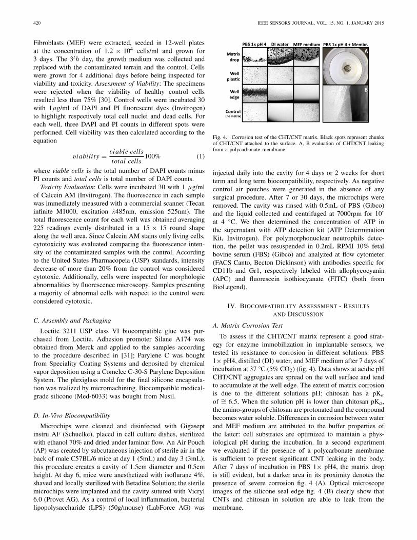
Fig. 4. Corrosion test of the CHT/CNT matrix. Black spots represent chunksof CHT/CNT attached to the surface. A, B evaluation of CHT/CNT leakingfrom a polycarbonate membrane.
injected daily into the cavity for 4 days or 2 weeks for shortterm and long term biocompatibility, respectively. As negativecontrol air pouches were generated in the absence of anysurgical procedure. After 7 or 30 days, the microchips wereremoved. The cavity was rinsed with 0.5mL of PBS (Gibco)and the liquid collected and centrifuged at 7000rpm for 10’at 4 °C. We then determined the concentration of ATP inthe supernatant with ATP detection kit (ATP DeterminationKit, Invitrogen). For polymorphonuclear neutrophils detec-tion, the pellet was resuspended in 0.2mL RPMI 10% fetalbovine serum (FBS) (Gibco) and analyzed at flow cytometer(FACS Canto, Becton Dickinson) with antibodies specific forCD11b and Gr1, respectively labeled with allophycocyanin(APC) and fluorescein isothiocyanate (FITC) (both fromBioLegend).
IV. BIOCOMPATIBILITY ASSESSMENT - RESULTS
AND DISCUSSION
A. Matrix Corrosion Test
To assess if the CHT/CNT matrix represent a good strat-egy for enzyme immobilization in implantable sensors, wetested its resistance to corrosion in different solutions: PBS1× pH4, distilled (DI) water, and MEF medium after 7 days ofincubation at 37 °C (5% CO2) (fig. 4). Data shows at acidic pHCHT/CNT aggregates are spread on the well surface and tendto accumulate at the well edge. The extent of matrix corrosionis due to the different solutions pH: chitosan has a pKa
of ∼= 6.5. When the solution pH is lower than chitosan pKa ,the amino-groups of chitosan are protonated and the compoundbecomes water soluble. Differences in corrosion between waterand MEF medium are attributed to the buffer properties ofthe latter: cell substrates are optimized to maintain a phys-iological pH during the incubation. In a second experimentwe evaluated if the presence of a polycarbonate membraneis sufficient to prevent significant CNT leaking in the body.After 7 days of incubation in PBS 1× pH4, the matrix dropis still evident, but a darker area in its proximity denotes thepresence of severe corrosion fig. 4 (A). Optical microscopeimages of the silicone seal edge fig. 4 (B) clearly show thatCNTs and chitosan in solution are able to leak from themembrane.

CAVALLINI et al.: SUBCUTANEOUS BIOCHIP FOR REMOTE MONITORING OF HUMAN METABOLISM 421
Fig. 5. Evaluation of materials cytotoxicity by calcein staining - averagefluorescence values. Error bars, standard deviation (1σ ).
B. In-Vitro Citotoxicity TestThe building blocks of the implantable sensor are partially
made from non-biocompatible or potentially toxic materials.For example, the receiving coil presents a copper metalliza-tion, notably cytotoxic [32], while materials of the integratedcircuit and of the auxiliary electric components, realized byexternal companies, are not totally disclosed, and have to beconsidered as potentially harmful. A conformal coating of3μm parylene C was employed to prevent both corrosion ofthe electrical parts and toxic metals leaking in the surroundingtissue. Parylene efficacy was evaluated by elution tests usingthe copper receiving coil as test substrate. When immersedin a fluid, the copper contained in the coil diffuses in thesolution, producing severe cytotoxicity even at very smallconcentrations. Cytotoxicity was assessed with elution testsaccording to the ISO 10993-1 guidelines. Each experiment wasconducted in triplicate and included a positive control to assurereproducibility and reliability of results. Fig. 5A presents theaverage fluorescence of Calcein-AM stained cells. After 4 daysof incubation with the contaminated medium, cells grown inpresence of the terrain eluted with the unprotected coil yieldedsevere cytoxicity, as demonstrated by very low fluorescence;On the other hand, the parylene C coating proved effectivein preventing severe cytotoxicity, as the fluorescence intensityresulted the 88% of the intensity of the control (cells grownin uncontaminated medium). It is worth mentioning that whilevariations up to 20% from the control are not consideredcytotoxic by the USP guidelines, such variability could beunacceptable in long term implants [30]. The protection of theparylene C coating can be enhanced improving its substrateadhesion and its thickness. Accelerated elution tests performedon our silicon electrodes (3 days at 70 °C in PBS 1× pH7.4),showed that coatings of 3μm on surfaces not treated withadhesion promoter, tend to form bubbles and eventually detachfrom the surface (fig. 7). In a second experiment we evaluatedthe cytotoxicity of the CHT/CNT matrix (fig. 6B). After 7 daysof elution and four days of incubation with healthy cells,no significant differences with the control cells have beenobserved, proving that in the considered period the CNTleaking is not cytotoxic. A possible explanation is that CNTtoxicity depends on many factors like length, functionalizationor aggregation [9], [33]. Also, the tight wrapping of chitosanto the nanotubes, which is also responsible for their dispersionin aqueous solutions [34], might shield the nanoparticle andits reactive groups, reducing the inherent toxicity and pre-venting their aggregation. The lack of short-term cytotoxicityof chitosan/CNT nanocomposites is a very promising resulttowards the employment of this nanomaterial in biomedical
Fig. 6. Morphology of MEF grown in different contaminated media.A) control, B) protected coil, C) CHT/CNT matrix, and D) unprotected coil.
Fig. 7. Accelerated elution test of the sensor platform covered by 3μm ofparylene C. Optical microscope images. A) Before the test; B) ater 1 weekat 37 °C in MEF medium; and C) after 3 additional days at 70 °C in MEFmedium.
applications; however, it is important to say that the applicationof carbon nanotubes in implantable devices relies stronglyon their long-term safety: at best of our knowledge, up todate there are no exhaustive studies concerning the long-term effects of functionalized CNT in living organisms or theimpact of these substances on the environment. As additionalcytotoxicity control we performed a morphological analysis ofthe cells exposed to the elution fluids. Fig. 6 shows that cellsfrom the protected coil (B) and from the CHT/CNT matrix(C) are similar in shape and distribution to the control (A).No living cells were found on the sample with fluid elutedfrom the unprotected coil (D, bright field image). The giantcells shown in the pictures are due to the initial cell density,which after 7 days resulted too low to give a confluent cover-age and the typical tight and elongated shape of fibroblasts.
C. Assembly and PackagingThe sensor building blocks were glued together using a
USP class VI biocompatible glue. Component interconnectionwas realized with Al wire bonding and protected with globtop. To further improve parylene C adhesion and moisturepenetration, the assembled platform was treated with silaneA-174 and coated with 16μm of parylene C. The outersilicon shell was realized by placing the implant into aplexiglass mold and injecting biocompatible medical-gradesilicone. To increase the host comfort, the outer shell wasmade 1mm thick and with rounded corners. In a designvariant, two wingsof 3 × 3mm placed along the main bodyhave been included to prevent the sensor to capsize afterimplantation, since misalignment between the receving coiland the external wearable device can compromise the remotepowering of the sensor array. 0.1μl of CHT/CNT suspensionwas then manually drop cast on the electrodes and dried in air.

422 IEEE SENSORS JOURNAL, VOL. 15, NO. 1, JANUARY 2015
Fig. 8. In-vivo biocompatibility results after 7 and 30 days. Top: neutrophilsinfiltration in the implant cavity (%); bottom, ATP concentration. Error bars,standard deviation (1σ ).
A polycarbonate membrane was placed above the electrodesand sealed to the external shell using fast curing medical gradesilicone. Without considering the external wings, the packageddevice measures 20 × 4.2 × 3mm. The final device is shownin fig. 1.
D. In-Vivo Biocompatibility
Potential sources of inflammation in implantable devices,can be attributed to the implant materials, shape and dimen-sions [35]. In order to investigate which elements are criticalin eliciting an inflammatory response, we fabricated devicesof different nature and shape to subcutaneously implant inmice. To account for inter-individual variability, each modelwas tested in eight different animals. Half of the mice carriedthe implant for 7 days, the other half for 30 days. At theend of the period, the implant site was washed with PBS,and levels of ATP and neutrophils in the elution liquid werecompared to follow the local inflammatory response. WhileATP release is a consequence of cellular necrosis and it istherefore a measure of the local cell damage, neutrophilsare recruited to the inflammation site by chemical signaling.Variations in neutrophil percentage at the implant site aretherefore informative of the status of tissue inflammation [36].Fig. 8 presents neutrophils and ATP variation in the liquidcollected from the implant site after 7 and 30 days. Sensorspackaged with a soft shell with or without external wings (W),were compared with dummy package replicas entirely madein biocompatible silicone. Bacterial lipopolysaccharide (LPS)was administered to a separate group of mice in order to inducean inflammatory response not caused by an artificial implant(positive control), while mice with air pouch only served asnegative control.
After 7 days, we measured high levels of ATP and neu-trophils, but both values substantially decreased after 30 daysin a way proportional to the insert complexity (presence of
wings and/or sensors), suggesting that the organism becametolerant to the implants. Data from neutrophils suggests thatthe insert complexity (presence of wings and/or sensors)tends to increase the short-term and long-term inflammatoryresponse. Considering average values, after 30 days, residualneutrophils in mice treated with winged inserts were 2-3 foldshigher than their counterparts, while the presence of the sensorplatform induced slightly higher inflammatory responses. ATPmeasurements in mice bearing the implant were affected bylarge statistical variability. A possible explanation is that uponremoval we found that several implants were displaced fromtheir original location. This may have led to internal tissuedamage due to mechanical friction, and therefore insurgenceof cellular damage and ATP release in some individuals [37].Unfortunately, this large variation in the ATP measurementsmakes difficult to establish a clear relationship betweenimplant complexity and extent of local cell death.
Values from LPS injection further suggest how after 30 daysthe host seems to accept the insert. Although LPS-inducedATP and neutrophils were detected in lower amounts comparedto the values obtained from implants, ATP concentration inanimals treated with LPS almost doubled after 30 days. Thepyrogenic effect of LPS is also evident with respect to the neu-trophils infiltration: in one month, LPS-treated mice presentedthe highest percentage of neutrophils and the slightest time-dependent reduction among the animals considered. From theneutrophils measurements it appears that the package shapehas a role in determining the inflammatory response, as thepresence of wings tends to increase the immune reaction.Although the introduction of stitching wings may be necessaryto hold the insert in place and correctly aligned with theexternal powering coil, their presence can exert an uncom-fortable localized pressure on some parts of the tissue, andangles where tissue stress might promote localized immuneresponses [38]. The presence of the electronic platform furtherincreased the inflammation response. However, it is importantto say that more than strong conclusions, these are trendssuggested by the average values obtained. In-vivo experimentstend to have large variability, and due to the reduced number ofanimals used, the overlap of error bars indicates that results arenot statistically different. A larger number of experiments, ordifferent investigation techniques are advised for a more pre-cise evaluation of the in-vivo biocompatibility. The removal ofcopper components from the sensor electronics, together withthicker parylene coatings may further reduce the inflammationcaused by the biosensor platform. The reduction of implantdimensions may also help increasing the host tolerance. In thisrespect, the bulkiest component of our platformis the inductivecoil, which with a thickness of 2mm, contributes to almost 4/5of the total platform volume. Our group is currently developinga single layer coil on silicon, which will reduce the platformthickness to 11.5mm.
V. CONCLUSION
This paper represent the extended version of the workdescrived in reference [16], and presents a novel, highlyintegrated system for human metabolism telemetry, focusing

CAVALLINI et al.: SUBCUTANEOUS BIOCHIP FOR REMOTE MONITORING OF HUMAN METABOLISM 423
on its packaging and biocompatibility. The system is made ofthree parts: a passive chip hosting 5 independent biosensors,a pH sensor and a temperature sensor; a CMOS integratedcircuit capable to perform the required electrochemical mea-surements, and a multi-layer antenna for the remote poweringof the device and the transmission of the sensing data. Theelectrodes of the passive chip can be functionalized withproper bio and nanostructures with high spatial precisiontowards electrodeposition of a solution of chitosan, carbonnanotubes and enzymes [16]. The CMOS IC showed goodperformance in simulations and measurements [24], [26], [39],while the receiving antenna resulted capable to collect enoughenergy to support the work of the IC [16]. The assembledsystem was packaged with an inner barrier of parylene C,an outer shell of biocompatible silicone and a polycarbonatemembrane to protect the sensor area. The biocompatibility ofthe device was then tested in-vitro and in-vivo.
In-vitro experiments on primary fibroblasts proved thatcoatings of 3M of parylene C are effective in preventingcopper leaking and cytoxicity for at least seven days; similartest on a chitosan/MWCNT matrix showed that release ofchitosan/MWCNT complexes in the growth terrain are notcytotoxic in the short term. This last result is in agreementwith recent studies demonstrating that the employ of short,functionalized CNT immobilized in a nanocomposite may beimplemented with a certain degree of safety. Although, inlast analysis, the fate of nanotubes in implantable devices isstill largely dependent by studies concerning their long-termtoxicity. In-vivo tests of the packaged sensor array demon-strated that the foreign body reaction significantly decreasedafter 30 days, suggesting normal recovery of the host. Packageshape is suspected to have a role in inducing the inflamma-tory response, suggesting that future implants must possesssmoother and simpler geometries.
Taken all together, these results demonstrate the feasibil-ity of the entire system. Future works will be directed inassessing the performance of the fully implantable systemin autonomous measurements in-vitro of different metabolitesand in promoting the long-term biocompatibility and biosta-bility of the device.
ACKNOWLEDGMENTS
J. O’Callaghan, K. Qian, and T. Miyazaki areacknowledged for the support and supervision duringthe in-vitro biocompatibility tests at Imec, Belgium.D. Sacchetto is acknowledged for the advices on themicrofabrication of the passive chip and M. De Marchi andC. Baj-Rossi for their help in formatting the text.
REFERENCES
[1] F. Valgimigli, F. Lucarelli, C. Scuffi, S. Morandi, and I. Sposato, “Eval-uating the clinical accuracy of GlucoMen day: A novel microdialysis-based continuous glucose monitor,” J. Diabetes Sci. Technol., vol. 4,no. 5, pp. 1182–1192, 2010.
[2] A. Poscia, D. Messeri, D. Moscone, F. Ricci, and F. Valgimigli, “A novelcontinuous subcutaneous lactate monitoring system,” Biosensors Bio-electron., vol. 20, no. 11, pp. 2244–2250, 2005.
[3] B. Yu, N. Long, Y. Moussy, and F. Moussy, “A long-term flexibleminimally-invasive implantable glucose biosensor based on an epoxy-enhanced polyurethane membrane,” Biosensors Bioelectron., vol. 21,no. 12, pp. 2275–2282, 2006.
[4] D. A. Gough, L. S. Kumosa, T. L. Routh, J. T. Lin, and J. Y. Lucisano,“Function of an implanted tissue glucose sensor for more than 1 yearin animals,” Sci. Translational Med., vol. 2, no. 42, pp. 42ra53–42ra53,2010.
[5] S. Carrara et al., “Single-metabolite bio-nano-sensors and system forremote monitoring in animal models,” in Proc. IEEE Sensors, Oct. 2011,pp. 716–719.
[6] A. Cavallini, G. De Micheli, and S. Carrara, “Comparison of three meth-ods of biocompatible multi-walled carbon nanotubes confinement forthe development of implantable amperometric adenosine-5’-triphosphatebiosensors,” Sensor Lett., vol. 9, no. 5, pp. 1838–1844, 2011.
[7] S. Carrara, A. Cavallini, V. Erokhin, G. Albini, and G. De Micheli,“Multi-panel drugs detection in human serum for personalized therapy,”Biosensors Bioelectron., vol. 26, no. 9, pp. 3914–3919, 2011.
[8] A. Cavallini, S. Carrara, G. De Micheli, and V. Erokhin, “P450-mediatedelectrochemical sensing of drugs in human plasma for personalizedtherapy,” in Proc. Conf. Ph.D. Res. Microelectron. Electron. (PRIME),Jul. 2010, pp. 1–4.
[9] S. K. Smart, A. I. Cassady, G. Q. Lu, and D. J. Martin, “The biocom-patibility of carbon nanotubes,” Carbon, vol. 44, no. 6, pp. 1034–1047,2006.
[10] D. L. Johnson, B. C. Lewis, D. J. Elliot, J. O. Miners, andL. L. Martin, “Electrochemical characterisation of the humancytochrome p450 cyp2c9,” Biochem. Pharmacol., vol. 69, no. 10,pp. 1533–1541, 2005.
[11] S. Carrara, A. Cavallini, A. Garg, and G. De Micheli, “Dynamical spotqueries to improve specificity in p450s based multi-drugs monitoring,”in Proc. Int. Conf. Complex Med. Eng. (ICME), Apr. 2009, pp. 1–6.
[12] S. Carrara, M. D. Torre, A. Cavallini, D. De Venuto, and G. De Micheli,“Multiplexing pH and temperature in a molecular biosensor,” in Proc.IEEE Biomed. Circuits Syst. Conf. (BioCAS), Nov. 2010, pp. 146–149.
[13] J. Olivo, S. Carrara, and G. De Micheli, “A study of multi-layer spiralinductors for remote powering of implantable sensors,” IEEE Trans.Biomed. Circuits Syst., vol. 7, no. 4, pp. 536–547, Aug. 2013.
[14] S. S. Ghoreishizadeh, S. Carrara, and G. De Micheli, “Circuit designfor human metabolites biochip,” in Proc. IEEE Biomed. Circuits Syst.Conf. (BioCAS), Nov. 2011, pp. 460–463.
[15] B. Krajewska, “Application of chitin-and chitosan-based materials forenzyme immobilizations: A review,” Enzyme Microbial Technol., vol. 35,no. 2, pp. 126–139, 2004.
[16] S. Carrara, A. Cavallini, S. Ghoreishizadeh, J. Olivo, and G. De Micheli,“Developing highly-integrated subcutaneous biochips for remote moni-toring of human metabolism,” in Proc. IEEE Sensors, Oct. 2012, pp. 1–4.
[17] P. R. N. Childs, J. R. Greenwood, and C. A. Long, “Reviewof temperature measurement,” Rev. Sci. Instrum., vol. 71, no. 8,pp. 2959–2978, Aug. 2000.
[18] T. V. Thamaraiselvi and S. Rajeswari, “Biological evaluation of bioce-ramic materials-a review,” Carbon, vol. 24, no. 31, p. 172, 2004.
[19] M. Knez, K. Nielsch, and L. Niinistö, “Synthesis and surface engineeringof complex nanostructures by atomic layer deposition,” Adv. Mater.,vol. 19, no. 21, pp. 3425–3438, 2007.
[20] A. Cavallini, C. Baj-Rossi, S. Ghoreishizadeh, G. De Micheli, andS. Carrara, “Design, fabrication, and test of a sensor array for perspectivebiosensing in chronic pathologies,” in Proc. IEEE Biomed. Circuits Syst.Conf. (BioCAS), Nov. 2012, pp. 124–127.
[21] M. Kimura, H. Fukushima, Y. Sagawa, K. Setsu, H. Hara, andS. Inoue, “An integrated potentiostat with an electrochemical cell usingthin-film transistors,” IEEE Trans. Electron Devices, vol. 56, no. 9,pp. 2114–2119, Sep. 2009.
[22] J. Zhang, Y. Huang, N. Trombly, C. Yang, and A. Mason, “Electro-chemical array microsystem with integrated potentiostat,” in Proc. IEEESensors, Oct./Nov. 2005.
[23] L. Li, W. A. Qureshi, X. Liu, and A. J. Mason, “Amperometric instru-mentation system with on-chip electrode array for biosensor applica-tion,” in Proc. IEEE Biomed. Circuits Syst. Conf. (BioCAS), Nov. 2010,pp. 294–297.
[24] S. S. Ghoreishizadeh, C. Baj-Rossi, S. Carrara, and G. De Micheli,“Nano-sensor and circuit design for anti-cancer drug detection,” in Proc.IEEE/NIH Life Sci. Syst. Appl. Workshop (LiSSA), Apr. 2011, pp. 28–33.
[25] M. M. Ahmadi and G. A. Jullien, “A wireless-implantable microsystemfor continuous blood glucose monitoring,” IEEE Trans. Biomed. CircuitsSyst., vol. 3, no. 3, pp. 169–180, Jun. 2009.

424 IEEE SENSORS JOURNAL, VOL. 15, NO. 1, JANUARY 2015
[26] S. S. Ghoreishizadeh, I. Taurino, S. Carrara, and G. De Micheli,“A current-mode potentiostat for multi-target detection tested withdifferent lactate biosensors,” in Proc. IEEE Biomed. Circuits Syst. Conf.(BioCAS), Nov. 2012, pp. 128–131.
[27] J.-Y. Xie, W.-Y. Yin, J. Shi, K. Kang, and Z. D. Chen, “Characterizationof on-chip miniature multi-layer spiral inductors for RFICs,” Microw.Opt. Technol. Lett., vol. 49, no. 12, pp. 2932–2936, 2007.
[28] A. Zolfaghari, A. Chan, and B. Razavi, “Stacked inductors and trans-formers in CMOS technology,” IEEE J. Solid-State Circuits, vol. 36,no. 4, pp. 620–628, Apr. 2001.
[29] J. Olivo, S. Carrara, and G. De Micheli, “Modeling of printed spiralinductors for remote powering of implantable biosensors,” in Proc.5th Int. Symp. Med. Inform. Commun. Technol. (ISMICT), Mar. 2011,pp. 29–32.
[30] M. O. de Beeck, K. Qian, P. Fiorini, K. Malachowski, and C. Van Hoof,“Design and characterization of a biocompatible packaging conceptfor implantable electronic devices,” J. Microelectron. Electron. Packag.,vol. 9, no. 1, pp. 43–50, 2012.
[31] C. Hassler, R. P. von Metzen, P. Ruther, and T. Stieglitz, “Characteri-zation of parylene C as an encapsulation material for implanted neuralprostheses,” J. Biomed. Mater. Res. B, Appl. Biomater., vol. 93, no. 1,pp. 266–274, 2010.
[32] M. E. Letelier et al., “Possible mechanisms underlying copper-induceddamage in biological membranes leading to cellular toxicity,” Chem.-Biol. Interactions, vol. 151, no. 2, pp. 71–82, 2005.
[33] M. A. Hussain, M. A. Kabir, and A. K. Sood, “On the cytotoxicity ofcarbon nanotubes,” Current Sci., vol. 96, no. 5, pp. 664–673, 2009.
[34] T. Takahashi, C. R. Luculescu, K. Uchida, T. Ishii, and H. Yajima, “Dis-persion behavior and spectroscopic properties of single-walled carbonnanotubes in chitosan acidic aqueous solutions,” Chem. Lett., vol. 34,no. 11, pp. 1516–1517, 2005.
[35] S. Arens, U. Schlegel, G. Printzen, W. J. Ziegler, S. M. Perren,and M. Hansis, “Influence of materials for fixation implants on localinfection an experimental study of steel versus titanium DCP in rabbits,”J. Bone Joint Surgery, Brit. Vol., vol. 78, no. 4, pp. 647–651, 1996.
[36] G. B. Ryan and G. Majno, “Acute inflammation. A review,” Amer. J.Pathol., vol. 86, no. 1, p. 183, 1977.
[37] P. Bodin and G. Burnstock, “Increased release of ATP from endothelialcells during acute inflammation,” Inflammation Res., vol. 47, no. 8,pp. 351–354, 1998.
[38] G. Melcher, B. Claudi, U. Schlegel, S. M. Perren, G. Printzen, andJ. Munzinger, “Influence of type of medullary nail on the developmentof local infection. An experimental study of solid and slotted nails inrabbits,” J. Bone Joint Surgery, Brit. Vol., vol. 76, no. 6, pp. 955–959,1994.
[39] S. S. Ghoreishizadeh, C. Baj-Rossi, A. Cavallini, S. Carrara, andG. De Micheli, “An integrated control and readout circuit for implantablemulti-target electrochemical biosensing,” IEEE Trans. Biomed. CircuitsSyst., to be published, Jun. 2014, doi: 10.1109/TBCAS.2014.2315157.
Andrea Cavallini received the master’s (Hons.) degree in biomolecularbiotechnology from the University of Bologna, Bologna, Italy, and thePh.D. degree from École Polytechnique Fédérale de Lausanne, Lausanne,Switzerland, with a focus on the development of implantable biosensors fordiagnostic applications. He was a recipient of the Best Poster Award at theSeventh Nanoeurope Symposium in 2009 and the Bronze Leaf Paper Awardat the PRIME Conference in 2010.
Tanja Rezzonico Jost received the master’s (Hons.) degree in veterinarianbiotechnology from the University of Milano, Milan, Italy. She is currentlyresponsible for in-vivo experiments with the T Cell Development Laboratory,Institute for Research in Biomedicine, Bellinzona, Switzerland. Her currentresearch interests include modeling human pathological conditions and iden-tifying biomarkers in chronic inflammatory diseases.
Seyedeh Sara Ghoreishizadeh received the M.Sc. degree in microelectronicscircuits from the Sharif University of Technology, Tehran, Iran. She is cur-rently pursuing the Ph.D. degree at École Polytechnique Fédérale de Lausanne,Lausanne, Switzerland. Her research focuses on design and implementationof low-power analog mixed-signal IC for electrical readout in implantablemultitarget biosensors.
Jacopo Olivo received the master’s (Hons.) degree in electrical engineeringfrom the University of Bologna, Bologna, Italy. During the master’s project,he was involved in the field of biosensors, by designing and developing anintegrable system for electrochemical measurements in point-of-care applica-tions for personalized medicine. He received the Ph.D. degree with a focuson energy scavenging techniques for implantable biosensors.
Maaike Op de Beeck received the Engineering degree and the Ph.D. degree inelectronics from the Catholic University of Leuven (KUL), Leuven, Belgium,in 1985 and 1993, respectively. She held several research positions at KUL,at Philips, Amsterdam, The Netherlands, at Mitsubishi Electric, Tokyo, Japan,and since 1992, she has been with IMEC, Leuven. During the first 20 yearsof her carrier, she specialized in advanced lithography. Since 2007, she hasbeen active in the field of biomedical applications, with a focus on packagingof wearable and implantable devices. She is currently the Program Managerof the HUMAN++ program at IMEC.
Benjamin Gorissen received the M.S. degree in mechanical engineeringdegree from Katholieke Universiteit Leuven, Leuven, Belgium, in 2010,where he is currently pursuing the Ph.D. degree in mechanical engineering.His current research interests include fluidic microactuation mechanisms andbiomedical MEMS.
Fabio Grassi is an Associate Professor of Biology with the Medical School,University of Milan, Milan, Italy. In 2002, he joined IRB as the Head of theT Cell Development Laboratory. His research is focused on various aspectsof T cell physiology, including protein and membrane trafficking, signaltransduction, control of cell growth and intercellular communications duringT cell development, and immunopathological conditions.
Giovanni de Micheli is a Professor and the Director of the Institute ofElectrical Engineering and the Integrated Systems Center at École Polytech-nique Fédérale de Lausanne, Lausanne, Switzerland, and a Program Leaderof the Nano-Tera Program. His current research interests include emergingtechnologies, NoCs, 3-D integration, and heterogeneous platform design. Heis a fellow of the Association for Computing Machinery and a member of theAcademia Europea. He was a recipient of the IEEE Emanuel Piore Award in2003 and the Golden Jubilee Medal from the IEEE CAS Society in 2000.
Sandro Carrara is a Lecturer and Scientist with the École PolytechniqueFédérale de Lausanne, Lausanne, Switzerland, the founder and Editor-in-Chief of BioNanoScience (Springer), the Topical Editor of the IEEE SENSORSJOURNAL, and an Associate Editor of the IEEE TRANSACTIONS ON BIO-MEDICAL CIRCUITS AND SYSTEMS. He is a member of the IEEE SensorsCouncil, and was a CASS Distinguished Lecturer from 2013 to 2014. Hisscientific interests are on electrical phenomena of nanobiostructured films,and CMOS design of biochips based on proteins and DNA. He has more then170 scientific publications (including Top-25 Hottest-Articles in 2004, 2005,2008, 2009, and two times in 2012) and holds 12 patents.