Embed Size (px)
Citation preview
Case ReportIgA Nephropathy in a Patient Presenting withPseudotumor Cerebri
Umair Syed Ahmed,1 Patrick Bacaj,2 Hafiz Imran Iqbal,1 and Songul Onder1
1Section of Nephrology, Department of Medicine, West Virginia University, Morgantown, WV 26506, USA2Department of Pathology, West Virginia University, Morgantown, WV 26506, USA
Correspondence should be addressed to Umair Syed Ahmed; [email protected]
Received 30 November 2015; Accepted 18 January 2016
Academic Editor: Yoshihide Fujigaki
Copyright © 2016 Umair Syed Ahmed et al. This is an open access article distributed under the Creative Commons AttributionLicense, which permits unrestricted use, distribution, and reproduction in any medium, provided the original work is properlycited.
IgA nephropathy is the most common glomerulonephritis worldwide and typically has minimal signs for chronicity inhistopathology at the time of initial presentation. Pseudotumor cerebri (PTC) is characterized by increased intracranial pressurein the absence of any intracranial lesions, inflammation, or obstruction. PTC has been reported in renal transplant and dialysispatients, but we are unaware of any reports of pseudotumor cerebri in patients with IgA nephropathy. We report a case of a youngfemale who presented with signs and symptoms of pseudotumor cerebri and was subsequently diagnosed with IgA nephropathyand end-stage renal disease. To our knowledge this is the first report of IgA nephropathy presenting as end-stage renal disease in apatient who presented with pseudotumor cerebri.
1. Introduction
IgA nephropathy is the most prevalent primary glomeru-lar disorder in the world [1–3], first described by Bergerand Hinglais in 1968 [4]. It is a mesangial proliferativeglomerulonephritis, characterized by diffuse mesangial IgAdeposition. Clinical features range from asymptomatic hema-turia to rapidly progressive glomerulonephritis (RPGN). It ismost often associated with microscopic or recurrent macro-scopic hematuria, proteinuria, and chronic kidney disease.Although it is a benign disease in most patients, chronickidney disease and end-stage renal disease (ESRD) occur inabout 20–40% of patients within decades of presentation.While IgA nephropathy typically involves only the kidneys,it has been reported in patients with liver cirrhosis [5],celiac disease [6], rheumatoid arthritis [7], and ankylosingspondylitis [8].
Idiopathic intracranial hypertension (IIH) or pseudotu-mor cerebri (PTC) is a neurological disorder that is alsoknown as benign intracranial hypertension (BIH). It ischaracterized by markedly elevated intracranial pressures
in the absence of an intracranial lesion, inflammation, orobstruction. PTC has been reported in association withkidney disease [9], including kidney transplantation [10],and patients on dialysis [11]. Despite its name, pseudotumorcerebri is not always benign and can be associated with debil-itating symptoms including severe headaches and permanentvisual loss.Therefore, the terms idiopathic intracranial hyper-tension and pseudotumor cerebri are more accurate than“benign” intracranial hypertension [12].
Pseudotumor cerebri most commonly occurs in obesewomen of childbearing age but may be seen in children,men, and older adults. The presentation is characterized byelevated cerebrospinal fluid (CSF) pressures of more than250mm H
2O, along with symptoms and signs of elevated
intracranial pressures. Symptoms include headaches, visualloss, and pulsatile tinnitus, while signs include papilledema,visual field defects, and sixth nerve palsy.
We present a case of a young female who presentedwith signs and symptoms of pseudotumor cerebri andwas subsequently diagnosed with IgA nephropathy withESRD.
Hindawi Publishing CorporationCase Reports in NephrologyVolume 2016, Article ID 5273207, 4 pageshttp://dx.doi.org/10.1155/2016/5273207
2 Case Reports in Nephrology
2. Case
Patient is a 31-year-old Caucasian female, with a BMI of33 and no known comorbid conditions, who presented toher ophthalmologist with a few-month history of headachesand blurred vision. The patient was not on any medications,including oral contraceptives. Her fundoscopic eye examrevealed papilledema. Patient was subsequently admittedto an outside hospital for an evaluation of any possibleintracranial lesions which could potentially be resulting inpapilledema.
On admission at the outside hospital, blood pressurewas 150/70mm Hg. Serum creatinine level was 3.7mg/dLon admission. Due to lack of previous medical evaluation,patient’s baseline serum creatinine was not known. Comput-erized tomography scan of the brain without contrast wasunremarkable.Magnetic resonance imaging of the brain donesubsequently showed minimal white matter changes, raisingconcern for posterior reversible encephalopathy syndrome. Alumbar puncture done revealed an elevated opening pressureof 460mm H
2O. CSF analysis was unremarkable. With
clinical concerns for pseudotumor cerebri, patientwas startedon Furosemide and Acetazolamide. Hydralazine was starteddue to elevated blood pressure which subsequently improved.Over the course of her stay, patient’s renal parametersworsened. An ultrasound of the kidneys was unremarkable,with normal kidney size, cortical thickness, and echogenic-ity. Duplex ultrasonography of the renal artery reportedlyshowed beading, raising concern for fibromuscular dysplasia.The patient was then transferred toWVUHospital for furtherevaluation.
On arrival at our hospital, serum creatinine was 5.15mg/dL. Microscopic urine analysis showed 5 red blood cellsper high power field, with an unremarkable urine sediment.A random urine protein/creatinine ratio showed 2.3 gramsof proteinuria. A CO
2angiogram was unremarkable for
any renal artery stenosis or microaneurysms. Repeat renalartery duplex ultrasonography at our institution did not showrenal artery stenosis. CT angiogram or renal arteriogramwas not pursued due to renal insufficiency. Both serumprotein electrophoresis and serum-free light chain assaywere unremarkable. Viral serologies and serum markers ofvasculitis were also negative.
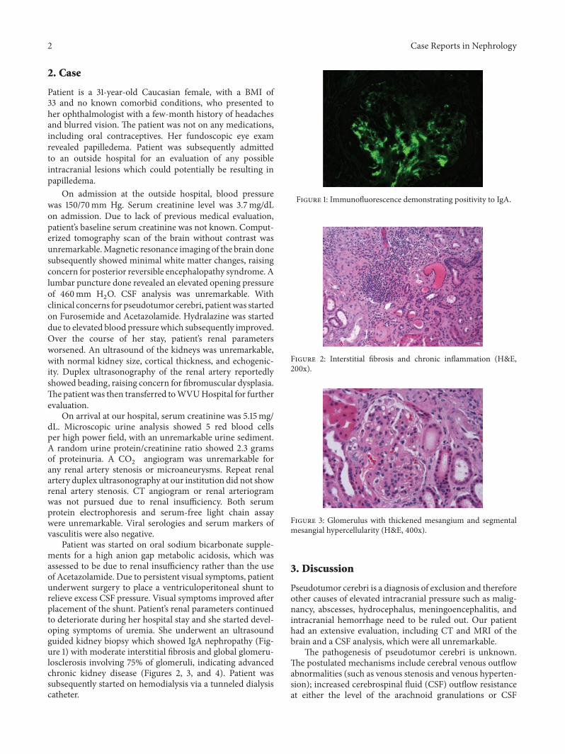
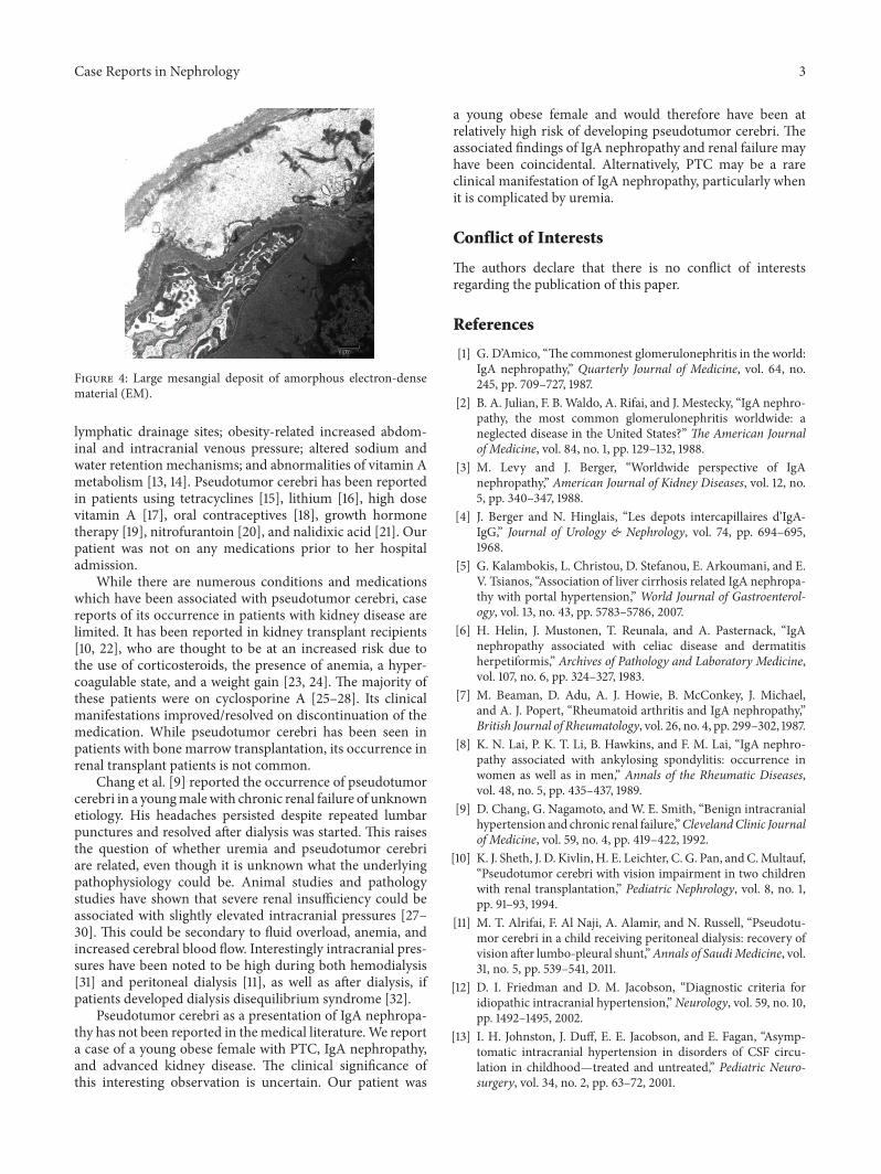
Patient was started on oral sodium bicarbonate supple-ments for a high anion gap metabolic acidosis, which wasassessed to be due to renal insufficiency rather than the useof Acetazolamide. Due to persistent visual symptoms, patientunderwent surgery to place a ventriculoperitoneal shunt torelieve excess CSF pressure. Visual symptoms improved afterplacement of the shunt. Patient’s renal parameters continuedto deteriorate during her hospital stay and she started devel-oping symptoms of uremia. She underwent an ultrasoundguided kidney biopsy which showed IgA nephropathy (Fig-ure 1) with moderate interstitial fibrosis and global glomeru-losclerosis involving 75% of glomeruli, indicating advancedchronic kidney disease (Figures 2, 3, and 4). Patient wassubsequently started on hemodialysis via a tunneled dialysiscatheter.
Figure 1: Immunofluorescence demonstrating positivity to IgA.
Figure 2: Interstitial fibrosis and chronic inflammation (H&E,200x).
Figure 3: Glomerulus with thickened mesangium and segmentalmesangial hypercellularity (H&E, 400x).
3. Discussion
Pseudotumor cerebri is a diagnosis of exclusion and thereforeother causes of elevated intracranial pressure such as malig-nancy, abscesses, hydrocephalus, meningoencephalitis, andintracranial hemorrhage need to be ruled out. Our patienthad an extensive evaluation, including CT and MRI of thebrain and a CSF analysis, which were all unremarkable.
The pathogenesis of pseudotumor cerebri is unknown.The postulated mechanisms include cerebral venous outflowabnormalities (such as venous stenosis and venous hyperten-sion); increased cerebrospinal fluid (CSF) outflow resistanceat either the level of the arachnoid granulations or CSF
Case Reports in Nephrology 3
Figure 4: Large mesangial deposit of amorphous electron-densematerial (EM).
lymphatic drainage sites; obesity-related increased abdom-inal and intracranial venous pressure; altered sodium andwater retention mechanisms; and abnormalities of vitamin Ametabolism [13, 14]. Pseudotumor cerebri has been reportedin patients using tetracyclines [15], lithium [16], high dosevitamin A [17], oral contraceptives [18], growth hormonetherapy [19], nitrofurantoin [20], and nalidixic acid [21]. Ourpatient was not on any medications prior to her hospitaladmission.
While there are numerous conditions and medicationswhich have been associated with pseudotumor cerebri, casereports of its occurrence in patients with kidney disease arelimited. It has been reported in kidney transplant recipients[10, 22], who are thought to be at an increased risk due tothe use of corticosteroids, the presence of anemia, a hyper-coagulable state, and a weight gain [23, 24]. The majority ofthese patients were on cyclosporine A [25–28]. Its clinicalmanifestations improved/resolved on discontinuation of themedication. While pseudotumor cerebri has been seen inpatients with bone marrow transplantation, its occurrence inrenal transplant patients is not common.
Chang et al. [9] reported the occurrence of pseudotumorcerebri in a youngmalewith chronic renal failure of unknownetiology. His headaches persisted despite repeated lumbarpunctures and resolved after dialysis was started. This raisesthe question of whether uremia and pseudotumor cerebriare related, even though it is unknown what the underlyingpathophysiology could be. Animal studies and pathologystudies have shown that severe renal insufficiency could beassociated with slightly elevated intracranial pressures [27–30]. This could be secondary to fluid overload, anemia, andincreased cerebral blood flow. Interestingly intracranial pres-sures have been noted to be high during both hemodialysis[31] and peritoneal dialysis [11], as well as after dialysis, ifpatients developed dialysis disequilibrium syndrome [32].
Pseudotumor cerebri as a presentation of IgA nephropa-thy has not been reported in themedical literature.We reporta case of a young obese female with PTC, IgA nephropathy,and advanced kidney disease. The clinical significance ofthis interesting observation is uncertain. Our patient was
a young obese female and would therefore have been atrelatively high risk of developing pseudotumor cerebri. Theassociated findings of IgA nephropathy and renal failure mayhave been coincidental. Alternatively, PTC may be a rareclinical manifestation of IgA nephropathy, particularly whenit is complicated by uremia.
Conflict of Interests
The authors declare that there is no conflict of interestsregarding the publication of this paper.
References
[1] G. D’Amico, “The commonest glomerulonephritis in the world:IgA nephropathy,” Quarterly Journal of Medicine, vol. 64, no.245, pp. 709–727, 1987.
[2] B. A. Julian, F. B. Waldo, A. Rifai, and J. Mestecky, “IgA nephro-pathy, the most common glomerulonephritis worldwide: aneglected disease in the United States?” The American Journalof Medicine, vol. 84, no. 1, pp. 129–132, 1988.
[3] M. Levy and J. Berger, “Worldwide perspective of IgAnephropathy,” American Journal of Kidney Diseases, vol. 12, no.5, pp. 340–347, 1988.
[4] J. Berger and N. Hinglais, “Les depots intercapillaires d’IgA-IgG,” Journal of Urology & Nephrology, vol. 74, pp. 694–695,1968.
[5] G. Kalambokis, L. Christou, D. Stefanou, E. Arkoumani, and E.V. Tsianos, “Association of liver cirrhosis related IgA nephropa-thy with portal hypertension,” World Journal of Gastroenterol-ogy, vol. 13, no. 43, pp. 5783–5786, 2007.
[6] H. Helin, J. Mustonen, T. Reunala, and A. Pasternack, “IgAnephropathy associated with celiac disease and dermatitisherpetiformis,” Archives of Pathology and Laboratory Medicine,vol. 107, no. 6, pp. 324–327, 1983.
[7] M. Beaman, D. Adu, A. J. Howie, B. McConkey, J. Michael,and A. J. Popert, “Rheumatoid arthritis and IgA nephropathy,”British Journal of Rheumatology, vol. 26, no. 4, pp. 299–302, 1987.
[8] K. N. Lai, P. K. T. Li, B. Hawkins, and F. M. Lai, “IgA nephro-pathy associated with ankylosing spondylitis: occurrence inwomen as well as in men,” Annals of the Rheumatic Diseases,vol. 48, no. 5, pp. 435–437, 1989.
[9] D. Chang, G. Nagamoto, and W. E. Smith, “Benign intracranialhypertension and chronic renal failure,”ClevelandClinic Journalof Medicine, vol. 59, no. 4, pp. 419–422, 1992.
[10] K. J. Sheth, J. D. Kivlin, H. E. Leichter, C. G. Pan, andC.Multauf,“Pseudotumor cerebri with vision impairment in two childrenwith renal transplantation,” Pediatric Nephrology, vol. 8, no. 1,pp. 91–93, 1994.
[11] M. T. Alrifai, F. Al Naji, A. Alamir, and N. Russell, “Pseudotu-mor cerebri in a child receiving peritoneal dialysis: recovery ofvision after lumbo-pleural shunt,”Annals of SaudiMedicine, vol.31, no. 5, pp. 539–541, 2011.
[12] D. I. Friedman and D. M. Jacobson, “Diagnostic criteria foridiopathic intracranial hypertension,”Neurology, vol. 59, no. 10,pp. 1492–1495, 2002.
[13] I. H. Johnston, J. Duff, E. E. Jacobson, and E. Fagan, “Asymp-tomatic intracranial hypertension in disorders of CSF circu-lation in childhood—treated and untreated,” Pediatric Neuro-surgery, vol. 34, no. 2, pp. 63–72, 2001.
4 Case Reports in Nephrology
[14] V. Biousse, B. B. Bruce, and N. J. Newman, “Update on thepathophysiology and management of idiopathic intracranialhypertension,” Journal of Neurology, Neurosurgery and Psychi-atry, vol. 83, no. 5, pp. 488–494, 2012.
[15] A. Kesler, Y. Goldhammer, A. Hadayer, and P. Pianka, “The out-come of pseudotumor cerebri induced by tetracycline therapy,”ActaNeurologica Scandinavica, vol. 110, no. 6, pp. 408–411, 2004.
[16] S. H. Levine and C. Puchalski, “Pseudotumor cerebri associatedwith lithium therapy in two patients,” Journal of ClinicalPsychiatry, vol. 51, no. 6, pp. 251–253, 1990.
[17] A. Drouet and J. Valance, “Benign intracranial hypertensionand chronic hypervitaminosis A,” Revue Neurologique, vol. 154,no. 3, pp. 253–256, 1998.
[18] J. Finsterer, E.-W. Kues, and S. Brunner, “Pseudotumour cerebriin a young obese woman on oral contraceptives,” EuropeanJournal of Contraception and Reproductive Health Care, vol. 11,no. 3, pp. 237–240, 2006.
[19] E. A. Koller, B. V. Stadel, and S. N. Malozowski, “Papilledema in15 renally compromised patients treated with growth hormone,”Pediatric Nephrology, vol. 11, no. 4, pp. 451–454, 1997.
[20] G. R. Mushet, “Pseudotumor and nitrofurantoin therapy,”Archives of Neurology, vol. 34, no. 4, p. 257, 1977.
[21] A. Mukherjee, P. Dutta, M. Lahiri, S. Sinha, A. K. Mitra, andS. K. Bhattacharya, “Benign intracranial hypertension afternalidixic acid overdose in infants,”TheLancet, vol. 335, no. 8705,p. 1602, 1990.
[22] P. J. Francis, S. Haywood, S. Rigden, D. M. Calver, and G. Clark,“Benign intracranial hypertension in children following renaltransplantation,” Pediatric Nephrology, vol. 18, no. 12, pp. 1265–1269, 2003.
[23] C. C.Mourani, S. G.Mallat,M. Y.Moukarzel, C. Y. Akatcherian,and P. Cochat, “Kidney transplantation after a severe form ofpseudotumor cerebri,” Pediatric Nephrology, vol. 12, no. 9, pp.709–711, 1998.
[24] T. Obeid, A. Awada, S. Huraib, K. Quadri, and S. Abu-Romeh, “Pseudotumor cerebri in renal transplant recipients: adiagnostic challenge,” Journal of Nephrology, vol. 10, no. 5, pp.258–260, 1997.
[25] R.Avery,D.A. Jabs, J. R.Wingard,G.Vogelsang, R. Saral, andG.Santos, “Optic disc edema after bone marrow transplantation:possible role of cyclosporine toxicity,” Ophthalmology, vol. 98,no. 8, pp. 1294–1301, 1991.
[26] O. A. Cruz, S. G. Fogg, and G. Roper-Hall, “Pseudotumorcerebri associated with cyclosporine use,” American Journal ofOphthalmology, vol. 122, no. 3, pp. 436–437, 1996.
[27] E. Rodrıguez, A. Delucchi, and F. Cano, “Neurotoxicity causedby cyclosporin A in renal transplantation in children,” RevistaMedica de Chile, vol. 120, no. 3, pp. 300–303, 1992.
[28] K. G. Gilliland and R.M. Hegstrom, “The effect of hemodialysison cerebrospinal fluid pressure in uremic dogs,” Transactions—American Society for Artificial Internal Organs, vol. 9, pp. 44–48,1963.
[29] O. Steen, “The brain in uremia,”Acta Psychiatrica Scandinavica,vol. 43, supplement 1, pp. 1–65, 1961.
[30] N. H. Raskin and R. A. Fishman, “Neurologic disorders in renalfailure, part 1,” The New England Journal of Medicine, vol. 294,pp. 143–148, 1976.
[31] C. M. Lin, J. W. Lin, J. T. Tsai et al., “Intracranial pressurefluctuation during hemodialysis in renal failure patients with
intracranial hemorrhage,” Acta Neurochirurgica. Supplement,vol. 101, pp. 141–144, 2008.
[32] P. Esnault, G. Lacroix, P.-J. Cungi, E. D’Aranda, J. Cotte, and P.Goutorbe, “Dialysis disequilibrium syndrome inneurointensivecare unit: the benefit of intracranial pressure monitoring,”Critical Care, vol. 16, article 472, 2012.


![Crescentic IgA Nephropathy[1][1]](https://img.pdfslide.net/doc/110x75/55cf9903550346d0339b030a/crescentic-iga-nephropathy11.jpg)