Embed Size (px)
Citation preview

Page 1
Baltazar Espiritu, MD Host Defense 2013
IgE Immunology
Revised 1/10/13
IgE IMMUNOLOGY
Date: Wednesday, May 1st, 2013 8:30 AM LH190
LEARNING GOALS You will be able to: • Distiguish the specialized functions of IgE • Describe how mast cell, basophils and eosinophils mediate allergic (Type 1
hypersensitivity) reactions • Predict and classify potential intervention points for control of allergic reactions and diseases.
BACKGROUND READING Janeway: 571-97
LECTURER Baltazar R. Espiritu, M.D.

Page 2
Baltazar Espiritu, MD Host Defense 2013
IgE Immunology
Revised 1/10/13
THE IMMUNOLOGY OF ALLERGY I. INTRODUCTION
A. Allergy: a disease following a response by the immune system to an otherwise innocuous antigen. An allergic response is termed a Type 1 hypersensitivity reaction.
B. Atopy - denotes the ability to transfer reactivity to allergens by means of
serum (the transfer agent formerly called was reagin and is now known as IgE.)
C. Allergic reactions affect up to 40% of the United States population and its
incidence had doubled over the past 10-15 years. Common causes are food, insect venom, latex and drugs. Manifestations of allergic reactions range from fatal anaphylactic shock to allergic rhinitis and chronic bronchial asthma.
D. IgE is the central mediator of these diseases. IgE drives an allergic reaction by activating a set of specialized cells which then generate a panoply of pharmacologic reactions characterized by smooth muscle spasm, increased vascular permeability and activation of inflammatory and coagulation cascades.
II. Components of an Allergic Response
A. IgE 1. Has a standard Ig structure of 2 heavy and 2 light 2. Is heavily glycosylated.
3. Has 2 Fc regions (CH3 + CH4) on the Fc that function as a binding site for high affinity Fcε receptors on mast cells and basophils and on antigen presenting cells, at much lower levels.
4. Is maintained in serum at a much lower lever than other Ig and is usually found in nanogram instead of milligram amounts/l.
5. Normal IgE responses occur to selective stimuli; for example, worms, certain parasites, and other large organisms with relatively biodegradable resistant antigens.
B. Mast Cells and Basophils
1. Both express high affinity IgE Fcε receptors constitutively. 2. Both have cytoplasmic stores of vasoactive mediators like histamine
and leukotrienes. 3. Both develop from hematopoietic precursors, but are of distinct
lineages and differ in phenotypic markers.

Page 3
Host Defense 2012Baltazar Espiritu, MD Host Defense 2013
IgE Immunology
Revised 1/10/13
4. Basophils are circulating leukocytes. 5. Human mast cells are divided into 2 major subtypes based on the
presence of tryptase (MCT cells) or tryptase and mast cell-specific chymase (MCTC cells), each predominating in different locations. Tryptase staining identifies all mast cells and is the primary method for identifying tissue mast cells. MCT cells are the prominent mast cell type within the mucosa of the respiratory and gastrointestinal tracts and increase with mucosal inflammation. MCTC cells are localized within connective tissues, such as the dermis, submucosa of the gastrointestinal tract, heart, conjunctivae, and perivascular tissues.
6. Mast cells contain large amounts of histamine, heparin and proteases. Newly synthesized leukotriene B4 (LTG4), and inflammatory cell cytokines that include IL 3, 4, 5, 6, 8, 10,13, TNF-α, chemokines and GM-CSF. These very potent mediators are important in allergic inflammation. Other diverse functions of mast cells include antigen presentation and has a direct effect on B-cells to induce IgE production.
C. Allergens (The antigens of allergic responses)
1. An infinite array of ubiquitous environmental proteins found in
insects, worms, shellfish and foods and fungi that can induce hypersensitivity responses in atopic individuals by inducing IgE instead of normal IgA/ IgG/IgM antibody formation.
Copyright: 2001 From: immunobiology, 5th edn. Author: Janeway, et al Reproduced by permission of Routledge, Inc., part of The Taylor Francis Group

Page 4
Baltazar Espiritu, MD Host Defense 2013
IgE Immunology
Revised 1/10/13
2. Types of Allergens - Aero-allergens such as pollen grains from trees, grasses and ragweed to mold spores, dog, cat, and rodent dander, dust mite feces and cockroach. The allergen profilin is responsible for cross-reactions between birch mugwort pollen-celery-spices, grass pollen-celery-carrots, and tree pollen-hazelnut. Food allergens are limited primarily to milk, egg, peanuts, nuts, shellfish, wheat and soy. Insect venoms are usually those from vespids (wasps, honeybees, yellow jackets and fire ants.) A common component of many of the environmental antigens is that they contain chitin-polysaccharide not found in mammals. Many drugs, especially antibiotics, can also act as allergens.
3. The immunodominant peptides of these allergens are usually
preferentially presented in selected (Class II) D MHC loci by dendritic cells. The D genes, in concert with other non-MHC genes, promote IgE production over IgG responses by influencing the type of TLR activated, type of T-helper cell and cytokine milieu present during allergen presentation by APC.
III. The Context and initiation of an allergic reaction-involves an unique
combination of circumstances
A. There is an obvious genetic dictation of the response. 1. 50% of individuals with 2 atopic (allergic) parents will also be atopic.
This is in contrast to the observation that only 15% of children of non-allergic parents will be atopic.
2. The genes are multiple and range from one that controls expression of a transcription factor, T-bet, that controls synthesis of INF-γ, one that controls bronchial reactivity, one that controls mast cell signaling and one that controls FcεR avidity and presence of “allergic” TLRs on DC. The bottom line: a multiplicity of genes must act in concert to produce an allergic reaction.
3. There is a direct relationship between serum IgE levels, allergic reactions and the atopic state. There is tight and complex control of IgE serum levels by MHC linked genes, maternal genes on chromosome 11 and other poorly described genetic loci.
B. Context of Antigen exposure:
1. Appropriate time of exposure. An unusually high level of exposure very early in life coupled with a relative LACK of exposure to infectious disease antigens that incite vigorous Th1 and Th2 responses will manifest by IgE production and allergy to some, but not all antigens, in lieu of an expected Th1 or IgG response.
2. Route. Delivery of antigens or allergens in very small amounts to mucosal surfaces facilitates allergic responses in the atopic host

Page 5
Host Defense 2012Baltazar Espiritu, MD Host Defense 2013
IgE Immunology
Revised 1/10/13
3. All the above must be coupled with exposure in a genetically predisposed individual.
IV. Sequence of an Allergic Response
A. Contact with an allergen is usually mucosal (respiratory or gastrointestinal), but can be cutaneous or systemic.
B. Uptake of allergen by antigen presenting cells (DC) via “allergic” TLR that
induces the DC to produce IL-4 instead of IL-12
C. Presentation of the allergen as an immunodominant peptide in a Class II MHC groove. The nature of the peptide and possibly its unique presentation dictate a dominant IgE response.
D. Allergic responses are dependent on Th-2 activation. 1. Presentation of this antigen by a DC in the absence of Th1-TLR binding
or by an “allergic” TLR 2. Ensuing absence of the Th1 initiator cytokine IL-12 or presence of IL-4
then leads to presentation to a Th2 by default. 3. Early release of IL-4 from mast cells or basophils- commonly caused by
innate immune responses of these cells to parasites 4. ANY situation where IL-4 is the dominant cytokine at the time of antigen
appearance.
E. Th-2 cell then provides the critical IL-4 signal to an allergen specific B-cell.
F. The accelerated production of Th-2 cytokines, especially IL-4 and IL-13, dominate the cytokine profile.
G. Promotion of IgE class switching occurs by up regulation of CD-23 (FcεRII receptor) on mast cells and basophils that increase their production of IL-4 and IL-13. This is strongly influenced by gene influenced polymorphisms.
H. IL-4 from Th-2, Basophils and mast cells activate ε germ-line transcription in B-cells and results in IgE synthesis.
V. Arming of Fcε Receptor Cells by Allergen Specific IgE
A. The allergen specific IgE then binds to the high affinity IgE Fc receptors on
mast cells and basophils. The FcεR is the ONLY FcR that can be occupied by antibody not previously complexed with antigen. These cells are now “armed”.

Page 6
Baltazar Espiritu, MD Host Defense 2013
IgE Immunology
Revised 1/10/13
FIRST STEP of an ALLERGIC REACTION
VI. Activation of Armed Fcε on Receptor Cells.
A. Subsequent exposure to the same allergen then cross-links the IgE previously
bound to FcεR receptors. B. The cross-linked Fcε receptors then aggregate and signal transduction occurs. C. Signal transduction activates calcium influx into armed mast cells and basophils
which then degranulate, releasing potent vasoactive, inflammatory and fibrogenic mediators.
Figure by John A. Robinson, MD

Page 7
Host Defense 2012Baltazar Espiritu, MD Host Defense 2013
IgE Immunology
Revised 1/10/13
VII. Temporal Sequence of Allergic Inflammation (also known as Type I Hypersensitivity
A. Immediate Reaction -defined as an allergic reaction occurring within 15 minutes of allergen exposure. This portion of the reaction is completely dependent on previous exposure and sensitization.
1. Multiple vasoactive mediators released from mast cells and basophils, 2. Prostaglandin and leukotriene synthesis and release.
3. Direct complement activation by tryptase (released from mast cells)

Page 8
Baltazar Espiritu, MD Host Defense 2013
IgE Immunology
Revised 1/10/13
cleavage. (Elevated tryptase levels can serve as a serum marker for massive mast cell activation that occurs in anaphylaxis)
VIII. The Late Phase (also known as slow-reacting phase) and defined as occurring
within hours of allergen contact. This phase is completely dependent upon T-cell activation and the presence of cytokines IL3, 4, 5, 13, TNF-α, GM-CSF and IL-10. A. The late phase is characterized by infiltration of the site of the response by
activated eosinophils, neutrophils, additional mast cells, basophils and lymphocytes.
B. IL-3, IL-5 and GM-CSF regulate growth and marrow release of eosinophils.
C. IL-5 stimulates their release from bone marrow and augments the chemotactic effect of a specific eosinophil chemokine called eotaxin.
D. IL-5 increases FcεR display and thereby augments the IgE reaction
E. Eosinophils produce several unique inflammatory enhancers, the major ones being major basic protein, leukotrienes and cationic proteins.
IX. The Clinical Manifestations of an Allergic Response are strongly dependent on the site of the reaction.
A. Immediate reactions can range from anaphylaxis with laryngeal and
bronchiolar constriction and generalized increase in vascular permeability. They can be fatal.
Do Not Memorize this! Provided simply to illustrate the # of mediators

Page 9
Host Defense 2012Baltazar Espiritu, MD Host Defense 2013
IgE Immunology
Revised 1/10/13
B. Allergic rhinitis occurs when allergen binds to cells in the nasal
submucosa and incites a chronic allergic inflammatory reaction driven by continuous aeroallergen exposure.
C. Urticaria(hives), which can be severely pruritic, are skin lesions that
occur when IgE armed mast cells are activated in the skin.
D. Acute and chronic bronchial asthma occurs when IgE armed cells are recruited to submucosal sites of the pulmonary bronchi.
Figure by John A. Robinson, MD X. Some particulars about asthma A. There is no question that asthma is a disease of the industrialized world and that it is on the increase. It is the poster child of a maladaptive immune reaction. B. The hygiene hypothesis is based on the following evidence:
1. Not only has there been a decrease in the number of childhood illnesses but that there has also been a change in the time of exposure (later in life) to acute childhood infections.
2. Large families and/or early day care-both situations provide an environment where infection is rampant and later allergy rare
3. There is increasingly compelling evidence that early exposure to childhood illnesses “sets” normal Th1 and Th2 responses to subsequent environmental antigen exposure.
4. The lack of early exposure to environmental and infectious antigens is associated with a lack of T regulator cells that control IgE synthesis to
Copyright: 2005 From: immunobiology, 6th edn. Author: Janeway, et al Reproduced by permission of Routledge, Inc., part of The Taylor Francis Group
Page 10
Baltazar Espiritu, MD Host Defense 2013
IgE Immunology
Revised 1/10/13
environmental antigens. How do we know this? i. Mice infected with gut worms are resistant to production of airway
reactivity by dust mite sensitization and the resistance is due to CD4 Tregs that have migrated from their gut. The worms invented a way to stimulate a Treg response in the gut so they could survive.
ii. Mice already sensitized to dust mites then infected with worms had significant decreases in eosinophil migration into their lungs
iii. In Gabon, successful treatment of worm infested children led to allergy to dust mites later on.
iv. Worm infested children rarely get autoimmune diseases (much more on this later)
C. There is a strong correlation between asthma and obesity also but the causal link is
unknown. The adipocyte tissue is an organ of sorts and has its own cytokine systems, i.e. leptin, among others, being pro-inflammatory and this may be an underlying factor.
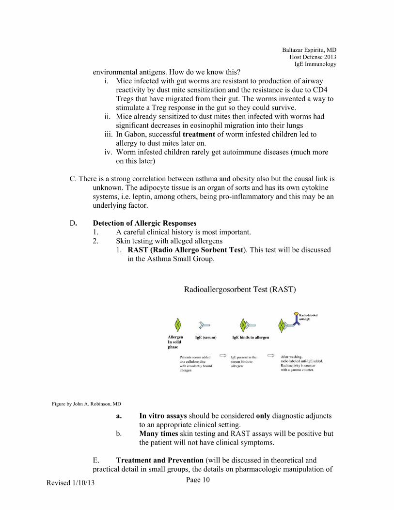
D. Detection of Allergic Responses 1. A careful clinical history is most important. 2. Skin testing with alleged allergens
1. RAST (Radio Allergo Sorbent Test). This test will be discussed in the Asthma Small Group.
a. In vitro assays should be considered only diagnostic adjuncts
to an appropriate clinical setting. b. Many times skin testing and RAST assays will be positive but
the patient will not have clinical symptoms.
E. Treatment and Prevention (will be discussed in theoretical and practical detail in small groups, the details on pharmacologic manipulation of
Figure by John A. Robinson, MD

Page 11
Host Defense 2012Baltazar Espiritu, MD Host Defense 2013
IgE Immunology
Revised 1/10/13
allergic responses will be next year!) 1. Allergen avoidance. Pharmacologic suppression moderately effective 2. Anti-IgE therapy-use of an anti-IgE monoclonal antibody that has been
engineered to bind to the site on circulating IgE that binds to the cell-bound IgE receptor. Anti-IgE therapy results in decrease in eosinophilic inflammation and IgE-bearing cells. Withdrawal of this therapy results in return on asthma symptoms and this correlates with increasing serum IgE.
3. “Desensitization”- rerouting of IgE response by constructing allergen antigens that promote either Th1 responses and macrophage destruction of the antigen or Th2 responses that culminate in IgG production-so-called blocking antibodies. This involves redirecting the immune response.
4. Vaccines also try and redirect the immune response. 5. The alert student has already figured out the "fear factor" approach to
treatment of allergy - infect the patient with worms or sensitize them with worm antigens- already being done!

2/12/2013
1
THE IMMUNOLOGY OF IgE
“The Itch, the Sneeze and the Wheeze”
“Mast Cells, Basophils and Eosinophils”
“Urticaria, Allergic rhinitis and Asthma”
Outline
HYPERSENSITIVITY DISEASES
– TYPE I - ALLERGIC RESPONSES -mediated by IgE
– TYPE II - ANTIBODY DIRECTED AGAINST TISSUE ANTIGENS - mediated by IgG
– TYPE III - IMMUNE COMPLEX MEDIATED DISEASE - mediated by antigen+IgG
– TYPE IV - DELAYED HYPERSENSIVITY-mediated by T Cells
DEFINITIONS
• ALLERGY - a disease induced by reaction to a usually innocuous antigen
• Atopy – predisposition to become IgE-sensitized to environmental allergens

2/12/2013
2
Components of an allergic response
• IgE• Standard Ig structure• heavily glycosylated and has binding sites for FcR
– normally very low serum concentrations-consider it a cell bound antibody found mainly at host-environmental interfaces
– binding sites for FcR on mast cells and basophils
Two (of 3) Major Cell Mediators are Mast cells and Basophils
• Both express HIGH affinity FcR
• Both contain histamine, TNF-and leukotrienes in cytoplasm
• Mast cells - tissue bound, compartmentalized as mucosal or connetive tissue, contain potent vasoactive compounds and cytokines
• Degranulation releases the mediators
Mast CellToxic mediator
HistamineHeparin
Lipid MediatorLeukotrienes - LTC4, LTD4,
LTE4Platelet activating factorEnzymes
Tryptase, chymase, cathepsin G,
carboxypeptidaseCytokines
IL-4, IL-13, IL- 3, IL-5, GM-CSF, TNFChemokines
MIP-1a
2 Types of Mast CellsMC -TryptaseMC- Tryptase and Chymase
2/12/2013
3
ALLERGENS
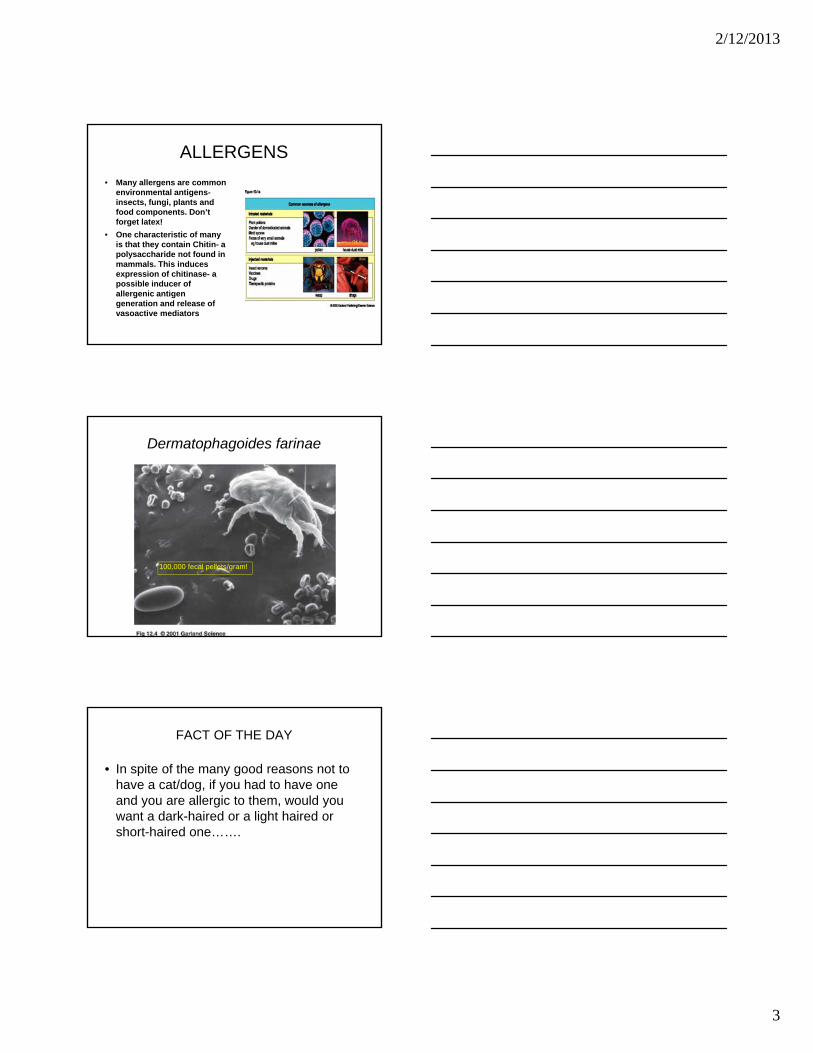
• Many allergens are common environmental antigens-insects, fungi, plants and food components. Don’t forget latex!
• One characteristic of many is that they contain Chitin- a polysaccharide not found in mammals. This induces expression of chitinase- a possible inducer of allergenic antigen generation and release of vasoactive mediators
Dermatophagoides farinae
100,000 fecal pellets/gram!
FACT OF THE DAY
• In spite of the many good reasons not to have a cat/dog, if you had to have one and you are allergic to them, would you want a dark-haired or a light haired or short-haired one…….

2/12/2013
4
THE MHC, GENES AND ALLERGENS
• ALMOST ANY PROTEIN CAN INDUCE AN ALLERGIC RESPONSE IN AN ATOPIC INDIVIDUAL
• Genes dictate allergic responses• 50% of children of 2 atopic parents will be atopic• Different MHC-II will present different peptides that differ
in their antigenic potency • Polymorphic expression of multiple genes culminate in
allergic responses• Examples:varying expression of IFN-via the T-Bet
gene, FcR avidity via a maternal gene, IgE synthesis and bronchial reactivity, IL-13 synthesis
THE MHC AND ALLERGENS
• There is a direct relationship between IgE levels, allergy and the atopic state.
• The end result is: a multiplicity of genes must act in concert to produce an allergic reaction.
Genesis of the allergic reaction
• Appropriate genetic background
• Type of Antigen exposure– Almost anything can be an allergen but there is a
trend towards proteins with enzymatic activity or ones that induce it
– Timing is important: decreased early exposure to infections in the genetically predisposed individual is associated with insufficient T regulator control of IgE (more later)
– route- mucosal exposures predominate

2/12/2013
5
Effect of Allergen Exposure
Text
FcR Affinity
ONLY FcRTHAT CANBE OCCUPIEDBY ANTIBODY WITHOUTANTIGEN IS THE FcR

2/12/2013
6
TEMPORAL SEQUENCE
• EARLY– WITHIN 15 MINUTES
– PROSTAGLANDINS & LEUKOTRIENE RELEASE
– DIRECT COMPLEMENT ACTIVATION
– CANNOT HAPPEN IF NO PREVIOUS EXPOSURE
Fig by John A Robinson

2/12/2013
7
EOSINOPHILS-the third major mediator cell
TEMPORAL SEQUENCE
• LATE– COMPLETELY
DEPENDENT ON Th2 ACTIVATION
– AND CYTOKINES IL3,4,5,13,AND 10
– EOTAXIN
– CHARACTERIZED BY EOSINOPHILS
Fig by John A Robinson

2/12/2013
8
THE CLINICAL MANIFESTATIONS OF AN ALLERGIC RESPONSE ARE DEPENDENT ON
THE SITE OF REACTION
• ANAPHYLAXIS
• ALLERGIC RHINITIS
• URTICARIA (HIVES)
• ATOPIC DERMATITIS
• ASTHMA
23
Anaphylaxis
Physiology of Anaphylaxis

2/12/2013
9
Physiology of Anaphylaxis
SENSITIZATION PHASE

2/12/2013
10
The Scheme of Things-asthma is a classic example of gene/environmental interaction
THE “RIGHT”GENES
ENVIRONMENT•RIGHT ALLERGEN AT THE RIGHT TIME•SMALL FAMILY•HYPERHYGIENE•ANTIBIOTICS EARLY
ATOPY
ALLERGIC SYNDROMES
TRIGGERS:•REEXPOSURE•VIRUSES•POLLUTANTS
IgE Antibodies
T REGULATOR CELL DEFICIENCY
Fig by John A Robinson
ASTHMA
• MARKED INCREASE IN ASTHMA IN THE INDUSTRIAL WORLD……WHY? The Hygiene hypothesis follows:
• Decreased childhood infection and later exposure
• Early exposure to infections less allergy
• Large families, rural residence and daycare associated with less allergy
• Lack of early exposure associated with Deficiency of T3 regulators that control IgE
• Worm infested children that are treated and cured develop allergies
• Mice infected with worms cannot be made atopic with dust mite antigens
• Mice with asthma-like pulmonary hypersensivity that are infected with worms have much less eosinophil migration into lungs on pulmonary rechallenge
• Counter- regulation hypothesis – infections causes decrease Th-1 and Th-2
DETECTION OF ALLERGIC RESPONSES
• Careful History
• Skin Testing
• RAST- KNOW THIS ASSAY FOR SMALL GROUPS!
• In vitro assays for allergy can be very misleading and occasionally dangerous

2/12/2013
11
Skin Test
Skin Test
RAST ASSAY
Fig by John A RobinsonCurrent technology:- 3D solid phase- enzyme labelled anti-IgE antibodies- flourogenic substrate

2/12/2013
12
Treatment of Allergic Diseases
TREATMENT AND PREVENTION
• SMALL GROUPS WILL ADDRESS THESE ISSUES BUT………….
• IF YOU CAN’T AVOID THE ALLERGEN, SUPPRESS THE SYMPTOMS WITH DRUGS
• BLOCK THE REACTION WITH A MONOCLONAL anti-IgE - Asthma
TREATMENT AND PREVENTION
• SMALL GROUPS WILL ADDRESS THESE ISSUES BUT………….
• DESENSITIZE
• VACCINATE- THE FUTURE (Maybe)

John A. Robinson, MD Host Defense 2013
Perturbations in the Super System
REV 10/30/12 Page 1
PERTURBATIONS in the SUPER SYSTEM Date: Thursday, May 2nd, 2013 Time: 10:30 AM
LH190
LEARNING GOALS You will be able to understand how the complexities of the immune response make it vulnerable to disruption and impaired regulation, which can then lead to immunopathologic diseases.
Describe superantigens and their potential for disease causation Understand how various pathogens, especially viruses, can manipulate the immune
response. In a future lecture you will learn how you can manipulate the immune response to the patient’s benefit!
Understand the significance of newly discovered mechanisms of autoimmunity
BACKGROUND READING FOR THIS LECTURE AND THE AUTO-IMMUNE SMALL GROUP 1. Janeway 8th edition: Pages 226-228; 209-210;512-514;612-615; 622-624; 631-635;figs 15.14-15.15. 2. If I want you to know something about a specific disease in order that you can understand an immunologic concept, that detail will be mentioned either in the lecture notes or discussed in small groups. (you do not need to go to a pathology text and read about a disease- save that for next year). 3. Articles posted on the Host defense site. DO NOT WORRY ABOUT THE TECHNICAL ASPECTS OF THE ARTICLES- YOU WON’T BE TESTED ON THEM! - BUT IF YOU UNDERSTAND THE CONCEPTS YOU WILL KNOW A LOT ABOUT IMMUNOLOGY IN GENERAL AND AUTOIMMUNITY IN PARTICULAR.
LECTURER John A. Robinson, MD

John A. Robinson, MD Host Defense 2013
Perturbations in the Super System
REV 10/30/12 Page 2
CONTENT SUMMARY
Run-away Immune Responses
The Diverse ways that Pathogens can Manipulate the Immune Response. Current concepts of autoimmunity: AIRE and FoxP3 defects Agonist autoantibodies Antagonist autoantibodies Autocytotoxicity Defective apoptosis Defective control by CD4, 25 FoxP3 cells “New” lineage of CD4 cells mediate autoimmunity

John A. Robinson, MD Host Defense 2013
Perturbations in the Super System
REV 10/30/12 Page 3
I. A Run-away Immune Response
1. Although there are multiple safeguards and fail/safe immune reaction
regulatory mechanisms characterized by agonist/antagonist cytokines relationships, some bacteria and viruses have developed ways to circumvent them. An important one, from a clinical standpoint, is the superantigen.
2. Superantigens differ in at least 3 major ways from conventional peptide
antigens: a. In contrast to conventional peptides that require uptake and cytosol processing prior to presentation in the context of MHC Class II determinants, superantigens can react with MHC Class II determinants in unprocessed form. b. The portion of the TCR that then reacts with them is not within the classic peptide binding groove or antigen specific antibody receptor on B-cells but on the ‘side’ of the mononuclear MHC Class II TCR complex. This nonconventional binding can activate up to 30% of peripheral T-cells. It is unclear whether the activated T cells are already committed Th1 (to another antigen) or naïve Th0s c. They elicit a massive immediate primary polyclonal response in T cells.
3. Bacterial pathogens that cause shock syndromes usually do so by producing
toxins that act as superantigens. The rapid activation of T cells leads to
Reprinted with permission

John A. Robinson, MD Host Defense 2013
Perturbations in the Super System
REV 10/30/12 Page 4
‘cytokine storm’ that causes massive release of vasoactive and proinflammatory cytokines. These, in turn, cause severe perturbations in organ function, especially of the liver and kidney, that are manifested as severe clinical diseases and the toxic shock syndrome. The intensity of the response is strongly dependent on the polymorphism of the host’s MHC class II and (probably) the TNF gene complex also. The importance of polymorphism became evident when it was discovered that the same bacterial strain might cause death in some patients and hardly any clinical disease in others. The individual’s MHC binding characteristics dictated the magnitude of T cell activation. You have heard this concept before.
4. The advantage to the organism is thought to be that the activated immune
effector cells undergo widespread apoptosis and lose their ability to react in regulated fashion. The organism then uses the host for propagation prior to death (host).
Figure by John A. Robinson, MD
a. The classic example is that of toxic shock caused by
Streptococcus/ Staphylococci- after the introduction of a “new and improved supertampon”- was a stark example of the law of unintended consequences
II. More sophisticated ways that pathogens have devised to either manipulate or
evade the Immune Response
1. Superantigens are not an elegant way to get around the immune response
TOXIC SHOCK SYNDROME
T
MAC
SAg
Massive Releaseof TNF-
INF--
Loss of endothelialintegrity, decreasedvascular resistance
SHOCK

John A. Robinson, MD Host Defense 2013
Perturbations in the Super System
REV 10/30/12 Page 5
because the host doesn’t last long. This is especially true for viruses because they require a living host for their own survival and propagation. If viruses alert the host’s alarm system, immune defenses will be turned on and will destroy them. They have thus been forced to develop ingenious ways to parasitize a host without activating the alarm systems. Successful viruses are those that have figured out ways to hijack host genes that are then used to modify or suppress immune responses. The following are some, but not all, strategies employed by viruses to evade destruction by immune system:
a. Bacteria and viruses can downregulate TLRs-remember the Toll
receptors and innate immunity? b. Interfering in the methods by which Class I MHC transports
antigen to the surface so it can be sampled by CD8 cells. By doing so, viral antigens remain camouflaged or hidden within the cell and cannot be deleted by antigen specific CD8 cytotoxic T-cells.
c. Piracy of the genes that produce inhibitor signals for NK cells and keep them turned off.
d. Preventing cytokine upregulation of MHC Class I and II antigens. e. Stealing genes that produce inhibitory cytokines-IL 10 or other IL
12 inhibitors-or induce genes that produce products that antagonize the effects of proinflammatory cytokines.
f. Successfully encode genes that produce soluble cytokine receptors, thus blindfolding cytokines generated during the immune response.
g. Block apoptosis by interfering with CASPASE activation or enhancing Bcl activity, ensuring that they continue to have a viable cell to live in.
h. Parasites can inactivate DC and prevent antigen presentation i. Parasites can switch on the Th1 system and prevent IgE
production. j. Recent evidence suggests that some viruses and bacteria can even
induce T regulator cells to prevent immune responses. k. Viruses can express suppressive micro-RNAs (non-coding RNAs) Every step in the figure below is vulnerable to a viral hacker! It is difficult to believe that we are surviving as a species- a real complement to the adaptability of our immune system.

John A. Robinson, MD Host Defense 2013
Perturbations in the Super System
REV 10/30/12 Page 6
III. The Immune System Reacting Against Self or AUTOIMMUNITY. [The
posted articles are the best way to understand many of the following concepts]
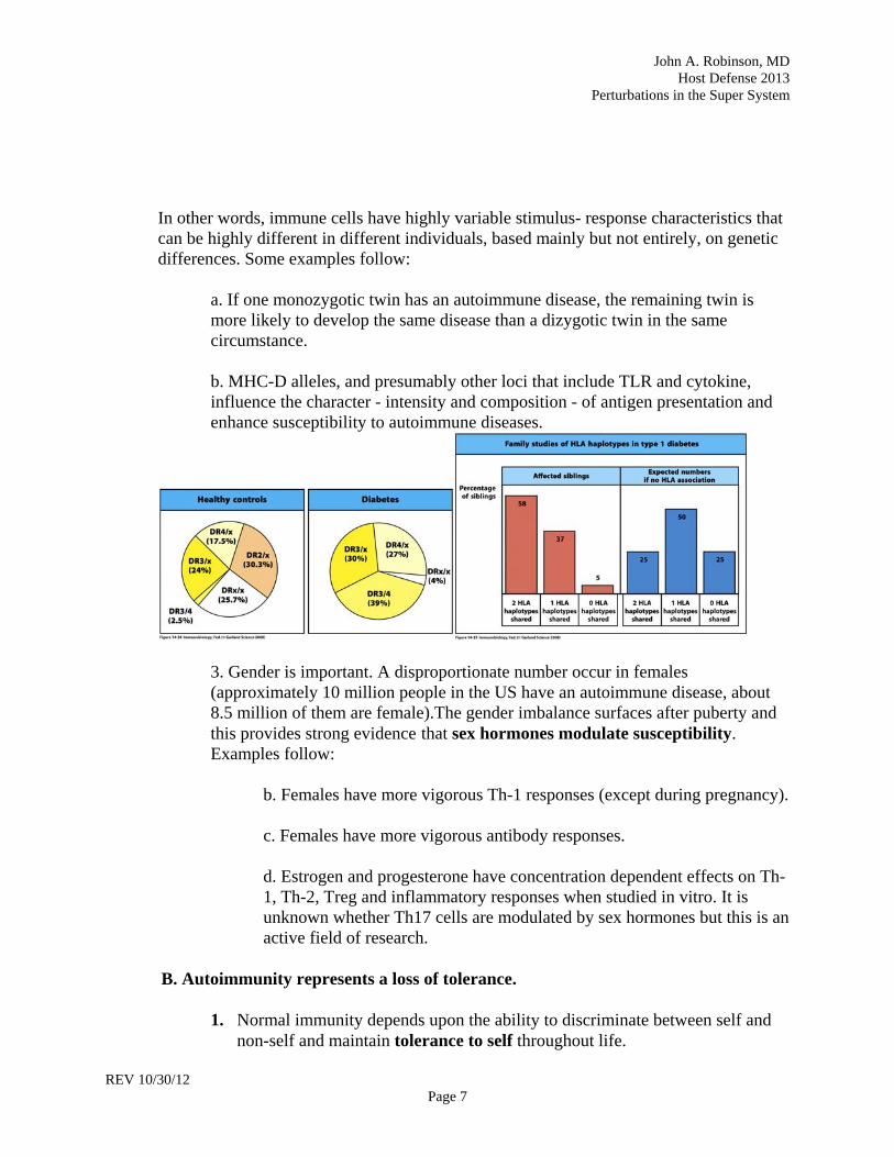
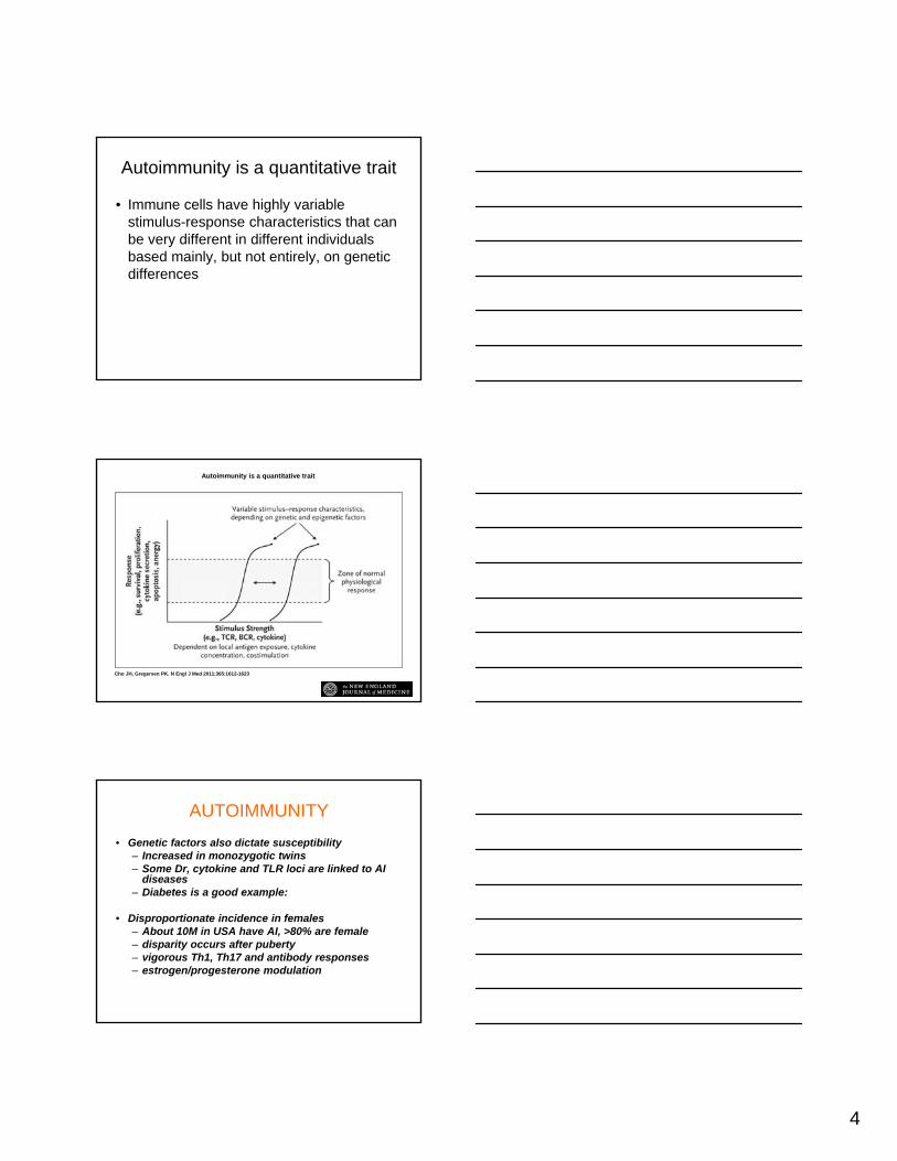
A. General overview of autoimmunity 1. Autoimmune Disorders are multifactorial in etiology. Three very common autoimmune diseases are rheumatoid arthritis, Crohn disease and diabetes mellitus. 2. The propensity to develop an autoimmune disease should be viewed conceptually as a quantitative trait.
Stimulus–Response Thresholds and Immune Recognition as a Quantitative Trait.
Cho JH, Gregersen PK. N Engl J Med 2011;365:1612-1623
Reprinted with permission
John A. Robinson, MD Host Defense 2013
Perturbations in the Super System
REV 10/30/12 Page 7
In other words, immune cells have highly variable stimulus- response characteristics that can be highly different in different individuals, based mainly but not entirely, on genetic differences. Some examples follow: a. If one monozygotic twin has an autoimmune disease, the remaining twin is more likely to develop the same disease than a dizygotic twin in the same circumstance. b. MHC-D alleles, and presumably other loci that include TLR and cytokine, influence the character - intensity and composition - of antigen presentation and enhance susceptibility to autoimmune diseases.
3. Gender is important. A disproportionate number occur in females (approximately 10 million people in the US have an autoimmune disease, about 8.5 million of them are female).The gender imbalance surfaces after puberty and this provides strong evidence that sex hormones modulate susceptibility. Examples follow: b. Females have more vigorous Th-1 responses (except during pregnancy). c. Females have more vigorous antibody responses. d. Estrogen and progesterone have concentration dependent effects on Th- 1, Th-2, Treg and inflammatory responses when studied in vitro. It is unknown whether Th17 cells are modulated by sex hormones but this is an active field of research.
B. Autoimmunity represents a loss of tolerance.
1. Normal immunity depends upon the ability to discriminate between self and
non-self and maintain tolerance to self throughout life.

John A. Robinson, MD Host Defense 2013
Perturbations in the Super System
REV 10/30/12 Page 8
2. There are 2 major ways that tolerance to self is enforced. C. Central tolerance is dependent on the thymus. 1. Tolerance to many self antigens occurs by virtue of negative selection and
clonal deletion of strongly self reactive thymocytes during thymic maturation. It is inevitable however that auto-reactive thymocytes with intermediate reactivity can escape from the thymus.
2. A second major intrathymic tolerance mechanism operates near in the cortico-medullary junction on T cells just before they emigrate to the periphery.
i. A gene complex, designated the autoimmune regulator complex
(Aire), is located in thymic medullary epithelial cells and peripheral lymphoid tissue (also weakly on DC). These genes appear to control the display of a wide variety of tissue antigens (especially endocrine gland) on thymic epithelium.
ii. If the TCR of any T cell reacts with the AIRE self antigens, it is converted to a protective CD4, CD25 regulatory cell (Treg). T regs can be further distinguished by the presence of the nuclear transcription factor-FoxP3. An alternate hypothesis to this mechanism is that AIRE deletes self reactive T cells and then programs another T cell to be a T reg specific for a tissue antigen. Recent data seems to support the first mechanism, not the alternate one. The following cartoons are highly conceptual but should get the point across!
T-HELPER T-REG T-CYTOTOXIC
T CELL TO BE
THYMUS CELLS
AUTOIMMUNESCREENING
Fig by J Robinson
APOPTOSIS
Figures by J. Robinson, MD

John A. Robinson, MD Host Defense 2013
Perturbations in the Super System
REV 10/30/12 Page 9
One of the ways the thymus prevents autoimmunity
THYROID
BETA CELL
OVARY
NERVE
AND SO ON
BLOW-UP OF A THYMICENDOCRINE DISPLAY CELL
What if an self-epitope is missing?
THYROID
BETA CELL
OVARY
NERVE
AND SO ON
BLOW-UP OF A THYMICENDOCRINE DISPLAY CELL
x
AUTOREACTIVETHYROID TCELL!
D. Peripheral tolerance 1. Peripheral tolerance is an active, antigen specific process enforced by CD4, 25 FoxP3 T regulatory cells (Treg) that have either emigrated from the thymus and prevent auto-attacks by self reactive T cells that have escaped deletion during thymic development or so-called inducible Treg cells that develop in the periphery as a normal regulatory step during an ongoing immunologic reaction. a. T regs cannot produce Il-2 but are dependent on critical levels of IL-2 for survival and proliferation. His is something that might be exploited clinically. b. T reg function is strongly influenced by the concentration ratio of TGF- relative to IL-6. The greater the amount of TGF- relative to Il-6, the more dominant is T reg function. Conversely, in the presence of increased IL-6 to TGF-, there will be decreased development of Tregs (implications are discussed below)
2. There is increasing evidence that AIRE is also expressed in peripheral
lymphoid tissue. This suggests that AIRE may have a regional role in both tolerance enforcement and controlling the shutdown of a normal immune

John A. Robinson, MD Host Defense 2013
Perturbations in the Super System
REV 10/30/12 Page 10
reaction.
3. A central tenet of the Course is that the character of an immune response is determined by TLR and DC that activate the immune response. a. There is strong evidence that a major site of peripheral tolerance is the gut and that tolerance there can be dictated by TLR and DC function b. This may explain how and why we tolerate so many forms (trillions) of bacteria in our gut. The many species of helpful bacteria that act as symbionts and are integral to human health co-exist with our gut mucosa because they activate TLR systems on T cells and DC that promote tolerance, not innate immunity, to their PAMPs.
E. Autoimmunity can occur when either central or peripheral (or both) tolerance breaks down.
1. How was this discovered? a. Strong hints were provided by the clinical awareness that patients with inherited or acquired T cell defects developed many autoimmune diseases. b. But the most important finding was that basic research uncovered the cause of a rare human disease that was characterized by the simultaneous occurrence of many autoimmune diseases in the same patient. Understanding this disease led directly to the identification of CD4, 25, FoxP3 regulatory cells –a finding that is revolutionizing immunology. c. The defect in the disease-loss of function mutation of the FoxP3 gene- led to loss of Tregs and endocrine autoimmune diseases. A similar gene deficiency was found in mice and reinsertion of the FoxP3 gene led to absence of autoimmune defects in the animal. d. the recent awareness that T regs are strongly dependent on exogenous IL-2 for survival and proliferation has led to the postulate that polymorphisms of IL-2 production may be associated with T reg functional deficiency. This has already prompted clinical investigators to increase T reg function by increasing IL-2 levels in patients with autoimmune diseases. 2. Another gene defect found in families with numerous autoimmune diseases led to understanding how the AIRE complex prevents autoimmunity. a. Here the most overt defect in tolerance occurs when there is no expression of tissue specific peptides by thymic epithelial cells because

John A. Robinson, MD Host Defense 2013
Perturbations in the Super System
REV 10/30/12 Page 11
of the complete absence of the Autoimmune regulator (Aire) gene complex. Families with a complete absence of this gene complex do not express self peptides in the thymus and autoreactive cells escape to the periphery. They subsequently develop multiple autoimmune diseases. The posted article from Science provides strong evidence for how critical the thymus is for preventing many autoimmune diseases. Less complete deficiencies in the Aire complex may explain many autoimmune diseases in humans.
3. Both Aire defects and FoxP3 loss of function mutations/polymorphisms are associated with loss of peripheral tolerance and defective function of regulatory CD25, 4 T cells that allow development of T cell and B cell mediated autoimmune reactions. These are becoming cardinal concepts in the pathogenesis of autoimmunity.
4. If a TLR is over-expressed, underexpressed, mutated or expressed as a tolerogenic signal, the immune response may be abnormal, absent (tolerant) or self directed. There is increasing evidence that TLR and DC are involved in many autoimmune diseases. It is becoming apparent that the quality and quantity of some immune responses may be dependent on what populations of bacteria are in the gut. Alterations in gut flora can induce autoimmune reactions! (Maybe yogurt freaks have been right all along!!)
5. Conversely, manipulations of gut bacteria and Treg offer new opportunities to control autoimmune diseases. F. Cellular mediators of auto-immune diseases. 1. A Th17 response is protective for certain bacterial, fungi and presumably some viruses also. However, Th17 cells have also identified as the predominant cell in the involved organs of patients with many different autoimmune diseases and this had led to a complete re-assessment of auto-immunity dogma. a. Many experimental models of autoimmune disease were thought to be mediated by Th1 IFN- producing cells-multiple sclerosis is a prime example. However, blocking IFN- not only did not prevent development of autoimmune diseases but sometimes even made it worse in animal models. If you remember that IFN- inhibits IL-17 and, that it now appears that MS is at least in part,mediated by IL-17, just think what a disaster would have occurred if patients with MS were treated by blocking IFN-!
b. The Th1 cells at the sites of autoimmune inflammation (for example,

John A. Robinson, MD Host Defense 2013
Perturbations in the Super System
REV 10/30/12 Page 12
rheumatoid arthritis) were not producing INF-, they were making exuberant amounts of IL-17- a pro-inflammatory cytokine, and they expressed the transcription factor ROR, not T-bet or GATA-3.
c. Subsequently, it was found that when an “autoimmune” DC presented antigen, it also produced large amounts of Il-6, IL-23 & TGF- to Th0 cells. In fact, any situation where TGF-, Il-6 & 23 were the dominant cytokines led to Th17 proliferation and high levels of Il-17
Th17 autoimmunity
DC
DC
Mac
Th17
Endo-thelial
Neutro-phil
Chond-rocyte
Osteo-blast
Th0 IL-17
“self”antigen
“IL-17” TLR
IL-23
IL-6, TGF-beta
Fibro-blast
d. There is intense interest in Th17 cells. They are inhibited by the “classic” Th1- INF- or Th2 - Il-4 subsets. They are also inhibited by Tregs and if the balance between T regs and Th17 is abnormal, chronic inappropriate inflammation and disease may ensue.
e. So the critical question now arises? What conditions favor IL6, TGF-
& Il-23 production by DC that then promotes TH17 development? Leading candidates would be viral or hormonal suppression of Tregs that would allow breakout of Th17 clones or abnormal TLR signaling that triggers DC to produce IL-23 and Il-6 instead of IL-12 (or Il-4). Genetic differences in TLR activation by some antigens most likely play a role. Whatever it is, once known new approaches to the treatment of autoimmune disease will open up.
G. Other possible mechanisms classically associated with autoimmunity.
1. Viral and bacterial infections may initiate or accelerate autoimmune diseases. It is extremely likely that viral infection is a contributing precipitating cause of Type 1 diabetes. Viruses have been shown to:

John A. Robinson, MD Host Defense 2013
Perturbations in the Super System
REV 10/30/12 Page 13
a. directly infect cells and initiate a CD8 attack against them b. exhibit antigens that mimic cell antigens. As CD8 cells attack the virus infected cells, they also mistakenly attack cells c. infect non- sites of a pancreas and incite enough collateral damage during the immune response to destroy cells as bystanders d. Unmask partial AIRE defects that are linked to loss of tolerance for cells e. Activate CD8 cells with dual antigen specificities. It is now well known that some CD8 cells recognize viruses and also self antigens (eg. myelin). When the host is infected with the virus, the CD8 cell develops cytotoxicity for both antigens. This has already been shown to occur in an animal model of multiple sclerosis. 2. B cell Tolerance is lost. i. Deletion of self- reactive B cells during their development in the bone marrow is not as stringent as the process that T cells are subjected to in the thymus.
ii. During antigen driven somatic hypermutation in the periphery, self- reactive B cells may develop. They usually die from “neglect” however, or are directly suppressed by CD4, 25 regulatory T cells. When self- reactive B cells are unchecked, they may synthesize antibodies that block functions of cells, may accelerate cell functions by mimicking agonists or promote cytotoxic responses. Be sure you understand these concepts in the small group
3. One of the NEJM articles should convince you however, that auto- antibodies are not a mandatory cause of Type 1 diabetes.
H. The clinical implications of understanding how autoimmune diseases occur are vast. Specific diseases that demonstrate some of the different immune mechanisms will be discussed in the autoimmunity small groups.

1
RUNAWAY IMMUNE RESPONSES-SUPERANTIGENS
• One of the earliest and least efficient ways bacteria used to circumvent immune responses
• Certain bacteria & viruses secrete toxins that bridge CD4 to MHC class II They do not bind to the conventional antigen binding site
• They do so in unprocessed form and can activate up to 20% of available CD4 cells
• Degree of activation varies with MHC-II locus polymorphism• They elicit a primary polyclonal T cell response
• There are B cell superantigens that bind to the heavy chain of surface Ig and cause apoptosis- don’t worry about these being on the test
SUPERANTIGENS
• Cause massive outpouring of pro-inflammatory cytokines
• Can lead to severe cytokine storm syndromes
• Systemic toxicity and shock with the paradoxical effect of depressed immune responses
• End result-patient becomes short term culture media for the pathogen
• This crude method might be OK for bacteria but not for viruses

2
TOXIC SHOCK SYNDROME
T
MAC
SAg
Massive Releaseof TNF-
INF--
Loss of endothelialintegrity, decreasedvascular resistance
SHOCK
Fig by J Robinson
Strategies used by pathogens to evade the immune response
• Just think with what they could do with antigen loading alone
• Points that can be exploited by a viral hacker
Other sophisticated ways that viruses and bacteria can evade the immune system
• Downregulate the TLR of choice
• Steal immune genes they can use to their advantage
• Inhibit apoptosis by increasing BcL display or blocking the CASPASE system
• Induce CD4,25 T cell production that specifically block responses against them
• Suppress DC function
• Worms can prevent IgE production
• Bacteria can “hide” their pathogenic proteins/genes until favorable time for infection arises

3
Strategies used to evade the immune response
• VIRUSES mediate many of their effects by:
• Increasing or decreasing production of cytokines
• upregulating or suppressing cytokine receptor display
• Making soluble decoys
Viruses are versatile
Do NotMemorize These
Autoimmunity
When the immune system reacts against self
4
Autoimmunity is a quantitative trait
• Immune cells have highly variable stimulus-response characteristics that can be very different in different individuals based mainly, but not entirely, on genetic differences
Autoimmunity is a quantitative trait
Cho JH, Gregersen PK. N Engl J Med 2011;365:1612-1623
AUTOIMMUNITY
• Genetic factors also dictate susceptibility– Increased in monozygotic twins– Some Dr, cytokine and TLR loci are linked to AI
diseases– Diabetes is a good example:
• Disproportionate incidence in females– About 10M in USA have AI, >80% are female– disparity occurs after puberty– vigorous Th1, Th17 and antibody responses– estrogen/progesterone modulation

5
Genetic Factors and Autoimmune Disease
CELLULAR & MOLECULAR ASPECTS OF AUTOIMMUNITY
• Normal immunity is dependent upon maintenance of self tolerance
• There are 2 major types of tolerance and these are also discussed in Drs Le and Iwashima lectures.
• A review:• Central:
– T cell related:• maintenance of central tolerance that develops by thymic
deletion of self-reactive thymocytes.• AIRE driven development of Tregs
Central and Peripheral Tolerance Mechanisms in the Adaptive Immune System.
Cho JH, Gregersen PK. N Engl J Med 2011;365:1612-1623

6
CELLULAR & MOLECULAR ASPECTS OF AUTOIMMUNITY
• Peripheral– Antigen specific process enforced by CD4,25,
FoxP3 cells that:• Have emigrated from the thymus or….
• Develop in the periphery as a normal regulatory step during an immune response
• The following is highly conceptual:
T-HELPER T-REG T-CYTOTOXIC
T CELL TO BE
THYMUS CELLS
AUTOIMMUNESCREENING
Fig by J Robinson
How the thymus prevents autoimmunity
THYROID
BETA CELL
OVARY
NERVE
AND SO ON
BLOW-UP OF A THYMICENDOCRINE DISPLAY CELL
Fig by J Robinson

7
What if an self-epitope is missing?
THYROID
BETA CELL
OVARY
NERVE
AND SO ON
BLOW-UP OF A THYMICENDOCRINE DISPLAY CELL
x
AUTOREACTIVETHYROID TCELL!
Peripheral Tolerance
• To summarize: Is an active, antigen specific process enforced by T regs that have either:– Emigrated from the thymus– Or developed in the periphery as a regulatory
step during an immune reaction by induction with TGF beta or IL-10
– There is also increasing evidence that AIRE is also expressed in peripheral lymphoid tissue and mandates regional tolerance
Peripheral Tolerance: a third way
• Another site of peripheral tolerance is the gut
• Bacteria that are symbionts (ie. they need us and we need them) exploit our TLR system with their PAMPs
• Symbiont PAMPs drive the TLRs to induce tolerance, not activation of the innate immune system.

8
CELLULAR & MOLECULAR ASPECTS OF AUTOIMMUNITY
• Autoimmunity occurs when either central tolerance or peripheral tolerance fails.
• Understanding of both mechanisms is directly related to understanding 2 rare recessive diseases in humans that were associated with numerous autoimmune diseases
• Pinpointing the defect in one of those diseases led directly to identification of CD4,25,FoxP3 cells and a current revolution in immunology.
Defects in T cell Tolerance
• Inadequate display of the Autoimmune regulator gene complex(AIRE) in the thymus
• Complete loss of AIRE function is associated with multiple auto-immune endocrine diseases because the endocrine antigens were not displayed in the thymic medulla
• Families with loss of the AIRE gene have multiple autoimmune diseases
Defects in T cell Tolerance
• Complete loss of FoxP3 function mutations and is associated with widespread T & B autoimmune reactions
• A third type of loss of function mutation can lead to autoimmunity and will be discussed in small groups

9
Tolerance also depends on:
• TLR function
• DC function
• Over- or under (or no) expression can lead to auto-immunity. Examples– Presentation of nucleic acid antigens
– Cross reactions to viruses- what does this mean?
Two scenarios where immune attack on host tissue could occur and auto-immune disease ensue
Betacell
Betacell
CD8CD4
MHC-I
NK
PancreasPancreas
Betacell
CD8 Antigen specific for greenVirus and beta cells
KILL
Cellular mediators of autoimmunity
The Th17 T cell

10
Th17 refresher course
• A new sub-lineage of Th cells has been described. Discovered when the T cells at auto-immune sites were Th1 but producing IL-17, not IFN- and the transcription factor was ROR, not T-bet
• They are CD3,4+ and will develop into Th17 IF IL-23, Il-6 & from DC are the initiation cytokines. TGF- is mandatory but the primary cell source is still unclear (most likely DC).
• IF ONLY TGF- present, T regs develop. The co-presence of IL-6 and IL-23 prevent T regs and allow Th17 differentiation
• CD3,4 cannot be forced to develop into IL-17+ cells ifeither IFN- and/or IL-4 are present.
• They can be found in very high concentrations at autoimmune inflammatory sites
Th17 refresher course
• The key question now becomes: what conditions favor DC signaling with IL-23?
• Whoever figures out why this happens will be famous– ?viral or hormonal induced loss of T reg
enforcement
– ?Genetic differences in TLR, DC
– ?Aire defects
Th17 immunity
DC
DC
Mac
Th17
Endo-thelial
Neutro-phil
Chond-rocyte
Osteo-blast
Th0 IL-17
Certain Fungi &bacteria
“IL-17” TLR
IL-23
IL-6, TGF-beta
Fibro-blast

11
Th17 autoimmunity
DC
DC
Mac
Th17
Endo-thelial
Neutro-phil
Chond-rocyte
Osteo-blast
Th0 IL-17
“self” antigen
“IL-17” TLR
IL-23
IL-6, TGF-beta
Fibro-blast
Loss of B cell tolerance
• Deletion of self reactive B cells in the bone marrow during development not as stringent as thymus
• B cells are constantly driven to make “better” antibody by somatic mutation- bound to make self reactive or cross reactive antibody in that quest
• In fact all of us have autoreactive B cell clones but they are usually not productive because…
• In most cases, a parallel auto-reactive T cell will not be there and a T reg will be there- so there is no T cell help and very little autoantibody formation
• In women especially, autoreactive clones can make functional antibodies and we will see what happens when that occurs in the small group
AUTOIMMUNE DISEASE
• At this point in time, you are expected to understand ONLY the concept of tolerance and how it protects one from autoimmunity.
• Specific examples of the mechanisms by which tolerance can be lost and lead to autoimmune diseases can only be understood by understanding the Small group cases!

John Clancy Jr., Ph.D. Host Defense - 2013
Functional Lymphoid Anatomy
1
FUNCTIONAL LYMPHOID ANATOMY
Date: May 3, 2013 8:30 AM – 9:30 AM
LEARNING GOAL You will be able to describe the various lymphoid cells and their functions during an immune response.
KEY CONCEPTS AND LEARNING OBJECTIVES
To attain the goal of this lecture you will be able to:
Describe the detailed morphology of the Immunological Synapse
Describe the steps in the path taken by T versus B lymphocytes as they recirculate through lymph nodes and spleen. Understand the role of the chemokines CCL21, CCL19 and CXCR5 in lymphocyte migration within these organs.
Describe the macrophages and other antigen presenting antigen retaining, antigen associated or antigen transporting cells involved in an immune response as well as their main functions.
Describe the six steps detailing the germinal center reaction after antigen entry
into a lymph node and their role in B cell maturation to plasma cells.
READING ASSINGMENTS Janeway, et al, Immunobiology, 8th Edition LECTURER John Clancy Jr., Ph.D.

John Clancy Jr., Ph.D. Host Defense - 2012
Functional Lymphoid Anatomy
2
CONTENT SUMMARY I. Immunological Synapse: Lymphocyte Morphology During Immune Responses II. Recirculation of Lymphocytes III. Macrophages and Other Antigen Presenting Cells or Antigen Associated Cells IV. Germinal Center Reaction V. Diseases Affecting T Cells

John Clancy Jr., Ph.D. Host Defense - 2012
Functional Lymphoid Anatomy
3
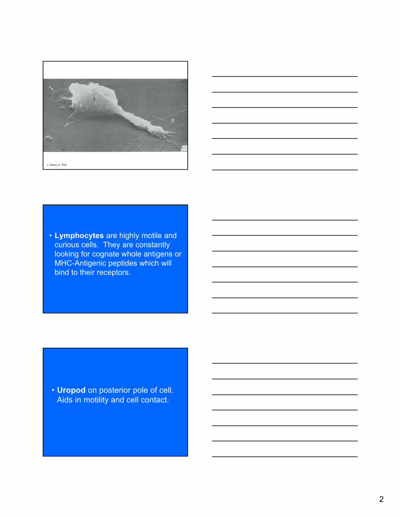
I. IMMUNOLOGICAL SYNAPSE: LYMPOCYTE MORPHOLOGY DURING
IMMUNE RESPONSES
• Lymphocytes are highly motile and curious cells. They are constantly looking for cognate whole antigens or MHC-Antigenic peptides which will bind to their receptors.
• Uropod on posterior pole of cell. Aids in motility and cell contact. • Receptor mediated dedifferentiation of small cells to blasts and then to effector cells.
T and B cells develop receptors for an antigen they have never seen before.
These receptors meet and recognize cognate antigens or MHC-peptides, on Antigen presenting cells.
- T: TCR (αβ; γδ) - CD3 → Peptide-MHC - B: mIg Igα Igβ → Ag
T and B cells have co-receptors
- T: LFA-1 → ICAM-1; CD28 (or CTLA-4) → B7
CD4 - MHC II CD8 - MHC I
- B: CD19, CD21
• Immunological Synapse: Intercellular junctions of approximately 20nms between T cells and antigen presenting cells (APC) which contain spatially segregated supramolecular activation clusters (SMACs) formed with TCR-CD3, CD4 or 8 and CD28 (or CTLA-4) in the center of a donut surrounded by LFA-1 on the T cell which binds ICAM-1 on the stimulating cell (APC). Requires cytoskeletal actin for formation. Cells initially bind the APC through low-affinity LFA-1: ICAM-1 interactions. Subsequent binding of TCR to peptide-MHC signals a conformational change in LFA-1 which increases affinity and prolongs cell-cell contact
(Figs 8.18, 8.19, 8.31)
• Membrane nanotubes have been seen between lymphoid cells upon disassembly of immunological synapses.

John Clancy Jr., Ph.D. Host Defense - 2012
Functional Lymphoid Anatomy
4
II. RECIRCULATION OF LYMPHOCYTES Lymph Node
High Endothelial Post-Capillary Venule (HEPCV)
- Both naïve T and B leave circulation because of low shear force and charge as well as adhesion molecules in the venule. (Fig 8.8).
Rolling: Process initiated by binding of lymphocyte L-selectin to sulfated
carbohydrates of the vascular addressins GlyCAM-1 and CD34. Rolling occurs after the initial binding.
Activation of LFA-1 by chemokines.
- Chemokines - small polypeptides which chemotactically attract
different types of leukocytes and regulate integrin expression (LFA-1) on leukocytes. Secondary lymphoid chemokine (SLC, CCL21 or 6 Ckine) present on HEPCV which binds to it’s receptor (CCR7) on the lymphocyte and activates it’s LFA-1.
Arrest/Adhesion of Lymphocyte by binding of LFA-1 to ICAM-1 on the
endothelial cells.
Migration (Diapedesis)
- Unzipping of Adhesion Molecules
Digestion of Basal lamina as lymphocytes move into and become part of the parenchyma of lymph nodes.
Fig. 8.8 Janeway 7th Edition. Lymphocytes in the blood enter lymphoid tissue by crossing the walls of high endothelial venules. The first step is the binding of L-selection on the lymphocyte to sulfated carbohydrates of GlyCAM-1 and CD34 on the high endothelial cells. Local chemokines activate LFA-1 on the lymphocyte and cause it to bind tightly to ICAM-1 on the endothelial cell, allowing migration across the endothelium. For the lymphocyte to cross the high endothelial barrier successfully, migration has to head to activation of matrix metalloproteinases, as with the migration of neutrophils out of the blood.
John Clancy Jr., Ph.D. Host Defense - 2012
Functional Lymphoid Anatomy
5
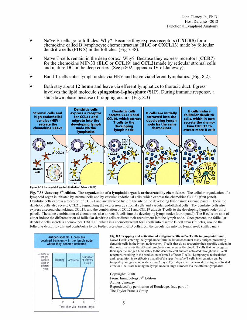
Naïve B-cells go to follicles. Why? Because they express receptors (CXCR5) for a chemokine called B lymphocyte chemoattractant (BLC or CXCL13) made by folicular dendritic cells (FDCs) in the follicles. (Fig 7.38).
Naïve T-cells remain in the deep cortex. Why? Because they express receptors (CCR7)
for the chemokine MIP-3β (ELC or CCL19) and CCL21made by reticular stromal cells and mature DC in the deep cortex. (See p.802, appendix IV of Janeway).
Band T cells enter lymph nodes via HEV and leave via efferent lymphatics. (Fig. 8.2).
Both stay about 12 hours and leave via efferent lymphatics to thoracic duct. Egress
involves the lipid molecule spingosine-1-phosphate (S1P). During immune response, a shut-down phase because of trapping occurs. (Fig. 8.3)
Fig. 7.38 Janeway 6th edition. The organization of a lymphoid organ is orchestrated by chemokines. The cellular organization of a lymphoid organ is initiated by stromal cells and by vascular endothelial cells, which express the chemokine CCL21 (first panel). Dendritic cells express a receptor for CCL21 and are attracted by it to the site of the developing lymph node (second panel). There the dendritic cells also secrete CCL21, augmenting the expression by stromal cells and vascular endothelial cells. The dendritic cells also express a second chemokines, CCL19, and the combination of CCL21 and CCL19 attracts T cells to the developing lymph node (third panel). The same combination of chemokines also attracts B cells into the developing lymph node (fourth panel). The B cells are able of either induce the differentiation of follicular dendritic cells or direct their recruitment into the lymph node. Once present, the follicular dendritic cells secrete a chemokines, CXCL13, which is a chemoattractant for B cells into discrete B-cell areas (follicles) around the follicular dendritic cells and contributes to the further recruitment of B cells from the circulation into the lymph node (fifth panel)
Fig. 8.3 Trapping and activation of antigen-specific naïve T cells in lymphoid tissue. Naïve T cells entering the lymph node form the blood encounter many antigen-presenting dendritic cells in the lymph node cortex. T cells that do no recognize their specific antigen in the cortex leave via the efferent lymphatics and reenter the blood. T cells that do recognize their specific antigen bind stably to the dendritic cell and are activated through their T-cell receptors, resulting in the production of armed effector T cells. Lymphocyte recirculation and recognition is so effective that all of the specific naïve T cells in circulation can be trapped by antigen in on node within 2 days. By 5 days after the arrival of antigen, activated effector T cells are leaving the lymph node in large numbers via the efferent lymphatics. Copyright: 2008 From: Immunology, 7th Edition Author: Janeway Reproduced by permission of Routledge, Inc., part of The Taylor Francis Group

John Clancy Jr., Ph.D. Host Defense - 2012
Functional Lymphoid Anatomy
6
• Spleen
5 x 10¹¹/day leave circulation, enter the spleen and return to the blood.
Cells go to marginal zone blood sinus
- B cells to follicles and marginal zone - T cells to PALS - Similar chemokine gradients to lymph nodes.
Both cells stay about 5 hours then leave via red pulp sinusoids in an open circulation
• Thoracic Duct
0.3 x 10¹¹/day enter the thoracic duct from efferent lymphatics of lymph nodes to return to the blood.
10 T, No NK, some plasmablasts which go to Gut (IgA) and Bone Marrow
(long lived IgG producing cells). Naive and Effector T cells home to different sites because of expression of different receptors on endothelium and on lymphocytes. Naïve cells (CD45RA) loose expression of L-selectin and increase expression of LFA-1 and VLA-4 (Fig 10.9) as they become armed effector T cells which are not readily found in secondary lymphoid organs.
Fig. 10.9 Janeway 7th Edition. Armed effector T cells change their surface molecules, allowing them to home to sites of infection. Naïve T cells home to lymph nodes through the binding of L- Selection to sulfated carbohydrates displayed by various proteins, such as CD34 and GlyCAM-1 on the high endothelial venule. If they encounter antigen and differentiate into armed effector T cells, many lose expression of L-section, leave the lymph node about 4-5 days later, and now express VLA-4 and increased levels of LFA-1. These bind to VCAM-1 and ICAM-1, respectively, on peripheral vascular endothelium at sites of inflammation. On differentiating into effector cells, T cells also alter splicing of the mRNA encoding the cell-surface molecule cd45. The CD45RO isoform expressed by naïve T cells, and make effector T cells more sensitive to stimulation by specific antigen.

John Clancy Jr., Ph.D. Host Defense - 2012
Functional Lymphoid Anatomy
7
III. MACROPHAGES AND OTHER ANTIGEN PRESENTING CELLS OR ANTIGEN
ASSOCIATED CELLS (Fig 8.16, 8.14) Macrophages: Bone Marrow → PB Monocytes → Tissue mononuclear phagocytic system → macrophages:
Kupffer cells (Liver)
Alveolar macrophages (Lung) Process and present antigenic peptides in association with MHC II (Fig 8.16). B cells can also take up antigen, process it and present it to T cells (Fig 8.16).
Dendritic (Branched) Cells: Sentinels of the immune system.
Originate from BM precursors under influence of GM-CSF, IL-4, Flt-3 ligand and TNFα. Migrate through bloodstream to almost every tissue where they become resident immature DC (see Fig 2 from Bone Marrow Lecture
3-6-06).
Immature are phagocytic with DEC205 receptors and E-cadherin molecules but ↓ MHC and ↓ co-stimulatory molecules.
↑ Ag Uptake. - e.g. Langerhans Cells (Fig 8.14) with Birbeck granules in the
epidermis. - After activation of their Toll-like receptors (TLR) by antigen
(Ag),they upregulate CCR7. - Traffic processed Ag to appropriate Lymphoid Organ where they
mature into Inter-digitating Dendritic Cells and initiate T cell responses.
- CCL19 and CCL21 bind to CCR7 to provide further maturation signals and direct their migration.
Mature cells (e.g. Interdigitating Dendritic Cells).
↑ Ag Presentation. - Express ↑ levels of MHC (10 - 100x B cells or monocytes); also
express costimulatory B7 (CD80 and 86), CD40 and ICAM as well as LFA molecules necessary for T cell responses.
- Produce and express CCL19 and CCL21 which attracts naïve T cells.
- 1 mature DC can stimulate 100-3000 T Cells! - Some mature Dendritic Cells go to the Thymus where they
function in negative selection of potentially auto-reactive thymocytes.
- Also act as sentinels along GI tract, particularly in the ileum.

John Clancy Jr., Ph.D. Host Defense - 2012
Functional Lymphoid Anatomy
8
Fig. 8.16 Janeway 7th Edition. The
properties of the various antigen-presenting cells. Dendritic cells, macrophages, and B cells are the main cell types involved in the initial presentation of foreign antigens to naïve T cells. These cells vary in their means of antigen uptake, MHC class II expression, co-stimulator expression, the type of antigen they present effectively, their locations in the body, and their surface adhesion molecules (not shown)
Fig. 8.14 Janeway 7th Edition. Dendritic cells mature through at least two definable stages to become potent antigen-presenting cells in peripheral lymphoid tissue. Dendritic cells originate from bone marrow progenitors and migrate via the blood to peripheral tissues and organs, where they are highly phagocytic via receptors such as DEC 205 and are actively macropinocytic but do not express co-stimulatory molecules (top panel). At sites of infection they pick up antigen and are induced to migrate to the regional lymph node. Here they are powerful activators of naïve T cells but are no longer phagocytic. Dendritic cells in lymphoid tissue express B7.1, B7.2, and high levels of MHC class I and class II molecules, as well as high levels of the adhesion molecules ICAM-1, ICAM-2, LFA-1 and CD58 (center panel). They also express high levels of the dendritic-cell-specific adhesions molecule DC-SIGN, which binds ICAM-3 with high DC-CK (CCL18), secreted chemokine affinity. The photograph courtesy of J. Barker.

John Clancy Jr., Ph.D. Host Defense - 2012
Functional Lymphoid Anatomy
9
• Antigen Associated Cell
Follicular Dendritic Cells (FDC) are antigen retaining.
- Found in Light Zone of Germinal Centers (GC).
- Probably not BM derived. Seem to be derived from reticular stromal cells
- ↓ MHC II
- ↑ FcγR, Complement R (Fig 9.14): thus Ag - Ab complexes
retained on them for years. - ↑ ICAM-1, VCAM-1
- They possess TNF - Receptors and are critical in GC formation.
In TNF-R knock-out mice there are no FDC and no GC.
- Make CXCL13 (BLC) which attracts B cells. - Iccosomes are morphological Ag - Ab complexes found on FDCs
(Fig 9.15) M cells are antigen transporting and are modified epithelial lining cells found in the
ileum of the GI tract and bronchi overlying bronchial associated lymphoid tissue. (Fig 11.8)
Copyright: 2008 From: Immunobiology, 7th edition Author: Janeway, et al Produced by permission of Routledge, Inc. part of the The Taylor Francis Group

John Clancy Jr., Ph.D. Host Defense - 2012
Functional Lymphoid Anatomy
10
IV. COURSE OF AN INFECTION AND GERMINAL CENTER REACTION (Figures 1
and 2) (Fig 10.2, 10.14, 9.10, 9.11, 9.3, 9.9)
• Step 1: Figure 1. Antigen reaches a regional lymph node via afferent lymphatics as either free antigen or after having been processed by a peripheral Langerhans Immature Dendritic Cell (IDC).
• Step 2: Antigen laden Langerhans Immature Dendritic Cell localizes in the deep cortex of lymph node where it up regulates its MHC and costimulatory molecules and becomes a mature Interdigitating Dendritic Cell (MDC). It then presents antigen to the Th cells in the deep cortex. The Th cell is now trapped in the deep cortex. An immunological synapse I is thus formed between the Th cell and the IDC activating the Th cell. - Antigen also binds to B cells (just arriving in the deep cortex through HEPCVs or in
the follicle) that have a cognate surface immunoglobulin receptor for the antigen.
• Step 3: 3-5 days after antigen exposure there is a synapse independent expansion of antigen-specific Th cells which down regulate their CCR7 receptor and migrate to the edge of the follicle. Follicular antigen activated B cells downregulate CXCR5 and move to the edge of the follicles to meet the Th cells.
• Step 4: Synapse II forms at the edge of the follicle between antigen primed B cells and
antigen-specific Th cells. B cells express peptide-class II, B7, CD40 and Th cells express TCR, CD28, CD40 Ligand.
• Step 5: Some B cells move to the medullary cords to become short-lived plasma cells
and produce IgM or IgG antibody for a few days (Fig 10.2). This antibody can combine with antigen to form some early protection and antigen-antibody complexes which are trapped on the surface of FDCs. Some CCR7- Ag primed Th cells migrate into the base of the follicle with the CXCR5↓ Ag-primed B cells.
• Step 6: Figure 2. At the base of the follicle the B cells involved in Synapse II are called
centroblasts and proliferate actively (dividing every 6 hours) and form a densely packed area called the dark zone within a few days. Within this area somatic hypermutation of rearranged immunoglobulin V-domain genes occurs.
- As these cells mature they stop dividing and move up into the less densely packed
light zone as non-dividing centrocytes along with their companion antigen specific Th cells. Thus centrocytes are the non-dividing progeny of centroblasts. As the newly formed centrocytes populate the light zone, B cells in the follicle not specific for the Ag are pushed outside to form the peripheral mantle zone. (Fig 9.10)
• Centrocyte selection and survival depends on antigen affinity and Bcl-xl expression.
Centrocytes come in contact with Ag/Ab complexes bound to Fc receptors or Ag/Ab/complement complexes bound to complement receptors on Follicular Dendritic cells (FDC). If centrocytes are making immunoglobulin receptors that do not bind Ag displayed on FDCs, they die by apoptosis and are engulfed by macrophages. (Fig 9.11)

John Clancy Jr., Ph.D. Host Defense - 2012
Functional Lymphoid Anatomy
11
Figure created by John Clancy, Jr., PhD

John Clancy Jr., Ph.D. Host Defense - 2012
Functional Lymphoid Anatomy
12
Fig. 10.14 Janeway 6th Edition. The specialized regions of peripheral lymphoid tissue provide an environment where antigen-specific naïve B cells can interact with armed helper T cells specific for the same antigen. The initial encounter of antigen-specific naïve B cells with the appropriate helper T cells occurs in the T-cell areas in lymphoid tissue and stimulates the proliferation of B cells in contact with the helper T cells to form a primary focus, as shown in the first 3 panels. This also results in some isotype switching in the antigen-specific B cells. Some of the activated B-cell blasts then migrate to medullary cords, where they divide, differentiate into plasma cells, and secret antibody for a few days (third panel. Other B-cell blasts migrate into primary lymphoid follicles, where they proliferate rapidly to form a germinal center under the influence of antigen and of helper T cells (fourth panel). The germinal center is the site of somatic hypermutation and selection of high-affinity B cells that are able to bind antigen better than lower-affinity cells and thus survive either because they are protected from apoptotic signals delivered by T cells or/and they are more capable of presenting antigen to T cells and thereby receiving positive signals such as IL-4 and CD40 ligand (fifth panel). FDC, follicle dendritic cell.
Fig. 9.10 Janeway 7th Edition. Germinal centers are formed when activated B cells enter lymphoid follicles. The germinal center is a specialized microenvironment in which B-cell proliferation, somatic hypermutation , and selection for antigen binding all occur. Closely packed centroblasts form the so-called “dark zone” of the germinal center, as can be seen in the lower part of the photomicrograph in the center, which shows a high-power view of a section through a human tonsillar germinal center. The photomicrograph on the right shows a lower-power view of a tonsillar germinal center; B cells are found in the dark zone, light zone, and mantle zone. Proliferating cells are stained green for K167, an antigen expressed in nuclei of dividing cells, revealing the centroblasts in the dark zone. The dense network of follicular dendritic cells, stained red, mainly occupies the light zone. Cells in the light zone are also proliferating, through to a lesser degree in most germinal centers. Small recirculating B cells occupy the mantle zone at the edge of the B-cell follicle. Large masses of CD4 T cells, stained blue, can be seen in the T-cell zones, which separate the follicles. There are also significant numbers of T cells in the light zone of the germinal center; CD4 staining in the dark zone is mainly associated with CD4-positive phagocytes. Photographs courtesy of I. MacLennan.

John Clancy Jr., Ph.D. Host Defense - 2012
Functional Lymphoid Anatomy
13
Copyright: 2008 From: Immunobiology, 7th edition Author : Janeway, et al Reproduced by permission of Routledge, Inc. part of The Taylor Francis Group
Fig. 9.11 Activated B cells undergo rounds of mutation and selection for higher-affinity mutants in the germinal center, resulting in high-affinity antibody-secreting plasma cells and high-affinity memory B cells. B cells are first activated outside follicles by the combination of antigen and T cells (top panel). They migrate to germinal centers (not shown), where the remaining events occur. Somatic hypermutation can result in amino acid replacements in immunoglobulin V regions that affect the fate of the B cell. Mutations that result in a B-cell receptor (BCR) of lower affinity for the antigen (left panels) will prevent the B cell from being activated as efficiently, because both B-cell receptor cross-linking and the ability of the B cell dying by apoptosis. In this way, low-affinity cells are purged from the germinal center. Most mutations are either negative or neutral (not shown) and thus the germinal center is a site of massive B-cell death as well as of proliferation. Some mutations, however, will improve the ability of the B-cell receptor to bind antigen. This increases the B cell’s chance of interacting with T cells, and thus of proliferating and surviving (right panels). Surviving cells undergo repeated cycles of mutation and selection during which some of the progeny B cells undergo differentiation to either memory B cells or plasma cells (bottom right panels) and leave the germinal center. The signals that control these differentiation decision are unknown.

John Clancy Jr., Ph.D. Host Defense - 2012
Functional Lymphoid Anatomy
14
Centrocytes with high affinity surface immunoglobulin receptors upregulate Bcl-xl expression and survive. This process of Affinity Maturation allows the selection of B cells (centrocytes) producing surface immunoglobulin with progressively higher affinity for the antigen to contribute to the response to that antigen.
A centrocyte that binds Ag then presents fragments of it to Ag-specific Th cells thus forming Synapse III. If the Th cell is a Th2 cell, it displays the B-cell stimulatory molecule CD40 Ligand and secretes the B-cell stimulatory cytokines IL4, IL5 and IL6 which drive further proliferation of B cells into either a blast cell (plasmablast) which will leave the lymph node and go to the bone marrow to become long lived IgG1 or IgE plasma cells or to enter the memory B-cell pool (Figs 9.3). If the Th cell is a Th1 cell which secretes interferon γ (IFNγ), the centrocyte will switch its isotype and become an IgG2a and IgG3 producer, which fixes complement promoting phagocytosis of opsonized microbes by binding to Fcγ receptors.
• Progeny of centrocytes are either memory B cells or plasmablasts.
Memory B cells are found in the mantle zone of follicle, some in marginal zone of
spleen. Long-lived memory B cells and IgG producing plasma cells go to the bone marrow (Fig 9.9). Remember that the spleen can generate an immune response to blood-borne antigens similar to a lymph node but antigen enters the white pulp through the marginal sinus.

John Clancy Jr., Ph.D. Host Defense - 2012
Functional Lymphoid Anatomy
15
Figure created by John Clancy, Jr. PhD

John Clancy Jr., Ph.D. Host Defense - 2012
Functional Lymphoid Anatomy
16
Fig. 9.3 Armed helper T cells stimulate the proliferation and then the differentiation of antigen-binding B cells. The specific interaction of an antigen-binding B cell with an armed helper T cell leads to the expression of the B-cell stimulatory molecule CD40 ligand (CD154) on the helper T-cell surface and to the secretion of the B-cell stimulatory cytokines IL-4, IL-5 and IL-6, which drive the proliferation and differentiation of the B cell into antibody-secreting plasma cells. An activated B cell can alternatively become a memory cell. Janeway 7th Edition

John Clancy Jr., Ph.D. Host Defense - 2012
Functional Lymphoid Anatomy
17
V. DISEASE AFFECTING T CELLS.
DiGeorge Syndrome - Status of lymphoid organs?
AIDS Patients - Where is the virus hiding?
Graft-Versus-Host Disease - A bone marrow cellular graft containing allogeneic T cells can reject the recipient.
1
Functional Lymphoid Anatomy
John Clancy, Ph.D.
4/27/2012
Content Summary
I. Immunological Synapse: Lymphocyte
Morphology During Immune Responses
II. Recirculation of Lymphocytes
III. Macrophages and Other Antigen Presenting Cells or Antigen Associated Cells
IV. Germinal Center Reaction
V. Diseases Affecting T Cells
I. Immunological Synapse: Lymphocyte Morphology During Immune Responses
2
J. Clancy, Jr., PhD
• Lymphocytes are highly motile and curious cells. They are constantly looking for cognate whole antigens or MHC-Antigenic peptides which will bind to their receptors.
• Uropod on posterior pole of cell. Aids in motility and cell contact.

3
J. Clancy, Jr., PhD
J. Clancy, Jr., PhDJ. Clancy, Jr., PhD
• Receptor mediated dedifferentiation of small cells to blasts and then to effector cells.

4
Immunological Synapse
• 20nm junction of T:APC
• First LFA-1(T):ICAM-I(APC)
• Then TCR: MHC-peptide
• LFA-1 binds ICAM-1 tighter
DONUT: T cell side
Center: TCR, CD4, CD28
Periphery-LFA-1
APC side
CENTER: MHC-peptide, B7
Periphery:ICAM-1
Janeway, Fig 8.19

5
Requires cytoskeletal actin for formation.
Membrane Nanotubes have been seen
between lymphoid cells upon disassembly of
immunological synapes.
John Clancy, Jr. PhD

6
John Clancy Jr., PhD
II Recirculation of Lymphocytes
Lymph NodeHigh Endothelial Post-CapillaryVenule (HEPCV)
- Both naïve T and B leave circulation because of lowshear force and charge as well as adhesion molecules in the venule (Fig. 8.8)
Janeway, Fig 8.4

7
Janeway Fig. 8.8
Rolling: Process initiated by bindingof lymphocyte L-selectin to sulfated
carbohydrates of the vascular addressins GlyCAM-1 and CD34. Rolling occurs after the initial binding.
Activation of LFA-1 by chemokines.
- Chemokines - small polypeptides which chemotactically attract different types of leukocytes and regulate integrin expression (LFA-1) on leukocytes. Secondary lymphoid chemokine (SLC, CCL21 or
6 Ckine) present on HEPCV which binds to it’s receptor (CCR7) on the lymphocyte and activates it’s LFA-1.

8
Arrest/Adhesion of Lymphocyte by binding of
LFA-1 to ICAM-1 on the endothelial cells.
Migration (Diapedesis)
- Unzipping of Adhesion Molecules
Digestion of Basal lamina as lymphocytes
move into and become part of the parenchyma
of lymph nodes.
Janeway Fig. 8.8
Naïve B-cells go to follicles.
Why? Because they express
receptors (CXCR5) for a
chemokine called B lymphocyte
chemoattractant (BLC or CXCL13)
made by folicular dendritic cells (FDCs) in the follicles. (Fig 7.38).

9
Janeway Fig. 7.38
Naïve T-cells remain in the
deep cortex. Why? Because they
express receptors (CCR7) for the
chemokine MIP-3 (ELC or
CCL19) and CCL21 made by reticular
stromal cells and mature DC in the
deep cortex. (See p.802, appendix IV
of Janeway).
Janeway, Fig 8.2

10
Both stay about 12 hours and
leave via efferent lymphatics to
thoracic duct. Egress involves the
the lipid molecule spingosine-1-
phosphate (S1P). Immunosuppressive drug FTY720 is an agonist to S1P receptors. It thus inhibits lymphocyte exit.
During immune response, a shut-
down phase because of trapping
occurs (Fig. 8.3). The mechanism
is a down regulation of S1P
receptors on activated T cells.
Janeway Fig 8.3

11
Junqueira Fig. 14-28
Spleen 5 x 10¹¹/day leave circulation, enter the spleen
and return to the blood.
Cells go to marginal zone blood sinus
- B cells to follicles and marginal zone
- T cells to PALS
- Similar chemokines gradients to
lymph nodes
Both cells stay about 5 hours then leave the
white pulp through marginal zone bridging
channels to the red pulp cords and red pulp
sinusoids in an open circulation.
John Clancy, Jr. PhD
Marginal Zone

12
• Thoracic Duct
0.3 x 10¹¹ /day enter the thoracic duct from efferent lymphatics of lymph nodes to return to the blood.
1° T, No NK, some plasmablasts which go to Gut (IgA) and Bone Marrow (long lived IgG producing cells).
Janeway Fig. 10.9
Naïve T (CD45RA) loose L-selectin
Armed effector T (CD45RO) gain
LFA-1, VLA-4

13
Naïve and Effector T cells home to
different sites because of expression
of different receptors on endothelium
and on lymphocytes. Naïve cells
(CD45RA) loose expression of L-selectin
and increase expression of LFA-1 and
VLA-4 (Fig. 10.9) as they become armed
effector T cells which are not readily found
in secondary lymphoid organs.
John Clancy Jr., PhD
III. Macrophages and other Antigen
Presenting Cells or Antigen Associated
Cells

14
John Clancy Jr., PhD
Macrophages: Bone Marrow → PB Monocytes→ Tissue mononuclear phagocytic system → macrophages.
Kupffer cell (Liver)
Alveolar macrophages (Lung)
Process and present antigenic peptides in
Association with MHC II
B cells can also take up antigen, process
It and present it to T cells.
Janeway Fig. 8.16

15
Dendritic (Branched) Cells: Sentinels of the immune system.
Originate from BM precursors under influence of GM-CSF, IL-4, Flt-3 ligand and TNF. Migrate through bloodstream to almost every tissue where they become resident immature DC (see Fig. 2 from Bone Marrow Lecture)
Flt-3 Ligand
John Clancy, Jr. PhD
Janeway Fig. 8.14

16
Janeway Fig. 1.9
Immature are phagocytic with DEC205 receptors and E-cadherin molecules but MHC and co-stimulatory molecules. ↑ Ag Uptake.
- e.g. Langerhans (Fig. 8.14) with Birbeckgranules in the epidermis.
- Traffic Ag to appropriate Lymphoid Organ where they mature into Inter- digitating Dendritic Cells and Initiate T cell responses.
How?
- After activation of their Toll-like receptors
by antigen (Ag), they upregulate CCR7.
- CCL19 and CCL21 bind to CCR7 to
provide further maturation signals and
direct their migration.

17
Mature cells (e.g. Interdigitating Dendritic Cells).↑ Ag Presentation.
- Express ↑ levels of MHC (10 – 100x B cells or monocytes); also express co-stimulatory B7 (CD80, 86), CD40 and ICAM as well as LFA molecules necessary for T cell responses.
- Contain low levels of proteases compared to macrophages. Thus slow Ag degradation.
Janeway Fig. 8.15
Janeway Fig. 8.9

18
- Produce and express MIP-3 (CCL19)
and CCL21 which attracts naïve T cells.
- 1 mature DC can stimulate 100-3000 TCells!
Also, can act as sentinels along the
GI tract, particularly in the ileum.
Janeway, Fig 11.9

19
Janeway Fig. 9.15
• Antigen Associated Cell
Follicular Dendritic Cells (FDC) are antigen retaining.
- Found in Light Zone of Germinal
Centers (GC).
- Probably not BM derived. Seem to
be derived from reticular stromal cells
- MHC II
- ↑ FcγR, Complement R (Fig. 9.14): thus
Ag – Ab complexes retained on them for
years.
Janeway Fig. 9.14

20
- ↑ ICAM-1, VCAM-1
- They possess TNF – Receptors and are critical in GC formation. In TNF-R knock-out mice there are no FDC and no GC.
- Make BLC (CXCL13)which attracts B cells.
- Iccosomes are morphological Ag – Ab complexes found on FDCs
Janeway Fig. 9.15
Iccosome
M cells are antigen transporting
and are modified epithelial lining cells
found in the ileum of the GI tract and
bronchi overlying bronchial
associated lymphoid tissue.

21
Janeway, Fig 11.8
IV. Course of an infection and
Germinal Center Reaction
Janeway, Fig. 10.2

22
IV. Germinal Center Reaction
• Step 1: Figure 1. Antigen reaches a regional lymph node via afferent lymphatics as either free antigen or after having been processed by a peripheral Langerhans Immature Dendritic Cell (IDC).
Step 1: Aff. Lymph
Free AG → B, Macrophages
Processed Ag within IDC
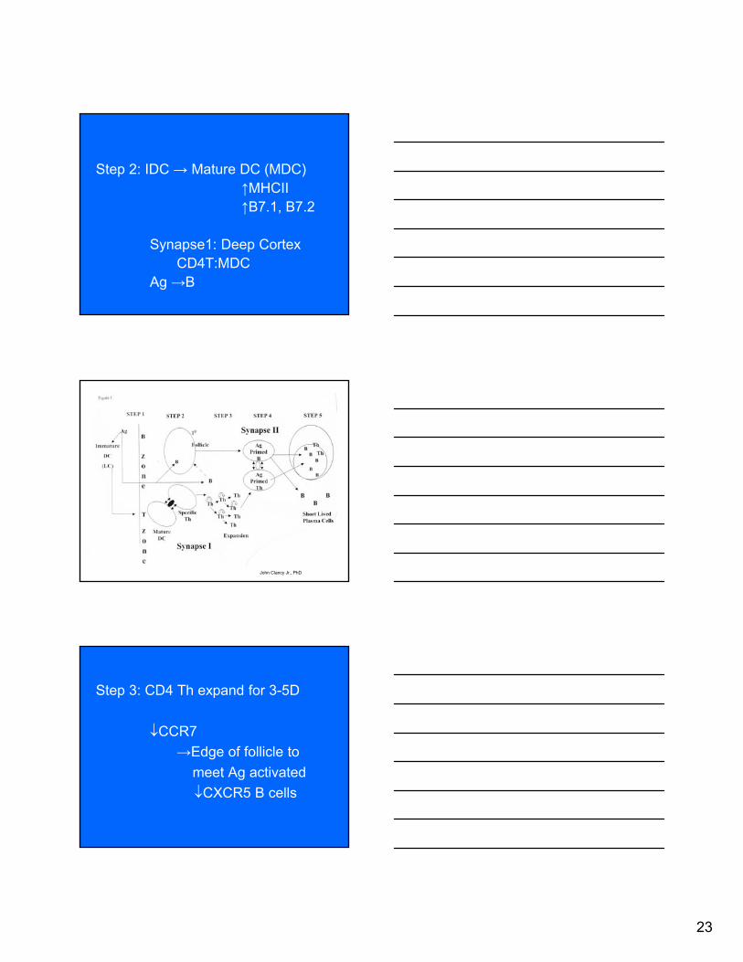
23
Step 2: IDC → Mature DC (MDC)↑MHCII↑B7.1, B7.2
Synapse1: Deep CortexCD4T:MDC
Ag →B
John Clancy Jr., PhD
Step 3: CD4 Th expand for 3-5D
CCR7
→Edge of follicle to
meet Ag activated
CXCR5 B cells

24
• Step 3: 3-5 days after antigen exposure there is a synapse independent expansion of antigen-specific Th cells which down regulate their CCR7 receptor and migrate to the edge of the follicle. Follicular antigen activated B cells
downregulate CXCR5 and move to the edge of the follicles to meet the Th cells.
Step 4: Synapse II at Follicular Edge
Th (TCR, CD28, CD40 Ligand)
B (peptide-MHCII, B7, CD40)
• Step 4: Synapse II forms at the edge of the follicle between antigen primed B cells and antigen-specific Th cells. B cells express peptide-class II, B7, CD40 and Th cells express TCR, CD28, CD40 Ligand.

25
John Clancy Jr., PhD
Janeway Fig. 10.14
• Step 5: Short-lived B → Plasma cells
(medulary cords)
Initial IgM or IgG burst
- Ag – Ab complexes FDC
Potential Long-lived B and some
CCR7-: Th → base of 1°F

26
J. Clancy, Jr., PhD
Light
Dark Zones
Janeway Fig. 9.10
Step 6: activated B → centroblasts (Dark Zone)
- GT: 6hrs
- Somatic hypermutation
Centroblasts → Centrocytes (Light Zone)
- no proliferation
- Affinity maturation or
Apoptosis (FDC)

27
Janeway Fig. 9.10
John Clancy Jr., PhD
plasmablasts
IL 21
Bcl-6
CXCR5
TFh
• Centrocyte selection and survival depends on antigen affinity and Bcl-xl expression. Centrocytes come in contact with Ag/Ab complexes bound to Fc receptors or Ag/Ab/complement complexes bound to complement receptors on Follicular Dendriticcells (FDC). If centrocytes are making immunoglobulin receptors that do not bind Ag displayed on FDCs, they die by apoptosis and are engulfed by macrophages. (Fig 9.11)

28
• Centrocytes with high affinity surface immunoglobulin receptors upregulate Bcl-xl expression and survive. This process of Affinity Maturation allows the selection of B cells (centrocytes) producing surface immunoglobulin with progressively higher affinity for the antigen to contribute to the response to that antigen.
Janeway Fig. 9.11
Synapse II
Synapse III
John Clancy Jr., PhD
Il 21 necessary for centrocyte differention, Class switching &Antibody secretion
IL-21
B Plasmablast
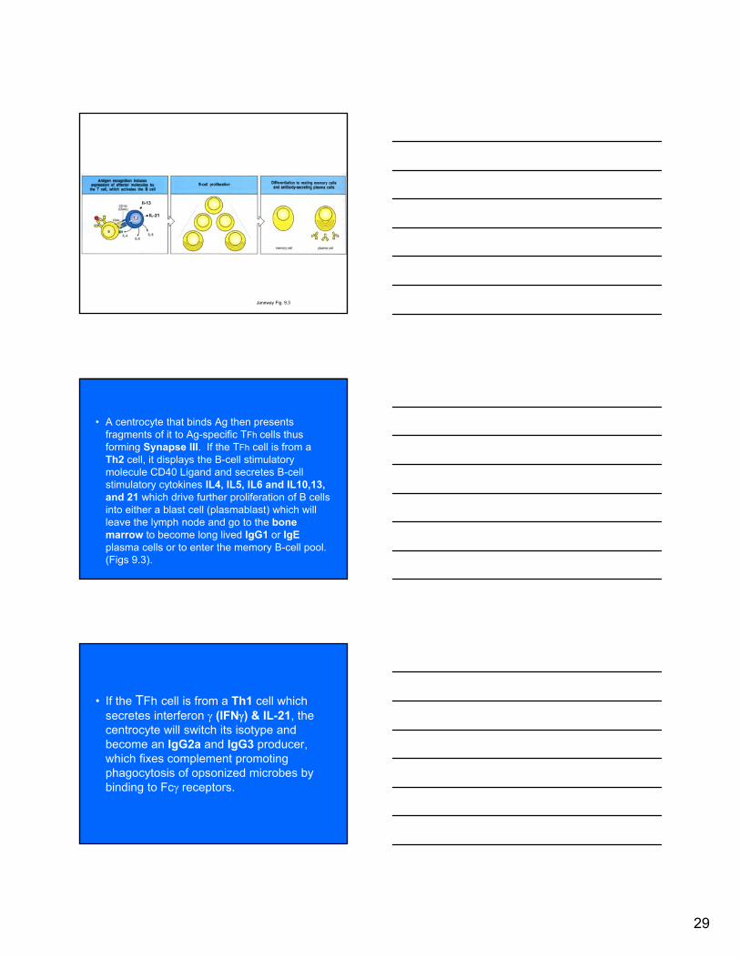
29
Janeway Fig. 9.3
Il-13
IL-21
• A centrocyte that binds Ag then presents fragments of it to Ag-specific TFh cells thus forming Synapse III. If the TFh cell is from a Th2 cell, it displays the B-cell stimulatory molecule CD40 Ligand and secretes B-cell stimulatory cytokines IL4, IL5, IL6 and IL10,13, and 21 which drive further proliferation of B cells into either a blast cell (plasmablast) which will leave the lymph node and go to the bone marrow to become long lived IgG1 or IgEplasma cells or to enter the memory B-cell pool. (Figs 9.3).
• If the TFh cell is from a Th1 cell which secretes interferon (IFN) & IL-21, the centrocyte will switch its isotype and become an IgG2a and IgG3 producer, which fixes complement promoting phagocytosis of opsonized microbes by binding to Fc receptors.

30
John Clancy Jr., PhD
plasmablasts
IL 21
CXCR5
Synapse III
↑ Affinity Centrocyte (↑Bcl-xl):TFh
Memory Cells
- Follicular Mantle
- Marg. Zone of Spleen
Plasmablasts (IgG1 or IgE)
Bone Marrow
IL10,13,21
IL4, 21
V. Diseases Affecting T Cells • Nude Mouse is similar to humanwith a DiGeorge Syndrome
Thymus agenesis because
developmental defect in the third
and fourth pharyngeal pouches
What would be the status of
lymphoid organs?

31
John Clancy Jr., PhD
John Clancy Jr., PhD
John Clancy Jr., PhD

32
John Clancy Jr., PhD
John Clancy Jr. PhD
John Clancy Jr., PhD
1° follicle

33
John Clancy Jr., PhD
AIDS PatientsWhere is the virus hiding?
John Clancy Jr., PhD

34
John Clancy, Jr., PhD
John Clancy Jr., PhD
• AIDS
CD4 antigen on Th cell binds Gp 120
on virus.
Follicular Dendritic Cells retain virus
antibody complexes for many years.
When FDC finally die, lethal veremia
ensues.

35
J. Clancy, Jr., PhD
J. Clancy, Jr., PhD
John Clancy Jr., PhD

36
• Graft-Versus-Host Disease in patientswith leukemia that receive a transplant from an allogenic donor.
Bone marrow transplant rejects host
Bone marrow graft contains allogeneic T
cells as well as CD 34+ cells. T cells of
donor react against host foreign MHC
antigens. Thus donor T cells rejecting
the recipient.

Host Defense 2013 Small Group Problem Solving Sessions
Cytokines/T-Cell Subsets
Rev 11/06/2012 Page 1
HOST DEFENSE
SMALL GROUP PROBLEM SOLVING SESSION
CYTOKINES/T CELL SUBSETS
Small Group Classrooms
LEARNING GOAL You will be able to explain how TLR, MHC & DC dictate the type of immune response and then how T-cells orchestrate it.
OBJECTIVES To achieve this goal, you will have to be able to: Differentiate T-cell functions. Understand how cytokine profiles interact with T-cell subsets to produce appropriate
responses to pathogens. Explain how deficiency of Th-1 function can cause disease. Explain the clinical consequences of the Th-2 reaction when a Th1-type infection is
present
BACKGROUND READING Janeway 8th edition: pages 364; 437-439; 516-517 and posted article
DEVELOPED BY John A. Robinson, MD

Host Defense 2013 Small Group Problem Solving Sessions
Cytokines/T-Cell Subsets
Rev 11/06/2012 Page 2
HOW TO SUCCEED IN SMALL GROUPS Before coming to class: 1. Read assigned chapters/ pages and develop answers for ALL the questions in the
4 clinical vignettes During the Small Group Session: 2. Each small group (should be 4-5 peers- please do not sort yourselves into large
groups-you will learn much less) should discuss the four case studies and decide the best solutions to the specific integrating questions associated with each case.
3. After approximately an hour of discussion by the subgroups, the facilitator will
recapitulate the answers to the integrating questions by selecting a subgroup to present a synthesis of their relevant discussions to the entire group. Facilitators will select, at their discretion, a small group for the discussion of the individual cases.
4. History has shown that students who don’t contribute to the Small Groups do not
do well in the Course (remember that about 25-30% of the final comes from small groups!) and also have been assaulted by their fellow group members
5. At the end of the session, a master answer sheet will be posted on the Host
Defense website.

Host Defense 2013 Small Group Problem Solving Sessions
Cytokines/T-Cell Subsets
Rev 11/06/2012 Page 3
CASE 1 Leprosy is caused by a mycobacterium related to the organism that causes tuberculosis. The disease has two major forms of expression. One, the tuberculoid form, is characterized by localized granulomas (clusters of macrophages, T cells, DC and necrotic debris). These patients are capable of mounting a normal Th1 response to the infection. The other form, lepromatoid, is characterized by widespread infection, sparse cellular reactions in the organs involved and a poor prognosis. These patients are incapable of mounting a Th1 response to the organism.
1. Why don’t all patients infected with M. Leprae manifest similar clinical states? How does the journal article assigned for this small group help explain the different disease manifestations? (again, do not worry about the technical details of the article and don’t memorize any new cell markers, molecules etc. in the article)
2. On occasion, patients with one form of leprosy will convert to the other. How could this happen and how could it be detected immunologically?

Host Defense 2013 Small Group Problem Solving Sessions
Cytokines/T-Cell Subsets
Rev 11/06/2012 Page 4
3. Clinicians must be aware of the potential dangers and benefits associated with
therapy that influences T-cell expression.
o Discuss mechanisms of action, cytokines influenced, and the advantages and pitfalls of the use of corticosteroids (these drugs suppress TMMI reactions- among other things) in leprosy patients.
o Clinical examples: a pineapple picker in Hawaii develops numbness and
paralysis in his right hand. Physical examination reveals a single, well localized mass in his right axilla which, upon biopsy, reveals leprosy organisms and marked inflammation in and about the nerves of the brachial plexus. Another patient has three small skin nodules but no symptoms and these are also positive for leprosy.
o Which pineapple picking patient might benefit the most from the use of
medications that suppress Th1 reactions?
4. If you wanted to develop a vaccine against leprosy, what cytokines or other
molecular structures (in theory) would be most logical to add to a living but attenuated leprosy preparation in order to mimic an actual infection?

Host Defense 2013 Small Group Problem Solving Sessions
Cytokines/T-Cell Subsets
Rev 11/06/2012 Page 5
CASE 2 Diphtheria is a bacterial infection that exerts many of its clinical effects by production of soluble protein exotoxins that circulate in the blood and damage the peripheral and cranial nervous system. 1. What strategies should dictate the development of a vaccine for this disease? 2. What should be the characteristics of the antigens in this type of vaccine?
3. The toxin can bind to nerve conducting tissue in the heart. If a patient with this
infection develops abnormal ventricular conduction detected by an EKG, what immunologic strategy, in theory, might be life saving?
4. If toxoid proteins were made for a vaccine but it was discovered that they couldn’t
evoke the cytokine profile necessary for a successful vaccine, what cytokines or other molecular structures could be added to the protein antigens to make it effective?

Host Defense 2013 Small Group Problem Solving Sessions
Cytokines/T-Cell Subsets
Rev 11/06/2012 Page 6
CASE 3 Four infants from the same small isolated mountain village died from disseminated infection with Bacillus Calmette-Guerin (BCG). BCG is an attenuated, living tubercular bacillus used extensively for vaccination against mycobacterial tuberculosis in European and Asian countries. All 4 children had a fulminating (rapidly progressive) systemic illness that ultimately proved fatal soon after vaccination. Two were sisters whose parents were first cousins; the third was related to the sisters as a fourth cousin through both parents. The fourth patient has no known link to the other three. The first cousin parents were healthy and had received BCG in the past, and had four other healthy children, all of whom had received BCG. 1. Discuss the possible epidemiologic reasons that a genetic mutation like this
occurred. Duh! But what would a typical pedigree of an affected child look like compared to a parent? And what are the odds that it is an autosomal recessive mutation versus a dominant one. Is the abnormal immune function likely to be epigenetic in origin?
2. In order of probability; discuss likely points in the immune response for a genetic
defect that would lead to this type of immuno deficiency.

Host Defense 2013 Small Group Problem Solving Sessions
Cytokines/T-Cell Subsets
Rev 11/06/2012 Page 7
3. Design experiments that would support your hypothesis that it may be a genetic
defect and identify the actual defect. 4. Design strategies to correct the defect.
5. The mechanism exploited by the physician to discern whether a patient has had or has TB is the TMMI reaction. Many normal patients receive BCG (an attenuated, live TB-like organism vaccine) and they develop a weakly positive TB skin test that can be very confusing when they are tested in the USA for past or current tuberculosis.
a. Be prepared to describe the immunologic mechanisms that give rise to a positive skin test in a first year incoming med student who has been exposed to TB. See if you can figure out how the test could be improved (Hint- do it in a test tube). b. Which of the Swiss family members would have developed a positive TB skin test or the new improved in vitro test after BCG vaccination? And if they didn’t, why not?

Host Defense 2013 Small Group Problem Solving Sessions
Cytokines/T-Cell Subsets
Rev 11/06/2012 Page 8
CASE 4 A In October 2001 (this is almost a true story), enthusiasts from 30 countries
attended the world championship of model airplane flying held at Lost Hills, CA (in the central valley)[up to this point, completely true] Two weeks later, after returning home, 27 of the participants developed flu-like symptoms characterized by fatigue, fever, mild joint pains and muscle aches. Chest x-rays were done in 12 and at least half of this group had bilateral pulmonary infiltrates (pneumonia). One patient required admission to intensive care. Epidemiologic analysis did not detect any specific activity or condition common to the group other than travel to CA and flying model airplanes in very dusty conditions. There was no clinical evidence in any of the patients of prior immunosuppressive therapy or immune defects. All the patients had serologic evidence of recent Coccidiomycosis infection (a fungus with an infectious form that lives in the soil).
B A large group of intravenous drug abusers shared a needle for heroin injection.
Fortunately none of them were HIV positive but, unfortunately, one was an asymptomatic Hepatitis B virus carrier. Four months later, 2 of the drug abusers were dead from fulminant hepatitis B, 6 were recovering from clinical hepatitis.
1. Why was there a broad spectrum of disease expression in both the model
plane flyers and the drug abusers? 2. Describe the defective or missing component of the immune response in
the patient in the ICU and the three patients that died from hepatitis.

Host Defense 2013 Small Group Problem Solving Sessions
Cytokines/T-Cell Subsets
Rev 11/06/2012 Page 9
3. Postulate ways that you could convert the carriers to a non-infectious state.
4. One of the patients with fulminant liver disease had an identical twin that is a dialysis technician and had been previously immunized with the
hepatitis B vaccine because of the occupational hazard. Devise creative ways that a physician could have exploited this stroke of luck.
5. In honor of George Santayana and serendipity How are T cells indebted to nudity?