Embed Size (px)
Citation preview

IHC Submission
All IHC slides submitted to the core should be accurately described on both form and slides. We can perform the experiment without rechecking them.

IHC Method
Indirect Alexa 488 or 568
Biotin-Streptavidin system
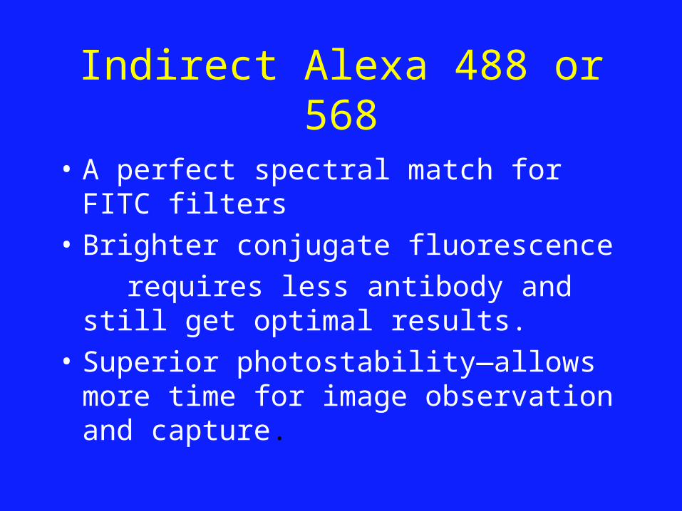
Indirect Alexa 488 or 568
• A perfect spectral match for FITC filters
• Brighter conjugate fluorescence
requires less antibody and still get optimal results.
• Superior photostability—allows more time for image observation and capture.
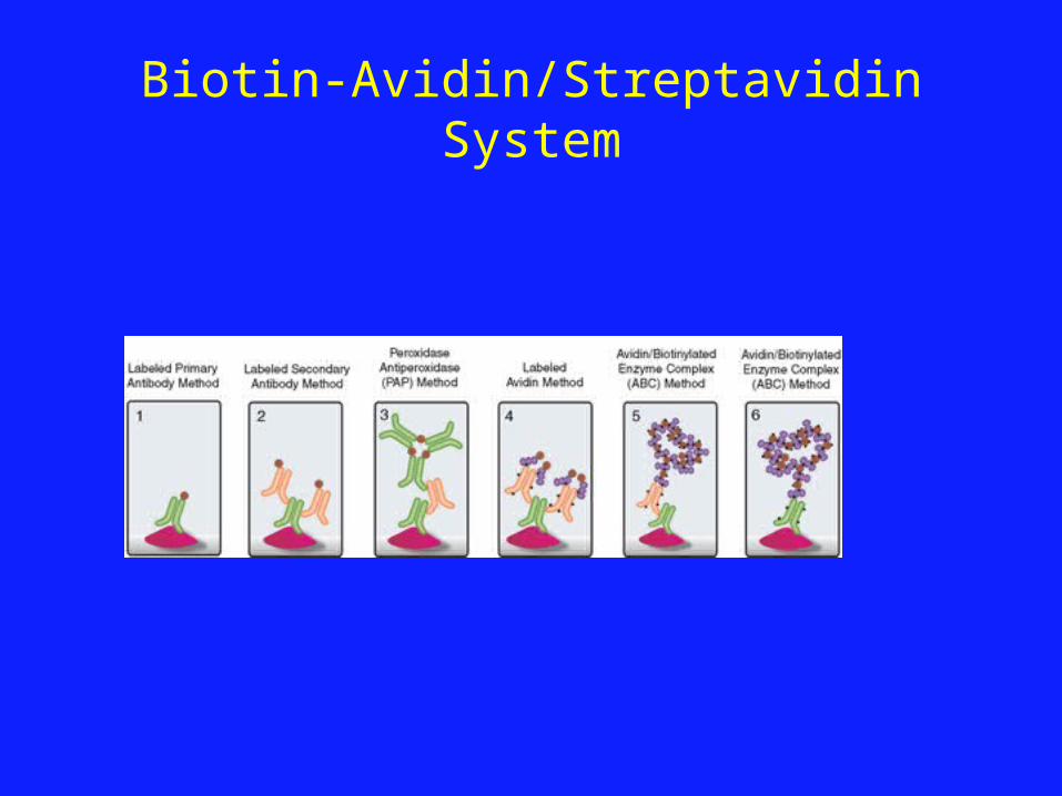
Biotin-Avidin/Streptavidin System

Biotin-Streptavidin system
• The Biotin-Avidin/Streptavidin System can improve sensitivity because of potential amplification with multiple site binding.
• Only a single labeled conjugate, avidin or streptavidin, need be kept on hand since it can be used with a variety of biotinylated lectins, antibodies or probes.
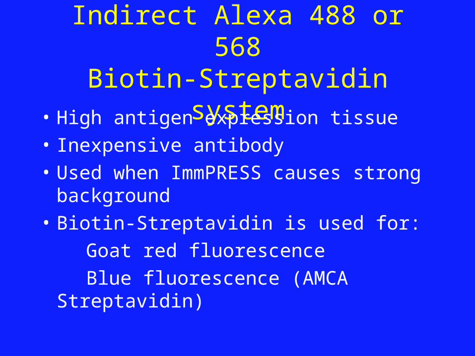
Indirect Alexa 488 or 568Biotin-Streptavidin system
• High antigen expression tissue
• Inexpensive antibody
• Used when ImmPRESS causes strong background
• Biotin-Streptavidin is used for:
Goat red fluorescence
Blue fluorescence (AMCA Streptavidin)

Avidin/Biotin Complex (ABC)

Avidin/Biotin Complex (ABC)
• ABC systems are extremely sensitive
• Can be used to detect any molecule that is biotinylated
• We use it when ImmPress is not available

ImmPRESS Polymer
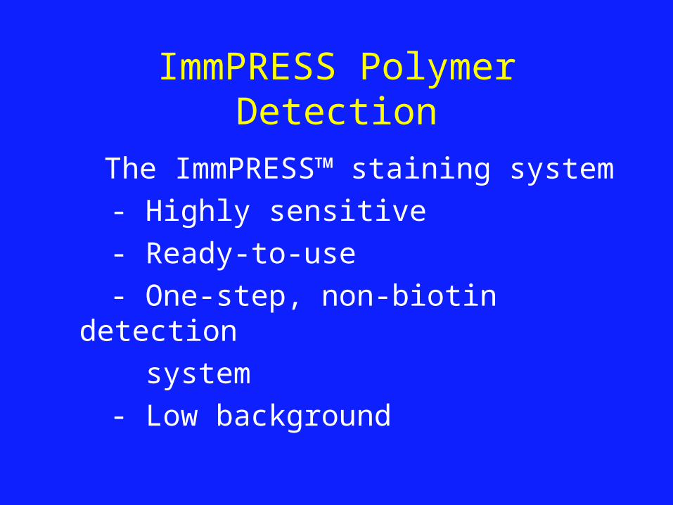
ImmPRESS Polymer Detection
The ImmPRESS™ staining system
- Highly sensitive
- Ready-to-use
- One-step, non-biotin detection
system
- Low background

ImmPRESS Polymer Detection
• DAB (Diaminobenzidine), brown
• Vector VIP, purple
• Vector SG, blue-gray
• AEC (3-amino-9-ethyl carbazole)*, red
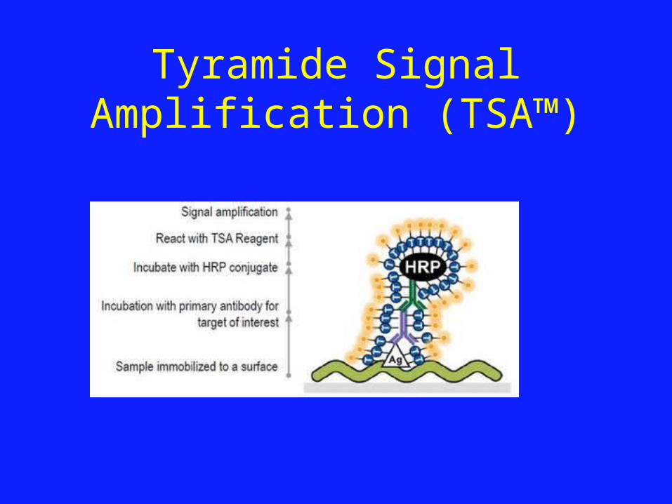
Tyramide Signal Amplification (TSA™)
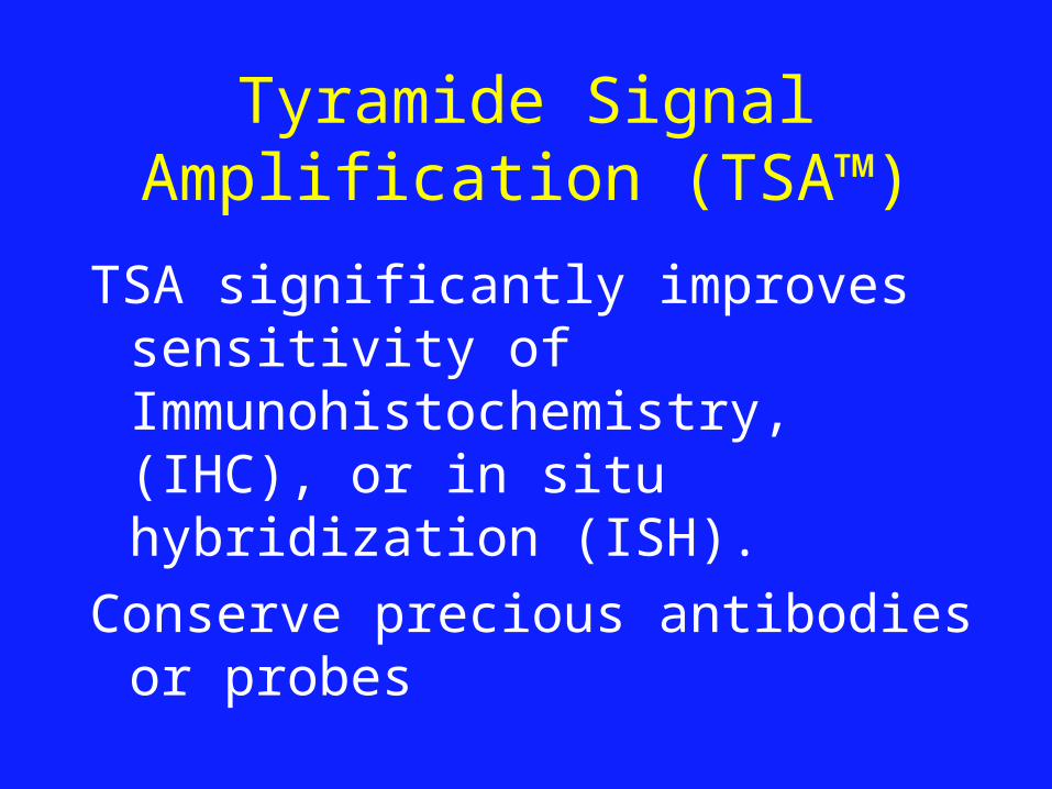
Tyramide Signal Amplification (TSA™)
TSA significantly improves sensitivity of Immunohistochemistry, (IHC), or in situ hybridization (ISH).
Conserve precious antibodies or probes
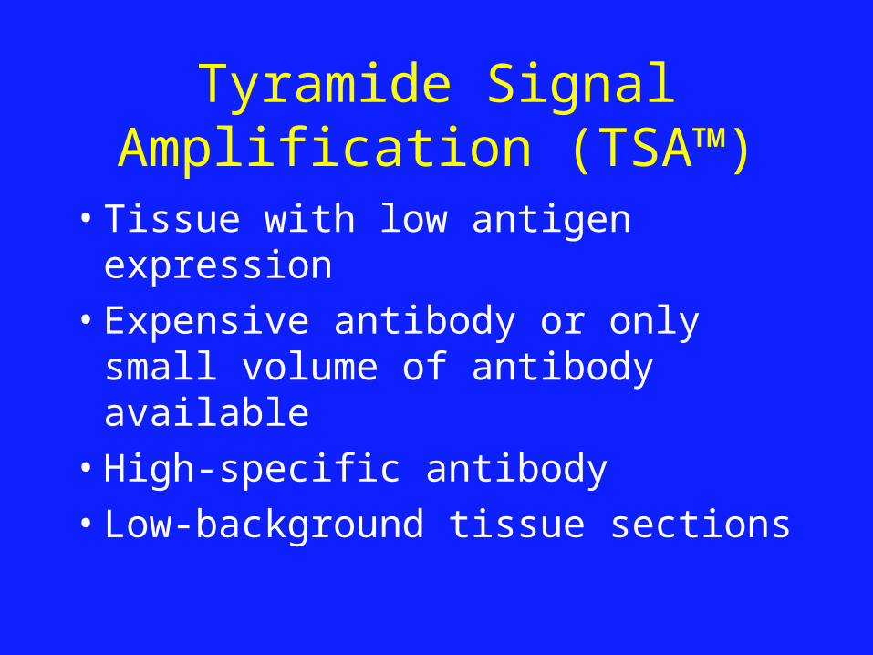
Tyramide Signal Amplification (TSA™)
• Tissue with low antigen expression
• Expensive antibody or only small volume of antibody available
• High-specific antibody
• Low-background tissue sections
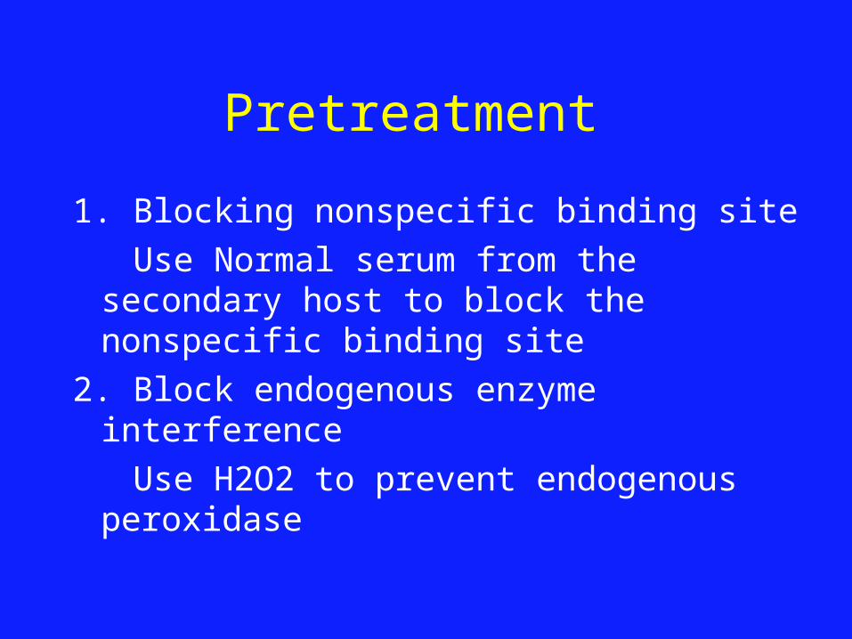
Pretreatment
1. Blocking nonspecific binding site
Use Normal serum from the secondary host to block the nonspecific binding site
2. Block endogenous enzyme interference
Use H2O2 to prevent endogenous peroxidase

Pretreatment
1. Heat Antigen Retrieval: Using a Pressure Cooker and different buffer Bull’s Eye decloaker pH 6.0 (PCBE) EDTA decloaker pH 8.5 (EDTA) Borg decloaker pH9.5 (Borg) Heat treat in pressure cooker at 120oC for
30 seconds, Cool down to 84-86oC.

Pretreatment
2. Protease Antigen Retrieval:
Using Proteinase K, Trypsin or Pepsin
Incubate time depend on the tissue 10-15 minutes
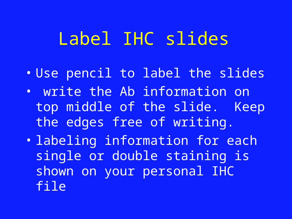
Label IHC slides
• Use pencil to label the slides • write the Ab information on top middle
of the slide. Keep the edges free of writing.
• labeling information for each single or double staining is shown on your personal IHC file

Label IHC slides
• Primary Antibody Abbreviation • Primary Antibody Dilution • Detection Method (add +H if you want nuclear
counter-stain)• Date (the Monday of the week of submission
– this must correspond to the date on the submission form)
• Name Initial

Label IHC slides
YAP R
1:50
ImmPF+H
3/12 KE

Label IHC slides
2H3 YAP R
1:50
ImmPF+H
3/12 KE
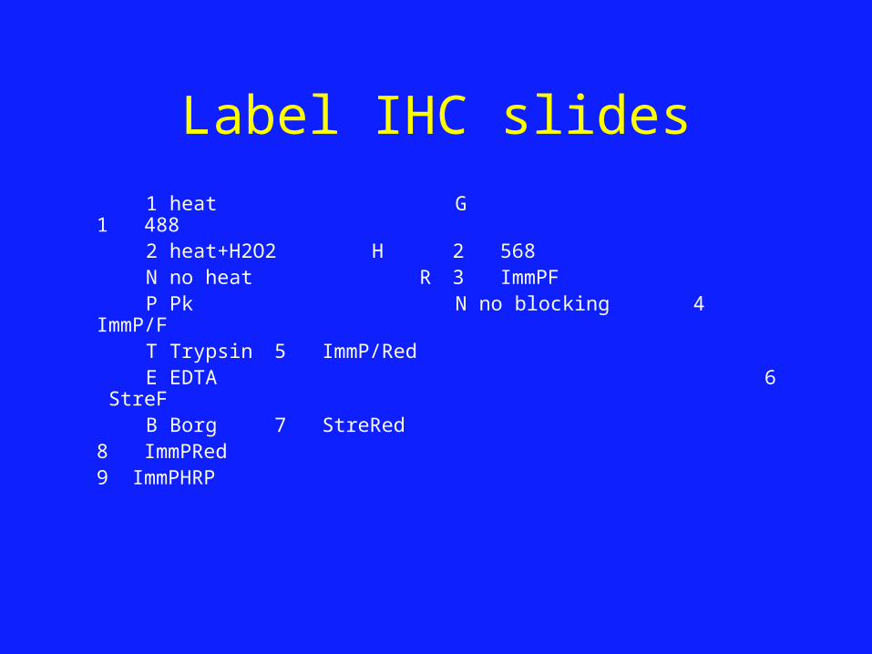
Label IHC slides
1 heat G 1 488 2 heat+H2O2 H 2 568 N no heat R 3 ImmPF P Pk N no blocking 4 ImmP/F T Trypsin 5 ImmP/Red E EDTA 6 StreF B Borg 7 StreRed
8 ImmPRed9 ImmPHRP

How to fill out IHC form
1. Import the files from template list to your personal IHC form
2. Duplicate the files from your personal IHC form
3. Submit a new antibody to Histology Core for testing.

Pick up right slides for IHC
1. Pick one slide from each sample for IHC
2. Look your slides under microscope to find your interested area and pick right slide from each sample for IHC
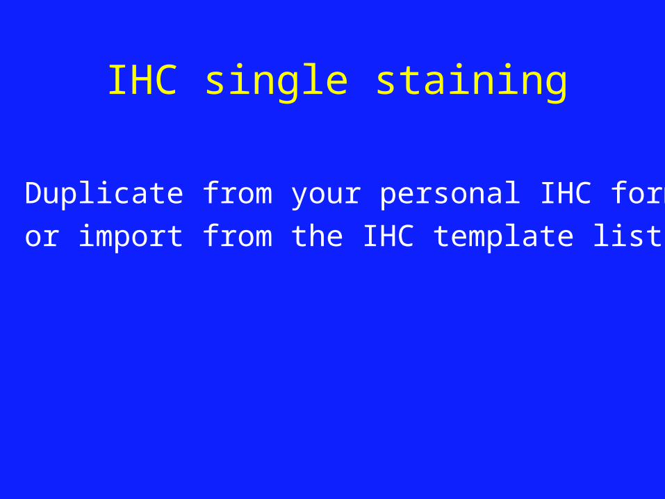
IHC single staining
Duplicate from your personal IHC form
or import from the IHC template list
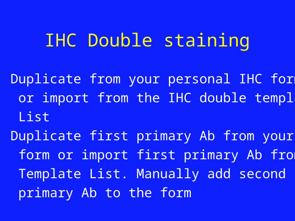
IHC Double staining
1. Duplicate from your personal IHC form
or import from the IHC double template
List
2. Duplicate first primary Ab from your IHC
form or import first primary Ab from
Template List. Manually add second
primary Ab to the form

Which Ab to use as first primary Ab
1. Primary Ab from goat
2. GFP, RFP
3. Strong IHC staining
4. No treatment

IHC Submission
IHC submission deadline is Thursday.Generally you will get the IHC slides one or two weeks.Fluorescent stained slides will be delivered to a slide box in each Lab -20oC freezer.HRP stained slides will be delivered on Friday to the shelf on Histo Core supply table.

Histology Submission
1. Send all histology submission to Lan and MinMin2. Write correct email subject on your email ( Tissue form, IHC form, InSitu form) Other questions should be specified3. Write priority on email subject and copy email to your PI If your histology request is urgent


![IHC PPT Ancillary Productsmy1hr-public.s3.amazonaws.com/documents/enroll/IHC PPT Ancillary Products[3].pdfAncillary Products From The IHC Group. The IHC Group Corporate Overview Ø](https://img.pdfslide.net/doc/110x75/5e38c9b5e1bb9a3e4e5b3bd8/ihc-ppt-ancillary-productsmy1hr-publics3-ppt-ancillary-products3pdf-ancillary.jpg)