Embed Size (px)
Citation preview
Biopac Student Lab® Lesson 4
ELECTROENCEPHALOGRAPHY (EEG) II
42 Aero Camino, Goleta, CA 93117
www.biopac.com
Procedure
Rev. 04012012
Richard Pflanzer, Ph.D. Associate Professor Emeritus
Indiana University School of Medicine Purdue University School of Science
William McMullen
Vice President, BIOPAC Systems, Inc.
Page P-1 ©BIOPAC Systems, Inc,
II. EXPERIMENTAL OBJECTIVES
1) To record an EEG from an awake, resting subject under the following conditions:
a) Relaxed with eyes closed;
b) Performing mental arithmetic with eyes closed;
c) Hyperventilating (breathing quickly and deeply) with eyes closed;
d) Relaxed with eyes open.
2) To examine differences in the level of alpha rhythm activity during mental arithmetic and hyperventilation, compared to the control condition of eyes closed and relaxed.
III. MATERIALS
BIOPAC Electrode Lead Set (SS2L)
BIOPAC Disposable Electrodes (EL503,) 3 electrodes per subject
BIOPAC Electrode Gel (GEL1) and Abrasive Pad (ELPAD) or Skin cleanser or alcohol prep
Lycra® swim cap (such as Speedo® brand) or supportive wrap (such as 3M Coban™ Self-adhering Support Wrap) to press electrodes against head for improved contact
Biopac Student Lab System: BSL 4 software, MP36, MP35 or MP45 hardware
Computer system (Windows 7, Vista, XP, Mac OS X 10.5 – 10.7)
IV. EXPERIMENTAL METHODS
A. SETUP
FAST TRACK Setup Detailed Explanation of Setup Steps
1. Turn the computer ON.
If using an MP36/35 unit, turn it OFF.
If using an MP45, make sure USB cable is connected and “Ready” light is ON.
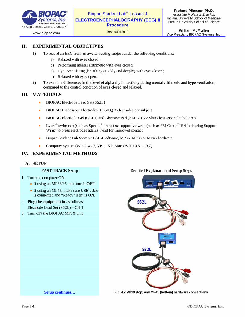
2. Plug the equipment in as follows:
Electrode Lead Set (SS2L)—CH 1
3. Turn ON the BIOPAC MP3X unit.
Setup continues…
Fig. 4.2 MP3X (top) and MP45 (bottom) hardware connections
Page P-2 L04 – Electroencephalography (EEG) II Biopac Student Lab 4
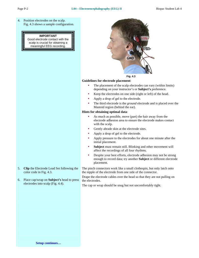
4. Position electrodes on the scalp. Fig. 4.3 shows a sample configuration.
IMPORTANT Good electrode contact with the scalp is crucial for obtaining a meaningful EEG recording.
Fig. 4.3
Guidelines for electrode placement:
The placement of the scalp electrodes can vary (within limits) depending on your instructor’s or Subject’s preference.
Keep the electrodes on one side (right or left) of the head.
Apply a drop of gel to the electrode.
The third electrode is the ground electrode and is placed over the Mastoid region (behind the ear).
Hints for obtaining optimal data:
As much as possible, move (part) the hair away from the electrode adhesion area to ensure the electrode makes contact with the scalp.
Gently abrade skin at the electrode sites.
Apply a drop of gel to the electrode.
Apply pressure to the electrodes for about one minute after the initial placement.
Subject must remain still. Blinking and other movement will affect the recordings of all four rhythms.
Despite your best efforts, electrode adhesion may not be strong enough to record data; try another Subject or different electrode placement.
5. Clip the Electrode Lead Set following the color code in Fig. 4.3.
6. Place cap/wrap on Subject’s head to press electrodes into scalp (Fig. 4.4).
Setup continues…
The pinch connectors work like a small clothespin, but only latch onto the nipple of the electrode from one side of the connector.
Drape the electrode cables over the head so that they are not pulling on the electrodes.
The cap or wrap should be snug but not uncomfortably tight.
©BIOPAC Systems, Inc L04 – Electroencephalography (EEG) II Page P-3
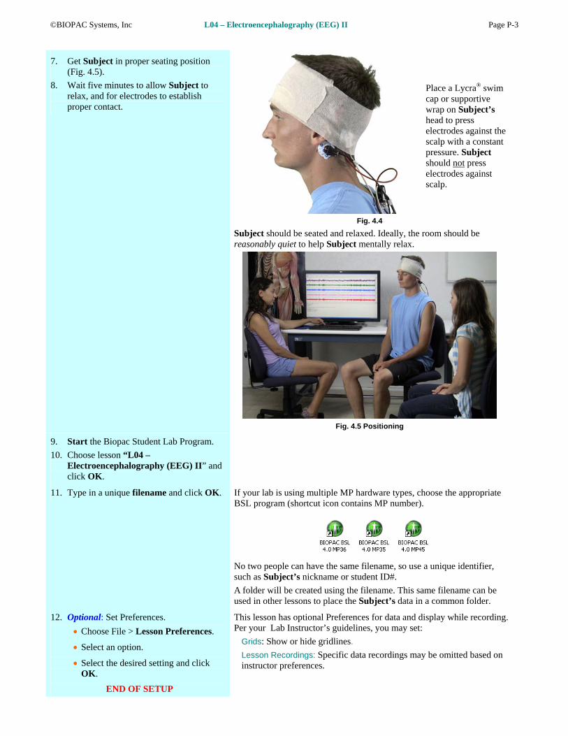
7. Get Subject in proper seating position (Fig. 4.5).
8. Wait five minutes to allow Subject to relax, and for electrodes to establish proper contact.
Place a Lycra® swim cap or supportive wrap on Subject’s head to press electrodes against the scalp with a constant pressure. Subject should not press electrodes against scalp.
Fig. 4.4
Subject should be seated and relaxed. Ideally, the room should be reasonably quiet to help Subject mentally relax.
Fig. 4.5 Positioning
9. Start the Biopac Student Lab Program.
10. Choose lesson “L04 – Electroencephalography (EEG) II” and click OK.
11. Type in a unique filename and click OK. If your lab is using multiple MP hardware types, choose the appropriate BSL program (shortcut icon contains MP number).
No two people can have the same filename, so use a unique identifier, such as Subject’s nickname or student ID#.
A folder will be created using the filename. This same filename can be used in other lessons to place the Subject’s data in a common folder.
12. Optional: Set Preferences.
Choose File > Lesson Preferences.
Select an option.
Select the desired setting and click OK.
END OF SETUP
This lesson has optional Preferences for data and display while recording. Per your Lab Instructor’s guidelines, you may set:
Grids: Show or hide gridlines.
Lesson Recordings: Specific data recordings may be omitted based on instructor preferences.
Page P-4 L04 – Electroencephalography (EEG) II Biopac Student Lab 4
B. CALIBRATION
The Calibration procedure establishes the hardware’s internal parameters (such as gain, offset, and scaling) and is critical for optimum performance. Pay close attention to Calibration.
FAST TRACK Calibration Detailed Explanation of Calibration Steps
1. Subject remains relaxed with eyes closed during Calibration.
2. Check Electrode Impedance. (Optional*)
*Only functional if your MP hardware is compatible with the Electrode Check feature. If your MP hardware is not compatible, this feature will not be available.
Please contact BIOPAC Technical Support for more
information on how to enable Electrode Check functionality.
IMPORTANT Certain subjects may not fall below the 10 K ohm reading.
This reading is subject to individual variations in skin conductivity and electrode
placement.
This step is optional and not applicable to MP45 hardware.
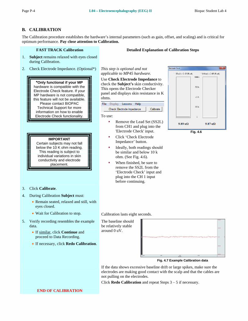
Use Check Electrode Impedance to check the Subject’s skin conductivity. This opens the Electrode Checker panel and displays skin resistance in K ohms.
To use:
Remove the Lead Set (SS2L)
s should
re to
from CH1 and plug into the 'Electrode Check' input.
Click ‘Check Electrode Impedance’ button.
Ideally, both readingbe similar and below 10 k ohm. (See Fig. 4.6).
When finished, be suremove the SS2L from the ‘Electrode Check’ input and plug into the CH 1 input before continuing.
Fig. 4.6
3. Click Calibrate.
4. During Calibration Subject must:
Remain seated, relaxed and still, with eyes closed.
Wait for Calibration to stop. alibration lasts eight seconds.
C
5. Verify recording resembles the example data.
If similar, click Continue and proceed to Data Recording.
If necessary, click Redo Calibration.
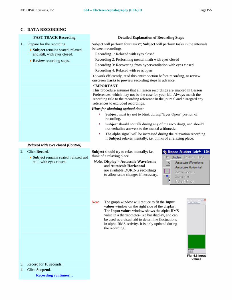
The baseline should be relatively stable around 0 uV.
Fig. 4.7 Example Calibration data
END OF CA IBRATION
If the data shows excessive baseli the
epeat Steps 3 – 5 if necessary.
L
ne drift or large spikes, make sureelectrodes are making good contact with the scalp and that the cables arenot pulling on the electrodes.
Click Redo Calibration and r
©BIOPAC Systems, Inc L04 – Electroencephalography (EEG) II Page P-5
C. DATA RECORDING
FAST TRACK Recording Detailed Explanation of Recording Steps
1. Prepare for the recording.
Subject remains seated, relaxed, and still, with eyes closed.
Review recording steps.
Subject will perform four tasks*; Subject will perform tasks in the intervals between recordings.
Recording 1: Relaxed with eyes closed
Recording 2: Performing mental math with eyes closed
Recording 3: Recovering from hyperventilation with eyes closed
Recording 4: Relaxed with eyes open
To work efficiently, read this entire section before recording, or review onscreen Tasks to preview recording steps in advance.
*IMPORTANT This procedure assumes that all lesson recordings are enabled in Lesson Preferences, which may not be the case for your lab. Always match the recording title to the recording reference in the journal and disregard any references to excluded recordings.
Hints for obtaining optimal data:
Subject must try not to blink during “Eyes Open” portion of recording.
Subject should not talk during any of the recordings, and should not verbalize answers to the mental arithmetic.
The alpha signal will be increased during the relaxation recording if Subject relaxes mentally; i.e. thinks of a relaxing place.
Relaxed with eyes closed (Control)
2. Click Record.
Subject remains seated, relaxed and still, with eyes closed.
Subject should try to relax mentally; i.e. think of a relaxing place.
Note: Display > Autoscale Waveforms and Autoscale Horizontal are available DURING recordings to allow scale changes if necessary.
Note The graph window will reduce to fit the Input values window on the right side of the display. The Input values window shows the alpha-RMS value in a thermometer-like bar display, and can be used as a visual aid to determine fluctuations in alpha-RMS activity. It is only updated during the recording.
Fig. 4.8 Input Values
3. Record for 10 seconds.
4. Click Suspend.
Recording continues…
Page P-6 L04 – Electroencephalography (EEG) II Biopac Student Lab 4
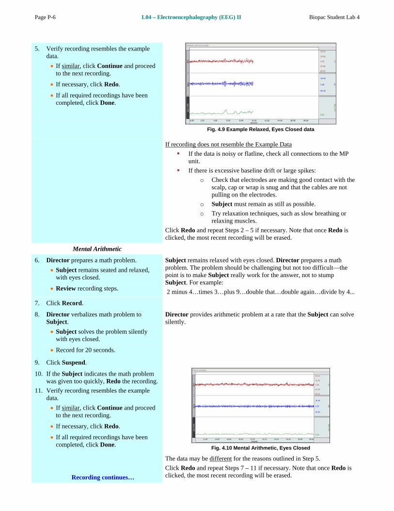
5. Verify recording resembles the example data.
If similar, click Continue and proceed to the next recording.
If necessary, click Redo.
If all required recordings have been completed, click Done.
Fig. 4.9 Example Relaxed, Eyes Closed data
es not resemble the Example DataIf recording do
trodes are making good contact with the rap is snug and that the cables are not
Subject must remain as still as possible.
If the data is noisy or flatline, check all connections to the MPunit.
If there is excessive baseline drift or large spikes:
Check that elecoscalp, cap or wpulling on the electrodes.
o
o Try relaxation techniques, such as slow breathing or relaxing muscles.
Click Redo and repeat Steps 2 – 5 if necessary. Note that once Redo is clicked, the most recent recording will be erased.
Mental Arithmetic
6. Director prepares a math problem.
Subject remains seated and relaxed, with eyes closed.
Review recording steps.
sed. Director prepares a math should be challenging but not too difficult—the
the answer, not to stump Sub
2 m at…double again…divide by 4...
Subject remains relaxed with eyes cloproblem. The problempoint is to make Subject really work for
ject. For example:
inus 4…times 3…plus 9…double th
7. Click Record.
8. Director verbalizes math problem to Subject.
Subject solves the problem silently with eyes closed.
Record for 20 seconds.
Director provides arithmetic problem at a rate that the Subject can solve silently.
9. Click Suspend.
10. If the Subject indicates the math problem was given too quickly, Redo the recording.
11. Verify recording resembles the example data.
If similar, click Continue and proceed to the next recording.
If necessary, click Redo.
If all required recordings have been
completed, click Done. Fig. 4.10 Mental Arithmetic, Eyes Closed
Recording continues…
The data may be different for the reasons outlined in Step 5.
Click Redo and repeat Steps 7 – 11 if necessary. Note that once Redo is clicked, the most recent recording will be erased.
©BIOPAC Systems, Inc L04 – Electroencephalography (EEG) II Page P-7
After Hyperventilation
12. Subject is seated.
Review recording steps.
Subject hyperventilate
Subject hyperventilates (by breathing rapidly and deeply through mouth) for two minutes with eyes closed.
It is important that recording be resumed as quickly as possible after Subject has hyperventilated. However, to avoid EMG artifact, make sure Subject has stopped hyperventilating prior to clicking Record.
s for two minutes with eyes closed.
WARNING Hyperventilation can make Subject dizzy and light he d. Subject adeshould be seated ith Director wwatching. Stop the procedure if Subject starts to feel sick or dizzy.
13. s soon as Subject stops hyperventilating A and is sitting still, Click Record immediately.
14. Record for 10 seconds.
15. Click Suspend.
16. Verify recording resembles the example data.
If similar, click Continue and proceed to the next recording.
If necessary, click Redo.
If all r ve been equired recordings hacompleted, click Done.
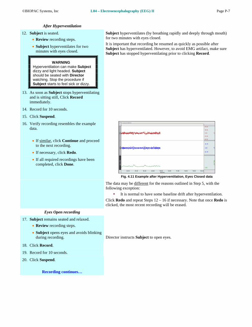
Fig. 4.11 Example after Hyperventilation, Eyes Closed data
The data may be different for the reasons outlined in Step 5, with the following exception:
It is normal to have some baseline drift after hyperventilation.
Click Redo and repeat Steps 12 – 16 if necessary. Note that once Redo is clicked, the most recent recording will be erased.
Eyes Open recording
17. Subject remains seated and relaxed.
Review recording steps.
Subject opens eyes and avoids blinking during recording.
Director instructs Subject to open eyes.
18. Click Record.
19. Record for 10 seconds.
20. Click Suspend.
Recording continues…

Page P-8 L04 – Electroencephalography (EEG) II Biopac Student Lab 4
21. Verify r ample ecording resembles the exdata.
If similar, click Continue
and proceed to optional recording section, or click Done to finish.
If necessary, click Redo.
Fig. 4.12 Example Relaxed, Eyes Open data
The data may be different for the reasons outlined in Step 5, with the following exception:
If the Subject blinked, it may have created a large spike in the data. If excessive, consider redoing the recording.
Click Redo and repeat Steps 17 – 21 if necessary. Note that once Redo is licked, the most recent recording will be erased. c
OPTIONAL ACTIVE LEARNING PORTION
With this lesson you may record additional data by clicking Continue following the last recording. Design an experiment to test or verify a scientific principle(s) related to topics covered in this lesson. Although you are limited to this lesson’s channel assignments, the electrodes may be moved to different locations on the Subject.
Design Your Experiment
Use a separate sheet to detail your experiment design, and be sure to address these main points:
A. Hypothesis
Describe the scientific principle to be tested or verified.
B. Materials
List .
rimental procedure—be sure to number each step
Set up the equipment and prepare the subject for your experiment.
Record
Use the Continue, Record and Suspend buttons to record as much data as necessary for your experiment.
ed all of the recordings required
Analyze Your Experiment
F. Set measurements relevant to your experiment and record the results in a Data Report.
the materials you will use to complete your investigation
C. Method
Describe the expeto make it easy to follow during recording.
Run Your Experiment
D. Set Up
E.
Click Done when you have completfor your experiment.
22. After clicking Done choose an option and , click OK.
fter clicking Done, dialog with options will be generated. Make a selection, and continue as directed.
If choosing the Record from another Subject option:
Repeat Setup Steps 5 – 9, and then proceed to Calibration.
A
23. Remove electrodes.
END OF RECORDING
Remove cap or wrap, the electrode cable pinch connectors, and peel off all electrodes. Discard the electrodes. (BIOPAC electrodes are not reusable.) Wash the electrode gel residue from the skin, using soap and water. The area around the electrode sites may remain red for a few hours, which is quite normal.
©BIOPAC Systems, Inc L04 – Electroencephalography (EEG) II Page P-9
V. DATA ANALYSIS
FAST TRACK Data Analysis Detailed Explanation of Data Analysis Steps
1. Enter the Review Saved Data mode. If entering Review Saved Data mode from the Startup dialog or lessons menu, make sure to choose the correct file.
Note Channel Number (CH) designations:
Channel Displays
CH 1 EEG
CH 40 a a lph
CH 41 al a RMS ph
Note measurement box settings:
Channel Measurement
CH 1 Stddev
CH 40 Stddev
CH 41 Mean
CH 40 Freq
The data should resemble Fig. 4.13.
Fig. 4.13 Example data
The measurement boxes are above the marker region in the data window. Each measurement has three sections: channel number, measurement type, and result. The first two sections are pull-down menus that are activated when you click them.
ments: Brief definition of measure
Stddev: Standard deviation is a measure of the variability of data points. The advantage of the Stddev measurement is that extreme values or artifacts do not unduly influence the measurement.
the average value in the selected area.
F quency in ond
The
Mean: Displays
req: Converts the time segment of the selected area to frecycles per sec
“selected area” is the area selected by the I-beam tool (includingendpoints).
2. Set up your display window for optimal viewing of the entire recording.
Note: The append event markers mark the beginning of each recording. Click on (activate) the event marker to display its
or changing view:
Disporward
Scro
Curs
Butt
Hide click” (Windows) or “Option + click” ( o toggle channel display.
label.
Useful tools f
lay menu: Autoscale Horizontal, Autoscale Waveforms, Zoom Back, Zoom F
ll Bars: Time (Horizontal); Amplitude (Vertical)
or Tools: Zoom Tool
ons: Overlap, Split, Show Grid, Hide Grid, -, +
/Show Channel: “Alt +Mac) the channel number box t
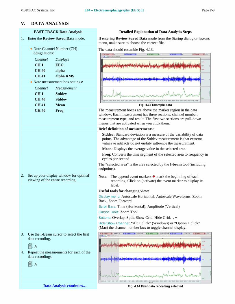
3. Use the I-Beam cursor to select the first
data recording.
A
4. Repeat the measurements for each of the data recordings.
A
Data Analysis continues… Fig. 4.14 First data recording selected

Page P-10 L04 – Electroencephalography (EEG) II Biopac Student Lab 4
5. Zoom in on a small section of the
RecBe sure to zofrequency ofording 1 data.
om in far enough so that you can easily measure the the alpha wave.
6. Use the I-Beam cursor to select an area from one peak to the next in the alpha band (CH 40).
B
Fig. 4.15 shows a sample setup for measuring the frequency in the alpha band (CH 40).
Fig. 4.15 Al ement pha wave frequency measur
7. Ans the end of the Data wer the questions at Report.
8. Save or Print the Data Report.
9. Quit the program.
END OF DATA ANALYSIS
ta at
An electronically editable Data Report is located in the journal (following the lesson summary,) or immediately following this DaAnalysis section. Your instructor will recommend the preferred formfor your lab.
Complete the rt that follows.
END OF LESSON 4 Lesson 4 Data Repo

©BIOPAC Systems, Inc L04 – Electroencephalography (EEG) II Page P-11
ELECTROENCEPHALOGRAPHY II
udent’s Name:
EEG II
DATA REPORT
St Lab Section: Date:
I. Data and Calculations
Subject Profile
Name: Height:
Age: Gender: Male / Female Weight:
Amplitudes ate the
e for the Alpha-RMS Mean between ether er (+,) smalle
T
A. Complete Table 4.1 with the amplitudes of thdifferenc
e recorded data in the control and experimental conditions. Calcul the Experimental Conditions and the Control, and then summarize whr (,) or the same (=) as the Control Mean.
able 4.1
the Experimental Mean was larg
EEG Alpha Alpha-RMS Alpha-RMS Alpha-RMS Condition
Difference Summary
(Exp. - Control) (+, , =)
Ey(Control)
es closed
Mental arithmetic
Recovering from hyperventilation
Eyes open
Frequency
B. What is the frequency of an alpha rhythm from “Eyes closed” data? = Hz
Does this agree with the expected values? Yes No
II. Questions
C. Refer to Table 4.1: When was the general amplitude of the EEG highest?
D. Refer to Table 4.1: When were the alpha wave levels highest?
E. Refer to Table 4.1: How do your results compare with the information presented in the Introduction?

Page P-12 L04 – Electroencephalography (EEG) II Biopac Student Lab 4
F. Did Subject need to concentrate during math problems? Yes No
would the level of concentration required affect the data? How
G. \hat might ac nt for the amplitude difference of waves recorded from a subject tested alone, in a darkened room, and subjects teste b full of students?
coud in a la
H. conditions prod ed the lowestWhich uc alpha activity?

©BIOPAC Systems, Inc L04 – Electroencephalography (EEG) II Page P-13
III.
A. thesis
OPTIONAL Active Learning Portion
Hypo
B. Materials
C. d
Metho
D. Set Up
E. Experimental Results
End of Lesson 4 Data Report