Embed Size (px)
Citation preview
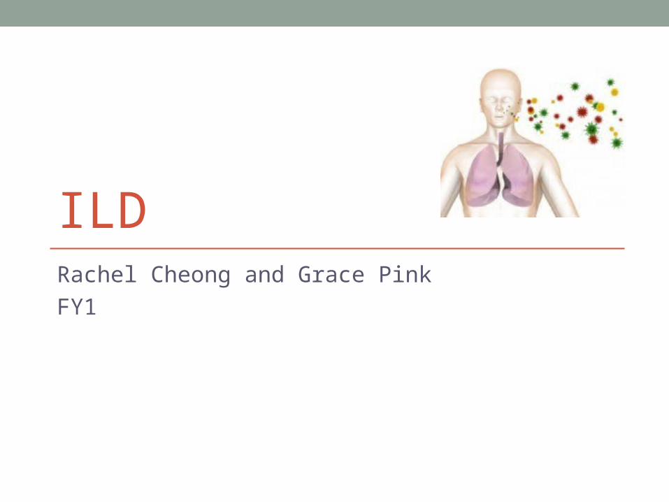
ILDRachel Cheong and Grace Pink
FY1

Introduction • Background/ Initial investigations/ Basic management • Case• Specific types:
• IPF• Hypersensitivity Pneumonitis (a.k.a Extrinsic Allergic Alveolitis) –
Organic Allergens• Industrial Dusts• Sarcoidosis
• Finals questions • Further reading

Background• Heterogeneous group of diseases which affect the lung
parenchyma
• Characterised by chronic inflammation and remodelling +/- progressive interstitial fibrosis, hyperplasia of type II epithelial cells and pneumocytes
• Grouped together on the basis of symptoms and radiological changes
• But very different in their aetiology and prognosis
• Getting the right diagnosis is important!

Classification
ILD
Idiopathic Known
Aetiology Associated systemic
disease
Idiopathic Pulmonary Fibrosis
Occupational dusts
Drugs- amiodarone, methotrexate, sulfasalazine, nitrofurantoin
Hypersensitivity Pneumonitis (organic)
Infection- TB
Sarcoid
RA
SLE
Sjogrens
UC
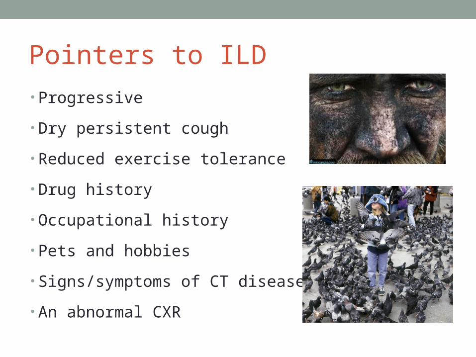
Pointers to ILD
• Progressive
• Dry persistent cough
• Reduced exercise tolerance
• Drug history
• Occupational history
• Pets and hobbies
• Signs/symptoms of CT disease
• An abnormal CXR

Case
Mr AF, retired 60 y/o man who has presented to his GP with progressive SOB over 6 months
He is now feeling SOB when walking to collect his grandchildren from school, which upsets him
He has been bothered by a dry cough and the GP trialled a “blue inhaler”
Think of one differential and 1 question that would most help you decide about that diagnosis

O/E
• Dyspnoeic
• Clubbing/ Cyanosis
• Reduced expansion
• Deviated trachea-
• towards pathological side
• Dull percussion
• Fine end-inspiratory crackles

How will you investigate?According to BTS guidelines:1.Urine dip2.FBC, U&E, LFTs3.Spirometry and gas transfer4.CXR and HRCT (especially if normal CXR)5.BAL and lung biopsy (before treatment)
Other tests depend on clinical context e.g. sputum culture, ABG , CRP/ESR, BNP, RF/ anti-CCP, ANA, ANCA, ACEEcho
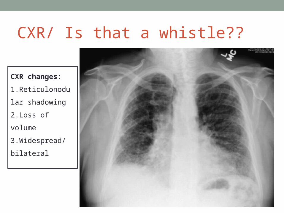
CXR/ Is that a whistle??
CXR changes:
1.Reticulonodular
shadowing
2.Loss of volume
3.Widespread/
bilateral

HRCT- What’s HR?
CT changes: honeycombing, GGO, loss of lung architecture, traction bronchiectasis

Spirometry
• All patients with ILD should have resting spirometric and gas transfer measurement at presentation reasonable measure of disease severity
• Carbon monoxide transfer factor (TLCO) levels at presentation are a more reliable guide to outcome than other resting lung function variables.
• Radiological changes correspond poorly to disease severity

Spirometry
Restrictive defect
FVC is reduced
FEV1 is reduced in proportion or slightly less
FEV1:FVC ratio normal or raised

General Management • Acute:
• ABC• ? ABx if infective exacerbation
• Conservative: • Lifestyle – exercise, weight loss, pulmonary rehab• Vaccinations
Smoking: All patients with ILD should be advised to stop smoking. Patients with IPF have an up to 10-fold increased risk of developing lung cancer whether they smoke or not
LTOT? No evidence that it influences long term survival What are the indications for LTOT?
LTOT:paO2 <7.3 on 2 occasions separated by 2-3 weeks when clinically stable paO2 7.3-8 if there is evidence of:1.Nocturnal hypoxia2.Seconday polycythaemia 3.Pulmonary HTN
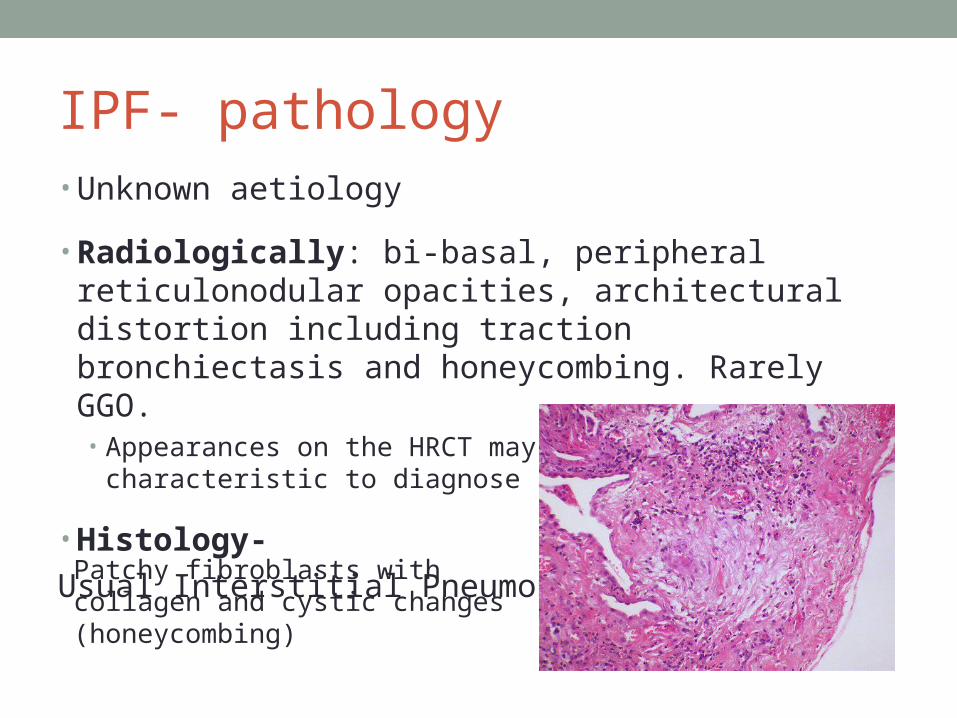
IPF- pathology• Unknown aetiology
• Radiologically: bi-basal, peripheral reticulonodular opacities, architectural distortion including traction bronchiectasis and honeycombing. Rarely GGO.• Appearances on the HRCT may be sufficiently characteristic to
diagnose
• Histology-
Usual Interstitial Pneumonia
Patchy fibroblasts with collagen and cystic changes (honeycombing)
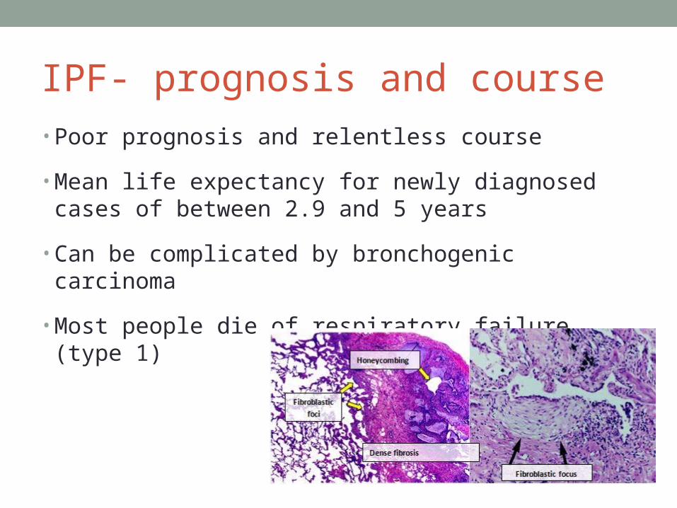
IPF- prognosis and course• Poor prognosis and relentless course
• Mean life expectancy for newly diagnosed cases of between 2.9 and 5 years
• Can be complicated by bronchogenic carcinoma
• Most people die of respiratory failure (type 1)
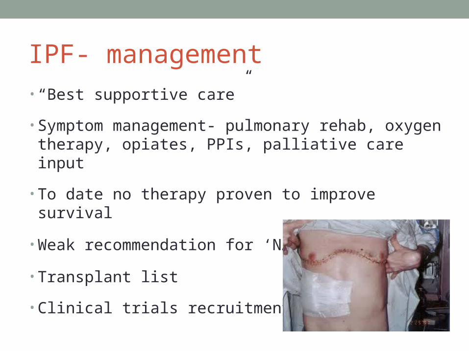
IPF- management • “Best supportive care”
• Symptom management- pulmonary rehab, oxygen therapy, opiates, PPIs, palliative care input
• To date no therapy proven to improve survival
• Weak recommendation for ‘NAP’
• Transplant list
• Clinical trials recruitment

“Pneumoconioses” Non-neoplastic lung disease caused by the reaction of the lung to inhalation of mineral or organic dusts.
The dust particles reach the terminal airways and settle on the epithelial lining. The inflammatory reaction caused by these leads to inflammation and scarring
Jobs to ask about: coal mining, sandblasting, miling, welding, foundry work, farming, working with grain
Hobbies to ask about: bird keeping, hot tubs/ sauna
Some of the unusual ones: cheese washer’s lung, thatched roof disease, Japanese summer house HP…
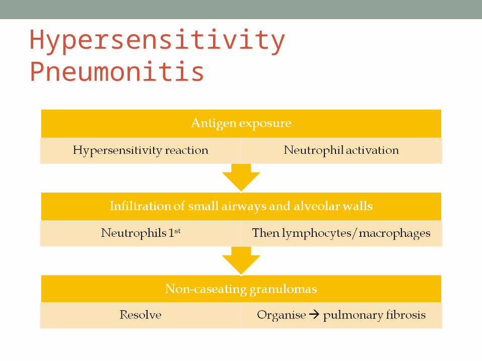
Hypersensitivity Pneumonitis
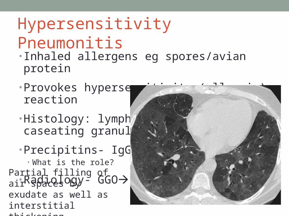
Hypersensitivity Pneumonitis • Inhaled allergens eg spores/avian protein
• Provokes hypersensitivity (allergic) reaction
• Histology: lymphocytes and non-caseating granulomas, bronchocentric
• Precipitins- IgG to allergens• What is the role?
• Radiology- GGO
Partial filling of air spaces by exudate as well as interstitial thickening
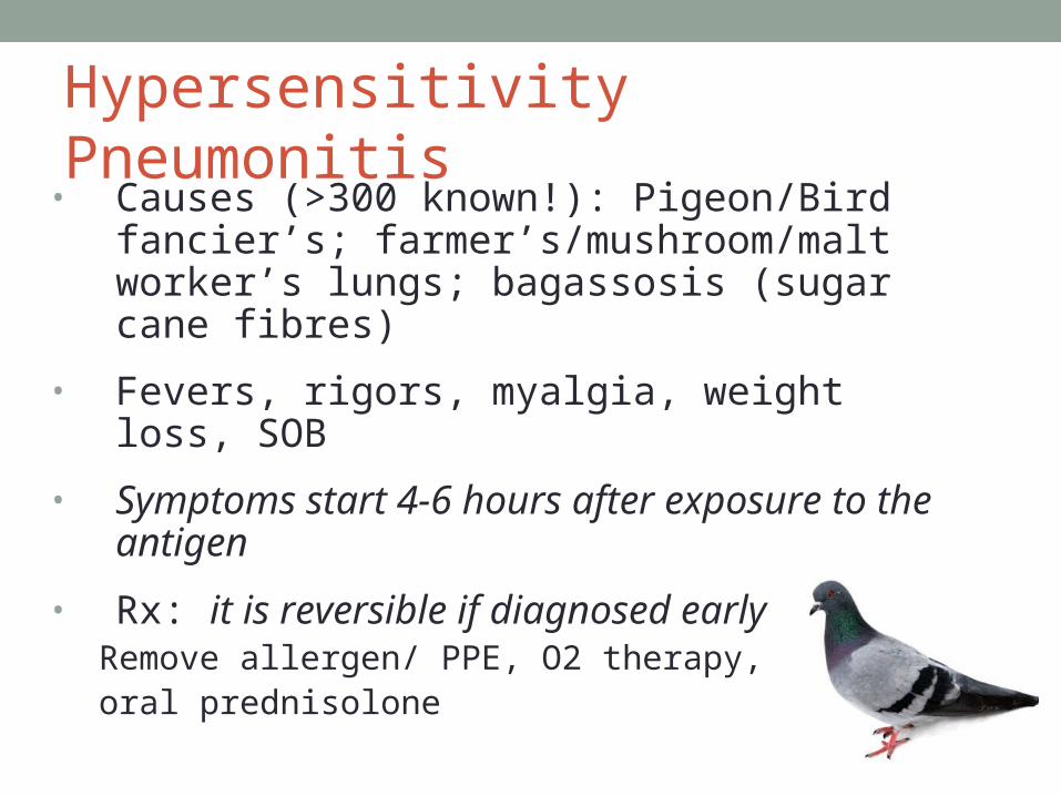
Hypersensitivity Pneumonitis • Causes (>300 known!): Pigeon/Bird fancier’s;
farmer’s/mushroom/malt worker’s lungs; bagassosis (sugar cane fibres)
• Fevers, rigors, myalgia, weight loss, SOB
• Symptoms start 4-6 hours after exposure to the antigen
• Rx: it is reversible if diagnosed earlyRemove allergen/ PPE, O2 therapy, oral prednisolone
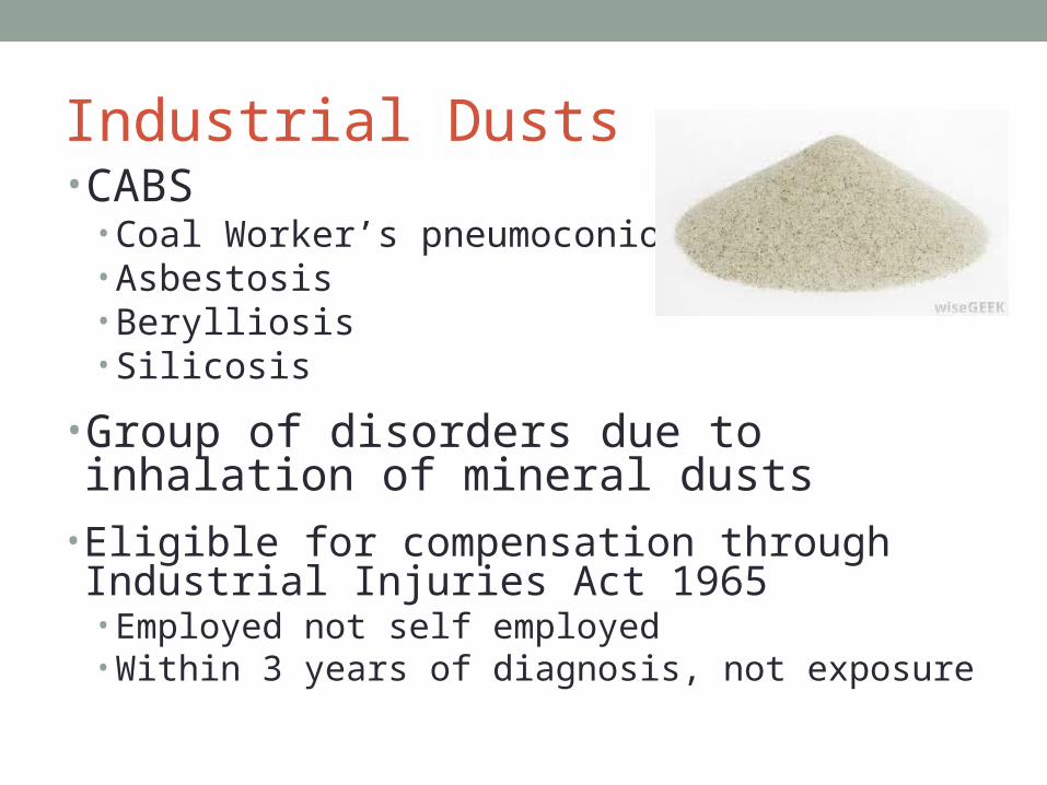
Industrial Dusts• CABS
• Coal Worker’s pneumoconiosis• Asbestosis• Berylliosis• Silicosis
• Group of disorders due to inhalation of mineral dusts
• Eligible for compensation through Industrial Injuries Act 1965• Employed not self employed• Within 3 years of diagnosis, not exposure

Asbestos Lung Disease
• Inhalation of asbestos fibres used in fireproofing/wire insulation, +++ latency period
• Fibrogenicity: White > Brown > Blue
• Exposure duration and degree also important
• Benign pleural plaques/ diffuse pleural thickening/ asbestosis/ malignant mesothelioma/ bronchial adenocarcinoma• Increased risk of adenocarcinoma with smoking + asbestos
Asbestosis- fibrosis of lungs +/- parietal or visceral pleura • dyspnoea, clubbing, fine bilat/ bi-basal end-insp creps
• Rx: Symptomatic

Asbestos CTs
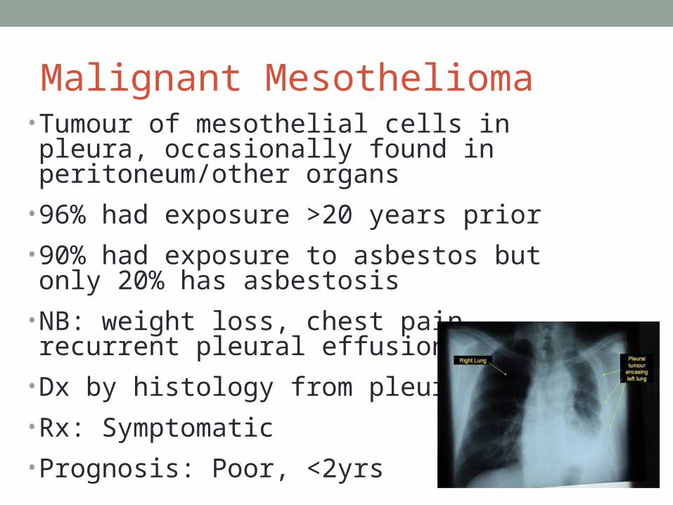
Malignant Mesothelioma • Tumour of mesothelial cells in pleura, occasionally found in peritoneum/other organs
• 96% had exposure >20 years prior
• 90% had exposure to asbestos but only 20% has asbestosis
• NB: weight loss, chest pain, recurrent pleural effusions, SOB
• Dx by histology from pleural Bx
• Rx: Symptomatic
• Prognosis: Poor, <2yrs

Sarcoidosis • Multi-system granulomatous disease of unknown aetiology
• Age 20-40yo, Afro-Caribbeans
• 90% have lung involvement
• Erythema nodosum/ uveitis/ keratoconjunctivitis sicca/ hepatosplenomegaly/ dysrhythmias/ CCF/ arthralgia/ polyneuropathy/ meningoencephalitis/ fever/ fatigue/ night sweats
• Rx: acute NSAIDs, can recover spontaneously
• Steroids if 1. symptomatic or static parenchymal disease, 2. uveitis, 3. hypercalcaemia, 4. neurological or cardiac involvement
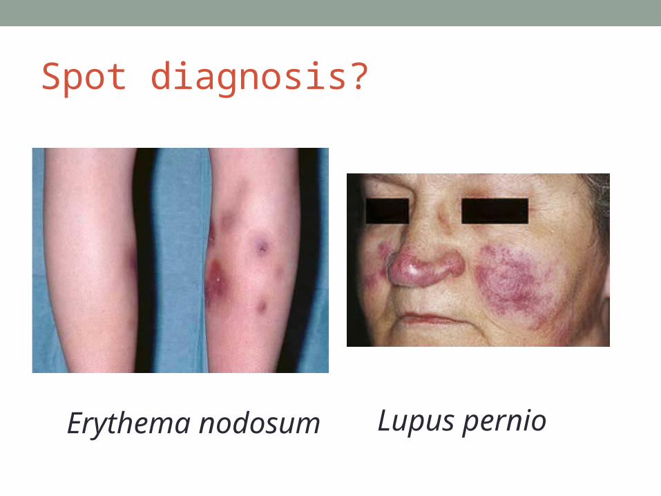
Spot diagnosis?
Lupus pernioErythema nodosum
Sarcoidosis
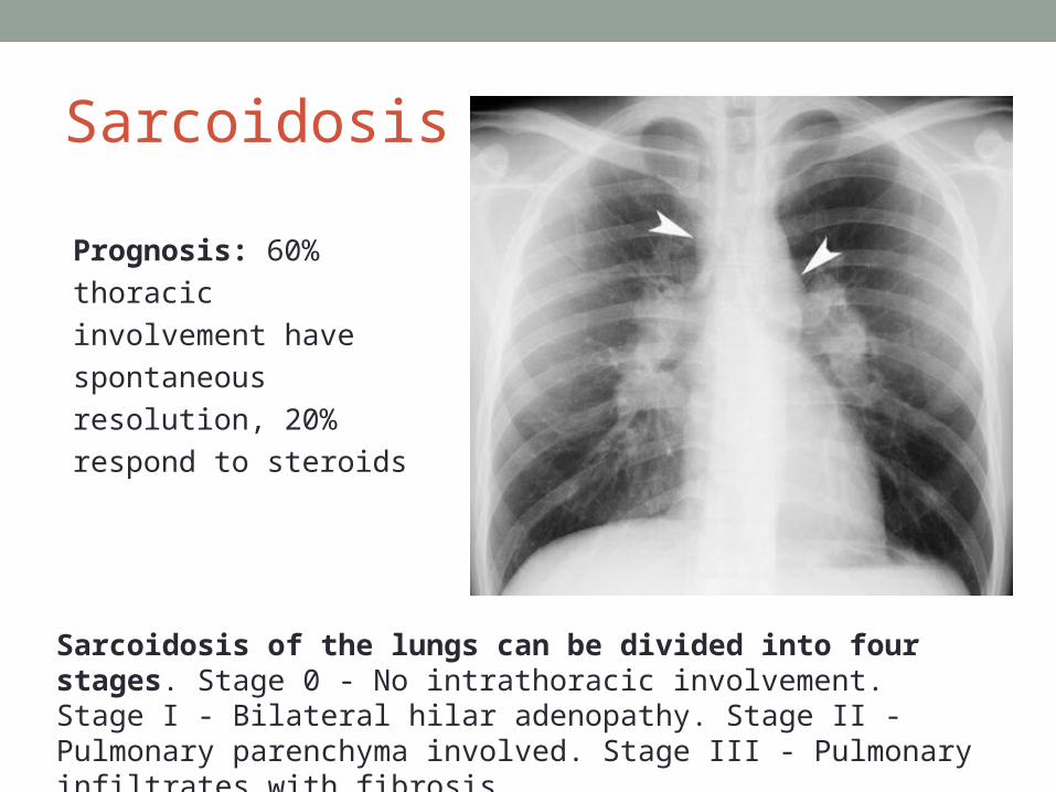
Sarcoidosis of the lungs can be divided into four stages. Stage 0 - No intrathoracic involvement. Stage I - Bilateral hilar adenopathy. Stage II - Pulmonary parenchyma involved. Stage III - Pulmonary infiltrates with fibrosis
Prognosis: 60% thoracic
involvement have
spontaneous resolution,
20% respond to steroids

Take Home Messages • Key symptoms- SOB, dry cough, reduced exercise
tolerance
• Flags- occupation, pets, hobbies, PMHx
• Investigations- think logically and start at the bedside, some specific tests for extra marks• You should be able to mention at least the essential tests the BTS
recommends
• Management- varies considerably, don’t forget the basics ABC/ stop smoking/ pulmonary rehab/ oxygen

More reading• http://www.brit-thoracic.org.uk/guidelines-and-quality-stan
dards/interstitial-lung-disease-guidelines/• Kumar and Clark page 935-947• http://www.pneumotox.com/pattern/view/8/I.g/pulmonary-fi
brosis/• OHCM • Patient.co.uk professional reference

Some Finals Questions• What are the clinical findings that would suggest ILD?• Explain to the patient what spirometry/ CT/ VATS biopsy/ Echo
involves• What is the role of echo in ILD?• What are the changes of ILD on CXR/ CT?• What is a HRCT?• What changes would you expect on spirometry?• Draw the flow volume loop in a normal patient and that of a patient
with restrictive lung disease• What are the indications for LTOT?• What is the role of serum precipitins?• What sort of jobs will you ask this patient about?• What are some of the lung diseases asbestos can cause?• What are the systemic manifestations of Sarcoid?• What are the types of inhaler you know and what colour are they?

My Finals Cases 1. SchizophreniaPredisposing factors, management including psychotherapy, what is risperidone, explain to patient why they should stay on their depot2. Unknown gastro complaintDifferentials, investigations, tell me about IBD, what is the difference between Crohn’s and UC, explain to the patient about a colonoscopy 3. Aortic stenosis (asymptomatic)What causes, draw the cardiac cycle in terms of pressures, endocarditis, explain to patient about AS4. MALT lymphomaDifferentials, investigations, different types lymphoma, staging, virus involved lymphoma, other diseases associated EBV, histological findings of HL, side effects of chemotherapy, side effects of steroids, explain to the patient about chemotherpay