Embed Size (px)
Citation preview

Image Diagnostic Technology LtdUnit GC, Westpoint, 36 Warple Way, London W3 0RG
Tel: +44 20 8600 3540 www.simplantscans.com email: [email protected]
What is the Radiation Dose
from
a CT or CBCT Scan?
Anthony Reynolds BA MSc PhDRegistered Clinical Scientist CS03469
Image Diagnostic Technology Ltd.

Who or what is IDT?
Image Diagnostic
Technology Ltd
IDT Dental Products Ltd
distributes
Irish Dental Tomography Ltd operates in Ireland
Three Companies:
since 1991
specialises in
arranging
CT scans and
3D processing

www.simplantscans.comwww.ctscan.co.uk

www.simplantscans.com

Cone Beam CT Scanner
i-CAT™ is a trademark of Imaging Sciences International LLC of Hatfield, USA

Gendex™ is a trademark of Gendex Dental Systems of Lake Zurich, USA
Medium Field Of View CBCT
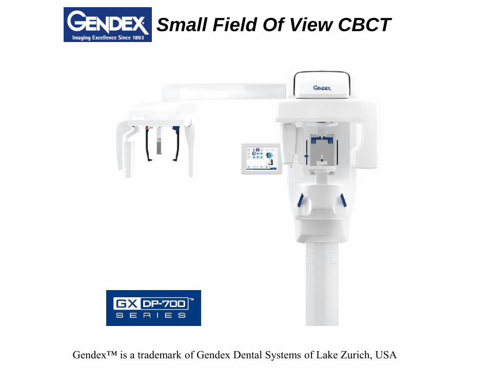
Small Field Of View CBCT
Gendex™ is a trademark of Gendex Dental Systems of Lake Zurich, USA

interactive implant planning software
SimPlant™ is a trademark of Materialise Dental NV of Belgium

Guide resting on:
• Bone
• Mucosa
• Teeth
Guiding
cylinders
The SurgiGuide controls:
• Position
• Orientation
• (Depth)
Surgical drill guide
SurgiGuide™ is a trademark of Materialise Dental NV of Belgium

Outline of Presentation
• Introduction / Disclosures
• Risk from Low Radiation Doses
• What do we mean by Effective Dose?
• How to evaluate the Risks?
• How does CT work?
• How does Dose affect Image Quality?
• What other factors affect Image Quality?


Transposition into UK Law
Ionisation Radiations Regulations 1999 – “IRR99”• Exposure of members of the public (e.g. staff and visitors)
• Enforced by Heath and Safety Executive (HSE)
Ionising Radiation (Medical Exposure) Regulations 2000 (amended in 2006) – “IR(ME)R 2000”
• Medical exposures (e.g. patients)
• Enforced by Care Quality Commission

What’s in ICRP103?
Fundamental Principles of Radiation Protection
• Justification (benefits must outweigh the risks)
• Optimisation (keep doses As Low As Reasonably Achievable)
• Dose Limits (20 mSv per year for members of the public)(no dose limits for medical exposures)

What else is in ICRP103?
• The distribution of risks to different organs/tissues is judged to
have changed somewhat since ICRP60 (1991)
• Overall estimate of deterministic effects remains the same
• Risk of hereditable effects is judged to be lower
• Risk of fatal cancer remains unchanged at just over 5% per Sv

Risk Coefficients for Stochastic Effects

Risk varies with Age
5% per Sievert at age 30

How do we know that exposure to
radiation results in harm?
Deterministic Effects are reproducible
• severity of the effect increases with the dose
• not observed below a threshold dose of about 500mSv
Stochastic Effects are random• known to occur above 20mSv or so
• the risk (not the severity) increases with the dose
• below about 20mSv we don’t know if they occur or not
Hereditary Effects are random but the incidence is very low


Estimated excess relative risk (±1 SE) of mortality (1950–1997) from solid cancers among
groups of survivors in the LSS cohort of atomic bomb survivors, who were exposed to low
doses (<500 mSv) of radiation (2).
Brenner D J et al. PNAS 2003;100:13761-13766
©2003 by National Academy of Sciences

Schematic representation of different possible extrapolations of measured radiation risks
down to very low doses, all of which could, in principle, be consistent with higher-dose
epidemiological data.
Brenner D J et al. PNAS 2003;100:13761-13766
©2003 by National Academy of Sciences
20 mSv


Duty Holders under IR(ME)R 2000
The Employer
• provides a framework of policies and procedures
The Referrer• must supply sufficient clinical information to allow the
exposure to be justified
The Practitioner• responsible for justifying the exposure
The Operator• responsible for carrying it out


The Problem
As Practitioners we have a duty to ensure the
benefits of exposing the patient to radiation
outweigh the risks
But we don’t know what the risks are
How can we address this issue in practice?
Use Effective Dose to assess the risks!

Effective Dose
We know the risks from high doses of radiation• e.g. Atom Bomb survivors
• Atom Bomb survivors received whole body doses
• Dental patients receive doses to a very small region
• How can we relate the risks?
Effective Dose is a way of describing the dose to a
limited region in terms of the whole body dose that
would result in the same risk to the patient
Effective Dose is a measure of risk!


Ludlow & Ivanovic, Oral & Maxillofacial Radiology 106,1 (July 2008)

Slice 0

Slice 1 – Mid brain superior

Slice 2 – Calvarium, APLR

Slice 3 – Orbit (skin dose),
mid brain

Slice 4 – lens of eye, orbits

Slice 5 – tip of nose (skin
dose), cerabellum, mid
palate, cheek (sd)

Slice 6 – back of neck, Mn
body, Ramus, Parotid

Slice 7 – Occipital, sub
mandibular gland

Slice 8 – Thyroid skin
dose, thyroid mid line

Slice 9 – Esophagus,
thyroid surface


Eur J Radiol 81,2,267-271 (February 2012)

Prof. Ria Bogaerts, Katholieke Universiteit Leuven, March 2011

Prof. Ria Bogaerts, Katholieke Universiteit Leuven, March 2011

Prof. Ria Bogaerts, Katholieke Universiteit Leuven, March 2011

AREA: 284cm² 95cm² 20cm²
FOV: 16dia x 18h 8dia x 12h 4dia x 5h
Estimating Effective Dose in practice
We can’t measure the Effective Dose for every patient
The SEDENTEXCT paper doesn’t cover every situation
SO
We need a practical way to calculate the Effective Dose.

How can we calculate the
Effective Dose?
kVp, mA, scan duration (s)
• can only use these to compare doses on the same machine
Dose Length Product (DLP)
• works very well for medical CT
Dose Area Product (DAP)• works reasonably well for cone beam CT

Dose Length Product (DLP)
CTDIvol is the dose per cm
DLP = CTDIvol x Irradiated Length
Effective Dose = DLP x F (where F is a conversion factor)
• works well for medical CT
• most CBCT manufacturers don’t display CTDIvol
(exception: J.Morita Accuitomo and Veraviewepocs)

Conversion Factor F
Table from “Radiation Exposure in Computed Tomography” edited by Hans Dieter Nagel
F can also by calculated from ImPACT CTDosimetry calculator www.impactscan.org
Roughly speaking, F = 0.002mSv / mGy.cm for Maxilla and 0.003mSv / mGy.cm for Mandible
Accuracy: ±50%2 µSv 3 µSv

Effective Dose for Medical CT Scanners
Multiply DLP by 2 for Maxilla or 3 for Mandible
to get the Effective Dose in microSieverts (µSv)
Mx 128µSvAccuracy: ±50%
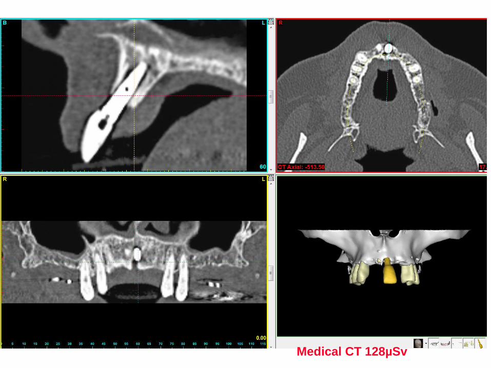
Medical CT 128µSv

IDT Physics Report

J.Morita Accuitomo and Veraviewepochs
DLP = CTDIvol x Irradiated Length = 4.6mGy x 4cm = 18.4mGy.cm
Effective Dose = 18.4 x 3 = 55 microSv

CBCT 55µSv


Effective Dose (µSv) = 0.08 x DAP (mGy.cm2)


Dose Area Product (DAP) for
Cone Beam CT Scanners
Multiply DAP by 0.1 for Maxilla or 0.15 for Mandible
to get the Effective Dose in microSieverts (µSv)
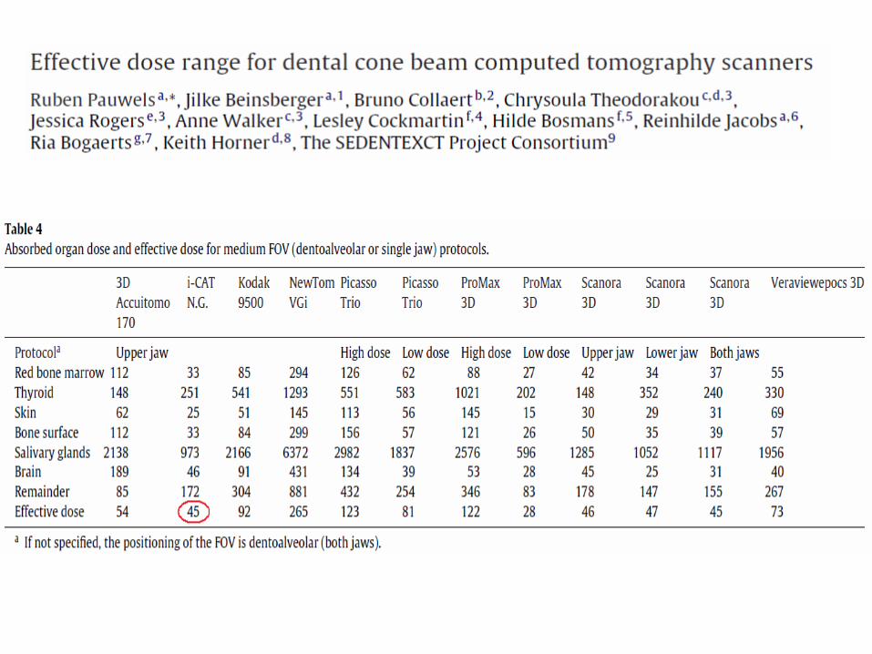
Mn 45µSvAccuracy: ±50%

CBCT 45µSv

CBCT 145µSv

* From: Pauwels et al, Effective dose range for dental CBCT scanners, Euro J Radiol 81, 2, 267-271, Feb 2012.
Effect of
Reducing
Beam
Height

Effect of Reducing Beam Width
• Reducing the beam height by 50% reduces the dose by approximately 50%
• Reducing the beam width by 50% reduces the dose by only about 25%
X-ray Tube Detector

Typical Doses from Dental X-Rays*
Lateral Ceph 4 µSv
Intraoral (E speed, round collimator) 6 µSv
Intraoral (F speed, rectangular collimator) 2 µSv
*ICRP103 weighting factors
Panoramic 24 µSv†
†
†
†Holroyd JR, Gulson AD, Guidance on the Safe Use of Dental Cone Beam CT
(Computed Tomography) Equipment, HPA-CRCE-010, November 2010
Cone Beam CT Scanner 48 - 1073 µSv
Medical CT Scanner 534 - 2100 µSv

Prof. Ria Bogaerts, Katholieke Universiteit Leuven, March 2011

Prof. Ria Bogaerts, Katholieke Universiteit Leuven, March 2011

Typical Doses from Dental X-Rays*
Lateral Ceph 10 µSv
Intraoral (E speed, round collimator) 6 µSv
Intraoral (F speed, rectangular collimator) 2 µSv
*ICRP103 weighting factors
Panoramic 24 µSv†
†
†
†Holroyd JR, Gulson AD, Guidance on the Safe Use of Dental Cone Beam CT
(Computed Tomography) Equipment, HPA-CRCE-010, November 2010
Cone Beam CT Scanner 48 - 1073 µSv20 400
Medical CT Scanner 534 - 2100 µSv

Typical Doses from Dental X-Rays*
Lateral Ceph 10 µSv
Intraoral (E speed, round collimator) 6 µSv
Intraoral (F speed, rectangular collimator) 2 µSv
*ICRP103 weighting factors
Panoramic 24 µSv†
†
†
†Holroyd JR, Gulson AD, Guidance on the Safe Use of Dental Cone Beam CT
(Computed Tomography) Equipment, HPA-CRCE-010, November 2010
Cone Beam CT Scanner 48 - 1073 µSv20 400
Medical CT Scanner 534 - 2100 µSv100 1000
(dental protocol)

What is the Risk from an Intraoral x-ray?
• Assume adult patient,⃰ F speed, rectangular collimation
• Effective Dose might be 2 microSieverts approx.
• Risk that patient might develop fatal cancer in 20 years time
= 5% (1 in 20) per Sievert (from ICRP103)
= 1 in 20 million for 1 microSievert
= 2 in 20 million for 2 microSieverts
Health & Safety people
would call this a
“Negligible Risk”
* If your patient is a child the risk is 3x more
= 1 in 10 million for 2 microSieverts

What is the Risk from a CBCT scan (worst case)?
• Assume adult patient ⃰
• Effective Dose might be 1073 microSieverts = 1.073 mSv
• Risk that patient might develop fatal cancer in 20 years time
= 5% (1 in 20) per Sievert (from ICRP103)
= 1 in 20 thousand for 1 mSv
= 1.073 in 20 thousand for 1.073 mSv
Health & Safety people
would call this a
“Very Low Risk”
* If your patient is elderly the risk is 3x less
= 1 in 18,639 for 1.073 mSv


The Risk from an Intraoral x-ray

The Risk from a CBCT scan (worst case)

Typical Doses from Dental X-Rays
Effective Dose
(µSv) RiskIntraoral (F speed, rect coll) 2 1 in 10 million Negligible
Intraoral (E speed, round coll) 6
Lateral Ceph 10
Panoramic 24
Cone Beam CT 48 to 1073 1 in 19 thousand Very Low
Medical CT 534 to 2100

Typical Doses from Dental X-Rays
Effective Dose
(µSv) RiskIntraoral (F speed, rect coll) 2 1 in 10 million Negligible
Intraoral (E speed, round coll) 6 1 in 3.3 million Negligible
Lateral Ceph 10 1 in 2 million Negligible
Panoramic 24 1 in 833 thousand Minimal
Cone Beam CT 48 to 1073
1 in 417 thousand
to 1 in 19 thousand
Mimimal
to Very Low
Medical CT 534 to 2100
1 in 37 thousand to
1 in 9.5 thousand
Very Low to
Low

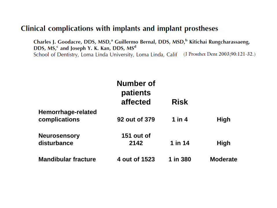
Number of
patients
affected Risk
Hemorrhage-related
complications 92 out of 379 1 in 4 High
Neurosensory
disturbance
151 out of
2142 1 in 14 High
Mandibular fracture 4 out of 1523 1 in 380 Moderate

Outline of Presentation
• Introduction / Disclosures
• Risk from Low Radiation Doses
• What do we mean by Effective Dose?
• How to evaluate Risks?
• How does CT work?
• How does Dose affect Image Quality?
• What other factors affect Image Quality?

how CT works…
Godfrey Hounsfield
Nobel prize in Medicine,
1979
Allan Cormack
Animation from
Demetrios J. Halazonetis
www.dhal.com

x-ray source
detectors
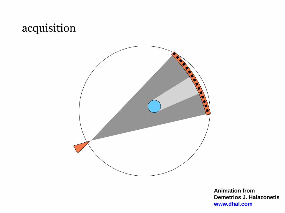
acquisition
Animation from
Demetrios J. Halazonetis
www.dhal.com

acquisition

acquisition

reconstruction

volume dataset
Animation from
Demetrios J. Halazonetis
www.dhal.com


Animation from
Demetrios J. Halazonetis
www.dhal.com

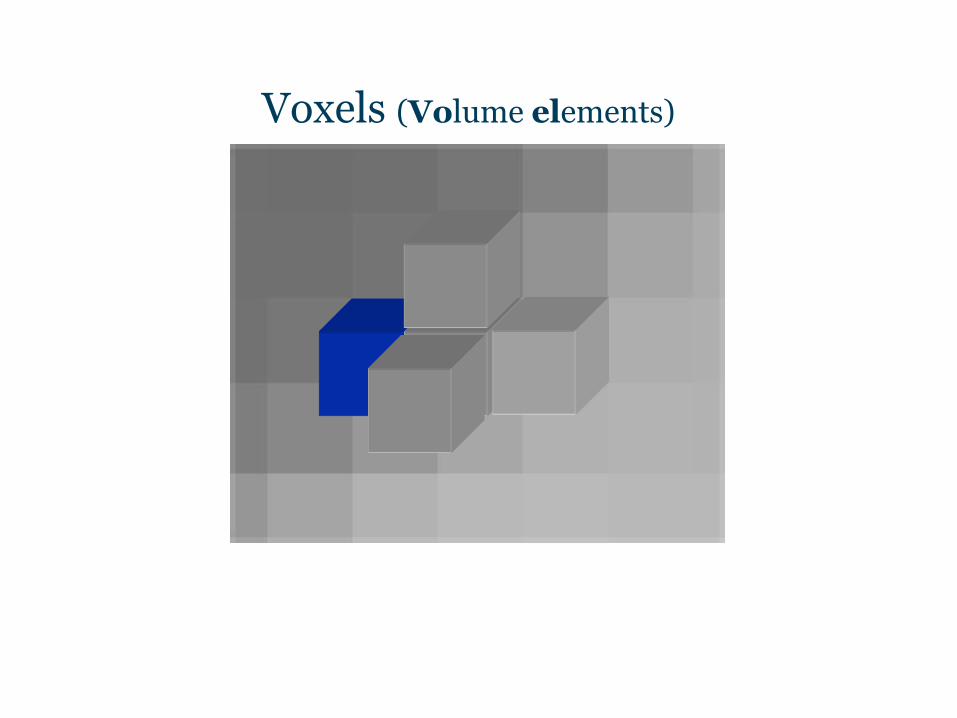
Voxels (Volume elements)

Voxels (Volume elements)
≈ 100 million voxels (200 Mb)400
slices512 x 512 x
density:0 to 4095
(-1000 to 3095Hounsfield Units)

From: Kalender WA. Computed Tomography. Munich: Publicis MCD Verlag, ISBN 3-89578-081-2, 2000.
The Hounsfield Scale was devised for medical CT scanners - 120kVp and Large Field Of View

• Segmentation – making physical models or drill guides
• Virtual 3D models e.g. in SimPlant
• Clinical application of bone densities e.g. Carl Misch scale
Why is Density Important?
Adapted from: Misch C. Contemporary Implant Dentistry. 2rd edn. Mosby, St Louis, 1999

Segmentation


Hyperdontia
Courtesy of Nicolette Schroeder

Third Molars
Courtesy of Barry Dace


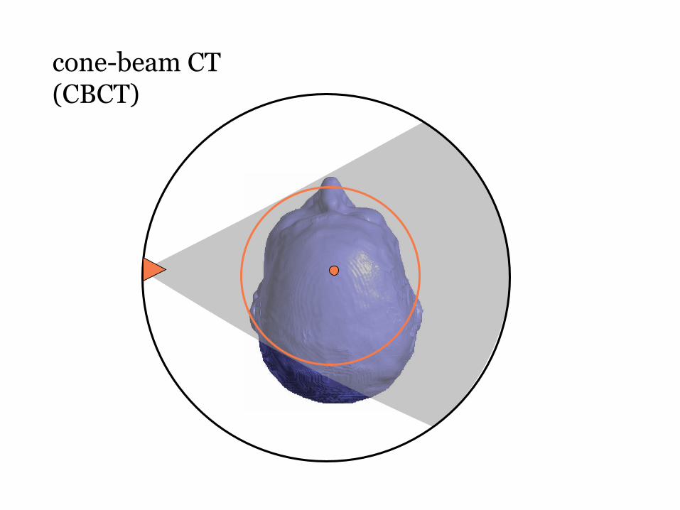
cone-beam CT(CBCT)
Animation from
Demetrios J. Halazonetis
www.dhal.com

cone-beam CT(CBCT)

cone-beam CT(CBCT)

cone-beam CT(CBCT)
cone-beam CT(CBCT)


3 small fields stitched together
each 5cm dia x 4cm height
Maxilla – Full Arch
Total: 60 µSv
20 µSv
20 µSv
20 µSv

one large field
16cm dia x 4cm height
Maxilla – Full Arch
Total: 40 µSv


Image Quality
- Noise• depends on radiation dose
- Artefact• metal objects within the patient• depends on machine calibration and operator technique
- Spatial Resolution (resolution at high contrast)• depends on machine design
(focal spot size, detector elements, sampling, mechanical stability)
• voxel size can only limit the resolution – cannot increase it!
- Contrast Resolution (resolution at low contrast)• depends on filtration and kVp• limited by the noise

Noise in CT / CBCT images
• Electronic noise (dark current)
• Photon noise (not enough x-rays)– Noise is proportional to √n
– Signal-to-Noise Ratio is proportional to n / √n = √n
– Where n is the number of x-ray photons
Noise = unstructured contribution to the image
which has no counterpart in the object.

If you halve (1/2) each side of a cube e.g. from 0.4mm to 0.2mm
Number of x-ray photons passing through it goes down by 8 (i.e. 1/8)
Noise goes up by √8 = 2.83
mAs (dose) may have to be increased to compensate
Noise depends on voxel size
x-rays(from all
directions)


• The noise increases as the voxel size gets smaller
• On most machines the operator may choose to increase
the dose (mA or scan duration) to compensate for this
• On some machines (e.g. i-CAT 17-19 and CB-500)
the operator must choose a longer scan duration
to obtain a smaller voxel size(e.g. 0.25mm voxels require a 23s scan duration on CB-500)
• Advantage of the longer scan duration is better spatial
resolution since the detector acquires more samples
• Disadvantages are: (a) more dose (b) patient movement.
Scan Duration versus Voxel Size

Other things that affect Image Quality
Noise• depends on radiation dose
- Artefact• metal objects within the patient• depends on machine calibration and operator technique
- Spatial Resolution (resolution at high contrast)• depends on machine design
(focal spot size, detector elements, sampling, mechanical stability)
• voxel size can only limit the resolution – cannot increase it!
- Contrast Resolution (resolution at low contrast)• depends on filtration and kVp• limited by the noise

Artefacts in CT images
• Motion artefact
• Spiral artefacts
• Cone beam artefacts
• Ring artefacts
• Starburst artefact
• Beam hardening
Artefact = structured contribution to the image
which has no counterpart in the object.


Motion Artefact – cone beam CT

Motion Artefact – cone beam CT

cone beam artefact

ring artefact

STARBURST ARTEFACT
• Starburst artefacts arise in CT scans when sharp changes in density are present, e.g. between air and bone or between bone and dense metals
• Starburst artefacts are caused bylimitations in high frequency sampling
• Starburst artefacts are not caused by scattered radiation


BEAM HARDENING ARTEFACT
• Beam Hardening artefacts also occur in CT scans when metals are present
• Metals cause the low energy x-rays to be filtered out of the x-ray beam
• The average energy becomes higher
• The CT numbers become lower
• Parts of the image appear black


High-Z materials cause the worst artefacts

• Titanium implants produce little artefact,gold produces a lot
• Remove dentures or other fixtures that include metal clasps, reinforcements or chrome cobalt bases
• Replace amalgam with composites, especially if the tooth will be sacrificed anyway.
HOW TO AVOID ARTEFACTS

Other things that affect Image Quality
Noise• depends on radiation dose
Artefact• metal objects within the patient• depends on machine calibration and operator technique
- Spatial Resolution (resolution at high contrast)• depends on machine design
(focal spot size, detector elements, sampling, mechanical stability)
• voxel size can only limit the resolution – cannot increase it!
- Contrast Resolution (resolution at low contrast)• depends on filtration and kVp• limited by the noise

Detail at high contrast
Spatial Resolution

Spatial Resolution
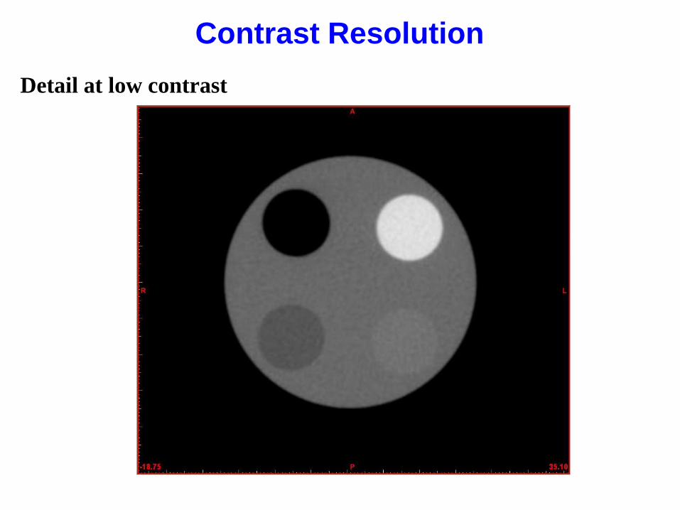
Detail at low contrast
Contrast Resolution

Contrast Resolution
LOW
CONTRAST

Spatial and Contrast Resolution are both important
Image 1 has good Spatial Resolution and good Contrast Resolution
Image 5 has poor Spatial Resolution and poor Contrast Resolution

Conclusions
• If your patient will truly benefit from a
CT or CBCT Scan the risks are likely to
be minimal or very low compared to the
benefits.
• A certain amount of Dose is essential
for good image quality but other factors
are important too.

5 things to discuss with CBCT salesmen
1. There’s no dose to the parts of the patientnot visible in the images.
2. A Small Field Of View (SFOV) always means a lower dose.
3. A CBCT scanner always has a lower dose than a medical CT scanner.
4. The dose from my SFOV scanner is so low that stitching 3 fields together is better than scanning the whole arch on a LFOV machine.
5. My CBCT scanner has a low kV so that means a lower dose.
FALSE
FALSE
FALSE
FALSE
FALSE
USUALLY BUT NOT ALWAYS.
USUALLY BUT NOT ALWAYS.

5 things to discuss with your colleagues
1. The smaller the voxel size, the better.
2. A smaller voxel size always means a higher dose.
3. A longer scan time can never be justified.
4. The CT images were non diagnostic but I shouldn’t ask for a repeat because of the dose.
5. My patient had a CT scan last week –she should wait at least 6 months beforeshe has another one.
FALSE
FALSE
FALSE
FALSE
FALSE
USUALLY BUT NOT ALWAYS.

Thank You!
• Any Questions?