Embed Size (px)
Citation preview

Image gently: Image quality and dose assessment in portable CXR in the NICU and PICU
before and after implementation of a high-kVp technique
Idris A. Elbakri1,2 PhD, MCCPM and Benjamin Z. Koplewitz MD3
1Department of Radiology, University of Manitoba, Winnipeg, Canada2Division of Medical Physics, CancerCare Manitoba, Winnipeg, Canada
3Dept. of Radiology, Hadassah-Hebrew University Medical Center, Jerusalem, Israel
The authors have no conflict of interest to report

Introduction
• Neonatal radiography is an essential tool in the care of patients in neonatal intensive care units (NICU).
• AP Chest and AP abdomen radiographs are the most common neonatal radiographs.
• Neonatal imaging is commonly carried out using portable radiography.
• Computed radiography (CR) has largely replaced film-screen cassettes in portable neonatal radiography
Introduction
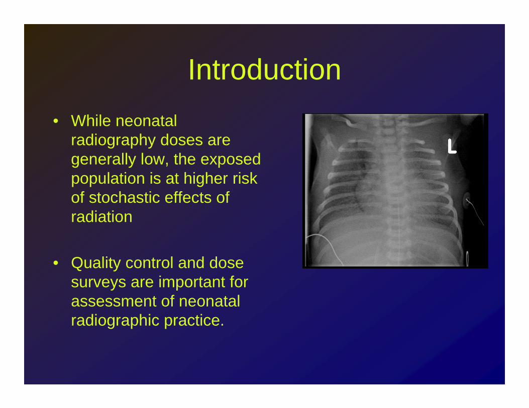
• While neonatal radiography doses are generally low, the exposed population is at higher risk of stochastic effects of radiation
• Quality control and dose surveys are important for assessment of neonatal radiographic practice.

Introduction and Motivation
• Quality control survey of neonatal radiography revealed the following:– No standardized technique chart was being followed– kVp/mAs and patient doses varied widely, depending
on operator experience and training– Protocol parameters were not adjusted after
introduction of CR. Low kVp (50-56) appropriate for film-screen cassettes still in use.

Quality Control SurveyNo clear relationship between kVp and patient weight
Wide kVp range for a given weight
49
50
51
52
53
54
55
56
57
0 1 2 3 4 5
Patient Weight (kg)
kVp

Quality Control Survey
0.0
10.0
20.0
30.0
40.0
50.0
60.0
0 1 2 3 4 5
Patient Weight (kg)
Pat
ien
t D
ose
(u
Sv)
Wide range of doses for a given weight highlights the lack of technique standardization

Purpose
• Implement weight-based technique parameters
• Reduce patient dose using a high-kVp technique
• Assess image quality
• Verify that image quality is not compromised

Methods
• Data collection (age, weight, gender, kVp, mAs) at pre-existing conditions for two months.
• Introduction of a weight based high-kVptechnique chart– Tube potentials - 60 to 76– Tube current fixed at 0.5 mAs
• Data collection at new conditions for two months
Methods
• GE AMX4 portable x-ray system • Fuji CR imaging plates and reader• Tracked AP chest and abdomen for
patients 0-3 months in the NICU and PICU at Hadassah Medical Organization
• Image quality assessment and dose estimation for high and low kVp image sets


Dose Estimation
• Portable GE AMX4 tube output characterized at various kVp settings
• Incident air kerma measured at 100 cm from x-ray tube using calibrated Pirahnasolid state dosimeter (RTI Electronics, Mölndal, Sweden)

Dose Estimation
• Effective dose for each images estimated using PCXMC 2.0 Monte Carlo software
• Software inputs: – weight, height, beam area, kVp, incident air
kerma, filtration, SID

PCXMC Dose Calculation Software

Image Quality Assessment• Two fellowship-trained pediatric radiologists blindly
assessed images before and after technique change.
• Evaluation criteria based on the CEC image quality standards1
• Criteria scored on a 4-point scale: (1) criterion definitely not defined, (2) criterion probably not defined, (3) criterion probably defined and (4) criterion definitely defined or (na) not applicable.
• Average score computed for each image
1. European Commission. European guidelines on quality criteria for diagnostic radiographic images in paediatrics. EUR 1626. July 1996.

Image Quality Criteria• Reproduction of the thorax without rotation and tilting• Reproduction of the chest must extend from the cervical trachea to T12/L1
(part of the abdomen maybe included for special purposes).• Reproduction of the vascular pattern in central two-thirds of the lungs• Reproduction of the trachea• Reproduction of the proximal bronchi• Visualization of the mediastinum• Visibility of the tip of the endotracheal tube• Visually sharp reproduction of the diaphragm• Visually sharp reproduction of the costophrenic angles• Reproduction of the spine• Visualization of the retrocardiac lung• Visibility of the tip of the umbilical catheter• Visibility of the tip of the long line• Visibility of bowel loops• Visibility of the nosagastric tube

Statistical Analysis
• We used the 2-tailed t-test to check significance of change in:– Patient dose– Patient weight – Reader 1 score – Reader 2 score
• We used ANCOVA analysis to check significance of change in effective dose with x-ray protocol, patient age and weight.

Results
12.532Abdomens
0.82Chest/Abdomen
100%254Total
76193Low kVp
2461High KVp
86.7221Chests
63.9/35.7163/91Gender (M/F)
Percentage %Number

Results - Averages
kVp mAsEffective
dose (uSv)Image quality
score
Low kVp
N=193 52.6 2.6 19.4±8.0 3.26±0.35
High kVpN=61
65.3 0.53 9.6±3.1 3.35±0.36

Image Quality Score vs Effective Dose
1.50
2.00
2.50
3.00
3.50
4.00
0.00 10.00 20.00 30.00 40.00 50.00 60.00
Effective Dose (µSv)
Ima
ge
Qu
ali
ty S
co
re (
IQ)
Low kVp
High kVp
Much narrower dose spread with new technique while maintaining similar IQ scores

Effective Dose (µSv) vs kVp
0.00
10.00
20.00
30.00
40.00
50.00
60.00
45 50 55 60 65 70 75
kVp
Eff
ect
ive
Do
se (
µS
v)
Low Kvp
High kVp
High-kVp method results in reduced dose and narrower dose range

Image Quality vs kVp
1.50
2.00
2.50
3.00
3.50
4.00
45 50 55 60 65 70 75
kVp
Ima
ge
Qu
ali
ty S
core
Low kVp
High kVp

Effective Dose vs Weight
0.00
10.00
20.00
30.00
40.00
50.00
60.00
0.00 1.00 2.00 3.00 4.00 5.00 6.00 7.00
Weight (Kg)
Eff
ect
ive
Do
se (
µS
v)
Low kVp
High kVp

Statistical Analysis
• 2-tailed t-test results:– Dose change is significant (p<-0.0001)
– Weight change is insignificant (p=0.072)– Reader 1 score change is significant (p=0.04)
– Reader 2 score change is significant (p<0.001)
• ANCOVA analysis showed that x-ray protocol is the only parameter that effects effective dose significantly (p<0.0001)

Summary of Results
• Clinical image rating is not affected by introducing weight-based higher-kVptechnique chart
• Average effective dose reduced by 50%• Effective dose range reduced from [7.0-
52.4] uSv to [5.9 – 19.9] uSv• The change in protocol parameters is the
single most significant factor contributing to dose reduction

Discussion
• Quality control survey revealed that the ALARA principle was not fully applied.
• Lack of standardized technique chart lead to wide variations in patient dose. The same patient could receive doses varying by a factor of 5 for the same examination.
• The dose-saving possibilities of digital imaging were not leveraged.

Discussion – Digital Imaging
• Film imaging is contrast limited. kVpchoice depends on:– Narrow exposure range required by film– Beam penetration (requires higher kVp) – Subject contrast (requires lower kVp).
• Digital imaging is noise limited. – Wide range of useful exposure
– Image Processing enhances image contrast– Enough exposure must reach the detector to
avoid a noisy image

Exposure
Imag
e R
ecep
tor
Res
pons
e
Film Digital
Digital detector has a wide dynamic range, make it more tolerant than film of variations in exposure
Film receptor gives optimal contrast over a narrow range of exposure
Increasing the kVp can deliver enough photons to the CR plate at lower mAs and lower patient exposure.

Discussion
• High-kVp protocol lowered patient dose significantly and reduced dose variations.
• The ‘significance’ in change in readers image quality scores is due to the narrow range of scores obtained.
• For all practical purposes, image quality not affected by change in kVp.

Conclusions / Lessons Learned
• Periodic quality control results in better patient care.
• “Imaging gently” is a team effort (physicists, radiologists, technologists, administration).
• Technique optimization should be carried out when new imaging modalities and techniques are implemented.

Conclusions / Lessons Learned
• Data is your best friend. We continue to record exposure and patient data for subsequent reviews.
• Data collected in this study will enable us to assess other aspects of quality control, such as positioning and collimation
• High-kV low-mAs technique enables marked dose reduction
• High-kV low-mAs technique dose not impair image quality